mutations and polymorphisms in gusb gene in mucopolysaccharidosis vii (sly syndrome)
TRANSCRIPT

Human MutationMUTATION UPDATE
Mutations and Polymorphisms in GUSB Genein Mucopolysaccharidosis VII (Sly Syndrome)
Shunji Tomatsu,1� Adriana M. Montano,1 Vu Chi Dung,1 Jeffrey H. Grubb,2 and William S. Sly2�
1Department of Pediatrics, Saint Louis University School of Medicine, St. Louis, Missouri2Edward A. Doisy Department of Biochemistry and Molecular Biology, Saint Louis University School of Medicine, St. Louis, Missouri
Communicated by Ronald J.A. WandersReceived 22 February 2008; accepted revised manuscript 29 April 2008.
Published online 17 February 2009 in Wiley InterScience (www.interscience.wiley.com). DOI 10.1002/humu.20828
ABSTRACT: Mucopolysaccharidosis VII (MPS VII; Slysyndrome) is an autosomal recessive disorder caused by adeficiency of b-glucuronidase (GUS, EC 3.2.1.31; GUSB).GUS is required to degrade glycosaminoglycans (GAGs),including heparan sulfate (HS), dermatan sulfate (DS),and chondroitin-4,6-sulfate (CS). Accumulation of unde-graded GAGs in lysosomes of affected tissues leads tomental retardation, short stature, hepatosplenomegaly,bone dysplasia, and hydrops fetalis. We summarizeinformation on the 49 unique, disease-causing mutationsdetermined so far in the GUS gene, including nine novelmutations (eight missense and one splice-site). Thisheterogeneity in GUS gene mutations contributes to theextensive clinical variability among patients with MPS VII.One pseudodeficiency allele, one polymorphism causing anamino acid change, and one silent variant in the codingregion are also described. Among the 103 analyzed mutantalleles, missense mutations accounted for 78.6%; nonsensemutations, 12.6%; deletions, 5.8%; and splice-site muta-tions, 2.9%. Transitional mutations at CpG dinucleotidesmade up 40.8% of all the described mutations. The fivemost frequent mutations (accounting for 44/103 alleles)were exonic point mutations, p.L176F, p.R357X, p.P408S,p.P415L, and p.A619 V. Genotype/phenotype correlationwas attempted by correlating the effects of certain missensemutations or enzyme activity and stability within pheno-types. These were in turn correlated with the location ofthe mutation in the tertiary structure of GUS. A total ofseven murine, one feline, and one canine model of MPSVII have been characterized for phenotype and genotype.Hum Mutat 30, 511–519, 2009. & 2009 Wiley-Liss, Inc.
KEY WORDS: GUS; GUSB; mucopolysaccharidosis VII;MPS VII; Sly syndrome; alignment
Introduction
Mucopolysaccharidosis VII (Sly Syndrome; MPS VII) is anautosomal recessive disease classified in the group of mucopoly-saccharide storage diseases. MPS VII (MIM 253220) is character-ized by the deficiency of activity of the enzyme b-glucuronidase(GUS: b-D-glucuronoside glucuronosohydrolase, EC 3.2.1.31;GUSB; MIM 611499) [Sly et al., 1973]. It is one of a class ofdiseases due to a deficiency of one of the dozen enzymes involvedin the stepwise degradation of glycosaminoglycans (GAGs). In theabsence of GUS, chondroitin sulfate (CS), dermatan sulfate (DS),and heparan sulfate (HS) are only partially degraded andaccumulate in the lysosomes of many tissues, eventually leadingto cellular and organ dysfunction. MPS VII is a rare disorder, andprecise epidemiologic data are scarce. MPS VII causes mentalretardation, hepatosplenomegaly, and skeletal dysplasia. MPS VIIpatients displayed a wide range of clinical variability, from themost severe type with hydrops fetalis to a milder phenotype withlater onset and normal intelligence. MPS VII patients with themost severe phenotype have hydrops fetalis at birth and often donot survive beyond a few months. Patients with mild manifesta-tions of MPS VII have survived into the fifth decade of life. MPSVII has also been reported in canine, feline, and murine species[Haskins et al., 1984; Birkenmeier et al., 1989; Sands andBirkenmeier, 1993; Gitzelmann et al., 1994; Gwynn et al., 1998;Ray et al., 1998; Fyfe et al., 1999; Sly et al., 2001; Vogler et al., 2001;Tomatsu et al., 2002b, 2003]. The initially described, naturalMPS VII mice (gusmps/mps) have a 1-bp deletion in exon 10 andhave similar morphologic, genetic, and biochemical characteristicsto human MPS VII patients, showing degenerative diseasewith progressive disability, widespread organ dysfunction,facial dysmorphism, growth retardation, deafness, behavioraldeficits, and shortened lifespan [Birkenmeier et al., 1989;Sands and Birkenmeier, 1993]. We produced mL175F (corre-sponding to p.L176F, the most common human mutation),mE536A, and mE536Q (active site nucleophile replacements,corresponding to p.E540A and p.E540Q in humans) knock-inmice [Tomatsu et al., 2002b]. These models reflect the variousclinical phenotypes of human MPS VII (Sly syndrome). Advancedtreatments such as enzyme replacement therapy (ERT) andgene therapy for MPS VII are currently being developed usingthese models. We have recently created MPS VII mouse modelstolerant to infused human GUS enzyme to test various treatmentprotocols using the human gene product [Sly et al., 2001; Tomatsuet al., 2003].
Characterization of GUS protein by X-ray crystallography andhomology comparisons among several species of GUS andbacterial b-galactosidases suggested R382, E451, and E540 as
OFFICIAL JOURNAL
www.hgvs.org
& 2009 WILEY-LISS, INC.
Contract grant sponsor: Austrian Research Society for Mucopolysaccharidoses
and Related Diseases; German MPS Society; Italian MPS Society; National MPS
Society.
�Correspondence to: William S. Sly, M.D., Department of Biochemistry, Saint Louis
University Doisy Research Center, 1100 South Grand Blvd., Room 533, St. Louis, MO
63104. E-mail: [email protected] or Shunji Tomatsu, M.D., Ph.D., Department of
Pediatrics, Saint Louis University Doisy Research Center, 1100 South Grand Blvd.,
Room 307, St. Louis, MO 63104. E-mail: [email protected]

active site residues [Jain et al., 1996; Islam et al., 1999]. Thesethree residues of human GUS are conserved among GUS and b-galactosidase proteins from bacterial species [Henrissat, 1991].E540 was identified experimentally as the active site nucleophile ofthe human enzyme [Wong et al., 1998].
Isolation and characterization of the human cDNA andgenomic gene made investigation of molecular lesions in theGUS gene of MPS VII patients feasible [Oshima et al., 1987; Milleret al., 1990; Shipley et al., 1991]. The GUS gene is located onchromosome arm 7 [Speleman et al., 1996] and spans approxi-mately 20 kb containing 11 introns and 12 exons. The 1,953-bpGUS mRNA encodes a 651–amino acid precursor. After cleavageof a 22–amino acid N-terminal signal peptide and glycosylation,the 78-kDa monomer is transported to lysosomes and cleaved inthe lysosome to become the 60-kDa and 18-kDa subunits of themature active enzyme [Brot et al., 1978; Oshima et al., 1987].
MPS VII Mutations and their BiologicalRelevance
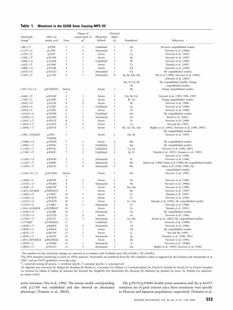
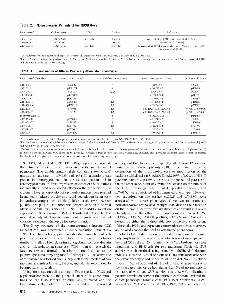
To date, 49 different mutations including nine novel mutationsin the GUS gene have been found in MPS VII patients. Thesemutations have been identified in 103 mutant alleles in a totalgroup of 56 patients by a variety of molecular techniques (92.0%of total investigated alleles) (Table 1). The numbers for thenucleotide changes are reported in accordance with GenBankentry NM_010368.1. Three nonpathogenic variants within thecoding sequence of the GUS gene have been also identified (onepseudodeficiency, one benign amino acid change, one silentchange) (Table 2). The DNA mutation numbering is based oncDNA sequence. For cDNA numbering, 11 corresponds to the Aof the ATG translation initiation codon in the reference sequence.The MPS VII patients were defined as attenuated if they did nothave hydrops fetalis and severe mental retardation leading to deathwithin a year.
The mutations are distributed along the whole gene and alltypes of mutations except insertion and rearrangement werefound. The total of 49 mutations includes 36 missense mutations,six nonsense, two splice site mutations, and five deletions. Thenumber of each type of mutation in a total of 103 mutant alleleswas 81 alleles for missense mutations (78.6%), 13 for nonsense(12.6%), six for deletions (5.8%), and three for splice-sitemutations (2.9%). Thus, missense mutations are the mostprevalent among GUS mutations. The five most frequentmutations (Table 1) are represented by single nucleotide changes.Together, they make up 36.9% of all described mutant alleles. Theremaining 63.1% of mutations each occur less than four times inthe mutant population, indicating extensive molecular hetero-geneity in GUS mutations.
Relation Between Transitions at CpG Sites and theMethylation Status of CpG Sites in the GUSB Gene
The variety, frequency, and location of point mutations causinghuman genetic disease are highly nonrandom. One importantfactor contributing to the nonrandomness at the DNA level is thelocal DNA sequence environment, especially CpG dinucleotides.DNA methylation at the cytosine residue of CpG dinucleotidesproduces 5-methylcytosine, which results in a C-to-T transitionalchange following deamination. The importance of CpG methyla-tion in the etiology of genetic diseases was deduced from theevidence that 10 to 60% of point mutations causing human
diseases in different genes result from transitions at CpGdinucleotides [Krawczak et al., 1998; Antonarakis et al., 2001].
There are 17 transitional mutations at CpG sites in the GUSgene. Transitions at CpG dinucleotides account for 40.8% ofdescribed mutant alleles and 44.7% of exonic point mutations thatcause MPS VII. This percentage is higher than that compiled frommany genes described previously [Krawczak et al., 1998;Antonarakis et al., 2001] and represents around a 30-fold higherprobability of a transitional mutation at a CpG dinucleotide thanexpected. These findings explain why many transitional mutationsat CpG sites are recurrent. No transitional mutation at CpG siteshas been detected in exon 1. To explain this discrepancy, weanalyzed the methylation pattern of the GUS coding region by asensitive bisulfite-based technique [Tomatsu et al., 2002a]. Wefound that methylation of the 67 individual CpG cytosines withinexons 2 to 12 was extensive while 24 CpG cytosines in exon 1 werecompletely unmethylated. All of the 17 transitional mutations atCpG sites out of the 42 exonic point mutations were locatedbetween exons 2 and 12, demonstrating the correlation ofnonmethylation of exon 1 with the absence of transitionalmutations at CpG sites in exon 1 and the reverse for exons 2 to12. One pseudodeficiency allele (p.D152N) and one benignpolymorphism allele (p.P649L), both of which change an aminoacid residue, are also derived from G-to-A or C-to-T transition atCpG dinucleotides, respectively [Tomatsu et al., 1991; Vervoortet al., 1995].
Missense Mutations
This is the most frequent group of GUS mutations, with 36changes including eight novel amino acid substitutions reportedhere (Tables 1 and 3; Fig. 1). Correlation of individual mutationwith disease severity is based on phenotype of the homozygotes,predicted change of tertiary structure of the protein, and theobserved level of enzyme activity on in vitro expression.
Several mutations are recurrent. Among the recurrent muta-tions, the most prevalent are: c.526C4T (p.L176F), c.1244C4T(p.P415L), c.1222C4T (p.P408S), c.1856C4T (p.A619 V),c.646C4T (p.R216W), c.1144C4T (p.R382C), and c.1429C4T(p.R477W), accounting for 20.4, 4.9, 4.9, 4.9, 3.9, 3.9, and 3.9%,respectively [Tomatsu et al., 1990, 1991; Fukuda et al., 1991;Vervoort et al., 1993; Wu et al., 1994; Islam et al., 1996, 1998;Vervoort et al., 1996, 1997; Schwartz et al., 2003]. The p.L176Fmutation has been identified in diverse ethnic populations whilethe p.P415L mutation and p.P415L/P408S double mutation, andthe p.A619 V mutation have been detected only in Mexican andJapanese populations, respectively.
The most prevalent c.526C4T transitional mutation (p.L176F),originally found in two Mennonite siblings, was identified in 21alleles of 11 patients from American (Caucasian), Brazilian, British,Chilean, French, Mexican, Polish, Spanish, and Turkish origins [Wuet al., 1994; Vervoort et al., 1996; Schwartz et al., 2003](Sly, unpublished results). A total of 10 of 11 patients werehomozygous for the mutation. Those homozygous patientsdeveloped an attenuated type of MPS VII with similarclinical symptoms and signs. The p.L176F conservative amino acidchange generates a subtle structural alteration of GUS protein [Wuet al., 1994]. Although the cultured fibroblasts homozygouswith p.L176F contained only 1.5 to 2.2% of normal GUSactivity, overexpression of the p.L176F cDNA in COS cells produced84% as much enzyme as the wild-type control cDNA. Thesefindings suggested that overexpression can drive the foldingreaction or the self-association of mutant monomers to form
512 HUMAN MUTATION, Vol. 30, No. 4, 511–519, 2009
active tetramers [Wu et al., 1994]. The mouse model correspondingwith p.L176F was established and also showed an attenuatedphenotype [Tomatsu et al., 2002b].
The p.P415L/p.P408S double point mutation and the p.A619 Vmutation are of great interest since these mutations were specificto Mexican and Japanese populations, respectively [Tomatsu et al.,
Table 1. Mutations in the GUSB Gene Causing MPS VII�
Nucleotide
changea
Effect on
amino acid Exon
Degree of
conservation of
aab
Phenotype
defined
Detected
alleles
(n) Populationc References
c.f8C4T p.P30S 1 2 Undefined 1 Un Vervoort (unpublished results)
c.112T4G p.C38G 1 4 Attenuated 2 It Vervoort et al. [1998a]
c.155C4T p.S52F 1 2 Severe 1 Cz Vervoort et al. [1997]
c.328C4T p.R110X 2 3 Severe 1 Be Vervoort et al. [1997]
c.406G4A p.G136R 3 2 Undefined 2 Bt Vervoort et al. [1996]
c.442C4T p.P148S 3 1 Severe 1 Cc Yamada et al. [1995]
c.448G4A p.E150K 3 1 Severe 1 Ch Vervoort et al. [1996]
c.455A4G p.D152G 3 3 Attenuated 1 Un Sly (unpublished results)
c.526C4T p.L176F 3 1 Attenuated 21 Sp, Br, Mn, Mx Wu et al. [1994]; Vervoort et al. [1996];
Schwartz et al. [2003]
Am, Fr-Ca, Bt,
Tu
Sly (unpublished results); Young
(unpublished results)
c.58111G4A p.K194fsX22 Intron
3
Severe 1 Pk Young (unpublished results)
c.646C4T p.R216W 4 1 Severe 4 Du, Be, Un Vervoort et al. [1993, 1996, 1997]
c.728T4C p.L243P 5 2 Severe 1 Bt, Tu Young (unpublished results)
c.935C4G p.S312X 6 4 Severe 1 Be Vervoort et al. [1996]
c.959A4G p.Y320C 6 1 Undefined 1 Ge Vervoort et al. [1996]
c.959A4C p.Y320S 6 1 Severe 1 Fi Vervoort et al. [1996]
c.1016A4G p.N339S 6 1 Undefined 1 Am Sly (unpublished results)
c.1050G4C p.K350N 6 2 Attenuated 1 Ge Storch et al. [2003]
c.1051C4T p.H351Y 6 1 Severe 1 Fi Vervoort et al. [1996]
c.1061C4T p.A354V 6 4 Severe 1 Cc Wu and Sly [1993]
c.1069C4T p.R357X 7 2 Severe 7 Be, Ch, Du, Am Shipley et al. [1993]; Vervoort et al. [1996, 1997];
Sly (unpublished results)
c.1081_1107del27 p.F361-
D369del
7 Severe 2 Du, Be Vervoort et al. [1997]
c.1084G4A p.D362N 7 1 Severe 2 Mx Sly (unpublished results)
c.1091C4T p.P364L 7 3 Undefined 1 Am Sly (unpublished results)
c.1120C4T p.R374C 7 2 Undefined 2 Cz, Ge Vervoort et al. [1996, 1997]
c.1144C4T p.R382C 7 1 Undefined 4 Jp, Fr Fukuda et al. [1991]; Tomatsu et al. [1991];
Vervoort et al. [1996]
c.1145G4A p.R382H 7 1 Attenuated 1 Fr Vervoort et al. [1996]
c.1222C4T p.P408S 7 2 Attenuated 5 Mx Islam et al. [1996]; Islam et al. [1998]; Sly (unpublished results)
c.1244C4T p.P415L 7 4 Attenuated 5 Mx Islam et al. [1996, 1998]; Sly
(unpublished results)
c.124411G4A p.P415fsX1 Intron
7
Severe 2 Mx Vervoort et al. [1997]
c.1304G4C p.R435P 8 2 Severe 2 Tu Vervoort et al. [1996]
c.1337G4A p.W446X 8 1 Attenuated 1 Sw Vervoort et al. [1998a]
c.1429C4T p.R477W 9 1 Severe 4 Kw, Ma Vervoort et al. [1996]
c.1454_1457del4 p.S485fsX13 9 Severe 1 Be Vervoort et al. [1997]
c.1484A4G p.Y495C 10 2 Severe 1 Cc Yamada et al. [1995]
c.1520G4A p.W507X 10 1 Severe 1 Fr Vervoort et al. [1996]
c.1521G4A p.W507X 10 1 Severe 2 Cc, Am Yamada et al. [1995]; Sly (unpublished results)
c.1523A4G p.Y508C 10 1 Attenuated 1 Fr Vervoort et al. [1996]
c.1616_1653del38 p.S539RfsX7 10 Severe 1 Cc Yamada et al. [1995]
c.1618G4A p.E540K 10 1 Attenuated 1 Un Sly (unpublished results)
c.1715G4A p.G572D 11 4 Severe 2 In Vervoort et al. [1996]
c.1730G4T p.R577L 11 2 Attenuated 2 Ge, Mx Storch et al. [2003]; Sly (unpublished results)
c.1775delT p.F592SfsX2 11 Undefined 1 Fr Vervoort et al. [1998b]
c.1818G4T p.K606N 12 2 Attenuated 2 Al Vervoort et al. [1996]
c.1820G4C p.G607A 12 1 Severe 1 Un Sly (unpublished results)
c.1831C4T p.R611W 12 1 Severe 1 Cc Wu and Sly [1993]
c.1856C4T p.A619V 12 1 Attenuated 5 Jp Tomatsu et al. [1990, 1991]
c.1874_1875delGA p.R625IfsX6 12 Severe 1 Be Vervoort et al. [1996]
c.1876T4C p.Y626H 12 1 Attenuated 1 It Vervoort et al. [1998a]
c.1881G4T p.W627C 12 3 Attenuated 2 Am Shipley et al. [1993]; Vervoort et al. [1996]
�The numbers for the nucleotide changes are reported in accordance with GenBank entry NM_010368.1, NP_034498.1.aThe DNA mutation numbering is based on cDNA sequence. Nucleotides are numbered from the ATG initiation codon as suggested by den Dunnen and Antonarakis et al.
[2001] and per HGVS guidelines (www.hgvs.org).b1, conserved among all species; 2, vertebrate specific; 3, mammal specific; 4, nonconserved.cAl, Algerian; Am, American; Be, Belgian; Br, Brazilian; Bt, British; Cc, Caucasian; Ch, Chilean; Cz, Czechoslovakian; Du, Dutch; Fi, Finnish; Fr, French; Fr-Ca, French-Canadian;
Ge, German; In, Indian; It, Italian; Jp, Japanese; Kw, Kuwaiti; Ma, Maghrebi; Mn, Mennonite; Mx, Mexican; Pk, Pakistani; Sp, Spanish; Sw, Swiss; Tu, Turkish; Un, unknown.
aa, amino acid(s).
HUMAN MUTATION, Vol. 30, No. 4, 511–519, 2009 513
1990, 1991; Islam et al., 1996, 1998] (Sly, unpublished results).Both founder mutations are associated with an attenuatedphenotype. The double mutant allele containing two C-to-Ttransitions resulting in p.P408S and p.P415L alterations waspresent in homozygous state in one Mexican patient and inheterozygous state in four. Expression of either of the mutationsindividually showed only modest effects on the properties of theenzyme. However, expression of the doubly mutant allele resultedin markedly reduced activity and rapid degradation in an earlybiosynthetic compartment (Table 4) [Islam et al., 1996]. Neitherp.P408S nor p.P415L mutation was present alone in a normalMexican population [Islam et al., 1998]. The p.A619 V mutationexpressed 9.1% of normal cDNA in transfected COS cells. Theresidual activity of these expressed mutant proteins correlatedwith the attenuated phenotype for those mutations.
The X-ray structure of the homotetrameric human GUS(332,000 Mr) was determined at 2.6-A resolution [Jain et al.,1996]. The tetramer had approximate dihedral symmetry and eachprotomer consisted of three structural domains with topologiessimilar to a jelly roll barrel, an immunoglobulin constant domainand a triosephosphateisomerase (TIM) barrel, respectively.Residues 179–204 formed a beta-hairpin motif similar to theputative lysosomal targeting motif of cathepsin D. The active siteof the enzyme was formed from a large cleft at the interface of twomonomers. Residues Glu 451, Tyr 504, and Glu 540 were shown tobe important for catalysis.
Using homology modeling among different species of GUS andb-galactosidase proteins, the potential effect of missense muta-tions on the GUS tertiary structure was estimated and thelocalization of the mutation site was correlated with the residual
activity and the clinical phenotype (Fig. 4). Among 12 missensemutations with a severe phenotype, 10 of these mutations involvedestruction of the hydrophobic core or modification of thepacking (p.S52F, p.P148S, p.E150 K, p.R216W, p.Y320S, p.H351Y,p.R435P, p.R477W, p.Y495C, p.G572D, p.K606N, and p.R611W).On the other hand, 5 out of 7 mutations located on the surface ofthe GUS protein (p.C38G, p.P415L, p.Y508C, p.R577L, andp.W627C) were associated with attenuated phenotypes. However,two mutations on the surface (p.S52F and p.Y495C) wereassociated with severe phenotypes. These two mutations arenonconservative amino acid changes that, despite their locationon the surface, disrupt the tertiary structure and result in a severephenotype. On the other hand, mutations such as p.D152N,p.L176F, p.A354 V, p.R382 H, p.P408S, p.A619 V, and p.Y626 H arelocated on either the hydrophobic core or involve a salt bridge[Jain et al., 1996], and represent conservative or semiconservativeamino acid changes that lead to attenuated phenotypes.
A total of 28 mutations, one pseudodeficiency, and one benignpolymorphism were analyzed by in vitro transient overexpression.We used COS cells for 23 mutations, MPS VII fibroblasts for threemutations, and BHK cells for two mutations (Table 4). GUSactivity was determined using 4-methylumbelliferyl-b-glucuro-nide as a substrate. A total of 8 out of 11 mutants associated withthe severe phenotype had under 3% of normal cDNA GUS activity(mean, 1.5%) while 13 out of 14 mutants found in patients withthe attenuated phenotype had higher than 3% of normal activity(3–112% of wild-type GUS activity; mean, 32.8%), indicating apositive correlation between the transient expression level and theclinical phenotype [Tomatsu et al., 1990, 1991; Shipley et al., 1993;Wu and Sly, 1993; Vervoort et al., 1995, 1996, 1998a; Yamada et al.,
Table 2. Nonpathogenic Variants of the GUSB Gene�
Base changea Codon change Effect Region Reference
c.454G4A GAC4AAC p.D152N Exon 3 Vervoort et al. [1995]; Vervoort et al. [1998a]
c.1740T4C TAT4TAC Exon 11 Vervoort et al. [1998a]
c.1946C4T CCG4CTG p.P649L Exon 12 Tomatsu et al. [1991]; Wu et al. [1994]; Vervoort et al. [1997];
Vervoort et al. [1998a]
�The numbers for the nucleotide changes are reported in accordance with GenBank entry NM_010368.1, NP_034498.1.aThe DNA mutation numbering is based on cDNA sequence. Nucleotides numbered from the ATG initiator codons as suggested by den Dunnen and Antonarakis et al. [2001]
and per HGVS guidelines (www.hgvs.org).
Table 3. Combination of Alleles Producing Attenuated Phenotypes�
Base changea (first allele) Amino acid changeb Factors defined as attenuated Base change (second allele) Amino acid change
c.112T4G p.C38G 3 c.1876T4C p.Y626H
c.455A4C p.D152G 4 c.1618G4A p.E540K
c.526C4T p.L176F 1,2,3 c.526C4T p.L176F
c.1050G4C p.K350N 3 c.1730G4T p.R577L
c.1061C4T p.A354V 3 c.1831C4T p.R611W
c.1144C4T p.R382C 1,3 c.1144C4T p.R382C
c.1145G4A p.R382H 2,3 c.1523A4G p.Y508C
c.1222C4T p.P408S 1,3 c.1244C4T, c.1222C4T p.P415L, p.P408S
c.1244C4T p.P415L 1,3 c.1244C4T, c.1222C4T p.P415L, p.P408S
IVS810.6kbdelTC ? 3 p.13376G4A p.P446X
c.1523A4G p.Y508C 3 c.1145G4A p.R382H
c.1730G4T p.R577L 3 c.1050G4C p.K350N
c.1856C4T p.A619V 1,3 c.1856C4T p.A619V
c.1876T4C p.Y626H 3 c.112T4G p.C38G
c.1881G4T p.W627C 3,4 c.1069C4T p.R357X
�The numbers for the nucleotide changes are reported in accordance with GenBank entry NM_010368.1, NP_034498.1.aThe DNA mutation numbering is based on cDNA sequence. Nucleotides numbered from the ATG initiator codons as suggested by den Dunnen and Antonarakis et al. [2001]
and per HGVS guidelines (www.hgvs.org).bThe correlation of a mutation with an attenuated phenotype is based on four factors: 1) homozygosity of the mutation in the patients with attenuated phenotypes; 2)
prediction from the likely structural change in the protein; 3) prediction from in vitro expression studies; and, 4) mutant allele permitting residual enzyme activity in primary
fibroblasts or leukocytes, which would be dominant over an allele permitting no activity.
514 HUMAN MUTATION, Vol. 30, No. 4, 511–519, 2009

1995; Storch et al., 2003]. One patient with an attenuatedphenotype had 2.3% of wild-type activity in COS cells.
The G-to-A transition (c.454G4A) in the coding region of theGUS gene, which resulted in an aspartic-acid-to-asparaginesubstitution at amino acid position 152 (p.D152N), produced apseudodeficiency allele that leads to greatly reduced levels of GUSactivity in vitro without apparent deleterious consequences[Vervoort et al., 1998a]. The c.454G4A mutation was foundinitially in the pseudodeficient mother of a child with MPS VII, butit was not on her disease-causing allele, which carried the p.L176Fmutation. Screening of 100 unrelated normal individuals for thec.454G4A mutation with a PCR method detected one carrier (arough estimate of frequency: 0.5%). Reduced GUS activityfollowing transfection of COS cells with the p.D152N cDNAsupported the causal relationship between the p.D152N allele andpseudodeficiency. The mutation reduced the fraction of expressedenzyme that was secreted. Pulse-chase experiments indicated thatthe reduced activity in COS cells was due to accelerated intracellularturnover of the p.D152N enzyme [Vervoort et al., 1998a]. Thepresence of the p.D152N mutation in combination with certainother MPS VII mutations might be more deleterious.
Nonsense Mutations
A total of six nonsense mutations have been reported:c.328C4T (p.R110X), c.935C4G (p.S312X), c.1069C4T
(p.R357X), c.1337G4A (p.W446X), c.1520G4A (p.W507X),and c.1521G4A (p.W507X) (Table 1) [Shipley et al., 1993;Yamada et al., 1995; Vervoort et al., 1996, 1997, 1998a](Sly, unpublished results). All of them should result in synthesisof truncated proteins without catalytic activity, predicting a severephenotype in MPS VII. The second most frequent p.R357Xmutation derived from a C-to-T transition at a CpG site occurredin diverse ethnic backgrounds suggesting a true recurrentmutation [Shipley et al., 1993; Vervoort et al., 1996, 1997] (Sly,unpublished results). Other nonsense mutations were sporadicand observed in only one patient.
Splice-Site Mutations
Two splice-site mutations including one novel mutation at thedonor site of intron 3 (c.58111G4A) were identified in the GUSgene (Table 1). One (c.124411G4A) is a homozygous mutationwhile the other one is in a compound heterozygote. Both splicing-site mutations in the GUS gene disrupt the consensus sequencebetween exon and intron. Both mutations at the acceptor sitecause complete deletion of the following exons. Skipping an exonwould result in a frameshift and the appearance of a prematurestop codon (p.K194fsX22, p.P415fsX1) and/or absence of a GUSactive catalytic site (E540).
In accordance with this prediction, a patient homozygous forthe C124411 G-to-A splice-site mutation developed a severe formof MPS VII and showed a complete loss of GUS activity infibroblasts [Vervoort et al., 1997] (Sly unpublished results).
Deletions
A total of five deletions were identified so far in the GUS gene(Table 1) (c.1081_1107del27, c.1454_1457del4, c.1616_1653del38,c.1775delT, and c.1874_1875delGA). Four deletions cause frame-shifts (c.1454_1457del4, c.1616_1653del38, c.1775delT, andc.1874_1875delGA) and the appearance of premature truncationcodons (p.S485fsX13, p.S539RfsX7, p.F592SfsX2, andp.R625IfsX6, respectively), probably leading to nonsense-mediateddecay of the mRNA and a complete loss of GUS activity in theaffected cells. Patients homozygous for the c.1081_1107del27 in-frame mutation manifested a severe form of MPS VII [Vervoortet al., 1997]. The mechanism of the c.1616_1653del38 deletion wasunique. The patient was a compound heterozygote of p.W507Xand a 38-bp deletion at position 1616–1653 in exon 10. The 38-bpdeletion was caused by a C-to-T transition in exon 10 thatgenerates a new, premature 50 splice-site. The resulting nucleotidesequence AGA/GTGAGT has a close homology to the 50 spliceconsensus sequence (A or C) AG/GT (A or G) AGT. Thisalteration interferes with normal splicing of the GUS genetranscript by forming a novel 50 splice-site.
Slipped mispairing can in principle account for the generationof 4 out of 5 deletions because of a run of identical bases or directrepeat (2 bp or more) (c.1081_1107del27, c.1454_1457del4,c.1775delT, and c.1874_1875delGA).
Vervoort et al. [1998b] reported a patient with an attenuatedphenotype whose paternal allele (IVS810.6kbdelTC) (Table 3)was claimed to create a new donor splice-site that activateda cryptic exon in an Alu-element of the GUS gene andled to skipping of exon 9. This allele confers the alternatephenotype, since the maternal allele (p.W446X) is a null allele(Table 1).
No insertions were identified so far in the GUS gene. The GUSgene spans approximately 20 kb, in addition to the 1-kb promoter
Figure 1. Location of GUS gene mutations in MPS VII patients. Theexons are presented by open boxes and the untranslated regions arefilled boxes. Clinical phenotypes associated with missense ornonsense mutations are described.
HUMAN MUTATION, Vol. 30, No. 4, 511–519, 2009 515

region located at 7q11.21, and contains 37 Alu repeats [Milleret al., 1990; Shipley et al., 1991; Speleman et al., 1996]. Neitherlarge deletions nor rearrangement were identified. Alu repeatsrepresented around 45% of the entire GUS gene (8.8 kb of 19.5 kbin total length), showing an extremely high percentage of Aluelements compared with the human genome (representing6–12%). In addition, over 20 pseudogenes of GUS gene were
observed in the entire human genome. Nevertheless, no largerearrangement has been reported so far.
Polymorphisms
A total of two benign genetic variants in the coding regions ofthe GUS gene have been reported (Table 2) [Tomatsu et al., 1991;
Table 4. In Vitro Mutagenesis Data of GUSB Missense Mutations
Mutation Phenotype
Percentage of normal
cDNA
Location of the
mutation
Conservativeness of amino acid
changes
Evolutionary
conservationa References
p.C38G Attenuated 7.0 Surface Nonconservative 4 Vervoort et al. [1998a]
p.S52F Severe 0.7 Surface Nonconservative 2 Vervoort et al. [1997]
p.G136R Undefined 0.6 Surface Nonconservative 2 Vervoort et al. [1996]
p.P148S Severe 1.2 Hydrophobic core Semiconservative 1 Yamada et al. [1995]
p.E150K Severe 4.0 Hydrophobic core Conservative 1 Vervoort et al. [1996]
p.D152N Pseudodeficiency 28 Hydrophobic core Conservative 3 Vervoort et al. [1995]
p.L176F Attenuated 84.1 Hydrophobic core Conservative 1 Wu et al. [1994]
p.R216W Severe 0.0 Hydrophobic core Nonconservative 1 Vervoort et al. [1996]
p.Y320C Undefined 1.4 Hydrophobic core Nonconservative 1 Vervoort et al. [1996]
p.Y320S Severe 1.0 Hydrophobic core Nonconservative 1 Vervoort et al. [1996]
p.K350N Attenuated 13 Hydrophobic core Nonconservative 2 Storch et al. [2003]
p.H351Y Severe 0.0 Hydrophobic core Semiconservative 1 Vervoort et al. [1996]
p.A354V Attenuated 37.7 Hydrophobic core Semiconservative 4 Wu and Sly [1993]
p.R374C Undefined 5.5 Surface Nonconservative 2 Vervoort et al. [1996]
p.R382C Attenuated 12.3 Salt bridge Nonconservative 1 Tomatsu et al. [1991]
p.R382H Attenuated 2.3 Salt bridge Conservative 1 Vervoort et al. [1996]
p.P408S Attenuated 61 Hydrophobic core Semiconservative 2 Islam et al. [1996]
p.P415L Attenuated 112 Surface Nonconservative 4 Islam et al. [1996]
p.R435P Severe 0.5 Hydrophobic core Nonconservative 2 Vervoort et al. [1996]
p.R477W Severe 0.7 Hydrophobic core Nonconservative 1 Vervoort et al. [1996]
p.Y495C Severe 0.8 Surface Nonconservative 2 Yamada et al. [1995]
p.Y508C Attenuated 19 Surface Nonconservative 1 Vervoort et al. [1996]
p.G572D Severe 2.1 Hydrophobic core Nonconservative 4 Vervoort et al. [1996]
p.R577L Attenuated 4.0 Surface Nonconservative 2 Storch et al. [2003]
p.K606N Severe 6.7 Hydrophobic core Nonconservative 2 Vervoort et al. [1996]
p.R611W Severe 0.0 Hydrophobic core Nonconservative 1 Wu and Sly [1993]
p.A619V Attenuated 9.1 Hydrophobic core Semiconservative 1 Tomatsu et al. [1990, 1991] Yamada
et al. [1995]
p.Y626H Attenuated 4.0 Hydrophobic core Semiconservative 1 Vervoort et al. [1998a]
p.W627C Attenuated 39 Surface Nonconservative 3 Shipley et al. [1993]
p.P408S/
p.P415L
Attenuated 12.5 Islam et al. [1996]
p.P649L Polymorphism 88.3 Nonconservative 4 Tomatsu et al. [1991]
a1, conserved among all species; 2, vertebrate species; 3, mammalian species; 4, nonconserved.
Figure 2. Multiple amino acid alignment of GUS from human (Hosa-GUS), chimpanzee (Patr-GUS), cow (Bota-GUS), pig (Susc-GUS), dog(Cafa-GUS), mouse (Mumu-GUS), rat (Rano-GUS), chicken (Gaga-GUS), frog (Xetr-GUS), fruit fly (Drme-GUS), mosquito (Anga-GUS), honey bee(Apme-GUS), red flour beetle (Trca-GUS), nematode (Cael-GUS), gram-positive bacteria (Arsp.-GUS), enterobacteria (Esco-GUS), and fungi(Scsp.-GUS), along with bacterial b-galactosidase (Esco-b-Gal). The arrow indicates the active site (residue E540) of human GUS. GenBankreference sequences: Homo sapiens, NM_010368.1, NP_034498.1; Pan troglodytes, XP_001138789.1; Macaca mulata, XM_001087699; Musmusculus, NM_010368.1, NP_034498.1; Rattus norvegicus, NP_058711; Felis catus, NM_001009310.1, NP_001009310.1; Canis familiaris,NM_001003191.1, NP_001003191.1; Bos taurus, NM_001083436.1; Sus scrofa, AK232674.1; Gallus gallus, NP_001034405; Xenopus tropicalis,CT030620; Danio renio, XM_695030; Drosophila melanogaster, NP_001014535.1; Anopheles gambiae, XP_320660.2; Apis mellifera, XM_393305;Tribolium castaneum, XM_964260.1; Caenorhabditis elegans, NP_493548.1; Arthrobacter sp., RP10, AAV91790; Scopulariopsis sp., RP38.3,AAV91788; Escherichia coli, AAB30197.
516 HUMAN MUTATION, Vol. 30, No. 4, 511–519, 2009

Wu et al., 1994; Vervoort et al., 1995, 1998a]. One polymorphismchanging an amino acid residue was identified in the normalpopulation (c.1946C4T, p.P649L). An in vitro expression studyshowed that this polymorphism provides 88.3% of normal GUScDNA activity.
Relations Among Genotypes and Phenotypes
The genotype/phenotype correlation for each of 38 single-nucleotide alterations has been examined based upon thefollowing four factors (Tables 1, 3, and 4): 1) the phenotype ofthe patient homozygous for the mutation; 2) the level of activityby in vitro expression study; 3) prediction of the likely change inthe protein structure; and 4) the presence of a second allelepermitting residual enzyme activity, which would be dominantover an allele permitting no activity. In all, 15 mutations wereassociated with a severe phenotype, 14 mutations were associatedwith an attenuated phenotype, and one with a normal phenotype(pseudodeficiency allele). The other seven mutations were notdefined by the current information. A total of 5 out of 6 nonsensemutations and 4 out of 5 deletions were associated with severephenotypes. One splicing site mutation was associated with asevere phenotype and the other was not defined.
Clinical and Diagnostic Relevance
Clinical diagnosis is based on findings typical of an MPSdisorder, including developmental delay and mental retardation,dysostosis multiplex, hepatosplenomegaly, and short stature.Biochemical diagnosis is based on demonstrating a deficiency ofGUS in serum, leukocyte lysates, or cultured fibroblasts [Glaserand Sly, 1973]. This assay is included in the diagnostic panel ofmost biochemical genetics laboratories. Genetests.org lists 14laboratories that offer diagnostic testing, three of which also offersequence analysis of the coding region. Molecular diagnosis andmutational analysis is possible by direct sequencing of mRNAfollowing RT-PCR [Tomatsu et al., 1991; Shipley et al., 1993;Vervoort et al., 1996]. Genomic sequencing is more challengingbecause of multiple unprocessed pseudogenes, but Shipley et al.
[1993] described conditions of amplifying and characterizinggenomic sequences of the true GUS gene, despite the backgroundof related sequences.
The analysis of GUS mutations in MPS VII reveals considerablemolecular heterogeneity, reflecting the diversity of clinicalphenotypes. A total of 5 out of 49 unique mutations occurredover five times, accounting for 36.9% of all the analyzed mutantalleles. The most prevalent mutation, p.L176F, accounted for20.4% of the analyzed mutant alleles. For diagnosis and prognosisin MPS VII, molecular testing should follow direct enzyme assayin leukocytes and cultured skin fibroblasts. The patients’ clinicalseverity generally can be correlated with their genotype, thepredicted effect of missense mutations on the tertiary structure ofthe enzyme, and the residual activity by in vitro expression study.Many of the reported patients had the severe form of MPS VII.These patients mainly demonstrate frameshifts or other mutationsresulting in premature truncations, as well as deletions andsplicing-site mutations. Other severe patients had missensemutations affecting conserved amino acid residues in thehydrophobic core or active site region for maintaining the tertiarystructure of the protein.
Patients who are compound heterozygotes, having a combinationof an attenuated and a severe mutation, manifest clinically mildersymptoms than patients homozygous for a severe mutation. Itappears that only a small percentage of normal GUS activityprovided by one allele (2–3%) can protect against severe phenotypes.The protective effect provided by small amounts of enzyme activityfrom enzyme replacement and/or gene therapies augers well foreffective treatments for MPS VII in the future.
MPS VII Models
Seven murine, one feline, and one canine model of MPS VII arenow available to experimentally test pharmaceutical agents, bonemarrow transplantation, ERT, and gene therapy (Table 5). Thesemodels result from missense mutations or deletions in the GUSgene, which are responsible for over 95% of mutant human alleles.These models should greatly contribute to evaluating theeffectiveness of treatment.
Table 5. Animal Models of MPS VII�
Animal Description Base changea Amino acid change Phenotype Reference
Murine gusmps/gusmps c.1470delC mP490RfsX8 Severe Birkenmeier et al. [1989];
Sands and Birkenmeier [1993]
Murine gusmps2J/gusmps2J Intron 8 ins5.4-kb NDb Attenuated Gwynn B et al. [1998];
Vogler et al. [2001]
Murine MPSVII/E540ATg c.1470delC 1 ins hGUSBcDNA
with c.1618A4C
p.E540A �mP490RfsX8 Severe Sly et al. [2001]
Murine Gustm(hE540A �mE536A)Sly c.1606A4C 1 intron 9 ins
hGUSBcDNA with c.1618A4C
p.E540A �mE536A Severe Tomatsu et al. [2003b]
Murine Gustm(E536A)Sly c.1606A4C mE536A Severe Tomatsu et al. [2002b]
Murine Gustm(E536Q)Sly c.1607G4C mE536Q Attenuated Tomatsu et al. [2002b]
Murine Gustm(L175F)Sly c.523C4T mL175F Attenuated Tomatsu et al. [2002b]
Feline NA c.1074G4A fE351K Severe Gitzelmann et al. [1994];
Fyfe et al. [1999]
Canine NA c.559G4A cR166H Severe Haskins et al. [1984];
Haskins et al. [1991];
Ray et al. [1998]
Canine NA c.559G4A cR166H Severe Silverstein et al. [2004]
�The numbers for the nucleotide changes are reported in accordance with GenBank entry human: NM_010368.1, NP_034498.1; murine: NM_010368.1, NP_034498.1; feline:
NM_001003910.1, NP_001003910.1; canine: NM_001003191.1, NP_001003191.1.aThe DNA mutation numbering is based on cDNA sequence. Nucleotides are numbered from the ATG initiation codon as suggested by den Dunnen and Antonarakis et al.
[2001] and per HGVS guidelines (www.hgvs.org).
NA, not available.
HUMAN MUTATION, Vol. 30, No. 4, 511–519, 2009 517

The original natural MPS VII murine model, gusmps/mps,showed a 1-bp deletion (c.1470delC), which created a frameshiftmutation in exon 10. This frameshift mutation introduces apremature stop codon at codon 497 in exon 10 (mP490RfsX8) andexplains the molecular, biochemical, and pathological abnormal-ities associated with the gusmps/mps phenotype [Birkenmeier et al.,1989; Sands and Birkenmeier, 1993]. The second natural MPS VIImouse model, gusmps2J/mps2J, is deficient in GUS because ofinsertion of an intracisternal A particle element into intron 8 ofthe gus structural gene. Mice with the gusmps2J/mps2J genotype hado1% of normal GUS activity and secondary elevations of otherlysosomal enzymes. The phenotype includes shortened life-span,dysmorphic features, and skeletal dysplasia. Lysosomal storage ofGAGs is widespread and affects the brain, skeleton, eye, ear, heartvalves, aorta, and the fixed tissue macrophage system. Thusthe phenotypic and pathologic alterations in gusmps2J/mps2J miceare similar to those in patients with MPS VII although milderthan those in gusmps/mps mice [Gwynn et al., 1998; Vogleret al., 2001].
To enhance the value of the gusmps/mps model for enzyme andgene therapy using the human GUS gene product, we produced atransgenic mouse expressing the human GUS cDNA with anamino acid substitution at the active site nucleophile (p.E540A)and bred it onto the MPS VII (gusmps/mps) background [Sly et al.,2001]. The mutant mice expressed the inactive human GUS fromthe mutant human transgene. We also used homologousrecombination to simultaneously introduce a human cDNAtransgene expressing inactive human GUS (p.E540A) into intron9 of the murine Gus gene and a targeted active site mutation(mE536A) into the adjacent exon 10 [Tomatsu et al., 2003]. Thesetwo models retained the clinical, morphological, biochemical,and histopathological characteristics of the original MPS VII(gusmps/mps) mouse. However, they were now tolerant to immunechallenge with human GUS. These tolerant MPS VII mousemodels became useful for preclinical trials evaluating theeffectiveness of enzyme and/or gene therapy with the humangene products likely to be administered to human patients withMPS VII [Vogler et al., 2001].
To study missense mutant models of murine MPS VII withphenotypes of varying severity, we used targeted mutagenesis toproduce mE536A and mE536Q, corresponding to active-sitenucleophile replacements p.E540A and p.E540Q in human GUS,and also mL175F, corresponding to the most common humanmutation, p.L176F. The mE536A mouse had no GUS activity inany tissue and displayed a severe phenotype like that of theoriginally described MPS VII mice carrying a deletion mutation(gusmps/mps). The mE536Q and mL175F mice had low levels ofresidual activity and milder phenotypes [Tomatsu et al., 2002b].
In the MPS VII feline model, there was a G-to-A transition inthe affected feline cDNA (c.1074G4A) that predicted a fE351 Ksubstitution, and eliminated GUS enzyme activity in expressionstudies. Multiple species comparisons with the crystal structure ofhuman GUS indicated that E351 is a highly conserved residuemost likely essential in maintenance of the enzyme’s conformation[Fyfe et al., 1999]. An affected male cat 12–14 weeks old hadwalking difficulties and an enlarged abdomen. Other findingsincluded facial dysmorphism, plump paws, corneal clouding,granulation of neutrophils, vacuolated lymphocytes, and apositive urine test for sulfated GAGs. Thus, the MPS VII cathad the phenotypic characteristics of human MPS VII patients[Gitzelmann et al., 1994].
In the MPS VII dog model, the G-to-A change at nucleotideposition 559 in the affected canine cDNA sequence (c.559G4A)
causes a cR166 H mutation. Introduction of the G-to-A substitutionat position 559 into the normal canine GUS cDNA nearly eliminatedthe GUS enzyme activity expressed in mammalian cells [Haskinset al., 1984, 1991; Ray et al., 1998]. The same cR166 H mutation wasfound in another German shepherd dog [Silverstein et al., 2004].This 12-week-old male German shepherd dog was evaluated becauseof a 3-week history of a progressive inability to ambulate. Clinicaland laboratory findings included skeletal deformities, cornealcloudiness, cytoplasmic granules in the neutrophils and lymphocytesof blood and CSF, and GAGs in a urine sample.
Future Prospects
Improvements in diagnostic techniques should allow identifica-tion of uncharacterized mutations in MPS VII patients (8% ofmutant alleles are undefined). To date, most investigations havebeen carried out by PCR-mediated strategies and subsequentanalysis by direct sequencing. Large and complex rearrangements,deletions, inversions, or mutations in the intronic sequence of theGUS gene escape detection by the published PCR-based strategies.These mutations might be detected by array comparative genomichybridization [Lu et al., 2007].
Defining the genotype/phenotype relationship remains one ofthe most challenging tasks for MPS VII professionals since theclinical manifestations of MPS VII patients are so variable. Stillneeded are long-term clinical observations as well as attempts tocharacterize the modifying factors that influence phenotype,posttranslational processing and stabilization of the mutantenzymes, and the efficient catabolism of DS, HS, and CS.
Longitudinal studies require a larger number of cases of thesame age and genotype to clarify the relationship betweengenotype, the chemical phenotype in blood and urine DS, HS,and CS levels, and the clinical course. Additionally, investigationson the relationship between the GUS residual activity in MPS VIIpatients and accumulation of each GAG, particularly in bone andbrain, will provide more precise information about the mechan-isms causing systemic bone dysplasia and central nervous system(CNS) involvement in each mutant form, and may provide arational basis for more efficient treatment.
Finally, recent development of identification of each GAG bytandem mass spectrometry will facilitate screening for MPS VIIand monitoring therapy [Oguma et al., 2007]. These techniqueswill also enhance prospects for newborn screening for lysosomalstorage disorders (LSDs), the importance of which is emphasizedby the correlation between early treatment and favorable responseto therapy.
Acknowledgments
We thank Tracey Baird for editorial assistance in the preparation of this
manuscript. W.S.S. and J.H.G. were also supported by National Institutes
of Health grant GM34182.
References
Antonarakis SE, Krawczak M, Cooper DN. 2001. The nature and mechanisms of
human gene mutation. In: Scriver CR, Beaudet AL, Sly WS, Valle D, editors. The
metabolic and molecular bases of inherited disease. New York: McGraw-Hill. p
343–377.
Birkenmeier EH, Davisson MT, Beamer WG, Ganschow RE, Vogler CA, Gwynn B,
Lyford KA, Maltais LM, Wawrzyniak CJ. 1989. Murine mucopolysaccharidosis
type VII. Characterization of a mouse with beta-glucuronidase deficiency. J Clin
Invest 83:1258–1266.
Brot FE, Bell CE, Jr, Sly WS. 1978. Purification and properties of beta-glucuronidase
from human placenta. Biochemistry 17:385–391.
518 HUMAN MUTATION, Vol. 30, No. 4, 511–519, 2009
Fukuda S, Tomatsu S, Sukegawa K, Sasaki T, Yamada Y, Kuwahara T, Okamoto H,
Ikedo Y, Yamaguchi S, Orii T. 1991. Molecular analysis of mucopolysacchar-
idosis type VII. J Inherit Metab Dis 14:800–804.
Fyfe JC, Kurzhals RL, Lassaline ME, Henthorn PS, Alur PR, Wang P, Wolfe JH, Giger
U, Haskins ME, Patterson DF, Sun H, Jain S, Yuhki N. 1999. Molecular basis of
feline beta-glucuronidase deficiency: an animal model of mucopolysaccharidosis
VII. Genomics 58:121–128.
Gitzelmann R, Bosshard NU, Superti-Furga A, Spycher MA, Briner J, Wiesmann U,
Lutz H, Litschi B. 1994. Feline mucopolysaccharidosis VII due to beta-
glucuronidase deficiency. Vet Pathol 31:435–443.
Glaser JH, Sly WS. 1973. Beta-glucuronidase deficiency mucopolysaccharidosis:
methods for enzymatic diagnosis. J Lab Clin Med 82:969–977.
Gwynn B, Lueders K, Sands MS, Birkenmeier EH. 1998. Intracisternal A-particle
element transposition into the murine beta-glucuronidase gene correlates with
loss of enzyme activity: a new model for beta-glucuronidase deficiency in the
C3 H mouse. Mol Cell Biol 18:6474–6481.
Haskins ME, Desnick RJ, DiFerrante N, Jezyk PF, Patterson DF. 1984. Beta-
glucuronidase deficiency in a dog: a model of human mucopolysaccharidosis
VII. Pediatr Res 18:980–984.
Haskins ME, Aguirre GD, Jezyk PF, Schuchman EH, Desnick RJ, Patterson DF. 1991.
Mucopolysaccharidosis type VII (Sly syndrome). Beta-glucuronidase-deficient
mucopolysaccharidosis in the dog. Am J Pathol 138:1553–1555.
Henrissat B. 1991. A classification of glycosyl hydrolases based on amino acid
sequence similarities. Biochem J 280 (Pt 2):309–316.
Islam MR, Vervoort R, Lissens W, Hoo JJ, Valentino LA, Sly WS. 1996. beta-
Glucuronidase P408S, P415L mutations: evidence that both mutations combine to
produce an MPS VII allele in certain Mexican patients. Hum Genet 98:281–284.
Islam MR, Gallegos Arreola MP, Wong P, Tomatsu S, Corona JS, Sly WS. 1998. Beta-
glucuronidase P408S, P415L allele in a Mexican population: population
screening in Guadalajara and prenatal diagnosis. Prenat Diagn 18:822–825.
Islam MR, Tomatsu S, Shah GN, Grubb JH, Jain S, Sly WS. 1999. Active site residues
of human beta-glucuronidase. Evidence for Glu(540) as the nucleophile and
Glu(451) as the acid-base residue. J Biol Chem 274:23451–23455.
Jain S, Drendel WB, Chen ZW, Mathews FS, Sly WS, Grubb JH. 1996. Structure of
human beta-glucuronidase reveals candidate lysosomal targeting and active-site
motifs. Nat Struct Biol 3:375–381.
Krawczak M, Ball EV, Cooper DN. 1998. Neighboring-nucleotide effects on the rates
of germ-line single-base-pair substitution in human genes. Am J Hum Genet
63:474–488.
Lu X, Shaw CA, Patel A, Li J, Cooper ML, Wells WR, Sullivan CM, Sahoo T,
Yatsenko SA, Bacino CA, Stankiewicz P, Ou Z, Chinault AC, Beaudet AL,
Lupski JR, Cheung SW, Ward PA. 2007. Clinical implementation of
chromosomal microarray analysis: summary of 2513 postnatal cases. PLoS
ONE 2:e327–e337.
Miller RD, Hoffmann JW, Powell PP, Kyle JW, Shipley JM, Bachinsky DR, Sly WS.
1990. Cloning and characterization of the human beta-glucuronidase gene.
Genomics 7:280–283.
Oguma T, Tomatsu S, Montano AM, Okazaki O. 2007. Analytical method for the
determination of disaccharides derived from keratan, heparan, and dermatan
sulfates in human serum and plasma by high-performance liquid chromato-
graphy/turbo ionspray ionization tandem mass spectrometry. Anal Biochem
368:79–86.
Oshima A, Kyle JW, Miller RD, Hoffmann JW, Powell PP, Grubb JH, Sly WS, Tropak
M, Guise KS, Gravel RA. 1987. Cloning, sequencing, and expression of cDNA
for human beta-glucuronidase. Proc Natl Acad Sci USA 84:685–689.
Ray J, Bouvet A, DeSanto C, Fyfe JC, Xu D, Wolfe JH, Aguirre GD, Patterson DF,
Haskins ME, Henthorn PS. 1998. Cloning of the canine beta-glucuronidase
cDNA, mutation identification in canine MPS VII, and retroviral vector-
mediated correction of MPS VII cells. Genomics 48:248–253.
Sands MS, Birkenmeier EH. 1993. A single-base-pair deletion in the beta-
glucuronidase gene accounts for the phenotype of murine mucopolysacchar-
idosis type VII. Proc Natl Acad Sci USA 90:6567–6571.
Schwartz I, Silva LR, Leistner S, Todeschini LA, Burin MG, Pina-Neto JM, Islam RM,
Shah GN, Sly WS, Giugliani R. 2003. Mucopolysaccharidosis VII: clinical,
biochemical and molecular investigation of a Brazilian family. Clin Genet
64:172–175.
Shipley JM, Miller RD, Wu BM, Grubb JH, Christensen SG, Kyle JW, Sly WS. 1991.
Analysis of the 50 flanking region of the human beta-glucuronidase gene.
Genomics 10:1009–1018.
Shipley JM, Klinkenberg M, Wu BM, Bachinsky DR, Grubb JH, Sly WS. 1993.
Mutational analysis of a patient with mucopolysaccharidosis type VII, and
identification of pseudogenes. Am J Hum Genet 52:517–526.
Silverstein D, Carmichael KP, Wang P, O’Malley TM, Haskins ME, Giger U. 2004.
Mucopolysaccharidosis type VII in a German Shepherd Dog. J Am Vet Med
Assoc 224:553–553.
Sly WS, Quinton BA, McAlister WH, Rimoin DL. 1973. Beta glucuronidase
deficiency: report of clinical, radiologic, and biochemical features of a new
mucopolysaccharidosis. J Pediatr 82:249–257.
Sly WS, Vogler C, Grubb JH, Zhou M, Jiang J, Zhou XY, Tomatsu S, Bi Y, Snella EM.
2001. Active site mutant transgene confers tolerance to human beta-
glucuronidase without affecting the phenotype of MPS VII mice. Proc Natl
Acad Sci USA 98:2205–2210.
Speleman F, Vervoort R, Van Roy N, Liebaers I, Sly WS, Lissens W. 1996. Localization
by fluorescence in situ hybridization of the human functional beta-glucuronidase
gene (GUSB) to 7q11.21 –4 q11.22 and two pseudogenes to 5p13 and 5q13.
Cytogenet Cell Genet 72:53–55.
Storch S, Wittenstein B, Islam R, Ullrich K, Sly WS, Braulke T. 2003. Mutational
analysis in longest known survivor of mucopolysaccharidosis type VII. Hum
Genet 112:190–194.
Tomatsu S, Sukegawa K, Ikedo Y, Fukuda S, Yamada Y, Sasaki T, Okamoto H,
Kuwabara T, Orii T. 1990. Molecular basis of mucopolysaccharidosis
type VII: replacement of Ala619 in beta-glucuronidase with Val. Gene
89:283–287.
Tomatsu S, Fukuda S, Sukegawa K, Ikedo Y, Yamada S, Yamada Y, Sasaki T, Okamoto
H, Kuwahara T, Yamaguchi S, Kiman T, Shintaku H, Isshiki G, Orii T. 1991.
Mucopolysaccharidosis type VII: characterization of mutations and molecular
heterogeneity. Am J Hum Genet 48:89–96.
Tomatsu S, Orii KO, Islam MR, Shah GN, Grubb JH, Sukegawa K, Suzuki Y,
Orii T, Kondo N, Sly WS. 2002a. Methylation patterns of the human
beta-glucuronidase gene locus: boundaries of methylation and general
implications for frequent point mutations at CpG dinucleotides. Genomics
79:363–375.
Tomatsu S, Orii KO, Vogler C, Grubb JH, Snella EM, Gutierrez MA, Dieter T,
Sukegawa K, Orii T, Kondo N, Sly WS. 2002b. Missense models [Gust-
m(E536A)Sly, Gustm(E536Q)Sly, and Gustm(L175F)Sly] of murine mucopoly-
saccharidosis type VII produced by targeted mutagenesis. Proc Natl Acad Sci
USA 99:14982–14987.
Tomatsu S, Orii KO, Vogler C, Grubb JH, Snella EM, Gutierrez M, Dieter T, Holden
CC, Sukegawa K, Orii T, Kondo N, Sly WS. 2003. Production of MPS VII mouse
(Gus(tm(hE540A x mE536A)Sly)) doubly tolerant to human and mouse beta-
glucuronidase. Hum Mol Genet 12:961–973.
Vervoort R, Lissens W, Liebaers I. 1993. Molecular analysis of a patient with hydrops
fetalis caused by beta-glucuronidase deficiency, and evidence for additional
pseudogenes. Hum Mutat 2:443–445.
Vervoort R, Islam MR, Sly W, Chabas A, Wevers R, de Jong J, Liebaers I, Lissens W.
1995. A pseudodeficiency allele (D152N) of the human beta-glucuronidase gene.
Am J Hum Genet 57:798–804.
Vervoort R, Islam MR, Sly WS, Zabot MT, Kleijer WJ, Chabas A, Fensom A, Young
EP, Liebaers I, Lissens W. 1996. Molecular analysis of patients with beta-
glucuronidase deficiency presenting as hydrops fetalis or as early mucopoly-
saccharidosis VII. Am J Hum Genet 58:457–471.
Vervoort R, Buist NR, Kleijer WJ, Wevers R, Fryns JP, Liebaers I, Lissens W. 1997.
Molecular analysis of the beta-glucuronidase gene: novel mutations in
mucopolysaccharidosis type VII and heterogeneity of the polyadenylation
region. Hum Genet 99:462–468.
Vervoort R, Gitzelmann R, Bosshard N, Maire I, Liebaers I, Lissens W. 1998a. Low
beta-glucuronidase enzyme activity and mutations in the human beta-
glucuronidase gene in mild mucopolysaccharidosis type VII, pseudodeficiency
and a heterozygote. Hum Genet 102:69–78.
Vervoort R, Gitzelmann R, Lissens W, Liebaers I. 1998b. A mutation (IVS810.6kbdelTC)
creating a new donor splice site activates a cryptic exon in an Alu-element in intron
8 of the human beta-glucuronidase gene. Hum Genet 103:686–693.
Vogler C, Barker J, Sands MS, Levy B, Galvin N, Sly WS. 2001. Murine
mucopolysaccharidosis VII: impact of therapies on the phenotype, clinical
course, and pathology in a model of a lysosomal storage disease. Pediatr Dev
Pathol 4:421–433.
Wong AW, He S, Grubb JH, Sly WS, Withers SG. 1998. Identification of Glu-540 as
the catalytic nucleophile of human beta-glucuronidase using electrospray mass
spectrometry. J Biol Chem 273:34057–34062.
Wu BM, Sly WS. 1993. Mutational studies in a patient with the hydrops fetalis form
of mucopolysaccharidosis type VII. Hum Mutat 2:446–457.
Wu BM, Tomatsu S, Fukuda S, Sukegawa K, Orii T, Sly WS. 1994. Overexpression
rescues the mutant phenotype of L176F mutation causing beta-glucuronidase
deficiency mucopolysaccharidosis in two Mennonite siblings. J Biol Chem
269:23681–23688.
Yamada S, Tomatsu S, Sly WS, Islam R, Wenger DA, Fukuda S, Sukegawa K, Orii T.
1995. Four novel mutations in mucopolysaccharidosis type VII including a
unique base substitution in exon 10 of the beta-glucuronidase gene that creates a
novel 50-splice site. Hum Mol Genet 4:651–655.
HUMAN MUTATION, Vol. 30, No. 4, 511–519, 2009 519