musculoskeletal system anatomy and assessment
TRANSCRIPT

ANATOMY AND PHYSIOLOGY
JOFRED M. MARTINEZ, RN, MANUniversity of San Agustin Review CenterIloilo City, Philippines

The skeletal system has four components:
bones, cartilage, tendons, and ligaments.
Functions
Support
Protection
Movement
Mineral homeostasis
Blood cell production
Triglyceride storage

Bone cells are categorized as osteoblasts, osteocytes, and osteoclasts.
Ossification or osteogenesis , is the formation of bone by osteoblasts.

Once an osteoblast becomes surrounded by bone matrix, it is referred to as an osteocyte.

Osteoclasts are bone-destroying cells.

Bone Structures
Compact (cortical) bone, forms the outer shell of a bone, has a densely packed, calcified intercellular matrix.
Cancellous (spongy) bone is found in the interior of bones and is composed of trabeculae, or spicules.

Bones can be classified according to the amount of bone matrix relative to the amount of space within the bone.
Spongy bone consists of interconnecting rods or plates of bone called trabeculae.

Compact bone consists mainly of osteons.
An osteon, or haversian system, consists of a single central canal, its contents, and associated concentric lamellae and osteocytes.

Bone ShapesIndividual bones are classified
according to shape: long, flat, short, or irregular
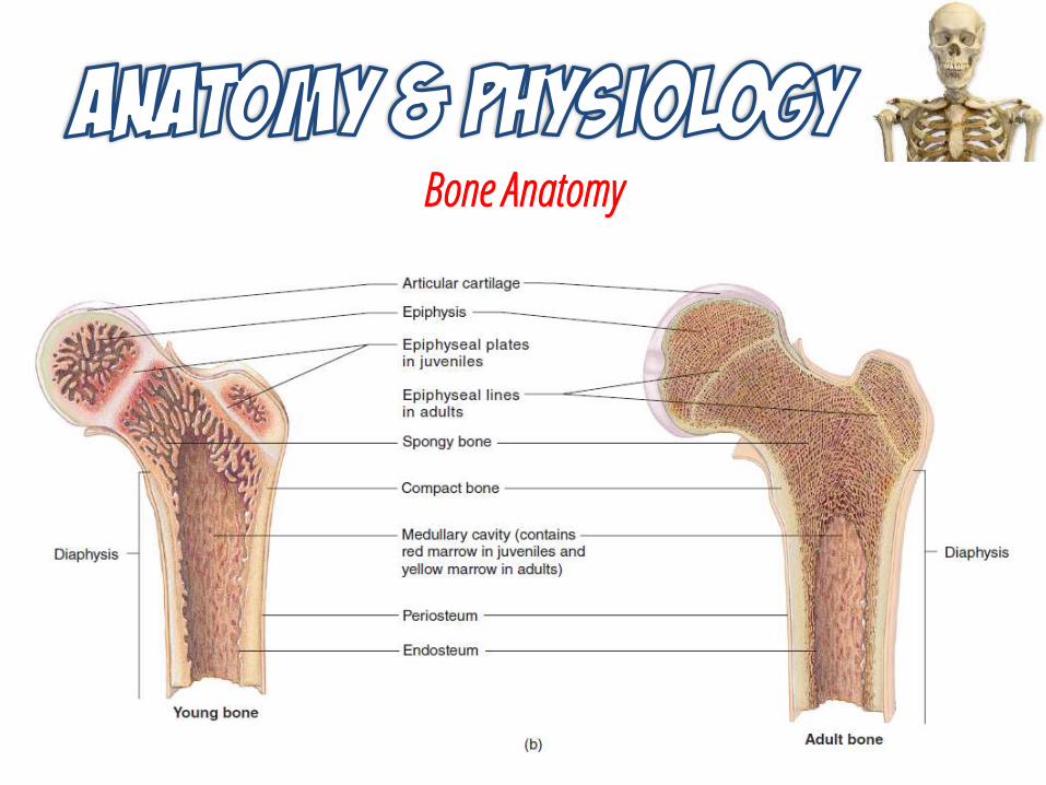
Bone Anatomy

Gross Anatomy

Factors Affecting Bone Growth
Minerals
Calcium and phosphorus make bone extracellular matrix hard.
Magnesium helps form bone extracellular matrix.
Fluoride helps strengthen bone extracellular matrix.
Manganese activates enzymes involved in synthesis of bone extracellular matrix.

Factors Affecting Bone Growth
Nutrition
Vitamin A needed for the activity of osteoblasts during remodeling of bone.
Vitamin D helps build bone by increasing absorption of calcium from gastrointestinal tract into blood.
Vitamin C is necessary for collagen synthesis by osteoblasts.
Vitamins K and B12 needed for synthesis of bone proteins.

Factors Affecting Bone Growth
Hormones
Growth hormone increases general tissue growth.
Insulin like growth factors (IGFs) promotes normal bone growth.
Thyroid hormone is required for normal growth of all tissues, including cartilage.
Insulin promotes normal bone growth by increasing the synthesis of bone proteins.

Factors Affecting Bone Growth
Hormones
Sex hormones (estrogens and testosterone); stimulate osteoblasts and promote the sudden “growth spurt” that occurs during the teenage years.
Parathyroid hormone (PTH) promotes bone resorption by osteoclasts; enhances recovery of calcium ions from urine; promotes formation of the active form of vitamin D (calcitriol).
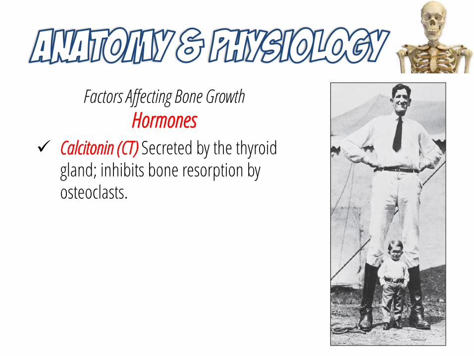
Factors Affecting Bone Growth
Hormones
Calcitonin (CT) Secreted by the thyroid gland; inhibits bone resorption by osteoclasts.

Factors Affecting Bone Growth
Exercise
Weight-bearing activities stimulate osteoblasts and, consequently, help build thicker, stronger bones and retard loss of bone mass that occurs as people age.

Bone Remodeling

Bone Healing

There are three types of muscle tissue: skeletal, smooth, and cardiac.
Functions
Movement of the body
Maintenance of posture
Respiration
Production of body heat
Communication
Constriction of organs and vessels
Contraction of the heart

The major characteristics of muscle tissue are skeletal, smooth, and cardiac muscle.
Skeletal muscle, with its associated connective tissue, constitutes about 40% of the body’s weight.
Is responsible for locomotion, facial expressions, posture, respiratory functions, and many other body movements.
Cardiac muscle is found only in the heart, and its contractions provide the major force for moving blood through the circulatory system.

The major characteristics of muscle tissue are skeletal, smooth, and cardiac muscle.
Smooth muscle is found in the walls of hollow organs and tubes, in the interior of the eye, and in the walls of blood vessels, among other areas.
Is responsible for propelling urine through the urinary tract, mixing food in the stomach and the small intestine, dilating and constricting the pupil of the eye, and regulating the flow of blood through blood vessels.

Properties of Muscular Tissue
Electrical excitability is the ability to respond to certain stimuli by producing electrical signals called action potentials.
Contractility is the ability of muscular tissue to contract forcefully when stimulated by an action potential.
Extensibility is the ability of muscular tissue to stretch without being damaged.
Elasticity is the ability of muscular tissue to return to its original length and shape after contraction or extension.

Skeletal Muscle Structure
Muscles are attached to bones by tendons.
Muscle contraction causes most body movements by pulling one of the bones toward the other across a movable joint.
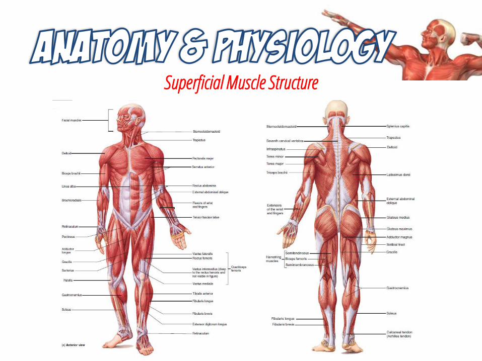
Superficial Muscle Structure

Skeletal Muscle Structure
A muscle is composed of muscle fasciculi, each surrounded by perimysium.
At the level of the perimysium, axons of neurons branch, and each branch extends to a muscle fiber.

Parts of a Muscle
The muscle fiber contains several myofibrils.
A single sarcomere of a myofibril is composed of actin myofilaments and myosin myofilaments.

Joints and MovementJoints are classified structurally as
fibrous, cartilaginous, or synovial.

Functional Classification of Joints
Type Description Examples
Synarthrosis Immovable joint Skull suturesEpiphyseal platesJoint between first rib and manubrium of sternum
Amphiarthrosis Slightly movable joint Vertebral jointsJoint of the symphysis pubis
Diarthrosis Freely movable joint Joints of the extremitiesShoulder jointsHip joints

Types of Synovial Joints
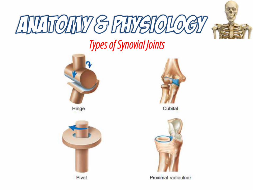
Types of Synovial Joints

Types of Synovial Joints

Range of Motion
Active range of motion is the amount of movement accomplished by contracting the muscles that normally act across a joint.
Passive range of motion is the amount of movement accomplished when the structures that meet at the joint are moved by an outside force.

Types of Movement

Types of Movement

Types of Movement
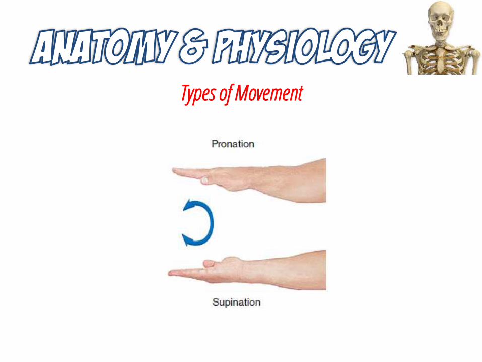
Types of Movement

Types of Movement

Types of Movement

Types of Movement

Types of Movement

Types of Movement

Types of Movement

Aging and the Joints
SYSTEM STRUCTURAL CHANGES FUNCTIONAL CHANGES
Bones • Gradual, progressive loss of bone mass after age 30
• Vertebrae collapse
• Bones fragile and prone to fracture: vertebrae, hip, wrist
Muscles • Increase in collagen and resultant fibrosis
• Muscles diminish in size (atrophy); wasting
• Tendons less elastic
• Loss of strength and flexibility• Weakness• Fatigue• Stumbling• Falls
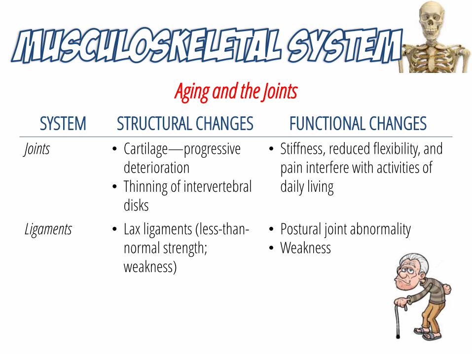
Aging and the Joints
SYSTEM STRUCTURAL CHANGES FUNCTIONAL CHANGES
Joints • Cartilage—progressive deterioration
• Thinning of intervertebral disks
• Stiffness, reduced flexibility, and pain interfere with activities of daily living
Ligaments • Lax ligaments (less-than-normal strength; weakness)
• Postural joint abnormality• Weakness

References
1. LeMone, P. et al. (2011). Medical-Surgical Nursing: Critical Thinking in Client Care. 5th Edition. New Jersey: Pearson Education, Inc.
2. Seeley. R. et al. (2014). Seeley’s Anatomy and Physiology. 10th
Edition. New York: McGraw-Hill Companies, Inc.3. Tortora, G. & Derrickson, B. (2009). Principles of Anatomy and
Physiology. 12th Edition. New Jersey: John Wiley and Sons, Inc.

ASSESSING MUSCULOSKELETAL FUNCTION
JOFRED M. MARTINEZ, RN, MANUniversity of San Agustin Review CenterIloilo City, Philippines

Diagnostic Tests
Test Purpose Nursing Interventions
Arthrocentesis Obtain synovial fluidfrom a joint for diagnosis or to remove excess fluid.
After the procedure, apply a compression dressing and tell the patient to report any bleeding and leakage of fluid.

Diagnostic Tests
Test Purpose Nursing Interventions
Arthroscopy Used to perform surgery and diagnose diseases of the patella, meniscus, and synovial and extrasynovialmembranes.
Fluid may be drained from the joint and tissue removed for biopsy.
If general anesthesia is used, tell the patient not to eat or drink fluids after midnight prior to the procedure.
Following the procedure, assess for bleeding and swelling, apply ice to the area if prescribed, and instruct patient to avoid excessive use of the joint for 2 to 3 days.

Diagnostic Tests
Test Purpose Nursing Interventions
Bone density (BD) Dual energy x-ray
absorptiometry (DEXA)
Quantitative ultrasound (QUS)
Bone mineral density (BMD)
Bone absorptiometry
Evaluate bone mineral density and to evaluate degree of osteoporosis. DEXA can calculate the size and thickness of bone.
Normal Value: 1 standard deviation below peak bone mass.
Instruct patient to remove all metal objects from the area to be scanned.
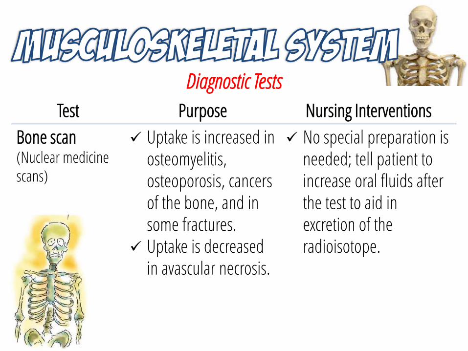
Diagnostic Tests
Test Purpose Nursing Interventions
Bone scan(Nuclear medicine scans)
Uptake is increased in osteomyelitis, osteoporosis, cancers of the bone, and in some fractures.
Uptake is decreased in avascular necrosis.
No special preparation is needed; tell patient to increase oral fluids after the test to aid in excretion of the radioisotope.

Diagnostic Tests
Test Purpose Nursing Interventions
Gallium/Thallium Scans
Gallium concentrates in areas of tumors, inflammation, and infections.
Thallium detects osteosarcoma.
Check if facility recommends that children and pregnant women stay a few feet away from the patient for the first 48 hours
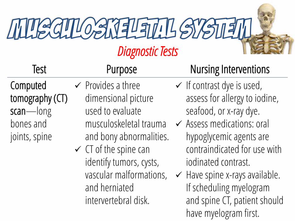
Diagnostic Tests
Test Purpose Nursing Interventions
Computed tomography (CT) scan—long bones andjoints, spine
Provides a three dimensional picture used to evaluate musculoskeletal trauma and bony abnormalities.
CT of the spine can identify tumors, cysts, vascular malformations, and herniated intervertebral disk.
If contrast dye is used, assess for allergy to iodine, seafood, or x-ray dye.
Assess medications: oral hypoglycemic agents are contraindicated for use with iodinated contrast.
Have spine x-rays available. If scheduling myelogramand spine CT, patient should have myelogram first.

Diagnostic Tests
Test Purpose Nursing Interventions
Computed tomography (CT) scan—long bones andjoints, spine
If long-bone and joint CT, nuclear medicine tests to locate “hot spots” should be done before CT.
After the test, if contrast dye was used, monitor for delayed allergic reaction (rash, itching, headache, vomiting) and instruct patient to increase fluid intake.

Diagnostic Tests
Test Purpose Nursing Interventions
Electromyogram (EMG)
Measures the electrical activity of skeletal muscles at rest and during contraction.
Tell the patient not to drink fluids containing caffeine or to smoke for 3 hours before the test, and not to take medications such as muscle relaxants, anticholinergics, or cholinergics.
If serum enzymes such as SGOT, CPK, or LDH are ordered, the specimen should be drawn before the EMG or 5 to 10 days after the EMG.

Diagnostic Tests
Test Purpose Nursing Interventions
Magnetic resonanceimaging (MRI)
Used in diagnosis and evaluation of avascular necrosis, osteomyelitis, tumors, disk abnormalities, and tears in ligament or cartilage.
Inform patient of need to lie still during the examination.
Assess for any metallic implants (such as pacemakers, clips on brain aneurysms, body piercings, tattoos, shrapnel). If present, notify imaging physician.
Remove transdermal medication patches (both OTC and prescribed) unless otherwise ordered (FDA, 2009).

Diagnostic TestsTest Purpose Nursing Interventions
Magnetic resonanceimaging (MRI)
Replace the patch following the procedure.
Tell the patient to inform the staff about the patch when making the appointment and when completing the admission information.
Ask if patient is pregnant; if so the test is not performed. Ask about claustrophobia; if a problem request patient to ask for a relaxing medication to take prior to the MRI.

Diagnostic Tests
Test Purpose Nursing Interventions
Skeletal x-ray Identify and evaluate bone density and structure.
Ask women if they are pregnant; x-rays should be avoided during the first trimester.
No special preparation is needed for skeletal x-rays.

Diagnostic Tests
Test Purpose Nursing Interventions
Somatosensory evoked potential (SSEP)
Measures nerve conduction along pathways to evaluate evoked potential of muscle contractions.
Used to identify dysfunction of lower motor neurons as well as muscle disease.
No special preparation is needed.

Diagnostic Tests
Test Purpose Nursing Interventions
Bone or MuscleBiopsy
Involves needle aspiration (closed) or surgical extraction (open) of bone or muscle tissue.
Monitor site of biopsy for bleeding.
Provide normal wound care for open biopsy.
Perform neurovascular assessments as needed.

Blood Tests
Test Purpose Normal Value
Alkaline phosphatase (ALP)
Identify bone diseases. Increased in bone cancer, Paget’s disease, healing fractures, rheumatoid arthritis, osteoporosis.
42–136 unit/L ALP120–130 unit/L ALP2(increases slightly with aging)
Calcium (Ca) Monitor calcium levels and detect calcium imbalances.Decreased with lack of calcium and vitamin D intake, and malabsorption from the gastrointestinal tract. Increased in bone cancer and multiple fractures.
4.5–5.5 mEq/L or9–11 mg/dL (serum)

Blood Tests
Test Purpose Normal Value
Phosphorus (P),phosphate (PO4)
To assess phosphorus levels. Increased with bone tumorsand healing fractures.
1.7–2.6 mEq/L or2.5–4.5 mg/dL
Rheumatoid factor (RF)
To diagnose rheumatoid arthritis (RA) (positive for RA at > 1:80).Also increased in lupus erythematosus and scleroderma.
< 1:20 titer

Blood Tests
Test Purpose Normal Value
Uric acid Diagnose and monitor the treatment of gout. Panic level considered > 12 mg/dL.
Male: 3.5–8.0 mg/dLFemale: 2.8–6.8 mg/dL
Human leukocyte antigen
Diagnose diseases such as juvenile RA or ankylosing spondylitis..
Match or no match;no normal values (HLA)
Creatine kinase (CK)
Diagnose muscle trauma or disease. Increased in muscular dystrophy and traumatic injuries (specifically, CPK-MM isoenzyme).
94%–100%

Subjective Health Assessment
Category What to Ask Rationale
Demographics Age, gender, socioeconomic status
Increased age, being female, and lower socioeconomic status increase risk of musculoskeletal injury/problems.
Occupation Enables nurse to begin planning for discharge teaching if the patient has to alter his or her employment.

Subjective Health Assessment
Category What to Ask Rationale
Previous Health History
Allergies Prevents exposure to medication or compounds used in diagnostic tests, treatments, and therapies.
Activities patient participates in
Provides information regarding the level of activity the patient had before the concern. Smoking and a sedentary lifestyle are risk factors for musculoskeletal problems.

Subjective Health Assessment
Category What to Ask Rationale
Previous Health History
Diet history Dietary intake such as calcium and vitamin D influences some musculoskeletal disorders.
Family history Some musculoskeletal conditions have genetic and familial tendencies.

Subjective Health Assessment
Category What to Ask Rationale
History of Injuryor PresentConcern
History of the injury (if there was one)
Provides information that helps in the diagnosis of the problem, as well as making you aware of possible complications of the injury.
Pain (use pain assessment scale)
Provides information about severity of the condition and effectiveness of the treatment and therapy.

Subjective Health Assessment
Category What to Ask Rationale
PsychosocialAssessment
Determine if deformities, changes in body image, self-concept, socialization, oremployment are present.
The patient may need assistance with strategies to cope with the stress of a possible chronicmusculoskeletal condition.
Determine coping skills. Some musculoskeletal conditions require lifestyle alterations that can cause increased stress anddifficulties in coping.

Objective Health Assessment
Category What to Ask Rationale
PhysicalExamination
Inspect, palpate, and observe range of motion(ROM) of affected areas.
Altered gait, tone, size, shape, posture, contractures, deformities, ROM, pain, and effects on ADLs can be determined.
Assess color, warmth, circulation, andmovement of affected areas.
Nerve function, sensation, movement, weakness, and the potential development of compartmentsyndrome can be determined.
Palpate all pulses below involved area.
Alterations may indicate altered vascular integrity of affected area or demonstrate developing compartment syndrome.

Objective Health Assessment

Objective Health Assessment

Muscle Grading Scale
Scale Assessment Description
0 (No visible) contraction; paralysis
1 Can feel contraction of muscle but there is no movement of limb
2 Passive ROM
3 Full ROM against gravity
4 Full ROM against some resistance
5 Full ROM against full resistance

Neurovascular Assessment
Monitor Report
Color Pallor, cyanosis, redness, or discoloration
Temperature Unusual coolness or warmth
Pain Pain that is worse on passive motion; pain that no longer responds to analgesics
Movement Alterations in movement
Sensation Alterations in feeling; tingling or paresthesias
Pulses Diminished or absent distal pulses
Capillary refill Nail bed that does not blanch in 3–5 seconds
References1. LeMone, P. et al. (2011). Medical-Surgical Nursing: Critical
Thinking in Client Care. 5th Edition. New Jersey: Pearson Education, Inc.
2. Smeltzer, S. C. et al. (2010). Brunner & Suddarth’s Textbook of Medical-Surgical Nursing. 12th Edition. Philadelphia: Lippincott Williams and Wilkins.
3. Williams. L. S. & Hopper, P. D. (2011). Understanding Medical-Surgical Nursing. 5th Edition. Philadelphia: F. A. Davis Company