muscular system
DESCRIPTION
Muscular System. Dr. Anderson GCIT. Function. Muscle is a collective term for tissue that causes movement of the body, or materials through the body Muscle can also serve other functions such as Stabilizing the body Producing heat. Muscle Tissue. Three main types of muscle - PowerPoint PPT PresentationTRANSCRIPT

Muscular System
Dr. Anderson GCIT

Function• Muscle is a collective term for tissue that
causes movement of the body, or materials through the body
• Muscle can also serve other functions such as – Stabilizing the body– Producing heat

Muscle Tissue• Three main types of
muscle– Skeletal – attaches to
skeleton and enables body movement - striated, voluntary
– Smooth – lines body cavities to move fluids throughout the body – smooth, involuntary
– Cardiac – makes up the heart – striated, involuntary

Physiology• Muscles function by
converting the energy stored in ATP into mechanical energy that the muscle can use to contract – muscles only expend energy to get shorter

Muscle Characteristics
• Excitability – the muscle’s ability to react to a stimulus
• Contractility – the ability to shorten forcibly
• Extensibility – the ability to be stretched
• Elasticity – the ability for a muscle to return to its pre-stretched length
Skeletal Muscle Anatomy
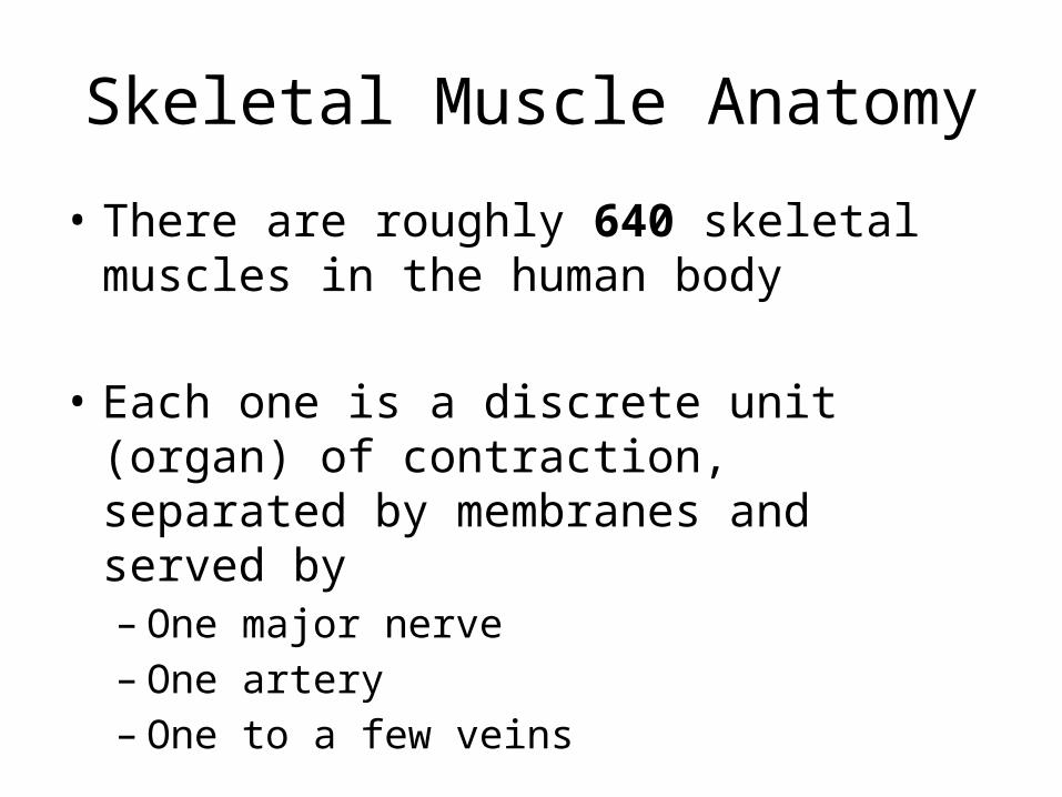
• There are roughly 640 skeletal muscles in the human body
• Each one is a discrete unit (organ) of contraction, separated by membranes and served by– One major nerve– One artery– One to a few veins
Muscle Connections/SeparationsIndividual muscles are separated by sheaths of connective tissue
• Epimysium – surrounds entire muscle
• Perimysium and fascicles – perimysium surrounds bundles of muscle fibers (fascicles)
• Endomysium – very thin sheath of tissue that surrounds each muscle fiber (cell)
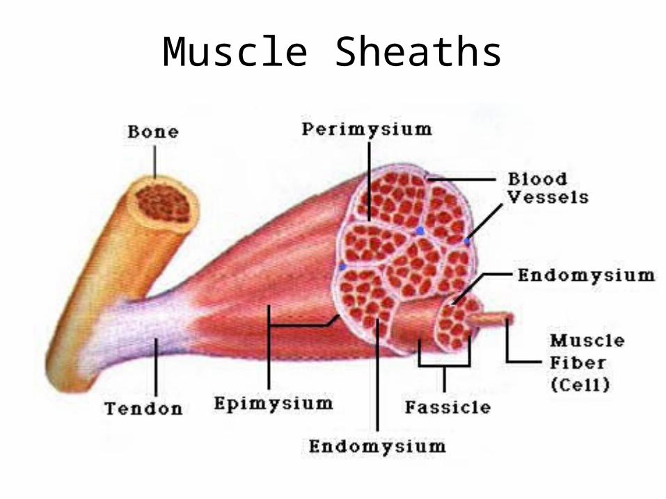
Muscle Sheaths

Skeletal Origins and Insertions• Skeletal muscles are connected to bone in at least
two places
• Origin – end connected to the immovable bone• Insertion – connected to moveable side
• Direct – Epimysium of muscle is fused directly to the periosteum of bone (less common)
• Indirect – The connective tissue connects to bone via a tendon (more common)

Direct vs. Indirect Muscle Attachment
Direct Attachment (to skull)
Indirect Attachments

Muscle Fiber Anatomy• Sarcolemma – cell membrane of muscle cell• Sarcoplasm – cytoplasm of muscle cell• Glycosomes – granules of stored glycogen• Myoglobin – red pigment that can store O2 in
muscles

Myofibrils• Complex organelle composed of bundles of
myofilaments in a muscle fiber (appear banded in skeletal muscle)
Sarcomere• The smallest contractile unit of the muscle
• Composed of protein filaments (myofilaments) that shorten the muscle upon stimulation
• Here’s where it gets complicated…..
Thick and Thin Filaments
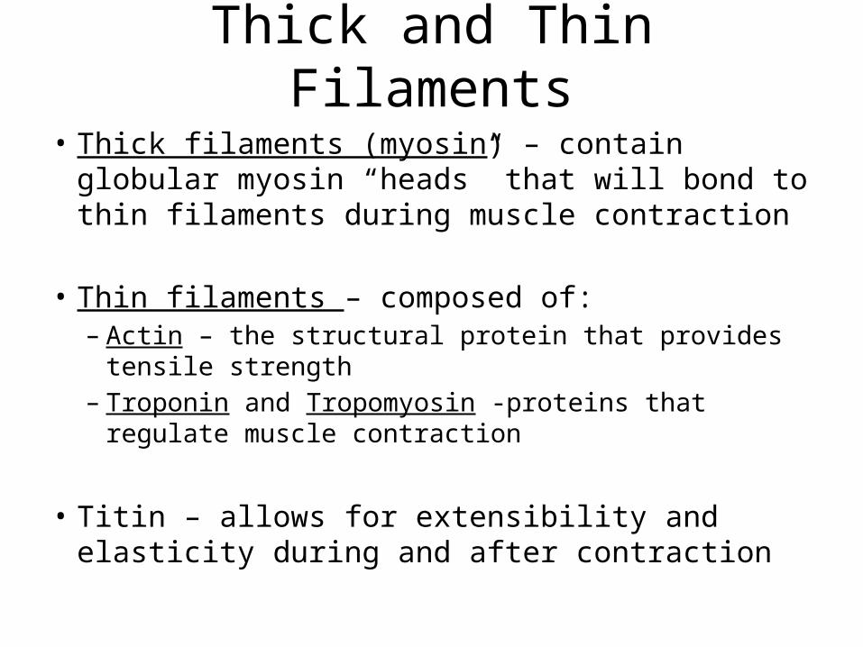
• Thick filaments (myosin) – contain globular myosin “heads” that will bond to thin filaments during muscle contraction
• Thin filaments – composed of:– Actin – the structural protein that provides tensile strength– Troponin and Tropomyosin -proteins that regulate muscle
contraction
• Titin – allows for extensibility and elasticity during and after contraction

Ultrastucture of Muscle Fibrils
Thick Filament (Myosin)
Thin Filament (Actin)
Titin
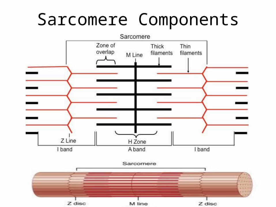
Sarcomere Components

Muscle Contraction
• Thick filament heads and thin filaments link and form cross-bridges– The heads swivel and pull the opposing thin
filaments of the sarcomere together
– Helpful video!– http://www.youtube.com/watch?v=ELyoJZom5N0

Regulatory Proteins
• Tropomyosin – bound within the actin filaments and block myosin-binding sites (when muscle is relaxed)
• Troponin – helps stabilize actin and binds Ca+ ions
Sarcoplasmic Reticulum
• Specialized smooth endoplasmic reticulum (sER) that surrounds each myofibril– Most run longitudinally along the length of the cell– However, between the thick filaments and the Z
disc, they form pairs of terminal cisternae
• This organelle is critical in that it stores and releases Ca+ ions, which are the triggers for muscle contraction

T- Tubules
• An invagination of the sarcolemma – located between the terminal cisternae in each sarcomere
• The structure of 2 Terminal cisternae divided by t-tubules is called a TRIAD
• Why is all of the surface area in triads necessary?
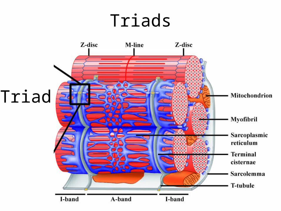
Triads
Triad
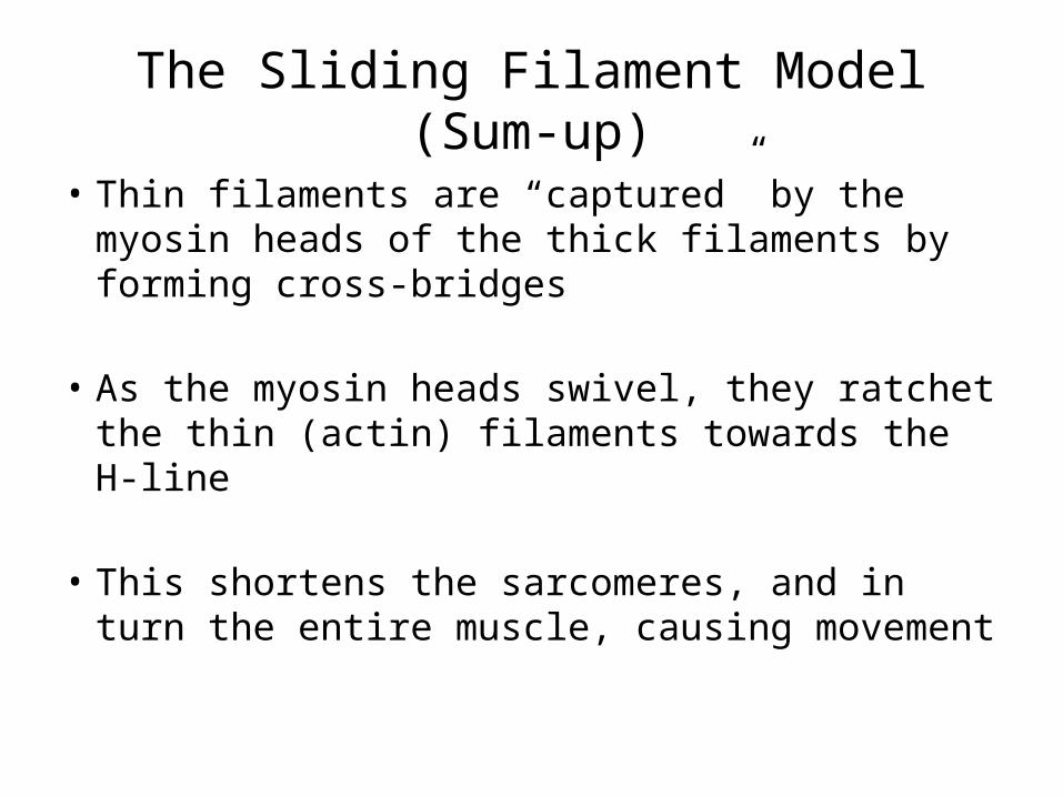
The Sliding Filament Model (Sum-up)
• Thin filaments are “captured” by the myosin heads of the thick filaments by forming cross-bridges
• As the myosin heads swivel, they ratchet the thin (actin) filaments towards the H-line
• This shortens the sarcomeres, and in turn the entire muscle, causing movement
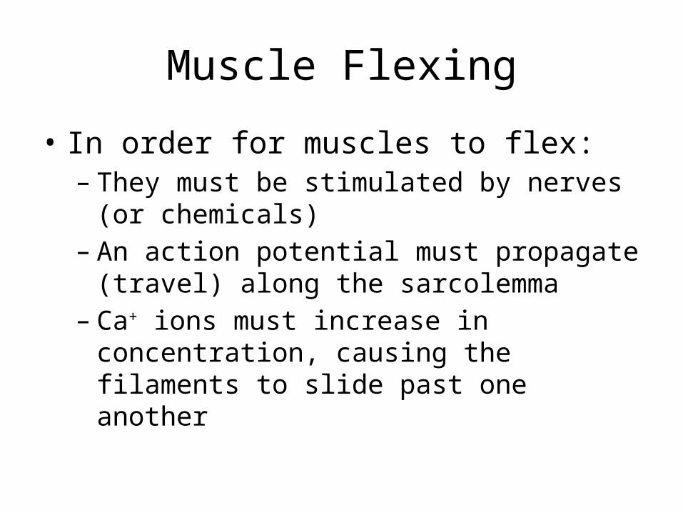
Muscle Flexing
• In order for muscles to flex:– They must be stimulated by nerves (or chemicals)– An action potential must propagate (travel) along
the sarcolemma– Ca+ ions must increase in concentration, causing
the filaments to slide past one another

Muscle Innervation (Motor Nerves)
• Motor nerves from the spinal cord innervate the muscle to communicate with the muscle fibers
• The fibers do not touch but come exceedingly close (1-2 nm apart) forming the synaptic cleft
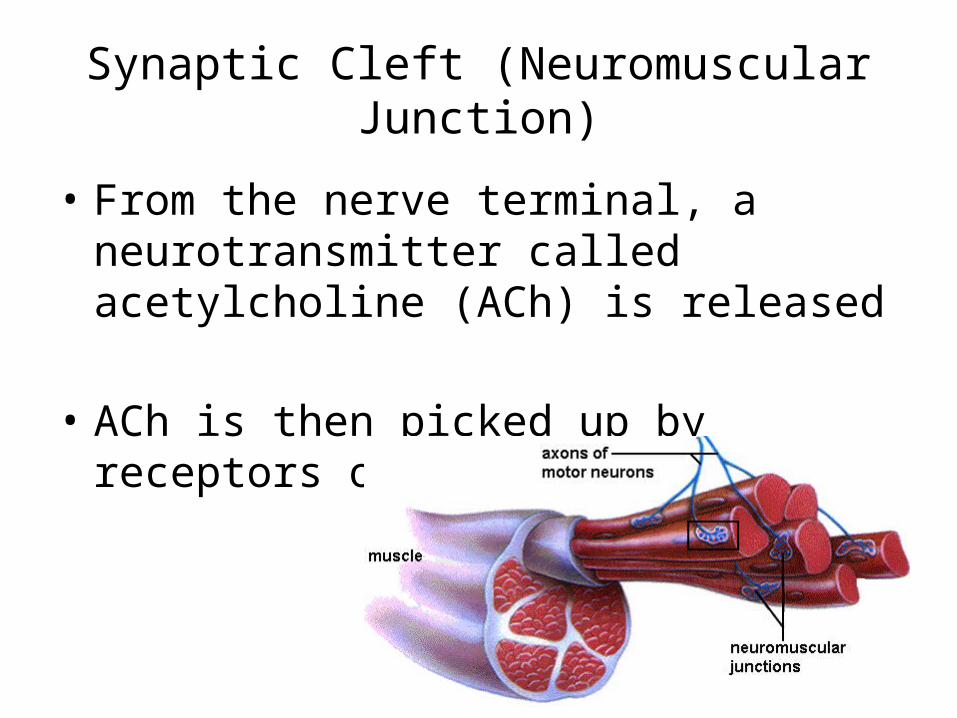
Synaptic Cleft (Neuromuscular Junction)
• From the nerve terminal, a neurotransmitter called acetylcholine (ACh) is released
• ACh is then picked up by receptors on the sarcolemma

Electrical Gradient• Normally, the sarcolemma is polarized (more
negative on the inside than the outside)
• When ACh binds to it receptors, channel proteins let ions (Ca+ and K+) pass
• More Ca+ ions stream into the cell than K+ ions stream out, therefore the inside of the cell becomes less negative – this is called depolarization

Synaptic Cleft Arrangement

Helpful Video
• http://www.youtube.com/watch?v=kvMFdNw35L0
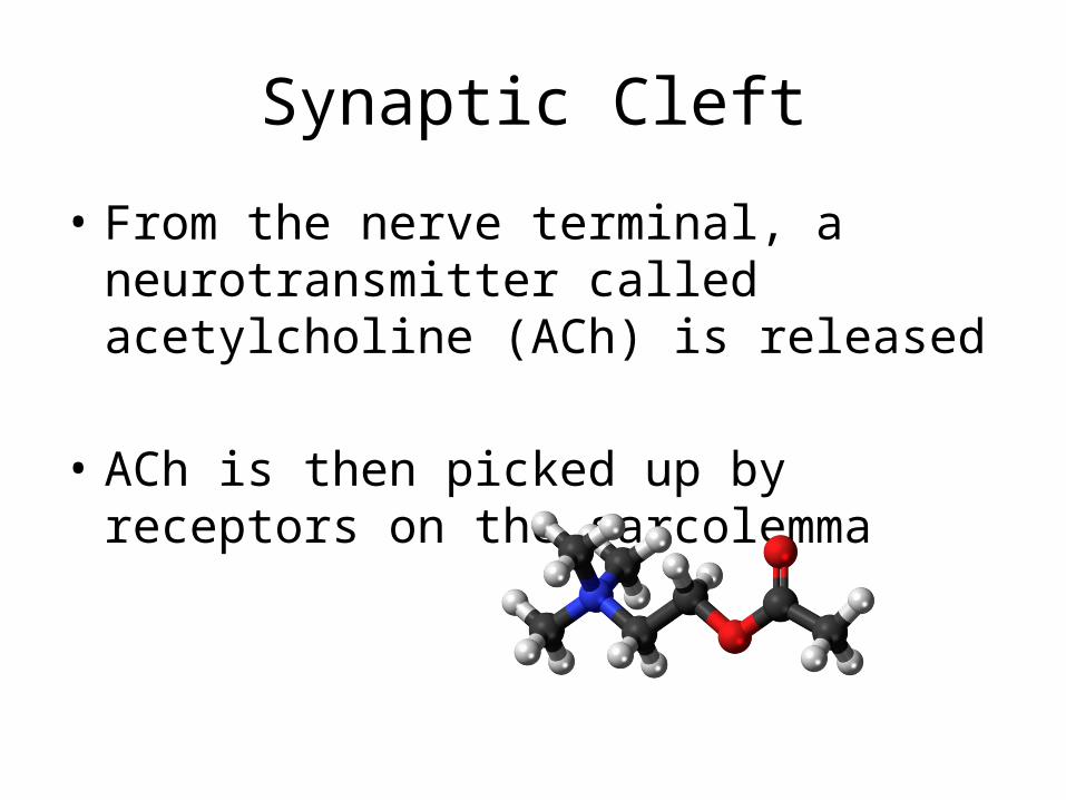
Synaptic Cleft
• From the nerve terminal, a neurotransmitter called acetylcholine (ACh) is released
• ACh is then picked up by receptors on the sarcolemma

Electrical Gradient• Normally, the sarcolemma is polarized (more
negative on the inside than the outside)
• When ACh binds to receptors, it opens channel proteins which lets ions (Ca+ and K+) pass
• More Ca+ ions stream into the cell than K+ ions stream out, therefore the inside of the cell becomes less negative – this is called depolarization
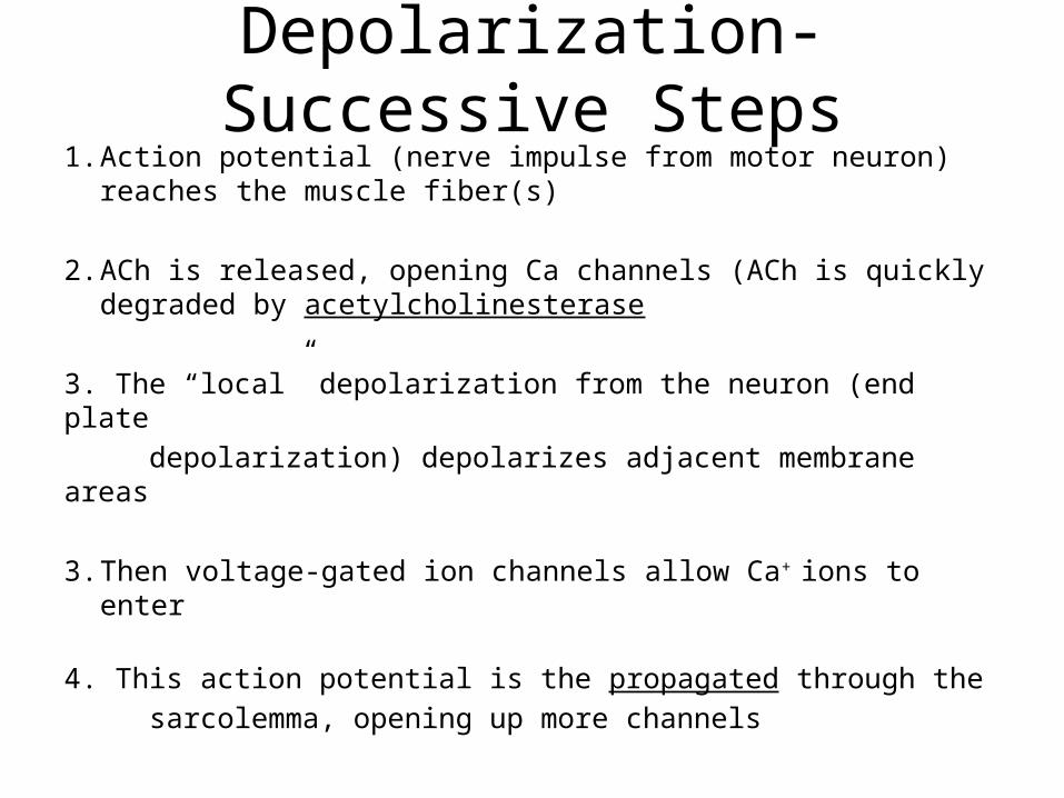
Depolarization- Successive Steps1. Action potential (nerve impulse from motor neuron)
reaches the muscle fiber(s)
2. ACh is released, opening Ca channels (ACh is quickly degraded by acetylcholinesterase
3. The “local” depolarization from the neuron (end plate depolarization) depolarizes adjacent membrane areas
3. Then voltage-gated ion channels allow Ca+ ions to enter
4. This action potential is the propagated through the sarcolemma, opening up more channels

• 5. Voltage-dependent gates open, and more Ca+ floods through the sarcolemma
• 6. The influx of Ca+ leads to muscle contraction by interacting with tropomyosin, freeing the myosin binding sites
Depolarization- Successive Steps
Repolarization
• K+ channels open, which allows K+ to flood out of the cell, restoring the negative conditions inside
• How do we restore the electrochemical balance of the cell?
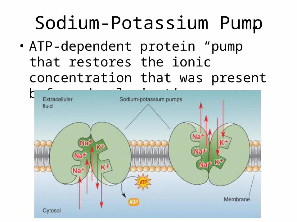
Sodium-Potassium Pump• ATP-dependent protein “pump” that restores
the ionic concentration that was present before depolarization
Excitation-Contraction Coupling• Calcium ion influx (through T-tubules and into
terminal cisternae)
• Calcium binds to troponin and removes the blocking action of tropomyosin, allowing myosin to bind to the thin (actin) filaments
• Myosin cross-bridges with actin, leading to contraction

Let’s Sum Up
• http://www.youtube.com/watch?v=70DyJwwFnkU

Muscle Innervation
• The single motor nerve that innervates a muscle can contain hundreds of motor neurons
• A motor unit is a motor neuron and all of the muscle fibers that it serves – the more motor units, the larger the force (more fibers contract)– Which muscles might have small motor units?
Large motor units?
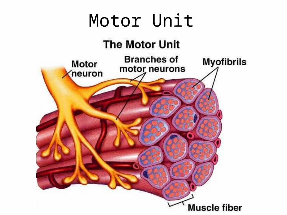
Motor Unit

Strength of Contraction• Increasing strength can be due to – Increased frequency of stimuli• The less time between nerve stimulation,
the greater the force of flexion due to Ca being left over in the muscle
– Increased number of stimulated motor units• The more motor units stimulated, the
greater the force of flexion

(Frequency of Stimuli)
TwitchLatent – Ca floods inContraction – Myosin heads pull and make sarcomeres shorterRelaxation – muscle activity stops as ions are replaced to normal
concentrations
Contraction Strength
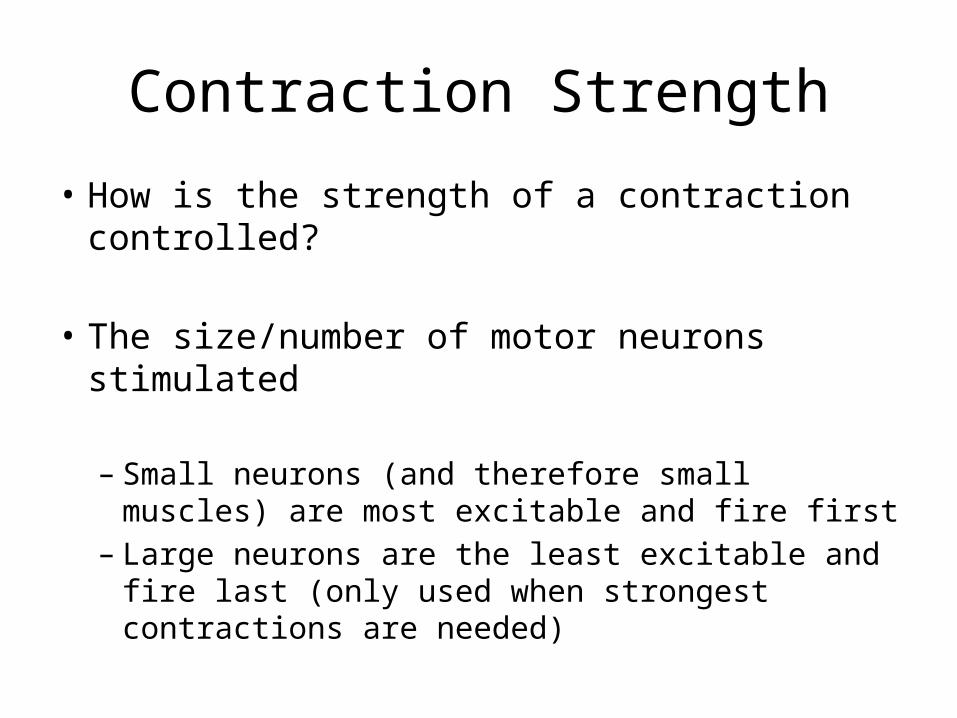
• How is the strength of a contraction controlled?
• The size/number of motor neurons stimulated
– Small neurons (and therefore small muscles) are most excitable and fire first
– Large neurons are the least excitable and fire last (only used when strongest contractions are needed)

Isotonic vs Isometric Contraction
• Isotonic – muscle moves when flexed– Concentric – muscle moves and shortens– Eccentric – muscle moves while lengthening
• Isometric – muscle flexes, but does not shorten or lengthen

Muscle Physiology
• Where do muscles get the energy to flex, and keep flexing?
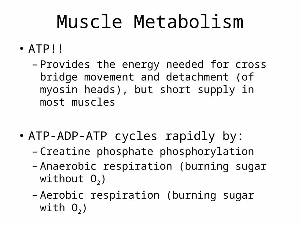
Muscle Metabolism• ATP!!– Provides the energy needed for cross bridge
movement and detachment (of myosin heads), but short supply in most muscles
• ATP-ADP-ATP cycles rapidly by:– Creatine phosphate phosphorylation– Anaerobic respiration (burning sugar without O2)
– Aerobic respiration (burning sugar with O2)

Creatine Phosphate
• Another high-energy molecule that converts ADP to ATP by direct phosphorylation via an enzyme called creatine kinase
ADP + creatine phosphate
creatine kinase
ATP + creatine

Rigor Mortis• Lack of ATP after
death causes contractures, where myosin heads cannot detach from the actin filaments, locking up the muscle until muscle proteins start to break down
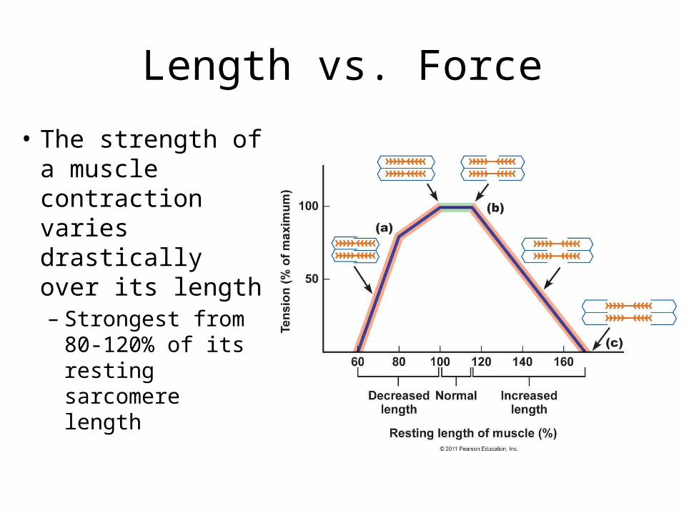
Length vs. Force
• The strength of a muscle contraction varies drastically over its length– Strongest from
80-120% of its resting sarcomere length
Velocity of Contraction
• “Fast twitch” fibers – contract rapidly– Large fibers that depend on stored glycogen reserves (does
not depend on delivery of nutrients)– Low myoglobin– Tires quickly
• “Slow fibers” – contract slowly– Uses nutrients delivered from blood, therefore rich capillary
supply– High myoglobin– High endurance
Smooth Muscle
• Different microstructure, but similar function as skeletal muscle
• Flexing is under control of the autonomic nervous system, but also under the control of local chemistry

Smooth Muscle
• Lines the:– Stomach and
intestines– Blood vessels– Lungs – Urinary system– Reproductive organs
Muscle Sheets
• In Digestive organs, smooth muscle sheets are arranged at 90 degree angles to each other
• What will this lead to during flexing?http://www.youtube.com/watch?v=pfNYbzGEgO8

Smooth Muscles• Smooth Muscle Cells
are:– Uninucleate– Spindle-shaped– Much narrower and
shorter than skeletal muscle cells
– Calveoli instead of T-tubules
– No sarcomeres!

Smooth Muscle Sheaths• Only a small amount of endomysium runs
between smooth muscle cells• This network is also perfused and innervated
Longitudinal section Transverse Section

Distribution
• Most smooth muscle occurs in walls of organs that transport fluids through the body (liquids and gases)
• Many organs have muscle cells arranged in 2 layers that run perpendicular to each other
– Why?

Peristalsis
• Rhythmic, alternate contractions between muscle sheets in digestive system
• By alternating contractions, the bolus of food (or waste) can be moved through the intestines
• http://www.youtube.com/watch?v=o18UycWRsaA

Stimulation
• Smooth muscles lack synaptic clefts, but neurotransmitter is released in the general area of muscle cells by nerve bundles called varicosities

Contraction – Smooth vs. Skeletal
• Thick and thin filaments arranged diagonally
• Calmodulin instead of troponin complex
• Thick filaments have myosin heads across their entire length

Anchoring of Smooth Muscle• Intermediate Filaments – non-contractile
filaments that anchor to dense bodies which are attached to adjacent sarcolemmas– Why is this important?
Smooth Muscle CellContraction vs. Extension
• Ca++ binds to calmodulin, which activates myosin kinase which phosphorylates the myosin heads, activating them
• Myosin is then able to form cross-bridges with actin, and contract the muscle
Response to Stimuli
• Smooth muscle takes up to 30 times longer to react to a stimulus than skeletal muscle
• Smooth muscles may stay in a contracted (flexed) state for long periods, as myosin heads remain attached to actin filaments for longer periods
Nerve- Based Contraction
• Contraction of smooth muscle is still due to the release of neurotransmitters– Highly dependent on receptors for
neurotransmitters– E.g. norepinephrine relaxes smooth muscle in
bronchioles
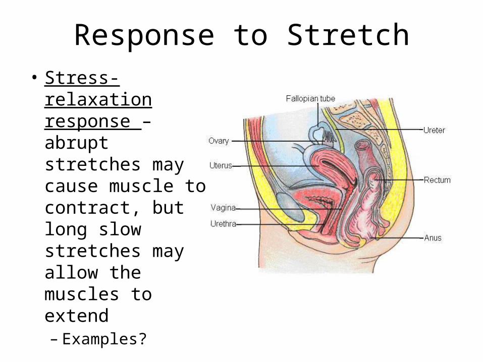
Response to Stretch• Stress-relaxation
response – abrupt stretches may cause muscle to contract, but long slow stretches may allow the muscles to extend– Examples?

Stretch and Tension
• Different arrangement of filaments allows smooth muscle to be stretched quite and still contract strongly
• Example?