multiple myeloma in older adults: effect of therapies on ... · multiple myeloma in older adults:...
TRANSCRIPT

Multiple Myeloma in Older Adults: Effect of Therapies on Nutritional Status
Sodexo Mid-Atlantic Dietetic Internship
Major Case Study
Amanda Schroeder Hege
January 27, 2014

Hege 2
ABSTRACT
A 90 year old Caucasian female, DL, was transferred to Hebrew Home of Greater
Washington (HHGW) on December 13, 2013, with IgG kappa multiple myeloma with worsening
renal function. Multiple myeloma is a neoplastic proliferation of plasma cells characterized by
an increase in calcium levels, renal dysfunction, anemia, and bone pain. DL visited her primary
care physician on November 8, 2013 with extreme fatigue, was found to be anemic, and a
subsequent workup revealed an IgG kappa paraprotein with bone marrow consistent with
multiple myeloma. She was admitted to Suburban Hospital from November 26th
to December
13th
for treatment. After stabilization, she began combination chemotherapy recognized as
CyBort-D therapy. Upon admission to HHGW, her nutrition diagnoses were inadequate energy
intake R/T increased nutrient needs due to catabolic illness, poor appetite, and dysgeusia AEB
stated energy intake <50% of estimated energy needs, along with unintended weight loss R/T
increased nutrient needs due to catabolic illness, dysgeusia, nausea AEB 7% weight loss x 10
days. After discussion with the patient, the intervention consisted of continuing a regular diet,
adding a 10am vanilla Resource 2.0 and 2pm chocolate pudding, and educating DL on methods
to manage signs and symptoms associated with cancer. Goals included tolerating the diet,
adequate nutrition/hydration to meet estimated energy needs, and greater than 50%-75% intake
of meals and supplements. The nutrition team monitored, evaluated, and followed-up on her
weight, % intake, signs and symptoms, and nutrition-related lab values.

Hege 3
INTRODUCTION
DL is a 90 year-old Caucasian female admitted to Suburban Hospital from November
26th
to December 13th
and transferred to Hebrew Home of Greater Washington (HHGW) on
December 13th
for further rehabilitation and care. Her primary diagnosis was IgG kappa multiple
myeloma (MM) with worsening renal function.
DISCUSSION OF THE DISEASE
Multiple myeloma is a neoplastic proliferation of the plasma cells1. Bone marrow
produces the three types of blood cells: red blood cells, white blood cells, and platelets. Plasma
cells are mature B-lymphocyte cells, which are a type of white blood cell. The physiology of
multiple myeloma involves the interaction between myeloma plasma cells and bone marrow
stroma cells. The interaction between these two cells produces growth factors and cytokines
including vascular endothelial growth factor (VEGF), basic fibroblast growth factor (bFGF),
interleukin-6 (IL-6), tumor necrosis factor (TNF-α), and interleukin-1β (IL-1 β). Overproduction
of IL-6 is central to the pathogenesis of multiple myeloma because it is the plasma cell growth
factor that promotes growth of myeloma cells. In a healthy individual, plasma cells consist of
less than 10% of bone marrow. In a patient with multiple myeloma, their bone marrow is
comprised of greater than 10% plasma cells. The overproduction of myeloma cells causes them
to “crowd out” healthy red blood cells, white blood cells, and platelets.2
A plasma cell’s main function is to produce antibodies that fight infection. Multiple
myeloma plasma cells produce one type of antibody: a monoclonal “M” protein that encodes the
structures of immunoglobulin G and A. Multiple myeloma is characterized by the
overproduction of monoclonal immunoglobulin G (IgG) and IgA, which is referred to as the “M”
spike. It is also characterized by destructive lytic bone lesions that appear on an x-ray as

Hege 4
“punched-out” holes on the bone and a decrease in bone density (Figure 1). This type of cancer
is not confined to a specific bone or location within a bone, may involve the entire skeleton, and
accounts for 10% of all hematologic cancers3.
ETIOLOGY
The precise etiology has yet to be
determined and many researchers believe
it occurs spontaneously. Research has
shown that most individuals with MM
have genetic abnormalities in plasma
cells that may contribute to cancer such
as a defect or translocation in
chromosome 14, extra copies of chromosomes, and an abnormality in chromosome 13 in which
part or all is missing. Genetic, environmental, or occupational factors may contribute to a patient
developing MM. It is typically thought to be a disorder in older adults with a median age of
approximately 65 years. Males and African-Americans have twice the risk and incidence of this
type of cancer as females and Caucasians. Exposure to ionizing radiation, uranium5, chronic
infection with a virus such as HIV or Herpes 8, or farmers exposed to herbicides, insecticides,
and the pesticide dioxin causes an individual to be at a high risk. Approximately 19% of patients
with the benign condition monoclonal gammopathy of undetermined significance (MGUS),
which is a disease that resembles multiple myeloma but with lower levels of plasma and
monoclonal plasma cells and no symptoms, will develop multiple myeloma within two to
nineteen years.6
Figure 1: Lytic MM Bone Lesions in Skull4

Hege 5
Complications associated with MM
are related to an overproduction of
malignant plasma cells in bone marrow.
One complication is an impaired immunity
due to malignant myeloma cells inhibiting
the production of healthy plasma cells and
antibodies needed to fight infection and promote normal immunity. Various bone problems
occur including the erosion of bone mass, fractures, lytic bone lesions, a compression of the
spinal cord, weakness or paralysis of legs, bone pain in the chest or spine, and “punched out”
areas of bone most commonly found in vertebral bodies, skull, ribs, humerus, and femur (Figure
2).4 Approximately 50% of patients experience altered renal function due to eroding bones
causing an increased level of calcium in the blood. Hypercalcemia interferes with the kidney’s
ability to filter blood’s waste and proteinuria is present in 90% of patients with MM. A further
complication is anemia due to bone marrow replacement of healthy plasma cells with malignant
myeloma cells. Erythropoietin and vitamin deficiencies may contribute to the development of
anemia in the body.7
SIGNS AND SYMPTOMS
MM may be expected in older adults presenting signs and symptoms known as the CRAB
complex; calcium elevation, renal dysfunction, anemia, and bone pain. Hypercalcemia occurs
may cause patients to feel nauseous, tired, confused, constipated, and/or experience frequent
urination. Renal dysfunction related to hypercalcemia leads to high levels of blood urea nitrogen
(BUN) and creatinine (Cr). Myeloma cells overcrowding healthy blood cells causes anemia,
which is characterized by low serum red blood cell, white blood cell, hemoglobin, and
Figure 2: Bone Lesions in Areas of the Leg4

Hege 6
Figure 3: Bone Marrow Biopsy8
© 2002 by the American Society of Hematology
hematocrit levels. Patients with anemia may feel weak, fatigued, and experience shortness of
breath during exercise. Bone tenderness occurs around the sternum and pelvis and is normally
accompanied by back and leg pain. Some patients may experience numbness or weakness in the
legs. These symptoms are due to compression of the spinal cord and it occurs in 10-15% of
patients. Pathologic fractures, those caused by tumors, occur frequently and most commonly in
the spine, but they also may occur in the ribs and pelvis. Advanced cases may experience weight
loss and recurrent infections.
DIAGNOSIS
Diagnosis of MM involves a series of tests that may include an x-ray, magnetic resonance
image (MRI), bone scan, bone biopsy, blood test, serum protein electrophoresis, and/or a
urinalysis. X-rays are used to diagnose multiple myeloma in most cases by looking for a
decrease in bone density and “punched out” holes in the bone. The lesions shown on an x-ray
are not surrounded by the white rim of the bone. Bone scans are used to show which bones are
involved with MM but will not show specific
bone lesions. If a bone scan is negative, the
next step is to order an MRI or skeletal survey
in order to determine where MM occurs in the
body. An MRI is more sensitive than an x-ray
and will be able to detect the exact location of
bone lytic lesions, if they are present.
Bone biopsies are used to determine if there are a large number of abnormal plasma cells in the
patient’s bone marrow (Figure 3). The serum blood test tests for anemia, leukopenia,
thrombocytopenia, hypercalcemia, and renal function by looking at levels of red blood cells,

Hege 7
white blood cells, platelets, calcium, BUN, and Cr. Serum protein electrophoresis (SPEP) will
show if the IgG or IgA levels are spiked, an indicator of the “M” spike associated with MM. The
IgG or IgA levels may be checked by a urinalysis.
REGIMEN OF THERAPIES
The treatment of MM is complex as there are a variety of treatment methods available
specific to the patient. There is no cure for MM and many patients undergo multiple different
treatment regimens during the course of their illness. Options of therapy include standard
chemotherapy medications, corticosteroids such as prednisone or dexamethasone, stem cell
transplantation, and supportive care. It is important to weigh the options carefully, and the first
step to deciding on a treatment plan is to determine if the patient is a candidate for a stem cell
transplant. Stem cell transplants involve a transfusion of previously collected immature blood
cells, stem cells, to replace the diseased/damaged marrow. The cells can be from the patient or
donor. Patients are not considered good candidates for stem cell transplants due to an advanced
age of greater than 77 years, elevated direct bilirubin levels above 2.0 mg/dL that indicate the
liver may not tolerate high dose of chemotherapy, and/or elevated serum creatinine levels above
2.5 mg/dL (unless on chronic, stable dialysis) that indicate the kidney may not tolerate high
levels of aggressive treatment. Once it is determined that the patient is a candidate, then the
patient and health care team weigh the options of combination treatments for chemotherapy.
Table 1 shows a variety of chemotherapeutic medications available (Appendix B).
If a patient is not a viable candidate for a stem cell transplant the initial therapy includes
melphalan and prednisone, which has been used on patients for over four decades. Melphalan is
used for those who are not candidates for stem cell transplantation because it may have a toxic
effect on stem cells that makes it impossible to collect enough cells. Combination therapy of

Hege 8
melphalan, prednisone, and thalidomide (MPT) is common for non-candidates and is normally
given for 12-18 months. Melphalan and prednisone are chemotherapy drugs that function by
killing and attacking tumor cells. Thalidomide functions by slowing the growth of new blood
vessels forming to a tumor and may slow or stop the growth of tumor cells. The median survival
rate with this treatment is around 2.5 years.
If the patient is a candidate for a stem cell transplant, then one of the combination
treatments available is dexamethasone, cyclophosphamide, and bortezomib (CyBort-D). This
three-drug combination therapy shows a rapid response for those with recently diagnosed
multiple myeloma, and patients experienced an 80% decline in monoclonal protein levels. It
demonstrates an improved response rate better than with melphalan and prednisone alone, has
manageable toxicities, and shows to improve renal function. The survival rate is approximately
three to five years.11
There are a variety of other combination treatments available to patients
with MM. After the patient has undergone three to four months of treatment with the initial
agents, they have a transfusion of previously collected stem cells to replace the damaged cells.
Table 1: Chemotherapeutic Regimens

Hege 9
Supportive care is another important type of therapy and focuses on improving quality of
life and managing signs and symptoms associated with the disease. One type of supportive
therapy is a medication, bisphosphonates, which binds to the surface of the bone in order to
manage bone pain and prevent further destruction of bone and spine fractures. Erythropoietin or
blood transfusions may stimulate the production of red blood cells and manage anemia. Patients
with recurrent infections can be treated with antibody infusions and vaccinations.
Corticosteroids such as prednisone 50mg every other day may decrease levels of calcium in the
blood and increase survival time. Pain can be managed with the use of narcotics, and patients
with severe kidney dysfunction may require the treatment of dialysis.
EFFECT ON NUTRITIONAL STATUS
One of the most severe and devastating aspects of the disease are the complications and
side effects that impact a patient’s nutritional status (Table 2). Common side effects include
depression, nausea, vomiting, diarrhea, constipation, dysgeusia, peripheral neuropathy, decreased
appetite, pain, and fatigue. If a patient is experiencing some or all of these side effects, then
he/she may also have a decrease in intake, altered GI absorption, or an inability to self-feed.
Alterations in a person’s intake may lead to cancer cachexia, advanced calorie protein
malnutrition associated with anorexia, muscle wasting, involuntary weight loss, weakness,
psychological distress, and a lower quality of life. Approximately 80% of patients with
advanced-stage cancer experience this debilitating condition and it is estimated to be the cause of
death in 20%-40% of cancer patients.12
Table 2: Side Effects’ Impact on Nutritional Status

Hege 10
Cancer cachexia is the primary condition in which dietitians address when treating
patients with cancer. The Academy of Nutrition and Dietetics’ Evidence Analysis Library
evaluated over forty-five research studies that studied the impact of nutritional status on
outcomes in adult oncology patients. The EAL concluded that poor nutritional status is
associated with higher rates of hospital admissions and re-admissions, length of hospital stay,
lower quality of life, mortality, and a decreased tolerance to chemotherapy.13
Iverson, Wisloff, and Gulbrandsen completed a prospective cohort study to determine the
nutritional status among unselected multiple myeloma patients undergoing intensive treatment
and examined if nutritional status was associated with a health-related quality of life. They
found that body mass index (BMI) remained constant throughout the period, and hand-grip
strength declined significantly during treatment. Vitamin D and vitamin E levels dropped
significantly during treatment and remained low until after stem cell transplant and bone marrow
recovery. There were no significant changes in vitamins B6, B12, or C. Thyroxin and thyroid
stimulating hormone fell significantly during treatment. The authors concluded that patients
with multiple myeloma undergoing intensive treatment experience a decline in nutritional status
as measured by anthropometrical changes.14
A cross-sectional study completed by Nourissat, et al. (2008) assessed the quality of life
as a function of nutritional status of patients with cancer. The researchers recruited patients with
cancer from six university hospitals in 23 departments. Indicators for nutritional status included
weight loss since the start of illness or onset of initial symptoms, weight loss over the last week,
and BMI. Their key findings showed that weight loss occurred for 22% of participants,
significant weight loss of more than 5% of usual body weight was reported in 24% of
participants, and weight loss of at least 10% since the start of the illness was reported in 30% of

Hege 11
the patients. A diagnosis of moderate to severe malnutrition was made for 43% of the patients.
Participants who lost less than 10% of weight since the start of the illness experienced a higher
quality of life compared to those who loss more than 10% of their usual body weight. The study
concluded that there is an association with significant weight loss and an impaired quality of life.
In order to improve the quality of life in patients with cancer, nutrition intervention is important
and necessary. Nutritional therapy should be part of the initial oncological support.15
The impact of nutritional status on quality of life of advanced cancer patients in hospice
care was evaluated in a cross-sectional study by Shahmoradi, Kandiah, and Peng. The research
participants were screened by palliative care physicians and nurses at selected hospice centers
and were evaluated based on weight, food intake, symptoms, activities, function, disease,
metabolic demand, and physical examination. Their nutritional status was categorized into three
groups: well-nourished, moderately malnourished, and severely malnourished. The authors
concluded that there is a significant correlation between poorer nutritional status and impaired
quality of life and that nutrition assessments should be carried out on cancer patients in hospice
care to plan a targeted intervention and improve a patient’s quality of life.16
NUTRITION INTERVENTIONS
Nutrition interventions help patients manage symptoms associated with cancer and
chemotherapy treatment (Table 3). Plastic utensils are helpful for those with dysgeusia and who
are suffering from a metallic taste. Patients should be aware of the effect temperature extremes
of hot and cold have on their sense of taste. Pineapple chunks between foods may help change
and stimulate taste sensation. Increased taste can occur by adding spices and flavorings such as
sugar, lemon, herbs, and wine. Patients can control nausea and vomiting by eating small,
frequent meals, foods that are cold or at room temperature, separating liquids and solid foods by

Hege 12
at least one hour, staying upright one to two hours after eating, and trying ginger ale and ginger
foods. The Inner Gate pressure point on the wrist is shown to be effective in fighting symptoms
of nausea.17
Patients can purchase wrist bands that use acupuncture to relieve nausea and
vomiting. Avoiding fatty foods, trying soluble fiber (bran, granola, nuts, seeds, vegetables), and
using low-lactose dairy products may reduce diarrhea.
Nutritional requirements of cancer patients are higher than that of healthy individuals due
to the catabolic illness. Estimated needs can be calculated by 25-35 kcal per kg body weight and
1.2-2.0 gm protein per kg of body weight. Nutritionally dense foods are important in order for
the patient to have a high-kilocalorie, high-protein diet and to prevent further weight loss. Tips
for patients to increase their intake include eating small, frequent meals, keeping snacks handy,
using nutrition supplements, and adding high-fat foods such as butter, whipped cream, half and
half, cream cheese, sour cream, salad dressings, mayonnaise, honey, jam, sugar, granola, dried
fruits, cheese, whole milk, yogurt, eggs, peanut butter, and guacamole to foods. The provision of
oral nutrition supplements (ONS) are shown to improve a patient’s body weight, total energy
Table 3: Managing Symptoms

Hege 13
intake, and body function in individuals with a BMI less than 20.18
Further research is needed to
determine the specific type of oral nutrition supplement recommended for patients with cancer.
DISCUSSION OF THE CASE
DL is a retired secretary/bookkeeper and has a high school education. The patient is
widowed with one daughter, with whom she lives. Her religious affiliation is Jewish. DL has no
history of alcohol, substance abuse, or cigarettes. Activities of daily living include needing
assistance with shopping, cooking, and cleaning.
PAST MEDICAL HISTORY
Past medical history was significant for a merkel cell tumor that was removed from her
right ear with no evidence of reoccurrence, as well as anemia, depression, hypothyroidism,
hyperlipidemia, dyslipidemia, osteoporosis, benign essential hypertension, generalized muscle
weakness, and an automatic implantable cardioverter-defibrillator, a device used to monitor the
heartbeat and can deliver an electrical impulse. Her family history was noncontributory.
She visited her primary care physician on November 8, 2013, due to extreme fatigue and
was found to be anemic. A subsequent workup revealed an IgG kappa paraprotein with bone
marrow consistent with multiple myeloma. At this time, she was noted to have a high serum
creatinine of 2.0 mg/dL (normal 0.5-1.1 mg/dL), which indicates reduced renal function. On
November 26th
, she was admitted to Suburban Hospital with deteriorating renal function with a
new diagnosis of IgG kappa multiple myeloma. Nephrology visited her to discuss the possibility
for plasma phoresis or hemodialysis and neither felt appropriate at the time. She was seen by
oncology and was further diagnosed with multiple myeloma with CRAB complex. DL had a
mediport placed to treat her with CyBort-D therapy. Due to the rapid progression of renal

Hege 14
failure, anemia, and the disease progression, she was initially treated with blood transfusions and
hydration. As soon as was feasible and after stabilization, she was treated with
cyclophosphamide 300 mg/m2, dexamethasone 40 mg total, and bortezomib 1.5 mg/m2. The
doses were based on body surface area of 1.53m2 calculated with a weight of 53.6 kg and a
height of 62 inches. She received bortezomib 2.3 mg subcutaneous and cyclophosphamide 460
mg orally on November 27, December 4, and December 11, which were days 1, 8, 15, and 22.
During her hospitalization at Suburban, she received a total of three units of packed red blood
cells with pretransfusion hemoglobin of 8.8.
DL was admitted to Hebrew Home of Greater Washington from Suburban Hospital for
further rehabilitation, treatment, and care. The dietetic intern completed the initial nutrition
assessment on December 16th
. Her weight was 108 pounds, 49.1 kg, for a height of 62 inches--
ideal body weight (IBW) of 120-158 (139) pounds for ages greater than 65 years; she was within
78% of IBW. DL’s body mass index was calculated to be 19.8 and indicated that she was
underweight for height and age. Her stated usual body weight (UBW) of 120 pounds, 54.4 kg
showed she was 90% of UBW. Upon admission, she was on a regular (no added salt), regular
texture, thin liquids diet and, per the nursing record, she had suboptimal PO intake of 25-75%.
She received a nighttime snack with 100% intake and 6 ounces of fluid between meals with
mostly 100% intake. DL’s skin was unremarkable with no rashes or suspicious lesions, no
pressure ulcers, no signs of edema, and the abdomen was soft, non-tender, non-distended. She
appeared under-nourished and skinny with mild temporal and clavicle wasting.
WEIGHT HISTORY
Over the course of DL’s hospital stay at Suburban and HHGW, her weight fluctuated
between 53.6 kg and 48.6 kg (Graph 1). On November 27, she experienced a significant weight

Hege 15
loss of 2.3kg/4% over the course of one day. The patient had another significant weight loss on
December 12th
of 3.8kg/7% over a 10-day period. The accuracy of the patient’s admitting
weight on December 13th
to HHGW was questionable because of the significant change in
weight, and identifying that it could have been stated instead of measured. The weight was
clarified by asking for a re-weigh on the 14th
that revealed a more accurate weight of 49.1 kg,
which is the weight used for the initial assessment.
LABORATORY TESTS
An ultrasound of the kidney, including the bladder, was completed on November 27th
,
and the findings showed that the kidneys were normal size and shape with no hydronephrosis or
stone disease. The urinary bladder was unremarkable and partially filled. Results were negative
for the kidneys, no findings of obstructive uropathy, and no explanation for the patient’s renal
failure. Immunoelectrophoresis on December 12th
showed a low IgA of 15 mg/dL (normal range
85-385 mg/dL), high IgG of 2128 mg/dL (normal range 565-1765 mg/dL), and low IgM of 19
mg/dL (normal 55-375 mg/dL). A low IgA indicates the use of immunosuppressive drugs or
steroid use, a high IgG indicates multiple myeloma, and low levels of IgM indicate drug
4546474849505152535455
We
igh
t (k
g)
Date
Graph 1: Weight (kg)
Weight (kg)

Hege 16
immunosuppressive use and multiple myeloma.20
On December 16th
, blood serum CBC without
differential and basic metabolic profile (BMP) tests were completed. Her CBC showed low
levels of white blood cells, red blood cells, hemoglobin, and hematocrit, which indicate anemia.
Her BMP was within normal limits, except for her BUN and Cr being high and trending up, and
her GFR was low at 8.1, which indicated worsening renal function (Figure 4). Urinalysis on
December 20th
showed urine color of red, hazy appearance, urine protein 100-200 mg/24hr
(high, normal 50-80 mg/24hr), large urine blood, moderate urine leak esterase, high >182 urine
RBC, and high 13 urine WBC.
MEDICATIONS
Medications necessary for the treatment of DL’s disease condition included treating her
hyperlipidemia, hypothyroidism, gastroesophageal reflux (GERD), hypertension, heartburn,
insomnia, nausea, constipation, and multiple myeloma (Table 4).
Figure 4: Serum Blood Test on 12/16

Hege 17
NUTRITION ASSESSMENT
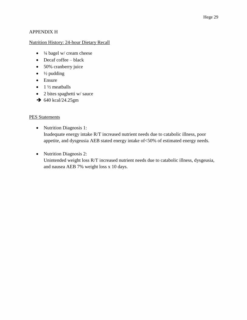
On December 16th
, the dietetic intern visited the patient and completed a 24-hour dietary
recall (Appendix H). The recall showed her average intake was approximately 650 kcal and 24
grams of protein per day. The patient stated that the recall was an adequate measure of her
current intake and that she was experiencing dysgeusia, a metallic taste, poor appetite, an
aversion to dairy, nausea, and significant weight loss related to chemotherapy treatment. The
dietetic intern estimated her needs to be 1348-1815 kcal/day based on the Mifflin St. Jeor
equation with an activity factor of 1.3-1.5 (light activity), and stress factor of 1.2-1.4 (cancer).
The estimate is similar to using 30-35 kcal/kg/day for cancer patients as recommend in The
Clinical Dietitian’s Essential Pocket Guide (2009), which is 1473-1719 kcal/day. Her protein
needs were 59-69 gm based on 1.2-1.4 gm/kg/day and fluid needs estimated were 1473 cc
fluid/day (30 cc/kg/day).21
DL’s nutrition diagnosis 1 is inadequate energy intake related to increased nutrient needs
due to catabolic illness, poor appetite, and dysgeusia as evidence by stated intake less than 50%
of estimated energy needs. Nutrition diagnosis 2 is unintended weight loss related to increased
nutrient needs due to catabolic illness, dysgeusia, and nausea as evidence by a 7% weight loss x
Table 4: Medications and their Use

Hege 18
10 days. Interventions included working with the patient to find oral nutrition supplements that
work well by bringing the nutrition supplements available to the patient for her to taste and try.
Education was provided by the dietetic intern in regard to using plastic utensils to relieve the
metallic taste, the importance in practicing oral hygiene, sitting upright before and after meals,
separating liquids and solids by an hour, eating small, frequent meals, trying ginger ale, and
avoiding dairy and high fat foods. After discussion with the patient, the decision was made to
continue her regular diet as ordered and add a 10am vanilla Resource 2.0 and 2pm chocolate
Boost pudding (715kcal/28gm protein) per the patient’s food preferences and to meet her
increased needs. Goals comprised of tolerating the diet, adequate nutrition/hydration to meet
estimated energy needs, and greater than 50%-75% intake of meals and supplements. The
dietetic intern monitored and evaluated the patient’s weight, percent intake, signs and symptoms,
and nutrition-related labs, and planned to follow-up in the next seven to ten days.
NUTRITION FOLLOW-UP
On December 27th
, the patient’s follow-up necessitated a change in her supplements
related to the patient stating that the Resource 2.0 was “too much to drink”, the Boost pudding
was good but “getting old”, and her family brought Ensure to her. The decision was made to
discontinue the Resource 2.0, change the Boost pudding to four times a week, and recommend
her drink an Ensure at least once a day. DL’s weight was trending down, which was to be
expected with the disease condition, and her laboratory values were improving with a BUN of 60
mg/dL, Cr of 4.7 mg/dL, and a GFR of 8.7ml/min. Her nutrition diagnoses from the initial
assessment were ongoing for continuing follow-up, monitoring, and evaluation.

Hege 19
CONCLUSION
Nutrition care provided to DL at HHGW corresponded to research associated with
managing symptoms of multiple myeloma and chemotherapy. DL was very receptive to the
suggestions in order to manage her dysgeusia, metallic taste, and nausea. The ability to taste
supplements in advance increased her total oral supplement intake. Several follow-up visits with
DL showed that her overall mood and quality of life improved while at HHGW. Providing her
with information to purchase an anti-nausea acupuncture bracelet, if interested, was the one piece
of advice that may have been beneficial. On January 7th
, DL was discharged home in her
daughter’s care with potential for provision for JSSA Hospice.

Hege 20
References
1. A clinical staging system for multiple myeloma. Correlation of measured myeloma cell
mass with presenting clinical features, response to treatment, and survival. Cancer. 36:
1975; 842-854.
2. Wei, A. Juneja, S. Bone marrow immunohistology of plasma cell neoplasms. J Clin
Pathol. 2003; 56:406-411.
3. Kyle RA, Rajkumar SV. Criteria for diagnosis, staging, risk stratification and response
assessment of multiple myeloma. Leukemia. Jan 2009;23(1):3-9.
4. Multiple Myeloma/Plasmacytoma. American Academy of Orthopaedic Surgeons. January
2011. Retrieved on January 15, 2014 from
http://orthoinfo.aaos.org/topic.cfm?topic=A00086.
5. Yiin JH, Anderson JL, Daniels RD, Seel EA, Fleming DA, Waters KM, et al. A nested
case-control study of multiple myeloma risk and uranium exposure among workers at the
Oak Ridge Gaseous Diffusion Plant.Radiat Res. Jun 2009;171(6):637-45 .
6. Wadhera RK, Kyle RA, Larson DR, et al. Incidence, clinical course, and prognosis of
secondary monoclonal gammopathy of undetermined significance in patients with
multiple myeloma. Blood. Sep 15 2011;118(11):2985-7.
7. Randomized, double-blind, placebo-controlled trial of recombinant human erythropoietin,
epoetin beta, in hematologic malignancies. J Clin Oncol. 20: 2002; 2486-2494.
8. Seiter, K. Multiple Myeloma. Medscape. 2013. Retrieved on January 22, 2013 from
http://emedicine.medscape.com/article/204369-overview#aw2aab6b2b3aa.
9. Baz, R. Bolwell, B. Multiple Myeloma. Cleveland Clinic. 2009. Retrieved on January 22,
2013 from
http://www.clevelandclinicmeded.com/medicalpubs/diseasemanagement/hematology-
oncology/multiple-myeloma/#bib12.
10. Raab MS, Podar K, Breitkreutz I, Richardson PG, Anderson KC. Multiple
myeloma. Lancet. Jul 25 2009;374(9686):324-39.
11. Reeder, et al. Cyclophosphamide, bortezomib and dexamethasone (CyBorD) induction
for newly diagnosed multiple myeloma: High response rates in a phase II clincal trial.
Leukemia. 2009. July; 23(7):1337-1341.
12. Akio, I. Cancer Anorexia-Cachexia Syndrome: Current issues in research and
management. CA Cancer J Clin 2002;52:72-91.

Hege 21
13. What is the relationship between nutrition status and quality of life in oncology patients?
Evidence Analysis Library, Academy of Nutrition and Dietetics. Retrieved on January 22,
2014 from http://andevidencelibrary.com/evidence.cfm?evidence_summary_id=251545.
14. Iversen, PO. Wisloff, F. Gulbrandsen, N. Reduced nutritional status among multiple
myeloma patients during treatment with high-dose chemotherapy and autologous stem
cell support. Clin Nutr. 2010 Aug; 29(4): 488-491.
15. Nourissat A, Vasson MP, Merrouche Y, Bouteloup C, Goutte M, Mille D, Jacquin JP,
Collard O, Michaud P, Chauvin F. Relationship between nutritional status and quality of
life in patients with cancer. Eur J Cancer. 2008 Jun; 44(9): 1,238-1,242.
16. Shahmoradi N, Kandiah M, Peng LS. Impact of nutritional status on the quality of life of
advanced cancer patients in hospice home care. Asian Pac J Cancer Prev. 2009; 10(6):
1,003-1,009.
17. Chao, L, et al. The efficacy of acupoint stimulation for the management of therapy-
related adverse events in patients with breast cancer: a systematic review. Breast Cancer
Res Treat. 2009; 118:255-267.
18. Strafton, R. Summary of a systematic review on oral nutritional supplement use in the
community. Proceedings of the Nutrition Society. 2000; 59:469-476.
19. Nutritional suggestions for symptom management. National Cancer Institute. Retrieved
on January 20, 2014 from
http://www.cancer.gov/cancertopics/pdq/supportivecare/nutrition/HealthProfessional/pag
e4#Section_117.
20. Pagana, K. Pagana, T. Diagnostic and Laboratory Test Reference. 9th
ed. Mosby Elsevie:
2009.
21. Width, M. Reinhard, T. The Clinical Dietitian’s Essential Pocket Guide. Lippincott
Williams & Wilkins: 2009.
Hege 22
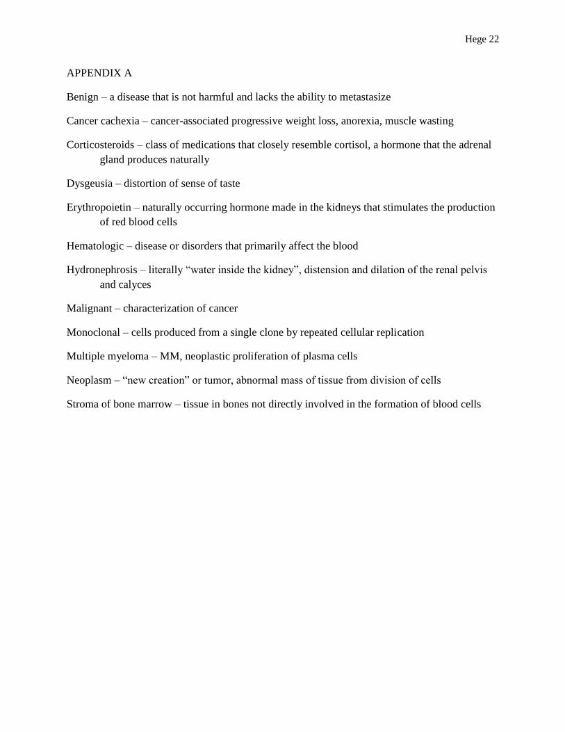
APPENDIX A
Benign – a disease that is not harmful and lacks the ability to metastasize
Cancer cachexia – cancer-associated progressive weight loss, anorexia, muscle wasting
Corticosteroids – class of medications that closely resemble cortisol, a hormone that the adrenal
gland produces naturally
Dysgeusia – distortion of sense of taste
Erythropoietin – naturally occurring hormone made in the kidneys that stimulates the production
of red blood cells
Hematologic – disease or disorders that primarily affect the blood
Hydronephrosis – literally “water inside the kidney”, distension and dilation of the renal pelvis
and calyces
Malignant – characterization of cancer
Monoclonal – cells produced from a single clone by repeated cellular replication
Multiple myeloma – MM, neoplastic proliferation of plasma cells
Neoplasm – “new creation” or tumor, abnormal mass of tissue from division of cells
Stroma of bone marrow – tissue in bones not directly involved in the formation of blood cells

Hege 23
APPENDIX B
Table 1: Chemotherapeutic Regimens

Hege 24
APPENDIX C
Table 2: Side Effects’ Impact on Nutritional Status

Hege 25
APPENDIX D
Table 3: Managing Symptoms

Hege 26
APPENDIX E
Graph 1: Weight (kg)
45
46
47
48
49
50
51
52
53
54
55
We
igh
t (k
g)
Date
Graph 1: Weight (kg)
Weight (kg)

Hege 27
APPENDIX F
Figure 4: Serum Blood Test on 12/16

Hege 28
APPENDIX G
Table 4: Medications and their Use
Hege 29
APPENDIX H
Nutrition History: 24-hour Dietary Recall
¼ bagel w/ cream cheese
Decaf coffee – black
50% cranberry juice
½ pudding
Ensure
1 ½ meatballs
2 bites spaghetti w/ sauce
640 kcal/24.25gm
PES Statements
Nutrition Diagnosis 1:
Inadequate energy intake R/T increased nutrient needs due to catabolic illness, poor
appetite, and dysgeusia AEB stated energy intake of<50% of estimated energy needs.
Nutrition Diagnosis 2:
Unintended weight loss R/T increased nutrient needs due to catabolic illness, dysgeusia,
and nausea AEB 7% weight loss x 10 days.