multiple mini punch grafts for extensive ulcer: acasereport
TRANSCRIPT
117
Wirohadidjojo, Multiple mini punch grafts for extensive ulcer: a case reportJ Med SciVolume 44, No. 2, June 2012: 117 - 123
* corresponding author: [email protected]
Multiple mini punch grafts for extensiveulcer: a case report
Yohanes Widodo Wirohadidjojo*Department of Dermato-Venereology, Faculty of Medicine, Gadjah Mada University/Dr. Sardjito General Hospital, Yogyakarta, Indonesia
ABSTRACTMultiple mini punch grafts is the placing of mini size of full thickness skins on to ulcer bed. Theyconsist of epidermal and dermal component composed with hair follicles and other skin appendiceswhere epidermal stem cells are located. The epidermal stem cells are the best source of epidermalcells in reconstruction of skin equivalent that is usually used for replacing classic split thicknessskin graft in recovering extensive ulcer. In this article, the application of multiple mini punchgrafts onto extensive ulcer is reported. A case of extensive ulcer was suffered by a 6-year-oldboy whose left foot is injured in a traffic accident. His toes had already been amputated bysurgeon but a classic skin graft failed to recover the ulcer. Multiple mini punch grafts had beenharvested from his inguinal and buttock skin and they were placed onto his ulcer. Pre and postmini punch grafting photographs were reviewed. After eight weeks, placed multiple mini punchtissues onto large ulcer reveals lateral extensions and more than 90% of epithelialization. Multiplemini punch grafts can be used as a method to cover large ulcer.
ABSTRAKPunch graft mini jamak merupakan penananaman potongan kulit berukuran kecil dengan ketebalanpenuh pada dasar ulkus. Potongan kulit seperti ini tersusun oleh epidermis, dermis yangmengandung folikel rambut dan apendik kulit lainnya tempat sel punca epidermal berada. Selpunca epidermal merupakan sumber terbaik sel-sel epidermal untuk menyusun kulit ekivalenyang dewasa ini sering dipakai sebagai pengganti split thickness skin graft untuk menutupulkus yang luas. Dalam artikel ini dilaporkan penggunaan punch graft mini jamak pada kasusulkus luas yang diderita oleh anak laki-laki berusia 6 tahun akibat kecelakaan lalu lintas pada kakikirinya. Jari-jari kaki telah diamputasi sebelumnya, tetapi skin graft klasik gagal menyembuhkanulkus luas pada kaki tersebut. Punch graft mini jamak yang diambil dari kulit lipat paha dan kulitbokong selanjutnya di tanamkan pada dasar ulkus tersebut. Setelah 8 minggu, kulit yang ditanam-kan tumbuh melebar kesamping dan menutup lebih dari 90 % permukaan ulkus. Disimpulkanbahwa teknik punch graft mini jamak dapat digunakan untuk menutup ulkus yang luas.
Key words: mini punch grafts-large ulcer-epithelialization-epidermal-stem cells
INTRODUCTION
In the traffic accident, full thickness of skinmay be extensively loose. The split thicknessskin graft may be failed to recover the losstissues because of limitation of skin donors and
harvesting large skin may cause a large scarformation, when skin graft is forced to use. Insuch cases, skin equivalent grafting can bechosen because epidermal equivalent of thisreconstructive skin reveals the capacity of life
118
J Med Sci, Volume 44, No. 2, June 2012: 117 - 123
organ as shown in its responding to physicalinjury in a staged and specific pattern of cellmigration, re-epithelialization, and cytokineexpression.1 The best source of epidermal cellsfor this purpose is epidermal stem cells that arelocated along basal layers, sebaceous glandsand around the hair follicles.2-5 Severalproblems in skin reconstructive transplantationare the handling of the fragile sheets, variable‘‘take rates’’,6 blister formation due to ulcer bedsecretion, scarring and contracture of the graft.7
Other disadvantages are: high cost and the factthat it can only be reconstructed by clinicianswho are familiar or have a relation with humancells culture technique researchers.
Multiple mini punch grafts are grafting ofmultiple mini size full thickness skins withmultipurpose. It is most popular in hairtransplantation for preparing follicular unittransplantation,8-9 but it is also used in acne scarcorrection,10-11 and re-pigmentation of vitiligo.12-
13 They consist of epidermal and dermalcomponent rich with hair follicles and other skinappendixes where epidermal stem cells arelocated. Placing mini punch tissues onto ulcerbed maybe similar with cultivation of stem cellsin hypoxia condition, a method for propagatingstem cells to release growth factors.14,15 Thispresumption is based on a report by Kirsner etal.16 Punch graft acts as a pharmacologic agentin stimulating granulation of the venous ulcer
bed as well as migration of previously dormantwound edges.16
Application of mini punch grafts onto largeulcer has never been published. It has beenreported in the treatment limited sixe of chronicleg ulcers.15 In this case report, it was reportedthe application of multiple mini punch grafts ontoextensive traffic accident ulcer.
CASE REPORT
A six-year-old boy who had a trafficaccident was referenced because ofepithelialization failures. Eight months before,he had the first operation performed byorthopedic surgeon for amputating the toes,fixing fragmented bones, and classic skingrafting. Unfortunately, the skin grafting was nottaking and skin grafted wound become anextensive chronic ulcer.
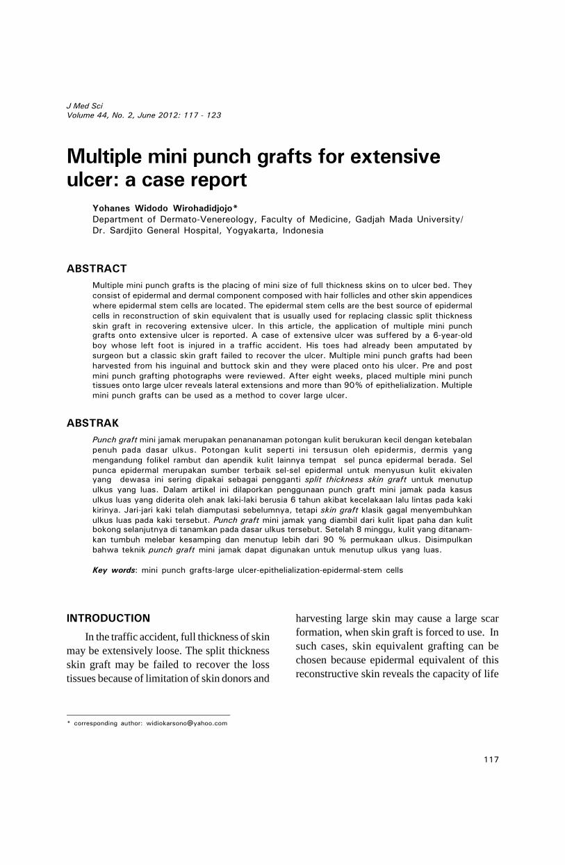
At the first visit, an extensive chronic ulceron both dorsal and plantar left foot wasobserved. The ulcer showed an easy bleedingof granulation tissue with profuse fluid secretioncovered by normal saline immersed gauges(FIGURE 1). Based on this condition, it wasdecided to take care of the ulcer by Sofratule®,0.1% gentamycine ointment in occlusivedressing technique to prevent new bleeding inthe replacing next new dressing. Cefixime 30mg twice a day and 150 mg of tranexamic acidtwice a day are also given for a week.
FIGURE 1. Ulcer condition in the first visit.

119
Wirohadidjojo, Multiple mini punch grafts for extensive ulcer: a case report
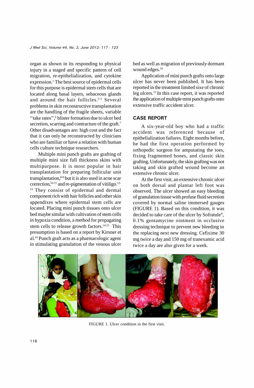
After a week of therapy, bleeding inreplaced dressing as well as fluid secretion wasnot observed anymore but epithelialization wasstill poor. The treatment has been continued butoral therapy was stopped. After a month of
treatment, epithelialization was just limited ina small area in the ankle as shown by blackarrows on FIGURE 2. Based on this condition,it was decided to perform multiple mini-punchgrafts.
FIGURE 2. Ulcer condition after a month of occlusive treatment.
Surgical ProceduresMultiple mini-punch grafts (MMPGs) were
performed in three phases. First, testing ofMMPGs taken from left inguinal skin wasplaced onto dorsal and inner side of feet.Second, MMGs taken from right inguinal skinwas placed onto lateral side of feet. Third,MMPGs taken from buttock skin was placedonto plantar side of feet. The third operationhas been performed after previous MMPGs ofdorsal feet showed confluence in skin sheet asa space for placing adhesive tape for fixingMMPGs onto plantar feet against gravitationforce.
Skin donor harvesting and graftingAfter disinfecting skin with povidone
iodine 10 % and placing sterile linen ontooperation area, the skin was injected with
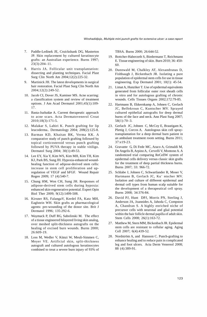
tumescent anesthetic (1: 300.000 epinephrineplus 0.2% lidocaine in sterile normal saline).Skin harvesting was performed by 0.2 mmdisposable punch device. MMPGs were thenharvested by cutting a piece of skin in dermal-fatlevel and placed them onto gauges immersed withsterile normal saline (to prevent skin dehydration)before they were placed onto ulcer bed.
Before grafting, ulcer bed was washed andimmersed with sterile normal saline. Placingof MMPGs onto ulcer bed was performed pieceby piece with blepharoplasty skin’s forceps. Allof procedure was performed without anyanesthetic drugs (FIGURE 3). The ulcer andMMPGs were then covered by netting gauze(Sofratule®) followed by an occlusive dressingfor a week. In second week, topical silverointment (Burnazin®) was then applied topromote lateral extension of MMPGs.

120
J Med Sci, Volume 44, No. 2, June 2012: 117 - 123
FIGURE 3. Harvesting (left) and placing MMPGS (right). Black arrow shows the lateral punched tissues expansionof previously grafting.
RESULTS
After a week of grafting, punched tissueislands showed that they were tightly attachedonto ulcer bed. They were proved by resistanceattachment in dry removing of sofratule. Thebeginning of their lateral extension was
observed in week two. At the same time, ulceredge tissue migration to the central area wasalso observed. Confluences of punched tissueto perform epithelial sheet were observed inweek three (FIGURE 4).
FIGURE 4. Ulcer Edge Tissue Migration and Confluences Of MMPGs. Black arrow shows new MMPGs.
The final result can be observed in FIGURE5. Eight weeks after the last operation, morethan 90% of epithelialization was observed on
recipient sites and slightly hypo andhyperchromic color was found on donor sites.

121
Wirohadidjojo, Multiple mini punch grafts for extensive ulcer: a case report
FIGURE 5. Eight weeks after the last operation
DISCUSSION
In case of extensive ulcer when enoughsource of skin donor is not available, such as incase of extensive burn, most of modern surgeonsmay perform grafting of reconstructive skin asskin substitute.17,18 This material is made froma small piece of patient’s own skin by in-vitrocultivating fibroblasts from dermal side andkeratinocytes from epidermal side. Fibroblastsmonolayer is then cultivated in the collagenmatrixes to perform dermal equivalent and inmedia-air interphase, keratinocytes are thendropped onto dermal equivalent surface toperform a large reconstructive skin.19
Donor can be taken from any site of skin.Epidermal cells are composed of threepopulation cells, namely: epidermal stem cells(SCs), transient amplifying cells, and nonproliferative basal cells. Epidermalconstruction from transient amplifying cellsformed faster than the others; however, the
epidermis from the SCs population showscontinuity in growing and expressing the reportergene for long time period.20 In a case of limitedsource of epidermal cells, autologous ofepidermal SCs can be generated from follicularouter root sheath cells as previously publishedby Limat and Hunziker.21
A controversy of this technique is arisendue to use of recombinant epidermal growthfactor (EGF) for keratinocytes and fetal bovineserum for fibroblast cultivation. The futureeffect of both materials in patient is not fullypredicted, especially in a case of bovine viralinfection. In another site, handling ofreconstructive skin grafting among surgeons isnot easy. It is fragile sheet and easy to bebroken.6 Clinical studies show that grafting takesrates are variable, blister formations aresometimes found, scarring and contractures arealso often observed.7 In addition, except forhighly cost, skin reconstructive skills is notalways available in developing countries.

122
J Med Sci, Volume 44, No. 2, June 2012: 117 - 123
Those problems lead researches to developa technique namely cells spray forepithelialization. The cells can be directlyharvested from a small piece (1: 100 of woundsize) of patient’s skin, or cultivated in vitro toachieve numbers of viable cells. In thistechnique, donor skin is cut and enzymaticdigestion with 2.5 unit/mL dispase followed by0.05% trypsin-EDTAin Ringer lactate solutionis used to harvest epidermal cells. Mixedepidermal cells are then sprayed onto woundbed. They reported that this technique shows asuccessful result of various depth of burnvictims.22,23 Compared to the classic graft, cellsspray showed similar functional and estheticoutcome.24 In order to mask scar formation ofdonor area, scalp skin can also be used.25
Different with reconstructive skin which needsa month for cultivation, the cells spray techniquemay only need time less than an hour if directcells isolation is chosen.
Mini punched tissues consist of fullthickness skin where epidermal stem cells arelocated. Epidermal stem cells have highplasticity to proliferate and difference to maturecells that are needed in damage tissues repair.26
They are also resistant against aging processagents that commonly exist in chronic ulcer.27
Placing stem cells in hypoxia condition, ascommonly found on ulcer bed surface, canpropagate them to release various growthfactors that are needed for healing process, withanti aging properties that are needed for agedcells in wound edge to proliferate and migrateto central area of ulcer. All of those theoriesare proved in our case. Placing MMPGs is notonly capable to proliferate and perform skinsheet but it also shows evidence of stimulatingulcer edge epthelialization and migration(FIGURE 4).
In limited facility of hospital such as ours,skin reconstructive and cells spray may bepracticed in the few next years. Unfortunately,limited available skin donor in extensive ulcerpatient, such as in this case, cannot wait forapplication of those techniques. Other moresimple techniques should be defined. In this
case, MMPGs have a proof to solve theproblem. According to Nordström andHansson28, MMPGs are very simple techniquesand they can be performed even by nurses inprimary health program
CONCLUSION
A case of extensive ulcer suffered by a 6–year-old boy that fails to be treated by classicskin graft has been presented. Due to limitedsource of skin donor, another skin grafting wasimpossible to be performed. This case can behandled by an easy technique of multi mini-punch grafts that was performed in two monthsof a three-step surgery
AKNOWLEDGMENT
Author would like thank Dr. Nurhidayah,Dr Meliana, Dr Sari and all of Nurses from Dr.Sardjito General Hospital, Yogyakarta forassisting the graftsand and also Kris Lantorofor taking the pictures.
REFERENCES
1. Falanga V, Isaacs C, Paquette D, Downing G,Kouttab N, Butmarc J, Badiavas E, Hardin-YoungJ. Wounding of bioengineered skin: cellular andmolecular aspect after injury. J Invest Dermatol2002; 119:653-60.
2. Fuchs E. Skin stem cells: rising to the surface. JCell Biol 2008; 180: 273–84.
3. Webb A, Li A, Kaur P. Location and phenotype ofhuman adult keratinocyte stem cells of the skin.Differentiation 2004; 72:387–95.
4. Kamstrup M, Faurschou A, Gniadecki R, Wulf HC.Epidermal stem cells – role in normal, woundedand pathological psoriatic and cancer skin. CurrStem Cell Res Ther 2008; 3(2): 146–50.
5. Pianigiani E, Ierardi F, Mazzanti B, Saccardi R,Cuciti C, Fimiani M. Human de-epidermizeddermis as a stem cell carrier. Transplant Proc2010; 42(6): 2244-6.
6. Chester DL, Balderson DS, Papini RP. A reviewof keratinocyte delivery to the wound bed. J BurnCare Rehabil 2004;25(3):266–75.
123
Wirohadidjojo, Multiple mini punch grafts for extensive ulcer: a case report
7. Paddle-Ledinek JE, Cruickshank DG, MastertonJP. Skin replacement by cultured keratinocytegrafts: an Australian experience. Burns 1997;23(3):204–11.
8. Harris JA. Follicular unit transplantation:dissecting and planting techniques. Facial PlastSurg Clin North Am 2004;12(2):225-32.
9. Martinick JH. The latest developments in surgicalhair restoration. Facial Plast Surg Clin North Am2004;12(2):249-52.
10. Jacob CI, Dover JS, Kaminer MS. Acne scarring:a classification system and review of treatmentoptions. J Am Acad Dermatol 2001;45(1):109-17.
11. Basta-Juzbašiæ A. Current therapeutic approachto acne scars. Acta Dermatovenerol Croat2010;18(3):171-5.
12. Malakar S, Lahiri K. Punch grafting for lipleucoderma.. Dermatology 2004; 208(2):125-8.
13. Barman KD, Khaitan BK, Verma KK. Acomparative study of punch grafting followed bytopical corticosteroid versus punch graftingfollowed by PUVA therapy in stable vitiligo.Dermatol Surg 2004; 30(1):49-53.
14. Lee EY, Xia Y, Kim WS, Kim MH, Kim TH, KimKJ, Park BS, Sung JH. Hypoxia-enhanced wound-healing function of adipose-derived stem cells:increase in stem cell proliferation and up-regulation of VEGF and bFGF. Wound RepairRegen 2009; 17 (4):540-7.
15. Chung HM, Won CH, Sung JH. Responses ofadipose-derived stem cells during hypoxia:enhanced skin-regenerative potential. Expert OpinBiol Ther 2009; 9(12):1499-508.
16. Kirsner RS, FalangaV, Kerdel FA, Katz MH,Eaglstein WH. Skin grafts as pharmacologicalagents: pre-wounding of the donor site. Brit JDermatol 1996; 135:292-6.
17. Waymack P, Duff RG, Sabolinski M. The effectof a tissue engineered bilayered living skin analog,over meshed split-thickness autografts on thehealing of excised burn wounds. Burns 2000;26:609-19.
18. Loss M, Wedler V, Künzi W, Meuli-Simmen C,Meyer VE. Artificial skin, split-thicknessautograft and cultured autologous keratinocytescombined to treat a severe burn injury of 93% of
TBSA. Burns 2000; 26:644-52.19. Bottcher-Haberzeth S, Biedermann T, Reichmann
E. Tissue engineering of skin. Burn 2010; 36: 450-60.
20. Dunnwald M, Chalkley AT, Alexandrunas D,Fishbaugh J, Bickenbach JR. Isolating a purepopulation of epidermal stem cells for use in tissueengineering. Exp Dermatol 2001; 10(1): 45-54.
21. Limat A, Hunziker T. Use of epidermal equivalentsgenerated from follicular outer root sheath cellsin vitro and for autologous grafting of chronicwounds. Cells Tissues Organs 2002;172:79-85.
22. Hartmann B, Ekkernkamp A, Johnen C, GerlachJC, Belfekroun C, Kuntscher MV. Sprayedcultured epithelial autografts for deep dermalburns of the face and neck. Ann Plast Surg 2007;58(1):70–3.
23. Gerlach JC, Johnen C, McCoy E, Brautigam K,Plettig J, Corcos A. Autologous skin cell spray-transplantation for a deep dermal burn patient inan ambulant treatment room setting. Burns 2011;37:e19-23.
24. Gravante G, Di Fede MC, Araco A, Grimaldi M,De Angelis B,Arpino A, Cervelli V, Montone A. Arandomized trial comparing ReCellW system ofepidermal cells delivery versus classic skin graftsfor the treatment of deep partial thickness burns.Burns 2007; 33: 966-72.
25. Schlabe J, Johnen C, Schwartlander R, Moser V,Hartmann B, Gerlach JC, Ku¨ ntscher MV.Isolation and culture of different epidermal anddermal cell types from human scalp suitable forthe development of a therapeutical cell spray.Burns 2008; 34:376-84.
26. David PJ, Hunt DPJ, Morris PN, Sterling J,Anderson JA, Joannides A, Jahoda C, CompstonA, Chandran S. A highly enriched niche ofprecursor cells with neuronal and glial potentialwithin the hair follicle dermal papilla of adult skin.Stem Cells 2008; 26(1):163-72.
27. Matthew M, Stern MM, Bickenbach JR. Epidermalstem cells are resistant to cellular aging. AgingCell 2007; 6(4):439-52.
28. Nordström A, and Hansson C. Punch-grafting toenhance healing and to reduce pain in complicatedleg and foot ulcers. Acta Derm Venereol 2008;88 (4):389-91.