multina - home | shimadzu
TRANSCRIPT
C297-E062E
MCE-202 Microchip Electrophoresis System for DNA/RNA Analysis
MultiNA
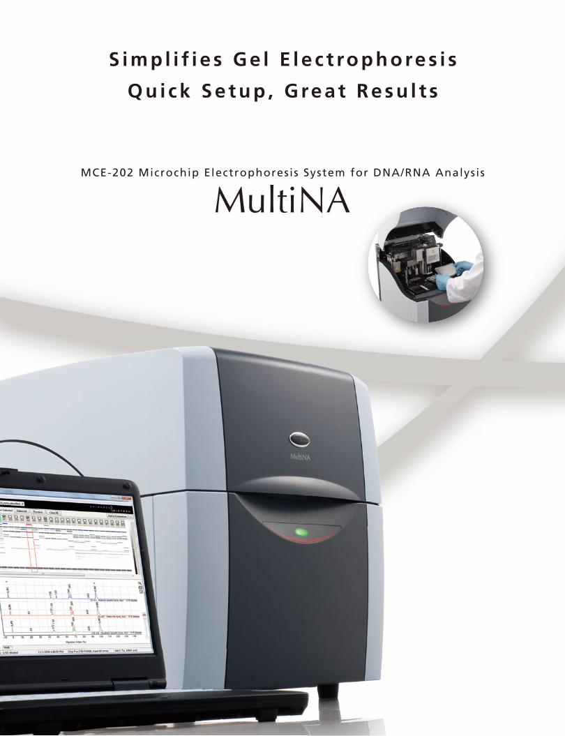
MCE-202 Microchip Electrophoresis System for DNA/RNA Analysis
S i m p l i f i e s G e l E l e c t ro p h o re s i s
Q u i c k S e t u p , G re a t R e s u l t s

RNA Analysisfor Genetic Research
Food Analysis Microbiological Analysis
Infectious DiseaseAnalysis
Genotypingisisisisearearearearchhhch
Food Analysis Microbiological Analysis
fectious DiseaseInfAnalysis
Genotyping
Wide Range of Applications
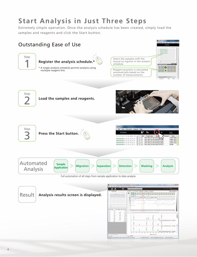
Start Analysis in Just Three Steps
Widely used for genetic research applications as well as food analysis, genotyping, microbiological analysis,
infectious disease analysis, and RNA analysis.
Automated Analysis From 1 to 108 Loaded Samples
Extremely simple operation. Once the analysis schedule has been created, simply load the samples and
reagents and click the Start button.
Fast analysis with up to four microchips in parallel.
Page 4
Page 8
Page 6
Page 10
Page 11
Consumables and Options
Specifications
4
Register the analysis schedule.*
Load the samples and reagents.
Press the Start button.
Analysis results screen is displayed.
1Step
2Step
3Step
Result
Full automation of all steps from sample application to data analysis.
Select the samples with the mouse to register in the analysis schedule.
Reagent quantity is calculated automatically based on the number of measurements.
SampleApplication
AutomatedAnalysis
Migration Separation Detection Washing Analysis
* A single analysis schedule permits analysis using multiple reagent kits.
Outstanding Ease of Use
Start Analysis in Just Three StepsExtremely simple operation. Once the analysis schedule has been created, simply load the
samples and reagents and click the Start button.
5Microchip Electrophoresis System for DNA/RNA Analysis
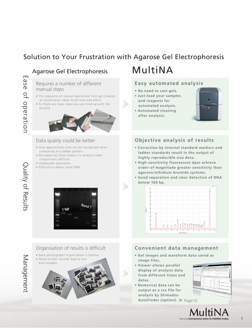
MultiNAAgarose Gel Electrophoresis
Requires a number of differentmanual steps
Easy automated analysis
Data quality could be better Objective analysis of results
Organisation of results is difficult Convenient data management
• Only approximate sizes can be recognized when comparing to a ladder pattern.
• Discrepancies from analysis to analysis make comparisons difficult.
• Inadequate separation.• Difficult to detect small DNA.
• The sequence of manual operations from gel creation to visualization takes much time and effort.
• As there are many steps you are tired up with the process.
• Correction by internal standard markers and ladder standards result in the output of highly reproducible size data.
• High-sensitivity fluorescent dyes achieve order-of-magnitude greater sensitivity than agarose/ethidium bromide systems.
• Good separation and clear detection of DNA below 100 bp.
• No need to cast gels.• Just load your samples
and reagents for automated analysis.
• Automated cleaning after analysis.
Ease of o
peratio
nQ
uality of ResultsM
anagement
?bpBand?
alysis
• Data (photograph) organization is tedious.• Hand-written records lead to loss and mistakes.
• Gel images and waveform data saved as image files.
• Viewer allows parallel display of analysis data from different times and dates.
• Numerical data can be output as a csv file for analysis by Shimadzu AutoFinder (option) .
Solution to Your Frustration with Agarose Gel Electrophoresis
Page10ge10
6
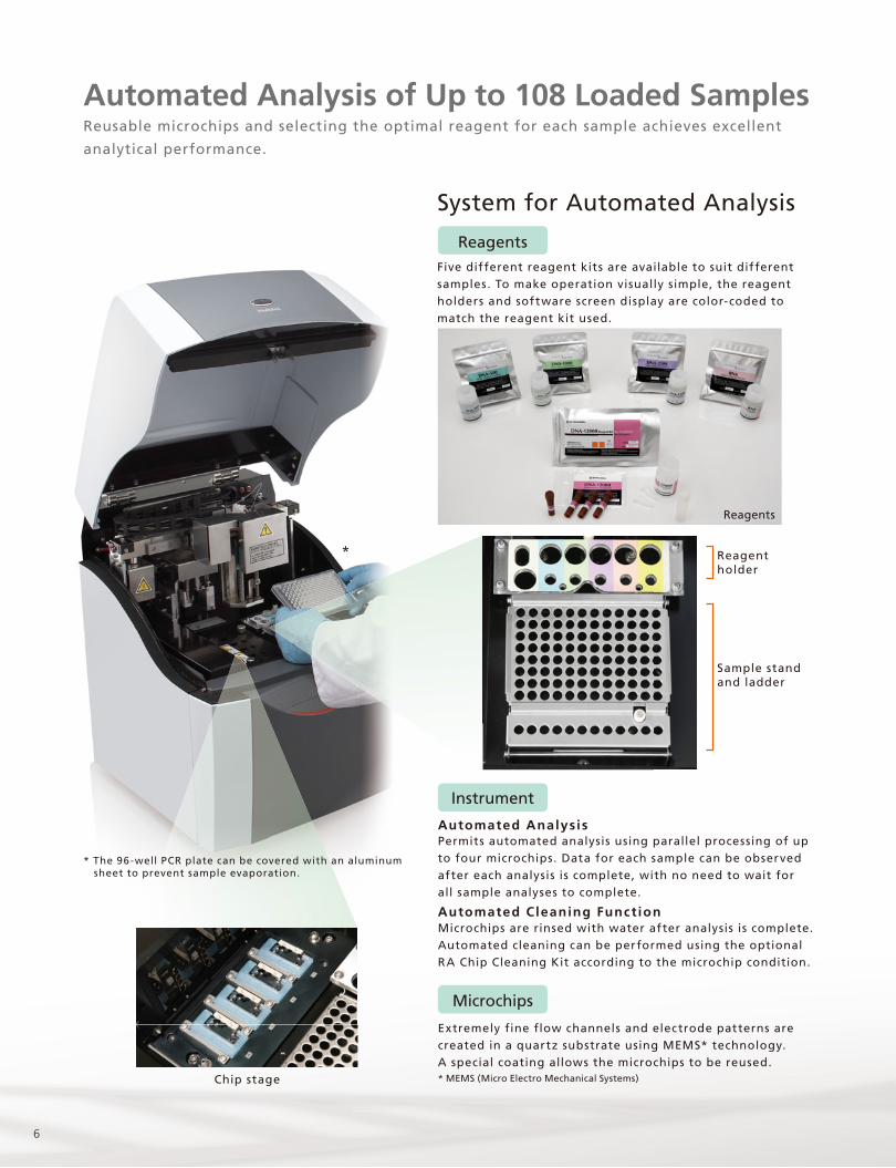
ReagentsFive different reagent kits are available to suit different samples. To make operation visually simple, the reagent holders and software screen display are color-coded to match the reagent kit used.
Permits automated analysis using parallel processing of up to four microchips. Data for each sample can be observed after each analysis is complete, with no need to wait for all sample analyses to complete.
System for Automated Analysis
Automated Analysis
Automated Cleaning Funct ionMicrochips are rinsed with water after analysis is complete. Automated cleaning can be performed using the optional RA Chip Cleaning Kit according to the microchip condition.
Extremely fine flow channels and electrode patterns are created in a quartz substrate using MEMS* technology. A special coating allows the microchips to be reused. * MEMS (Micro Electro Mechanical Systems)
Instrument
Microchips
Reagents
Chip stage
Reagentholder
Sample standand ladder
*
* The 96-well PCR plate can be covered with an aluminum sheet to prevent sample evaporation.
Automated Analysis of Up to 108 Loaded SamplesReusable microchips and selecting the optimal reagent for each sample achieves excellent
analytical performance.
7Microchip Electrophoresis System for DNA/RNA Analysis
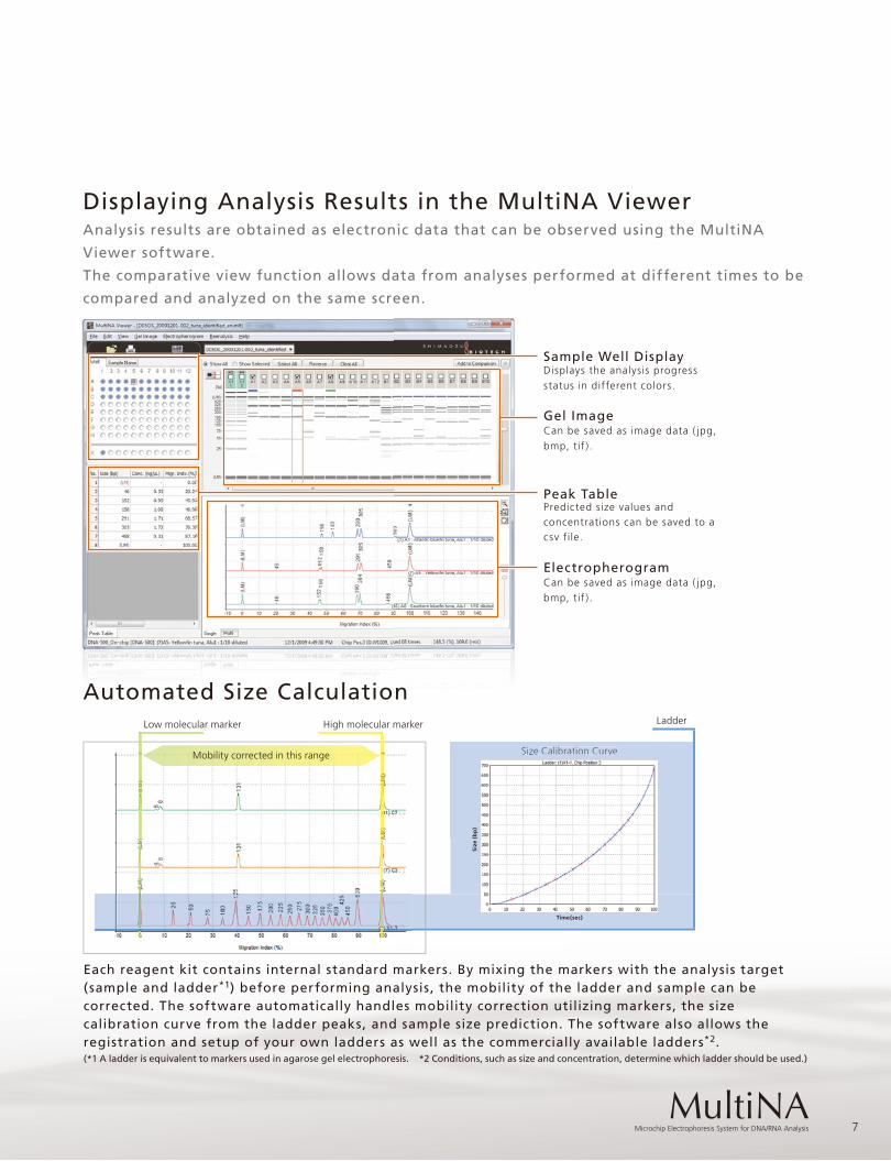
Gel Image
Peak Table
Sample Well Display
Electropherogram
Automated Size Calculation
Each reagent kit contains internal standard markers. By mixing the markers with the analysis target (sample and ladder*1) before performing analysis, the mobility of the ladder and sample can be corrected. The software automatically handles mobility correction utilizing markers, the size calibration curve from the ladder peaks, and sample size prediction. The software also allows the registration and setup of your own ladders as well as the commercially available ladders*2.(*1 A ladder is equivalent to markers used in agarose gel electrophoresis. *2 Conditions, such as size and concentration, determine which ladder should be used.)
Displaying Analysis Results in the MultiNA Viewer
Displays the analysis progress status in different colors.
Can be saved as image data ( jpg, bmp, tif ) .
Can be saved as image data ( jpg, bmp, tif ) .
Predicted size values and concentrations can be saved to a csv file.
Analysis results are obtained as electronic data that can be observed using the MultiNA
Viewer software.
The comparative view function allows data from analyses performed at different times to be
compared and analyzed on the same screen.
Size Calibration Curve
Low molecular marker High molecular marker
Calibration CurveSize Mobility corrected in this range
Size
(b
p)
Ladder
Time(sec)

8
PCRPrimer specific for
each sample
Analysis ofPCR products
MCE-202 MultiNA
Restriction enzymeprocessing
DNA extractionIon-exchange resin kit
DNA Purification
PCR Products
Detection ofAllergenic Substance
Sample
PCR
DNA extraction
DNA Purification
PCR Products
PCR-RFLP Products
MultiNAelectrophoresis
Sample
: Ladder Marker(25bp DNA Ladder): Wheat
Lane 2 : BuckwheatLane 3 : PeanutsLane 4 : PrawnLane 5 : Crab
Gel Images
• Tuna Species Identification Manual (Food and Agricultural Materials Inspection Center; Fisheries Research Agency, Japan)
Mse I Processing Tsp 509 I ProcessingAlu I Processing
Gel Images of PCR-RFLP Separation Patterns
Japan was the world's earliest adopter of a labeling system for foods containing allergens. DNA analysis by qualitative PCR can be performed on five (wheat, buckwheat, peanuts, prawn, and crab) of the seven specified raw materials (excluding egg and milk) .
Detection of Allergenic Substances Application News: No. B23Application to Food Analysis
Application to Genotyping
The tuna-specific genetic sequence in mitochondrial DNA is amplified using PCR. This amplified DNA is cleaved with a restriction enzyme and the pattern used to identify the tuna species. * PCR-RFLP:(Polymerase Chain Reaction-Restriction Fragment Polymorphism)
Identification of Thunnus Using PCR-RFLP Method Application News: No. B28
LM
LM Lane 1
1 2 3 4 5
Identification ofTuna Varieties
Yellow
fin tuna
Southern bluefin tuna
Ladder
Albacore tuna
Yellow
fin tuna
Atlantic bluefin tuna
Yellow
fin tuna
Ladder
Ladder
α Bigeye tuna
β Bigeye tuna
α Bigeye tuna
Southern bluefin tuna
α Bigeye tuna
Wide Range of ApplicationsWidely used for genetic research as well as food analysis, genotyping, microbiological
analysis, infectious disease analysis, and RNA analysis.
Samples
Result
Application Example
Purified DNA
Electrophoresis
DetectionPCR Products
Sample PreparationNucleic AcidExtraction PCR

9Microchip Electrophoresis System for DNA/RNA Analysis
Multiplex-PCRPCR
Analysis of PCR productsMCE-202 MultiNA
DNA
PCR Products
Example of RNA Analysis Rat Total RNA Analysis
Application News: No. B32
Application News: No. 6
During research using RNA, it is important to continuously monitor the RNA quality to ensure that the RNA used is not affected by degradation by RNase. MultiNA is able to accurately recognize 18S-rRNA and 28S-rRNA based on the calibration curve information acquired from the ladder.
PCR is performed on DNA extracted from four types of white kidney bean. The white kidney bean variety can then be identified by comparing the pattern obtained against patterns for each variety. * RAPD-STS:(Random Amplified Polymorphic DNA-Sequence Tagged Sites)
Identification of Common Bean Cultivars by RAPD-STSApplication to RAPD-STS Method
Epidemiological studies to gain a detailed grasp of the virus type during an influenza epidemic are important in medical facilities and designated regions. The Seeplex FluA ACE Subtyping Kit allows the detection of type A influenza and the simultaneous detection of four major influenza A subtypes (H3: Hong Kong; H1: Soviet ; H1(HA) : swine flu ; H5: avian flu) . It extensively covers clinical research in laboratories , examination rooms, and infection control departments.
Analysis with Seeplex®FluA ACE Subtyping Kit (from Seegene)
Multiplex-PCR is performed on four sets of samples using a variety identification kit (from Kokken). The rice variety can then be identified by comparing the pattern obtained against patterns for each rice variety.
Identification of Rice Varieties Application News: No. B30
Lad
de
r
Lad
de
r
Co
ntro
l
Co
ntro
l
Ko
shih
ikari
Ko
shih
ikari
Ak
itakom
ach
i
Ak
itakom
ach
i
Kin
uh
ikari
Kin
uh
ikari
Kirara 3
97
Kirara 3
97
Hito
me
bo
re
Hito
me
bo
re
Multiplex PCR by VarietyIdentification Kit
DNA extraction
Analysis of PCRproducts-MultiNA
Acquisition ofappearance patterns
of PCR products
Extracted DNA
Collation of patterns
Sample
PCR Products
Sample
Application to Multiplex-PCR
Application to Multiplex-PCR
Reverse TranscriptaseReaction
Nucleic Acid Extraction
Negative control
Positive control
4
3
2
1
Ne
gat
ive
con
tro
l
Po
siti
ve c
on
tro
l
4 3 2 1
Gel Images Electropherogram
Universal Influenza A
Swine H5
Avian H5
Seasonal H3
Seasonal H1
Internal control
Set A Set C Set DSet BIdentification ofRice Varieties
Influenza VirusType Evaluation
Lad
de
r
Co
ntro
l
Ko
shih
ikari
Ak
itakom
ach
i
Kin
uh
ikari
Kirara 3
97
Hito
me
bo
re
Lad
de
r
Co
ntro
l
Ko
shih
ikari
Ak
itakom
ach
i
Kin
uh
ikari
Kirara 3
97
Hito
me
bo
re
10
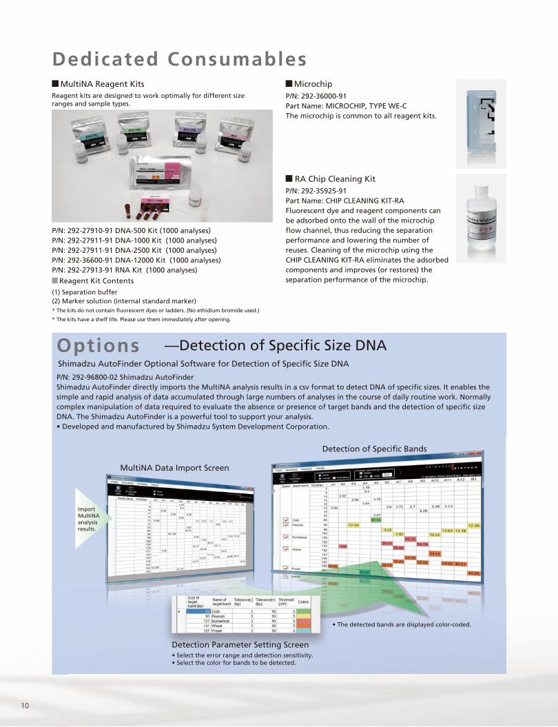
MultiNA Reagent Kits
RA Chip Cleaning Kit
—Detection of Specific Size DNA
Reagent kits are designed to work optimally for different size ranges and sample types.
Reagent Kit Contents
(1) Separation buffer(2) Marker solution (internal standard marker)* The kits do not contain fluorescent dyes or ladders. (No ethidium bromide used.)
* The kits have a shelf life. Please use them immediately after opening.
P/N: 292-35925-91Part Name: CHIP CLEANING KIT-RAFluorescent dye and reagent components can be adsorbed onto the wall of the microchip flow channel, thus reducing the separation performance and lowering the number of reuses. Cleaning of the microchip using the CHIP CLEANING KIT-RA eliminates the adsorbed components and improves (or restores) the separation performance of the microchip.
P/N: 292-36000-91Part Name: MICROCHIP, TYPE WE-CThe microchip is common to all reagent kits.
P/N: 292-27910-91 DNA-500 Kit (1000 analyses)P/N: 292-27911-91 DNA-1000 Kit (1000 analyses)P/N: 292-27911-91 DNA-2500 Kit (1000 analyses)P/N: 292-36600-91 DNA-12000 Kit (1000 analyses)P/N: 292-27913-91 RNA Kit (1000 analyses)
Shimadzu AutoFinder Optional Software for Detection of Specific Size DNA
P/N: 292-96800-02 Shimadzu AutoFinderShimadzu AutoFinder directly imports the MultiNA analysis results in a csv format to detect DNA of specific sizes. It enables the simple and rapid analysis of data accumulated through large numbers of analyses in the course of daily routine work. Normally complex manipulation of data required to evaluate the absence or presence of target bands and the detection of specific size DNA. The Shimadzu AutoFinder is a powerful tool to support your analysis. • Developed and manufactured by Shimadzu System Development Corporation.
Detection of Specific Bands
ImportMultiNAanalysisresults.
• The detected bands are displayed color-coded.
MultiNA Data Import Screen
Detection Parameter Setting Screen• Select the error range and detection sensitivity. • Select the color for bands to be detected.
Options
Microchip
Dedicated Consumables
11Microchip Electrophoresis System for DNA/RNA Analysis
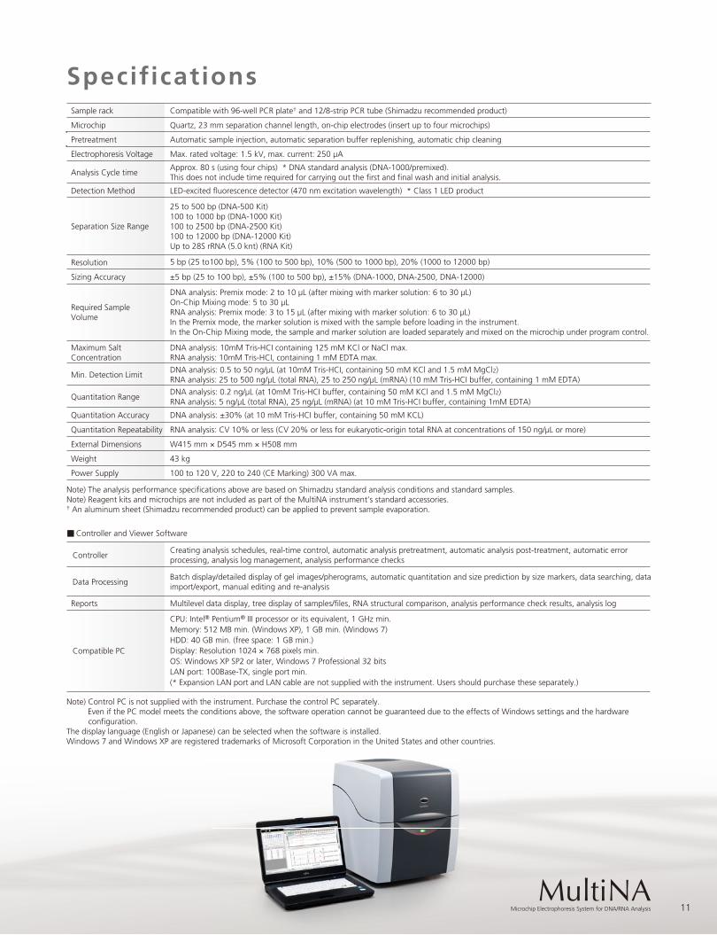
SpecificationsSample rack Compatible with 96-well PCR plate† and 12/8-strip PCR tube (Shimadzu recommended product)
Microchip Quartz, 23 mm separation channel length, on-chip electrodes (insert up to four microchips)
Pretreatment Automatic sample injection, automatic separation buffer replenishing, automatic chip cleaning
Electrophoresis Voltage Max. rated voltage: 1.5 kV, max. current: 250 µA
Analysis Cycle time
Detection Method LED-excited fluorescence detector (470 nm excitation wavelength) * Class 1 LED product
Separation Size Range
Resolution 5 bp (25 to100 bp), 5% (100 to 500 bp), 10% (500 to 1000 bp), 20% (1000 to 12000 bp)
Sizing Accuracy ±5 bp (25 to 100 bp), ±5% (100 to 500 bp), ±15% (DNA-1000, DNA-2500, DNA-12000)
Required SampleVolume
External Dimensions W415 mm × D545 mm × H508 mm
Weight 43 kg
Power Supply 100 to 120 V, 220 to 240 (CE Marking) 300 VA max.
Note) The analysis performance specifications above are based on Shimadzu standard analysis conditions and standard samples.Note) Reagent kits and microchips are not included as part of the MultiNA instrument’s standard accessories.† An aluminum sheet (Shimadzu recommended product) can be applied to prevent sample evaporation.
Quantitation Accuracy DNA analysis: ±30% (at 10 mM Tris-HCI buffer, containing 50 mM KCL)
Quantitation Repeatability RNA analysis: CV 10% or less (CV 20% or less for eukaryotic-origin total RNA at concentrations of 150 ng/µL or more)
ControllerCreating analysis schedules, real-time control, automatic analysis pretreatment, automatic analysis post-treatment, automatic errorprocessing, analysis log management, analysis performance checks
Data ProcessingBatch display/detailed display of gel images/pherograms, automatic quantitation and size prediction by size markers, data searching, dataimport/export, manual editing and re-analysis
Reports Multilevel data display, tree display of samples/files, RNA structural comparison, analysis performance check results, analysis log
Compatible PC
CPU: Intel® Pentium® III processor or its equivalent, 1 GHz min.Memory: 512 MB min. (Windows XP), 1 GB min. (Windows 7)HDD: 40 GB min. (free space: 1 GB min.)Display: Resolution 1024 × 768 pixels min.OS: Windows XP SP2 or later, Windows 7 Professional 32 bitsLAN port: 100Base-TX, single port min.(* Expansion LAN port and LAN cable are not supplied with the instrument. Users should purchase these separately.)
Note) Control PC is not supplied with the instrument. Purchase the control PC separately. Even if the PC model meets the conditions above, the software operation cannot be guaranteed due to the effects of Windows settings and the hardware
configuration. The display language (English or Japanese) can be selected when the software is installed. Windows 7 and Windows XP are registered trademarks of Microsoft Corporation in the United States and other countries.p
Controller and Viewer Software
DNA analysis: Premix mode: 2 to 10 µL (after mixing with marker solution: 6 to 30 µL)On-Chip Mixing mode: 5 to 30 µLRNA analysis: Premix mode: 3 to 15 µL (after mixing with marker solution: 6 to 30 µL)In the Premix mode, the marker solution is mixed with the sample before loading in the instrument. In the On-Chip Mixing mode, the sample and marker solution are loaded separately and mixed on the microchip under program control.
25 to 500 bp (DNA-500 Kit)100 to 1000 bp (DNA-1000 Kit)100 to 2500 bp (DNA-2500 Kit)100 to 12000 bp (DNA-12000 Kit)Up to 28S rRNA (5.0 knt) (RNA Kit)
Approx. 80 s (using four chips) * DNA standard analysis (DNA-1000/premixed).This does not include time required for carrying out the first and final wash and initial analysis.
Min. Detection Limit
Maximum SaltConcentration
DNA analysis: 10mM Tris-HCI containing 125 mM KCl or NaCl max.RNA analysis: 10mM Tris-HCI, containing 1 mM EDTA max.
DNA analysis: 0.5 to 50 ng/µL (at 10mM Tris-HCI, containing 50 mM KCl and 1.5 mM MgCl2)RNA analysis: 25 to 500 ng/µL (total RNA), 25 to 250 ng/µL (mRNA) (10 mM Tris-HCI buffer, containing 1 mM EDTA)
Quantitation RangeDNA analysis: 0.2 ng/µL (at 10mM Tris-HCI buffer, containing 50 mM KCl and 1.5 mM MgCl2)RNA analysis: 5 ng/µL (total RNA), 25 ng/µL (mRNA) (at 10 mM Tris-HCI buffer, containing 1mM EDTA)

MCE®-202 MultiNA is not available in the United States.
MultiN
A
Printed in Japan 3295-02203-30ANS
Company names, product/service names and logos used in this publication are trademarks and trade names of Shimadzu Corporation or its affiliates, whether or not they are used with trademark symbol “TM” or “®”.Third-party trademarks and trade names may be used in this publication to refer to either the entities or their products/services. Shimadzu disclaims any proprietary interest in trademarks and trade names other than its own.
For Research Use Only. Not for use in diagnostic procedures. The contents of this publication are provided to you “as is” without warranty of any kind, and are subject to change without notice. Shimadzu does not assume any responsibility or liability for any damage, whether direct or indirect, relating to the use of this publication.
© Shimadzu Corporation, 2012www.shimadzu.com/an/