multifunctional coatings to simultaneously promote...
TRANSCRIPT

lable at ScienceDirect
Biomaterials 84 (2016) 301e314
Contents lists avai
Biomaterials
journal homepage: www.elsevier .com/locate/biomater ia ls
Review
Multifunctional coatings to simultaneously promote osseointegrationand prevent infection of orthopaedic implants
Jordan Raphel a, Mark Holodniy b, c, Stuart B. Goodman d, Sarah C. Heilshorn a, *
a Department of Materials Science and Engineering, Stanford University, Stanford, CA, USAb Division of Infectious Diseases & Geographic Medicine, Stanford University, Stanford, CA, USAc Veterans Affairs Palo Alto Health Care System, Palo Alto, CA, USAd Department of Orthopaedic Surgery and Bioengineering, Stanford University, Stanford, CA, USA
a r t i c l e i n f o
Article history:Received 23 September 2015Received in revised form22 December 2015Accepted 1 January 2016Available online 18 January 2016
Keywords:Orthopaedic implantsFunctional coatingsOsseointegrationAntimicrobial treatments
* Corresponding author. 476 Lomita Mall, McCull94305, USA.
E-mail address: [email protected] (S.C. Heils
http://dx.doi.org/10.1016/j.biomaterials.2016.01.0160142-9612/© 2016 Elsevier Ltd. All rights reserved.
a b s t r a c t
The two leading causes of failure for joint arthroplasty prostheses are aseptic loosening and peri-prosthetic joint infection. With the number of primary and revision joint replacement surgeries on therise, strategies to mitigate these failure modes have become increasingly important. Much of the recentwork in this field has focused on the design of coatings either to prevent infection while ignoring bonemineralization or vice versa, to promote osseointegration while ignoring microbial susceptibility.However, both coating functions are required to achieve long-term success of the implant; therefore,these two modalities must be evaluated in parallel during the development of new orthopaedic coatingstrategies. In this review, we discuss recent progress and future directions for the design of multifunc-tional orthopaedic coatings that can inhibit microbial cells while still promoting osseointegration.
© 2016 Elsevier Ltd. All rights reserved.
1. Introduction
Orthopaedic implant use for joint replacements has been on therise, with significant increases still projected over the next 15 years[1]. Themajority of procedures are knee and hip replacements, withover 700,000 knee and 300,000 hip replacements done annually inthe United States [2]. While these surgeries have a track record ofdecades of positive outcomes, approximately 10% of these implantsfail prematurely, within the first 10e20 years, thereby affectingmany tens of thousands of patients annually [3]. Furthermore, asthe US population continues to age and as life expectancy continuesto increase, premature failures are not the only concern; manypatients are now outliving their implants. This combination offactors leads to projections of a dramatic increase in implant fail-ures in the near future.
The two leading causes of implant failure are aseptic looseningand infection. While the reported rates of these failures varydepending on the study, approximately 18% of implant failures aredue to aseptic loosening while 20% of failures are attributed toinfection [4,5]. Additionally, these issues become even more
ough Rm 246, Stanford, CA,
horn).
prevalent in revised total joint arthroplasties. Aseptic loosening canoriginate from a variety of sources. These include micromotion ofthe implant relative to the bone during loading, the generation ofimplant wear particles that lead to inflammation and boneresorption, and poor osseointegration e the functional interfacebetween the implant and the patient's bone [6]. Implant site in-fections occur as microbes, particularly bacteria, become sessileand adhere to implant surfaces. These solid interfaces providesurfaces for bacterial attachment, proliferation, and biofilm for-mation, in which the adherent bacteria produce a protective,polymeric, extracellular substance, rendering these bacteria sub-stantially more difficult to eradicate than individual suspendedplanktonic bacteria floating around the body [7,8]. Awide variety ofbacteria can infect an implant, but a small subset of species makesup the majority of pathogens. Staphylococcus bacteria, mostprominently Staphylococcus aureus and Staphylococcus epidermidis,account for close to 70% of orthopaedic implant infections, whilePseudomonas aeruginosa accounts for another 8% of infections [9].
Aseptic loosening and implant infection appear to be mutuallyexclusive, particularly given the use of the word ‘aseptic’. However,recent studies point to the potential connection between implantsthat have been reported to fail aseptically and latent occult in-fections that may have been missed prior to the time of diagnosis[10]. Therefore, even in cases of implant failure where infectionwas

J. Raphel et al. / Biomaterials 84 (2016) 301e314302
not the primary cause, microbial presence may still play a criticalrole in initiating or accelerating the failure pathway.
Independently, the problems of aseptic loosening and infectionare pressing for the orthopaedics field, and many excellent reviewarticles cover the fields of osseointegration and infection preven-tion individually [11e18]. However, the two issues are intimatelyrelated, as laid out by Gristina in his description of the “race for thesurface”; if the host's cells can reach and occupy the implant surfacefirst, not only will stronger tissue integration be achieved, but adefensive barrier will also be established against microbialattachment and colonization [19]. Strong osseointegration andprevention of infection are both required for a successful implant,necessitating that implant designs consider both criteria simulta-neously. In this review we describe several of the specific under-lying mechanisms that lead to implant failure either by asepticloosening or infection and potential design strategies to addressthese challenges (summarized in Table 1). In particular, with recentprogress in understanding the connections between aseptic loos-ening and infection, this article will highlight recent works thataddress both problems in concert.
2. Challenges and potential solutions for osseointegration
Implant osseointegration relies on two distinct requirements.The first is obtaining initial implant stability during surgery, whichthen lays the groundwork for subsequent osseointegration of theimplant as the patient heals. Ensuring implant stability is largelythe responsibility of the surgeon and her/his team. Even techno-logical solutions to improve initial implant fit, including automatedimaging and robotic arm assistance platforms only assist, ratherthan replace, the surgery team. The second requirement is theprevention of later-stage loosening of the implant, which can becaused by a variety factors. These include lack of bone in-growthduring healing, implant micromotion relative to the bone,adverse bone remodeling around the implant, and the formation ofimplant wear particles. This section will further investigate thechallenges that can lead to later-stage implant loosening, technol-ogies that have been explored to mitigate these factors or may beconsidered as the field progresses, and the successes of thesetechnologies at simultaneously limiting infection.
2.1. Challenge: gaps at prosthesisebone interface
A primary cause of aseptic loosening of joint replacement devices
Table 1Causes of orthopaedic implant failure and potential solutions to mitigate eac
Causes of implant failure Poten
Gaps at prosthesis-bone interface � Ga
Poor bone in-growth on implants � Ca� En� Bio� 3D
Poor bone deposition on implant surfaces � Bio� Bio� Re
Initial microbial adhesion and infection � En� En
Late-stage infection � Lo� Slo� Co
Infection leading to osteolysis � Tre� Im
is implant micromotion due to gaps at the prosthesisetissue inter-face [6,20]. Increasing bone-implant contact reduces the size andnumber of gaps surrounding the implant, stabilizing the jointreplacement prosthesis and limiting micromotion. As orthopaedicjoint replacements are load-bearing implants, small micromotionmay worsen over timewith implant use, which can further progresstowards greater micromotion, eventually leading to implant failure[21]. Particularly large gaps are slow to be filled by bony in-growthduring osteogenesis, further emphasizing the importance of initiallylimiting and eliminating the gaps. Gaps at the bone-implant inter-face can also lead to aseptic loosening in combination with implantdebris, such as polymeric or metallic wear particles [22]. The gapscan serve as conduits for wear particles to flow along the length ofthe implant, building up at the interface, and inhibiting directprosthesis-bone contact [6]. Furthermore, wear particles cannot beeasily phagocytosed by macrophages, leading the cells to adopt anactivated inflammatory state, in which they secrete a series of cy-tokines. These cytokines, such as tumor necrosis factor a (TNF-a),can lead to the generation of osteoclasts and the local resorption ofbone tissue, effectively forming and enlarging gaps at the prosthe-sisetissue interface [23]. Local inflammation may lead to an alteredimmunological state that makes the implant more susceptible tomicrobial colonization. Illustrations of the different aseptic failuremodes are presented in Fig. 1. The potential implant instabilitycaused by these gaps can progress in severity to the point that animplant revision is required.
2.1.1. Potential solution: gap-bridging coatingsOne potential solution for gaps at the prosthesisetissue inter-
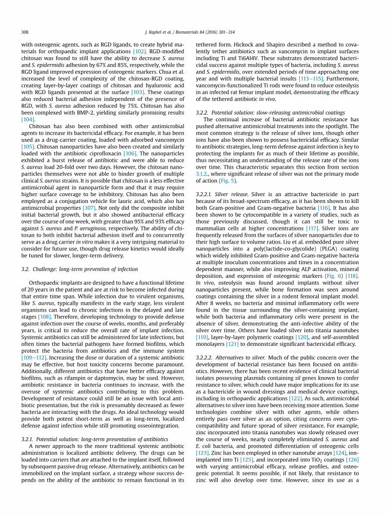
face is a coating that can swell or expand upon implantation inorder to reduce micromotion and wear particle-induced osteolysis.Dynamic coatings can respond to small gaps on the micron scalearound the implant in order to provide more direct contact be-tween the implant and bone, thereby increasing the chance for astable interface to form. A variety of different materials could beemployed for these coatings, including hydrogels, foams, ordeformable elastic metallic structures. Fournier et al. created a wireknit using nickel titanium (nitinol) for use as a flexible, gap-bridging implant coating [24]. Thin nitinol wires were knit into aporous, superelastic structure that could be attached to the surfaceof an orthopaedic implant by a brazing process (Fig. 2). In itsexpanded form, the knit coating was approximately 750 mm thick,large enough to bridge large gaps surrounding an implant, butcould be compacted down approximately 90% during implantation
h cause and improve implant efficacy and lifetime.
tial solutions
p-bridging coatings
lcium phosphate coatingsgineering surface topographyactive glass coatings-printed coatings
molecular coatings incorporating extracellular matrix proteinsmolecular coatings incorporating growth factorscruiting osteogenic cells to the implant surface
gineering surfaces to inhibit bacterial adhesiongineering bactericidal surfaces
ng-term presentation of antibioticsw-releasing antimicrobial coatingsatings containing antimicrobial peptides
atments to block inflammation and differentiation signaling cascadesmunomodulatory treatments
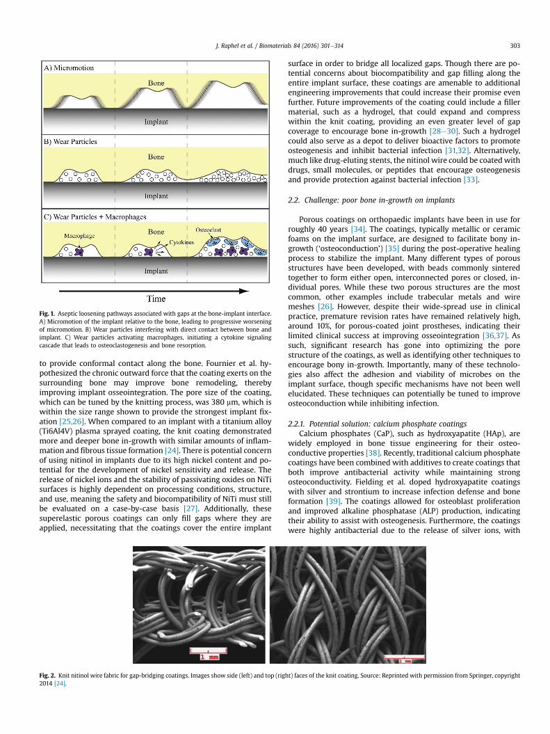
Fig. 1. Aseptic loosening pathways associated with gaps at the bone-implant interface.A) Micromotion of the implant relative to the bone, leading to progressive worseningof micromotion. B) Wear particles interfering with direct contact between bone andimplant. C) Wear particles activating macrophages, initiating a cytokine signalingcascade that leads to osteoclastogenesis and bone resorption.
J. Raphel et al. / Biomaterials 84 (2016) 301e314 303
to provide conformal contact along the bone. Fournier et al. hy-pothesized the chronic outward force that the coating exerts on thesurrounding bone may improve bone remodeling, therebyimproving implant osseointegration. The pore size of the coating,which can be tuned by the knitting process, was 380 mm, which iswithin the size range shown to provide the strongest implant fix-ation [25,26]. When compared to an implant with a titanium alloy(Ti6Al4V) plasma sprayed coating, the knit coating demonstratedmore and deeper bone in-growth with similar amounts of inflam-mation and fibrous tissue formation [24]. There is potential concernof using nitinol in implants due to its high nickel content and po-tential for the development of nickel sensitivity and release. Therelease of nickel ions and the stability of passivating oxides on NiTisurfaces is highly dependent on processing conditions, structure,and use, meaning the safety and biocompatibility of NiTi must stillbe evaluated on a case-by-case basis [27]. Additionally, thesesuperelastic porous coatings can only fill gaps where they areapplied, necessitating that the coatings cover the entire implant
Fig. 2. Knit nitinol wire fabric for gap-bridging coatings. Images show side (left) and top (righ2014 [24].
surface in order to bridge all localized gaps. Though there are po-tential concerns about biocompatibility and gap filling along theentire implant surface, these coatings are amenable to additionalengineering improvements that could increase their promise evenfurther. Future improvements of the coating could include a fillermaterial, such as a hydrogel, that could expand and compresswithin the knit coating, providing an even greater level of gapcoverage to encourage bone in-growth [28e30]. Such a hydrogelcould also serve as a depot to deliver bioactive factors to promoteosteogenesis and inhibit bacterial infection [31,32]. Alternatively,much like drug-eluting stents, the nitinol wire could be coatedwithdrugs, small molecules, or peptides that encourage osteogenesisand provide protection against bacterial infection [33].
2.2. Challenge: poor bone in-growth on implants
Porous coatings on orthopaedic implants have been in use forroughly 40 years [34]. The coatings, typically metallic or ceramicfoams on the implant surface, are designed to facilitate bony in-growth (‘osteoconduction’) [35] during the post-operative healingprocess to stabilize the implant. Many different types of porousstructures have been developed, with beads commonly sinteredtogether to form either open, interconnected pores or closed, in-dividual pores. While these two porous structures are the mostcommon, other examples include trabecular metals and wiremeshes [26]. However, despite their wide-spread use in clinicalpractice, premature revision rates have remained relatively high,around 10%, for porous-coated joint prostheses, indicating theirlimited clinical success at improving osseointegration [36,37]. Assuch, significant research has gone into optimizing the porestructure of the coatings, as well as identifying other techniques toencourage bony in-growth. Importantly, many of these technolo-gies also affect the adhesion and viability of microbes on theimplant surface, though specific mechanisms have not been wellelucidated. These techniques can potentially be tuned to improveosteoconduction while inhibiting infection.
2.2.1. Potential solution: calcium phosphate coatingsCalcium phosphates (CaP), such as hydroxyapatite (HAp), are
widely employed in bone tissue engineering for their osteo-conductive properties [38]. Recently, traditional calcium phosphatecoatings have been combined with additives to create coatings thatboth improve antibacterial activity while maintaining strongosteoconductivity. Fielding et al. doped hydroxyapatite coatingswith silver and strontium to increase infection defense and boneformation [39]. The coatings allowed for osteoblast proliferationand improved alkaline phosphatase (ALP) production, indicatingtheir ability to assist with osteogenesis. Furthermore, the coatingswere highly antibacterial due to the release of silver ions, with
t) faces of the knit coating. Source: Reprinted with permission from Springer, copyright

J. Raphel et al. / Biomaterials 84 (2016) 301e314304
activity expected to persist as silver releasewas still increasing afterone week. Others have also doped silver into HAp coatings withsimilar results [40].
Alternative calcium phosphate strategies combining the osteo-conductive coating with bactericidal agents, such as antimicrobialpeptides (AMP, discussed further in section 3.2.3.) also have beenexplored. In one example, AMPs were physically adsorbed onto thecoatings to allow for temporal release [41]. The AMP-CaP was adhe-sive to osteoblasts and, importantly, showed significantly improvedbone-implant contact in a rabbit tibial model, providing in vivo evi-dence for better osseointegration compared with an uncoated tita-nium control. Furthermore, the AMP provided rapid and completeprotection against both S. aureus and P. aeruginosa, killing all bacteriawithin 150 min. While this technology shows great promise, therewas a significant burst release of the AMP, thus limiting the potentialeffective lifetime of the coating. Others incorporated the antibioticnorvancomycin into an HAp coating, which showed positive osteo-genic and antibacterial abilities against S. aureus, but also sufferedfrom a short-term burst release [42]. To slow down the release ki-netics of the bactericides, degradable polymer capping agents havebeen employed [43]. While each example showcases the strongosteoconductive capabilities of calcium phosphate coatings andshows promise as an anti-infective, longer-term protection frombacterial infection is desirable for clinical use.
2.2.2. Potential solution: engineering surface topographyAnother area of interest has been engineering of the micro- and
nanostructure of the implant surface to improve interactions withosteogenic cells while simultaneously inhibiting bacterial adhesion.Increasing the surface area and porosity of the implant can improvebone in-growth and the coefficient of friction between the boneand implant, thereby reducing micromotion and increasingosseointegration. For example, an anchor-like surface architecturewas fabricated on implant surfaces using a direct metal laser sin-tering process. This anchor-like surface topography, together with asecondary interconnected pore coating, significantly improvedprimary implant fixation and bone in-growth and decreasedmicromotion amplitude in both in vitro and large animal in vivostudies [44,45]. Another method for altering the surface structure isto create nanopits through acid etching. Lan et al. combined acidetching with UV exposure to alter structure and surface energy[46]. The resulting surface improved osteoblast ALP production anddepositedmineralizationwhile decreasing the presence of S. aureusand S. epidermidis by approximately 70%. Etching has also beencombined with techniques to alter microstructure, giving a hier-archically structured titanium surface [47]. These surfaces showedimproved in vivo mechanical stability and osseointegration in arabbit tibial screw implantation model. The nano-micro-hierarchyprovided defense against multiple Staphylococcus and Pseudo-monas species over months, which is promising for addressing late-onset infections [48]. One possible mechanism for this improvedantimicrobial activity is an increase of surface free energy on thehierarchical surfaces, which was shown to alter the membranestructure of adhered bacteria, potentially inhibiting cellular activ-ities [47]. Developing a strategy that can maintain both the anti-microbial and osseointegrative capabilities long-term is critical forthe viability of the implant technologies, making the early resultsfor engineered surface topographies quite promising.
2.2.3. Potential solution: bioactive glass coatingsBioactive glasses are synthetic, degradable ceramic materials
containing key elements and molecules to encourage osteogenesis,and these materials have been in use for nearly 50 years [49,50].They have found continued use in the orthopaedics field due totheir ability to deliver a surface apatite layer during dissolution,
known as hydroxycarbonate apatite, which promotes osteogenesis.On their own, bioactive glasses have been shown to possess limitedantimicrobial character by altering local pH during dissolution.However, when combined with additional components, bioactiveglasses can both promote osteogenesis and provide significantantimicrobial activity [49]. Ordikhani and Simchi created a com-posite implant coating by pairing bioactive glass with antibiotic-loaded chitosan [51]. Chitosan, a popular coating material whoseantimicrobial uses will be discussed in greater detail in section3.1.2., was loaded with vancomycin and deposited in conjugationwith bioactive glass nanoparticles onto titanium surfaces. Thebioactive glass facilitated calcium phosphate mineral depositionand osteoblast adhesion. The antibiotic release rapidly inhibitedS. aureus and continued over four weeks. Similar results were foundusing an ampicillin-loaded chitosan/bioactive glass hybrid toinhibit S. mutants [52].
2.2.4. Potential solution: 3D-Printed coatingsRecent advances in 3D printing technology have improved its
flexibility and precision to the point where it is now being exten-sively employed to create printed surfaces and coatings that may beutilized for orthopaedic applications. Though there is room forcontinued development and printer resolution to allow finerstructures to be processed, 3D printing holds promise for creatingcustomized coatings to improve the osseointegration of orthopaedicimplants [53]. One example of these coatings is 3D-printed trabec-ular titanium, described by Regis et al. [54]. The hexagonal, meso-porous structure was generated by specific electron beam melting,an additive manufacturing process. Trabecular titanium coatingsenhanced osteogenic activity of human adipose-derived stem cellsand osteoblasts in vitro and coated acetabullar cups showed strongosseointegration and survival rates in clinical studies. Additionalexamples of 3D-printed coatings and scaffolds utilizing calciumphosphate components have also been presented and show excel-lent promise for customizable implant surface engineering [55e57].
2.3. Challenge: poor bone deposition on implant surfaces
An ideal orthopaedic implant would not only allow for bony in-growth to secure the prosthesis, but would also encourage newbone deposition on its surface (‘osteoinduction’) [35] to speed upthe stabilization process and to provide a seamless bone-implantinterface. Without osteoinductive functionality, implants can suf-fer from poor deposition of new bone tissue. Bony in-growth isrequired for successful stabilization, which can be compromised inpatients with poor local bone quality, slow healing rates, and otherconfounding factors. Myriad surface coatings, additives, andbioactive cues have been incorporated with implants to renderthem osteoinductive, including growth factors and chemokines.There are additional operational concerns with these coatings,particularly around long-term storage, biomolecular activity, andadhesion strength. Still, given the roles of many of these bio-molecules in native osteogenesis, they serve as attractive compo-nents for potential osteoinductive coating design.
2.3.1. Potential solution: biomolecular coatings incorporatingextracellular matrix proteins
Extracellular matrix (ECM) proteins have found use in bonetissue engineering applications in part because of their presence inphysiological bone. Bone is a compositematerial containing both aninorganic mineral phase as well as an organic matrix phase(‘osteoid’). Collagen I comprises approximately 90% of the osteoidphase, making it an obvious starting point for an osteoinductivebiomolecular coating; however, this ECM is also known to be ad-hesive to multiple microbes. Engineered, collagen-mimetic peptide

J. Raphel et al. / Biomaterials 84 (2016) 301e314 305
implant coatings have been used in place of human collagen I tolimit bacterial adhesion and colonization while still providingosteoinductive cues [58]. The collagen mimic was engineered toinclude specific ligand sequences that recognize highly expressedintegrins on osteogenic cells. The coatings were adhesive for mul-tiple osteogenic cell types with a similar level of adhesion as foundon human collagen I coatings. Importantly, the collagen-mimeticcoatings showed reductions in adhesion of S. aureus andS. epidermidis between 100 and 1000 fold over human collagen Icoatings, and 10e100 fold versus uncoated titanium. This collagen-mimetic protein serves as an excellent example of an osteoinduc-tive biomolecular coating that also provides defense againstimplant infection. Other groups utilized collagen coatings thatrelease adsorbed antibiotics [59] and silk coatings coupled with ananti-fouling surface [60] as combinatorial protein coatings toimprove osteoinduction.
2.3.2. Potential solution: biomolecular coatings incorporatinggrowth factors
Some of the most widely used cues to initiate bone depositionare bone morphogenetic proteins (BMP), a family of potent osteo-genic growth factors [61]. In particular, BMP-2 has been used inconjunction with antimicrobial components to create osteoinduc-tive coatings for orthopaedic implants. For example, the growthfactor has been grafted to an anti-infective chitosan layer to create amultifunctional coating [62]. Though the BMP-2 only mildlyimproved adhesion of osteogenic cells, it greatly improved bothcalcium mineral deposition and ALP activity compared to coatingslacking BMP-2. Furthermore, Shi et al. demonstrated that thegrafted BMP-2 remained localized to the coating, implying that itcould be osteoinductive long-term. In an alternative approach,BMP-2 was delivered as a soluble cue, using a biomolecular heparincarrier to control release. While this BMP-2 delivery strategyresulted in similar osteoblast adhesion, ALP activity, and mineraldeposition results as the grafted BMP-2 study, it is expected thatthe osteoinductive potential would diminish over time as the BMP-2 was released from the coating [63].
2.3.3. Potential solution: recruiting osteogenic cells to the implantsurface
A newer strategy in the osteoinductive field is the recruitment ofosteogenic cells through released signaling factors. Instead of solelyrelying on cells to passively find the osteoinductive coating, cellscan be induced to “home” to the implant surface by creating agradient of chemoattractive factors. One of the most well studiedrecruitment cues is the cytokine stromal cell-derived factor 1a(SDF-1a), also known as CXCL12, which can mobilize multiple stemcell populations, includingmesenchymal stem cells (MSCs) throughtheir CXCR4 surface receptor [64e67]. While a recruitment strategymight work independently, it could be most valuable when used inconjunction with localized osteogenic cues to encourage new bonedeposition from the cells that get trafficked to the implant (Fig. 3).For example, Zwingenberger, et al. found that delivery of SDF-1aenhanced the efficacy of BMP-2 in generating new bone [68]. Themigration of MSCs to the site of a bone injury and bone volumewere significantly improved using the cytokine combination at theinjury site, resulting in substantially more bone regrowth in a ro-dent femoral defect model. Others have found similar results usingthe same combination of SDF-1a and BMP-2, delivered eitherconcurrently or sequentially [69,70]. To the best of our knowledge,this strategy has not been used to improve the osseointegration ofan orthopaedic implant, though its success in the literature sug-gests that it could be hugely beneficial, especially considering thelocal environment around orthopaedic implants. For instance, thestem portion of a hip prosthesis is implanted into the femoral
medullary cavity, which contains a significant amount of bonemarrow. Similarly, bone marrow-derived MSCs are found in themetaphysis, where the posts of knee replacement prostheses areimplanted. Additionally, SDF-1amay also help prevent infection, asit is a chemoattractant for neutrophils [71]. Summoning neutro-phils to the implant site could limit the occurrence of infectionthrough their ability to phagocytose microbes and recruitmacrophages.
3. Challenges and potential solutions for bacterial infectionof implants
Infection of orthopaedic implants can have a plethora of con-sequences, including hospitalization and costly revision surgery[11]. Implant infection has been reported to be the new leadingcause of orthopaedic implant removal, overtaking aseptic loosening[4,72e74]. Despite the biocompatibility of titanium and its alloysused for implants, bacteria easily and readily colonize these sur-faces. Once bacteria adhere to the surface, they begin to proliferate,eventually reaching a high enough density to form a biofilm. Thebiofilm protects the bacterial colony with an extracellular poly-meric substance, rendering it largely resistant to antibiotics, im-mune cells, and other potential infection defense mechanisms [75].Therefore, it is critical to address pathogenic bacteria as early aspossible in order to minimize the chance of biofilm formation.
A myriad of strategies have been explored to limit microbialinfections. The research can generally be broken down intoaddressing infections over two different time frames; techniquesthat can rapidly inhibit infection and techniques designed forlonger-term defense. Both timescales are important from a clinicalperspective; infections occurring within the first three months aredefined as early while those manifesting after the initial period aredefined as delayed and late [76]. The first 6 hours after surgery areviewed as the particularly decisive period for preventing earlyinfection, as the introduced pathogens have not yet begun rapidproliferation [77].
Limiting the initial microbial adhesion to implant surfaces iscritical to defend against early infection. During surgery and for afew days after, patients receive systemic antibiotics to addressconcerns of post-operative infection. However, many types ofbacteria have developed resistance to various antibiotics, makingalternative and redundant strategies attractive to further protectthe patients. Strategies to limit bacterial adhesion are one way ofaddressing early infection. Another solution is bactericidal surfacesthat can actively kill bacteria as they contact the implant surface.Within bactericidal techniques, some protect only the implant site,typically by directly coupling an antimicrobial agent to the surface,while others allow for the release of the antimicrobial agent,creating a zone of protection greater than just the implant surfacebut still much more localized than a systemic treatment. Anentirely different strategy looks to stimulate and interact with thepatient's own immune system. This immunomodulatory approachcan either antagonize the immune system in a controlled mannerto increase native defense against infection or down regulate anoveractive defense thatmay be fighting infection but at the expenseof implant function.
Many antimicrobial strategies have been explored and reportedin the literature. However, the evaluation of these strategies fororthopaedic implant applications is often limited only to theirability to prevent infections and does not consider their effects onosseointegration. Since both infection prevention and osseointe-gration are requirements for a successful implant, we will highlightantimicrobial strategies that also assess the impact on osseointe-gration, focusing specifically on multifunctional strategies.

Fig. 3. Potential strategy for improving osteogenesis using a combination of stem cell “homing” and osteogenic cues. A) Mesenchymal stem cell recruitment using chemoattractivegradient of SDF-1. B) Osteogenesis using BMP-2 to induce new bone formation from present mesenchymal stem cells. C) Combined strategy using both SDF-1 and BMP-2 to recruitmesenchymal stem cells and induce new bone formation concurrently.
J. Raphel et al. / Biomaterials 84 (2016) 301e314306
3.1. Challenge: initial microbial adhesion and infection
The first phase of a deep implant infection is the initial adhesionof microbes at the site of implantation, beginning the process ofinfection-associated implant failure. To inhibit this first step, theimplant surface can be rendered non-adhesive (‘anti-fouling’) orbactericidal. Each strategy has potential advantages and disad-vantages. Anti-fouling coatings may be efficacious for longer pe-riods of time because they are passive rather than active. However,many coatings that inhibit bacterial adhesion also limit mammaliancell adhesion, which is desired for osteogenesis and implant fixa-tion [78]. Conversely, bactericidal surfaces not only inhibit thedevelopment of infection but can also kill the pathogen, decreasingthe risk of a future infection. Nonetheless, even potent antimicro-bial agents will rarely kill all local bacteria, leaving open the chancefor a later infection or the development of resistance to thebactericide. Furthermore, anti-infective coatings will typically havea limited lifetime as the active agents are consumed or degradedover time, and the bactericide (and its potential degradationproducts) may have unknown, negative interactions with osteo-genic cells. In this section, passive and active strategies to mitigateearly-stage infection are highlighted.
3.1.1. Potential solution: engineering surfaces to inhibit bacterialadhesion3.1.1.1. Polymer brushes. Engineering surfaces to inhibit bacterial
adhesion has been a consistent pushwithin the biomedical industry.One technique commonly employed is creating an inert polymerbrush layer on the substrate. The most widely studied polymer forthis purpose is polyethylene glycol (PEG), which has been physicallyadsorbed,directlygrafted, andgrafted through intermediate bondinglayers onto surfaces [79]. In one example, an anti-fouling grafted PEGbrushwasmodifiedwith the cell-adhesive arginine-glycine-asparticacid (RGD) ligand to regain interactions with osteoblasts [80].S. aureus adhesionwas significantly reduced and, thoughHarris et al.did not directly assess osteoblast interactions, RGD-modifiedPEGhasbeen shown to facilitate interactions with osteogenic cells [81].
Other types of polymers have also been explored for anti-foulingsurfaces, including synthetic and biopolymers. Dextran has beenused in part due to its multiple sites for additional modification,such as tethering BMP-2 [82]. The anti-fouling nature of thedextran coating was confirmed, as adhesion of S. aureus andS. epidermidis were reduced by approximately 50% compared tobare Ti6Al4V. Highlighted previously, Zhang et al. used a poly(-methacrylic acid) (PMAA)-silk copolymer to inhibit S. aureus andS. epidermidis adhesion [60]. The PMAA coating alone hinderedadhesion of both bacteria and osteoblasts, demonstrating its anti-fouling functionality, but the inclusion of the grafted silk proteinpartially rescued osteoblast functionality.
Polymer brush coatings are passive solutions to create inertlayers that limit protein adsorption and cell adhesion. However,uniform brush layers can be difficult to fabricate, and polymer

J. Raphel et al. / Biomaterials 84 (2016) 301e314 307
brushes may degrade over time and are particularly susceptible tocleavage at the grafting point [83]. Therefore, while polymerbrushes can slow down the process of bacterial adhesion, they areunlikely to entirely eliminate infections on their own.
3.1.1.2. Nanotube coatings. Titanium dioxide (TiO2, ‘titania’) nano-tube coatings are an interesting direction that has been pursuedrecently to limit bacterial adhesion. Additionally, titania nanotubeshave been shown to have beneficial interactions with osteogeniccell types, making them a potentially useful tool for concurrentlyimproving osseointegration [84]. Das et al. synthesized titaniananotubes from a Ti surface using an anodization process in thepresence of sodium fluoride and sulfuric acid. In order to provideantimicrobial efficacy, the nanotubes were then anodized withsilver nitrate [85]. While human osteoblast precursor cells adheredequally well to nanotube coatings with and without the silver ni-trate coating, P. aeruginosa viability was decreased one thousandfold on the silver coated nanotubes. Subsequent research on titaniananotubes has yielded additional promising results. Peng et al.applied a simple nanotube array to Ti substrates and measured theosteogenic and antibacterial properties of the coating [86]. Thepresence of the nanotubes improved the adhesion of osteogeniccells, with better adhesion on nanotubes of smaller diameter.Interestingly, S. epidermidis adhesion followed an opposite trend,with smaller tubes providing better inhibition. The results implythat nanotube optimization may be used to generate an osteogenicand anti-infective coating. A similar system was introduced byIzquierdo-Barba et al., who created titania nanocolumns on Ti6Al4Vsurfaces oriented approximately normal to the substrate [87]. Thedensely packed columns had only a minimal effect on attachmentof osteoblasts, but were able to significantly reduce the percentsurface coverage and biofilm formation of multiple clinical S. aureusstrains.
Nanotubes can also be modified to provide alternative antimi-crobial and osteogenic mechanisms. For instance, zinc (Zn) isrequired for multiple steps of bone formation, and its ions havebeen found to be bactericidal. Therefore, zinc was loaded onto atitania nanotube surface coating to simultaneously enhanceosseointegration and prevent infection (Fig. 4) [88]. Along withimprovements in the in vitro osteogenic markers of ALP activation,mineral deposition, and enhanced mRNA expression levels ofcollagen I, osteocalcin, and osteoprotegerin; the Zn-loaded nano-tubes significantly increased osseointegration in vivo in a rodenttibial insert model. Additionally, the Zn inhibited S. aureus growth,with longer-term bactericidal activity expected given the slowrelease of ions over one month. Other modification strategies haveincluded the incorporation of silver [89] and copper [90] intonanotubes to serve as bactericidal agents, again taking advantage of
Fig. 4. Scanning electron micrographs of titania nanotubes loaded with increasing amountswith increasingly long exposure to zinc solution of 1 h (middle) and 3 h (right). Source: Re
the high surface area and loading capacity of nanotubes.
3.1.2. Potential solution: engineering bactericidal surfacesMultiple approaches have been used to kill bacteria on contact.
One advantage of bactericides compared to anti-fouling coatings isthe elimination of the pathogen; whereas anti-fouling coatingsmay allow the bacteria to return, bactericidal coatings can actuallyeradicate the root cause of infection. However, in order to entirelyeliminate the risk of future infection, all of the pathogenic microbeswould need to be purged, which is still unlikely using today'sstrategies. Instead, bactericidal coatings can simply mitigate theimmediate risk of infection, and may eliminate the risk of infectionaltogether in conjunctionwith a healthy immune system. The mostcommon bactericidal agent currently in use, both clinically and inresearch, is silver.
3.1.2.1. Silver implantation. Silver has long been known to possessbroad spectrum antimicrobial activity, and hence has been widelyemployed to fight infections within the medical device field [91]. Acommon strategy to introduce antimicrobial character to ortho-paedic materials without significantly altering or coating theimplant surface is through silver implantation, specifically silverplasma immersion ion implantation (Ag-PIII) [92e95]. Ag-PIIIantimicrobial surfaces have been shown to have bactericidal effi-cacy against relevant bacterial species and to promote osteogenesisboth in vitro and in vivo. The Ag-PIII process creates and embedssilver nanoparticles in substrates to ensure their long-term locali-zation. The surfaces have been proven to be bactericidal, as opposedto anti-fouling, by collecting adherent bacteria and growing themin fresh culture, where a 99% reduction in viability for S. aureuswasobserved. [94] Meanwhile, the Ag-PIII surfaces showed goodcytocompatibility with osteoblasts and facilitated higher bonevolume, bone mineral density, and trabecular thickness than non-implanted surfaces in a dog model [95]. The results further sug-gested that an optimal silver concentration exists, and exceedingthat amount can be detrimental to bone growth, emphasizing theinterplay between antimicrobial functionality and osseointegra-tion. Ag-PIII has also been combined with other elemental im-plantation, including magnesium, zinc, and calcium, for additionalantibacterial and osteogenic efficacy [93,96,97].
3.1.2.2. Chitosan coatings. An interesting bactericidal alternative tosilver is the biopolymer chitosan. Chitosan is a cationic, linearpolysaccharide derived from chitin, an abundant biopolymer ininsect and crustacean exoskeletons. Chitosan has been widelystudied in both bone tissue engineering and antimicrobial appli-cations. Multiple reviews of the properties and uses of chitosanexist in the literature [98e101]. Typically, chitosan is combined
of zinc. Images show top-down views of nanotubes without exposure to zinc (left), andprinted with permission from John Wiley and Sons, copyright 2013 [88].
J. Raphel et al. / Biomaterials 84 (2016) 301e314308
with osteogenic agents, such as RGD ligands, to create hybrid ma-terials for orthopaedic implant applications [102]. RGD-modifiedchitosan was found to still have the ability to decrease S. aureusand S. epidermidis adhesion by 67% and 85%, respectively, while theRGD ligand improved expression of osteogenic markers. Chua et al.increased the level of complexity of the chitosan-RGD coating,creating layer-by-layer coatings of chitosan and hyaluronic acidwith RGD ligands presented at the surface [103]. These coatingsalso reduced bacterial adhesion independent of the presence ofRGD, with S. aureus adhesion reduced by 75%. Chitosan has alsobeen complexed with BMP-2, yielding similarly promising results[104].
Chitosan has also been combined with other antimicrobialagents to increase its bactericidal efficacy. For example, it has beenused as a drug-carrier coating, loaded with adsorbed vancomycin[105]. Chitosan nanoparticles have also been created and similarlyloaded with the antibiotic ciprofloxacin [106]. The nanoparticlesexhibited a burst release of antibiotic and were able to reduceS. aureus load 20-fold over two days. However, the chitosan nano-particles themselves were not able to hinder growth of multipleclinical S. aureus strains. It is possible that chitosan is a less effectiveantimicrobial agent in nanoparticle form and that it may requirehigher surface coverage to be inhibitory. Chitosan has also beenemployed as a conjugation vehicle for lauric acid, which also hasantimicrobial properties [107]. Not only did the composite inhibitinitial bacterial growth, but it also showed antibacterial efficacyover the course of oneweek, with greater than 95% and 93% efficacyagainst S. aureus and P. aeruginosa, respectively. The ability of chi-tosan to both inhibit bacterial adhesion itself and to concurrentlyserve as a drug carrier in vitromakes it a very intriguing material toconsider for future use, though drug release kinetics would ideallybe tuned for slower, longer-term delivery.
3.2. Challenge: long-term prevention of infection
Orthopaedic implants are designed to have a functional lifetimeof 20 years in the patient and are at risk to become infected duringthat entire time span. While infection due to virulent organisms,like S. aureus, typically manifests in the early stage, less virulentorganisms can lead to chronic infections in the delayed and latestages [108]. Therefore, developing technology to provide defenseagainst infection over the course of weeks, months, and preferablyyears, is critical to reduce the overall rate of implant infection.Systemic antibiotics can still be administered for late infections, butoften times the bacterial pathogens have formed biofilms, whichprotect the bacteria from antibiotics and the immune system[109e112]. Increasing the dose or duration of a systemic antibioticmay be effective, but host toxicity concerns become paramount.Additionally, different antibiotics that have better efficacy againstbiofilms, such as rifampin or daptomycin, may be used. However,antibiotic resistance in bacteria continues to increase, with theoveruse of systemic antibiotics contributing to this problem.Development of resistance could still be an issue with local anti-biotic presentation, but the risk is presumably decreased as fewerbacteria are interacting with the drugs. An ideal technology wouldprovide both potent short-term as well as long-term, localizeddefense against infection while still promoting osseointegration.
3.2.1. Potential solution: long-term presentation of antibioticsA newer approach to the more traditional systemic antibiotic
administration is localized antibiotic delivery. The drugs can beloaded into carriers that are attached to the implant itself, followedby subsequent passive drug release. Alternatively, antibiotics can beimmobilized on the implant surface, a strategy whose success de-pends on the ability of the antibiotic to remain functional in its
tethered form. Hickock and Shapiro described a method to cova-lently tether antibiotics such as vancomycin to implant surfacesincluding Ti and Ti6Al4V. These substrates demonstrated bacteri-cidal success against multiple types of bacteria, including S. aureusand S. epidermidis, over extended periods of time approaching oneyear and with multiple bacterial insults [113e115]. Furthermore,vancomycin-functionalized Ti rods were found to reduce osteolysisin an infected rat femur implant model, demonstrating the efficacyof the tethered antibiotic in vivo.
3.2.2. Potential solution: slow-releasing antimicrobial coatingsThe continual increase of bacterial antibiotic resistance has
pushed alternative antimicrobial treatments into the spotlight. Themost common strategy is the release of silver ions, though otherions have also been shown to possess bactericidal efficacy. Similarto antibiotic strategies, long-term defense against infection is key toprotecting the implants for as much of their lifetime as possible,thus necessitating an understanding of the release rate of the ionsover time. This characteristic separates this section from section3.1.2., where significant release of silver was not the primary modeof action (Fig. 5).
3.2.2.1. Silver release. Silver is an attractive bactericide in partbecause of its broad-spectrum efficacy, as it has been shown to killboth Gram-positive and Gram-negative bacteria [116]. It has alsobeen shown to be cytocompatible in a variety of studies, such asthose previously discussed, though it can still be toxic tomammalian cells at higher concentrations [117]. Silver ions arefrequently released from the surfaces of silver nanoparticles due totheir high surface to volume ratios. Liu et al. embedded pure silvernanoparticles into a poly(lactide-co-glycolide) (PLGA) coatingwhich widely inhibited Gram-positive and Gram-negative bacteriaat multiple inoculum concentrations and times in a concentrationdependent manner, while also improving ALP activation, mineraldeposition, and expression of osteogenic markers (Fig. 6) [118].In vivo, osteolysis was found around implants without silvernanoparticles present, while bone formation was seen aroundcoatings containing the silver in a rodent femoral implant model.After 8 weeks, no bacteria and minimal inflammatory cells werefound in the tissue surrounding the silver-containing implant,while both bacteria and inflammatory cells were present in theabsence of silver, demonstrating the anti-infective ability of thesilver over time. Others have loaded silver into titania nanotubes[119], layer-by-layer polymeric coatings [120], and self-assembledmonolayers [121] to demonstrate significant bactericidal efficacy.
3.2.2.2. Alternatives to silver. Much of the public concern over thedevelopment of bacterial resistance has been focused on antibi-otics. However, there has been recent evidence of clinical bacterialisolates possessing plasmids containing sil genes known to conferresistance to silver, which could have major implications for its useas a bactericide in wound dressings and medical device coatings,including in orthopaedic applications [122]. As such, antimicrobialalternatives to silver ions have been receivingmore attention. Sometechnologies combine silver with other agents, while othersentirely pass over silver as an option, citing concerns over cyto-compatibility and future spread of silver resistance. For example,zinc incorporated into titania nanotubes was slowly released overthe course of weeks, nearly completely eliminated S. aureus andE. coli bacteria, and promoted differentiation of osteogenic cells[123]. Zinc has been employed in other nanotube arrays [124], ion-implanted into Ti [125], and incorporated into TiO2 coatings [126]with varying antimicrobial efficacy, release profiles, and osteo-genic potential. It seems possible, if not likely, that resistance tozinc will also develop over time. However, since its use as a

Fig. 5. Proposed bactericidal mechanisms of embedded and released silver. Microgalvanic interactions of embedded silver nanoparticles lead to the generation of protons forapplications where ion generation is slow (left). Free silver ions and nanoparticles interact with membranes, transport proteins, and DNA to compromise bacteria (right). Source:Adapted and reproduced with permission from Elsevier, copyright 2011 (left) and Springer, copyright 2010 (right) [92,116].
Fig. 6. S. aureus growth after exposure to silver nanoparticle coatings. Increasing amounts of silver were used from left to right, with bacterial exposure times of 1 and 24 h. Afterexposure, bacteria adherent to the coating surfaces were detached and plated. SNPSA: silver nanoparticle/PLGA-coated stainless steel alloy. Source: Reprinted with permission fromElsevier, copyright 2012 [118].
J. Raphel et al. / Biomaterials 84 (2016) 301e314 309
bactericidal agent is recent, the hope is that resistance will notdevelop quickly. Still, one way of fighting resistant bacteria is tocombine multiple agents together for broader defense.
Svensson et al. applied a nanostructured coating to titanium,containing silver, gold, and palladium, which had previously beenused to render catheters antimicrobial [127]. The coatings inhibitedS. aureus adhesion to the implant while not interfering with in vivomeasures of osseointegration e new bone formation, total bonearea, and bone-implant contact area. The combination of multiplebactericidal agents may not only produce a strong defense againstinfection, but could slow down the development of bacterialresistance. A combination of zinc and silver nanoparticles was usedto compromise both S. aureus and E. coli on the implant surfacethrough direct microgalvanic interactions with the silver nano-particles. These same nanoparticles were also found to be active ina rat femoral model against planktonic pathogens in the local
region of the implant through interactions with leached zinc ions[96]. Again, by combining multiple strategies, a wider spectrum ofpotential infections can be addressed. Conceivably, the most potentstrategy for creating an antimicrobial coating that would have long-term efficacy involves the combination of multiple bactericidalagents with varying acute and chronic release profiles and/or pre-sentation modes.
3.2.2.3. Triggered release. Antibiotics can also be released overtime, such as from titania nanotube carriers [128]. One issue withdiffusive release of antibiotics is the difficulty in controlling therelease profile, and these systems often suffer from an initial burstrelease. Burst release is suboptimal because it represents a rapiddepletion of the antimicrobial agent and because the high localconcentrations upon release may be cytotoxic to host cells. Activedrug release triggered by the presence of pathogenic microbes
J. Raphel et al. / Biomaterials 84 (2016) 301e314310
would be an immense step towards the development of long-termmicrobial defense. This strategy could help reduce the developmentof antibiotic resistance, as doses would only be applied in the eventof microbial insult. Furthermore, defense lifetime could beincreased, since the drug reservoir would only be depleted atspecific times, rather than continuously. Bioresponsive, ‘smart’carriers have been utilized in other biomedical applications[129e131] but are just beginning to be used for the selective de-livery of antibiotics. For example, liposomes have been engineeredto release an antibiotic payload in the presence of toxins secretedby S. aureus. [132] Another potential solution would be utilizingmultiple layers of passive release carriers, such as loaded nano-particles or layer-by-layer assemblies, capped with either a slowlydegrading coating or an active release trigger, combining some ofthe previously outlined strategies. By using multiple layers, aninitial burst release could be designed to protect the implant fromimmediate acute infections while the longer release portions wouldprovide a chronic local defense.
3.2.3. Potential solution: coatings containing antimicrobial peptidesAntimicrobial peptides represent a new class of antimicrobial
agents that are derived from nature's various microbial defensemechanisms, particularly from organisms' innate immune defense.AMPs are typically short, amphiphilic, cationic peptides that caninterfere with microbial membranes and, upon entry into the cellcytoplasm, intracellular targets [133,134]. Since AMPs require aphysical interaction with cell membranes, they can be active eitherfloating free in solution or grafted onto a surface, assuming theypossess enough conformational flexibility to properly reach andinteract with their targets. For example, Kazemzadeh-Narbat et al.demonstrated that release of the AMPs Tet213 and HHC36 fromcalcium phosphate coatings had immense antimicrobial efficacyagainst S. aureus and P. aeruginosawithin hours, killing virtually allbacteria [41,135]. To achieve slower release kinetics, AMPs werethen combined with titania nanotube-calcium phosphate layer-by-layer coatings with a phospholipid capping agent [136]. With thisstrategy, the release lifetime was extended to several days, thoughfurther development to increase both the initial loading and releasetimeline would be desired to provide longer-term protection. AMPshave also been immobilized on hydrophilic polymer brushes,creating surfaces that are inhibitory to both P. aeruginosa viabilityand surface adhesion [137]. With their mechanisms of action stillbeing elucidated and a huge number of potential peptide sequencesleft to explore, the use of AMPs will likely greatly increase in thenear future. Furthermore, given the current belief that AMPsphysically interact with microbes, they may provide longer-termprotection than other agents that become depleted over time.However, the AMPs themselves will be subject to proteolyticdegradation. One proposed solution is engineering AMPs using theD-form of amino acids to inhibit protease attack [134]. Theseenantiomer-substituted AMPs have been shown to maintain theirbactericidal properties [138e140].
3.3. Challenge: infection leading to osteolysis
Throughout this article, the concepts of improved osseointe-gration and prevention of bacterial infection have been presentedin parallel. However, they can have a direct relationship, particu-larly when infection leads to osteolysis. This process begins whenmacrophages recognize lipopolysaccharide (LPS, also known asendotoxin) within bacterial cell walls, causing the macrophages tobecome activated [141]. Activated macrophages and many subse-quent signaling molecules, including toll-like receptors, play sig-nificant roles in both infection-mediated osteolysis and asepticloosening through osteoclastogenesis, highlighting the overlap
between osseointegration and infection pathways [142].
3.3.1. Potential solution: treatments to block inflammation anddifferentiation signaling cascades
Macrophages home to sites of infection by detecting compo-nents of bacterial cells, such as LPS [143]. Once at the infection site,the macrophages start their own signaling cascade, in combinationwith bone marrow stromal cells, which eventually leads to differ-entiation into osteoclasts and hence local bone resorption [144].Potential strategies to limit infection-mediated osteolysis andimplant loosening would include the disruption of either thesignaling that homes the macrophages to the implant site ormacrophage differentiation into osteoclasts. The latter may bemore beneficial for the patient overall, since having macrophagespresent will help address the underlying infection. Osteoclasto-genesis requires signaling cues such as colony-stimulating factor-1(CSF-1) and receptor activator of nuclear factor-kB ligand (RANKL)[144,145]. It has been shown that osteoclastogenesis can be miti-gated in vivo by inhibiting the pathway leading to NF-kB productionby use of a soluble protein [146]. By incorporating a released ortethered inhibitor to an implant coating, similar inhibition ofosteoclastogenesis may be achieved. Local and targeted delivery ofosteoprotegerin or related peptides from a coating could also beused to block differentiation, as it has been successful at inhibitingosteoclastogenesis when delivered systemically [147]. Additionally,hindering osteoclastogenesis could be done in a ‘smart’ fashion.Macrophages responding to infection secrete a variety of bio-molecules, including proteases, which could be used as triggers forthe release of an interfering peptide.
3.3.2. Potential solution: immunomodulatory treatmentsMuch of the initial research on implantable biomaterials was
focused on creating bioinert surfaces that would have minimalinteraction with the host. However, there is currently significantinterest in materials that can generate a consistent yet controlledlevel of immune response, termed ‘immunomodulation’. In addi-tion to speeding up tissue regeneration [148], immunomodulationcan also be used to prime or trigger the body's innate immunesystem, and hence indirectly prevent infections. However, sincechronic stimulation of the innate immune system can lead to sepsis[149], it is important that immunomodulatory responses be care-fully engineered and that the response remain localized. Still, thepotential to stimulate the body's innate immune system to protectagainst infection, as opposed to attempting to directly interferewith the pathogenic microbes, would be very valuable in creatinglong-term, broad-spectrum defense. As a demonstration of thispotential, LL-37, a cathelicidin peptide, was shown to have nospecific antimicrobial functionality of its own against S. aureus, butwas antimicrobial in vivo due to its immunomodulatory effects,protecting rodents from acquiring S. aureus and Salmonella in-fections [150]. The anti-infective mechanism could be similar tothat of the IDR-1 peptide, which has no independent antimicrobialcharacter but is anti-infective due to its ability to stimulate factorswithin the innate immune system, including toll-like receptors,while controlling inflammatory response [151]. There has been anincreasing understanding of the role of AMPs in immunomodula-tory activities, suggesting that they may be good candidates forthese types of immune system-instructive treatments in additionto their direct bactericidal roles (Fig. 7) [152]. Modifying ortho-paedic implants with coatings that could present or releaseimmunomodulatory agents could serve as an antimicrobial de-fense, providing both acute and chronic protection, thereby limitingthe chances for infection leading to osteolysis and implantloosening.

Fig. 7. Schematic of the multiple possible modes of action for antimicrobial peptides(AMPs) in protecting against infection. AMPs can act as direct bactericides (right) orcan serve as immunomodulatory agents (left). Source: Adapted and reprinted withpermission from Nature Publishing Group, copyright 2006 [134].
J. Raphel et al. / Biomaterials 84 (2016) 301e314 311
4. Future direction
Addressing the clinical challenges of aseptic loosening andimplant infection have clearly drawn much attention from theresearch community. While significant advances have been madein coating technologies to reduce failure either by loosening orinfection, the development of technologies to address both chal-lenges simultaneously is ongoing. A clear future direction for thefield is the development of multifunctional implant coatings thatcan effectively balance osseointegration and microbial challenges.
Since there is significant interaction between mammalian cellsand microbes in situ, tailoring a solution to improve the adhesionand function of the former or hinder the adhesion and function ofthe latter may lead to unintended consequences. Ideally, an implantcoating would only need to include a single design element thatcould simultaneously promote interactions with host tissue cellswhile inhibiting microbial interactions. However, until these spe-cific design elements are identified, new implant coatings willlikely require the combination of multiple functional elements inorder to address both the aseptic loosening and the implantinfection failure modes simultaneously. Myriad technologies thatbegin to address these failure modes in concert have been pre-sented in this review. These can serve as the initial building blocksfor future implant coatings, but none are yet sufficient on their own.Instead, combinations of these technologies will likely need to beexploited. For example, the idea of combining a gap-bridging elasticmesh with a hydrogel depot containing growth factors waspostulated. Similarly, the long-term, anti-infective properties oftitania nanotube coatings could potentially be improved by graftingimmunomodulatory peptides to the coating surface, therebygenerating coatings that protect against infection in both the short-and long-term.
Along with combining design elements to make multifunctionalcoatings, the ideal coating should also be cost-effective, applicableto any implant geometry, and maintain the shape and feel of theimplants. The shifting focus in healthcare to better preventive careand fee-for-value models has placed increased scrutiny on costsand effectiveness of treatments. While an additional premiumwould certainly be warranted for a coating technology thatincreased implant efficacy and reduced the revision rate, the coststill needs to be balanced with the additional benefits in order toensure widespread reimbursement from healthcare payers. Thecoatings should also be processable in a scalable and implant-
agnostic manner to ensure extensive availability to patients.While the joint replacement prostheses market is fragmentedamong a handful of key manufacturers, each has exclusive implantgeometries, sizes, recommended implantation procedures, andsurfaces. Creating coatings that could be universally applied andeffective on devices from any manufacturer would allow the mostpatients to benefit from the technology and have the greatestoverall impact on reducing implant failures. Finally, the coatingsshould minimally affect the size, shape, and feel of the implants. Bymaintaining implant fidelity on the macroscopic level, the amountof additional surgeon training and experience needed to effectivelyutilize the coated implants will be minimized. This will speed upthe adoption of the new coating technology without generating asteep learning curve, again providing the greatest benefit topatients.
5. Conclusions
Orthopaedic implants are widely used and highly successfultreatments for musculoskeletal issues. Their success can beundermined by poor osseointegration and infection, the twoleading causes of implant failure and revision surgeries. A variety ofstrategies have been studied to improve osseointegration and toprevent infection, though typically proposed solutions have onlyaddressed one of the two issues. In this review, we highlightedtechnologies that have the potential to address these issues intandem to improve the overall function and lifetime of orthopaedicimplants. Certain technologies, such as osteoconductive calciumphosphate and antimicrobial silver coatings, have a longer trackrecord and are now being used in combination with other treat-ments. Others, such as gap-bridging coatings to reduce micro-motion and immunomodulatory treatments to prime the innateimmune system, are in their earliest stages but possess great po-tential for future success. The ideal orthopaedic implant coatingwill likely be multifunctional, combining different technologies tosimultaneously promote osseointegration while inhibiting micro-bial infection. Our hope is that new understanding and new di-rections of research will result in patients receiving these idealimplants, thereby reducing premature implant failures and alsoextending overall implant lifetimes.
Acknowledgments
We acknowledge the Robert L. and Mary Ellenburg Professor-ship at Stanford University (SBG) and funding from the NationalScience Foundation (DMR 0846363 to SCH) and the National In-stitutes of Health (U19 AI116484e01, R21 EB018407e01, R21AR062359-01 to SCH).
References
[1] S. Kurtz, K. Ong, E. Lau, F. Mowat, M. Halpern, Projections of primary andrevision hip and knee arthroplasty in the United States from 2005 to 2030,J. Bone Jt. Surg. Am. 89 (2007) 780.
[2] Cdc. National hospital discharge survey, Available from: http://www.cdc.gov/nchs/fastats/inpatient-surgery.htm accessed: August, 2015.
[3] S. Kurtz, F. Mowat, K. Ong, N. Chan, E. Lau, M. Halpern, Prevalence of primaryand revision total hip and knee arthroplasty in the United States from 1990through 2002, J. Bone Jt. Surg. Am. 87 (2005) 1487.
[4] K.J. Bozic, S.M. Kurtz, E. Lau, K. Ong, V. Chiu, T.P. Vail, et al., The epidemiologyof revision total knee arthroplasty in the United States, Clin. Orthop. Relat.Res. 468 (2010) 45.
[5] K.J. Bozic, S.M. Kurtz, E. Lau, K. Ong, T.P. Vail, D.J. Berry, The epidemiology ofrevision total hip arthroplasty in the United States, J. Bone Jt. Surg. Am. 91(2009) 128.
[6] M. Sundfeldt, L.V. Carlsson, C.B. Johansson, P. Thomsen, C. Gretzer, Asepticloosening, not only a question of wear: a review of different theories, ActaOrthop. 77 (2006) 177.
[7] J.W. Costerton, P.S. Stewart, E.P. Greenberg, Bacterial biofilms: a common

J. Raphel et al. / Biomaterials 84 (2016) 301e314312
cause of persistent infections, Science 284 (1999) 1318.[8] L. Hall-Stoodley, J.W. Costerton, P. Stoodley, Bacterial biofilms: from the
natural environment to infectious diseases, Nat. Rev. Microbiol. 2 (2004) 95.[9] D. Campoccia, L. Montanaro, C.R. Arciola, The significance of infection related
to orthopedic devices and issues of antibiotic resistance, Biomaterials 27(2006) 2331.
[10] E.M. Greenfield, Y. Bi, A.A. Ragab, V.M. Goldberg, J.L. Nalepka, J.M. Seabold,Does endotoxin contribute to aseptic loosening of orthopedic implants?J. Biomed. Mater. Res. B Appl. Biomater. 72 (2005) 179.
[11] L. Zhao, P.K. Chu, Y. Zhang, Z. Wu, Antibacterial coatings on titanium im-plants, J. Biomed. Mater. Res. B Appl. Biomater. 91 (2009) 470.
[12] D. Campoccia, L. Montanaro, C.R. Arciola, A review of the biomaterialstechnologies for infection-resistant surfaces, Biomaterials 34 (2013) 8533.
[13] F. Costa, I.F. Carvalho, R.C. Montelaro, P. Gomes, M.C. Martins, Covalentimmobilization of antimicrobial peptides (AMPs) onto biomaterial surfaces,Acta Biomater. 7 (2011) 1431.
[14] M.L. Knetsch, L.H. Koole, New strategies in the development of antimicrobialcoatings: the example of increasing usage of silver and silver nanoparticles,Polymers 3 (2011) 340.
[15] A. Simchi, E. Tamjid, F. Pishbin, A.R. Boccaccini, Recent progress in inorganicand composite coatings with bactericidal capability for orthopaedic appli-cations, Nanomedicine 7 (2011) 22.
[16] R. Branemark, P.I. Branemark, B. Rydevik, R.R. Myers, Osseointegration inskeletal reconstruction and rehabilitation: a review, J. Rehabil. Res. Dev. 38(2001) 175.
[17] S.B. Goodman, Z. Yao, M. Keeney, F. Yang, The future of biologic coatings fororthopaedic implants, Biomaterials 34 (2013) 3174.
[18] A. Wennerberg, T. Albrektsson, Effects of titanium surface topography onbone integration: a systematic review, Clin. Oral Implants Res. 20 (Suppl 4)(2009) 172.
[19] A.G. Gristina, Biomaterial-centered infection: microbial adhesion versustissue integration, Science 237 (1987) 1588.
[20] S.B. Goodman, The effects of micromotion and particulate materials on tissuedifferentiation. Bone chamber studies in rabbits, Acta Orthop. Scand. Suppl.258 (1994) 1.
[21] E.R. Valstar, R.G.H.H. Nelissen, J.H.C. Reiber, P.M. Rozing, The use of Roentgenstereophotogrammetry to study micromotion of orthopaedic implants, ISPRSJ. Photogramm. Remote Sens. 56 (2002) 376.
[22] T.P. Schmalzried, M. Jasty, W.H. Harris, Periprosthetic bone loss in total hiparthroplasty. Polyethylene wear debris and the concept of the effective jointspace, J. Bone Jt. Surg. Am. 74 (1992) 849.
[23] H.C. Amstutz, P. Campbell, N. Kossovsky, I.C. Clarke, Mechanism and clinicalsignificance of wear debris-induced osteolysis, Clin. Orthop. Relat. Res. 7(1992).
[24] E. Fournier, R. Devaney, M. Palmer, J. Kramer, R. El Khaja, M. Fonte,Superelastic orthopedic implant coatings, J. Mater. Eng. Perform. 23 (2014)2464.
[25] J.D. Bobyn, R.M. Pilliar, H.U. Cameron, G.C. Weatherly, The optimum pore sizefor the fixation of porous-surfaced metal implants by the ingrowth of bone,Clin. Orthop. Relat. Res. 263 (1980).
[26] G. Ryan, A. Pandit, D.P. Apatsidis, Fabrication methods of porous metals foruse in orthopaedic applications, Biomaterials 27 (2006) 2651.
[27] S. Shabalovskaya, J. Anderegg, J. Van Humbeeck, Critical overview of nitinolsurfaces and their modifications for medical applications, Acta Biomater. 4(2008) 447.
[28] C. Von See, N.C. Gellrich, U. Jachmann, M.W. Laschke, K.H. Bormann,M. Rucker, Bone augmentation after soft-tissue expansion using hydrogelexpanders: effects on microcirculation and osseointegration, Clin. Oral Im-plants Res. 21 (2010) 842.
[29] S. Srouji, A. Rachmiel, I. Blumenfeld, E. Livne, Mandibular defect repair byTGF-beta and IGF-1 released from a biodegradable osteoconductive hydro-gel, J. Craniomaxillofac. Surg. 33 (2005) 79.
[30] J.L. Drury, D.J. Mooney, Hydrogels for tissue engineering: scaffold designvariables and applications, Biomaterials 24 (2003) 4337.
[31] N. Saito, T. Okada, H. Horiuchi, N. Murakami, J. Takahashi, M. Nawata, et al.,A biodegradable polymer as a cytokine delivery system for inducing boneformation, Nat. Biotechnol. 19 (2001) 332.
[32] G. Laverty, S.P. Gorman, B.F. Gilmore, Antimicrobial peptide incorporatedpoly(2-hydroxyethyl methacrylate) hydrogels for the prevention of Staph-ylococcus epidermidis-associated biomaterial infections, J. Biomed. Mater.Res. A 100 (2012) 1803.
[33] R. Ahmed, S. Fadl-Allah, N. El-Bagoury, S. Gad El-Rab, Improvement ofcorrosion resistance and antibacterial effect of NiTi orthopedic materials bychitosan and gold nanoparticles, App Surf. Sci. 292 (2014) 390.
[34] C.A. Engh, J.D. Bobyn, A.H. Glassman, Porous-coated hip replacement. Thefactors governing bone ingrowth, stress shielding, and clinical results, J. BoneJt. Surg. Br. 69 (1987) 45.
[35] T. Albrektsson, C. Johansson, Osteoinduction, osteoconduction and osseoin-tegration, Eur. Spine J. 10 (Suppl 2) (2001) S96.
[36] H. Kawamura, M.J. Dunbar, P. Murray, R.B. Bourne, C.H. Rorabeck, The porouscoated anatomic total hip replacement. A ten to fourteen-year follow-upstudy of a cementless total hip arthroplasty, J. Bone Jt. Surg. Am. 83-A (2001)1333.
[37] N.P. Hailer, S. Lazarinis, K.T. Makela, A. Eskelinen, A.M. Fenstad, G. Hallan, etal., Hydroxyapatite coating does not improve uncemented stem survival
after total hip arthroplasty!, Acta Orthop. 86 (2015) 18.[38] R.Z. Legeros, Properties of osteoconductive biomaterials: calcium phos-
phates, Clin. Orthop. Relat. Res. 81 (2002).[39] G.A. Fielding, M. Roy, A. Bandyopadhyay, S. Bose, Antibacterial and biological
characteristics of silver containing and strontium doped plasma sprayedhydroxyapatite coatings, Acta Biomater. 8 (2012) 3144.
[40] W. Chen, S. Oh, A.P. Ong, N. Oh, Y. Liu, H.S. Courtney, et al., Antibacterial andosteogenic properties of silver-containing hydroxyapatite coatings producedusing a sol gel process, J. Biomed. Mater. Res. A 82 (2007) 899.
[41] M. Kazemzadeh-Narbat, S. Noordin, B.A. Masri, D.S. Garbuz, C.P. Duncan,R.E. Hancock, et al., Drug release and bone growth studies of antimicrobialpeptide-loaded calcium phosphate coating on titanium, J. Biomed. Mater.Res. B Appl. Biomater. 100 (2012) 1344.
[42] C.J. Pan, Y.X. Dong, Y.Y. Zhang, Y.D. Nie, C.H. Zhao, Y.L. Wang, Enhancing theantibacterial activity of biomimetic HA coatings by incorporation of nor-vancomycin, J. Orthop. Sci. 16 (2011) 105.
[43] V. Uskokovic, C. Hoover, M. Vukomanovic, D.P. Uskokovic, T.A. Desai, Oste-ogenic and antimicrobial nanoparticulate calcium phosphate and poly-(D,L-lactide-co-glycolide) powders for the treatment of osteomyelitis, Mater. Sci.Eng. C Mater. Biol. Appl. 33 (2013) 3362.
[44] N. Harrison, P.E. Mchugh, W. Curtin, P. Mc Donnell, Micromotion and frictionevaluation of a novel surface architecture for improved primary fixation ofcementless orthopaedic implants, J. Mech. Behav. Biomed. Mater. 21 (2013)37.
[45] N. Harrison, J.R. Field, F. Quondamatteo, W. Curtin, P.E. Mchugh, P. McDonnell, Preclinical trial of a novel surface architecture for improved primaryfixation of cementless orthopaedic implants, Clin. Biomech. Bristol. Avon. 29(2014) 861.
[46] G. Lan, M. Li, Y. Tan, L. Li, X. Yang, L. Ma, et al., Promoting bone mesenchymalstem cells and inhibiting bacterial adhesion of acid-etched nanostructuredtitanium by ultraviolet functionalization, J. Mater. Sci. Technol. 31 (2015)182.
[47] Y. Huang, G. Zha, Q. Luo, J. Zhang, F. Zhang, X. Li, et al., The construction ofhierarchical structure on Ti substrate with superior osteogenic activity andintrinsic antibacterial capability, Sci. Rep. 4 (2014) 6172.
[48] Q. Luo, Y. Huang, G. Zha, Y. Chen, X. Deng, K. Zhang, et al., Topography-dependent antibacterial, osteogenic, and anti-aging properties of pure tita-nium, J. Mater. Chem. B 3 (2015) 784.
[49] J.R. Jones, Review of bioactive glass: from Hench to hybrids, Acta Biomater. 9(2013) 4457.
[50] L.L. Hench, The story of bioglass, J. Mater. Sci. Mater. Med. 17 (2006) 967.[51] F. Ordikhani, A. Simchi, Long-term antibiotic delivery by chitosan-based
composite coatings with bone regenerative potential, App. Surf. Sci. 317(2014) 56.
[52] K.D. Patel, A. El-Fiqi, H. Lee, R.K. Singh, D. Kim, H. Lee, et al., Chitosan-nanobioactive glass elecrophoretic coatings with bone regenerative and drugdelivering potential, J. Mater. Chem. 22 (2012) 24945.
[53] A. Bandyopadhyay, S. Bose, S. Das, 3D printing of biomaterials, MRS Bull. 40(2015) 108.
[54] M. Regis, E. Marin, L. Fedrizzi, M. Pressacco, Additive manufacturing oftrabecular titanium orthopedic implants, MRS Bull. 40 (2015) 137.
[55] S. Tarafder, N.M. Davies, A. Bandyopadhyay, S. Bose, 3D printed tricalciumphosphate scaffolds: effect of SrO and MgO doping on osteogenesis in a ratdistal femoral defect model, Biomater. Sci. 1 (2013) 1250.
[56] S. Tarafder, V.K. Balla, N.M. Davies, A. Bandyopadhyay, S. Bose, Microwave-sintered 3D printed tricalcium phosphate scaffolds for bone tissue engi-neering, J. Tissue Eng. Regen. Med. 7 (2013) 631.
[57] S. Maleksaeedi, J.K. Wang, A. El-Hajje, L. Harb, V. Guneta, Z. He, et al., Toward3D printed bioactive titanium scaffolds with bimodal pore size distributionfor bone ingrowth, Procedia CIRP 5 (2013) 158.
[58] J.K. Bronk, B.H. Russell, J.J. Rivera, R. Pasqualini, W. Arap, M. Hook, et al.,A multifunctional streptococcal collagen-mimetic protein coating preventsbacterial adhesion and promotes osteoid formation on titanium, Acta Bio-mater. 10 (2014) 3354.
[59] J. Tu, M. Yu, Y. Lu, K. Cheng, W. Weng, J. Lin, et al., Preparation and antibioticdrug release of mineralized collagen coatings on titanium, J. Mater. Sci.Mater. Med. 23 (2012) 2413.
[60] F. Zhang, Z. Zhang, X. Zhu, E.T. Kang, K.G. Neoh, Silk-functionalized titaniumsurfaces for enhancing osteoblast functions and reducing bacterial adhesion,Biomaterials 29 (2008) 4751.
[61] P.C. Bessa, M. Casal, R.L. Reis, Bone morphogenetic proteins in tissue engi-neering: the road from laboratory to clinic, part II (BMP delivery), J. TissueEng. Regen. Med. 2 (2008) 81.
[62] Z. Shi, K.G. Neoh, E.T. Kang, C.K. Poh, W. Wang, Surface functionalization oftitanium with carboxymethyl chitosan and immobilized bone morphoge-netic protein-2 for enhanced osseointegration, Biomacromolecules 10 (2009)1603.
[63] D.W. Lee, Y.P. Yun, K. Park, S.E. Kim, Gentamicin and bone morphogenicprotein-2 (BMP-2)-delivering heparinized-titanium implant with enhancedantibacterial activity and osteointegration, Bone 50 (2012) 974.
[64] Z.J. Liu, Y. Zhuge, O.C. Velazquez, Trafficking and differentiation of mesen-chymal stem cells, J. Cell Biochem. 106 (2009) 984.
[65] T. Kitaori, H. Ito, E.M. Schwarz, R. Tsutsumi, H. Yoshitomi, S. Oishi, et al.,Stromal cell-derived factor 1/CXCR4 signaling is critical for the recruitmentof mesenchymal stem cells to the fracture site during skeletal repair in a

J. Raphel et al. / Biomaterials 84 (2016) 301e314 313
mouse model, Arthritis Rheum. 60 (2009) 813.[66] A. Kortesidis, A. Zannettino, S. Isenmann, S. Shi, T. Lapidot, S. Gronthos,
Stromal-derived factor-1 promotes the growth, survival, and development ofhuman bone marrow stromal stem cells, Blood 105 (2005) 3793.
[67] E.L. Fong, C.K. Chan, S.B. Goodman, Stem cell homing in musculoskeletalinjury, Biomaterials 32 (2011) 395.
[68] S. Zwingenberger, Z. Yao, A. Jacobi, C. Vater, R.D. Valladares, C. Li, et al.,Enhancement of BMP-2 induced bone regeneration by SDF-1alpha mediatedstem cell recruitment, Tissue Eng. Part A 20 (2014) 810.
[69] K. Higashino, M. Viggeswarapu, M. Bargouti, H. Liu, L. Titus, S.D. Boden,Stromal cell-derived factor-1 potentiates bone morphogenetic protein-2induced bone formation, Tissue Eng. Part A 17 (2011) 523.
[70] H.D. Hwang, J.T. Lee, J.T. Koh, H.M. Jung, H.J. Lee, T.G. Kwon, Sequentialtreatment with SDF-1 and BMP-2 potentiates bone formation in calvarialdefects, Tissue Eng. Part A 21 (2015) 2125.
[71] H. Nagase, M. Miyamasu, M. Yamaguchi, M. Imanishi, N.H. Tsuno,K. Matsushima, et al., Cytokine-mediated regulation of CXCR4 expression inhuman neutrophils, J. Leukoc. Biol. 71 (2002) 711.
[72] S. Oussedik, K. Gould, I. Stockley, F.S. Haddad, Defining peri-prostheticinfection: do we have a workable gold standard? J. Bone Jt. Surg. Br. 94(2012) 1455.
[73] S.M. Jafari, C. Coyle, S.M. Mortazavi, P.F. Sharkey, J. Parvizi, Revision hiparthroplasty: infection is the most common cause of failure, Clin. Orthop.Relat. Res. 468 (2010) 2046.
[74] D.H. Le, S.B. Goodman, W.J. Maloney, J.I. Huddleston, Current modes of failurein TKA: infection, instability, and stiffness predominate, Clin. Orthop. Relat.Res. 472 (2014) 2197.
[75] R. Kargupta, S. Bok, C.M. Darr, B.D. Crist, K. Gangopadhyay, S. Gangopadhyay,et al., Coatings and surface modifications imparting antimicrobial activity toorthopedic implants, Wiley Interdiscip. Rev. Nanomed. Nanobiotechnol. 6(2014) 475.
[76] W. Zimmerli, P.E. Ochsner, Management of infection associated with pros-thetic joints, Infection 31 (2003) 99.
[77] M. Emmerson, A microbiologist's view of factors contributing to infection,New Horiz. 6 (1998) S3.
[78] K.G. Neoh, X. Hu, D. Zheng, E.T. Kang, Balancing osteoblast functions andbacterial adhesion on functionalized titanium surfaces, Biomaterials 33(2012) 2813.
[79] P. Kingshott, J. Wei, D. Bagge-Ravn, N. Gadegaard, L. Gram, Covalentattachment of poly(ethylene glycol) to surfaces, critical for reducing bacterialadhesion, Langmuir 19 (2003) 6912.
[80] L.G. Harris, S. Tosatti, M. Wieland, M. Textor, R.G. Richards, Staphylococcusaureus adhesion to titanium oxide surfaces coated with non-functionalizedand peptide-functionalized poly(L-lysine)-grafted-poly(ethylene glycol) co-polymers, Biomaterials 25 (2004) 4135.
[81] F. Yang, C.G. Williams, D.A. Wang, H. Lee, P.N. Manson, J. Elisseeff, The effectof incorporating RGD adhesive peptide in polyethylene glycol diacrylatehydrogel on osteogenesis of bone marrow stromal cells, Biomaterials 26(2005) 5991.
[82] Z. Shi, K.G. Neoh, E.T. Kang, C. Poh, W. Wang, Titanium with surface-grafteddextran and immobilized bone morphogenetic protein-2 for inhibition ofbacterial adhesion and enhancement of osteoblast functions, Tissue Eng. PartA 15 (2009) 417.
[83] H.-A. Klock, J. Genzer, Expanding the polymer mechanochemistry toolboxthrough surface-initiated polymerization, ACS Macro Lett. 4 (2015) 636.
[84] K.C. Popat, L. Leoni, C.A. Grimes, T.A. Desai, Influence of engineered titaniananotubular surfaces on bone cells, Biomaterials 28 (2007) 3188.
[85] K. Das, S. Bose, A. Bandyopadhyay, B. Karandikar, B.L. Gibbins, Surfacecoatings for improvement of bone cell materials and antimicrobial activitiesof Ti implants, J. Biomed. Mater. Res. B Appl. Biomater. 87 (2008) 455.
[86] Z. Peng, J. Ni, K. Zheng, Y. Shen, X. Wang, G. He, et al., Dual effects andmechanism of TiO2 nanotube arrays in reducing bacterial colonization andenhancing C3H10T1/2 cell adhesion, Int. J. Nanomed. 8 (2013) 3093.
[87] I. Izquierdo-Barba, J.M. Garcia-Martin, R. Alvarez, A. Palmero, J. Esteban,C. Perez-Jorge, et al., Nanocolumnar coatings with selective behavior towardsosteoblast and Staphylococcus aureus proliferation, Acta Biomater. 15 (2015)20.
[88] Y. Li, W. Xiong, C. Zhang, B. Gao, H. Guan, H. Cheng, et al., Enhancedosseointegration and antibacterial action of zinc-loaded titania-nanotube-coated titanium substrates: in vitro and in vivo studies, J. Biomed. Mater. Res.A 102 (2014) 3939.
[89] L. Zhao, H. Wang, K. Huo, L. Cui, W. Zhang, H. Ni, et al., Antibacterial nano-structured titania coating incorporated with silver nanoparticles, Bio-materials 32 (2011) 5706.
[90] R. Hang, A. Gao, X. Huang, X. Wang, X. Zhang, L. Qin, et al., Antibacterialactivity and cytocompatibility of Cu-Ti-O nanotubes, J. Biomed. Mater. Res. A102 (2014) 1850.
[91] S. Eckhardt, P.S. Brunetto, J. Gagnon, M. Priebe, B. Giese, K.M. Fromm,Nanobio silver: its interactions with peptides and bacteria, and its uses inmedicine, Chem. Rev. 113 (2013) 4708.
[92] H. Cao, X. Liu, F. Meng, P.K. Chu, Biological actions of silver nanoparticlesembedded in titanium controlled by micro-galvanic effects, Biomaterials 32(2011) 693.
[93] H. Cao, T. Cui, G. Jin, X. Liu, Cellular responses to titanium successivelytreated by magnesium and silver PIII&D, Surf. Coat. Tech. 256 (2014) 9.
[94] H. Cao, Y. Qiao, X. Liu, T. Lu, T. Cui, F. Meng, et al., Electron storage mediateddark antibacterial action of bound silver nanoparticles: smaller is not alwaysbetter, Acta Biomater. 9 (2013) 5100.
[95] S. Qiao, H. Cao, X. Zhao, H. Lo, L. Zhuang, Y. Gu, et al., Ag-plasma modificationenhances bone apposition around titanium dental implants: an animal studyin Labrador dogs, Int. J. Nanomed. 10 (2015) 653.
[96] G. Jin, H. Qin, H. Cao, S. Qian, Y. Zhao, X. Peng, et al., Synergistic effects of dualZn/Ag ion implantation in osteogenic activity and antibacterial ability of ti-tanium, Biomaterials 35 (2014) 7699.
[97] R. Huang, Y. Han, S. Lu, Enhanced osteoblast functions and bactericidal effectof Ca and Ag dual-ion implanted surface layers on nanograined titaniumalloys, J. Mater. Chem. B 2 (2014) 4531.
[98] A. Di Martino, M. Sittinger, M.V. Risbud, Chitosan: a versatile biopolymer fororthopaedic tissue-engineering, Biomaterials 26 (2005) 5983.
[99] C. Shi, Y. Zhu, X. Ran, M. Wang, Y. Su, T. Cheng, Therapeutic potential ofchitosan and its derivatives in regenerative medicine, J. Surg. Res. 133 (2006)185.
[100] M. Dash, F. Chiellini, R.M. Ottenbrite, E. Chiellini, Chitosan e a versatile semi-synthetic polymer in biomedical applications, Prog. Polym. Sci. 36 (2011)981.
[101] E.I. Rabea, M.E. Badawy, C.V. Stevens, G. Smagghe, W. Steurbaut, Chitosan asantimicrobial agent: applications and mode of action, Biomacromolecules 4(2003) 1457.
[102] Z. Shi, K.G. Neoh, E.T. Kang, C. Poh, W. Wang, Bacterial adhesion and oste-oblast function on titanium with surface-grafted chitosan and immobilizedRGD peptide, J. Biomed. Mater. Res. A 86 (2008) 865.
[103] P.H. Chua, K.G. Neoh, E.T. Kang, W. Wang, Surface functionalization of tita-nium with hyaluronic acid/chitosan polyelectrolyte multilayers and RGD forpromoting osteoblast functions and inhibiting bacterial adhesion, Bio-materials 29 (2008) 1412.
[104] D. Zheng, K.G. Neoh, Z. Shi, E.T. Kang, Assessment of stability of surfaceanchors for antibacterial coatings and immobilized growth factors on tita-nium, J. Colloid Interface Sci. 406 (2013) 238.
[105] F. Ordikhani, E. Tamjid, A. Simchi, Characterization and antibacterial per-formance of electrodeposited chitosan-vancomycin composite coatings forprevention of implant-associated infections, Mater. Sci. Eng. C Mater. Biol.Appl. 41 (2014) 240.
[106] M. Mattioli-Belmonte, S. Cometa, C. Ferretti, R. Iatta, A. Trapani, E. Ceci, et al.,Characterization and cytocompatibility of an antibiotic/chitosan/cyclodex-trins nanocoating on titanium implants, Carbohydr. Polym. 110 (2014) 173.
[107] L. Zhao, Y. Hu, D. Xu, K. Cai, Surface functionalization of titanium substrateswith chitosan-lauric acid conjugate to enhance osteoblasts functions andinhibit bacteria adhesion, Colloids Surf. B 119 (2014) 115.
[108] J.L. Del Pozo, R. Patel, Clinical practice. Infection associated with prostheticjoints, N. Engl. J. Med. 361 (2009) 787.
[109] H. Van De Belt, D. Neut, W. Schenk, J.R. Van Horn, H.C. Van Der Mei,H.J. Busscher, Infection of orthopedic implants and the use of antibiotic-loaded bone cements. A review, Acta Orthop. Scand. 72 (2001) 557.
[110] W.M. Dunne Jr., Bacterial adhesion: seen any good biofilms lately? Clin.Microbiol. Rev. 15 (2002) 155.
[111] E.M. Hetrick, M.H. Schoenfisch, Reducing implant-related infections: activerelease strategies, Chem. Soc. Rev. 35 (2006) 780.
[112] J.W. Costerton, K.J. Cheng, G.G. Geesey, T.I. Ladd, J.C. Nickel, M. Dasgupta, etal., Bacterial biofilms in nature and disease, Annu. Rev. Microbiol. 41 (1987)435.
[113] V. Antoci Jr., C.S. Adams, J. Parvizi, P. Ducheyne, I.M. Shapiro, N.J. Hickok,Covalently attached vancomycin provides a nanoscale antibacterial surface,Clin. Orthop. Relat. Res. 461 (2007) 81.
[114] V. Antoci Jr., C.S. Adams, J. Parvizi, H.M. Davidson, R.J. Composto,T.A. Freeman, et al., The inhibition of Staphylococcus epidermidis biofilmformation by vancomycin-modified titanium alloy and implications for thetreatment of periprosthetic infection, Biomaterials 29 (2008) 4684.
[115] N.J. Hickok, I.M. Shapiro, Immobilized antibiotics to prevent orthopaedicimplant infections, Adv. Drug Deliv. Rev. 64 (2012) 1165.
[116] C. Marambio-Jones, E.M.V. Hoek, A review of the antibacterial effects of silvernanomaterials and potential implications for human health and the envi-ronment, J. Nanopart. Res. 12 (2010) 1531.
[117] P.V. Asharani, G. Low Kah Mun, M.P. Hande, S. Valiyaveettil, Cytotoxicity andgenotoxicity of silver nanoparticles in human cells, ACS Nano 3 (2009) 279.
[118] Y. Liu, Z. Zheng, J.N. Zara, C. Hsu, D.E. Soofer, K.S. Lee, et al., The antimicrobialand osteoinductive properties of silver nanoparticle/poly (DL-lactic-co-gly-colic acid)-coated stainless steel, Biomaterials 33 (2012) 8745.
[119] A. Gao, R. Hang, X. Huang, L. Zhao, X. Zhang, L. Wang, et al., The effects oftitania nanotubes with embedded silver oxide nanoparticles on bacteria andosteoblasts, Biomaterials 35 (2014) 4223.
[120] A. Agarwal, T.L. Weis, M.J. Schurr, N.G. Faith, C.J. Czuprynski, J.F. Mcanulty, etal., Surfaces modified with nanometer-thick silver-impregnated polymericfilms that kill bacteria but support growth of mammalian cells, Biomaterials31 (2010) 680.
[121] C.M. Tilmaciu, M. Mathieu, J.P. Lavigne, K. Toupet, G. Guerrero, A. Ponche, etal., In vitro and in vivo characterization of antibacterial activity andbiocompatibility: a study on silver-containing phosphonate monolayers ontitanium, Acta Biomater. 15 (2015) 266.
[122] P.J. Finley, R. Norton, C. Austin, A. Mitchell, S. Zank, P. Durham, Unprece-dented silver resistance in clinically isolated enterobacteriaceae: major

J. Raphel et al. / Biomaterials 84 (2016) 301e314314
implications for burn and wound management, Antimicrob. Agents Che-mother. 59 (2015) 4734.
[123] H. Hu, W. Zhang, Y. Qiao, X. Jiang, X. Liu, C. Ding, Antibacterial activity andincreased bone marrow stem cell functions of Zn-incorporated TiO2 coatingson titanium, Acta Biomater. 8 (2012) 904.
[124] K. Huo, X. Zhang, H. Wang, L. Zhao, X. Liu, P.K. Chu, Osteogenic activity andantibacterial effects on titanium surfaces modified with Zn-incorporatednanotube arrays, Biomaterials 34 (2013) 3467.
[125] G. Jin, H. Cao, Y. Qiao, F. Meng, H. Zhu, X. Liu, Osteogenic activity and anti-bacterial effect of zinc ion implanted titanium, Colloids Surf. B 117 (2014)158.
[126] M. Tsai, Y. Chang, H. Huang, J. Hsu, Y. Chen, A. Wu, Characterization andantibacterial performance of bioactive Ti-Zn-O coatings deposited on tita-nium implants, Thin Solid Films 528 (2013) 143.
[127] S. Svensson, F. Suska, L. Emanuelsson, A. Palmquist, B. Norlindh, M. Trobos, etal., Osseointegration of titanium with an antimicrobial nanostructured noblemetal coating, Nanomedicine 9 (2013) 1048.
[128] K.C. Popat, M. Eltgroth, T.J. Latempa, C.A. Grimes, T.A. Desai, DecreasedStaphylococcus epidermis adhesion and increased osteoblast functionalityon antibiotic-loaded titania nanotubes, Biomaterials 28 (2007) 4880.
[129] R.V. Ulijn, N. Bibi, V. Jayawarna, P.D. Thornton, S.J. Todd, R.J. Mart, et al.,Bioresponsive hydrogels, Mater. Today 10 (2007) 40.
[130] D.A. Wang, C.G. Williams, F. Yang, N. Cher, H. Lee, J.H. Elisseeff, Bioresponsivephosphoester hydrogels for bone tissue engineering, Tissue Eng. 11 (2005)201.
[131] N. Singh, A. Karambelkar, L. Gu, K. Lin, J.S. Miller, C.S. Chen, et al., Bio-responsive mesoporous silica nanoparticles for triggered drug release, J. Am.Chem. Soc. 133 (2011) 19582.
[132] D. Pornpattananangkul, L. Zhang, S. Olson, S. Aryal, M. Obonyo, K. Vecchio, etal., Bacterial toxin-triggered drug release from gold nanoparticle-stabilizedliposomes for the treatment of bacterial infection, J. Am. Chem. Soc. 133(2011) 4132.
[133] C.D. Fjell, J.A. Hiss, R.E. Hancock, G. Schneider, Designing antimicrobialpeptides: form follows function, Nat. Rev. Drug Discov. 11 (2012) 37.
[134] R.E. Hancock, H.G. Sahl, Antimicrobial and host-defense peptides as newanti-infective therapeutic strategies, Nat. Biotechnol. 24 (2006) 1551.
[135] M. Kazemzadeh-Narbat, J. Kindrachuk, K. Duan, H. Jenssen, R.E. Hancock,R. Wang, Antimicrobial peptides on calcium phosphate-coated titanium forthe prevention of implant-associated infections, Biomaterials 31 (2010)9519.
[136] M. Kazemzadeh-Narbat, B.F. Lai, C. Ding, J.N. Kizhakkedathu, R.E. Hancock,R. Wang, Multilayered coating on titanium for controlled release of antimi-crobial peptides for the prevention of implant-associated infections,
Biomaterials 34 (2013) 5969.[137] G. Gao, D. Lange, K. Hilpert, J. Kindrachuk, Y. Zou, J.T. Cheng, et al., The
biocompatibility and biofilm resistance of implant coatings based on hy-drophilic polymer brushes conjugated with antimicrobial peptides, Bio-materials 32 (2011) 3899.
[138] R. Rathinakumar, W.F. Walkenhorst, W.C. Wimley, Broad-spectrum antimi-crobial peptides by rational combinatorial design and high-throughputscreening: the importance of interfacial activity, J. Am. Chem. Soc. 131(2009) 7609.
[139] W.L. Maloy, U.P. Kari, Structure-activity studies on magainins and other hostdefense peptides, Biopolymers 37 (1995) 105.
[140] K. Hamamoto, Y. Kida, Y. Zhang, T. Shimizu, K. Kuwano, Antimicrobial ac-tivity and stability to proteolysis of small linear cationic peptides with D-amino acid substitutions, Microbiol. Immunol. 46 (2002) 741.
[141] M.J. Sweet, D.A. Hume, Endotoxin signal transduction in macrophages,J. Leukoc. Biol. 60 (1996) 8.
[142] J. Pajarinen, E. Jamsen, Y.T. Konttinen, S.B. Goodman, Innate immune re-actions in septic and aseptic osteolysis around hip implants, J. Long. Term.Eff. Med. Implants 24 (2014) 283.
[143] M. Benoit, B. Desnues, J.L. Mege, Macrophage polarization in bacterial in-fections, J. Immunol. 181 (2008) 3733.
[144] S.L. Teitelbaum, Bone resorption by osteoclasts, Science 289 (2000) 1504.[145] W.J. Boyle, W.S. Simonet, D.L. Lacey, Osteoclast differentiation and activation,
Nature 423 (2003) 337.[146] E. Jimi, K. Aoki, H. Saito, F. D'acquisto, M.J. May, I. Nakamura, et al., Selective
inhibition of NF-kappa B blocks osteoclastogenesis and prevents inflamma-tory bone destruction in vivo, Nat. Med. 10 (2004) 617.
[147] S. Morony, C. Capparelli, R. Lee, G. Shimamoto, T. Boone, D.L. Lacey, et al.,A chimeric form of osteoprotegerin inhibits hypercalcemia and boneresorption induced by IL-1beta, TNF-alpha, PTH, PTHrP, and 1, 25(OH)2D3,J. Bone Min. Res. 14 (1999) 1478.
[148] S. Franz, S. Rammelt, D. Scharnweber, J.C. Simon, Immune responses to im-plants - a review of the implications for the design of immunomodulatorybiomaterials, Biomaterials 32 (2011) 6692.
[149] A. Oberholzer, C. Oberholzer, L.L. Moldawer, Sepsis syndromes: under-standing the role of innate and acquired immunity, Shock 16 (2001) 83.
[150] D.M. Bowdish, D.J. Davidson, Y.E. Lau, K. Lee, M.G. Scott, R.E. Hancock, Impactof LL-37 on anti-infective immunity, J. Leukoc. Biol. 77 (2005) 451.
[151] M.G. Scott, E. Dullaghan, N. Mookherjee, N. Glavas, M. Waldbrook,A. Thompson, et al., An anti-infective peptide that selectively modulates theinnate immune response, Nat. Biotechnol. 25 (2007) 465.
[152] A.L. Hilchie, K. Wuerth, R.E. Hancock, Immune modulation by multifacetedcationic host defense (antimicrobial) peptides, Nat. Chem. Biol. 9 (2013) 761.