multidetector computed tomography of mesenteric ischaemia · 2017-08-28 · multidetector computed...
TRANSCRIPT

PICTORIAL REVIEW
Multidetector computed tomography of mesenteric ischaemia
Andreu F. Costa &Vijay Chidambaram & Jonghun J. Lee &
John Asquith & Elie R. Skaff & Seng Thipphavong
Received: 16 April 2014 /Revised: 28 August 2014 /Accepted: 15 September 2014 /Published online: 31 October 2014# The Author(s) 2014. This article is published with open access at Springerlink.com
AbstractMesenteric ischaemia comprises a broad, heterogeneousgroup of diseases characterised by inadequate blood supplyto the small or large bowel. Acute mesenteric ischaemia is asurgical emergency, with significant associated morbidity andmortality. Because the clinical presentation of mesenteric is-chaemia is variable and often nonspecific, a high index ofclinical and radiologic suspicion is required for early diagno-sis. The severity of mesenteric ischaemia ranges from tran-sient, localised ischaemia to frank necrosis of the bowel. Themost common causes of acute mesenteric ischaemia are em-bolic and thrombotic occlusion of the superior mesentericartery, whereas chronic mesenteric ischaemia is almost alwaysassociated with generalised atherosclerotic disease.Multidetector computed tomography (MDCT) angiographyis the preferred imaging test for acute and chronic mesentericischaemia. MDCT is useful in making a prompt, more precisediagnosis of mesenteric ischaemia, as well as identifying the
cause and potential complications, which are key to reducingpatient morbidity and mortality. In this article, we review theclinical features and aetiologies of mesenteric ischaemia andillustrate the imaging manifestations on MDCT.
Main Messages• Acute and chronic mesenteric ischaemia are morbid condi-tions challenging to diagnose.
• MDCT is the first-line imaging test for evaluating patientswith suspected mesenteric ischaemia.
• Bowel findings include wall thickening, abnormal enhance-ment, pneumatosis and luminal dilation.
• Vascular occlusion, portomesenteric venous gas, mesentericcongestion and free air can be seen.
Keywords Mesenteric ischaemia . Acute mesentericischaemia . Chronicmesenteric ischaemia . Ischaemic colitis .
Multidetector computed tomography
Introduction
Mesenteric ischaemia comprises a complex, heterogeneousgroup of disorders that result in inadequate blood supply to thesmall or large bowel. In the acute setting, mesenteric ischaemiais a surgical emergency, with mortality rates ranging between 30and 90 % [1]. Because the clinical presentation and imagingmanifestations of mesenteric ischaemia are variable and oftennonspecific, a high index of clinical and radiologic suspicion isrequired for prompt diagnosis and treatment [2]. The severity ofmesenteric ischaemia ranges from localised, transient ischaemiato frank necrosis of the gastrointestinal tract and is a function ofmultiple factors, including the degree of vascular compromise,duration of the insult, metabolic requirements of the affectedbowel and capacity of the underlying systemic circulation,including collateral flow [2]. In this article, we provide an
A. F. Costa : J. J. Lee : S. Thipphavong (*)Joint Department of Medical Imaging, University Health Network,Mount Sinai Hospital, Women’s College Hospital, 76 Grenville St,Rm 2238, 2nd Floor, Toronto, ON M5B 1S2, Canadae-mail: [email protected]
A. F. Costa : J. J. Lee : S. ThipphavongUniversity of Toronto, Toronto, ON, Canada
V. ChidambaramDepartment of Radiology, The Royal Liverpool University Hospital,Liverpool, UK
J. AsquithDepartment of Radiology, University Hospital of NorthStaffordshire, Stoke-on-Trent, UK
E. R. SkaffDepartment of Medicine, University of Western Ontario, London,ON, Canada
Insights Imaging (2014) 5:657–666DOI 10.1007/s13244-014-0361-1

overview of the epidemiology, aetiology, clinical presentationand imaging manifestations of acute and chronic mesentericischaemia.
Epidemiology
Bowel ischaemia accounts for approximately 0.1 % of all hos-pital admissions and 1.0 % of admissions for an acute abdomen[1, 2]. Mesenteric ischaemia predominantly affects the elderly,particularly those with comorbid conditions such as congestiveheart failure, cardiac arrhythmias, valvular heart disease, coro-nary artery disease, peripheral vascular disease, dyslipida, recentmyocardial infarction or hypotensive episode [2, 3]. Tobaccouse is a strongly associated risk factor [2]. In chronic mesentericischaemia, women are affected more often than men by a factorof 3–4:1 [2, 3], and there is invariably a background of athero-sclerotic disease and often smoking. Younger patients whodevelop acute or chronic mesenteric ischaemia usually have apredisposing condition such as vasculitis, collagen vasculardisease, hypercoagulable state or vasoactive medications [3].
Clinical features
The clinical and laboratory features of acute mesenteric is-chaemia (AMI) are nonspecific and make early diagnosis achallenge. Patients present with vague symptoms, such asabdominal pain, nausea, vomiting, diarrhoea and bloating [1,2]. The physical examination is often benign, with an initiallysoft and nontender abdomen; the classic hallmark is pain outof proportion to the physical examination [1, 3]. Laboratorytests are neither sensitive nor specific; leukocytosis with left-ward shift, acidosis with a high anion gap and elevated amy-lase may occur late [3, 4]. Though suggestive of ischaemia, anelevated lactate value is also nonspecific and a late marker [1,2]. Stools may contain occult blood and potentially fatalhaemorrhage can occur with bowel infarction [3].
Patients with chronic mesenteric ischaemia have a moreinsidious onset of disease, with subtle or nonspecific symp-toms, an unremarkable physical examination and nonspecificlaboratory tests. The classic clinical triad of postprandial ab-dominal pain, sitophobia (aversion to food) and weight lossmay be present [5]. There may also be a history of nausea,vomiting, diarrhoea and signs of malabsorption [6].
Aetiology
A detailed discussion of the complex pathophysiology regard-ing mesenteric ischaemia is outside the scope of this article,but available elsewhere [2, 7, 8]. Mesenteric ischaemia pre-sents acutely in 95 % of cases and can be arterial or venous in
origin according to the following four categories [2]: embolicocclusion of the superior mesenteric artery (SMA), 40–50 %of cases; acute mesenteric arterial thrombosis, 20–30 %;nonocclusive mesenteric ischaemia (NOMI), 25 %; mesenter-ic venous thrombosis (MVT), 5–15 % [6, 9].
Mesenteric emboli usually originate from left-sided cardiacchambers or valves [2, 3]. Emboli most commonly affect theSMA because of its high flow rate and acute angle with theaorta [2, 4], and they typically lodge distal to the origin of themiddle colic artery, resulting in sparing of the duodenum andtransverse colon [1]. Twenty per cent of patients may havesynchronous emboli to other viscera, such as the spleen orkidneys [2].
Acute mesenteric arterial thrombosis carries the worstprognosis and typically occurs at or near the ostia of themesenteric arteries. There is usually a background of general-ised atherosclerosis and possibly chronic mesenteric ischae-mia [1, 2, 10]. NOMI results from low cardiac output andsubsequent splanchnic vasoconstriction in such settings asmyocardial infarction, congestive heart failure, arrhythmias,shock, sepsis, hypovolaemia and certain drugs [2, 3, 10].NOMI is associated with a high mortality rate and is oftenunderdiagnosed. Treatment includes administration ofvasodilating agents.
MVT involves the SMV in 95 % of cases. MVT may beprimary (idiopathic), but is more often secondary to predis-posing hypercoagulable conditions such as portal hyperten-sion, trauma, inflammatory or neoplastic processes, and bowelobstruction [3, 9]. With venous thrombosis, bowel ischaemiamay be acute, subacute or chronic, with the clinical presenta-tion varying from relatively asymptomatic to acutely ill pa-tients [2]. Thrombosis of small veins draining close to thebowel are more likely to cause bowel infarction [9].
Chronic mesenteric ischaemia (CMI) accounts for 5 % ofall mesenteric ischaemia cases and is almost always secondaryto severe atherosclerotic disease. Less common,nonatherosclerotic causes of CMI include fibromuscular dys-plasia, median arcuate ligament syndrome, radiation-induced injury, tumours encasing or obstructing majorvessels, arterial dissection, Takayasu’s arteritis and othervasculitides [3, 5, 11]. As atherosclerotic disease pro-gresses slowly over time, collateral vessels developthroughout the splanchnic circulation. Because of thesecollateral vessels, intestinal infarction is rare in CMI [3],and typically two of the three main vessels must beaffected for symptoms to occur [6].
Mesenteric anatomy
Three major arteries arise from the ventral abdominal aorta tosupply the splanchnic organs: the coeliac artery (CA), superiormesenteric artery (SMA) and inferior mesenteric artery
658 Insights Imaging (2014) 5:657–666
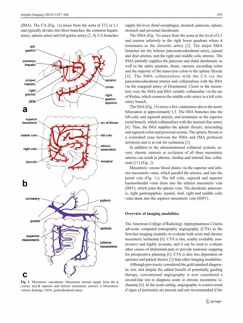
(IMA). The CA (Fig. 1a) arises from the aorta at T12 or L1and typically divides into three branches: the common hepaticartery, splenic artery and left gastric artery [2, 4]. CA branches
supply the liver, distal oesophagus, stomach, pancreas, spleen,stomach and proximal duodenum.
The SMA (Fig. 1b) arises from the aorta at the level of L1and courses inferiorly to the right lower quadrant where itterminates as the ileocolic artery [2]. The major SMAbranches are the inferior pancreaticoduodenal artery, jejunaland ileal arteries, and the right and middle colic arteries. TheSMA partially supplies the pancreas and distal duodenum, aswell as the entire jejunum, ileum, caecum, ascending colonand the majority of the transverse colon to the splenic flexure[4]. The SMA collateralises with the CA via thepancreaticoduodenal arteries and collateralises with the IMAvia the marginal artery of Drummond. Closer to the mesen-teric root, the SMA and IMAvariably collateralise via the arcof Riolan, which connects the middle colic artery to a left colicartery branch.
The IMA (Fig. 1b) arises a few centimetres above the aorticbifurcation at approximately L3. The IMA branches into theleft colic and sigmoid arteries, and terminates as the superiorrectal branch, which collateralises with the internal iliac artery[4]. Thus, the IMA supplies the splenic flexure, descendingand sigmoid colon and proximal rectum. The splenic flexure isa watershed zone between the SMA and IMA perfusionterritories and is at risk for ischaemia [2].
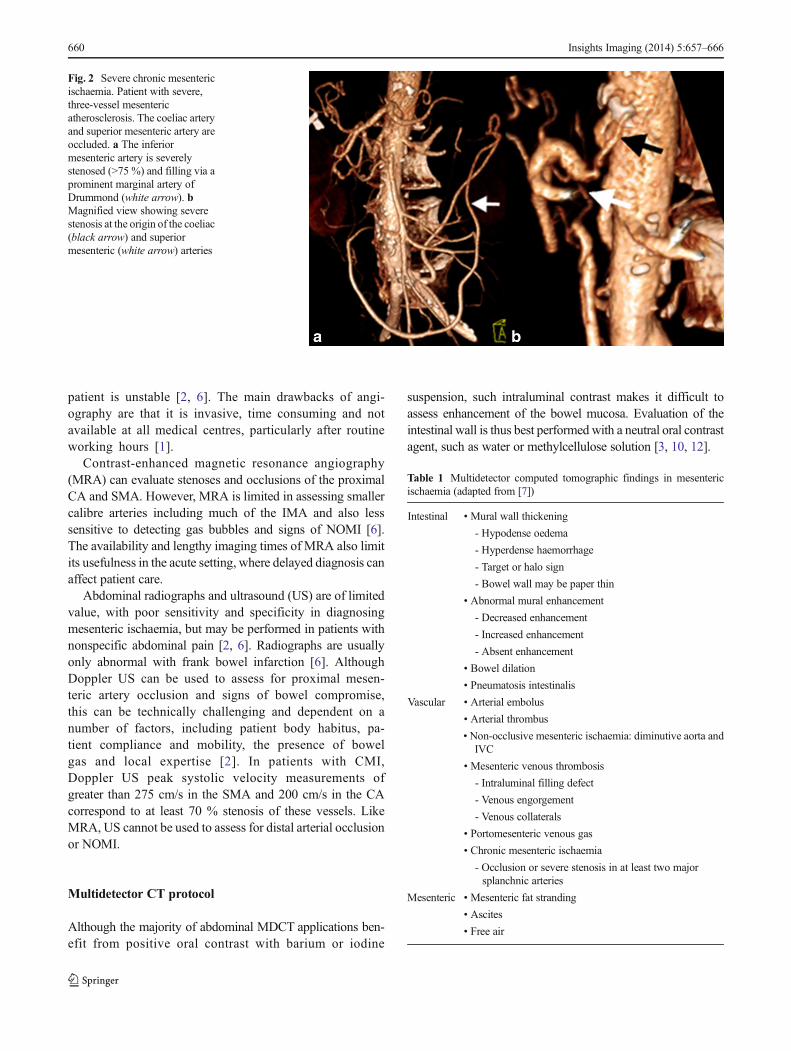
In addition to the aforementioned collateral systems, se-vere, chronic stenosis or occlusion of all three mesentericarteries can result in phrenic, lumbar and internal iliac collat-erals [11] (Fig. 2).
Mesenteric venous blood drains via the superior and infe-rior mesenteric veins, which parallel the arteries, and into theportal vein (Fig. 1c). The left colic, sigmoid and superiorhaemorrhoidal veins drain into the inferior mesenteric vein(IMV), which joins the splenic vein. The duodenal, pancreat-ic, right gastroepiploic, jejunal, ileal, right and middle colicveins drain into the superior mesenteric vein (SMV).
Overview of imaging modalities
The American College of Radiology Appropriateness Criteriaadvocate computed tomography angiography (CTA) as thefirst-line imaging modality to evaluate both acute and chronicmesenteric ischaemia [6]. CTA is fast, readily available, non-invasive and highly accurate, and it can be used to evaluateother causes of abdominal pain or provide anatomic mappingfor preoperative planning [6]. CTA is also less dependent onoperator and patient factors [2] than other imaging modalities.
Although previously considered the gold standard diagnos-tic test, and despite the added benefit of potentially guidingtherapy, conventional angiography is now considered asecond-line test to diagnose acute or chronic mesenteric is-chaemia [6]. In the acute setting, angiography is controversialif signs of peritonitis are present and not recommended if the
Fig. 1 Mesenteric vasculature. Mesenteric arterial supply from the acoeliac and b superior and inferior mesenteric arteries. c Mesentericvenous drainage. GDA, gastroduodenal artery
Insights Imaging (2014) 5:657–666 659
patient is unstable [2, 6]. The main drawbacks of angi-ography are that it is invasive, time consuming and notavailable at all medical centres, particularly after routineworking hours [1].
Contrast-enhanced magnetic resonance angiography(MRA) can evaluate stenoses and occlusions of the proximalCA and SMA. However, MRA is limited in assessing smallercalibre arteries including much of the IMA and also lesssensitive to detecting gas bubbles and signs of NOMI [6].The availability and lengthy imaging times of MRA also limitits usefulness in the acute setting, where delayed diagnosis canaffect patient care.
Abdominal radiographs and ultrasound (US) are of limitedvalue, with poor sensitivity and specificity in diagnosingmesenteric ischaemia, but may be performed in patients withnonspecific abdominal pain [2, 6]. Radiographs are usuallyonly abnormal with frank bowel infarction [6]. AlthoughDoppler US can be used to assess for proximal mesen-teric artery occlusion and signs of bowel compromise,this can be technically challenging and dependent on anumber of factors, including patient body habitus, pa-tient compliance and mobility, the presence of bowelgas and local expertise [2]. In patients with CMI,Doppler US peak systolic velocity measurements ofgreater than 275 cm/s in the SMA and 200 cm/s in the CAcorrespond to at least 70 % stenosis of these vessels. LikeMRA, US cannot be used to assess for distal arterial occlusionor NOMI.
Multidetector CT protocol
Although the majority of abdominal MDCT applications ben-efit from positive oral contrast with barium or iodine
suspension, such intraluminal contrast makes it difficult toassess enhancement of the bowel mucosa. Evaluation of theintestinal wall is thus best performedwith a neutral oral contrastagent, such as water or methylcellulose solution [3, 10, 12].
Fig. 2 Severe chronic mesentericischaemia. Patient with severe,three-vessel mesentericatherosclerosis. The coeliac arteryand superior mesenteric artery areoccluded. a The inferiormesenteric artery is severelystenosed (>75 %) and filling via aprominent marginal artery ofDrummond (white arrow). bMagnified view showing severestenosis at the origin of the coeliac(black arrow) and superiormesenteric (white arrow) arteries
Table 1 Multidetector computed tomographic findings in mesentericischaemia (adapted from [7])
Intestinal • Mural wall thickening
- Hypodense oedema
- Hyperdense haemorrhage
- Target or halo sign
- Bowel wall may be paper thin
• Abnormal mural enhancement
- Decreased enhancement
- Increased enhancement
- Absent enhancement
• Bowel dilation
• Pneumatosis intestinalis
Vascular • Arterial embolus
• Arterial thrombus
• Non-occlusive mesenteric ischaemia: diminutive aorta andIVC
• Mesenteric venous thrombosis
- Intraluminal filling defect
- Venous engorgement
- Venous collaterals
• Portomesenteric venous gas
• Chronic mesenteric ischaemia
- Occlusion or severe stenosis in at least two majorsplanchnic arteries
Mesenteric • Mesenteric fat stranding
• Ascites
• Free air
660 Insights Imaging (2014) 5:657–666

Although initial CT protocols for mesenteric ischaemiarecommended an initial, unenhanced phase to assess for in-tramural haematoma, calcified plaque and bowel enhance-ment, recent articles suggest that the unenhanced phase canbe omitted [9, 10, 13, 14], as a loop of normally enhancingbowel can often be found to compare and act as an internalcontrol [13]. The multiphasic MDCT imaging protocol in-cludes both arterial and portal venous phase acquisitions: thearterial phase is required for optimal assessment of the mes-enteric arterial supply, as thromboembolic disease may ac-count for 60–80 % of acute mesenteric ischaemia cases, andthe venous phase is used for assessing bowel wall enhance-ment and venous drainage. Typical CT parameters are asfollows: 120 ml of non-ionic iodinated contrast material ispower injected at a rate of 3–5 ml/s, followed by a salinechaser; 120 kVp; 270–300 mAs with automatic tube currentmodulation whenever possible; as thin a collimation as possi-ble (e.g. 0.625 mm in 64-slice scanners) because of the smallsize of mesenteric branches [9, 10, 12]. The arterial and portal-venous phases are acquired at approximately 30 and 60 s afterinjection, respectively, often triggered by a specific attenua-tion threshold in a region of interest placed over the abdominalaorta [13, 14].
Raw images are reconstructed into 3–5-mm-thick slices forreview of the abdominal viscera. Sagittal and coronalreformatted images are generated, as well as three-dimensional maximum-intensity projection (MIP) andvolume-rendered images. Sagittal reformats are particularly
useful for assessing the origin of the mesenteric arteries fromthe aorta [10].
MDCT imaging findings of mesenteric ischaemia
MDCT imaging features of mesenteric ischaemia can be clas-sified as intestinal, vascular and mesenteric (Table 1) [9].
Bowel wall thickening
Bowel wall thickening is the most common intestinal CTfinding in mesenteric ischaemia (Fig. 3) [3]. Mural thickeningis typically circumferential and measures approximately0.8 cm, but can reach up to 1.5 cm, particularly in the settingof venous thrombosis [10]. A “target” or “halo” sign may bepresent, representing a two- or three-layer striated pattern ofenhancement. This sign is assumed to represent hypodenseoedema or inflammation in the submucosal layer, sandwichedby enhancing mucosa and muscularis propria [3, 15]. If
Fig. 4 Bowel wall thickening and haemorrhage. Axial CT image in apatient with an occluded superior mesenteric artery. A short segment ofjejunum shows circumferential thickening and hyperattenuation,favoured to represent bowel wall heamatoma
Fig. 5 Hyper-enhancement of the bowel mucosa. Axial contrast-enhanced CT image demonstrates hyper-enhancement of the right lowerquadrant ileal mucosa (arrow) secondary to superior mesenteric arteryocclusion (not shown). The degree of mucosal enhancement can becompared to left-sided bowel loops as an internal control
Fig. 3 Bowel wall thickening in ischaemic colitis. Contrast-enhancedcoronal reformatted image of a patient with severe chronic mesentericischaemia and diarrhoea. There is circumferential, oedematous wallthickening of the descending colon, pathologically proven to representischaemic colitis
Insights Imaging (2014) 5:657–666 661
present, acute haemorrhage will appear as hyperattenuatingmaterial in the bowel wall (Fig. 4) [9, 10]. The absence ofbowel wall thickening does not exclude mesenteric ischaemia,however. In cases of acute arterio-occlusive transmural infarc-tion, the bowel wall can become paper thin [10].
The distribution of bowel wall thickening depends on theaetiology of ischaemia. In SMA or SMVocclusion, for exam-ple, the small bowel, right colon and proximal transversecolon are thickened [3, 10], whereas in NOMI, findings areoften much more diffuse. Segments of ischaemic bowel maybe multifocal with mesenteric emboli [3].
Bowel wall enhancement
Bowel wall enhancement may be increased, decreased orabsent with mesenteric ischaemia (Fig. 5). Complete lack of
mural enhancement is a highly specific but insensitive findingfor acute mesenteric ischaemia [3, 9, 10]. Abnormal muralenhancement is often more subtle, such as delayed arterial-
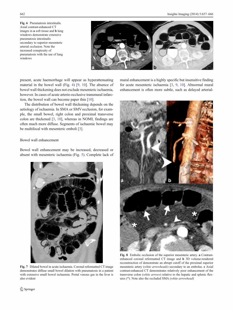
Fig. 6 Pneumatosis intestinalis.Axial contrast-enhanced CTimages in a soft tissue and b lungwindows demonstrate extensivepneumatosis intestinalissecondary to superior mesentericarterial occlusion. Note theincreased conspicuity ofpneumatosis with the use of lungwindows
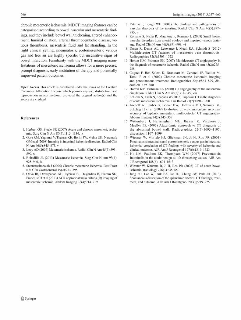
Fig. 7 Dilated bowel in acute ischaemia. Coronal reformatted CT imagedemonstrates diffuse small bowel dilation with pneumatosis in a patientwith extensive small bowel ischaemia. Portal venous gas in the liver isalso evident
Fig. 8 Embolic occlusion of the superior mesenteric artery. a Contrast-enhanced coronal reformatted CT image and b 3D volume-renderedreconstruction of demonstrate an abrupt cutoff of the proximal superiormesenteric artery (white arrowheads) secondary to an embolus. c Axialcontrast-enhanced CT demonstrates relatively poor enhancement of thetransverse colon (white arrows) relative to the hepatic and splenic flex-ures (*). Note also the occluded SMA (white arrowhead)
662 Insights Imaging (2014) 5:657–666

phase enhancement and persistent portal-venous phase en-hancement [9]. Hyper-enhancement of the bowel wall maybe caused by impaired venous drainage of contrast, such as inMVT or strangulated hernia, luxury reperfusion afterarterial occlusion or in cases of reduced arterial perfusionand venous drainage, such as NOMI or shock bowel. Incontrast to decreased or absent mural enhancement,hyperaemia is probably a good prognostic sign as itlikely indicates viable bowel [11].
Bowel wall gas
In the setting of mesenteric ischaemia, pneumatosis intestinalis(mural gas) often indicates transmural infarction, particularly ifit is associated with bandlike portomesenteric venous gas(Fig. 6) [10, 16]. However, pneumatosis and venous gas mayalso be seen with reversible ischaemia [9, 16], as well as aplethora of non-ischaemic disorders such as chronic pulmonarydisease, medications and infectious, inflammatory or neoplasticcauses of intestinal mucosal disruption [3, 17]. There is inaddition a primary (idiopathic), asymptomatic form termedpneumatosis cystoides intestinalis, which is characterised bycircular, bubble-like gas collections in the bowel wall andmesentery; it almost always affects the colon [17].
Bowel lumen
Bowel dilation (Fig. 7) is often present and secondary to eitheraperistalsis from ischaemic injury or complete loss of contrac-tility from transmural infarction [18]. Severe dilation is seen inthe setting of irreversible, transmural ischaemia or infarction[10].
Mesenteric arteries
Emboli typically lodge in the proximal SMA, distal to theorigin of the middle colic artery. SMA embolism appears as acentrallylocated, hypodense intraluminal filling defect
Fig. 9 SMA thrombosis. a Contrast-enhanced, coronal reformatted CTimages demonstrate an eccentric filling defect in the proximal SMA,corresponding to thrombus formation. b Axial contrast-enhanced CTimage shows oedematous bowel wall thickening of small bowel loops(white arrowheads) and mesenteric congestion. This patient subsequentlyimproved with conservative anticoagulation therapy
Fig. 10 Spontaneous coeliacartery dissection. a Contrast-enhanced sagittal reformattedimage demonstrates a dissectionflap of the coeliac artery (whitearrow). b Axial CT image showsthe dissection flap continues intothe common hepatic artery withthrombosis of the false lumen.There was no evidence of bowelischaemia in this patient and theappearance remained stable onfollow-up imaging
Insights Imaging (2014) 5:657–666 663

(Fig. 8). In contrast, SMA thrombosis typically developseccentrically at or near the ostium, on a background of ath-erosclerosis (Fig. 9).
Another potential, albeit less common cause of AMI isdissection of a splanchnic artery, often as a continuation ofaortic dissection. The most common CT findings include anintimal flap, aneurysmal dilation and a thrombosed false lumen(Fig. 10) [19]. In dissections causing subtotal luminal occlu-sion, the branching smaller arteries are at higher risk of beingaffected. Malignant encasement and narrowing of the mesen-teric arteries is another uncommon cause of AMI (Fig. 11).
Portomesenteric veins
SMV thrombosis is demonstrated as a partial or completehypoattenuating filling defect (Figs. 11 and 12) [9]. Care mustbe taken to avoid mistaking delayed venous filling and otherflow-related phenomena for thrombi. The affected venousbranch may be enlarged, particularly if the thrombus is acuteor malignant tumour thrombus. The associated venous con-gestion can result in engorged mesenteric veins (Fig. 12) [9].As mentioned previously, in the setting of ischaemiaportomesenteric venous gas and bandlike pneumatosis are
highly associated with transmural bowel infarction (Figs. 13and 14) [16].
MVT secondary to malignancy typically occurs with hepa-tocellular carcinoma and pancreatic adenocarcinoma, but canbe seen with other malignancies as well [9].
Non-occlusive mesenteric ischaemia
Because it is a systemic disorder, MDCT findings ofNOMI are typically diffusely thickened and fluid-distended bowel loops and markedly attenuated, prunedblood vessels [10].
Shock bowel is a subtype of NOMI (Fig. 15). TypicalMDCT features of shock bowel include diffuse small bowelthickening with relative sparing of the colon, prolonged muralenhancement, fluid or gas-filled small bowel loops, and othersigns of hypovolaemia, such as a flattened inferior vena cava(IVC) [3].
Chronic mesenteric ischaemia
The diagnosis of CMI is based on clinical symptoms andsupported by imaging findings, following exclusion of other
Fig. 11 Mesenteric ischaemia secondary to vascular compression by asmall bowel carcinoid. a Coronal contrast-enhanced reformatted imageshows mesenteric metastatic disease from the small bowel carcinoidencasing distal branches of the SMA (white arrow). b Coronal image
from an octreotide scan shows uptake by the metastatic disease (whitearrow), indicating tumoral somatostatin receptors. c Three-dimensionalvolume-rendered reconstruction shows attenuated calibre of the ileocolicartery (white arrow)
Fig. 12 SMV thrombosis. aContrast-enhanced coronalreformatted CT demonstratesocclusive thrombus in the SMVnear the portal confluence (whitearrowhead). b Axial CT imagedemonstrates diffuse small bowelwall thickening secondary tovenous congestion and ischaemia
664 Insights Imaging (2014) 5:657–666

potential intestinal disorders [3]. CT accurately demonstratescalcified and noncalcified plaque causing arterial stenosis orocclusion, typically in the proximal CA and SMA [10]. Small,attenuated vessels and large collateral vessels are importantsupportive findings (Fig. 2) [10].
Mesenteric stranding, fluid and gas
With the exception of free peritoneal gas, mesenteric MDCTfindings in bowel ischaemia are nonspecific, as they arecommonly seen in any acute abdominal process [3]. Ascitesand stranding or haziness of the mesenteric fat are seen tovariable degrees, and, as these processes often result fromvenous congestion, are much more common in MVT thanarterio-occlusive disorders [9].
In contrast to free fluid and fat stranding, free air in thesetting of mesenteric ischaemia is indicative of bowel infarc-tion and perforation.
Conclusion
Acute and chronic mesenteric ischaemia are morbid condi-tions that are challenging to diagnose. Patients present withvariable, nonspecific signs and symptoms, and the physicalexamination is often benign. A high index of clinical andradiologic suspicion is thus required for diagnosis. Contrast-enhanced, multidetector CT angiography is the first-line im-aging test for evaluating patients with suspected acute or
Fig. 13 SMV thrombosis with mesenteric congestion. a Contrast-enhanced coronal reformatted CT of an occlusive thrombus in the SMV(white arrowhead) extending to the portal confluence. b Axial contrast-
enhanced CT image demonstrates mesenteric congestion with engorgedmesenteric veins and trace fluid (white arrowhead). The small bowel isthickened because of the venous stasis and probable ischaemia
Fig. 14 Portomesenteric venous gas. a Contrast-enhanced coronalreformatted CT demonstrates occlusion of the SMA (arrowhead). Portalvenous gas is seen in the liver. bAxial CT shows portal venous gas in theanti-dependent liver. c The small bowel demonstrates luminal dilation, apaper-thin wall and poorly enhancing mucosa. There is mesenteric ve-nous gas (arrowhead) as well as pneumatosis in the dependent bowelwall. The patient had established bowel necrosis and died shortly afterimaging
Fig. 15 Shock bowel. Contrast-enhanced axial CT image demonstratesdiffuse bowel wall thickening with mural hyper-enhancement. Note thesecondary sign of a flattened inferior vena cava, indicating hypovolaemia
Insights Imaging (2014) 5:657–666 665
chronic mesenteric ischaemia.MDCT imaging features can becategorised according to bowel, vascular and mesenteric find-ings, and they include bowel wall thickening, altered enhance-ment, luminal dilation, arterial thromboembolic disease, ve-nous thrombosis, mesenteric fluid and fat stranding. In theright clinical setting, pneumatosis, portomesenteric venousgas and free air are highly specific but insensitive signs ofbowel infarction. Familiarity with the MDCT imaging mani-festations of mesenteric ischaemia allows for a more precise,prompt diagnosis, early institution of therapy and potentiallyimproved patient outcomes.
Open Access This article is distributed under the terms of the CreativeCommons Attribution License which permits any use, distribution, andreproduction in any medium, provided the original author(s) and thesource are credited.
References
1. Herbert GS, Steele SR (2007) Acute and chronic mesenteric ische-mia. Surg Clin N Am 87(5):1115–1134, ix
2. Gore RM,Yaghmai V, Thakrar KH, Berlin JW,Mehta UK, NewmarkGM et al (2008) Imaging in intestinal ischemic disorders. Radiol ClinN Am 46(5):845–875, v
3. Levy AD (2007) Mesenteric ischemia. Radiol Clin NAm 45(3):593–599, x
4. Bobadilla JL (2013) Mesenteric ischemia. Surg Clin N Am 93(4):925–940, ix
5. Sreenarasimhaiah J (2005) Chronic mesenteric ischemia. Best PractRes Clin Gastroenterol 19(2):283–295
6. Oliva IB, Davarpanah AH, Rybicki FJ, Desjardins B, Flamm SD,Francois CJ et al (2013) ACR appropriateness criteria (R) imaging ofmesenteric ischemia. Abdom Imaging 38(4):714–719
7. Paterno F, Longo WE (2008) The etiology and pathogenesis ofvascular disorders of the intestine. Radiol Clin N Am 46(5):877–885, v
8. Romano S, Niola R, Maglione F, Romano L (2008) Small bowelvascular disorders from arterial etiology and impaired venous drain-age. Radiol Clin N Am 46(5):891–908, vi
9. Duran R, Denys AL, Letovanec I, Meuli RA, Schmidt S (2012)Multidetector CT features of mesenteric vein thrombosis.Radiographics 32(5):1503–1522
10. Horton KM, Fishman EK (2007) Multidetector CT angiography inthe diagnosis of mesenteric ischemia. Radiol Clin N Am 45(2):275–288
11. Cognet F, Ben Salem D, Dranssart M, Cercueil JP, Weiller M,Tatou E et al (2002) Chronic mesenteric ischemia: imagingand percutaneous treatment. Radiographics 22(4):863–879, dis-cussion 879–880
12. Horton KM, Fishman EK (2010) CT angiography of the mesentericcirculation. Radiol Clin N Am 48(2):331–345, viii
13. Schieda N, Fasih N, ShabanaW (2013) Triphasic CT in the diagnosisof acute mesenteric ischaemia. Eur Radiol 23(7):1891–1900
14. Aschoff AJ, Stuber G, Becker BW, Hoffmann MH, Schmitz BL,Schelzig H et al (2009) Evaluation of acute mesenteric ischemia:accuracy of biphasic mesenteric multi-detector CT angiography.Abdom Imaging 34(3):345–357
15. Wittenberg J, Harisinghani MG, Jhaveri K, Varghese J,Mueller PR (2002) Algorithmic approach to CT diagnosis ofthe abnormal bowel wall. Radiographics 22(5):1093–1107,discussion 1107–1099
16. Wiesner W, Mortele KJ, Glickman JN, Ji H, Ros PR (2001)Pneumatosis intestinalis and portomesenteric venous gas in intestinalischemia: correlation of CT findings with severity of ischemia andclinical outcome. AJR Am J Roentgenol 177(6):1319–1323
17. Ho LM, Paulson EK, Thompson WM (2007) Pneumatosisintestinalis in the adult: benign to life-threatening causes. AJR AmJ Roentgenol 188(6):1604–1613
18. Wiesner W, Khurana B, Ji H, Ros PR (2003) CT of acute bowelischemia. Radiology 226(3):635–650
19. Jung SC, Lee W, Park EA, Jae HJ, Chung JW, Park JH (2013)Spontaneous dissection of the splanchnic arteries: CT findings, treat-ment, and outcome. AJR Am J Roentgenol 200(1):219–225
666 Insights Imaging (2014) 5:657–666