mucosal ablation therapy of barrett esophagus
TRANSCRIPT

Mayo Clin Proc, April 2001, Vol 76 Mucosal Ablation 433
Mayo Clin Proc. 2001;76:433-437 433 © 2001 Mayo Foundation for Medical Education and Research
From the Division of Gastroenterology and Hepatology and InternalMedicine, Mayo Clinic, Rochester, Minn (K.K.W.); and Departmentof Gastroenterology, University of Arizona, Tucson (R.E.S.).
Supported in part by National Institutes of Health grants CA72541and CA78870 and by Mayo Foundation.
Individual reprints of this article are not available. The entire Alan J.Cameron Symposium on Barrett Esophagus and GastroesophagealReflux Disease will be available for purchase as a bound bookletfrom the Proceedings Editorial Office at a later date.
Cameron Symposium on Barrett Esophagus and GERD
Mucosal Ablation Therapy of Barrett Esophagus
KENNETH K. WANG, MD, AND RICHARD E. SAMPLINER, MD
Barrett esophagus is defined by the metaplasia of existingsquamous mucosa into a specialized intestinal-type mu-cosa. The importance of this metaplasia is the associationof this condition with the development of adenocarcinomaof the esophagus. Elimination of the metaplastic mucosamay decrease the cancer risk. Currently, several forms oftherapy have evolved with the goal of replacing the special-ized mucosa with normal squamous mucosa. These pro-posed treatments include photodynamic therapy and ther-mal techniques. The effectiveness of photodynamictherapy varies depending on the pharmaceutical photo-sensitizer used and the wavelength of light applied to acti-vate the drug. Thermal techniques include multipolar co-agulation, argon plasma coagulation, KTP:YAG laser
therapy, Nd:YAG laser therapy, and argon laser therapy.Finally, mucosal resection has been attempted through theendoscope to remove large areas of the Barrett mucosa. Allof these ablative strategies attempt to destroy the meta-plastic mucosa and promote the regrowth of squamousepithelium. These therapies have demonstrated the abilityto “reverse” the metaplasia to varying degrees, but a de-crease in cancer risk has not been demonstrated conclu-sively with any of these treatment methods.
Mayo Clin Proc. 2001;76:433-437
ALA = aminolevulinic acid; HpD = hematoporphyrin deriva-tive; MPEC = multipolar electrocoagulation
Management of Barrett esophagus is still a clinicalchallenge. Although the incidence of cancer appears
to be low, occurring in 0.5% of patients with Barrettesophagus, the risk in an individual patient is more difficultto ascertain.1 Up to 50% of patients with high-grade dyspla-sia may have endoscopically undetectable cancer,2 and astandard endoscopic biopsy may not detect endoscopicallyoccult cancers in 43% of patients prior to esophagectomyfor high-grade dysplasia.3 This situation leaves the clini-cian with difficult management decisions. The majority ofpatients with Barrett esophagus do not have any decrease intheir life expectancy.4 However, the ability to determinecancer risk in an individual patient is imprecise. In addi-tion, the clinician must determine at which point interven-tion should be attempted. Individuals with Barrett esopha-gus without dysplasia are not likely to have serious risk ofmalignancy. Outcomes studies that use the most recentfigures for incidence of cancer (0.4%) suggest that surveil-lance strategies should be based on performance of anendoscopy at 5-year intervals. This frequency of endos-copy, however, may not be effective in identifying patientswith early neoplasms who may be most likely to benefit
from intervention.1 Surgery is advocated in patients withhigh-grade dysplasia who are reasonable candidates foresophagectomy.2 Surgical resection of early cancers is usu-ally associated with good outcomes, with 5-year survival of93% if the tumor is intraepithelial and 72.8% if the tumor isintramucosal. Operative mortality in a cohort of Europeanpatients was 9.1%5 and is related directly to the number ofesophagectomies performed in the institution.6,7 Surgicalmortality rates in specialized cancer institutions or at institu-tions where the number of esophagectomies performed ishigh were significantly lower (3%-4.2%) than in those hos-pitals performing fewer esophagectomies (12.2%-13.3%).6,7
Since Barrett esophagus is a disease process that isdefined by endoscopy, gastroenterologists have been usingmultiple strategies in an attempt to eliminate this metaplas-tic mucosa. Early therapy was focused on acid control,since acid formation seemed to be the primary pathogenicfactor, but a number of studies have found that acid controltherapy has not led to regression of metaplastic mucosa.8,9
More recent efforts have focused on combining destructionof the metaplastic mucosa with some degree of acid con-trol.10 These treatments can be divided into 3 basic tech-niques: (1) photodynamic therapy, (2) thermal ablationtherapies, and (3) endoscopic mucosal resection. The endpoint of each of these therapies is elimination of the risk ofadenocarcinoma, although none of these techniques has yetbeen proved to accomplish this goal.
PHOTODYNAMIC THERAPYPhotodynamic therapy or photochemotherapy involves thecombination of 3 elements. These include a drug that is a
For personal use. Mass reproduce only with permission from Mayo Clinic Proceedings.

Mucosal Ablation Mayo Clin Proc, April 2001, Vol 76434
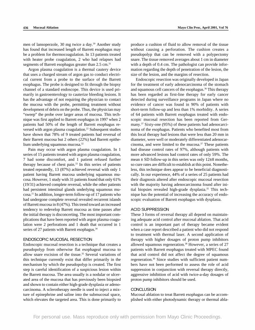
photosensitizer, light, and oxygen. In clinical practice, thephotosensitizer is administered intravenously 2 days priorto irradiation with light. This time delay is needed to permitthe photosensitizer to reach an optimal level within neo-plastic tissue compared with the surrounding normal tis-sue.11 Once this is achieved, the patient undergoes an endos-copy during which the Barrett esophagus can be irradiatedwith red light. The red light is usually produced with use ofa tunable dye laser, which can generate sufficient light toradiate large portions of the esophagus with a single appli-cation of a laser fiber. The red light activates the photosen-sitizer in the neoplastic tissue, and the photosensitizer inturn interacts with oxygen. The interaction creates singletoxygen, which mediates the damage to the neoplastic tis-sue. The absence of any of the 3 elements—red light, photo-sensitizer, or oxygen—prevents the photodynamic effect.Tissue necrosis is delayed and evolves over the ensuing 2 to5 days (Figure 1).
Photodynamic therapy for Barrett esophagus has in-volved a number of photosensitizing agents, includingaminolevulinic acid (ALA), hematoporphyrin derivative(HpD), porfimer sodium (a refined version of HpD), andtemoporfin.12-15 The difference in these agents relates pri-marily to the depth of tissue destruction that is achievableand the duration of subsequent skin phototoxicity that oc-curs. Porfimer sodium is the only one of these agentscommercially available in the United States for use in thegastrointestinal tract.
Photodynamic therapy has primarily been used in thetreatment of Barrett esophagus with high-grade dysplasiaor superficial cancer. This method has been favored forthese applications because of the possible selectivity of the
treatment for neoplastic tissue and the ease with which theentire mucosal surface can be treated. The agents used fortreatment of Barrett esophagus appear to have similar effi-cacy in eliminating dysplasia within Barrett esophagus asassessed by endoscopic biopsy. In patients who have re-ceived photodynamic therapy, high-grade dysplasia ap-pears to be eliminated in 88% to 100%.12,15 Given orally,ALA is selectively retained within the dysplastic mucosa ofBarrett esophagus16 and is converted into the fluorescentcompound protoporphyrin IX, the active photosensitizer.This drug appears to be retained strictly within the mucosa.In the series reported to date,17 although elimination ofhigh-grade dysplasia is fairly common, reversal of mucosato squamous mucosa is less likely. This may be due to thesuperficial nature of the injury. The ability to reverse meta-plasia may be related to the depth of tissue injury producedby the therapy. Strictures have not been reported with ALAas they have been with porfimer sodium. The most exten-sive experience with mucosal ablation therapy has beenwith use of porfimer sodium–based photodynamic therapy.Photodynamic therapy with this agent can cause completereversion to normal-appearing squamous mucosa in abouta third of cases. As with ALA, porfimer sodium may haveselectivity for dysplasia. High-grade dysplasia appears torespond in about 88% of treated cases (Table 1).12
One concern with photodynamic therapy, as with any ofthe thermal ablative techniques, is the unfortunate tendencyof the squamous mucosa to overgrow small areas of Barrettmucosa. This phenomenon seems to be relatively uncom-mon, occurring in 4% to 6% of patients treated with photo-dynamic therapy.17 The relatively high percentage of pa-tients developing this condition in 1 series was actually due
Figure 1. Left, This endoscopic photograph of the esophagus shows the positioning of acylindrical diffusing fiber within the lumen of a segment of Barrett esophagus. Right, Theappearance of the esophageal mucosa 48 hours after therapy with evidence of tissue necrosis.
For personal use. Mass reproduce only with permission from Mayo Clinic Proceedings.
Mayo Clin Proc, April 2001, Vol 76 Mucosal Ablation 435
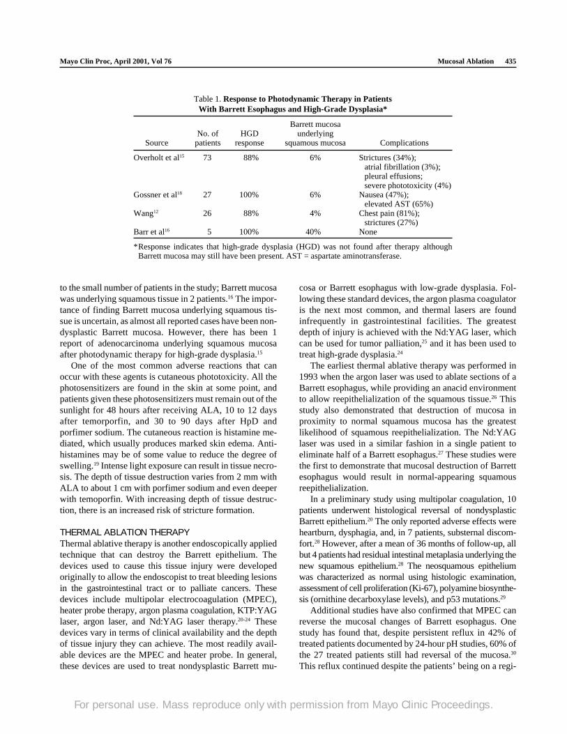
Table 1. Response to Photodynamic Therapy in PatientsWith Barrett Esophagus and High-Grade Dysplasia*
Barrett mucosaNo. of HGD underlying
Source patients response squamous mucosa Complications
Overholt et al15 73 88% 6% Strictures (34%);atrial fibrillation (3%);pleural effusions;severe phototoxicity (4%)
Gossner et al18 27 100% 6% Nausea (47%);elevated AST (65%)
Wang12 26 88% 4% Chest pain (81%);strictures (27%)
Barr et al16 5 100% 40% None
*Response indicates that high-grade dysplasia (HGD) was not found after therapy althoughBarrett mucosa may still have been present. AST = aspartate aminotransferase.
to the small number of patients in the study; Barrett mucosawas underlying squamous tissue in 2 patients.16 The impor-tance of finding Barrett mucosa underlying squamous tis-sue is uncertain, as almost all reported cases have been non-dysplastic Barrett mucosa. However, there has been 1report of adenocarcinoma underlying squamous mucosaafter photodynamic therapy for high-grade dysplasia.15
One of the most common adverse reactions that canoccur with these agents is cutaneous phototoxicity. All thephotosensitizers are found in the skin at some point, andpatients given these photosensitizers must remain out of thesunlight for 48 hours after receiving ALA, 10 to 12 daysafter temorporfin, and 30 to 90 days after HpD andporfimer sodium. The cutaneous reaction is histamine me-diated, which usually produces marked skin edema. Anti-histamines may be of some value to reduce the degree ofswelling.19 Intense light exposure can result in tissue necro-sis. The depth of tissue destruction varies from 2 mm withALA to about 1 cm with porfimer sodium and even deeperwith temoporfin. With increasing depth of tissue destruc-tion, there is an increased risk of stricture formation.
THERMAL ABLATION THERAPYThermal ablative therapy is another endoscopically appliedtechnique that can destroy the Barrett epithelium. Thedevices used to cause this tissue injury were developedoriginally to allow the endoscopist to treat bleeding lesionsin the gastrointestinal tract or to palliate cancers. Thesedevices include multipolar electrocoagulation (MPEC),heater probe therapy, argon plasma coagulation, KTP:YAGlaser, argon laser, and Nd:YAG laser therapy.20-24 Thesedevices vary in terms of clinical availability and the depthof tissue injury they can achieve. The most readily avail-able devices are the MPEC and heater probe. In general,these devices are used to treat nondysplastic Barrett mu-
cosa or Barrett esophagus with low-grade dysplasia. Fol-lowing these standard devices, the argon plasma coagulatoris the next most common, and thermal lasers are foundinfrequently in gastrointestinal facilities. The greatestdepth of injury is achieved with the Nd:YAG laser, whichcan be used for tumor palliation,25 and it has been used totreat high-grade dysplasia.24
The earliest thermal ablative therapy was performed in1993 when the argon laser was used to ablate sections of aBarrett esophagus, while providing an anacid environmentto allow reepithelialization of the squamous tissue.26 Thisstudy also demonstrated that destruction of mucosa inproximity to normal squamous mucosa has the greatestlikelihood of squamous reepithelialization. The Nd:YAGlaser was used in a similar fashion in a single patient toeliminate half of a Barrett esophagus.27 These studies werethe first to demonstrate that mucosal destruction of Barrettesophagus would result in normal-appearing squamousreepithelialization.
In a preliminary study using multipolar coagulation, 10patients underwent histological reversal of nondysplasticBarrett epithelium.20 The only reported adverse effects wereheartburn, dysphagia, and, in 7 patients, substernal discom-fort.28 However, after a mean of 36 months of follow-up, allbut 4 patients had residual intestinal metaplasia underlying thenew squamous epithelium.28 The neosquamous epitheliumwas characterized as normal using histologic examination,assessment of cell proliferation (Ki-67), polyamine biosynthe-sis (ornithine decarboxylase levels), and p53 mutations.29
Additional studies have also confirmed that MPEC canreverse the mucosal changes of Barrett esophagus. Onestudy has found that, despite persistent reflux in 42% oftreated patients documented by 24-hour pH studies, 60% ofthe 27 treated patients still had reversal of the mucosa.30
This reflux continued despite the patients’ being on a regi-
For personal use. Mass reproduce only with permission from Mayo Clinic Proceedings.
Mucosal Ablation Mayo Clin Proc, April 2001, Vol 76436
men of lansoperzole, 30 mg twice a day.30 Another studyhas found that increased length of Barrett esophagus maybe a problem for thermocoagulation. In 13 patients treatedwith heater probe coagulation, 2 who had relapses hadsegments of Barrett esophagus greater than 2.5 cm.31
Argon plasma coagulation is a thermal cautery devicethat uses a charged stream of argon gas to conduct electri-cal current from a probe to the surface of the Barrettesophagus. The probe is designed to fit through the biopsychannel of a standard endoscope. This device is used pri-marily in gastroenterology to cauterize bleeding lesions. Ithas the advantage of not requiring the physician to contactthe mucosa with the probe, permitting treatment withoutdevelopment of debris on the probe. Thus, the physician may“sweep” the probe over larger areas of mucosa. This tech-nique was first applied to Barrett esophagus in 1997 when 2patients had 50% of the length of Barrett esophagus re-versed with argon plasma coagulation.22 Subsequent studieshave shown that 78% of 9 treated patients had reversal oftheir Barrett mucosa but 2 others (22%) had Barrett epithe-lium underlying squamous mucosa.32
Pain may occur with argon plasma coagulation. In 1series of 15 patients treated with argon plasma coagulation,7 had some discomfort, and 1 patient refused furthertherapy because of chest pain.33 In this series of patientstreated repeatedly, 13 (87%) achieved reversal with only 1patient having Barrett mucosa underlying squamous mu-cosa. However, 1 study with 31 patients found that only 61%(19/31) achieved complete reversal, while the other patientshad persistent intestinal glands underlying squamous mu-cosa.21 In addition, longer-term follow-up of 17 patients whohad undergone complete reversal revealed recurrent islandsof Barrett mucosa in 8 (47%). This trend toward an increasedtendency to redevelop Barrett mucosa as time passes afterthe initial therapy is disconcerting. The most important com-plications that have been reported with argon plasma coagu-lation were 2 perforations and 1 death that occurred in 1series of 27 patients with Barrett esophagus.34
ENDOSCOPIC MUCOSAL RESECTIONEndoscopic mucosal resection is a technique that creates apseudopolyp from otherwise flat esophageal mucosa toallow snare excision of the tissue.35 Several variations ofthis technique currently exist that differ primarily in themechanism by which the pseudopolyp is created. The firststep is careful identification of a suspicious lesion withinthe Barrett mucosa. The area usually is a nodular or ulcer-ated area of the mucosa that has previously been biopsiedand shown to contain either high-grade dysplasia or adeno-carcinoma. A sclerotherapy needle is used to inject a mix-ture of epinephrine and saline into the submucosal space,which elevates the targeted area. This is done primarily to
produce a cushion of fluid to allow removal of the tissuewithout causing a perforation. The cushion creates apseudopolyp that can be removed with a polypectomysnare. The tissue removed averages about 1 cm in diameterwith a depth of 0.4 cm. The pathologist can provide infor-mation regarding the depth of penetration of the lesion, thesize of the lesion, and the margins of resection.
Endoscopic resection was originally developed in Japanfor the treatment of early adenocarcinoma of the stomachand squamous cell cancers of the esophagus.36 This therapyhas been regarded as first-line therapy for early cancerdetected during surveillance programs in Japan where noevidence of cancer was found in 90% of patients withshort-term follow-up and less than 1% morbidity. A seriesof 64 patients with Barrett esophagus treated with endo-scopic mucosal resection has been reported from Ger-many.37 Sixty-one (95%) of these patients had adenocarci-noma of the esophagus. Patients who benefited most fromthis local therapy had lesions that were less than 20 mm indiameter, were well or moderately differentiated adenocar-cinoma, and were limited to the mucosa.37 These patientshad disease control rates of 97%, although patients withmore advanced lesions had control rates of only 59%. Themean ± SD follow-up in this series was only 12±8 months,so cure rates are difficult to establish at this point. Nonethe-less, this technique does appear to be beneficial diagnosti-cally. In our experience, 44% of a series of 25 patients hadtheir diagnosis altered after endoscopic mucosal resectionwith the majority having adenocarcinoma found after ini-tial biopsies revealed high-grade dysplasia.35 This tech-nique has the potential of increasing the accuracy of endo-scopic evaluation of Barrett esophagus with dysplasia.
ACID SUPPRESSIONThese 3 forms of reversal therapy all depend on maintain-ing adequate acid control after mucosal ablation. That acidcontrol is an important part of therapy became evidentwhen a case report described a patient who did not respondto treatment with thermal laser. A second application oftherapy with higher dosages of proton pump inhibitorsallowed squamous regeneration.38 However, a series of 27patients with Barrett esophagus treated with MPEC foundthat acid control did not affect the degree of squamousregeneration.30 Since studies with sufficient patient num-bers have not been performed to assess the role of acidsuppression in conjunction with reversal therapy directly,aggressive inhibition of acid with twice-a-day dosages ofproton pump inhibitors should be used.
CONCLUSIONMucosal ablation to treat Barrett esophagus can be accom-plished with either photodynamic therapy or thermal abla-
For personal use. Mass reproduce only with permission from Mayo Clinic Proceedings.
Mayo Clin Proc, April 2001, Vol 76 Mucosal Ablation 437
tive therapy. Patients with lesions such as high-grade dys-plasia or superficial adenocarcinoma who are not candi-dates for traditional surgical therapy can be approachedwith photodynamic therapy and endoscopic mucosal resec-tion. Nd:YAG laser therapy can also be applied althoughthis is not widely available. Nondysplastic Barrett mucosaor low-grade dysplasia can be treated with thermal tech-niques, including argon plasma coagulation or MPEC. Mac-roscopic abnormalities within Barrett esophagus should beremoved with endoscopic mucosal resection for diagnosticpurposes. None of these strategies has been proven yet toprovide long-term control of Barrett esophagus.
REFERENCES1. Provenzale D, Schmitt C, Wong JB. Barrett’s esophagus: a new
look at surveillance based on emerging estimates of cancer risk. AmJ Gastroenterol. 1999;94:2043-2053.
2. Pera M, Trastek VF, Carpenter HA, Allen MS, Deschamps C,Pairolero PC. Barrett’s esophagus with high-grade dysplasia: anindication for esophagectomy? Ann Thorac Surg. 1992;54:199-204.
3. Nigro JJ, Hagen JA, DeMeester TR, et al. Occult esophageal ad-enocarcinoma: extent of disease and implications for effective ther-apy. Ann Surg. 1999;230:433-438.
4. Van der Veen AH, Dees J, Blankensteijn JD, Van Blankenstein M.Adenocarcinoma in Barrett’s oesophagus: an overrated risk. Gut.1989;30:14-18.
5. Bonavina L, Group Europeen pour l’Etude des Maladies de l’Oesoph-age. Early oesophageal cancer: results of a European multicentresurvey. Br J Surg. 1995;82:98-101.
6. Patti MG, Corvera CU, Glasgow RE, Way LW. A hospital’s annualrate of esophagectomy influences the operative mortality rate. JGastrointest Surg. 1998;2:186-192.
7. Swisher SG, Deford L, Merriman KW, et al. Effect of operativevolume on morbidity, mortality, and hospital use after esophagec-tomy for cancer. J Thorac Cardiovasc Surg. 2000;119:1126-1132.
8. Sampliner RE. Effect of up to 3 years of high-dose lansoprazole onBarrett’s esophagus. Am J Gastroenterol. 1994;89:1844-1848.
9. Sharma P, Sampliner RE, Camargo E. Normalization of esophagealpH with high-dose proton pump inhibitor therapy does not result inregression of Barrett’s esophagus. Am J Gastroenterol. 1997;92:582-585.
10. Sampliner RE. Ablative therapies for the columnar-lined esopha-gus. Gastroenterol Clin North Am. 1997;26:685-694.
11. Dougherty TJ, Gomer CJ, Henderson BW, et al. Photodynamictherapy. J Natl Cancer Inst. 1998;90:889-905.
12. Wang KK. Current status of photodynamic therapy of Barrett’sesophagus. Gastointest Endosc. 1999;49(3, pt 2):S20-S23.
13. Gossner L, May A, Sroka R, Stolte M, Hahn EG, Ell C. Photody-namic destruction of high grade dysplasia and early carcinoma ofthe esophagus after the oral administration of 5-aminolevulinicacid. Cancer. 1999;86:1921-1928.
14. Gossner L, May A, Sroka R, Ell C. A new long-range through-the-scope balloon applicator for photodynamic therapy in the esopha-gus and cardia. Endoscopy. 1999;31:370-376.
15. Overholt BF, Panjehpour M, Haydek JM. Photodynamic therapyfor Barrett’s esophagus: follow-up in 100 patients. GastrointestEndosc. 1999;49:1-7.
16. Barr H, Shepherd NA, Dix A, Roberts DJ, Tan WC, Krasner N.Eradication of high-grade dysplasia in columnar-lined (Barrett’s)oesophagus by photodynamic therapy with endogenously gener-ated protoporphyrin IX. Lancet. 1996;348:584-585.
17. Loh CS, Vernon D, MacRobert AJ, Bedwell J, Bown SG, BrownSB. Endogenous porphyrin distribution induced by 5-amino-laevulinic acid in the tissue layers of the gastrointestinal tract. JPhotochem Photobiol B. 1993;20:47-54.
18. Gossner L, Stolte M, Sroka R, et al. Photodynamic ablation of high-grade dysplasia and early cancer in Barrett’s esophagus by meansof 5-aminolevulinic acid. Gastroenterology. 1998;114:448-455.
19. Baas P, Oppelaar H, van der Valk MA, van Zandwijk N, StewartFA. Partial protection of photodynamic-induced skin reactions inmice by N-acetylcysteine: a preclinical study. Photochem Photo-biol. 1994;59:448-454.
20. Sampliner RE, Fennerty B, Garewal HS. Reversal of Barrett’sesophagus with acid suppression and multipolar electrocoagula-tion: preliminary results. Gastrointest Endosc. 1996;44:532-535.
21. Van Laethem JL, Cremer M, Peny MO, Delhaye M, Deviere J. Eradi-cation of Barrett’s mucosa with argon plasma coagulation and acidsuppression: immediate and mid term results. Gut. 1998;43: 747-751.
22. Dumoulin FL, Terjung B, Neubrand M, Scheurlen C, Fischer HP,Sauerbruch T. Treatment of Barrett’s esophagus by endoscopicargon plasma coagulation. Endoscopy. 1997;29:751-753.
23. Gossner L, May A, Stolte M, Seitz G, Hahn EG, Ell C. KTP laserdestruction of dysplasia and early cancer in columnar-linedBarrett’s esophagus. Gastrointest Endosc. 1999;49:8-12.
24. Sharma P, Jaffe PE, Bhattacharyya A, Sampliner RE. Laser andmultipolar electrocoagulation ablation of early Barrett’s adenocar-cinoma: long-term follow-up. Gastrointest Endosc. 1999;49(4, pt1):442-446.
25. Alexander GL, Wang KK, Ahlquist DA, Viggiano TR, Gostout CJ,Balm R. Does performance status influence the outcome ofNd:YAG laser therapy of proximal esophageal tumors? Gastro-intest Endosc. 1994;40:451-454.
26. Berenson MM, Johnson TD, Markowitz NR, Buchi KN, SamowitzWS. Restoration of squamous mucosa after ablation of Barrett’sesophageal epithelium. Gastroenterology. 1993;104:1686-1691.
27. Sampliner RE, Hixson LJ, Fennerty MB, Garewal HS. Regressionof Barrett’s esophagus by laser ablation in an anacid environment.Dig Dis Sci. 1993;38:365-368.
28. Sharma P, Bhattacharyya A, Garewal HS, Sampliner RE. Durabil-ity of new squamous epithelium after endoscopic reversal ofBarrett’s esophagus. Gastrointest Endosc. 1999;50:159-164.
29. Garewal H, Ramsey L, Sharma P, Kraus K, Sampliner R, Fass R.Biomarker studies in reversed Barrett’s esophagus. Am JGastroenterol. 1999;94:2829-2833.
30. Kovacs BJ, Chen YK, Lewis TD, DeGuzman LJ, Thompson KS.Successful reversal of Barrett’s esophagus with multipolar electro-coagulation despite inadequate acid suppression. GastrointestEndosc. 1999;49:547-553.
31. Michopoulos S, Tsibouris P, Bouzakis H, Sotiropoulou M, KraliosN. Complete regression of Barrett’s esophagus with heat probethermocoagulation: mid-term results. Gastrointest Endosc. 1999;50:165-172.
32. Grade AJ, Shah IA, Medlin SM, Ramirez FC. The efficacy andsafety of argon plasma coagulation therapy in Barrett’s esophagus.Gastrointest Endosc. 1999;50:18-22.
33. Mork H, Barth T, Kreipe HH, et al. Reconstitution of squamousepithelium in Barrett’s oesophagus with endoscopic argon plasmacoagulation: a prospective study. Scand J Gastroenterol. 1998;33:1130-1134.
34. Byrne JP, Armstrong GR, Attwood SE. Restoration of the normalsquamous lining in Barrett’s esophagus by argon beam plasmacoagulation. Am J Gastroenterol. 1998;93:1810-1815.
35. Nijahwan PK, Wang KK. Endoscopic mucosal resection for lesionswith endoscopic features suggestive of malignancy and high-gradedysplasia within Barrett’s esophagus. Gastrointest Endosc. 2000;52:328-332.
36. Inoue H, Tani M, Nagai K, et al. Treatment of esophageal andgastric tumors. Endoscopy. 1999;31:47-55.
37. Ell C, May A, Gossner L, et al. Endoscopic mucosal resection ofearly cancer and high-grade dysplasia in Barrett’s esophagus. Gas-troenterology. 2000;118:670-677.
38. Brandt LJ, Blansky RL, Kauvar DR. Repeat laser therapy of recur-rent Barrett’s epithelium: success with anacidity [letter]. Gastro-intest Endosc. 1995;41:267.
For personal use. Mass reproduce only with permission from Mayo Clinic Proceedings.