mss lecture 6 -...
TRANSCRIPT

MSS LECTURE 6OSTEOMYELITIS

• Osteomyelitis, an infection of bone that leads to tissuedestruction and often to debility and formation ofsequestra(dead necrotic bone).
• Caused by a wide variety of bacteria (including mycobacteria)but can also be caused by fungi and may be associated withviral infections.
• Management is tailored for each individual
• Tailored management depends on man factors that include:• Causative organism
• Which bone is involved
• State of the vascular supply
• State of nerve function
• Presence of foreign bodies
• Recent injury,
• The status of the host and any associated comorbidities.

• The spectrum of osteomyelitis can range from extensive
(such as tibial or vertebral osteomyelitis) to localized (such asbone invasion following a tooth abscess).
• Due to the many factors mentioned so far, the syndrome isidentified as a spectrum, and two major classification systemsare used (mainly to making therapeutic decisions).
• 1) Lee and Waldvogel system: used three main criteria• a) acute or chronic
• b) hematogenous or contiguous
• c) with or without vascular compromise.
• 2)The Cierny and Mader system: used for long boneosteomyelitis takes into account the location and extent ofinfection (+other factors)
§

Osteomyelitis
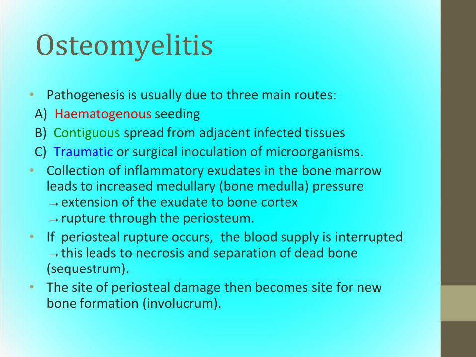
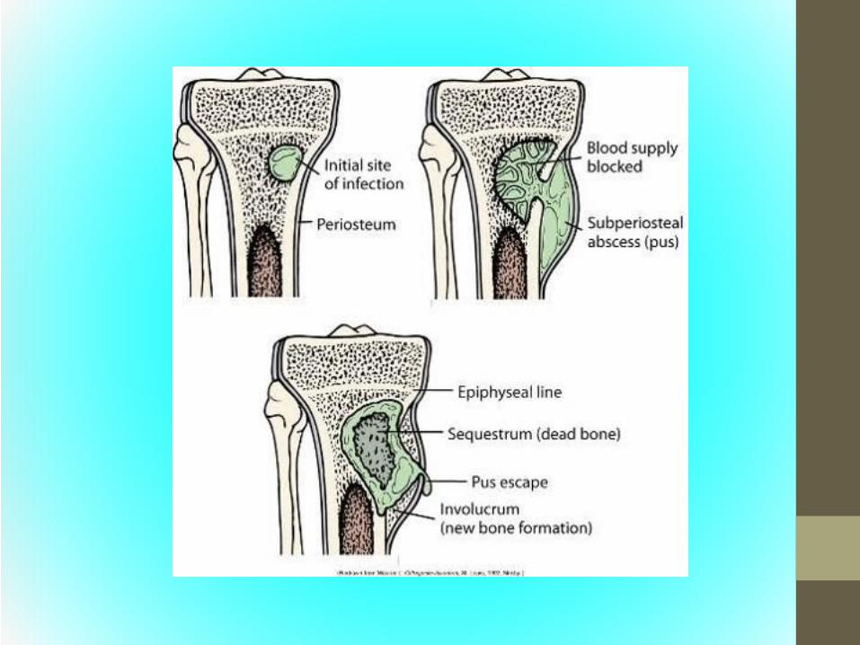
• Pathogenesis is usually due to three main routes:A) Haematogenous seedingB) Contiguous spread from adjacent infected tissuesC) Traumatic or surgical inoculation of microorganisms.• Collection of inflammatory exudates in the bone marrow
leads to increased medullary (bone medulla) pressure→extension of the exudate to bone cortex→rupture through the periosteum.
• If periosteal rupture occurs, the blood supply is interrupted→this leads to necrosis and separation of dead bone(sequestrum).
• The site of periosteal damage then becomes site for newbone formation (involucrum).
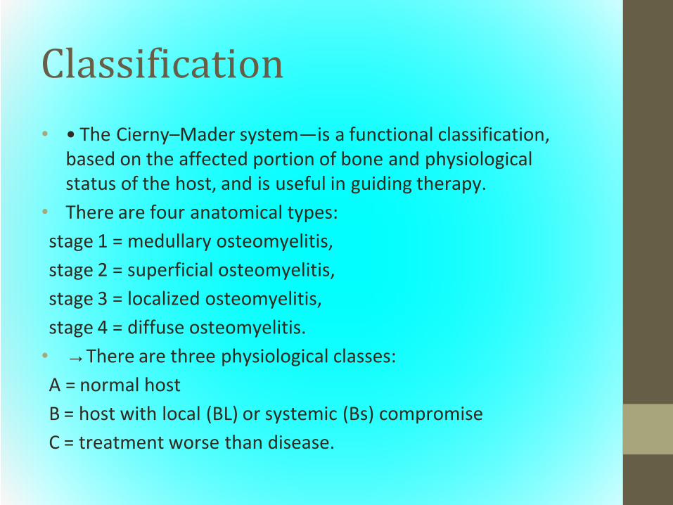
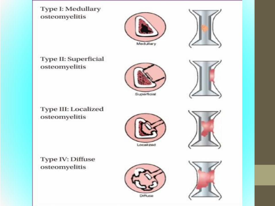
Classification
• • The Cierny–Mader system—is a functional classification,based on the affected portion of bone and physiologicalstatus of the host, and is useful in guiding therapy.
• There are four anatomical types:
stage 1 = medullary osteomyelitis,
stage 2 = superficial osteomyelitis,
stage 3 = localized osteomyelitis,
stage 4 = diffuse osteomyelitis.
• →There are three physiological classes:
A = normal host
B = host with local (BL) or systemic (Bs) compromise
C = treatment worse than disease.

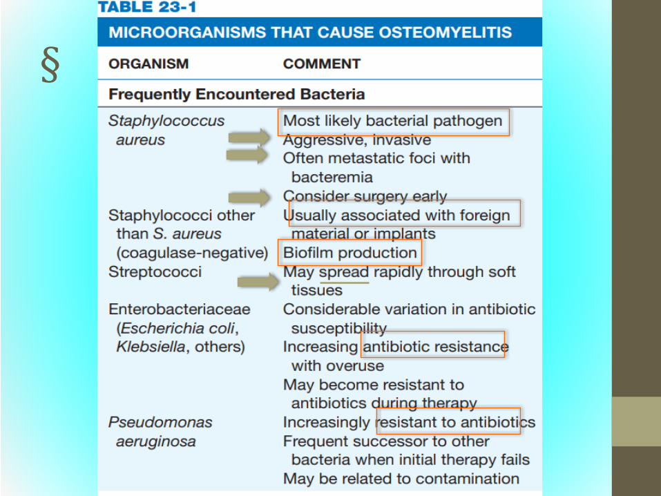
Etiology
• • Haematogenous osteomyelitis → usually monomicrobialcontiguous osteomyelitis → monomicrobial or polymicrobial.
• In patients with sinuses, the superficial flora may notrepresent the true pathogen.
• •The most common bacteria (>50%) cause of osteomyelitis isStaphylococcus aureus and CoNS .
• Gramve organisms such as Pseudomonas aeruginosa andEscherichia coli , enterococci, and propionibacteria may alsobe found.
Etiology cont.
• Mycobacterium tuberculosis is a common cause in countrieswith limited medical resources (other mycobacterial speciesthat infect bone include M. marinum , M. chelonei , and M.fortuitum) .
• Fungi may include Candida , Coccidioides , Histoplasma , andAspergillus species.

• The precipitating factors can vary according to route ofinfection:
• 1 Prosthetic joint implants and stabilization devices (allforeign objects) are being used more frequently in orthopedicsurgery and are associated with complex infections.
• 2 Trauma if a wound is involved with trauma that leads tocontamination of bone or surrounding tissue withsignificant tissue damage or destruction.
• Not necessary to have an open wound or a compoundfracture. In a similar fashion to what is seen in pyomyositis :→ damaged tissue and internal bleeds slows down thecirculation which creates favorable condition for bacterialgrowth.
• In these damaged tissues, bacteria from peripheral veins orlymphatic channel (low level bacteremia) maybe sufficient tocause infection

• Bacteremia—is a frequent cause of osteomyelitis, maybearising from endocarditis or from seeding of other infection sites(abscess, boils..etc)
• A Prosthetic joints and S. aureus: Studies show that S. aureusbacteremia cause a rate of metastatic osteomyelitis approaching28% if there is a prosthetic joint in place. can be complicated bythe involvement of methicillinresistant strains (MRSA), which areprogressively replacing strains that are more susceptible toantibiotics.
• BUrinary tract circulation: The overlapping circulations of theurinary tract and the spine is suggested to be the source ofvertebral osteomyelitis especially due to UTI causing pathogens (E.coli and Klebsiella).
• Climited vascular supply: other predisposing factors →limitedarterial and venous blood supply →limit perfusion to bone to thepoint of an inadequate response and poor healing.
• DDiabetes and other host factors contribute significantly to thedevelopment of osteomyelitis through impaired immunity withhyperglycemia, loss of sensation, vascular disease, and renalfailure.

Epidemiology
• In the United States, acute osteomyelitis affects ∼0.1–1.8% ofthe otherwise healthy adult population.
• After a foot puncture, 30–40% of adults with diabetesdevelop osteomyelitis.
• MRSA has been steadily replacing MSSA over the last fewdecades.
• The morbidity and economic burden is greater for MRSAosteomyelitis that that causes by MSSA.
• Is MRSA more aggressive because it can evade antimicrobialsso has more time to cause damage? Or is it cause there bugssurvive longer and get more virulence factors??

• Certain countries that have more aging population and / orpopulations with more DM and obesity all contribute to thefrequency of osteomyelitis in these areas.
• Any type of instrumentation may lead to infection in a smallproportion of cases.
• Richer countries have more orthopedic related Osteomyelitis ,whereas poorer countries have more TB and brucella orsignificant wounds in the society (wars, accidents)→lesshealthcare service (micro labs, Abx..etc).
Pathogenesiscan be applied to all pathogensmentioned in this module
• The most common predisposing factor for osteomyelitis is anarea of bone ( or contiguous surrounding tissue) that isdefective in in viability, blood supply, sensation.
• This damaged tissue suffers from reduced oxygenatedarterials supply and hindered venous and lymph out flow(less in , less out), these are prime factors that providebacteria with optimal growth conditions (O2, nutrients, lessinflammatory cytokines and WBC..etc).
• Host factors such as poor nutrition and immunosuppressionmay also be relevant.
• As mentioned, Diabetes in adults poses the most significantrisk (and further accentuates the above factors).

• Diabetic neuropathy makes progression of the diseasemuch worse, as the patient would be unaware or anysymptoms (pain sensation reduced)
• → makes DM a significant cause for many amputationdue to OM.
• In a similar fashion, other causes ofimmunosuppression will predispose to serious andfrequent infections and OM is no exception.

• Bacterial pathogens that cause OM perpetuatethemselves (the maintain their presence)
• → they do this by secreting toxins that continuallydamages surrounding tissue.
• S. aureus is especially strong in this respect, whereit colonizes the nasal area in about onethird ofhealthy populace and can produce a variety ofcytokines, enzymes, and toxins that destroy tissueand affect neutrophil response.
Bacterial pathogenesis:

• Certain strains of S. aureus can survive uptake into thephagocytic vacuoles of macrophages, this enables themto keep causing tissue damage by consistently evadinghost defenses
• Basically → two populations of S. aureus, intra andextra cellular, where intra cellular keeps replenishingthe extra cellular pathogens.
• S. aureus has the capacity to remain dormant(sometimes called NCBVviable but not culturable form)
• → that are resistant forms that hibernate and remaininactive for decades before infection erupts at sites ofold injuries (especially penetrating wounds, shrapnel…)

• Although CoNS are typically less virulent than S. aureusbut they have been found to persist by producing abiofilm that protects them from the host and is thoughtto be the mechanism that allows them to persist formany years on especially prosthetic joints, with minimalsymptoms.
• In CoNS it is not uncommon for prosthetic joints to showno symptoms and suddenly show infection a year oreven more later.
• How much other organisms use their biofilm to theiradvantage is not fully understood, but biofilmproduction probably plays an important role inosteomyelitis, especially in chronic forms.

• Multiple bacteria may be recovered from cultures,especially when there is an entry wound.
• This makes the decision which one to target in antibiotictherapy difficult.
• At this point → Typically common skin flora andcolonizing bacteria are not targeted (if they are , it mightmake them more aggressive and resistant).
• Anaerobic bacteria can often be recovered and can playsynergistic role with other pathogens → these areusually targeted with specific therapy.

Clinical features
• • Acute osteomyelitis presentation usually in pediatricpatients and due to hemtogenous spread
• Whereas: subacute to chronic usually in adults
• Onset of pain around the affected site.
• Local and systemic signs of inflammation such asswelling, tenderness, warmth, and erythema may ormay NOT be present! (especially in vertebra, hip orpelvisnot long bones)
• •
• Chronic osteomyelitis presentation may begin localsigns of inflammation and/or presence of a sinus tract,or even fracture).
•• If skin ulcers are present that are prolonged that fail
to heal with antibiotic therapy may indicateunderlying osteomyelitis.
• In such case, if bone is felt when palpating an ulcercan be sufficient to diagnose osteomyelitis

Diagnosis 1
• Usually based on clinical suspicion and then confirmed by aradiology, microbiology and pathology.
• • Blood tests—
• White cell count may be raised but can be normal.
• Inflammatory markers (ESR and CRP) are usually high
→ CRP changes occur earlier in bacterial infection.
However, ESR and CRP are not specfic and can be elevated inother conditions other than osteomyelitis
• Blood cultures are more likely positive in vertebral infectionand in haematogenous spread (clavicle, pubis).

Dx 2• • Radiology—plain Xray may show changes after 12 weeks.
• may eliminate the need for further imaging studies.
• Bone loss, sequestra, periosteal elevation or swelling (whichcan develop early on), and shadows around foreign bodiesare hallmarks of bone infection.
• CT or MRI scans are the investigations of choice (MRI is bothspecific and sensitive).
• MRI may be contraindicated in patients with metalware;these may also cause artefacts on CT.

http://www.stepwards.com/?page_id=4028


Dx 3• • Biopsy—an open or percutaneous bone biopsy should be
taken and sent to microbiology and histology labs.
• Needle aspiration of pus collection is both Rx and Dx forthe pathogen
• Biopsy can be taken in open surgery with debridment of allnecrotic tissue, which is again both Rx and Dx.
• antibiotics should be stopped 4872 hours prior to biopsyto improve the yield of culture.
• Swabs from sinus tracts are of questionable value, andmay often just be presenting the local flora.

• PCR, and sequencing technologies are becoming morestandard Dx to detect and identify specific organisms
• (even their sensitivity to Abx) within hours instead of days orweeks.

Management
• • General principles—
• The aim of treatment is to eradicate the causative agentand restore ( or at least preserve) function of the bone.
• OM in adults is usually treated with a combination ofsurgical debridement and antibiotic therapy.
• • Surgery—the principles of surgical therapy aredebridement of infected tissue, removal of metalware,management of dead space (using a flap), woundclosure, and stabilization of infected fractures.

Antimicrobial therapy
• Choice of ABx therapy is based on culture andsensitivity results , duration, however, duration isunknown and most experts treat for 46 weeks IVtherapy!
• The addition of rifampicin to β-lactams was shownto be effective in certain staphylococcal OM animalmodels is often used in infections, particularly thoseinvolving prosthetic material.
• Patients are usually discharged once they are clinicallystable and treated as outpatient with an IV antimicrobialcatheter.
• Hyperbaric oxygen has been shown to be effective inanimal studies (no data in humans) and can be used asadjunctive therapy.
• Negative pressure wound therapy (vacuumassistedclosure) is being increasingly used and may acceleratewound healing in complex wounds and in diabeticpatients.


• There is still controversy about the optimal route andduration of therapy.
• However a 4 to 6week course of IV therapy remainsthe standard and is the usual recommended minimum.
• Although in pediatric population, some studies aresuggestive adequate treatment with somewhat shorterduration + oral therapy.

• Because some of the active agents reach comparablelevels when given by mouth, a switch from therecommended IV administration to oral therapy may beappropriate in some situations.
• Duration is increased for more extensive disease or withpatients with additional comorbidity (see previousclassificationCierny Mader) + vertebral OM.
Complications
• • Sinus tract formation.
• • Pathological fractures, as the sequestra make that specificarea of bone less able to bear weight and is prone to fracture.
• • Haematogenous spread and sepsis, especially in aggressivedisease.
• • Tumours in patients with longstanding (4–5 years)
• In rare instances, chronic inflammation and infection maylead to malignant transformation into squamous cellcarcinoma or sarcoma
• osteomyelitis, e.g. squamous cell carcinoma (commonest),fibrosarcoma, myeloma, lymphoma, plasmacytoma,angiosarcoma, rhabdomyosarcoma, and malignant fibroushistiocytoma
Prognosis
• Varies, on all the factors that are included in theclassfication systems.
• Veretbral immunecompromisedlate Dx..etc →poorer prognosis
• Mandible following tooth extraction, early propertreatment→ better prognosis.

Prevention
• Osteomyelitis can be prevented with better preopertiveinfection and prevention measures.
• Agents such as mupirocin and chlorhexidine have beenshown to be successful in preventing operativeinfections (which are a common cause in prostheticjoins OM).
• Early Dx and treatment of other infection routes(abscess, bactermia, boil…etc).
• Early surgical treatment of wounds (esp extensive ones)have better outcome.
• Sacral ulcers, can be often a point of infection –bedridden patients and easily overlooked.

Septic Arthritis

Defined as : An inflammatory reaction of the joint spacecaused by an infectious agent.
→ Usually caused by bacteria but may be caused bymycobacteria or fungi.
→ Very common and hard to treat due to use ofprosthetic joints (210% of all prosthetic joints!)
→ Also common among immune compromised and eldery(45% of ppl with Septic arthritis are above 65 years and56% are male)
Etiology:
• • S. aureus : commonest cause overall, especially in acute cases, theincrease in incidence here matches that of increase use ofProsthetic joints.
• • Streptococci → groups A, B, C, and G streptococci, + S. pneumoniae and viridans groups (20%) of all cases.
• • CoNS.• • E. coli.• • H. influenzae.• • N. gonorrhoeae (the commonest cause in sexually active young
adults)• • N. meningitidis.• • P. aeruginosa.(sternoclavicular joints, sacroilliac joints)• • Salmonella spp.• + other causes, brucella, polymicrobial.

Prosthetic join infection
• According to presentation:
• Acute (S. aureus) within 3 months
• Subacute within 324 months
• Chronic >24 months
• Overall S.aureus, but CoNS and Gve aerobes causethe delayed cases more

Epidemiology
• The reported incidence of septic arthritis varies from twoto five cases per 100000 population or 8–27% of adultspresenting with painful joints (20,000 cases /year in US)
• Risk factors for septic arthritis include• Age >80 years,• Diabetes mellitus• Rheumatoid arthritis• Prosthetic joint• Recent joint surgery• Skin infection/ulcers• Intraarticular corticosteroid infectioninjection drug use, and alcoholism.
Pathogenesis
• Septic arthritis usually occurs after haematogenousseeding of pathogenic bacteria this is the mostcommon route.
• But like osteomyelitis contiguous spread or directinoculation can also be causes.
• Healthy synovial cells have phagocytic activity andnormally able to clear any seeding from outside sources.
• Any weakness to immune system (SLE, Rhuematoicarthritis..etc) increases risk (hence old age!)
• Previously damaged joints are most susceptible toinfection (arthritis)
• These joints show neovascularization and and adhesionfactors, which promote bacteremia and consequentinfection.
• S. aureus especially, binds to articular sialoprotein,collage, elastic and prosthetic materials visa tissieadhesion factors that they possess.
• Infection typically damages the cartilage (chondrocyteproteases of S. aureus , the inflammation in turn causesfurther damage to the cartilage)
• Gonococcal arthritis exhibits much less influx of WBCinto the joint, which explains why it is not as destructiveto joints as other bacteria.
Clinical features
• • Children and adults with acute septic arthritis usuallypresent with fever (60–80%) and monoarticularinvolvement (90%).
• • The knee is the most commonly affected joint,followed by the hip.
• Clinical features include pain, swelling, and reducedmobility in the joint.
• • Polyarticular infections occur in 10–20% of patients,especially those with rheumatoid arthritis and viralcauses.
• • Infections with mycobacteria or fungi usually have aninsidious onset.
Diagnosis
• Laboratory investigations frequently show a raisedWCC and inflammatory markers.
• Joint aspiration shows purulent synovial fluid, with anelevated WCC (50 000–100 000 cells/mm3), mostlyneutrophils.
• Gram stain is positive in 29–50%, and culture ispositive in 80–90% of cases (synovial fluids in bloodculture bottles may improve yield)
• Samples should also be sent for microscopy forcrystals. BCs are positive in 75% of cases.

Imaging
• Radiographs of the affected joint may be normal atpresentation.
• Typical changes are periarticular soft tissue swelling, fatpad edema, periarticular osteoporosis, loss of jointspace, periosteal reactions, erosions, and loss ofsubchondral bone.
• Ultrasound can be used to confirm an effusion andguide aspiration.
• CT and MRI are highly sensitive for imaging early septicarthritis. CT is better for imaging bone lesions.
• MRI may not distinguish septic arthritis frominflammatory arthropathies.

Management
• • Drainage of the joint, either by closed aspiration orarthroscopic washout, should be performed urgently.
• Open drainage may be required either when repeateddrainage has failed to control the infection or fordrainage of hip joints.
• Prosthetic joint infections often require removal of theprosthesis.
Antimicrobial therapy
According to the initial Gram stain findings.
Empirically IV piperacillin–tazobactam ± vancomycin.
• Definitive therapy is tailored to culture and sensitivityresults
• • Adjunctive therapy with a shortcourse systemiccorticosteroid treatment has been shown to be ofbenefit in children with haematogenous bacterialarthritis.

End