mri in urology
TRANSCRIPT

MRI in Urology
Mohamed SamirLecturer of Radiology

What is MRI?


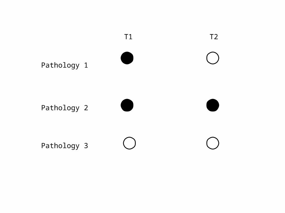
Pathology 1
Pathology 2
Pathology 3
T1 T2

• Prostate• Urinary bladder• Kidneys• Testis • Adrenal Gland
MRI in Urology

Prostate

• Diagnosis of carcinoma + Local Staging
Prostate MRI
• Conventional MRI
• Dynamic MRI
• MRI Diffusion
• MR Spectroscopy
Prostate MRI
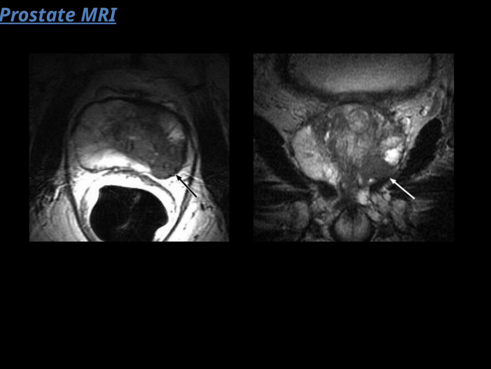
(a) T1-weighted axial imaging of the pelvic region is used for detecting nodal disease and postbiopsy intraglandular hemorrhage.
(b) thin-section T2-weighted imaging with a smaller field of view (14 cm) in the axial, sagittal, and coronal planes is used for tumor detection, localization, and staging.
Prostate MRI
Conventional MRI

Prostate MRI
Prostate MRI
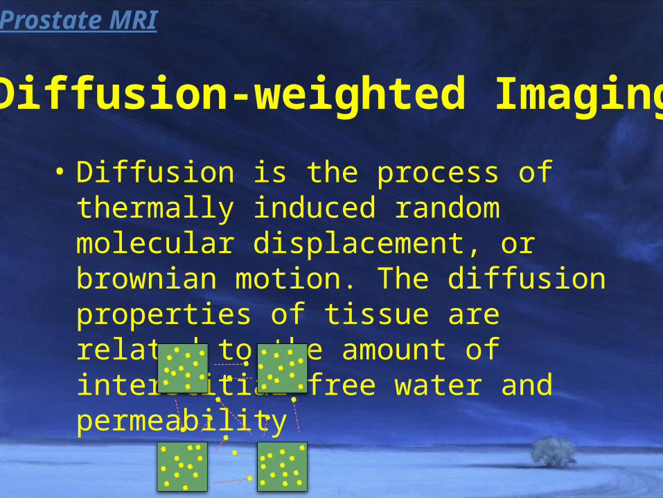
• Diffusion is the process of thermally induced random molecular displacement, or brownian motion. The diffusion properties of tissue are related to the amount of interstitial free water and permeability
Diffusion-weighted Imaging
Prostate MRI

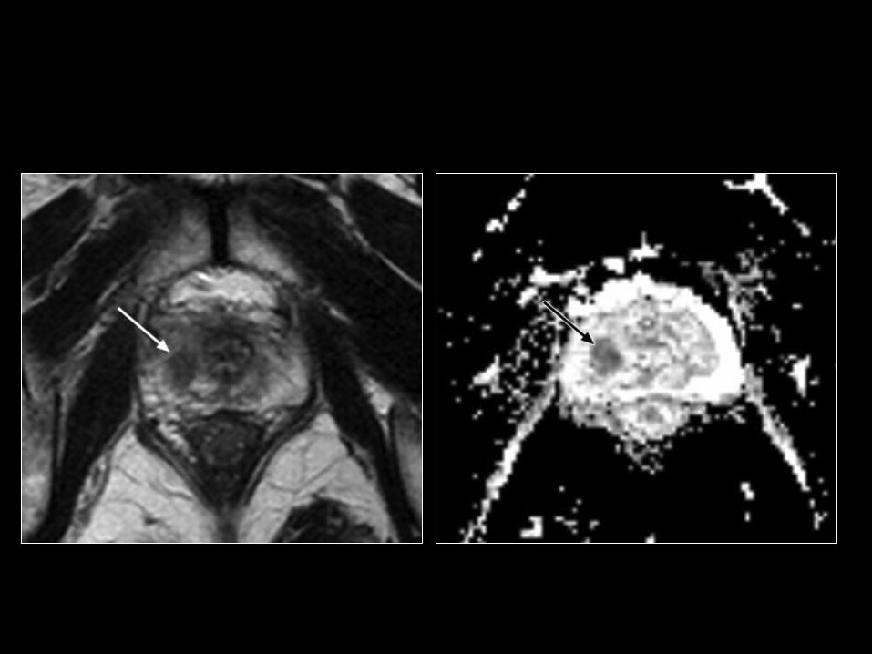
• In general, cancer tends to have more restricted diffusion than does normal tissue because of the high cell densities and abundance of intra-and intercellular membranes in cancer
Diffusion-weighted Imaging
Prostate MRI

• the use of diffusion-weighted imaging in
addition to T2-weighted imaging significantly
improved the accuracy of tumor detection
beyond that achieved with T2-weighted
imaging alone.
Diffusion-weighted Imaging
Prostate MRI

• The rationale for dynamic contrast-enhanced MR imaging is the expectation that increased microvascular density in prostate cancer would result in different contrast enhancement than that seen in normal tissue.
Dynamic MRIProstate MRI
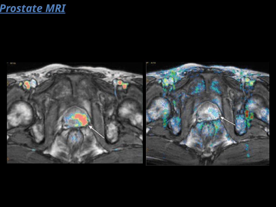
Prostate MRI

• However, the limitations of this technique include unsatisfactory depiction of transitional zone cancer in patients with hypervascular benign prostatic hyperplasia.
Dynamic MRIProstate MRI

• MR spectroscopy provides metabolic information about prostate tissue by demonstrating the relative concentration of chemical compounds.
• Normal prostate tissue contains a high level of citrate.
MR SpectroscopyProstate MRI

• In prostate cancer, the citrate level decreases as the citrate-producing metabolism of normal tissue is converted to a citrate-oxidating metabolism. At the same time, the level of choline in cancer is elevated because of a high turnover of phospholipid in cell membranes in the proliferating tissue.
MR SpectroscopyProstate MRI


• The combined use of MR spectroscopy and MR
imaging has been shown to improve cancer detection
and localization in the peripheral zone with a
sensitivity and specificity for cancer detection of 91%
and 95% for combined MR spectroscopy and MR
imaging, but 77%–81% and 46%–61% for MR imaging
alone and 63% and 75% for MR spectroscopy alone.
MR SpectroscopyProstate MRI

• MR spectroscopy also is more useful than conventional MR imaging for detecting transitional zone cancer. However, the cancer metabolite ratio in the transitional zone varies broadly, and thus there may be overlap in metabolite ratios between cancerous and benign tissues in the transitional zone .
MR SpectroscopyProstate MRI
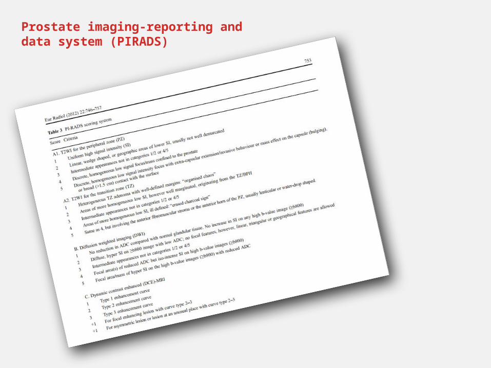
Prostate imaging-reporting and data system (PIRADS)

Urinary Bladder
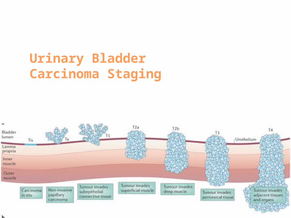
Urinary Bladder Carcinoma Staging

• High resolution MRI
• Diffusion MRI
• Dynamic MRI

Kidneys

• Multiphasic Renal MRI
• MR Urography
• MR Renogram
• Non-contrast MRI ?
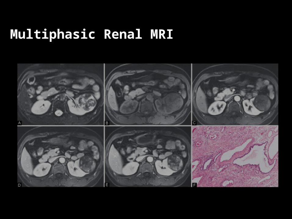
Multiphasic Renal MRI
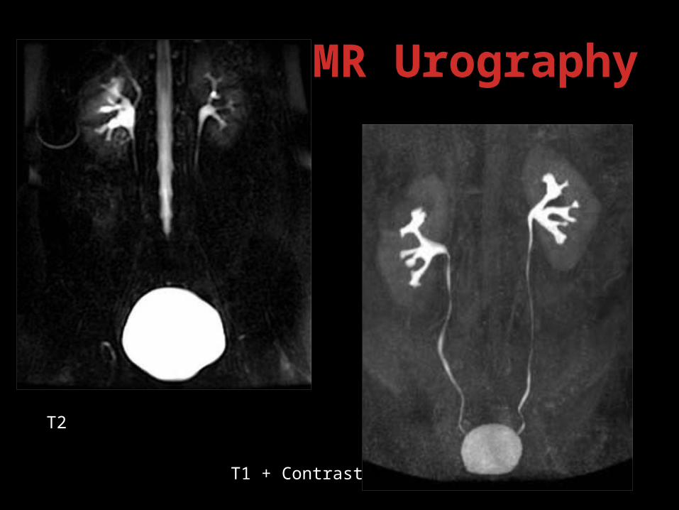
MR Urography
T2
T1 + Contrast
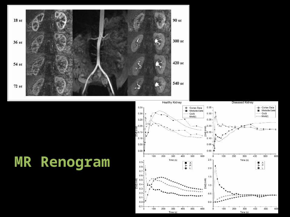
MR Renogram

Adrenal Glands
• Dynamic Adrenal MRI
• In-Phase/Opposed-Phase MRI
• Heavy T2

MRI In-Phase/Opposed-Phase MRI
in-phase out-of-phase
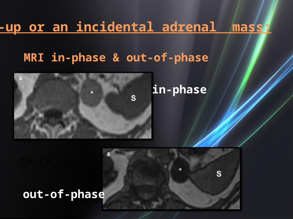
MRI in-phase & out-of-phase
Work-up or an incidental adrenal mass:
in-phase
out-of-phase
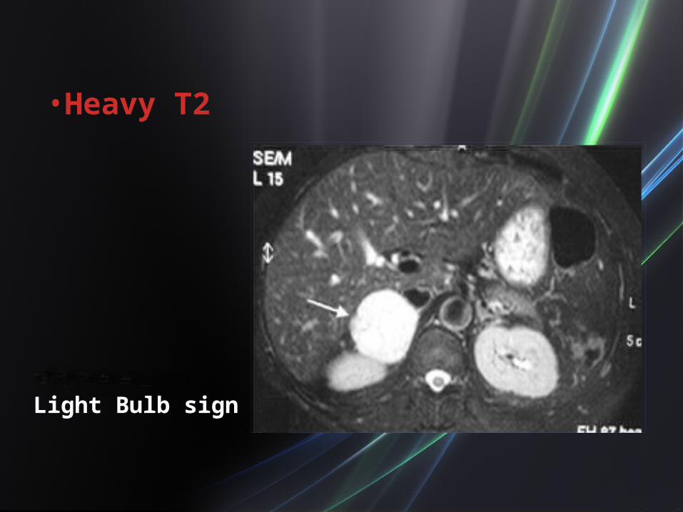
• Heavy T2
Light Bulb sign

Testis

Testis

• MRI can be useful in the evaluation of scrotal masses as a problem-solving technique:
1. Discrepancies between US and clinical findings.
2. Diffuse, non-specific testicular involvement seen on US scanning.
3. Fibrous lesions, lipomas or hemorrhage are suspected.

I Love Uroradiology