mri basics
TRANSCRIPT


Felix Bloch Edward PurcellJoseph Fourier Joseph Larmor
Raymond Damadian C Lauterbur Peter Mansfield Seiji OgawaRichard Ernst
The first Nuclear magnetic resonance
experiment was conducted
independently by two scientist in 1946.
Felix Bloch working at Stanford
University
Edward Purcell working at Harvard
University
Bloch and Purcell were awarded Nobel
Prize for Physics in1952
Joseph Fourier
1768- 1830
Joseph Larmor
1857-1942
Published his
collected papers on
electromagnetism
in 1900 in a famous
book entitled
“Aether and Matter”
In 1970 Raymond Damadian found
that it is possible to characterize
different body tissues using NMR
Technology
In 1977 he completed the
construction of the first MRI scanner
In 1978 he founded the FONAR
corporation which manufactured the
first commercial MRI
In 1973 Paul C Lauterbur discovered the
possibility to create a two dimensional
picture by introducing gradients in the
magnetic field. He used the back
projection technique to reconstruct the
image. He termed his new imaging
technique zeumatography. He shared
the 2003 Nobel prize for medicine with
Peter Mansfield
Peter Mansfield
developed the technique
of detecting the emitted
signals rapidly
mathematically analyzing
them and turning them
into an image.
He evolved a very fast
imaging technique known
as Echo Planar Imaging
In 1975 Richard Ernst
introduced 2D NMR
using phase and
frequency encoding
and the Fourier
transform instead of
Lauterbur’s back
projection technique
In 1992 Functional
MRI was developed
by Seiji Ogawa.
FIRST MRI MACHINE AND MRI IMAGE
In 1890 Roy and Sherrington’s paper ‘On the regulation of
blood supply of brain’ suggested neural activity was
accompanied by a regional increase in cerebral blood flow.
In 1992 Ogawa and Lee at AT and T Bell laboratories working
on rodents discovered that oxygenation level of blood act as
contrast agent in MR images which finally led to the fMRI

Structure of Atom Magnetic Dipole moment
ELECTROMAGNETISM
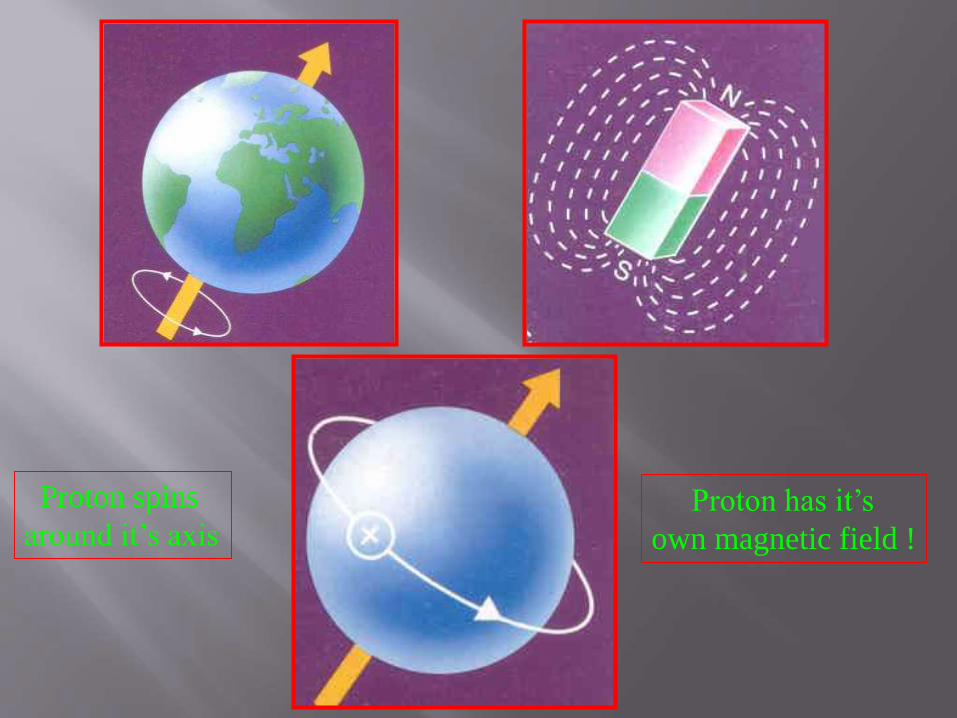
Proton has it’s
own magnetic field !
Proton spins
around it’s axis

History
Ogawa and lee?Found
1. Neural activity is proportional to
oxygenation
2. O2 level acts as contrast agent
3. EP imaging technique
4. Experimented in human MR
imaging

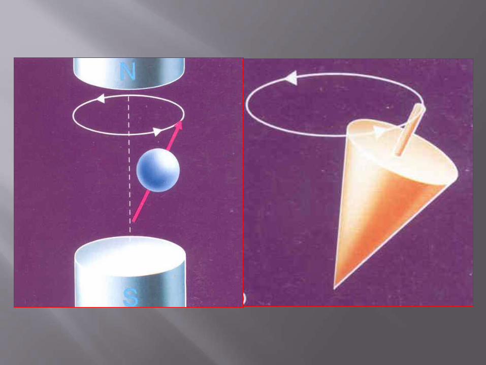
Precession in Magnetic Field

PRECESSIONAL FREQUENCY
In our MRI (1.5 T) ,Protons precess at around 63.75 MHz


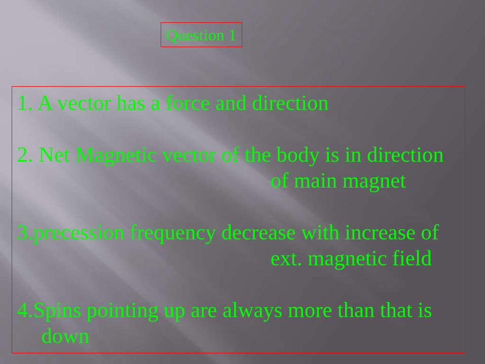
1. A vector has a force and direction
2. Net Magnetic vector of the body is in direction
of main magnet
3.precession frequency decrease with increase of
ext. magnetic field
4.Spins pointing up are always more than that is
down
Question 1


Precession frequency
1. Is the freqeuncy of the nucleus
2. Is the freqeuncy of the protons
3. Is directly proportional to strength of
main magnet(B0)
4. Is the frequency of the spins
Question 2

How to measure magnetisation?

Transversal Relaxation
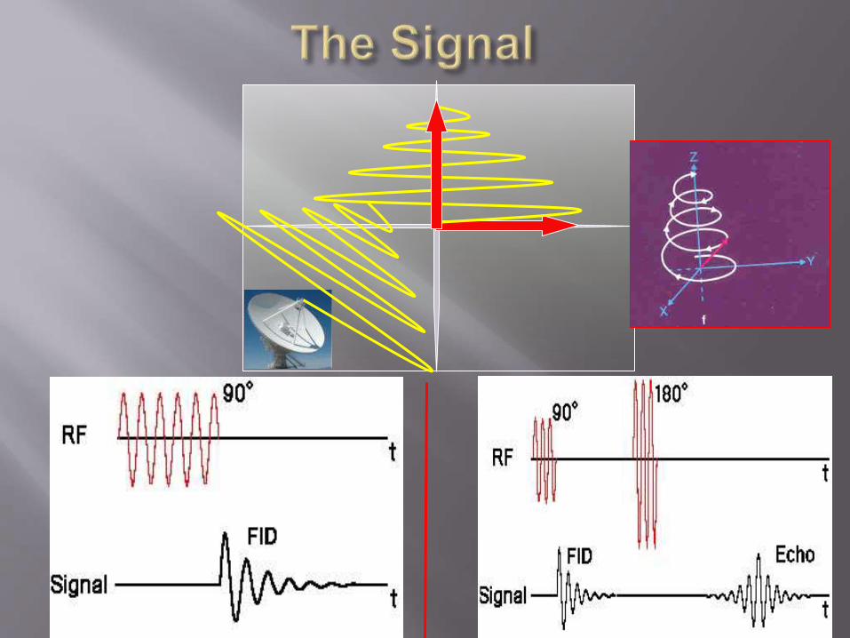
RF PULSE !
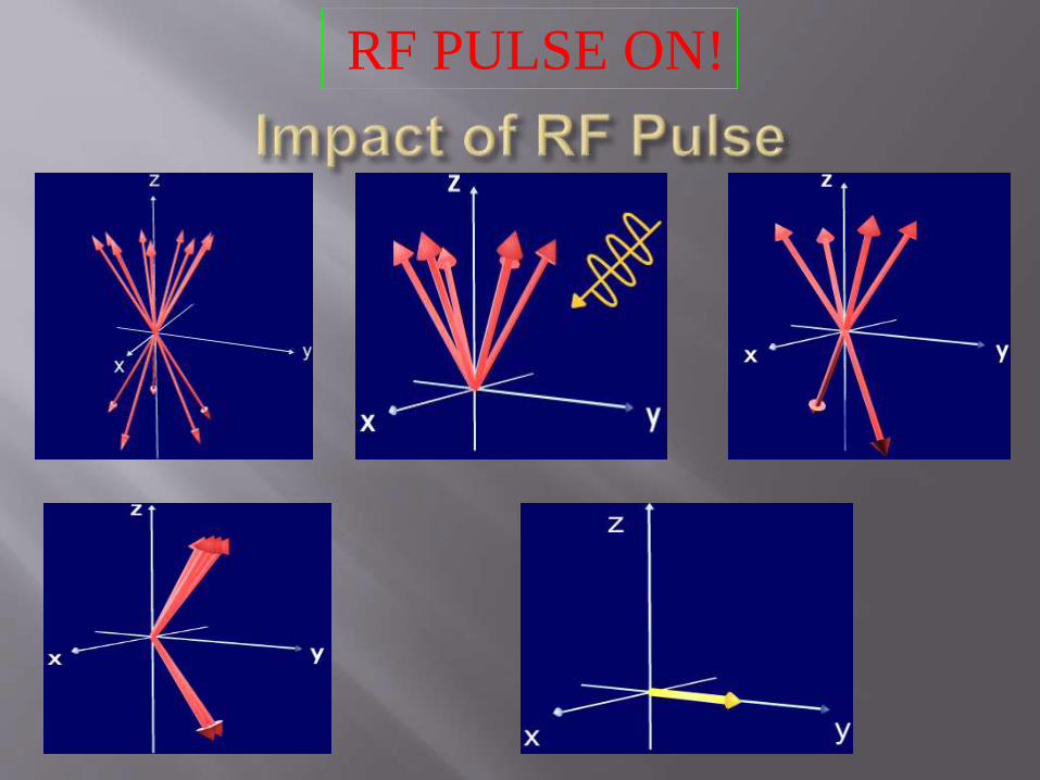
RF PULSE ON!

RF PULSE OFF !

** Change in magnetic force moment
** Induces a current
** Signal captured
* Decays when RF pulse is off
Free induction decay

Question 3
RF PULSE
1. When RF pulse is off SPINS DEPHASE
2. RF pulse has same frequency as that of spins
3. In phase vector is always in transverse direction
4. A moving magnetic field induces a current

PRECESSION FREQUENCY
SPEED !

IN PHASE !

SPIN ECHO SEQUENCE
90°--TE /2 --- 180° --- TE/2 -- RECORD SIGNAL

RF PULSE OFF
PROTONS
DEPHASE
AFTER 180° PULSE
AT TE / 2, PR0TONS REPHASE
WHY ANOTHER 180° PULSE ?
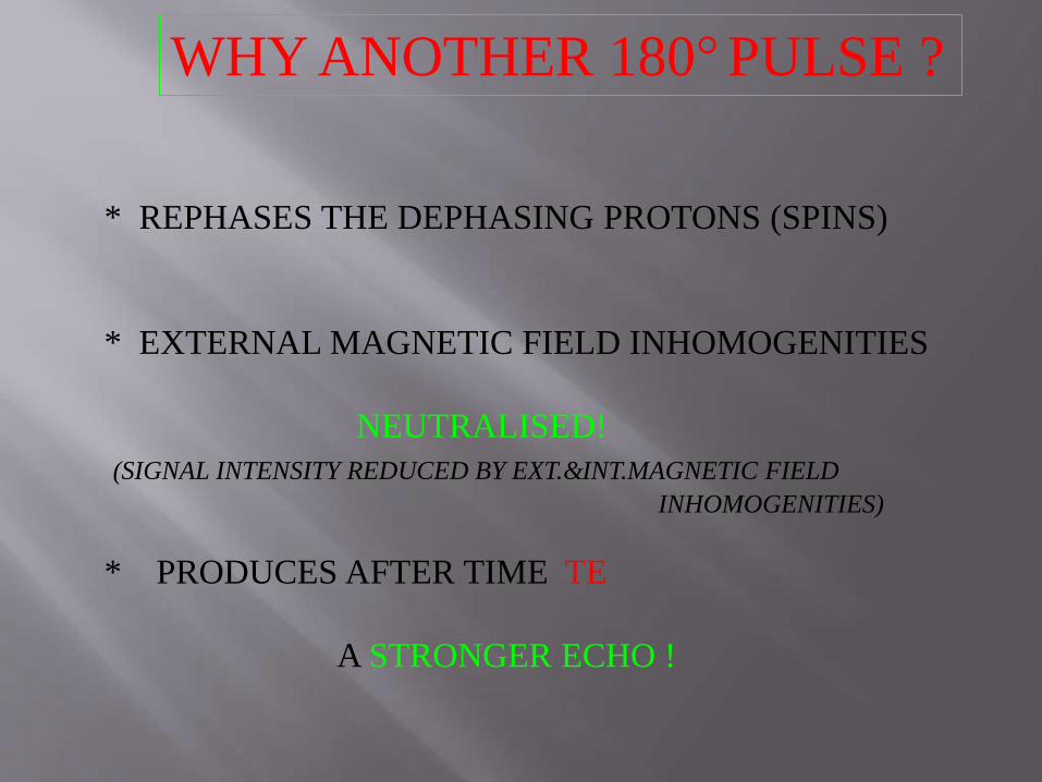
WHY ANOTHER 180° PULSE ?
* REPHASES THE DEPHASING PROTONS (SPINS)
* EXTERNAL MAGNETIC FIELD INHOMOGENITIES
NEUTRALISED!
(SIGNAL INTENSITY REDUCED BY EXT.&INT.MAGNETIC FIELD
INHOMOGENITIES)
* PRODUCES AFTER TIME TE
A STRONGER ECHO !

T2* EFFECT

T1 = t(63 % of original magnetization)
1/T1 = Longitudinal relaxation rateSpin Lattice Relaxation
T1 (water) > T1 (fat)

T2= t(37 % of its initial value)
1/T2 = Transversal Relaxation Rate
T2(water) > T2(fat)
RF PULSE REMOVED!
SPIN-SPIN RELAXTION

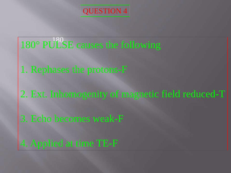
180180° PULSE causes the following
1. Rephases the protons
2. Ext. Inhomogenity of magnetic field reduced
3. Echo becomes weak
4. Applied at time TE.
QUESTION 4


F
90 90TR
GΦ
TE/2TE
GZ
GX
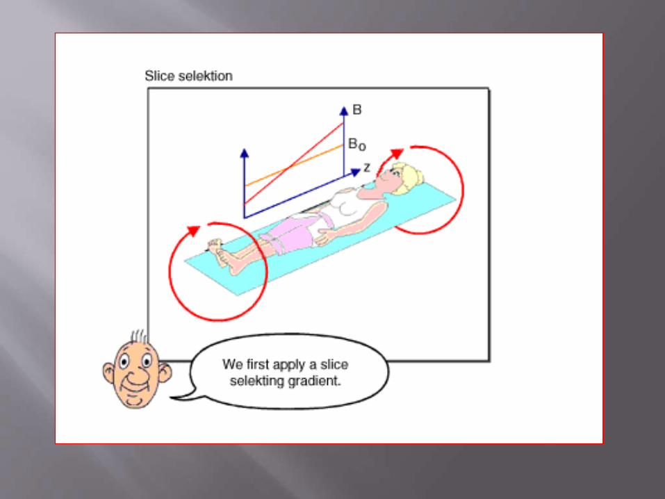
IMAGE FORMATION
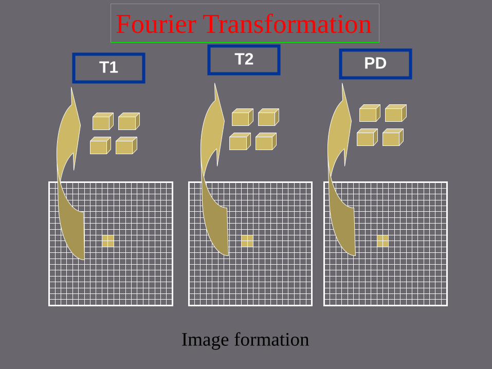
T1T2 PD
Fourier Transformation
Image formation

GRADIENT
COILS
RF Coils
Depending on the working
-Transmit and Receive Coil
-Transmit only Coil
-Receive only Coil
Depending on the application
-Volume Coil
-Surface Coil
-Internal Coil
INSTRUMENTATION
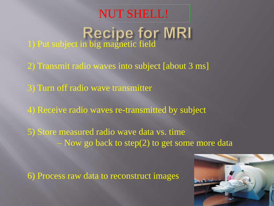
1) Put subject in big magnetic field
2) Transmit radio waves into subject [about 3 ms]
3) Turn off radio wave transmitter
4) Receive radio waves re-transmitted by subject
5) Store measured radio wave data vs. time
– Now go back to step(2) to get some more data
6) Process raw data to reconstruct images
NUT SHELL!

1. A vector has a force and direction -T
2. Net Magnetic vector of the body is in direction
of main magnet-T
3.precession frequency decrease with increase of
ext. magnetic field-F
4.Spins pointing up are always more than that is down-T
Question 1

Precession frequency
1. Is the freqeuncy of the nucleus -F
2. Is the freqeuncy of the protons-T
3. Is directly proportional to strength of
main magnet(B0)-T
4. Is the frequency of the spins-T
Question 2

Question 3
RF PULSE
1. When RF pulse is off SPINS DEPHASE-T
2. RF pulse has same frequency as that of spins-T
3. Inphase vector is always in transverse direction-F
4. A moving magnetic field induces a current-T
180180° PULSE causes the following
1. Rephases the protons-F
2. Ext. Inhomogenity of magnetic field reduced-T
3. Echo becomes weak-F
4. Applied at time TE-F
QUESTION 4
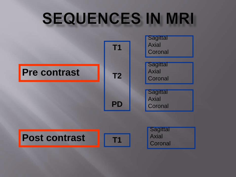
Pre contrast
T1
T2
PD
Post contrast T1
Sagittal
Axial
Coronal
Sagittal
Axial
Coronal
Sagittal
Axial
Coronal
Sagittal
Axial
Coronal
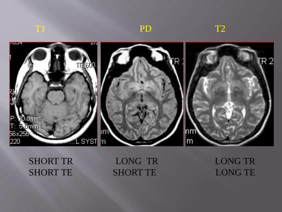
T1 PD T2
SHORT TR LONG TR LONG TR
SHORT TE SHORT TE LONG TE
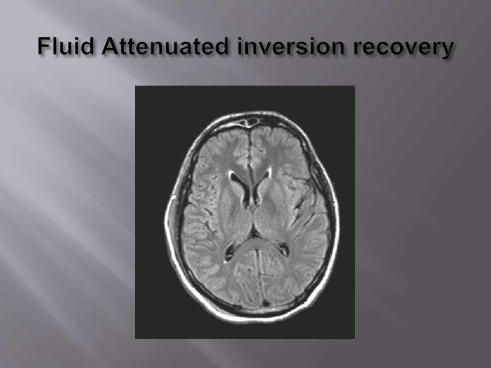

• Long T1 value (1800-2500msec)
• Fluid suppression technique
(CSF appears dark)
• Fast flair sequences
Use
• Acute subarachnoid hge
• WM lesion in spinal cord
• Flair sequence in unco-operative patients


To see internal auditory canal
CP angle structures – Cr Nn
Short T1 inversion recovery
T1 – inversion time
Fat suppression
Useful in Orbit, Optic N
Bone marrow imaging – Metastasis
(BM & Fat suppressed – Met; lesion more conspicuous)


Strong T2 Wted Image
In 3D mode – High signal from fluids (CSF)
High spatial resolution
Cisternal portions of Cr.Nns
Workup of acoustic tumours
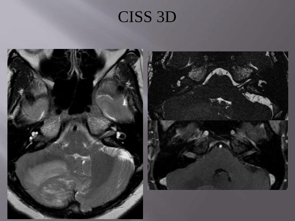
CISS 3D

STIR
1. Is a FAT SUPPRESSED SEQUENCE
2. Ideal in CSF RHINORREA
3. FAT HAS SHORT TI VALUE
4. TISSUE WITH LONG T1 APPEAR DARK


Hypo intensity in GRE1. Haemorrhage – Deoxy Hb
Meth HbFerritin (Haemosiderin other Iron forms)
2. Calcification – Diamagnetic ca saltsAssociated paramagnetic ions
3. Air containing PNS4. Normal brain iron5. Intratumoral melanin6. Paramagnetic devices, Fn bodies7. Ferromagnetic devices, Fn bodies8. Intravascular deoxygenated blood.

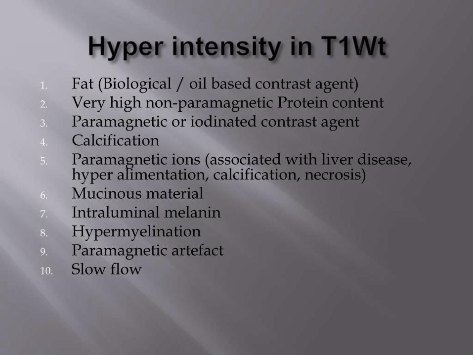
1. Fat (Biological / oil based contrast agent)2. Very high non-paramagnetic Protein content3. Paramagnetic or iodinated contrast agent4. Calcification5. Paramagnetic ions (associated with liver disease,
hyper alimentation, calcification, necrosis)6. Mucinous material7. Intraluminal melanin8. Hypermyelination9. Paramagnetic artefact10. Slow flow

Iron with out hemorrhage
Calcification or bone
Air
Very high paramagnetic Protein content
Deoxy Hb in patent veins
Mucinous material
Rapid or turbulent flow
Ferromagnetic artefact


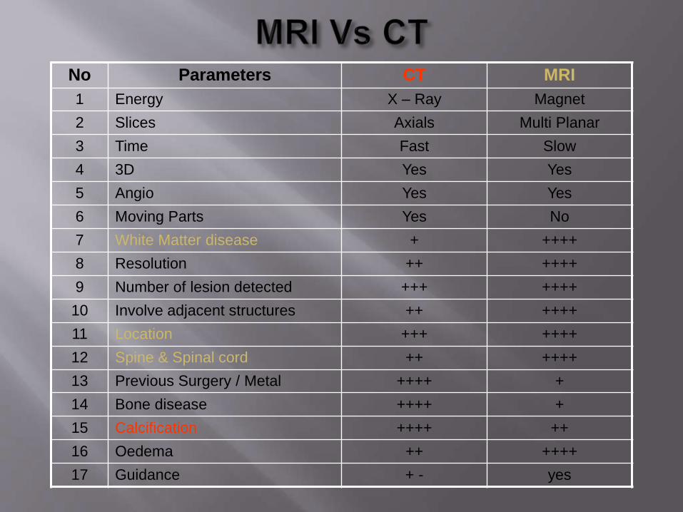
No Parameters CT MRI
1 Energy X – Ray Magnet
2 Slices Axials Multi Planar
3 Time Fast Slow
4 3D Yes Yes
5 Angio Yes Yes
6 Moving Parts Yes No
7 White Matter disease + ++++
8 Resolution ++ ++++
9 Number of lesion detected +++ ++++
10 Involve adjacent structures ++ ++++
11 Location +++ ++++
12 Spine & Spinal cord ++ ++++
13 Previous Surgery / Metal ++++ +
14 Bone disease ++++ +
15 Calcification ++++ ++
16 Oedema ++ ++++
17 Guidance + - yes

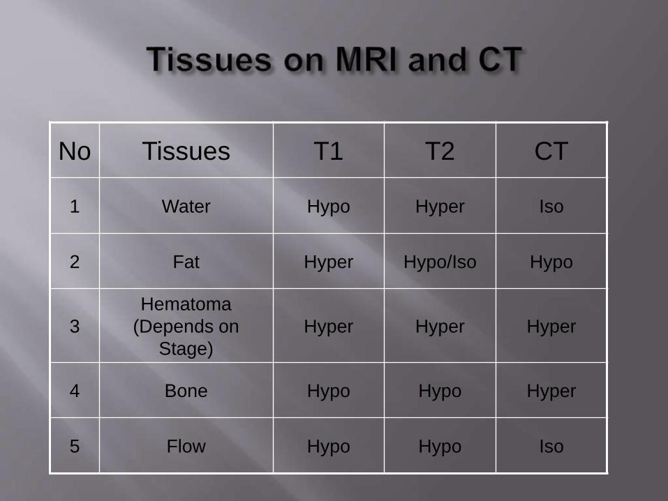
No Tissues T1 T2 CT
1 Water Hypo Hyper Iso
2 Fat Hyper Hypo/Iso Hypo
3
Hematoma
(Depends on
Stage)
Hyper Hyper Hyper
4 Bone Hypo Hypo Hyper
5 Flow Hypo Hypo Iso
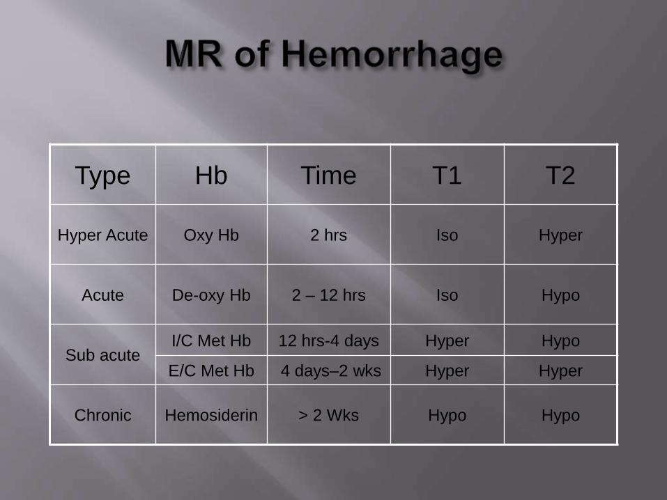
Type Hb Time T1 T2
Hyper Acute Oxy Hb 2 hrs Iso Hyper
Acute De-oxy Hb 2 – 12 hrs Iso Hypo
Sub acuteI/C Met Hb 12 hrs-4 days Hyper Hypo
E/C Met Hb 4 days–2 wks Hyper Hyper
Chronic Hemosiderin > 2 Wks Hypo Hypo

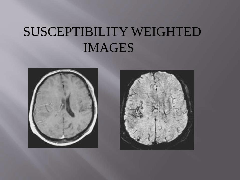
SUSCEPTIBILITY WEIGHTED
IMAGES

DIA MAGNETIC PARAMAGNETIC Super Paramagnetic
Disperse EXT.
Magnetic field
CONCENTRATE
EXT. Magnetic field
CONCENTRATE
EXT. Magnetic field
Water, Org. molecule Ions , simple salts, Ferric sulphate ,
Salts of non-metals Chelates of metals Alloys( Fe, Ni, Co)
Inert gases

Magnetic Susceptibility

Sections of skull vault
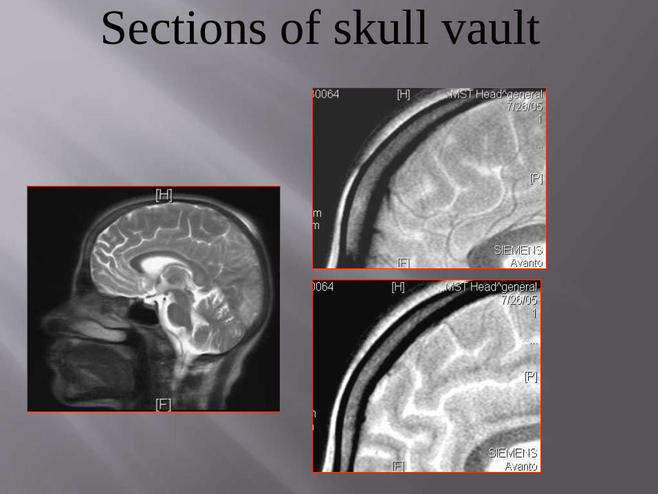
Sections of skull vault


MRI basicshttp://pss100.psi.ch/~kuehne/MR_Basics.html
Basics MRIhttp://howstuffworks.com/mri.htm
MRI Basicshttp://www.wellweb.com/Diagnost/arnief/mri.htm
Magnetic Resonance Advisory Panelhttp://www.giant.net.au/air/mrpanel.htm
MRI Educationhttp://www.mrieducation.com/usa.htm
MRI Review dotcomhttp://www.mrireview.com/index2.html
Hesselink Basic Principles Of MRImaginghttp://spinwarp.ucsd.edu/NeuroWeb/Text/br-100.htm
MRI Educationhttp://www.mrieducation.com/mainb.htm
A Selection of Slides on MRI Basicshttp://porkpie.loni.ucla.edu/BMD_HTML/SharedCode/slides/SlideFiles.html
Animated Basic MRIhttp://www.t2star.com/basic_mr/Basic.html

THANKYOU