mri and psma pet scan in pca - …abg2018.conferenceworks.com.au/wp-content/uploads/... · pi-rads...
TRANSCRIPT

Mira Keyes MD FRCPCClinical Professor Radiation Oncology Department of Surgery UBCHead Provincial Prostate Brachytherapy ProgramBC Cancer, Vancouver Canada
Vancouver BC, Canada
MRI and PSMA PET scan in PCaThe Game Changers or a Revolution?

mpMRI Use of multiple anatomical and functional parameters
T2W•Bx artefact can overestimate TU volume
Dynamic contrast-enhanced imaging (DCE)• IV gadolinium. Tumours of higher-grade or larger-volume take up
contrast (vascularity). The contrast typically leaks out, giving a steep “wash in -wash out” curve. This can be measured.
Diffusion-weighted imaging (DWI), •Assesses the movement of water within a given volume. Higher
grade cancer, with its disorganized cellular structure, tends to restrict the movement of water
Proton spectroscopic imaging (MRSI)•high ratio of choline to citrate is suspicious for cancer. Less favoured.
Need endorectal coil, not all MRIs have the software

Mp-MRI-defined radiological phenotypeincludes volume and characteristics of lesions on both anatomical and functional sequences.
T2-weighted sequence, T2WContrast-enhanced sequence DCEDiffusion coefficient map. DWI
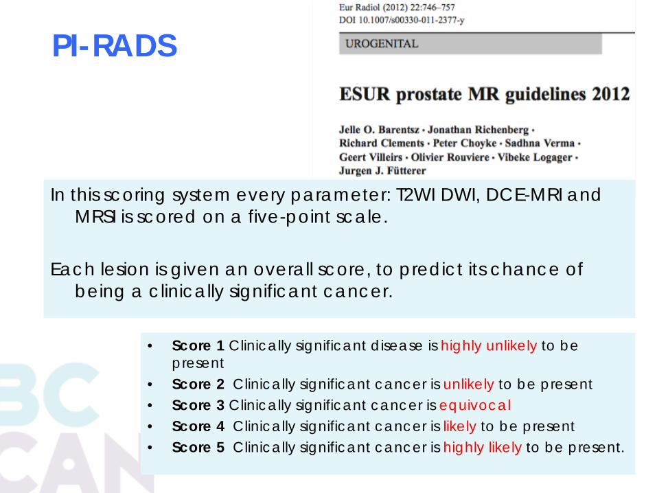
PI-RADS
• Score 1 Clinically significant disease is highly unlikely to be present
• Score 2 Clinically significant cancer is unlikely to be present • Score 3 Clinically significant cancer is equivocal• Score 4 Clinically significant cancer is likely to be present• Score 5 Clinically significant cancer is highly likely to be present.
In this scoring system every parameter: T2WI DWI, DCE-MRI and MRSI is scored on a five-point scale.
Each lesion is given an overall score, to predict its chance of being a clinically significant cancer.

PI-RADS

PI-RADS
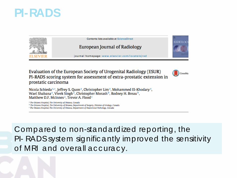
Compared to non-standardized reporting, the PI- RADS system significantly improved the sensitivity of MRI and overall accuracy.
PI-RADS

MRI Meta-analysis

SensitivityTrue positive
SpecificityTrue negative
ECE 0.57 0.91SVT 0.58 0.96T3 0.61 0.88
Improved sensitivity for ECE and SVI1. Functional imaging2. Higher field strength (3T)3. Not with endo-rectal coil use.
75 studies (9796 patients) - pooled data for ECE (45 studies, 5681 patients), SVI (34 studies, 5677 patients), T3 detection (38 studies, 4001 patients)
MRI has high specificity but poor and heterogeneous sensitivity for local PCa staging.

High Specificity reading Minimize unnecessary exclusion of men from curative treatment. This is probably why the meta-analysis revealed high specificity and low sensitivity for MRI.
High Sensitivity reading Nowadays, urologists become more interested in high-sensitivity reading to reduce positive surgical margins and preserve neurovascular bundles.
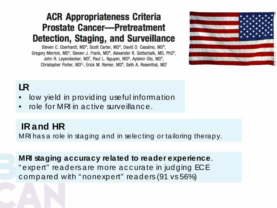
LR • low yield in providing useful information • role for MRI in active surveillance.
IR and HR MRI has a role in staging and in selecting or tailoring therapy.
MRI staging accuracy related to reader experience. “expert” readers are more accurate in judging ECE compared with “nonexpert” readers (91 vs 56%)

Review of 62 MR studies95% studies consistently demonstrated upstaging by MRI
Up to 43% of patients are experiencing increased therapy due to MRI stagingThe correctness of treatment changes was under-studied.
Overall sensitivity of ~ 50‒60% and specificity of >85%

Use for staging is a reasonable for assessment of EPE in IR and HR (knowledge of EPE will alter management)
Quality assurance
Recommendations
MRI AND EBRT
To evaluate the accuracy of preoperative 3TmMRI for staging of prostate cancer and Evaluate its influence on the decision to change the • CTV• RT Dose • Use of ADT

N=103 EBRT N=47 Prostatectomy
Sensitivity Specificity PPV NPV accuracy
ECE 57% 95% 66% 92% 89%
94% modification of Clinical Stage34% change in RT and ADT
RT treatment (CTV, total dose and hormonal therapy) was initially determined on the basis of the clinical information
Radiation therapy plan was reevaluated after MRI
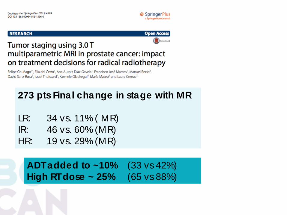
273 pts Final change in stage with MR
LR: 34 vs. 11% ( MR)IR: 46 vs. 60% (MR)HR: 19 vs. 29% (MR)
ADT added to ~10% (33 vs 42%) High RT dose ~ 25% (65 vs 88%)

10-30% - Risk group change 20-30% - Change in RT dose, ADT CTV
Literature review table of other published studies
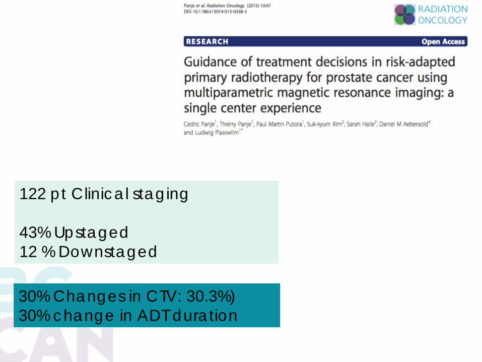
122 pt Clinical staging
43% Upstaged12 % Downstaged
30% Changes in CTV: 30.3%)30% change in ADT duration

MRI and Brachytherapy
21
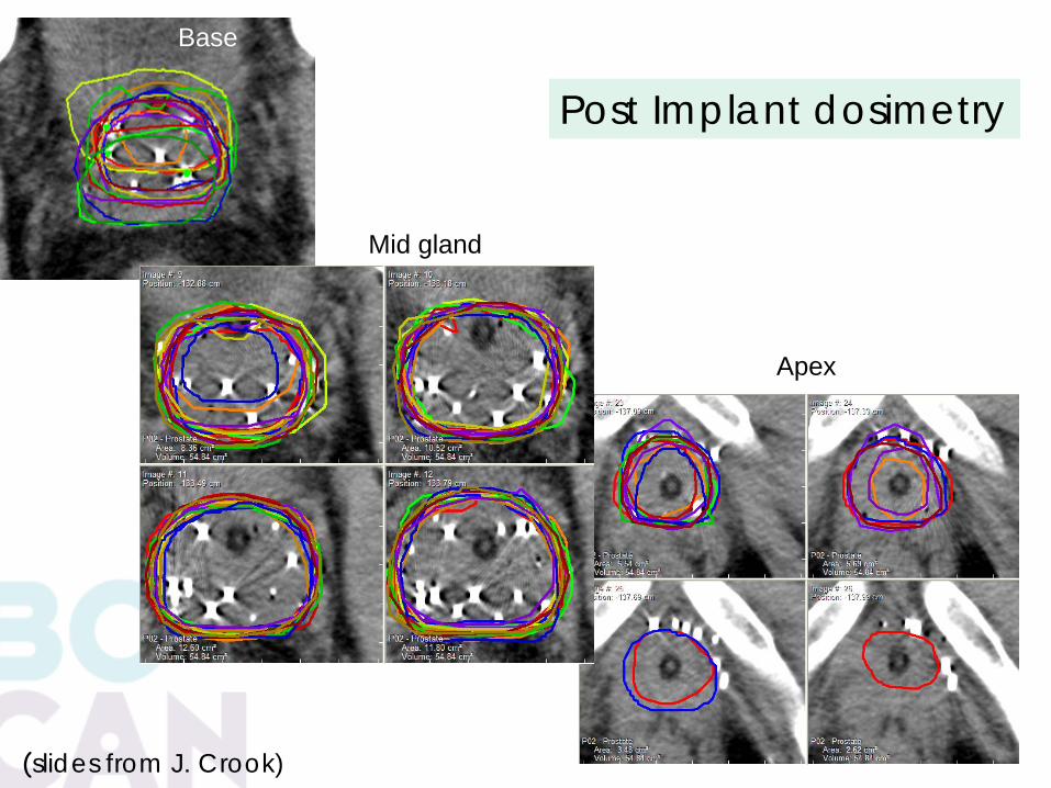
Apex
Mid gland
Base
(slides from J. Crook)
Post Implant dosimetry

22
Base on MRI

Post implant dosimetry - QA
Dosimetry more accurateLearning curve shorterDose to normal structure accurateLess inter-observer variabilityHigher quality brachytherapy

MRI in Brachytherapy Planning
• More accurate anatomical delineating of prostate, bladder neck, apex and urethra
• Pre-operative or intra operative US MRI image fusion


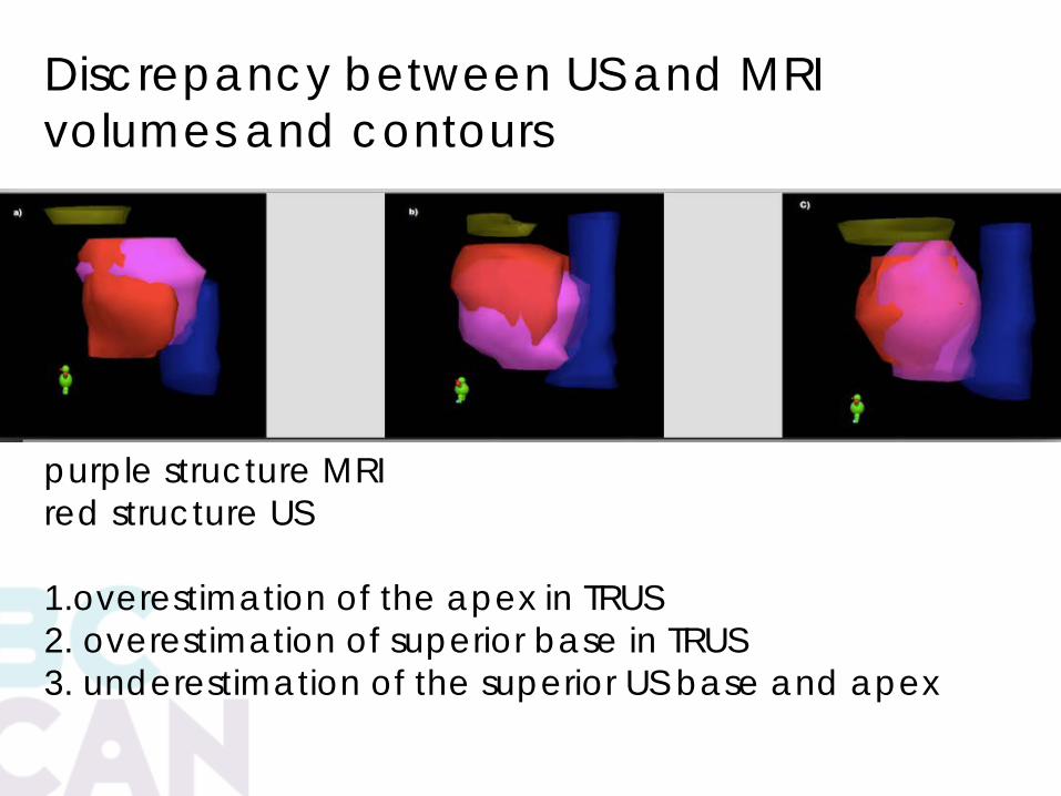
purple structure MRI red structure US
1.overestimation of the apex in TRUS2. overestimation of superior base in TRUS3. underestimation of the superior US base and apex
Discrepancy between US and MRI volumes and contours

MRI contours transferred to US. Green dots are the locations of the planned seeds. - no seeds in the tumour to allow for urethral sparing
If you know where the tumour is would you not use this information?Location of DIL
Dose escalation Dose de- escalation
MRI can alter intraoperative and pre planning

Variation in bladder neck

UG diaphragmInternal sphincter variations

Anterior venous plexuson MR and CT

MR SIMMR LINACSMR only work flow


PSMA PET Challenge to standard target volumesChallenge to treatment concepts
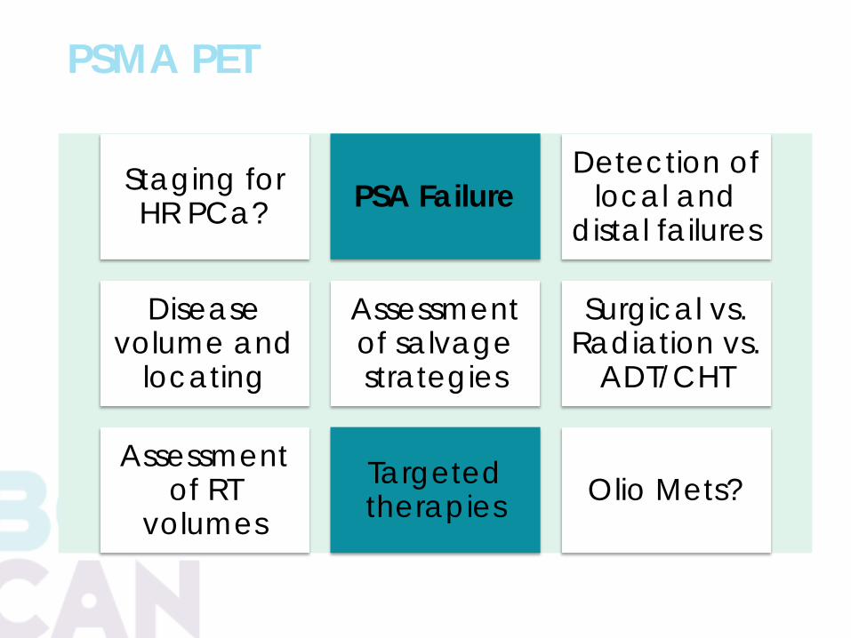
PSMA PET
Staging for HR PCa? PSA Failure
Detection of local and
distal failures
Diseasevolume and
locating
Assessment of salvage strategies
Surgical vs. Radiation vs.
ADT/CHT
Assessment of RT
volumesTargeted therapies Olio Mets?

ASTRO 2017 ( abstract) 212 Location of Recurrence by Gallium-68 PSMA PET Scan in Prostate Cancer Patients Eligible for Salvage Radiation Therapy UCLA
LOCATION OF LN AND RT FIELDS51pts, PSA< 2, post RP, uIR and HR pts
62% had positive scans PSA <1 - 50% scans were positive PSA 1-2 - 85% of scans were positive
8 – prostate bed23 - LN (4 PA, 1 inguinal LN)11 - bones
Only 40% LN would have been covered by standard pelvic LN
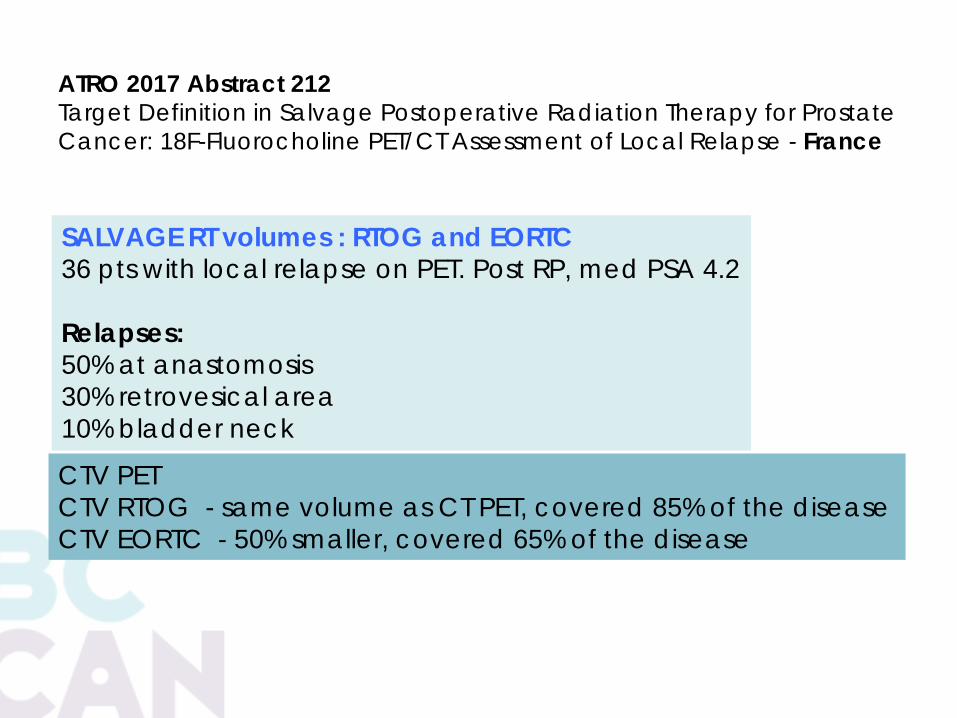
ATRO 2017 Abstract 212Target Definition in Salvage Postoperative Radiation Therapy for Prostate Cancer: 18F-Fluorocholine PET/CT Assessment of Local Relapse - France
SALVAGE RT volumes : RTOG and EORTC36 pts with local relapse on PET. Post RP, med PSA 4.2
Relapses: 50% at anastomosis30% retrovesical area10% bladder neck
CTV PETCTV RTOG - same volume as CT PET, covered 85% of the diseaseCTV EORTC - 50% smaller, covered 65% of the disease
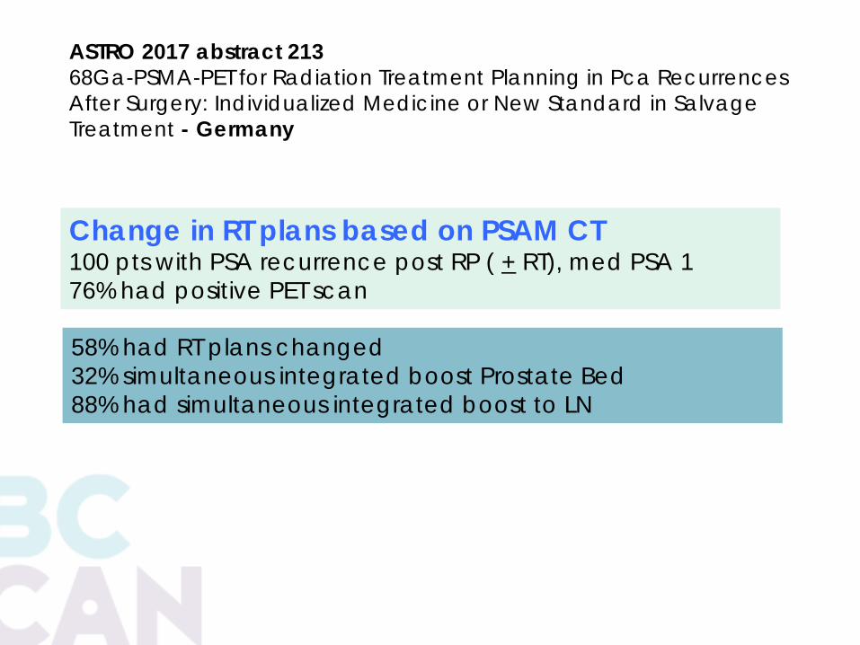
ASTRO 2017 abstract 21368Ga-PSMA-PET for Radiation Treatment Planning in Pca Recurrences After Surgery: Individualized Medicine or New Standard in Salvage Treatment - Germany
Change in RT plans based on PSAM CT100 pts with PSA recurrence post RP ( + RT), med PSA 176% had positive PET scan
58% had RT plans changed32% simultaneous integrated boost Prostate Bed88% had simultaneous integrated boost to LN


Conclusions: More than one-third of the PET positive lymph nodes in patients with no prior treatment and post RP would not have been treated adequately using the RTOG CTV.

2010

PSMA report – 15mm SVU 16 upper prostate gland near the base lesion -local recurrence, No mets or LN
2018
Prostate Bx: 8 cores and targeted 3 cores form L TZ and base: TZ 3 cores:GS4+3=7 80% pattern 4The rest of the cores benign

PSAM PET scan LN
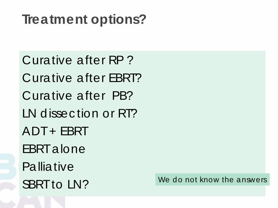
Treatment options?
Curative after RP ?Curative after EBRT?Curative after PB?LN dissection or RT?ADT + EBRTEBRT alonePalliativeSBRT to LN? We do not know the answers

Summary
1.Standard post op CTV is wrong ( >35%) EORTC and RTOG
2. Pelvic LN RTOG CTV is wrong ( >35%)3. >50% of the plans need to be
changed ( CTV or dose or both)4. + Lymph Nodes are everywhere5. New treatment algorithms

Thank You!