mr ga68-psma prostate imaging brochure...
TRANSCRIPT

Prostate cancer staging with
68Ga-PSMA
Gallium-labelled prostate-specific membrane antigen ligand (68Ga-PSMA), is a groundbreaking PET scan which is rapidly gaining popularity worldwide.
Mercy PET-CT is now able to offer this state of the art, fusion examination as a superior modality for staging and restaging of prostate cancer patients.
References
1. Eiber et al. Evaluation of Hybrid 68Ga-PSMA ligand PET/CT in 248 patients with biochemical recurrence after radical prostatectomy. J Nucl Med 2015; 56:668-674.
2. Afshar-Oromieh et al. The diagnostic value of PET/CT imaging with the 68Ga-labelled PSMA ligand HBED-CC in the diagnosis of recurrent prostate cancer. Eur J Nucl Med Mol Imaging 2015; 42:197-209.
100 Mountain Rd, Epsom, Auckland
PO Box 9056, Newmarket, Auckland 1149
Tel 09 623 5862, Fax 09 623 5863
Email [email protected]
www.radiology.co.nz
Dr Remy Lim, MB ChB (Auckland), FRANZCR
Specialising in: Nuclear, Oncologic and Cross Sectional Imaging
Nuclear Medicine and PET fellowship; Body Oncology fellowship (Memorial Sloan-Kettering Cancer Center)
68Ga-PSMA has been shown to be highly effective in the detection of prostate cancer cells in regional nodes and distant metastatic sites as well as early detection of site of relapse following definitive treatment of the disease.
Lesions suspicious for metastatic prostate cancer present with high tumour to background contrast resulting in superior detection rate even when the level of PSA is low.
Tumour specifi c Prostate specifi c membrane antigen is
a cell surface protein over-expressed in prostate cancer cells compared to benign prostatic tissue.
68Ga-PSMA detects presence of prostate cancer cells directly, rather than indirect indicators of disease such as increased bone turnover (bone scan) or enlarged lymph node.
High sensitivity and specifi city Superior tumour to background contrast
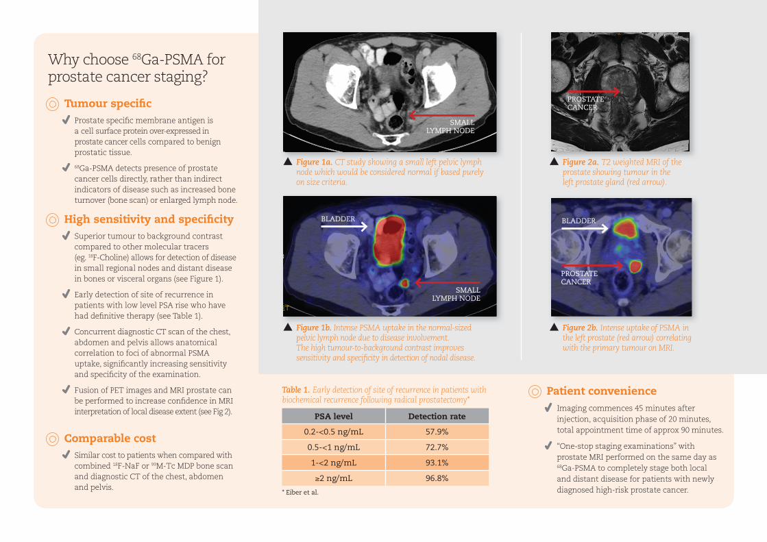
compared to other molecular tracers (eg. 18F-Choline) allows for detection of disease in small regional nodes and distant disease in bones or visceral organs (see Figure 1).
Early detection of site of recurrence in patients with low level PSA rise who have had defi nitive therapy (see Table 1).
Concurrent diagnostic CT scan of the chest, abdomen and pelvis allows anatomical correlation to foci of abnormal PSMA uptake, signifi cantly increasing sensitivity and specifi city of the examination.
Fusion of PET images and MRI prostate can be performed to increase confi dence in MRI interpretation of local disease extent (see Fig 2).
Comparable cost Similar cost to patients when compared with
combined 18F-NaF or 99M-Tc MDP bone scan and diagnostic CT of the chest, abdomen and pelvis.
Figure 1a. CT study showing a small left pelvic lymph node which would be considered normal if based purely on size criteria.
Figure 2a. T2 weighted MRI of the prostate showing tumour in the left prostate gland (red arrow).
Figure 2b. Intense uptake of PSMA in the left prostate (red arrow) correlating with the primary tumour on MRI.
Figure 1b. Intense PSMA uptake in the normal-sized pelvic lymph node due to disease involvement. The high tumour-to-background contrast improves sensitivity and specifi city in detection of nodal disease.
Table 1. Early detection of site of recurrence in patients with biochemical recurrence following radical prostatectomy*
* Eiber et al.
PSA level Detection rate
0.2-<0.5 ng/mL 57.9%
0.5-<1 ng/mL 72.7%
1-<2 ng/mL 93.1%
≥2 ng/mL 96.8%
Why choose 68Ga-PSMA for prostate cancer staging?
BLADDERBLADDER
PROSTATE CANCER
SMALL LYMPH NODE
SMALL LYMPH NODE
PROSTATE CANCER
Patient convenience Imaging commences 45 minutes after
injection, acquisition phase of 20 minutes, total appointment time of approx 90 minutes.
“One-stop staging examinations” with prostate MRI performed on the same day as 68Ga-PSMA to completely stage both local and distant disease for patients with newly diagnosed high-risk prostate cancer.