mr contrast enhancement: an experimental study in ... · mr contrast enhancement: an experimental...
TRANSCRIPT
MR Contrast Enhancement: An Experimental Study in Postlaminectomy Epidural Fibrosis
Canh M . Nguyen, 1 Victor M. Haughton , 1 Khang-Cheng Ho,2 and Howard S. An3
PURPOSE: To measure the effect of contrast medium dose, time elapsed since injection, and
maturity of ep idural scar tissue on the enhancement of scar ti ssue in MR imaging. METHODS:
We imaged 12 beagle dogs with MR at 10 to 60 days after lum bar laminectom y, and at necropsy
we obtained exactly correlating histologic sections. Contrast enhancement of scar tissue at 2, 15,
40, and 60 minutes after 0 .1 and 0.3 mmol of paramagnetic contrast medium per ki logram was
measured. Contrast enhancement was analyzed wi th respect to the dose of contrast medium, the
time of imaging, and the maturity of scar tissue. RESULTS: Epidural scar tissue enhanced more
intensely at 2 and at 15 minutes than at 40 or at 60 m inutes. Consistently greater enhancement
was observed with the dose of 0.3 mmol/ kg than with the dose of 0. 1 mmol/kg. Regions of
loosely organized scar ti ssue enhanced less intensely and less quickly than did more organized
scar tissue. CONCLUSION: Contrast enhancement in scar tissue can be heightened by increasing
the dose of contrast medium from 0.1 to 0.3 mmol/kg and by obtaining images within 15 minutes
of injection .
Index terms: Magnetic resonance, contrast enhancement; Magnetic resonance, experimental;
Magnetic resonance, t issue characterization; Contrast media, comparative studies; A nimal studies
AJNR 14:997-1002, July/ August 1993
Despite numerous studies on magnetic resonance (MR) imaging of postlaminectomy patients, the differentiation of recurrent herniated disk and epidural fibrosis is imperfect (1-8). Ross et al showed that the timing of the images affected the intensity of the enhancement in epidural scar after the intravenous injection of gadopentetate dimeglumine (Magnevist; Berlex Laboratories, Secaucus, NJ) (8). A new contrast medium, gadoteridol (ProHance, Squibb Diagnostics, Princeton , NJ) , has been developed and is approved for larger doses than the conventional 0.1 mmol/kg approved for gadopentetate. Increasing the dose of paramagnetic contrast medium increases con-
Received June 16, 1992; rev ision requested July 31, received Septem
ber 10, and accepted September 15. This work was supported by a grant-in-a id from Bristol-Myers Squibb
Company. Departments of 'Radiology, 2Pathology, and ' Orthopaedic Surgery ,
The Medical College of Wisconsin , Froedtert Memorial Lutheran Hospital,
9200 West Wisconsin Avenue, Milwaukee, WI 53226. Address requests
for reprints to Victor M . Haughton, MD.
AJNR 14:997- 1002, Jui/Aug 1993 0195-6108/ 93/ 1404- 0997 © A merican Society of Neuroradiology
997
trast enhancement only within a limited range of doses. In some tissues and with some dosage ranges, increasing the dose may decrease the amount of enhancement. The optimal dose with which to differentiate scar and disk has not been determined. Therefore , we measured the effect of dose, timing , and maturity of scar tissue on the enhancement of epidural scar tissue.
Materials and Methods
Twelve beagle dogs (six female, six male) weighing between 9 and 14 kg underwent laminectomy and then MR imaging on a 1.5-T Signa imager with boHr a 0.1 and a 0.3 mmol dose of contrast medium per k ilogram.
Each animal underwent a left hem ilam inectomy at the L3 level. After the animal was fasted for 12 hours, it was premedicated with acepromazine (1 mg/kg intramuscularly) and atropine (0.05 mg/kg intramuscularly) and anesthetized wi th pentobarbital (25 mg/ kg intravenously) and innovarvet (0.1 ml/kg int ramuscularly). T he skin over the lumbar area was shaved and prepared surgically with betadine. With an electrocautery blade, a 7-cm-long midline incision was made along the spinous processes from midL 1 to mid-L5 . With a periosteal elevator , the left paraspinal
998 NGUYEN
A
"' "' E: "' '"' "' "' .c "' UJ
"' ~ "' 0
'-'
2
8
Contrast Enhan cement in Post Laminectomy Scar Tissue Gadote ridol 0.3 mmo l/kg
3 0
2 5 • • 2 0
D 0 0 i!J
' 5 ~ ' • ' 0
05
0 0
''
•
t
!I! • D
30 J O
MINUTE S
,o
I 0 10 days
D15 days
• 20 days
• 30 days
• .&. .J O days
t t 60 days
• • 8
60
co <1> E: <1>
'"' "' "' .c
"' UJ
-;;; ~
"' 0
'-'
3
AJNR : 14, July/ August 1993
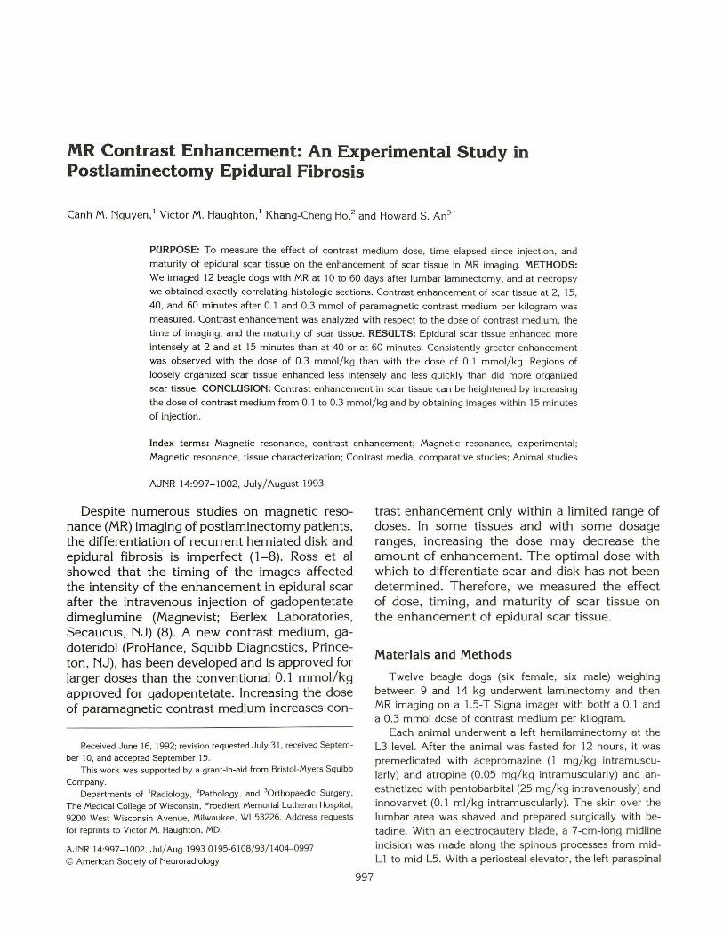
Fig. 1. Contrast enhancement at 2 minutes after 0.1 mmol/ kg (A) and 0.3 mmol/kg (B). The more-enhancing regions correspond to organized scar tissue, and the poorly enhancing regions to correspond poorly organized scar.
Contrast Enhancement in Post Laminectomy Scar Tissue Gadope ntetate 0. 1 mmo l/ kg
3.0
2.5
2.0 0
I .5 D
D
l'
I 0 l t
' • 0.5
0.0 15
6
~ •
30 40
MINU TES
50
6
i 60
_l 0 10 days
D1s days
• 20 da ys
• 30 days
.t. 40 da ys
t 5o da ys
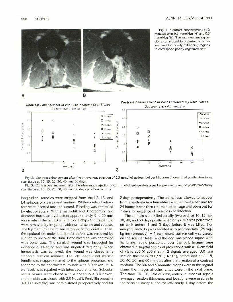
Fig. 2. Contrast enhancement after the intravenous injection of 0.3 mmol of gadoteridol per ki logram in organized postlaminectomy scar tissue at 10, 15, 20 , 30, 40, and 60 days.
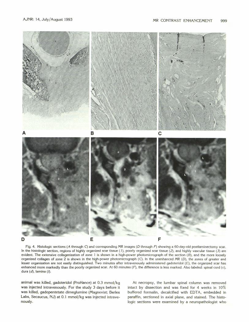
Fig . 3 . Contrast enhancement after the intravenous injection of 0.1 mmol of gadopentetate per kilogram in organized postlaminectomy scar ti ssue at 10, 15 , 20, 30, 40, and 60 days postlaminectom y.
longitudinal muscles were stripped from the L2, L3, and L4 spinous processes and laminae. Whitemoreland retractors were inserted into the wound. Bleeding was controlled by electrocautery . With a microdrill and decort icating and diamond burrs, an oval defect approximately 9 X 20 mm was made in the left L3 lamina. Bone chips and tissue fluid were rem oved by irrigation with normal saline and suction. The ligamentum flavum was rem oved with a curette. Then , the epidural fat under the lamina defect was removed by suction to uncover the dura. Bone bleeding was controlled with bone wax. T he surgical wound was inspected for evidence of bleeding and was irrigated frequently. When hemostasis was achieved, the wound was closed in a standard surgical manner. The left longitudinal muscle bundle was reapprox imated to the spinous processes and anchored to the contralateral muscle with 3.0 dexon. Muscle fascia was repaired with interrupted stitches. Subcutaneous t issues were closed with a continuous 3.0 dexon, and the skin was closed wi th 2.0 Novafil. Penicillin procaine (40,000 units/ kg) was administered preoperatively and for
2 days postoperatively. The animal was allowed to recover from anesthesia in a humidified warmed Kertscher unit for 24 hours; it was then returned to its cage and observed for 7 days for evidence of weakness or infection.
The animals were killed serially (two each at 10, 15, 20, 30, 40, and 60 days postlaminectomy). MR was performed on each animal 1 and 3 days before it was killed . For imaging, each dog was sedated with pentobarbital (25 mg/ kg intravenously). A 3-inch round surface coil was placed on the scanner table , and the dog was placed supine with its lumbar spine positioned over the coil. Images were obtained in sagittal and axial projections with a 1 0-cm field of view, 256 X 256 matrix , 2 signals averaged , 3 .0 mm section thickness, 500/ 30 (TR/ TE), before and at 2 , 15, 30, 40, 50, and 60 minutes after the injection of a contrast medium. The 30- and 50-minute images were in the sagittal plane; the images at other times were in the axial plane. The same TR , TE, field of view , matrix , number of signals averaged, section thickness, and locations were used as in the baseline images. For the MR study 1 day before the
AJNR: 14, July/ August 1993 MR CONTRAST ENHANCEMENT 999
D E F
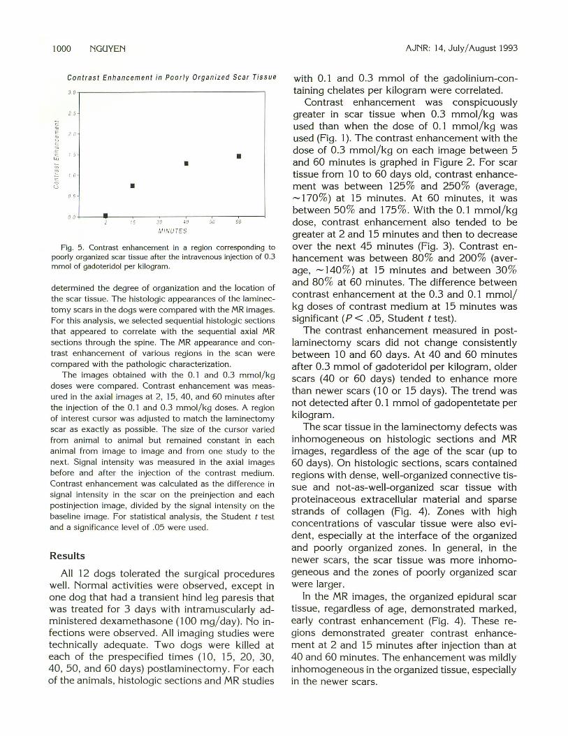
Fig. 4. Histologic sections (A through C) and corresponding MR images (D through F) showing a 60-day-old postlaminectomy scar. In the histologic section, regions of highly organized scar tissue ( 7), poorly organized scar tissue (2), and highly vascular tissue (3) are evident. The extensive collagenization of zone 1 is shown in a high-power photomicrograph of the section (B), and the more loosely organized collagen of zone 2 is shown in the high-power photomicrograph (C). In the unenhanced MR (D), the zones of greater and lesser organization are not easily distinguished. T wo minutes after intravenously administered gadoteridol (£), the organized scar has enhanced more markedly than the poorly organized scar. At 60 minutes (F), the difference is less marked. Also labeled: spinal cord (c), dura (d), lamina(/).
animal was killed , gadoteridol (ProHance) at 0.3 mmol/kg was injected intravenously. For the study 3 days before it was killed, gadopentetate dimeglumine (Magnevist; Berlex Labs, Secaucus, NJ) at 0 .1 mmol/kg was injected intravenously.
At necropsy , the lumbar spinal column was removed intact by dissection and was fixed for 4 weeks in 10% buffered formalin , decalcified with EDTA, embedded in paraffin , sectioned in axial plane, and stained. The histologic sections were examined by a neuropathologist who
1000 NGUYEN
Contrast Enhancement in Poorly Organized Scar Tissue
30 ,---------------------------------,
c: 0
<..:>
2 5
I 5
I 0
0 5
0 0 •
•
15
•
30 oo MINUTES
•
50 60
Fig. 5. Contrast enhancement in a region corresponding to poorly organized scar tissue after the intravenous injection of 0.3 mmol of gadoteridol per kilogram.
determined the degree of organization and the location of the scar tissue. The histologic appearances of the laminectomy scars in the dogs were compared with the MR images. For this analysis, we selected sequential histologic sections that appeared to correlate with the sequential axial MR sections through the spine. The MR appearance and contrast enhancement of various regions in the scan were compared with the pathologic characterization.
The images obtained with the 0.1 and 0.3 mmol/kg doses were compared . Contrast enhancement was measured in the axial images at 2, 15, 40, and 60 minutes after the injection of the 0 .1 and 0 .3 mmol/kg doses. A region of interest cursor was adjusted to match the laminectomy scar as exactly as possible. The size of the cursor varied from animal to animal but remained constant in each animal from image to image and from one study to the next. Signal intensity was measured in the axial images before and after the injection of the contrast medium. Contrast enhancement was calculated as the difference in signal intensity in the scar on the preinjection and each postinjection image, divided by the signal intensity on the baseline image. For statistical analysis, the Student t test and a significance level of .05 were used.
Results
All 12 dogs tolerated the surgical procedures well. Normal activities were observed, except in one dog that had a transient hind leg paresis that was treated for 3 days with intramuscularly administered dexamethasone (100 mg/day). No infections were observed. All imaging studies were technically adequate. Two dogs were killed at each of the prespecified times (10, 15, 20, 30, 40, 50, and 60 days) postlaminectomy. For each of the animals , histologic sections and MR studies
AJNR: 14, July/ August 1993
with 0.1 and 0.3 mmol of the gadolinium-containing chelates per kilogram were correlated.
Contrast enhancement was conspicuously greater in scar tissue when 0.3 mmol/kg was used than when the dose of 0. 1 mmol/kg was used (Fig. 1 ). The contrast enhancement with the dose of 0.3 mmol/kg on each image between 5 and 60 minutes is graphed in Figure 2. For scar tissue from 10 to 60 days old, contrast enhancement was between 125% and 250% (average, ~170%) at 15 minutes. At 60 minutes, it was between 50% and 175%. With the 0.1 mmol/kg dose, contrast enhancement also tended to be greater at 2 and 15 minutes and then to decrease over the next 45 minutes (Fig. 3). Contrast enhancement was between 80% and 200% (average, ~140%) at 15 minutes and between 30% and 80% at 60 minutes. The difference between contrast enhancement at the 0.3 and 0.1 mmol/ kg doses of contrast medium at 15 minutes was significant (P < .05, Student t test).
The contrast enhancement measured in postlaminectomy scars did not change consistently between 10 and 60 days. At 40 and 60 minutes after 0.3 mmol of gadoteridol per kilogram, older scars (40 or 60 days) tended to enhance more than newer scars (10 or 15 days). The trend was not detected after 0. 1 mmol of gadopentetate per kilogram.
The scar tissue in the laminectomy defects was inhomogeneous on histologic sections and MR images, regardless of the age of the scar (up to 60 days). On histologic sections, scars contained regions with dense, well-organized connective tissue and not-as-well-organized scar tissue with proteinaceous extracellular material and sparse strands of collagen (Fig. 4). Zones with high concentrations of vascular tissue were also evident, especially at the interface of the organized and poorly organized zones. In general, in the newer scars, the scar tissue was more inhomogeneous and the zones of poorly organized scar were larger.
In the MR images, the organized epidural scar tissue, regardless of age, demonstrated marked, early contrast enhancement (Fig. 4). These regions demonstrated greater contrast enhancement at 2 and 15 minutes after injection than at 40 and 60 minutes. The enhancement was mildly inhomogeneous in the organized tissue, especially in the newer scars.
AJNR: 14, July/ August 1993
Regions of poorly organized scar tissue had less rapid and less intense enhancement than did better organized scar tissue. After the intravenous administration of contrast medium, these regions increased in signal intensity progressively over 60 minutes. At 5 and 15 minutes, they were conspicuously hypointense with respect to the moreorganized scar (Fig. 4), and by 40 or 60 minutes, they were isointense or hyperintense. These regions sometimes had a lower signal intensity on the precontrast T1-weighted images. The contrast enhancement measured serially in one of the focal regions of poor organization is shown in Figure 5.
Discussion
The study shows that contrast enhancement in epidural scar tissue is greater with the 0.3 mmol/kg than with the 0.1 mmol/kg dose of contrast medium, that contrast enhancement in laminectomy scar tissue reaches a maximum before 30 minutes, and that the maturity of the scar tissue is a factor in the homogeneity and rapidity of contrast enhancement. The study suggests that a trial of 0.3 mmol of gadoteridol per kilogram in the differentiation of scar versus disk is warranted. It shows that scar tissue, like cavernous sinus, pituitary gland, and infundibulum, but unlike falx and sinus mucosa , is enhanced more effectively with the larger dose of contrast medium (V.M. Haughton and R. Lindsey, unpublished data) .
Our study has technical limitations. The measurement of signal intensity is inexact because of the use of surface coils. Therefore, the measurements of contrast enhancement were normalized to diminish the effect of the inhomogeneous sensitive volume of the coil. The measurements used for the graphs represent averages of two dogs and weighted averages of more and less mature portions of the scar. Although the number of animals used was relatively small, consistent results with statistical significance were obtained. Although no animals were monitored for longer than 60 days, the scars observed in dogs at 60 days appeared histologically to be well organized and mature. The molecular structure as well as the dose of the two contrast media differed . To what degree the differences in the molecular structure of the contrast media (ionic versus nonionic) determined the degree of enhancement is
MR CONTRAST ENHANCEMENT 1001
not known . Our model of an epidural scar does not duplicate the scarring seen anteriorly in the spinal canal of patients who have had laminectomy. To what degree that anterior scarring results from the surgical exploration from the preexisting degenerative disease or from invasion of the disc is unknown. Scar tissue ventral to the dural sac probably does not have different properties than scar tissue dorsal to the dural sac, although some investigators have suggested that there are differences (7).
Our results with the 0 .1 mmol/kg dose were similar to those obtained by Ross et al (8). Enhancement occurred within 2 to 15 minutes after injection . In the first 15 minutes after the injection of the contrast medium, the contrast enhancement of scar tissue is maximal. We found a substantial increase in enhancement (about 100%) with the threefold increase in contrast medium dose. The lack of proportionality between enhancement and dose suggests the confounding effect of T2 shortening in the enhanced images. Therefore , doses of more than 0.3 mmol/ kg would not likely result in a marked increase in contrast enhancement.
The problem in distinguishing scar from disk by contrast enhancement is that sometimes the scar enhances less or the recurrent disk tissue enhances more than expected. This study suggests one explanation for the inhomogeneous or variable degree of enhancement in scar tissue. Regions of poorly organized scar tissue may demonstrate poor enhancement compared with mature scar tissue . Immature scar tissue may explain regions of poorly enhancing tissue in the epidural space of patients recovering from laminectomy such as Bodin et al (9) observed. It may also suggest an explanation other than herniated disk material for tissue that fails to enhance promptly or intensely with contrast med ium in postlaminectomy patients.
References
1. Hueftle M, Modic MT, Ross JS, et al. Lumbar spine: postoperative
MR imaging with Gd-DT PA. Radiology 1988;167:817-824.
2. Frocrain L , Duvauferrier R, Husson JL, Noel J , Ramee A , Pawlotsky
Y. Recurrent postoperative sciatica: evaluation with MR imaging and
enhanced CT. Radiology 1989;170:531-540.
3. Bundschuh CV, Modic MT, Ross JS, et al. Epidural fibrosis and
recurrent disk herniation in the lumbar spine: assessment with MR.
AJNR: Am J Neuroradiol 1988;9: 169-178.
1002 NGUYEN
4. Ross JS, Masaryk T J, Schrader M , et al. MR imaging of the postop
erative lumbar spine: assessment with gadopentetate dimeglumine.
AJNR: Am J Neuroradiol 1990; 11:771-776.
5. Ross JS, Modic MT, Masaryk T J, et al. Assessment of extradural
degenerative disease with Gd-DTPA-enhanced MR imaging: correla
tion with surgica l and pathologic findings. AJNR: Am J Neuroradiol
1989; 10:1243-1249.
6. Bundschuh CV, Stein L, Slusser JH , Schinco FP, Ladaga LE, Dillon
JD. Distinguishing between scar and recurrent herniated disk in
postoperative patients: value of contrast enhanced CT and MR
imaging. AJNR: Am J Neuroradio/ 1990; 11 :949-958.
AJNR: 14, July/ August 1993
7. Ross JS, Blaser S, Masaryk T J , et al. Gd-DTPA enhancement of
posterior epidura l scar: an experimental model. AJNR: Am J Neuro
radio/ 1989; 10:1083-1088.
8. Ross JS, Delamarter R, Hueftle MG, et al. Gadolinium-DTPA-en
hanced MR imaging of the post-operative lumbar spine: time course
and mechanism of enhancement. AJNR: Am J Neuroradio/ 1989;
10:37-46.
9. Bodin SD, Davis DO, Dina TS, Parker DP, O'Malley S, Sunner JL,
Weisel SW. Contrast enhanced MR imaging performed after success
ful lumbar disk surgery: prospective study. Radiology 1992;182:
59-64.