movement strategies and dynamic knee control after...
TRANSCRIPT

Movement strategies and dynamic knee control after anterior cruciate ligament injury
A three-dimensional biomechanical analysis
Jonas Markström
Department of Community Medicine and Rehabilitation, Physiotherapy, Umeå University, Sweden
Umeå 2019

Responsible publisher under Swedish law: the Dean of the Medical Faculty This work is protected by the Swedish Copyright Legislation (Act 1960:729) Dissertation for PhD ISBN: 978-91-7855-066-1 ISSN: 0346-6612 New Series No 2040 Front page illustrate a side hop by one of the participants. Screenshot taken from Visual 3D, C-motion, Inc (www.c-motion.com) Electronic version available at: http://umu.diva-portal.org/ Printed by: Cityprint i Norr AB Umeå, Sweden, 2019

i
"All right," said Deep Thought. "The Answer to the Great Question..."
"Yes..!"
"Of Life, the Universe and Everything..." said Deep Thought.
"Yes...!"
"Is..." said Deep Thought, and paused.
"Yes...!"
"Is..."
"Yes...!!!...?"
"Forty-two," said Deep Thought, with infinite majesty and calm.”
“Forty-two!" yelled Loonquawl. "Is that all you've got to show for seven and a half million years' work?"
"I checked it very thoroughly," said the computer, "and that quite definitely is the answer. I think the problem, to be quite honest with you, is that you've never actually known what the question is.”
― Douglas Adams, The Hitchhiker's Guide to the Galaxy

ii
Table of Contents
Abstract ............................................................................................ iv Svensk sammanfattning .................................................................... v Abbreviations ................................................................................... vi Definitions ...................................................................................... vii Original papers .............................................................................. viii Thesis at a glance .............................................................................. x Preface ............................................................................................ xii Introduction ....................................................................................... 1
Motor control and biomechanics of the knee joint in relation to ACL injury ................ 1 Motor control of the knee joint with relevant definitions ........................................ 1 Anatomy and biomechanics of the knee and ACL ....................................................3
General background about ACL injuries ......................................................................... 5 ACL injury and reinjury epidemiology ..................................................................... 5 Common context of ACL injury mechanism ............................................................ 6 Treatment and rehabilitation after ACL injury ....................................................... 7 Personal consequences of ACL injury ....................................................................... 7 Assessment of knee function after ACL injury ......................................................... 8
Biomechanical assessment after ACL injury .................................................................. 9 Quantitative and qualitative components ............................................................... 9 Previous biomechanical work ................................................................................. 11 Assessment of neuromuscular landing control ...................................................... 13
Rationale for this thesis .................................................................................................. 15 Aims ................................................................................................................................ 16
Materials and Methods .................................................................... 17 Design and overview of the papers ................................................................................ 17 Participants ..................................................................................................................... 18
Paper I ....................................................................................................................... 18 Papers II – V ............................................................................................................. 19
Ethics .............................................................................................................................. 20 Test procedure ............................................................................................................... 22
Questionnaires ......................................................................................................... 22 Anterior tibial translation as an estimate of knee laxity ...................................... 23 Functional tests ....................................................................................................... 23
Data acquisition ............................................................................................................. 26 Motion capture and integrated force plates .......................................................... 26 Surface electromyography, EMG ............................................................................ 27 Knee isometric strength ........................................................................................... 27
Data processing .............................................................................................................. 28 Kinematic and kinetic data ..................................................................................... 28 Time to stabilization, TTS ....................................................................................... 32 Surface electromyography, EMG ........................................................................... 33

iii
Functional tests ....................................................................................................... 33 Statistical analyses ......................................................................................................... 33
Analyses of discrete data ........................................................................................ 34 Analyses of functional data .................................................................................... 36
Results ............................................................................................. 37 Comparisons between groups (specific aims A, D and E)............................................. 37
Dynamic knee robustness (papers III and IV) ....................................................... 37 Joint angles (papers I, III and IV) ......................................................................... 39 Joint moments (papers III and IV) ........................................................................ 42 Average EMG amplitudes and cocontraction ratio (paper IV) ........................... 43 Knee laxity and functional performances (papers I, III, IV) ................................ 44
Comparisons between legs within groups (specific aims A and E) ............................. 46 Dynamic knee robustness (paper IV)..................................................................... 46 Joint angles (papers I and IV) ................................................................................ 46 Joint moments (paper IV) ....................................................................................... 46 Knee laxity and functional performances (papers I and IV) ............................... 46
Comparisons between hop landings (specific aim F) .................................................... 47 Outcomes compared between landings (paper V) ................................................. 47 Outcomes correlated between landings (paper V) ................................................ 49
Reliability and agreement of the novel SRSH test (specific aims B and C) ................. 49 Description of angles and moments during SRSH landings (paper II) .............. 49 Within-session results for ACLR and CTRL (paper II) ......................................... 49 Test-retest results for CTRL (paper II) .................................................................. 50 Within-session results of FHA-1 inclination angle (papers III, IV and V)............ 51
Discussion ....................................................................................... 52 Main findings ................................................................................................................. 52 Assessment of neuromuscular landing control ............................................................ 52
The development and evaluation of the SRSH ...................................................... 52 Dynamic knee robustness ........................................................................................53 Adopted movement strategy to decrease knee loading ........................................ 54 Adopted movement strategy to avoid knee internal rotation .............................. 58 Neuromuscular landing control and athletic level ............................................... 59 The need to adapt a whole-body perspective ........................................................ 60 Different knee joint demands between landings ................................................... 60 Functional performances and neuromuscular control .......................................... 61
Methodological considerations ..................................................................................... 62 Participants ............................................................................................................. 62 Data collection and processing .............................................................................. 63 Outcome measures .................................................................................................. 64 Statistical analyses .................................................................................................. 64
Clinical implications and future research ..................................................................... 65 Conclusions ......................................................................................67 Acknowledgements ......................................................................... 68 References ...................................................................................... 69 Appendix ......................................................................................... 84

iv
Abstract
Background: Rupture of the anterior cruciate ligament (ACL) is common and mainly occurs in non-contact situations in sports, often due to momentarily poor movement control. Assessment of movement quality during sport-like tasks is crucial to understand how to decrease the high risk of reinjury for ACL-injured persons, but also how to prevent primary injury. This thesis addresses movement quality after ACL injury and includes development and evaluation of a novel standardized rebound side hop test (SRSH) for reliability and agreement of landing mechanics, and compares these outcomes between asymptomatic persons with different athletic levels, and between different hop tests.
Methods: This thesis involves five papers based on two separate data collections performed in a motion analysis laboratory. Paper I is a long-term follow up of ACL-injured persons treated with or without ACL reconstruction (ACLR) compared to asymptomatic persons (total N = 99, age 35-63), while papers II-V included ACLR persons, and asymptomatic elite athletes and non-athletes (total N = 79, age 17-34). A motion capture system synchronized with force plates and surface electromyography (EMG) registered trunk, hip and knee angles and moments and knee muscle activity during the hop for distance, vertical hop, and SRSH. Novel measures of dynamic knee robustness were also evaluated using finite helical axis inclination angles extracted from knee rotation intervals of 10˚.
Results: On average 23 years after injury, ACL injured persons performed the vertical hop with diverse angles compared to controls and their non-injured leg. The younger groups of ACLR persons and controls generally displayed excellent reliability and agreement for SRSH landing mechanics. These outcomes differed between the groups, and between legs for ACLR persons, despite similar dynamic knee robustness and acceptable knee function outcomes. Curve analyses further displayed differences between athletes and non-athletes, mainly with greater hip moments for athletes, although with similar values for dynamic knee robustness. Finally, greater knee angles and moments considered strenuous for the ACL were evident during the first rebound landing in SRSH compared to the other landings.
Conclusions: Persons who have suffered an ACL injury, regardless of whether treated with ACLR or not, appear to use task-coping strategies in preparation for and during landings to decrease knee joint loading, probably to preserve dynamic knee robustness. More attention should be given to the trunk and hip in clinics when evaluating movement quality after ACL injury to reduce the risk of future injuries due to movement compensation. High-level athletic training may also improve the ability to maintain dynamic knee robustness whilst performing a sport-like side-to-side task more efficiently through increased engagement of the hip. Finally, side hop landings should be assessed when evaluating and correcting for erroneous landing mechanics to improve knee landing control.

v
Svensk sammanfattning
Bakgrund: Främre korsbandsskada (ACL-skada) är en vanlig idrottsskada som huvudsakligen uppstår i situationer utan kontakt med annan spelare till följd av en tillfälligt dålig rörelsekontroll. Utvärdering av rörelsekvalitet under idrottsliknande tester behövs för att bättre förstå hur risken för ACL-skador och återskador kan minskas. Denna avhandling är inriktad på rörelsekvalitet efter ACL-skada och behandlar utveckling samt utvärdering av ett nytt standardiserat sidohopp (SRSH). Tillförlitlighet och överensstämmelse av ledvinklar och moment utvärderas och jämförs mellan personer med och utan ACL-skada, mellan personer med olika atletbakgrund, samt mellan olika hopptester.
Metoder: Denna avhandling omfattar fem studier, vilka är baserade på två separata datasamlingar utförda i ett rörelsesanalyslaboratorium. Studie I är en långtidsuppföljning av personer med ACL-skada behandlade både med och utan ACL-rekonstruktion, vilka jämförs med knäfriska kontroller (totala N = 99, 35-63 år). Studie II-V inkluderade personer med ACL-rekonstruktion, knäfriska kontroller och elitatleter (totala N = 79, 17-34 år). Ett rörelseanalyssystem synkroniserat med kraftplattor och ytelektromyografi registrerade bål, höft och knävinklar och moment, samt lårmuskelaktivitet under enbenshopp (på längden, på höjden, samt SRSH). Även nya utfallsmått som utvärderar knäets robusthet under rörelse analyserades med helixvinklar från intervaller av knärörelse på 10˚.
Resultat: I genomsnitt 23 år efter ACL-skada utförde båda grupperna ett enbenshopp på höjden med olika ledvinklar, både jämfört med kontroller samt deras oskadade ben. De yngre ACL-skadade personerna och kontrollerna visade generellt utmärkt tillförlitlighet och överensstämmelse av ledvinklar och moment under SRSH. Dessa utfallsmått skiljde sig mellan grupperna och mellan benen för ACL-skadade personer, trots att lika resultat av knäets robusthet samt acceptabla knäfunktionsresultat visades. Kurvanalyser visade även på skillnader mellan atleter och icke-atleter, främst med större höftmoment för atleter, trots lika resultat av knäets robusthet. Den första landningen i SRSH visade större knävinklar och moment som anses belasta ACL jämfört med övriga landningar.
Slutsatser: Personer med ACL-skada, oavsett om de behandlats med ACL-rekonstruktion eller ej, verkar tillämpa rörelsestrategier för att hantera landningar från enbenshopp genom att minska belastningen på knäleden, troligen för att bevara knäets robusthet. I klinik bör ett större fokus läggas på bål- och höftrörelser vid utvärdering av rörelsekvaliteten efter ACL-skada. Detta för att minska risken för framtida skador på grund av rörelsekompensation. Vidare förbättrar idrottsträning på hög nivå troligen förmågan att upprätthålla knäets robusthet samtidigt som utförandet av sidohoppstester blir effektivare genom ett ökat engagemang av höften. Slutligen bör sidhoppslandningar användas vid utvärdering och korrigering av landningsmekanik för en förbättrad knäkontroll.

vi
Abbreviations
The abbreviations below are found in the main text. 3D Three-dimensional
ACL Anterior cruciate ligament
ACLD20 ACL deficient persons injured over 20 years ago
ACLR20 ACL reconstructed persons injured over 20 years ago
ACLR ACL reconstructed persons (another younger cohort)
ANOVA Analysis of variance
ATH Asymptomatic elite athletes
BF Biceps femoris
CTRL20 Asymptomatic controls matched to ACLD20 and ACLR20
CTRL Asymptomatic controls matched to ACLR
EMG Electromyography
ES Effect size (partial eta squared were used)
FHA Finite helical axis
Hz Hertz, unit of frequency
ICC Intraclass correlation coefficient
MANOVA Multivariate analysis of variance
ms Millisecond
N Newton, unit of force
Non-ATH Asymptomatic non-athletes
n.s. Non-significant
OLHD One-leg hop for distance
OLVH One-leg vertical hop
SD Standard deviation
SRSH Standardized rebound side hop
ST Semitendinosus
SW Within-person standard deviation
TTS Time to stabilization
VL Vastus lateralis
VM Vastus medialis
vii
Definitions
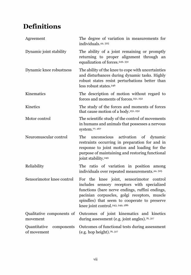
Agreement The degree of variation in measurements for individuals.44, 325
Dynamic joint stability The ability of a joint remaining or promptly returning to proper alignment through an equalization of forces.249, 331
Dynamic knee robustness The ability of the knee to cope with uncertainties and disturbances during dynamic tasks. Highly robust states resist perturbations better than less robust states.246
Kinematics The description of motion without regard to forces and moments of forces.251, 252
Kinetics The study of the forces and moments of forces that cause motion of a body.251, 252
Motor control The scientific study of the control of movements in humans and animals that possesses a nervous system.77, 267
Neuromuscular control The unconscious activation of dynamic restraints occurring in preparation for and in response to joint motion and loading for the purpose of maintaining and restoring functional joint stability.249
Reliability The ratio of variation in position among individuals over repeated measurements.44, 325
Sensorimotor knee control For the knee joint, sensorimotor control includes sensory receptors with specialized functions (bare nerve endings, ruffini endings, pacinian corpuscles, golgi receptors, muscle spindles) that seem to cooperate to preserve knee joint control.243, 249, 286
Qualitative components of movement
Outcomes of joint kinematics and kinetics during assessment (e.g. joint angles).76, 317
Quantitative components of movement
Outcomes of functional tests during assessment (e.g. hop height).76, 317

viii
Original papers
The thesis is based on the following papers. They will be referred to in the text by their respective Roman numerals.
I. Markström JL, Tengman E, Häger CK. ACL-reconstructed and ACL-deficient individuals show differentiated trunk, hip, and knee kinematics during vertical hops more than 20 years post-injury. Knee Surg Sports Traumatol Arthrosc. 2018;26(2):358-367.
II. Markström JL, Schelin L, Häger CK. A novel standardised side hop test reliably evaluates landing mechanics for anterior cruciate ligament reconstructed persons and controls. Sports Biomech. 2018 Dec 10; [Epub ahead of print] DOI: 10.1080/14763141.2018.1538385.
III. Markström JL, Grip H, Schelin L, Häger CK. Dynamic knee control and movement strategies in athletes and non-athletes in side hops: Implications for knee injury. Scand J Med Sci Sports, 2019 Apr 10; [Epub ahead of print] DOI: 10.1111/sms.13432.
IV. Markström JL, Grip H, Schelin L, Häger CK. Individuals with anterior cruciate ligament reconstruction adopt different movement strategies but display robust knees during side hop landings. (Manuscript)
V. Markström JL, Tengman E, Häger CK. One-leg lateral side-hops induce greater demands on knee landing control than hops in other directions as demonstrated in athletic and non-athletic females with or without injury of the anterior cruciate ligament. (Submitted)
Original papers have been reproduced with attributions according to creative commons (CC) license CC BY 4.0.

ix
x
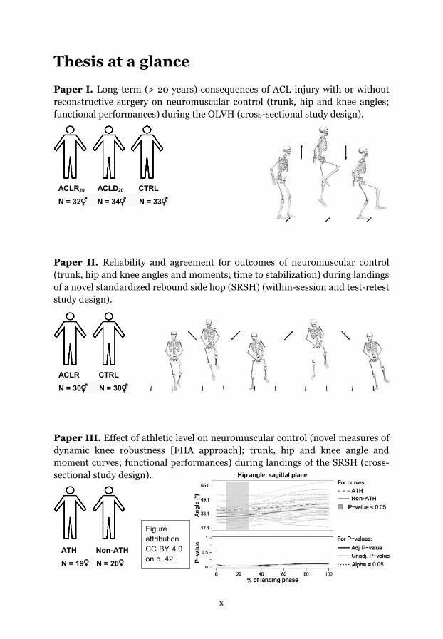
Thesis at a glance
Paper I. Long-term (> 20 years) consequences of ACL-injury with or without reconstructive surgery on neuromuscular control (trunk, hip and knee angles; functional performances) during the OLVH (cross-sectional study design).
Paper II. Reliability and agreement for outcomes of neuromuscular control (trunk, hip and knee angles and moments; time to stabilization) during landings of a novel standardized rebound side hop (SRSH) (within-session and test-retest study design).
Paper III. Effect of athletic level on neuromuscular control (novel measures of dynamic knee robustness [FHA approach]; trunk, hip and knee angle and moment curves; functional performances) during landings of the SRSH (cross-sectional study design).
ACLR20 ACLD20 CTRL N = 32⚥ N = 34⚥ N = 33⚥
ACLR CTRL N = 30⚥ N = 30⚥
ATH Non-ATH N = 19♀ N = 20♀
Figure attribution CC BY 4.0 on p. 42.

xi
Paper IV. Consequences of ACL reconstructive surgery on neuromuscular control (novel measures of dynamic knee robustness [FHA approach]; trunk, hip and knee angles and moments; functional performances) during landings of the SRSH (cross-sectional study design).
Paper V. Comparisons between hop landings for knee outcomes of neuromuscular control (novel measures of dynamic knee robustness [FHA approach]; knee abduction and internal rotation angles and moments) (cross-sectional study design).
ACLR CTRL N = 32⚥ N = 32⚥
ACLR ATH Non-ATH N = 21♀ N = 19♀ N = 20♀

xii
Preface
For a long time, I have had a significant interest in human movement and the highly specialized motor tasks that humans are able to perform. This interest grew when I took up climbing as a recreational sport during my bachelor’s studies. I was particularly fascinated by how climbing routes were solved by persons in different ways by using the strengths and weaknesses of the person attacking it. I adapted a view that there are always alternative ways to solve any problem at hand, which (unbeknownst to me at the time) is an issue that would return to me when trying to make sense of my research. My interest in research grew during my master’s studies, particularly when I worked with my master’s thesis that was also published in an international peer-reviewed journal. During this time period, I suffered a knee injury that increased my interest for this specific joint, particularly towards injuries of the anterior cruciate ligament (ACL), since my injury resembled a partly-ruptured ACL but also because my father had ruptured his ACL. When I later got the chance to be involved in a project that focused on biomechanical assessment related to ACL injury, I got to combine my personal interests with my curiosity for research.
The main problem that underlies this thesis is the high frequency of ACL injuries and re-injuries, mainly in sports participation. Most injuries and re-injuries occur in non-contact situations, which implies that neuromuscular control is important for avoiding injury. The research associated with the ACL is vast due to a wide variety of perspectives that are adopted to better understand the main problem stated above. A simple search prior to my thesis for “anterior cruciate ligament” on PubMed, Scopus, and Web of Science resulted in over 20 000 hits. Nevertheless, further knowledge of how to optimally evaluate neuromuscular control related to injury of the ACL was needed. This thesis includes five studies that address the topic of assessment of neuromuscular control related to injury of the ACL. A three-dimensional motion capture system synchronized with force plates and a surface electromyography system are used. Data are evaluated during sport-similar hop task landings to simulate situations where ACL injuries often occur. The introduction first supplies general background information and then review previous and relevant biomechanical work that are necessary to provide a full picture of the specific problems handled in each of the five studies. With this thesis I hope to add further knowledge of how to better assess neuromuscular landing control related to ACL injury, which contributes to practical implications that may result in fewer ACL injuries and re-injuries.

Introduction
1
Introduction
Motor control and biomechanics of the knee joint in relation to ACL injury
Motor control of the knee joint with relevant definitions Humans are bipedal and as such it is the joints in the lower extremities that are most exposed to forces and to injury. Particularly the knee joint is exposed to injury due to the competing biomechanical constraints between knee joint flexibility and stability that are needed for optimal movement control. A non-contact knee injury implicates a failure of knee joint motor control, since sensory integration and complex motor planning accurately should have predicted the joint loads. Therefore, the motor control of the knee joint is closely related to injury of the anterior cruciate ligament (ACL) that is the main focus of this thesis.
Motor control is the scientific study of the control of movements in humans and animals that possesses a nervous system.77, 267 The nervous system is specialized on fast transmission of information throughout the body and has conventionally been anatomically divided into central and peripheral components. The central nervous system comprises the brain (cerebral hemispheres, cerebellum, brainstem) and the spinal cord.77, 244 The peripheral nervous system contains sensory nerve cells that connects sensory receptors with relevant processing circuits in the central nervous system. The motor system of the peripheral nervous system consists in turn of the somatic motor system, that contains motor axons that connect the brain and spinal cord to skeletal muscles, and the autonomic motor system, that contains cells and axons that innervate smooth muscles, cardiac muscle, and glands.77, 244
A commonly used term used to describe the relevant components involved in maintaining joint movement control is sensorimotor control.249 The sensorimotor control of the knee joint includes sensory receptors with specialized functions, which consists of bare nerve endings (registers deformation, pain, inflammation), ruffini endings (slowly-adapting, registers deformation), pacinian corpuscles (fast-adapting, registers forces and pressure deformation), golgi receptors (registers high forces), and muscle spindles (registers muscle elongation, velocity, acceleration).243, 249, 286 These are located in the knee joint in articular surfaces, ligaments, menisci, tendons, capsule, and in relevant muscles that cross the knee joint. All of these seem to cooperate to preserve knee joint control, even though muscle spindles (located in muscles) have been argued to be the major kinaesthetic (sensations of limb position and movement) sensors.243

Introduction
2
Another common term is neuromuscular control, which when applied to the knee joint has been defined as the unconscious activation of dynamic restraints that occur in preparation for and in response to joint motion and loading for the purpose of maintaining and restoring functional joint stability.249 In the context of dynamic knee joint control, neuromuscular control may be considered as the motor component of sensorimotor control. As such, it involves kinesthetic-mediated activation of muscles to protect the non-contractile tissues (bones, capsule, ligaments, hyaline cartilage, menisci) from excessive forces, and facilitate optimal knee joint movement.249 Since the general theme of this thesis is the analysis of movement with a particular emphasis on the knee joint, it is the motor system that mainly is of interest here. As such, the term neuromuscular control will be used throughout the thesis.
Delving further into relevant biomechanical terms for the knee joint relevant for this thesis, both stability and robustness need to be defined. Dynamic joint stability is an overarching term and has been defined as the ability of a joint remaining or promptly returning to proper alignment through an equalization of forces.249, 331 This definition include components of non-contractile tissue integrity (mechanical integrity) and efficient sensorimotor control mechanisms.249, 250 When applied to the knee joint, the knee can be considered a stable system until injury occur.246 Dynamic knee stability is provided by the passive restraints of ligaments, joint capsule, cartilage, bony geometry, and friction, while active restraints are provided from feedforward and feedback neuromuscular control of skeletal muscles, and joint compressive forces.208, 249 However, to properly assess neuromuscular knee joint control there is a need to also grade the ability to cope with uncertainties and disturbances as better or poorer during dynamic tasks. This ability has been defined as robustness, where highly robust states resists perturbations more than less robust states (Figure 1), thus reducing the risk of injury.246 As such, particularly dynamic knee robustness relate to the evaluation of neuromuscular knee joint control, although lack objective measures for proper assessment.
Figure 1A-C. Illustration of stability and robustness, which should be translated to knee joint motor control. In A, the ball is not stable and not robust to perturbation. In B, the ball is stable but not robust to perturbation. In C, the ball is both stable and robust to perturbation, which is the optimal scenario when translated to knee motor control during dynamic tasks.

Introduction
3
Figure 2. Knee joint translations, angles and external moments of forces, which are defined in relation to the femur as reference segment. A, posterior drawer; B, anterior drawer; C, lateral shift; D, medial shift; E, distraction; F, compression; G, flexion; H, extension; I, abduction; J, adduction; K, external rotation; L, internal rotation.
Anatomy and biomechanics of the knee and ACL The knee joint has main articulations between the tibia, the posterior surface of the patella, and the femur. The knee joint has six degrees of freedom, three rotations and three translations, which is the result by movement of the shank relative femur. The rotations are described as flexion/extension, abduction/adduction (also termed valgus/varus, respectively) and internal/external, while the translations are described as anterior/posterior drawer, medial/lateral shift, and distraction/compression.61, 106, 189 These movements can be described in relation to three perpendicular axes, with flexion/extension defined along the femoral epicondylar axis (X-axis), abduction/adduction defined along the anterior-posterior axis (Y-axis), and internal/external rotation defined along the tibial shaft axis (Z-axis). The associated planes are in sagittal (YZ), frontal (XZ), and transversal (XY) plane, and will be used throughout this thesis (Figure 2).
The femoral condyles are elongated and the articular surface of the medial condyle is shorter and wider than the lateral condyle, thus enabling the rotation around the tibial shaft axis during knee flexion and extension.134 As the tibia glides on the femur from full flexion to full extension, it descends and then ascends the curves of the medial femoral condyle and simultaneously rotates externally (in relation to the femur). The motion is reversed as the tibia moves back into the fully flexed position.245 Such a mechanism provides more stability to the knee than would a simple hinge configuration and has been termed the screw-home mechanism. Tibiofemoral joint motion can therefore best be

Introduction
4
described as spiral during flexion and extension.134 The medial femoral condyle lies slightly distal to the lateral femoral condyle which results in knee abduction during extension.164 Knee joint ROM is limited to about 130-140° in sagittal plane, 15-20° in frontal plane, and 35-45° in transversal plane, although with variation in frontal and transversal plane movement due to flexion angle.32, 164
Ligaments in the knee provide tensile forces when stretched to keep the joint stable. There are four main ligaments in the knee, the ACL, the posterior cruciate ligament, and the medial and lateral collateral ligaments.109 These ligaments function as primary or secondary restraints to knee motion depending on joint position.109, 208 An injury to any of these ligaments may therefore result in knee instability in more than one plane-of-motion, where altered loadings of the knee joint may trigger the development of osteoarthritis.109 (Figure 3)
The ACL has a proximal attachment site to a fossa on the posteromedial edge of the lateral femoral condyle, follows an oblique course in the anterior-medial-distal direction and attaches distally to the anterior intercondylar fossa on the tibial plateau.28, 153 The ACL is usually described as consisting of two bundles, the anteromedial and posterolateral bundles named from their respective tibial insertion sites. This construction provide unique biomechanical properties that restrain anterior tibial translation (knee laxity) and excessive knee internal rotation that naturally occur during flexion and extension.5, 28, 153, 248 The ACL also contains kinaesthetic receptors that are considered important for proper sensorimotor control of the knee.1, 270, 271, 341
Figure 3. The four main ligaments in the knee, and a rupture of the ACL. ACL, anterior cruciate ligament; LCL, lateral collateral ligament; MCL, medial collateral ligament; PCL, posterior cruciate ligament. Attribution according to CC license: “AcL CAUSES”, by The Joint clinic is licensed under CC BY-SA 4.0 (https://creativecommons.org/licenses/by-sa/4.0/deed.en). This Figure has been modified from original by the addition of abbreviations.

Introduction
5
General background about ACL injuries
ACL injury and reinjury epidemiology Lower extremity injuries account for roughly 67% of all sports injuries, where the knee is the most exposed joint and represents 22-23% of these sports injuries.126 Specifically rupture of the ACL is common,64 with roughly 70% of all ACL ruptures occurring during sports participation.103 The ACL injury incidence rate per 10 000 athlete-exposures (soccer and basketball) was recently reported to be 0.63-0.70 for males and 1.95-2.55 for females.288 However, these incidences are likely underestimated since not all injuries are reported. As indicated by these numbers, females have a greater risk of ACL injury than males.201, 242
The risk of re-injury is unfortunately also high, where females again display a greater risk of re-injury than males.227 ACL reconstructed persons aged 14-62 that participate in more demanding compared to less demanding sports have shown a 2.1 increased odds of injuring the graft and a 9.8 increased odds for contralateral ACL injury.260 Among the ACL reconstructed persons in the demanding sports, 8% ruptured their graft while 10% ruptured their contralateral ACL within a five year period. Higher injury incidences have been found among younger individuals (10-25 years old), where 9% of ACL reconstructed persons injured their graft and 21% injured their contralateral ACL over the course of two years after return to sport.228 This equalled a 5.7 times higher incidence than their non-injured counterparts.228 Higher injury occurrences for the contralateral leg than for the graft are also corroborated by two recent publications by Schilaty and colleagues.265, 266
Moreover, a recent study by Niederer and colleagues show that ACL injured male elite soccer players (from 1st or 2nd leagues of the five European top leagues) had a 10% re-injury risk over a time period of five years, which equalled to a 25 times higher risk than their non-injured elite soccer player counterparts.205 Also, a systematic review and meta-analysis published in 2016 show contralateral reinjury rates of 11.3% and ipsilateral reinjury rates of 10%, for persons younger than 25 years.328 These rates increases among persons that return to sports, with pooled contralateral reinjury rates of 12% and ipsilateral reinjury rates of 10%, for persons younger than 25 years.328 The combined re-injury rates equals a 38 times greater risk relative to a population-based cohort of Finnish adolescents226 and a 22 times greater risk relative to American Collegiate and High School athletes.288 To conclude, primary and secondary ACL injuries constitute a major problem among sport active persons at all athletic levels, and it is important to decrease the risk of injury occurrences.

Introduction
6
Common context of ACL injury mechanism ACL injuries occur in non-contact or indirect contact with another player or equipment in roughly 70-80% of cases.35, 103, 135, 149, 200, 266 These percentages are still valid for high-collision sports such as American football, where 72.5% of all ACL injuries have been reported as non-contact.135 Most cases of non-contact ACL injuries occur in situations with momentarily poor movement control. Common injury situations involve multi-plane knee loading with the knee in a relatively straight (often less than 30˚ flexion), abducted and rotated position during an eccentric movement with a rapid deceleration of the body, such as during side-cuttings and jump landings (Figure 4). These findings are verified by video analysis,36,
55, 122, 135, 151, 156, 217, 291, 312 interviews,35 medical records,149 and magnetic resonance examinations of bone contusions within the tibia and femur.82, 224
The multifactorial injury mechanism are corroborated by experimental in vitro or in situ research that show higher peak ACL strain when combining external moments of knee abduction and internal tibial rotation (relative femur), compared to the strain of each of the uniplanar loadings alone.20, 146, 165, 215, 277 Adding anterior shear force further increases peak ACL strain.146, 165 Studies with an in vivo or in situ design also show greater ACL strain with less knee flexion angle,27, 72, 166 a longer ACL (thus larger ACL strain) with the knee more extended, abducted, and internally rotated,254 and that peak ACL strain occur with the knee extended at landing.48 These injury situations
are attributed to the anatomy of the knee, where a small knee flexion angle increases the patellar tendon insertion angle (shown in vivo)68 and decreases the hamstrings insertion angle, which results in an increased anterior tibial shear force for a given anterior tibial shear load.29 Peak ACL strain have also been estimated to occur as early as 7-50 milliseconds (ms) after initial contact during hop landings.48, 145, 156, 160, 165, 234
To summarize, the ACL is particularly subjected to a high risk of injury and re-injury when the knee (tibia relative femur) is extended, abducted, internally rotated and anteriorly translated, and occur shortly after initial contact during one-leg landings or side-cutting maneuvers.
Figure 4. Common ACL injury mechanism with the knee (tibia relative femur) being relatively extended, abducted and internally rotated, while the hip (femur relative pelvis) is adducted and internally rotated, during a landing or side-cutting maneuver.

Introduction
7
Treatment and rehabilitation after ACL injury An ACL injury is a serious knee injury and often results in decreased knee function despite rehabilitation programs longer than 6-12 months.204, 303 Treatment is either physiotherapy in combination with reconstructive surgery, or solely physiotherapy, and aims to improve knee function and stability. Treatment with ACL reconstruction is common for physically active persons171 and is performed in approximately 50-60% of cases in Sweden.336 Treatment with ACL reconstruction has increased over the years, with a 22% increase reported in the state of New York between 1997 and 2006171 and a 32% increase reported in the USA between 1994 and 2006.177 In Scandinavia, ACL reconstructive surgery have a documented annual incidence of 70-106 per 100 000 persons 10-39 years old.101
Rehabilitation protocols following ACL reconstruction199, 317 316 describe several phases with predefined criteria where at least the following outcomes show deficits and need attention: pain, inflammation, knee range of motion, muscle coordination and strength, gait symmetry, patellar mobility, neuromuscular control of dynamic tasks, functional performances, and self-rated knee function and stability. However, a systematic review from 2011 reported that time after ACL reconstruction is the most common (and often the only) criteria used before return to sport.17 Such an approach is problematic since no relation between time after ACL reconstruction and functional outcome measures (in this case from the one-leg vertical hop) have been shown.198 Another meta-analysis from 2014 similarly showed that 90% of identified randomized controlled trials did not use objective criteria to permit return to sport, while 65% of studies failed to use any criteria.111 Consensus statements on return to sport argue that at least the following aspects need consideration when estimating knee function after ACL injury: strength, range of motion, neuromuscular control, psychological factors and skill execution.8, 303
Personal consequences of ACL injury Personal consequences in both the short and the long term irrespective of treatment may include: reduced mechanical stability,248 lower self-rated knee function42, 194, 333 and quality of life,84 decreased muscle strength,132, 268 poorer functional performances,108, 133, 300 decreased sport-specific performances,174 increased body mass,300 and eventually knee osteoarthritis.19, 53, 99, 168, 334 Altogether, the persons often experience fear of movement (kinesiophobia) due to fear of re-injury, or a feeling of knee instability, which may generate a failure to return to sport.9, 10, 86, 157 Indeed, less than half and in some cases less than one third of athletes report that they returned to the same level of sport after ACL reconstruction with a minimum of 12-24 months after surgery.9, 10, 86, 182 These numbers are higher among elite athletes, where over 80% are reported to return to their pre-injury level or sport within 12-24 months after ACL reconstruction.159,
311 Over a longer time-period of five years, 25% of ACL injured male elite soccer

Introduction
8
players (1st or 2nd leagues of the five European top leagues) continued at their pre-injury level while 35% of their non-injured elite soccer player counterparts continued (relative rate of 72%).205
Further, the risk of developing knee osteoarthritis is 10 times greater after an ACL injury.99 Roughly 80% of individuals shows radiographic changes and 40-70% shows advanced changes (similar to Kellgren-Lawrence grade 2 or higher) at 12168 and 14 years19, 334 after ACL injury. This was irrespective if they had ACL reconstructive surgery or not. Such findings are corroborated by results from a meta-regression that show a prevalence of osteoarthritis after ACL reconstruction of 11% after five years, 21% after 10 years and 52% after 20 years.53 The high prevalence of osteoarthritis is indeed concerning since most individuals suffer an ACL injury already when 10-25 years old. A high degree of osteoarthritis already at ages of 30-40 inevitable affects quality of life to the worse and, in the most severe cases of osteoarthritis, may result in a total knee replacement to an artificial joint. Moreover, ACL injuries with or without ACL reconstruction also bear great societal costs, where solely one reconstructive surgery is estimated to roughly 4000 US dollars and an ACL reconstruction revision to 20 000 US dollars.98 Even though ACL reconstruction have higher costs in the short-term than treatment without surgery, such treatment are considered more cost-effective for persons where return to sport are related to greater quality of life.261,
290 The total costs for rehabilitation, sick leave, complications and later care per person are estimated to roughly 90 000 US dollars.181
Assessment of knee function after ACL injury Common functional high-intensive performance tests used to assess knee function involve side and cross-cutting, bilateral and unilateral hops, and strength tests. Unilateral tests have greater similarities to sport-specific scenarios and generally display greater (ACL injury prone) knee abduction angles and moments than bilateral tests.113, 154, 225, 295 Among these, hop tests are easy to administer, are fast to perform, and are clinician-friendly.116 Common one-leg hop performances include maximal performances of distance, height, or number of side hops over 30 s or the time to complete 10 side hops. One-leg hop tests also provide the advantage of evaluating symmetry of outcomes between legs.74, 116, 218 This is usually performed by presenting the result of the injured leg as a ratio to the contralateral non-injured leg in percent (100% equal similar performances). Evaluation of symmetry from a test battery of hop tests has presented a high ability to discriminate between the ACL deficient or reconstructed leg and the non-injured leg.108, 133 Current recommendations of minimum asymmetries on strength and hop performances vary between 80-90%.12, 18, 207 However, ACL injured persons that aim to return to pivoting or contact sports, or sports on competitive level, should display equal knee strength and at least 90% on two maximum as well as one endurable hop test for their injured leg.108, 303 Further, it

Introduction
9
is recommended that high-intensity tests that put high loads on the knee joint should include elements of side-cutting, similar to sport situations.8 Thus, to successfully return to sports or leisure activities that place considerable load on the lower limb (and in particular the knee joint), ACL injured individuals should demonstrate sufficient lower limb control during tasks that simulate sport maneuvers. This may reduce the risk of injuring the graft but also menisci etc.
However, a problem when evaluating symmetry of functional performances is that also the contralateral non-injured leg may exhibit a decreased neuromuscular function following injury.11, 304, 326 Wellsandt and colleagues showed a significantly better sensitivity of 82% compared to 27% to predict a second ACL injury when using a return to sport criteria with data from the non-injured leg collected within two months after injury but before surgery compared to data collected at 6 month after surgery.326 Sousa and colleagues further showed that ACL reconstructed persons who met return to sport criteria at 6 month after surgery had similar relative rates of graft ruptures and greater relative numbers of contralateral ACL injuries at a follow-up at least two years post-surgery, compared to those that did not meet the criteria.287 As such, further components of neuromuscular control not captured by functional performances are needed to better evaluate the rehabilitation before recommending a return to sport for ACL injured persons.76, 317 Corroborating such arguments are findings showing that ACL injured persons have presented acceptable symmetry, or acceptable performances to non-injured equals, while at the same time self-estimating their injured knee as unstable,42, 194, 333 or showing altered hip and knee angles and moments.218, 335 Further investigation of how to optimally assess neuromuscular control among ACL injured persons are warranted.
Biomechanical assessment after ACL injury
Quantitative and qualitative components It is important to evaluate both quantitative and qualitative components of movement during high-intensity functional tests for ACL injured persons that aim to return to physical activity, to prepare them for a successful return.76, 317 Quantitative components may be the distance hopped or peak muscle strength, while qualitative components include outcomes from analyses of in vivo joint kinematics and kinetics, as well as electromyography (EMG).76, 317 Kinetics is the study of the forces and moments of forces that cause motion of a body, and kinematics is the description of motion without regard to these forces and moments.251, 252 Joint moments (derived from inverse dynamics) are used as surrogate measures of joint loading since the articular loads cannot be measured with non-invasive methods. Invasive methods have the drawback of possible alterations in joint movement due to the surgery.

Introduction
10
A common method that evaluates in vivo kinematics is motion cameras that uses infra-red light to capture the three-dimensional (3D) motion of reflective markers put on anatomical landmarks of the body.144 This system may be synchronized with force plates to gain information of kinetics, and to EMG systems to gain information of muscle electrical activity, generated in muscle fibres in response to the activation provided by innervating motor neurons.80, 251 Kinematics and kinetics are closely aligned with the EMG signal since it provides information about the control and execution of voluntary (and reflexive) movements.80, 251 These qualitative outcomes are important tools for a thorough understanding of normal and pathological joint function during human movements.
Regarding motor control of the knee joint, it seems that an ACL injury results in a loss of mechanically sensitive receptors originally found in the ruptured ACL.1,
270, 271, 341 It appears that the CNS adapts to the injury by an altered engagement between brain areas as shown through electroencephalography21 and functional magnetic resonance imaging.107, 141, 142 Considering these alterations in relation to the common functional deficits and psychological aspects of an ACL injury mentioned earlier (see section Personal consequences of ACL injury and references therein), it may be expected that certain movement strategies are adapted among ACL injured persons to cope with high-intensity tasks that load the lower limb. Indeed, strategies to unload the injured knee are commonly displayed by shifting load to the hip and ankle on the injured leg46, 79, 212, 214, 241, 256-
258, 335 and by shifting load to the non-injured contralateral leg.79, 212, 257 The shift in load to the nearby joints has been explained by greater trunk flexion angle due a more anterior position of the ground reaction force vector closer to the knee joint axis and longer from the hip and ankle joint axes.212, 214 Landing with a greater trunk flexion angle also result in a softer landing as displayed by lower peak vertical ground reaction force and peak knee and hip flexion moments.275
One problem with adapted strategies to unload the injured leg is insufficient preparation for a return to sports, resulting in a subsequent injury. In sports, the injured knee will inevitable be exposed to high loads due to rapid and unpredictable scenarios that constantly occur out of the patients’ control. Such scenarios may explain the increased risk of injuring the graft compared to the ACL injury rate among non-injured counterparts.205, 228, 260, 328 Also, an active strategy to transfer load onto the contralateral leg when possible would also explain the even higher amount of contralateral injuries.228, 260, 265, 266, 328 Investigations of neuromuscular landing control among ACL injured persons therefore need to adapt a multi-joint perspective to thoroughly capture compensational strategies that may be adapted. Significant associations between restricted hip rotation to an increased risk of ACL injury for both the injured and the non-injured leg,318 and significant positive relations between hip adduction and hip rotation to (ACL injury prone) knee abduction angle and moment,129, 187 further argue for such a standpoint.
Introduction
11
Previous biomechanical work The assessment of high-intensity tasks, such as hop tests, among ACL reconstructed persons (both with and without subjective evidence of dysfunction) show that especially the capabilities of force absorption (i.e. landing) rather than force generation (i.e. take-off) is problematic.140 Since landing scenarios also precedes ACL injury in most cases (see section ACL injury situations and reference therein), this phase seems well suited for further assessment of neuromuscular control. Multiple studies have investigated kinematics and kinetics during the force absorption phase of functional unilateral hop tests mainly performed in vertical or forwards directions, among ACL reconstructed persons. Particularly the one-leg hop for distance (OLHD) has been evaluated, both with a standardized forward hop distance60, 74, 124, 212, 213, 322, 323 and for maximal distance.46, 100, 148, 218, 256, 269, 335 In 2015 it was the most common hop test for knee evaluation in research.116 Other tests that have been evaluated are drop landing,132, 269 220, 322, 323 diagonal hop landing,69 drop vertical hop,148, 232 one-leg vertical hop (OLVH),79, 125, 209 multiple hopping,232 and stop-jump.45, 247 However, these tests have been argued not to challenge the lower limbs enough in comparison to the demands found in pivoting or contact sports.93, 229, 303 The evaluation of maximal hop length (in OLHD) or hop height (in OLVH) mainly evaluate artificial performances indicative of knee function that generally not is found in sports. This argument is corroborated by research that show greater hip and knee frontal and transversal plane angles and moments when adding an emphasized side-to-side component to unilateral hop tasks.26, 137, 154, 202, 273, 283, 295 Lateral and diagonal landings also requires longer times to gain control of the landing167, 330 and display different hip-knee motion coordination than forward directions.284
While numerous studies have investigated kinematics and kinetics during tests that emphasise side-to-side movement on asymptomatic persons,22, 26, 59, 63, 83, 91,
188, 190, 203, 211, 281, 282, 294, 313 but at the time that this thesis were outlined (late autumn 2014) only a few studies included ACL reconstructed persons. The existing studies compared ACL reconstructed individuals to asymptomatic persons during side-step cutting161, 240, 289 and side hop,221 and present no differences between groups for hip221 and knee161, 221 angles but greater knee abduction angles289 for ACL reconstructed persons. Different results are shown for kinetics with decreased knee joint flexion moment161, 221 and greater moments in frontal161,
221, 289 and transversal planes161 among ACL reconstructed persons. Other findings include greater intra-limb coupling variability for ACL reconstructed female soccer players than for asymptomatic female soccer players.240 However, these studies had only small sample sizes of 10-13 ACL reconstructed persons, and no between-leg comparisons within groups for asymmetry were investigated.
After the initiation of this project a few additional studies have been published: a case-control study that compared landing mechanics collected before injury and

Introduction
12
again 27 months after ACL reconstructive surgery during an unanticipated side-cutting;262 a multicentre study that investigated tibiofemoral contact forces with an EMG-driven neuro-musculoskeletal model during side-cutting for 104 ACL reconstructed persons tested two to three years after surgery and compared to asymptomatic persons;264 and a study that evaluated trunk and lower limb asymmetry for 156 ACL reconstructed males at nine months after surgery during a side hop test.148 Results from these studies show differences in lower limb kinematics and kinetics, either between groups or between legs. It is evident that the existing material on this subject is insufficient and in need of further research.
Regarding EMG, the injured leg of ACL reconstructed persons have shown, either compared to asymptomatic persons or to the contralateral non-injured leg, earlier onset of muscle activity in preparation for landing for lower limb muscles,100, 232 greater cocontraction for spinae muscles37 and greater preparatory amplitude for impact among lower limb muscles.71, 209 Such neuromuscular strategies have also been related to higher sports capabilities when compared to the pre-injury status.210 The altered muscle activity patterns seems to serve a purpose to protect the knee joint against injury, although how this relates to dynamic knee robustness or landing mechanics incorporating multiple joints is unknown.
Even fewer studies have investigated long-term consequences of ACL injury on lower limb kinematics, kinetics, and muscle activity outcomes. The few existing studies show inconclusive results.220, 221, 333 Among these studies, von Porat and colleagues reported similar knee kinematic and kinetic results for ACL-injured men including both ACL deficient (n = 6) and ACL reconstructed (n = 6) persons and matched asymptomatic persons during gait, step up, and cross-over hop tests at 16 years post-ACL injury.333 The ACL deficient and ACL reconstructed males did however display worse clinical status by KOOS scales and lower isokinetic knee extensor strength than the controls. Further, Ortiz and colleagues published two papers that both evaluated hip and knee landing mechanics and EMG outcomes during the drop jump and up-down tests220 and during a side-to-side hop test221 for ACL reconstructed active females (n = 13) tested 1-16 years after surgery, and asymptomatic active females. They found similar kinematics between groups and between legs for all tasks, although altered knee kinetics and EMG outcomes. As such, the body of literature investigating landing mechanics in the long term also suffers from inconsistent results.
In conclusion, there is still a need to investigate neuromuscular control and closely related muscle activity patterns and functional performances among ACL reconstructed persons both in the short-term for sport-like tests that emphasises side-to-side movement, but also in the longer term, to better elucidate consequences of an ACL injury.

Introduction
13
Assessment of neuromuscular landing control Common practice to evaluate landing mechanics is to target specific variables at certain points in time, such as peak angles and moments for a specific joint, and to analyse these one by one thus neglecting possible inter-relations. More information of movement strategies and dynamic knee control may be gained if applying methods that summarizes motion from multiple motion planes or joints, or measures that describes the whole curve rather than single extracted values. The general lack of objective measures that evaluate dynamic knee robustness hinder the assessment of neuromuscular knee joint landing control. An appropriate method to gain such a measure may be to describe the knee joint motion as an instantaneous rotation about an axis as performed using finite helical axis (FHA) methods.33, 104, 105 By relating the inclination of the knee’s FHA to the flexion-extension axis of the knee over specific helical rotation intervals, information is provided of how much the motion diverges from pure sagittal plane movement. Such an approach present a realistic evaluation of dynamic knee robustness in relation to ACL injury mechanics, since sagittal-plane flexion torque are argued incapable of rupturing the ACL186 and that knee frontal and transversal plane motion induce loading that strains the ACL the greatest when combined.20, 146, 165, 215, 277 Persons with good dynamic knee robustness display small motions in frontal and transversal planes without compromising with task performance, thus presenting low FHA inclination angles (discrete values) over consecutive rotation intervals. Previous research have used FHA methods to discriminate between high and low intensity tests,104 between persons with a history of ACL injury (the 20 year follow-up) from matched asymptomatic persons,105 and between different knee pathologies.332 However, FHA methods has not been used to properly evaluate dynamic knee robustness among ACL reconstructed persons or asymptomatic high-level athletes. Such knowledge is important to further elucidate neuromuscular knee control in relation to ACL injury mechanics.
Another discrete measure that has been used to evaluate dynamic postural control or dynamic stability in research is called time-to-stabilization (TTS). This is a temporal measure that evaluates the time of fluctuations of the ground reaction force or center of pressure to stabilize, where a longer TTS indicate poorer dynamic postural control. Both ACL deficient and ACL reconstructed persons have shown longer TTS compared to asymptomatic equals during one-leg landing tasks.229, 235, 321 Even though several measures have been introduced for assessing dynamic stability, TTS was argued in 2013 to be the most commonly used.167 Such a measure may contribute to a greater understanding of neuromuscular landing control among ACL injured persons.
An alternative methodological approach that provide more information than selected discrete values is to apply inferential statistical methods for functional data. As such, the variable of interest include the whole curve while controlling

Introduction
14
for the overall type-1 error rate. These methods are found within the statistical area of functional data analysis, and has only been applied to assess human movement in a few studies. Selected research include topics of sports performance,319, 320 functional developmental stages for children,112, 259 and consequences of injury to the anterior cruciate ligament (the 20 year follow-up).114, 115 Such analyses are well suited to evaluate angle and moment curves and make better use of the data that is collected.
To summarize, the application of methods that evaluate multivariate data (for movement strategies), FHA inclination angles (for dynamic knee robustness), TTS (for dynamic postural control) and functional data (for angle and moment curves) hold promising potential in further contributing to the assessment of neuromuscular control that is related to ACL injury.
However, the usefulness of biomechanical measures need to be evaluated for reliability and agreement before further exploration. Reliability is the ratio of variation in position among individuals over repeated measurements, while agreement is the degree of variation in measurements for individuals.44, 325 No study to date have investigated reliability and agreement of landing mechanics during side-to-side emphasised tests for ACL reconstructed persons, although a few studies included asymptomatic athletes during sidestep cutting tasks.3, 26, 180,
193, 263, 279 In comparison to side or crosscutting tasks, a one-leg side-hop test has the advantage of being easier to administer, requires less space (sidestep cutting has been limited to analyses of only one direction due to restricted lab-space),3,
137 eliminates compensational strategies of the contralateral leg in preparation to the cutting, and provides a direct between-leg comparison. These arguments are particularly valid if the test is well standardized, which is considered important for dynamic high-intensity tests.52 The conventionally used side hop test was designed to particularly target capacity and endurance since the outcome is either the number of hops performed during 30 s108, 150 or the time to complete 10 hops.222, 337 As such, there is a need for a standardised side hop test that specifically aim to evaluate joint-specific angles and moments. Particularly without the onset of e.g. fatigue, which is known to negatively alter knee stability191 and that may alter lower limb mechanics during hop tests16, 24, 185, 324 thus affecting the data collected during testing.
Introduction
15
Rationale for this thesis The high incidences of primary ACL injuries and secondary ACL re-injuries (to both the contralateral knees and to the reconstructed graft) represent significant individual consequences in both short and long term perspectives. Further consequences are high societal costs with respect to the health-care system. The common ACL injury situations in non-contact during sports participation due to momentarily poor movement control implies that assessment of neuromuscular landing control is important. An ACL injury seems to result in alterations with adapted movement strategies that affects motion of the trunk and the nearby joints during high-intensity landing tasks. These compensational movement strategies suggest that a multi-joint perspective should be applied, which often is not the case today in research. Additional information of how dynamic knee robustness presents itself after ACL reconstruction or among asymptomatic elite athletes, and how to evaluate it, may further contribute to the understanding of neuromuscular control of hop landings. Such knowledge could lead to implementation of more effective rehabilitation regimens for ACL injured persons with safer return to sports, but also for primary injury preventive purposes among athletes. A requirement of such assessment are valid and reliable tests that are specifically designed to evaluate movement control. An increased knowledge of movement strategies among ACL injured persons in the longer term is also of interest due to few and inconclusive results.

Introduction
16
Aims The general aim of this interdisciplinary 3D biomechanical investigation was to contribute with new perspectives on how to analyse and assess neuromuscular control related to injury of the ACL. For this purpose, this study evaluated novel measures of dynamic knee robustness, joint-specific angles and moments from multiple joints, and general functional performances for different populations during various hop tests.
Specific aims were:
A. To analyse long-term consequences of ACL injury on neuromuscular control by comparing trunk, hip, and knee angles during the OLVH ~20 years post-ACL injury between ACL reconstructed persons, ACL deficient persons, and matched asymptomatic persons, and to determine if there was asymmetry in these measures within groups (paper I)
B. To develop and assess the usefulness of a novel standardized rebound side hop (SRSH) test for improved biomechanical evaluation of joint-specific kinematics and kinetics, by evaluating ACL reconstructed persons and asymptomatic persons for within-session reliability and agreement for trunk, hip, and knee angles and moments, and measures of TTS, during landings of this test (paper II)
C. To further investigate the usefulness of the SRSH by evaluating asymptomatic persons for test-retest reliability and agreement for the same outcomes mentioned above (aim B), during landings of this test (paper II)
D. To evaluate the effect of athletic level on neuromuscular control by comparing asymptomatic female athletes and non-athletes for dynamic knee robustness and trunk, hip and knee angle and moment curves during landings of the SRSH, in relation to knee laxity and functional performances (paper III)
E. To analyse consequences of ACL reconstruction on neuromuscular control by comparing ACL reconstructed persons and matched controls for dynamic knee robustness, trunk, hip, and knee angles and moments, and EMG outcomes during landings of the SRSH, in relation to knee laxity and general functional performances, and to determine if there was asymmetry in these measures within groups (paper IV)
F. To evaluate hop landings for outcomes related to ACL-injury by comparing dynamic knee robustness and knee-specific angles and moments between landings of the SRSH, OLHD and OLVH for females in separate groups of ACL reconstructed individuals and asymptomatic elite athletes and non-athletes, and to evaluate how well these outcomes correlate between the landings (paper V)

Materials and Methods
17
Materials and Methods
Design and overview of the papers
This thesis consists of five papers based on two data collections, both performed at U-motion lab at the Department of Community Medicine and Rehabilitation, Umeå University, Sweden. Paper I was based on data from a long-term follow up after ACL injury with or without ACL reconstructive surgery, and asymptomatic persons, where all persons were between the ages of 35-63. Papers II-V were based on data from a second data collection where ACL reconstructed persons, asymptomatic elite athletes and non-athletes were between the ages of 17-34.
• Paper I: a cross-sectional study including ACL reconstructed persons (n = 32, 12 females), ACL deficient persons (n = 34, 13 females, named ACLPT in paper I) and age and sex matched asymptomatic persons (n = 33, 11 females). The ACL injured persons suffered their injury 17-28 years prior to testing. This data collection have previously been described in detail in a previous thesis that primarily reported on knee function, overall physical activity level and capacity, and knee kinematics during the OLHD for these groups.299 These groups are names as ACLR20, ACLD20 and CTRL20 throughout the thesis to clearly separate them from the younger and more recently injured ACL reconstructed persons included in papers II, IV and V (named ACLR).
• Paper II: a reliability study with a test-retest design including ACL reconstructed persons (n = 30, 22 females) and activity-matched controls (n = 30, 22 females) that were evaluated for within-session reliability and agreement while controls (n = 25, 22 females) were also evaluated for test-retest reliability and agreement on two separate occasions with a mean (range) of 16.4 (7-30) days between tests. These groups are named as ACLR and CTRL throughout the thesis.
• Paper III: a cross-sectional study including 19 elite athlete females and 20 non-athlete females. These groups are mentioned as ATH and non-ATH, respectively, throughout the thesis.
• Paper IV: a cross-sectional study including 32 ACL reconstructed persons (24 females) and 32 asymptomatic activity-matched persons (24 females, from ATH and non-ATH in paper III). These groups are mentioned as ACLR and CTRL throughout the thesis (thus same as for paper II, but the results from each paper are clearly distinguished in the thesis).
• Paper V: a cross-sectional study including females in groups of 21 ACLR, 19 ATH and 20 non-ATH (same ATH and non-ATH included in paper III).

Materials and Methods
18
Table 1. Main outcomes analysed for the tests performed in papers I-V. Paper I Paper II Paper III Paper IV Paper V
Joint angles OLHD X OLVH X X SRSH X X X X
Joint moments OLHD X OLVH X SRSH X X X X
Time to stabilization SRSH X
Surface EMG outcomes SRSH X
Functional performances OLHD X X OLVH X X X SRSH X X X Isometric knee strength X X
EMG, electromyography; OLHD, one-leg hop for distance; OLVH, one-leg vertical hop; SRSH, standardized rebound side hop.
Participants
Paper I The sample size for paper I was calculated with a power analysis based on pilot tests including five ACL-injured persons and five asymptomatic persons, which suggested that 32 persons/group were needed for a power of 80 % to detect a significant difference in knee joint flexion angle between groups with a variance of 10° and a significance level of 5 %. The ACL injured persons that were contacted for paper I consisted of two cohorts from 113 individuals who suffered an ACL injury 17-28 years previously. Individuals were treated at two separate hospitals either with physiotherapy in combination with reconstructive surgery (ACLR20, n = 62) or solely with physiotherapy (ACLD20, n = 51). A subset of 42 persons in ACLR20 and 39 persons in ACLD20 was eligible for the present study according to the following inclusion criteria: unilateral ACL injury, not having any surgical total hip or knee replacement (prosthesis), no inflammatory or rheumatic disease or neurological pathology. Eleven persons declined to participate due to time constraints and logistical reasons, resulting in 33 persons in ACLR20 and 37 persons in ACLD20. Details of treatments have been presented previously.299 All persons in ACLR20 had a patellar tendon autograft. Radiological knee OA mostly in stage 1-2, but in some cases up to 4,143 was detected in ~90 % of the participants in both ACL groups at the time of testing.

Materials and Methods
19
One person from ACLR20 and three persons from ACLD20 were excluded from the analyses due to lost marker data in sensitive parts of the OLVH, resulting in a total of 32 persons in ACLR20 and 34 persons in ACLD20. The CTRL20 group consisted of 33 persons matched for age and sex with no previous knee injuries and with normal results from a clinical knee examination prior to testing.
Papers II – V Recruitment of ACLR were performed through the local University Hospital and private clinics, advertisement and convenience sampling, while ATH and non-ATH were recruited through the two latter strategies.
For paper II, a power analysis was performed that relied on the test-retest approximation by Walter and colleagues314 revealed that at least 22 participants per group were needed to achieve an intraclass correlation (ICC) of 0.8 as previously shown for the angles and moments of interest3, 180, 193 that differ from an ICC of 0.5 with a type I error of 0.05 and type II error of 0.20. Hence, 30 participants in each group were included for within-session analyses and 25 persons in CTRL were considered sufficient for test-retest analyses.
For papers III, IV and V, power analyses on subgroup data for the SRSH for maximal hip adduction and knee abduction were performed (variables considered relevant with regard to knee stability and ACL injury risk).117, 120, 129, 187 These analyses were conducted for one-way ANOVA analyses with fixed effects from mean values of maximal angles from each group with three ACLR, four ATH and four non-ATH. Results showed a group sample size of nine persons for hip adduction and six persons for knee abduction, with a wanted power of 0.8. These results are similar to the number of participants that are commonly used in movement analysis studies, including studies that show good to excellent reliability in dynamic tasks with as few as 8-12,175, 192, 263 and studies showing significant results of kinematic differences between groups using 10-14 ACLR.70,
212, 218 However, groups of nine persons are considered small samples so the power analysis for paper I that suggested 32 persons in ACLR for paper IV was adhered to. Moreover, 20 persons in each group for papers III and V was considered sufficient. However, one of the athletes that was planned to participate at the end of the data collection phase sustained an injury two days before planned testing, thus resulting in 19 persons in ATH.
Inclusion criteria for ACLR in papers II-V were: 17-34 years of age, unilateral ACL injury, a hamstring surgical graft, no complete tear of any other knee ligament, no major menisci or articular damage, no severe ankle sprain the last 6 months, no musculoskeletal or neurological pathology that would affect their ability to execute any of the hop tests or strength tests, and that they had returned to physical activity after recommendations from physician and physiotherapist and felt comfortable and confident in performing the different hop tests and strength

Materials and Methods
20
tests. The same (relevant) inclusion criteria were applicable for ATH and non-ATH, although with a few additional criteria to clearly separate these groups for paper III. To participate, ATH had to regularly conduct knee-specific training with the aim of improving lower limb control in multi-directional movements every week, which they were questioned for both at recruitment and again at time of testing. Such exercises included lunges, jumps and hops, side-cutting movements with changes of direction, as well as agility and speed drills. Non-ATH were physically active but were not included if they participated in any recreational physical activity more than 4 days per week or performed knee-specific training outside of gym or workout classes specifically to improve knee movement control with the aim to decrease the risk of injury. All participants had a clinical knee examination prior to testing for screening of inclusion criteria by an experienced physiotherapist.
For papers II and IV, both sexes were included in ACLR, even though more females than males were recruited, to achieve the sample sizes required to analyse asymmetries between the injured and non-injured legs and differences to CTRL. For papers III and V, on females were included since, relative to males, they have an approximately three times greater risk of sustaining an ACL injury242 and after ACL reconstruction show lower physical activity levels, poorer self-estimated knee function scores and lower rate of return to sport.293 An overview of participants included in papers I-V is presented in Table 2.
Ethics
The projects were approved by the Regional Ethical Review Board in Umeå for paper I (Dnr. 08-211M) and paper II-V (Dnr. 2015/67-31). All participants provided written informed consent in accordance with the declaration of Helsinki (2013) before partaking. A separate written informed consent was also provided for the two video cameras that recorded all tests in the lab to visually check performance during analysis, one mounted from the side and one in front of the participants. Any major risks were not foreseen, although asking participants to perform challenging hop tasks poses potential risks for injury, especially for ACL-injured persons where the knee has been previously injured. The inclusion criteria ensured that these persons felt comfortable and confident in performing the different hop tests and strength tests before partaking. These persons did, or had, practiced similar hops in their rehabilitation before participation. Comparable tests are often performed before a return to sport are recommended by their main physiotherapist. Careful considerations were undertaken during the physical testing in the lab, and not a single incident have been observed despite physical testing of several hundred persons in the motion laboratory over the last two decades. There is a first aid kit available in the lab and at least one of the researchers’ present was trained to perform first aid should an injury occur.
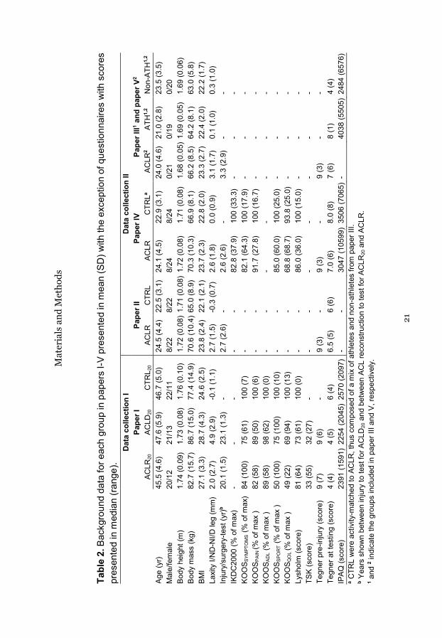
Mat
eria
ls a
nd M
etho
ds
21
Tabl
e 2.
Bac
kgro
und
data
for e
ach
grou
p in
pap
ers
I-V p
rese
nted
in m
ean
(SD
) with
the
exce
ptio
n of
que
stio
nnai
res
with
sco
res
pres
ente
d in
med
ian
(rang
e).
a CTR
L w
ere
activ
ity-m
atch
ed to
AC
LR, t
hus
com
pose
d of
a m
ix o
f ath
lete
s an
d no
n-at
hlet
es fr
om p
aper
III.
b Yea
rs s
how
n be
twee
n in
jury
to te
st fo
r AC
LD20
and
bet
wee
n AC
L re
cons
truct
ion
to te
st fo
r AC
LR20
and
AC
LR.
1 and
2 ind
icat
e th
e gr
oups
incl
uded
in p
aper
III a
nd V
, res
pect
ivel
y.
D
ata
colle
ctio
n I
Dat
a co
llect
ion
II
Pape
r I
Pape
r II
Pape
r IV
Pape
r III1 a
nd p
aper
V2
AC
LR20
AC
LD20
C
TRL 2
0 AC
LR
CTR
L AC
LR
CTR
La AC
LR2
ATH
1,2
Non
-ATH
1,2
Age
(yr)
45.5
(4.6
) 47
.6 (5
.9)
46.7
(5.0
) 24
.5 (4
.4)
22.5
(3.1
) 24
.1 (4
.5)
22.9
(3.1
) 24
.0 (4
.6)
21.0
(2.8
) 23
.5 (3
.5)
Mal
e/fe
mal
e 20
/12
21/1
3 22
/11
8/22
8/
22
8/24
8/
24
0/21
0/
19
0/20
Bo
dy h
eigh
t (m
) 1.
74 (0
.09)
1.
73 (0
.08)
1.
76 (0
.10)
1.
72 (0
.08)
1.7
1 (0
.08)
1.7
2 (0
.08)
1.
71 (0
.08)
1.
68 (0
.05)
1.6
9 (0
.05)
1.
69 (0
.06)
Bo
dy m
ass
(kg)
82
.7 (1
5.7)
86
.7 (1
5.0)
77
.4 (1
4.9)
70
.6 (1
0.4)
65.
0 (8
.9)
70.3
(10.
3)
66.9
(8.1
) 66
.2 (8
.5)
64.2
(8.1
) 63
.0 (5
.8)
BMI
27.1
(3.3
) 28
.7 (4
.3)
24.6
(2.5
) 23
.8 (2
.4)
22.1
(2.1
) 23
.7 (2
.3)
22.8
(2.0
) 23
.3 (2
.7)
22.4
(2.0
) 22
.2 (1
.7)
Laxi
ty I/
ND
-NI/D
leg
(mm
) 2.
0 (2
.7)
4.9
(2.9
) -0
.1 (1
.1)
2.7
(1.5
) -0
.3 (0
.7)
2.6
(1.8
) 0.
0 (0
.9)
3.1
(1.7
) 0.
1 (1
.0)
0.3
(1.0
) In
jury
/sur
gery
-test
(yr)b
20.1
(1.5
) 23
.1 (1
.3)
- 2.
7 (2
.6)
- 2.
6 (2
.6)
- 3.
3 (2
.9)
- -
IKD
C20
00 (%
of m
ax)
- -
- -
- 82
.8 (3
7.9)
10
0 (3
3.3)
-
- -
KOO
S SYM
PTO
MS
(% o
f max
) 84
(100
) 75
(61)
10
0 (7
) -
- 82
.1 (6
4.3)
10
0 (1
7.9)
-
- -
KOO
S PAI
N (%
of m
ax )
82 (5
8)
89 (5
0)
100
(6)
- -
91.7
(27.
8)
100
(16.
7)
- -
- KO
OS A
DL (
% o
f max
) 89
(58)
98
(62)
10
0 (0
) -
- -
- -
- -
KOO
S SPO
RT (
% o
f max
) 50
(100
) 75
(100
) 10
0 (1
0)
- -
85.0
(60.
0)
100
(25.
0)
- -
- KO
OS Q
OL (%
of m
ax )
49 (2
2)
69 (9
4)
100
(13)
-
- 68
.8 (6
8.7)
93
.8 (2
5.0)
-
- -
Lysh
olm
(sco
re)
81 (6
4)
73 (6
1)
100
(0)
- -
86.0
(36.
0)
100
(15.
0)
- -
- TS
K (s
core
) 33
(55)
32
(27)
-
- -
- -
- -
- Te
gner
pre
-inju
ry (s
core
) 9
(7)
9 (6
) -
9 (3
) -
9 (3
) -
9 (3
) -
- Te
gner
at t
estin
g (s
core
) 4
(4)
4 (5
) 6
(4)
6.5
(5)
6 (6
) 7.
0 (6
) 8.
0 (8
) 7
(6)
8 (1
) 4
(4)
IPAQ
(sco
re)
2391
(159
1) 2
254
(204
5) 2
570
(209
7)
- -
3047
(105
99)
3506
(706
5) -
40
38 (5
505)
248
4 (6
576)

Materials and Methods
22
Test procedure Common for both data collections is that participants first completed self-rated questionnaires. Then they had a clinical knee examination and were inquired about any history of injuries by an experienced physiotherapist, and their passive tibial anterior translation (knee laxity) assessed followed by measurement of body height and mass. Leg dominancy was also evaluated to enable comparisons between groups, where the dominant leg was defined as the preferred leg to kick a ball, since the same leg has been shown to be preferred as leading leg when stepping up on a 40 cm platform and when stepping out to prevent a fall when suddenly pushed in the back.66 After these initial assessments, the EMG and marker setup were placed and controlled for and the functional tests were performed. Details of these test procedures are described below.
Questionnaires Established questionnaires were used to assess self-rated knee function, health, physical activity and fear of movement, and are presented briefly below.
International Knee Documentation Committee Subjective Knee Form 2000
A self-administered questionnaire of valid and reliable measures of symptoms, daily function and sports activities, addressing many of the symptoms and patient-reported responses identified as being predictive of poor satisfaction, including pain, stiffness, welling, instability, sport, activities of daily living and overall rating of knee joint function.130 The results were presented as a percentage of max score where a higher score indicate better knee function. This questionnaire was used to describe the groups in paper IV.
Knee injury and Osteoarthritis Outcome Score
A self-administered questionnaire in five subscales: symptoms, pain, activity in daily living, function in sports and recreational activities, and knee-related quality of live.255 The subscale activity in daily living was only used for participants evaluated in paper I, since the subscale is not considered relevant to young and active persons as those evaluated in papers II-V.84 The results from each subscale were presented as a percentage of the max score where a higher score indicate better knee function. This questionnaire was used to describe the groups in papers I and IV.

Materials and Methods
23
Lysholm knee scoring scale
A self-administered eight-item questionnaire developed to asses knee symptoms and functional disabilities resulting from an ACL injury.172 It provides a score between 0-100 where a higher score indicate better knee function. This questionnaire was used to describe the groups in papers I, IV and V.
Tegner activity level scale
An activity score ranging from 1-10 that describe the patient activity level including activities in daily life and recreational and competitive sports (a higher score indicate a higher level).296 A score of seven indicate e.g. recreational soccer, while a score of eight indicate e.g. competitive floorball. This questionnaire was used to describe the groups in papers I-V.
International Physical Activity Questionnaire, short form
A self-administered questionnaire designed primarily for population surveillance of physical activity and inactivity among adults.58 This short form estimates physical activity (vigorous, moderate and walking activities) during the last seven days. This questionnaire was used to describe the groups in papers I, III and IV.
Tampa Scale for Kinesiophobia
A self-administered questionnaire designed to evaluate fear of movement that provide a score between 17-68, where a higher score indicate greater fear of movement.170 This questionnaire was only used to describe the two ACL groups in paper I.
Anterior tibial translation as an estimate of knee laxity The anterior tibial translation was assessed with a KT1000 arthrometer (Medmetric Corporation, San Diego, Ca, USA) at anterior pull forces of 15 lb. (67 N), 20 lb. (89 N), and 30 lb. (133 N). Three to five trials for each leg were assessed to attain a reliable mode value. The 30 lb. load was used to describe the groups in papers I-IV since it reveals the greatest between-leg asymmetry.123
Functional tests In data collection for paper I, the test procedure began with a 6-minute warm-up on a bicycle ergometer at moderate intensity. Then a test battery of functional tasks was performed (see Tengman for more details299) of which only the OLVH was of interest for paper I (performed after four balance tasks, a two-leg squat test, OLHD, and rise from chair). In data collection for papers II – V, the test procedure began with a test battery of functional tasks of which the OLHD,

Materials and Methods
24
OLVH, SRSH and isometric strength tests were of interest for papers II – V (performed in this order after two knee joint position sense tests, two stair descent tests, a balance test, a two-leg squat test, and a step-over test). In both data collections, participants performed all hop tests barefoot and alternated legs between trials to avoid a possible onset of fatigue. This protocol was used since fatigue may negatively affect reflex components and alter tibial anterior translational stability,191 and affect lower limb mechanics during hop tests.16, 24,
185, 324 ACLR started on the non-injured leg and CTRL on the dominant leg. During hop tests, participants in data collection for paper I held their arms across their chest while participants in data collection for papers II-V held their arms behind their back while holding a 25 cm short rope (with knots). These instructions were given for standardisation, to avoid obstructing the markers that generated the model, and to emphasize lower limb control. Restricting arms by keeping them closer to the body emphasizes lower limb landing control by resulting in e.g. greater knee impulse110 and greater knee abduction moment.51 Successful trials in all hop tests had requirements of a 2-3 s single leg stance after landing without removing their arms from their chest (paper I) or letting go of the rope (papers II-V), not putting the contralateral foot down and not making significant adjustments with the ipsilateral foot to maintain balance. The time between each trial was approximately five seconds. These hop tests and strength tests are further described below.
One-leg vertical hop, OLVH
The OLVH was performed on a force plate with the participants’ initially standing upright on one leg before hopping vertically as high as possible and landing with the same leg on the force plate (Figure 5A). This is a test that involves the change between eccentric and concentric force over the knee joint and requires explosive muscle strength, balance and confidence in the capacity of the knee. This hop test was analysed in papers I, III, IV and V. One to three (paper I) and one or two (papers II-V) practice trials were allowed for familiarization, followed by three to four (paper I) and three to five (papers II-V) trials for each leg.
One-leg hop for distance, OLHD
The OLHD was performed with the participants’ initially standing upright on one leg on the floor before hopping horizontally as far as possible and landing with the same leg on a force plate (Figure 5B). This is a test that involves the change between eccentric and concentric force over the knee joint and requires explosive muscle strength, balance and confidence in the capacity of the knee. This hop test was analysed in papers III, IV and V. One or two practice trials were allowed for familiarization and followed by three to five trials for each leg.

Materials and Methods
25
One-leg standardized rebound side hop, SRSH
The SRSH is a novel test that was developed for reliable evaluation of joint-specific kinematics and kinetics. The test was performed with the participants’ initially standing upright on one leg on a force plate before hopping laterally straight to the side to another force plate over a distance normalized to 25% of body height, and immediately hopping back to their starting position. The 25% hop distance normalisation was based on the common 40 cm distance used in the conventional side hop test108, 150 for a woman with a height of 160 cm, to provide a similar individual challenge indifferent of body height. This standardization enables better comparisons of kinematics and kinetics between persons and between groups. The tailored distance was marked by two parallel tape strips on the floor (Figure 5C). The first landing is called Rebound while the second landing is called 2nd landing. This is a test that involves the change between eccentric and concentric force over the knee joint and requires coordination, muscle strength, balance and confidence in the capacity of the knee. This hop test was analysed in papers II-V. One or two practice trials were allowed for familiarization and followed by 10 trials for each leg.
Figure 5A-C. A schematic description of the hop tests analysed in this thesis. In A, the one-leg hop for distance (OLHD), where participants aimed to hop as far as possible while aiming to maintain control at landing. In B, the one-leg vertical hop (OLVH), where participants aimed to hop as high as possible while aiming to maintain control at landing. In C, the standardized rebound side hop (SRSH), where participants hopped laterally and immediately back again over a standardized distance of 25% of body height, while aiming to maintain control at landing. The lines on the ground indicate tape that marked the starting position and distance to hop over (in SRSH).

Materials and Methods
26
Knee strength
Isometric peak knee extensor and flexor strength were tested using an isokinetic dynamometer (Kinetic communicator 125 Auto Positioning, The Chattanooga group inc.) following the retailer’s recommended settings with participants seated upright with a back angle of 78˚, a seat bottom angle of 10˚, the knee at ~65˚ (0˚ defined by the lever arm in a horizontal position) and secured using straps around the hip, both shoulders and the thigh being tested. The 65˚ angle at the knee was chosen for maximal isometric strength output for both extensors and flexors.206, 219 The dynamometer axis was aligned with the lateral femoral epicondyle and with the lowest part of the resistance pad placed ~10 mm proximal to the medial malleolus. After a warm up of two trials for two seconds each with submaximal contraction, three maximal five seconds trials were conducted, with five seconds rest between repetitions. Knee extensors were tested first with participants maximally contracting their quadriceps by trying to extend their leg, followed by testing the knee flexors with participants maximally contracting their hamstrings by trying to bend their leg. Similar to the hop tests, the non-injured leg for ACLR and the dominant leg for ATH and non-ATH was tested first for knee extension strength followed by knee flexion strength. A similar procedure for isometric quadriceps strength evaluation with 60˚ knee flexion has shown high test-retest reliability with a high ICC of 0.97.85
Data acquisition
The same test leader applied markers and instructed participants on all test occasions in data collection for paper I, while another test leader applied markers, prepared for EMG and instructed participants on all test occasions in data collection for papers II-V.
Motion capture and integrated force plates Movements was registered at 240 Hz using a motion capture system with eight cameras (Oqus 300, Qualisys AB, Gothenburg, Sweden). Ground reaction forces were collected in paper I by using a custom-built force plate (Dept. of Biomedical Engineering and Informatics, Umeå University Hospital, Sweden), and in papers II-V using 3D force plates (Kistler Instrument AG, model 9260AA, Winterthur, Switzerland) installed on the floor. A rate of 1200Hz was used to sample raw data for both the custom-built and Kistler force plates, but these were digitally resampled at 1680Hz to match the kinematics sampling rate. A six degree-of-freedom model was constructed from passive spherical markers attached with double-coated adhesive tape on the skin at anatomical landmarks (see Appendix for details of the model used and for marker positions). Participants wore rigid clusters (lightweight rigid plastic shells) on thighs and shanks for tracking to improve construct validity by reducing effects of soft tissue artefacts.57 This

Materials and Methods
27
method is suggested to be more accurate with higher precision for tracking motion compared to the use of individual markers.47 Cluster markers also allows less theoretical assumptions throughout the entire model including absence of joint constraints and independence between segments, and have reported better construct validity than a modified Helen Hayes set for biomechanical analysis of gait, thus increasing reliability and precision.57 The cluster sets were attached using Velcro-elasticated wraps for papers I-V, while double-coated adhesive tape were additionally used on insides of clusters in papers II-V. The hip joint centers were defined using a modified (for the pelvis) Helen Hayes model65 in paper I and a functional joint method from hip circumduction movement with the pelvis as reference272 in papers II-V. The functional joint algorithm searches for a point that is stationary relative to movement of the thigh cluster, and is considered more accurate than the Helen Hayes model for hip angles.57 Marker placements on femur epicondyles and malleoli defined knee and ankle joint centres, respectively. A stationary standing trial was recorded for anatomical modelling before removal of placement-sensitive markers (see Appendix).
Surface electromyography, EMG Surface EMG was sampled at 1680 Hz (TeleMyo Direct transmission System, model 542 DTS EMG sensor, Noraxon USA Inc., US) using silver-silver chloride, pre-gelled bipolar surface electrodes (Ambu® BlueSensor N, Ballerup, Denmark) from muscles biceps femoris (BF), semitendinosus (ST), vastus medialis (VM) and vastus lateralis (VL) on the injured leg for ACLR and the non-dominant leg for CTRL. After shaving, scrubbing and cleaning of skin surface with isopropyl alcohol, electrodes were placed over the muscle belly in line with the direction of muscle fibres with an inter-electrode distance of 20 mm (SENIAM guidelines).
Knee isometric strength The raw dynamometer data (in N) were recorded at 1500 Hz after application of a zero baseline correction for each participant’s leg weight using Visual3D software (v.5.02.19, C-Motion Inc., Germantown, MD, USA).
Missing data
For participants included in papers II-V, data are lacking for one person in ACLR and one person in ATH for KT1000, questionnaires and strength, one person in non-ATH lack data for KT1000 and strength, and one person in ACLR and non-ATH lack data for KT1000, all due to mixed technical and logistical circumstances not related to hop performance. For two persons in ACLR, one person in non-ATH and two persons in ATH there were technical errors for one of the four muscles during EMG data collection, thus excluded from the EMG analyses.

Materials and Methods
28
Data processing
Kinematic and kinetic data The software Qualisys Track Manager (v.2.2, Qualisys AB, Gothenborg, Sweden) and Visual3D were used for data processing and calculation. Marker trajectories were filtered at 15 Hz with a critically damped digital filter before further calculations. An eight-segment six degrees-of-freedom model consisting of feet, shanks, thighs, pelvis, and trunk was constructed for paper I, while the head segment was added to the model for papers II-V.104 Trunk, hip and knee angles and moments were calculated using joint coordinate systems with the Cardan rotation sequence of XYZ (X, medial-lateral axis; Y, anterior-posterior axis; Z, vertical axis).56 Trunk angles were defined relative to the vertical axis of the lab coordinate system, hip angles from motion of the thigh relative to the pelvis, and knee angles from motion of the shank relative to the thigh. Hip and knee moments normalized to body mass were calculated using inverse dynamics and presented as external moments, e.g. an external flexion moment would tend to bend the knee. Kinematic and kinetic data were filtered with the same cut-off frequency of 15 Hz using a fourth-order bidirectional low-pass Butterworth digital filter. This was performed since application of the same filter to motion and force data are recommended in high-intensity tests (such as hop tests).155 This approach have been used in previous research for tasks with high impacts, although with slight variation with 12-12 Hz,88, 89 15-15 Hz,184, 193 and 18-18 Hz.54
Two main movement phases of interest were evaluated in this thesis: 1) Take-off, defined from peak knee flexion to force plate signal registration below 20 N and, 2) Landing, defined from initial contact where the force signal registration exceeds 20 N to peak knee flexion. The take-off phase was evaluated in paper I during OLVH, while the landing phase was the main focus in papers I-V since the majority of ACL injuries occur in the eccentric phase of landings.36, 55, 122, 135, 151, 156,
217, 291, 312 The injured leg of ACL injured persons was compared to the non-dominant leg of non-injured persons in papers I-V.
Dynamic knee robustness
In papers III, IV and V, dynamic knee robustness was evaluated using an FHA approach104, 105 where a discrete FHA inclination angle value (0-90˚) was extracted for each 10˚ of helical rotation over the landing phase (from initial contact) (Figure 6A-D). This computes how much the knee motion differs from strict flexion-extension movement at certain motion intervals of the landing phase, regardless of whether this difference results from frontal or transversal plane movement. A lower inclination angle thus indicates greater dynamic knee robustness (less movement in frontal and/or transversal planes).

Materials and Methods
29
Figure 6A-D. In A, the knee Euler angle curves for frontal (grey solid line) and transversal (grey dashed line) planes relative to the knee flexion angle. In B, the knee motion curves during the Rebound landing. The thick black solid line is the helical axis rotation, the thin black solid line is the sagittal plane angle curve, the grey solid line is the frontal plane angle curve, and the grey dashed line is the transversal plane angle curve. For both A and B, rings indicate the start of the knee helical motion for a new FHA, and crosses indicate when 10˚ of helical rotation has occurred which generate the discrete FHA inclination angles. The more dissimilar the knee sagittal plane curve is to the helical axis curve, the greater movement occurs in the frontal and/or transversal planes. This generates greater inclination angles thus indicative of less knee robustness. In C and D, the FHA inclination angles displayed as viewed from above and posteriorly, respectively, show how the helical axes are rotated in relation to the knee medial-lateral axis. The FHA inclination angles for this hop landing trial were: 50.5˚ for FHA-1, 30.3˚ for FHA-2, 5.3˚ for FHA-3 and 37.9˚ for FHA-4, respectively. FHA, finite helical axis.
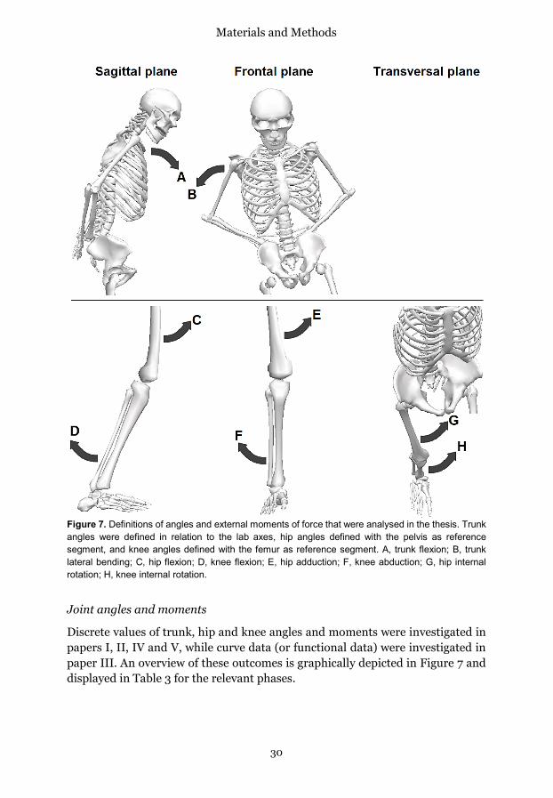
Materials and Methods
30
Figure 7. Definitions of angles and external moments of force that were analysed in the thesis. Trunk angles were defined in relation to the lab axes, hip angles defined with the pelvis as reference segment, and knee angles defined with the femur as reference segment. A, trunk flexion; B, trunk lateral bending; C, hip flexion; D, knee flexion; E, hip adduction; F, knee abduction; G, hip internal rotation; H, knee internal rotation.
Joint angles and moments
Discrete values of trunk, hip and knee angles and moments were investigated in papers I, II, IV and V, while curve data (or functional data) were investigated in paper III. An overview of these outcomes is graphically depicted in Figure 7 and displayed in Table 3 for the relevant phases.
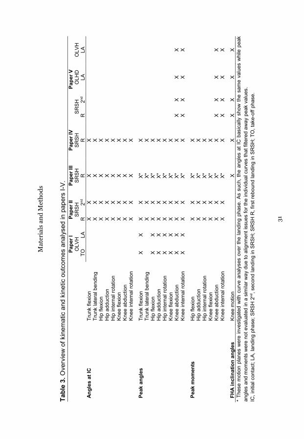
Mat
eria
ls a
nd M
etho
ds
31
Tabl
e 3.
Ove
rvie
w o
f kin
emat
ic a
nd k
inet
ic o
utco
mes
ana
lyse
d in
pap
ers
I-V.
Pape
r I
Pape
r II
Pape
r III
Pape
r IV
Pape
r V
OLV
H
SRSH
SR
SH
SRSH
SR
SH
OLH
D
OLV
H
TO
LA
R
2nd
R
R
R
2nd
LA
LA
Angl
es a
t IC
Trun
k fle
xion
X
X X
X
Trun
k la
tera
l ben
ding
X
X X
X
Hip
flex
ion
X X
X X
H
ip a
dduc
tion
X X
X X
H
ip in
tern
al ro
tatio
n
X
X X
X
Knee
flex
ion
X X
X X
Kn
ee a
bduc
tion
X X
X X
Kn
ee in
tern
al ro
tatio
n
X
X X
X
Peak
ang
les
Trun
k fle
xion
X
X X
X X*
X
Tr
unk
late
ral b
endi
ng
X X
X*
X
Hip
flex
ion
X X
X X
X*
X
Hip
add
uctio
n X
X X
X X*
X
H
ip in
tern
al ro
tatio
n X
X X
X X*
X
Kn
ee fl
exio
n X
X X
X X*
X
Kn
ee a
bduc
tion
X X
X X
X*
X X
X X
X
Knee
inte
rnal
rota
tion
X X
X X
X*
X X
X X
X
Peak
mom
ents
H
ip fl
exio
n
X
X X*
X
H
ip a
dduc
tion
X X
X*
X
Hip
inte
rnal
rota
tion
X X
X*
X
Knee
flex
ion
X X
X*
X
Knee
abd
uctio
n
X
X X*
X
X X
X X
Kn
ee in
tern
al ro
tatio
n
X
X X*
X
X X
X X
FHA
incl
inat
ion
angl
es
Knee
mot
ion
X X
X X
X X
* Th
ese
mot
ion
plan
es w
ere
inve
stig
ated
with
cur
ve a
naly
ses
over
the
land
ing
phas
e. A
s su
ch, t
he a
ngle
s at
IC b
asic
ally
sho
w th
e sa
me
valu
es w
hile
pea
k an
gles
and
mom
ents
wer
e no
t eva
luat
ed in
a s
imila
r way
due
to a
lignm
ent i
ssue
s fo
r the
indi
vidu
al c
urve
s th
at fi
ltere
d aw
ay p
eak
valu
es.
IC, i
nitia
l con
tact
; LA,
land
ing
phas
e; S
RSH
2nd
, sec
ond
land
ing
in S
RSH
; SR
SH R
, firs
t reb
ound
land
ing
in S
RSH
; TO
, tak
e-of
f pha
se.

Materials and Methods
32
Figure 8. A graphic depiction of time to stabilization in one direction (either anterior-posterior or medial-lateral). The thin black line depicts the rectified ground reaction force velocity signal, the grey bold line is the filtered signal, and the horizontal grey dashed line is the cut-off value of 2.5 SD extracted from one-leg standing balance. The time to stabilization is the time from initial contact to the time where the filtered signal reaches and remains below the threshold for at least 0.5 s. GRFv, ground reaction force velocity; SD, standard deviation; TTS, time to stabilization.
Time to stabilization, TTS In paper II, calculation of TTS 235, 321 was performed for the SRSH 2nd Landing by filtering the ground reaction force velocity using a second-order, recursive low-pass Butterworth digital filter with a cut-off frequency of 40 Hz using Visual3D software. It was calculated in anterior-posterior and medial-lateral directions by differentiating their excursion signals from peak ground reaction force, then rectified and filtered using a moving average window of 0.25 s.
The TTS reflects the time taken for the ground reaction force velocity in each direction to reach and remain below a stability threshold for at least 0.5 s (Figure 8). A resultant value was also calculated by taking the square root of the sum of TTS in anterior-posterior direction squared and medial-lateral direction squared.95 The stability threshold was calculated from a one-leg standing balance test for 10 s (with a 5 s pre-stabilisation period) from three trials for 10 asymptomatic persons. The overall standard deviation (SD) from the ground reaction force velocity was averaged across trials and participants, with the threshold set at 2.5 SD in line with previous work.235, 321

Materials and Methods
33
Surface electromyography, EMG The EMG raw data were filtered with a low pass filter of 500 Hz, a high pass filter of 20 Hz and with a root mean square moving window of 20 ms (SENIAM guidelines) using Visual3D software. The peak EMG value was then extracted for each muscle and trial during the landing phase to calculate an average peak value used for normalization. This method consider the force-velocity and length-tension relationships in the muscles, often with higher EMG levels than in maximal isometric contractions,14 and has been used previously.25, 220 The maximal trial-to-trial values provided high ICCs of 0.92-0.99 for all muscles in both groups using ICC(3,5) (model: two-way mixed, form: average measures, type: consistency). Similar to previous research,25 the average activation during a period of 50 ms prior to initial contact and the average activation during the landing phase, were investigated. Outcomes were the average activation of BF, ST, VM, VL and a co-contraction ratio. The co-contraction ratio was calculated using the equation EMGlower/EMGhigher, where EMGlower is the level of activity in the less active of flexors or extensors.25
Functional tests Functional performances of OLHD, OLVH and SRSH were evaluated. Hop length in OLHD was calculated from the displacement of a marker on the foot between starting position to landing, while hop height in OLVH was calculated from the displacement of the pelvis center of mass between standing position to peak height. The single highest peak value for length and for height from trials deemed successful were used in the analyses of functional performances. For the Rebound phase in SRSH, contact time was considered a performance measure and was defined from initial contact to next take-off (force signal < 20 N). Dynamometer raw data were filtered with a moving average of 60 ms using Visual 3D software. The single highest peak value from the three trials completed were extracted and normalized to body mass and multiplied with the dynamometer lever arm. These peak values in Nm/kg for knee extensors and flexors were used in analyses.
Statistical analyses
For the biomechanical analyses, outcome variables for the ACL injured leg was compared to the non-dominant leg for asymptomatic persons in papers I, II, IV and V, while the dominant leg was used to compare ATH and non-ATH in paper III. The successful trial with maximal OLVH height for each participant were used for analyses in paper I, while the first five successful trials for SRSH (out of the ten performed) were used to calculate individual means for analyses in papers II, III and IV. The use of five trials is considered well sufficient for sports specific side-to-side tasks for reliability evaluations.193 In paper V, all successful trials for OLHD, OLVH and SRSH were used to calculate individual mean values for analyses since the number of trials varied between tests.

Materials and Methods
34
Analyses of discrete data
Body anthropometrics and self-reported questionnaires (papers I-V)
For the long-term follow-up (paper I), body anthropometrics were analysed between groups with ANOVAs and questionnaires with Kruskal-Wallis H tests. For papers II-V, body anthropometrics and questionnaires were mainly presented as background information.
Dynamic knee robustness (papers III, IV and V) and EMG outcomes (paper IV)
The mean values for FHA inclination angles and their time occurrences from initial contact and the mean motion curves of the first five successful trials for each person were calculated. The successive FHAs were numbered FHA-1, FHA-2, FHA-3, etc., each representing a knee helical rotation interval of 10˚. Since each trial may result in a different number of FHAs (depending on range of knee motion), at least three of the five trials had to generate the same specific FHA (e.g., FHA-2) to be included in the analyses. Thus, FHAs provided by two or less trials were not considered representative for that person’s knee movement and were excluded from analyses. FHA inclination angles and EMG outcomes (average amplitudes, cocontraction ratios) were compared between groups with independent t-tests since normally distributed (Shapiro-Wilk test).
Joint angles and moments (papers I, IV and V)
Multivariate analyses of variance (MANOVAs) were used to compare blocks of discrete kinematic and kinetic variables between ACL injured persons and matched controls, and if significant, further investigated with direct discriminant analysis with correlations ≥ 0.32 interpreted (papers I and IV).38, 292 These blocks of variables (Table 3) were: angles at initial contact for trunk, hip and knee variables (paper IV), peak angles of trunk, hip and knee variables (papers I and IV), and peak moments of hip and knee variables (paper IV). Since body mass index differed between both ACLD20 and ACLR20 to CTRL20, relations between body mass index and kinematic variables in the sagittal plane were investigated but displayed no significant correlations (Pearson). The effect of body mass index was also investigated as a covariate for the same variables, although with no differences in results, thus removed as a covariate. Main effects of asymmetry within groups for these blocks of variables were investigated with repeated-measures MANOVAs and, if significant, followed by paired t-tests (papers I and IV). Assumptions of absence of multivariate outliers and multicollinearity, linearity, and homogeneity of variance-covariance were met.

Materials and Methods
35
Regarding comparisons between landings for ACLR, ATH and non-ATH, repeated-measures ANOVAs with group as between-subject factor were used to investigate the effect of type of landing, group belonging and their interaction for the chosen outcome measures (paper V). Bonferroni post-hoc tests (to correct for multiple comparisons) were applied to further elucidate significant main effects. Pearson correlations r were also used to investigate the relationships of the outcome variables between the landings (paper V). Partial eta2 as effect sizes (ES, 0.01 = small, 0.1 = medium, 0.25 = large)310 were presented for main effects of MANOVAs (papers I and IV) and repeated-measures ANOVAs (paper V).
Functional performances (papers I, III and IV)
Hop performances of the OLVH (paper I) were investigated between legs within groups with paired t-tests, and between groups with one-way ANOVAs where Bonferroni post hoc tests were used if significant.
Functional performance outcomes (papers III and IV) were analysed with MANOVAs and followed with Bonferroni Post-Hoc tests (correction for multiple comparisons). Partial eta2 as effect sizes (ES, 0.01 = small, 0.1 = medium, 0.25 = large)310 were presented. Further, relations between hop and strength performances were investigated with r2-values to evaluate if these indicated different aspects of functional performances.
Reliability and agreement of joint angles and moments (paper II) and FHA inclination angles (papers III, IV and V)
Reliability were investigated using intraclass correlations (ICCs) for within-session analyses (ACLR and CTRL) using ICC(3,5) (model: two-way mixed, form: average measures, type: consistency) and in test-retest (CTRL) with ICC(3,1) (model: two-way mixed, form: single measures, type: consistency) using the average of the trials for each test session. The classification of Fleiss87 was adapted for interpretation (ICC < 0.4 = poor, ICC 0.4 – 0.75 = fair to good, ICC > 0.75 = excellent). Bland-Altman plots were used to screen for systematic bias, outliers, and heteroscedasticity for test-retest data. Agreement was evaluated with the within-person SD (denoted SW) calculated by taking the square root of the average variances on group level, to provide clinically relevant values for expected errors in an individual.31, 325 Minimal differences for each variable was also assessed in test-retest by constructing a 95% confidence interval for the SW that take both test sessions into account.
Considering that ACL injuries have been estimated to occur as fast as 30-50 ms after impact,151, 156 the initial FHA-1 inclination angle was considered most important and thus also evaluated for trial-to-trial reliability using ICC model(3,5) for the data analysed in papers III, IV and V.

Materials and Methods
36
Statistical software and level of significance
The Statistical Package for the Social Sciences (v.23, IBM SPSS Statistics, Armonk, New York, USA) were used for all analyses of discrete data, with a 5% level for statistical significance set a priori.
Analyses of functional data Joint angle and moment curves (paper III)
The kinematic and kinetic mean curves (representing the individuals) were analysed with a statistical method from the area of functional data analysis, that investigated the equality of the mean functions from the functional populations of ATH and non-ATH. The curves were aligned within the landing phase to account for individual differences in time between the aforementioned landing events. Prior to the analysis, the landing phase was discretized in 51 points. This adjustment enabled continuous data series to be compared between persons and groups using identical relative time points. The curves were analysed between ATH and non-ATH by applying a functional t-test, based on the interval-wise testing procedure.237 Such an approach enabled the identification of time-intervals where the groups differed. The unadjusted P-values correspond to a point-wise control of the probability of wrongly detecting a significant difference. The interval-wise testing-adjusted P-values ensured that the probability of wrongly rejecting any interval (i.e., false positive, or type I error) was below the chosen significance level, within each analysis.
Statistical software and level of significance
All computations and statistical analyses of functional data were conducted using R (v.3.2.0), with a 5% level for statistical significance set a priori.

Results
37
Results
The comparative results between groups (specific aims A, D and E handled in papers I, III and IV, respectively) and between legs within groups for asymmetry (specific aims A and E handled in papers I and IV, respectively) are compiled and presented with subheadings that relate to each theme of outcome measures. Results of dynamic knee robustness (FHA inclination angles) are presented separate from the cardan joint angles for clarity. The comparative results between landings (specific aim F handled in paper V) and results of reliability and agreement (specific aims B and C handled mainly in paper II but also to some extent for FHA-inclination angles for papers III, IV and V) are presented in separate headings. As such, this structure diverge from the specific aims previously described (e.g. reliability and agreement of the SRSH was evaluated before group comparisons were performed) although is considered more appropriate here.
General findings
General findings show significant differences in angles, moments and functional performances between ACL groups to controls, between athletes and non-athletes, and between legs (asymmetry) for ACL groups. Significant differences in knee-specific angles and moments were also displayed between the landings, and excellent reliability were shown for most joint-specific angles and moments during landings of the novel SRSH. Specific results are presented below.
Comparisons between groups (specific aims A, D and E)
Dynamic knee robustness (papers III and IV) ACLR and CTRL had a similar range of knee motion which provided an equal number of FHAs (3.2 ± 0.9 for ACLR and 3.5 ± 0.9 for CTRL) during the first landing of SRSH (n.s., P = 0.175). The number of participants that provided FHA-1 – FHA-5 occurrences (each consecutive 10° helical rotation interval) were 31, 31, 24, 10 and 2 respectively for ACLR (n = 32) and 32, 32, 28, 14 and 5 respectively for CTRL (n = 32). Thus, one person among ACLR had less than 10˚ of helical knee motion for four of the five trials (thus no FHA for analyses). Similar results were found among ATH and non-ATH during the first landing of the SRSH, where both groups displayed an equal range of knee motion thus the same amounts of FHAs (3.1 ± 1.0 for ATH and 3.5 ± 0.7 for non-ATH, n.s., P = 0.213). The number of participants that provided FHA-1 – FHA-5 occurrences were 19, 17, 16, 6, and 1 respectively for ATH and 20, 20, 18, 11 and 0 respectively for non-ATH.

Results
38
Figure 9A-D. Average FHA inclination angles (dynamic knee robustness) and their times shown in mean values with 95% confidence intervals. Similar FHA inclination angles and times were displayed between ACLR and CTRL in A and B, and between ATH and Non-ATH in C and D. Each FHA inclination angle represents a knee helical motion of ~10˚. ACLR, ACL reconstructed persons; ATH, elite athletes; CTRL, controls; FHA, finite helical axis; Non-ATH, non-athletes.
Further statistical analyses of group comparisons between both ACLR and CTRL and between ATH and non-ATH were therefore restricted for inclination angles of FHA-1 – FHA-3, since too few persons (in most cases less than half) displayed FHA-4 or FHA-5. However, no significant differences were found between ACLR and CTRL (P-values ≥ 0.321, Figure 9A-B), or between ATH and non-ATH (P-values ≥ 0.150, Figure 9C-D). Further, these FHA inclination angles occurred at similar times after initial contact for both groups.

Results
39
Joint angles (papers I, III and IV) For the long-term follow up, significant main effects (shown via MANOVA) of group differences for peak trunk, hip and knee angles were found for the OLVH during both take-off (P < 0.001, ES = 0.21 [medium]) and landing (P < 0.001, ES = 0.25 [large]). During take-off, follow-up analyses (discriminant analysis) showed that ACLD20 differed to CTRL20, where ACLD20 displayed a combination of less knee flexion and external knee rotation (instead of internal rotation that was evaluated). ACLR20 was also differentiated to both ACLD20 and CTRL20, and displayed a combination of greater trunk and hip flexion, greater hip internal rotation, and less knee abduction than both of the latter groups. During landing, discriminant analysis separated between ACLD20 to both ACLR20 and CTRL20, where they demonstrated a combination of less hip and knee flexion, greater hip adduction, and greater external (instead of internal) knee rotation than both ACLR20 and CTRL20. Further, ACLR20 was also distinguished to both ACLD20 and CTRL20, where they displayed a combination of greater trunk flexion and hip internal rotation than the latter two groups (Table 4).
Significant main effects between groups were also found when comparing peak trunk, hip and knee angles between the non-injured leg of ACL groups to the dominant leg of CTRL20 during take-off (P = 0.004, ES = 0.16 [medium]) but not during landing (n.s., P = 0.301). Follow-up analysis discriminated ACLD20 from both ACLR20 and CTRL20, where ACLD20 displayed less hip and knee flexion, less hip internal rotation, and greater knee abduction than the other two groups (Table 4).
Regarding comparison for the younger and more recently injured ACLR compared to the matched CTRL during the first landing of the SRSH, significant main effects (MANOVA) were found for trunk, hip and knee angles at initial contact (P = 0.005, ES = 0.32 [large]) and for peak angles (P = 0.002, ES = 0.35 [large]). Follow-up analyses (discriminant analysis) revealed that ACLR displayed a combination of greater hip and knee flexion angles at initial contact than CTRL (5.6˚ and 7.2˚ greater, respectively). For peak angles, ACLR demonstrated a combination of greater knee flexion (3.6˚ greater) and less knee internal rotation (actually 2.4˚ more external rotation) than CTRL (Table 5).

Results
40
Table 4. Peak angles during take-off and landing presented in mean (SD) for ACLR20 (n = 32), ACLD20 (n = 34) and CTRL20 (n = 33).
ACLD20, ACL deficient persons; ACLR20, ACL reconstructed persons; CTRL20, controls; I/ND leg, injured leg for ACL20 groups and non-dominant leg for CTRL20; NI/D leg, non-injured leg for ACL20 groups and dominant leg for CTRL20. Positive values for flexion, adduction, internal (int.) rotation and negative values for extension, abduction and external rotation. a Differentiated from CTRL20 ND-leg through discriminant analysis. b Differentiated from ACLD20 I-leg through discriminant analysis. c Significantly different to contralateral leg within groups at 0.05 level, only found for ACL20 groups.
Take-off Landing I/ND leg NI/D leg I/ND leg NI/D leg Trunk flexion (+) (˚) ACLR20 44.2 (12.7)abc 40.6 (11.2) 30.0 (8.9)abc 26.0 (9.7) ACLD20 37.7 (11.0)c 35.2 (11.5) 23.8 (7.7) 23.0 (9.4) CTRL20 39.1 (9.6) 36.6 (9.4) 23.6 (9.9) 24.8 (9.7)
Hip flexion (+) (˚) ACLR20 73.3 (15.4)ab 71.7 (12.4)b 52.4 (12.0)b 50.1 (11.9) ACLD20 65.4 (16.7) 63.1 (15.5)a 43.0 (11.5)a 44.8 (13.1) CTRL20 70.7 (12.9) 72.3 (12.5) 52.0 (10.7) 49.8 (10.9)
Hip adduction (+) (˚) ACLR20 9.8 (6.6) 10.6 (6.8) 3.3 (7.8)b 4.7 (6.9) ACLD20 12.3 (7.5) 10.2 (6.9) 7.8 (7.6)a 5.8 (8.7) CTRL20 11.0 (5.6) 11.0 (6.9) 5.5 (5.6) 5.6 (8.0)
Hip int. rotation (+) (˚) ACLR20 10.6 (8.2)abc 6.7 (6.4)b 10.6 (8.1)ab 7.6 (7.0) ACLD20 6.6 (9.3)c 2.9 (8.4)a 6.5 (7.8) 4.0 (8.5) CTRL20 7.6 (6.3) 6.1 (7.1) 7.5 (7.5) 6.8 (9.1)
Knee flexion (+) (˚) ACLR20 71.3 (10.5)bc 74.3 (8.7)b 57.0 (10.6)b 58.1 (9.2) ACLD20 65.3 (9.7)a 66.6 (7.9)a 50.0 (9.3)ac 54.7 (8.9) CTRL20 73.6 (8.8) 74.1 (8.1) 59.9 (9.1) 58.5 (9.0)
Knee abduction (-) (˚) ACLR20 -1.2 (5.1)ab -2.1 (5.1)b 0.0 (4.7) -1.7 (5.1) ACLD20 -4.1 (5.5) -5.7 (4.1)a -3.4 (6.3) -4.8 (5.2) CTRL20 -2.3 (5.0) -2.0 (4.9) -1.6 (6.2) -0.8 (5.1)
Knee int. rotation (+) (˚) ACLR20 -0.1 (7.0)b 1.4 (6.8) 0.4 (6.8)b 2.8 (6.7) ACLD20 -1.5 (5.5)ac 2.0 (7.0) -1.6 (6.7)ac 3.3 (7.8) CTRL20 4.4 (7.3) 1.3 (5.0) 5.1 (7.0) 2.8 (5.3)

Results
41
Table 5. Joint angles presented in mean (SD) for ACLR and CTRL. ACLR (n = 32) CTRL (n = 32) I-leg NI-leg ND-leg D-leg
Angles at IC (˚) Trunk flexion (+) 18.4 (4.3)b 17.1 (4.8) 16.4 (6.2) 16.7 (5.5) Trunk lat. bending (-) -7.6 (4.0) -8.8 (3.0) -7.1 (2.5) -7.0 (3.1) Hip flexion (+) 40.3 (7.8)ab 37.8 (9.1) 34.7 (7.6) 34.2 (7.3) Hip adduction (+) -11.4 (4.8) -12.4 (4.9) -11.1 (5.1) -11.0 (3.5) Hip int. rotation (+) 4.2 (5.1) 3.2 (5.4) 3.8 (8.8) 5.8 (6.9) Knee flexion (+) 37.2 (6.2)a 36.5 (7.2) 30.0 (6.6) 30.1 (6.4) Knee abduction (-) -2.6 (5.0) -2.6 (5.3) -2.3 (5.7) -1.6 (5.0) Knee int. rotation (+) -10.7 (5.0) -9.6 (5.0) -9.8 (6.8) -11.6 (5.8)
Peak Angles (˚) Trunk flexion (+) 22.2 (5.3)b 20.8 (5.7) 21.1 (8.9) 21.7 (7.7) Trunk lat. bending (-) -9.2 (4.4) -9.7 (3.3) -7.9 (2.5) -7.8 (3.4) Hip flexion (+) 49.1 (9.0)b 45.9 (9.1) 45.4 (9.0) 45.3 (9.0) Hip adduction (+) -3.0 (7.0) -5.0 (6.8) -0.7 (6.8) -0.2 (6.2) Hip int. rotation (+) 10.7 (5.3) 9.4 (5.7) 10.8 (7.6) 12.7 (5.5) Knee flexion (+) 63.0 (6.8)a 62.9 (6.8) 59.4 (7.2) 59.9 (7.7) Knee abduction (-) -3.2 (5.0) -2.9 (5.1) -3.0 (5.6) -1.9 (5.0) Knee int. rotation (+) -3.2 (4.3)a -1.8 (5.3) -0.8 (5.0) -2.9 (4.1)
ACLR, ACL reconstructed persons; CTRL, controls; D-leg, dominant leg; IC, initial contact; I-leg, injured leg; ND-leg, non-dominant leg; NI-leg, non-injured leg; SD, standard deviation. Positive values for flexion, adduction, internal (int.) rotation and negative values for extension, lateral (lat.) bending, abduction and external rotation. a Differentiated from CTRL ND-leg through discriminant analysis. b Significantly different to contralateral leg within groups at 0.05 level, only found for ACLR.
Comparisons between ATH and non-ATH for trunk, hip and knee angle curves revealed that only the hip flexion angle (among the angle curves) was significantly different between the groups. ATH displayed greater flexion angles than non-ATH (37-38˚ versus 30-31˚) between 9-29% of the landing phase. The unadjusted P-value also identified a significant difference immediately at initial contact, which would have been found if analysing that specific value without applying the interval-wise testing procedure (Figure 10).
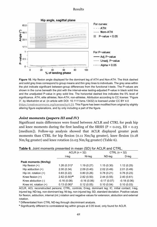
Results
42
Figure 10. Hip flexion angle displayed for the dominant leg of ATH and Non-ATH. The thick dashed and solid grey lines correspond to group means and thin grey lines to individuals. The grey area within the plot indicate significant between-group differences from the functional t-tests. The P-values are shown in the curve beneath the plot with the interval-wise testing-adjusted P-value in black solid line and the unadjusted P-value in grey solid line. The horizontal dashed line indicates the 5% level of significance. ATH, elite athletes; Non-ATH, non-athletes. Attribution according to CC license: “Figure 3”, by Markström et al. (in article with DOI: 10.1111/sms.13432) is licensed under CC BY 4.0 (https://creativecommons.org/licenses/by/4.0/). This Figure has been modified from original by slightly altering figure explanations, and by only including a part of the figure.
Joint moments (papers III and IV) Significant main differences were found between ACLR and CTRL for peak hip and knee moments during the first landing of the SRSH (P = 0.015, ES = 0.23 [medium]). Follow-up analysis showed that ACLR displayed greater peak moments than CTRL for hip flexion (0.11 Nm/kg greater), knee flexion (0.18 Nm/kg greater) and knee rotation (0.03 Nm/kg greater) (Table 6).
Table 6. Joint moments presented in mean (SD) for ACLR and CTRL. ACLR (n = 32) CTRL (n = 32) I-leg NI-leg ND-leg D-leg
Peak moments (Nm/kg) Hip flexion (+) 1.26 (0.31)a 1.19 (0.27) 1.15 (0.30) 1.12 (0.25) Hip adduction (+) 2.00 (0.34) 2.09 (0.40) 2.02 (0.49) 2.01 (0.49) Hip int. rotation (+) 0.83 (0.22) 0.80 (0.26) 0.78 (0.21) 0.76 (0.23) Knee flexion (+) 2.62 (0.50)ab 2.82 (0.50) 2.44 (0.55) 2.40 (0.61) Knee abduction (-) -0.16 (0.08) -0.16 (0.06) -0.17 (0.07) -0.18 (0.06) Knee int. rotation (+) 0.13 (0.06)a 0.12 (0.05) 0.10 (0.04) 0.10 (0.03)
ACLR, ACL reconstructed persons; CTRL, controls; D-leg, dominant leg; IC, initial contact; I-leg, injured leg; ND-leg, non-dominant leg; NI-leg, non-injured leg; SD, standard deviation. Positive values for flexion, adduction, internal (int.) rotation and negative values for extension, abduction and external rotation. a Differentiated from CTRL ND-leg through discriminant analysis. b Significantly different to contralateral leg within groups at 0.05 level, only found for ACLR.

Results
43
Figure 11. Hip adduction (+)/abduction (-) moment shown in top left, hip internal (+)/external (-) rotation moment in bottom left and knee flexion (+)/extension (-) moment in bottom right for the dominant leg of ATH and Non-ATH. The thick dashed and solid grey lines correspond to group means and the thin grey lines to individuals. The grey areas within the plots indicate significant between-group differences from the functional t-tests. These P-values are shown as curves beneath each plot with the interval-wise testing-adjusted P-value in black solid line and the unadjusted P-value in grey solid line. The horizontal dashed line indicates the 5% level of significance. ATH, elite athletes; Non-ATH, non-athletes. Attribution according to CC license: “Figure 3”, by Markström et al. (in article with DOI: 10.1111/sms.13432) is licensed under CC BY 4.0 (https://creativecommons.org/licenses/by/4.0/). This Figure has been modified from original by slightly altering figure explanations, and by only including a part of the figure.
Comparisons between ATH and non-ATH revealed significantly different hip and knee moment curves during the first landing of SRSH. ATH had greater moments of hip adduction between 59-99%, hip internal rotation between 83-89%, and knee flexion between 79-93% of the landing phase. These moments were 1.2-1.4 times greater than those of non-ATH throughout the identified time intervals of significant differences (Figure 11).
Average EMG amplitudes and cocontraction ratio (paper IV) No significant differences were shown between ACLR and CTRL for average EMG amplitudes (relative average peak values) for BF, ST, VM and VL during the 50 ms period prior to initial contact or during the first landing phase of the SRSH.

Results
44
Figure 12. Average EMG amplitudes presented in means with 95% confidence intervals. Groups displayed similar amplitudes both during the 50 ms period prior to initial contact at landing (pre) and during the landing phase (post). ACLR, ACL reconstructed persons; BF, biceps femoris; CC ratio, cocontraction ratio; CTRL, controls; ST, semitendinosus; VL, vastus lateralis; VM, vastus medialis.
There was however a tendency to a higher average amplitude for BF during landing for ACLR compared to CTRL (29% vs 23%) nearing the statistical significance threshold (P = 0.075) (Figure 12). For the 50 ms period, the average EMG amplitudes for both ACLR and CTRL were higher for knee flexors (33-39%) than for knee extensors (16-19%). This pattern changed during the landing phase, where lower average EMG amplitudes for knee flexors were displayed for both ACLR and CTRL (23-29%) than for knee extensors (36-39%).
Knee laxity and functional performances (papers I, III, IV) In the long-term follow up, a significant difference for hop height was found for the injured legs of ACLD20 and ACLR20 compared to the non-dominant legs of CTRL20 (P = 0.007), but not for the non-injured compared to the dominant legs (n.s., P = 0.195). Follow-up analyses showed that the maximal hop height (on group level) for ACLD20 (0.17 ± 0.03 m) were lower than for both ACLR20 (0.20 ± 0.04 m, p = 0.037) and CTRL20 (0.20 ± 0.04 m, p = 0.010), while the latter two groups did not differ (n.s., P = 1.000).
Comparisons between ACLR and CTRL revealed a significant main effect (MANOVA) for laxity and knee function outcomes (P < 0.001, ES = 0.56 [large]). However, this finding was only attributed to a significantly higher knee laxity for ACLR (9.6 vs 6.3 mm) (Table 7). Nevertheless, these functional performances indicate different aspects of function since low to moderate relations were found between strength and hop performances for both legs among ACLR (r2-values ≤ 0.26) and CTRL (r2-values ≤ 0.36).

Results
45
Table 7. Knee laxity and functional performances presented in mean (SD) for ACLR and CTRL.
ACLR CTRL I-leg NI-leg ND-leg D-leg
Knee laxity 30 Ib (mm) 9.6 (2.0)ab 7.0 (2.1) 6.3 (2.2) 6.4 (2.2) Maximal OLHD (m) 1.26 (0.18) 1.28 (0.19) 1.26 (0.23) 1.29 (0.22) Maximal OLVH (m) 0.22 (0.04)b 0.24 (0.03) 0.24 (0.04) 0.25 (0.04) Hops SRSH (No. out of 10) 8.3 (1.2) 8.1 (1.6) 8.5 (1.7) 8.9 (1.4) Contact time in SRSH (s) 0.45 (0.13)b 0.43 (0.13) 0.55 (0.26) 0.54 (0.25) Peak vGRF landing SRSH 2.23 (0.21)b 2.33 (0.24) 2.30 (0.34) 2.27 (0.28) Peak extensor torque (Nm/kg) 2.57 (0.52)b 2.67 (0.49) 2.44 (0.48) 2.54 (0.54) Peak flexor torque (Nm/kg) 1.05 (0.23)b 1.19 (0.24) 1.10 (0.23) 1.13 (0.22)
ACLR, ACL reconstructed persons; CTRL, controls; D-leg, dominant leg; I-leg, injured leg; ND-leg, non-dominant leg; NI-leg, non-injured leg; OLHD, one-leg hop for distance; OLVH, one-leg vertical hop; SRSH, standardized rebound side hop; vGRF, vertical ground reaction force. a Significantly different to CTRL ND-leg at 0.05 level. b Significantly different to contralateral leg within groups at 0.05 level, only found for ACLR.
Comparisons between ATH and non-ATH also revealed a significant main effect for laxity and knee function outcomes (P-value = 0.007, ES = 0.45 [large]). ATH displayed significantly less laxity, longer maximal OLHD, greater number of successful hops in SRSH, shorter contact time in the first landing (Rebound) in SRSH and greater knee extensor strength than non-ATH (Table 8). ATH also displayed tendencies to greater maximal OLVH and knee flexor torque (9-11% larger) nearing the statistical significance threshold. These functional performances indicate different aspects of function since low to moderate relations were found between strength and hop performances among ATH (r2-values ≤ 0.39) and non-ATH (r2-values ≤ 0.29).
Table 8. Knee laxity and functional performances presented in mean (SD) for female athletes and non-athletes (dominant leg).
ATH, elite athletes; ES, effect size; Non-ATH, non-athletes; OLHD, one-leg hop for distance; OLVH, one-leg vertical hop; SD, standard deviation; SRSH, standardized rebound side hop. Bold P-values indicate significant differences (main effect and post-hoc) at 0.05 level.
ATH (n = 19) Non-ATH (n = 20) P-value (ES) Main effect Post-hoc 0.007 (0.45) Knee laxity 30 Ib (mm) 5.6 (2.1) 7.3 (1.6) 0.009 (0.18) Maximal OLHD (m) 1.34 (0.17) 1.13 (0.18) 0.001 (0.28) Maximal OLVH (m) 0.24 (0.03) 0.22 (0.03) 0.077 (0.08) Hops SRSH (No. out of 10) 9.1 (1.1) 7.9 (1.8) 0.037 (0.12) Contact time in SRSH (s) 0.41 (0.14) 0.70 (0.32) 0.001 (0.26) Peak extensor torque (Nm/kg) 2.71 (0.44) 2.23 (0.57) 0.007 (0.19) Peak flexor torque (Nm/kg) 1.20 (0.22) 1.08 (0.22) 0.097 (0.07)

Results
46
Comparisons between legs within groups (specific aims A and E)
Dynamic knee robustness (paper IV) Regarding asymmetry in FHA-inclination angles for FHA-1 – FHA-3, no significant differences between legs within groups were found among ACLR or among CTRL during the first landing in SRSH (P-values ≥ 0.146).
Joint angles (papers I and IV) For the long-term follow-up, significant main effects (MANOVA) of asymmetry for peak trunk, hip and knee angles were found for both ACLD20 and ACLR20 when performing the OLVH during both Take-off (ACLR20: P < 0.001, ES = 0.63 [large]; ACLD20: P = 0.004, ES = 0.51 [large]) and Landing (ACLR20: P = 0.026, ES = 0.44; ACLD20: P = 0.020, ES = 0.43 [large]). CTRL did not display such asymmetries in any of the phases (n.s., P-values ≥ 0.559). During Take-off, the injured leg (relative the non-injured) for both ACLD20 and ACLR20 had greater trunk flexion and hip internal rotation, while only ACLR20 displayed less knee flexion and only ACLD20 showed an externally (even though peak internal rotation angles was sought) rotated knee. During Landing, the injured leg (relative the non-injured) among ACLR20 had greater trunk flexion while the injured leg among ACLD20 had less knee flexion and an externally instead of an internally rotated knee (Table 4).
Similarly, the younger ACLR displayed significant main effects of asymmetries in trunk, hip and knee angles during the first landing of the SRSH for both angles at initial contact (P = 0.024, ES = 0.48 [large]) and peak angles (P = 0.015, ES = 0.51 [large]). The injured leg of ACLR displayed (relative the non-injured leg) a combination of greater angles of trunk and hip flexion both at initial contact (1.3˚ and 2.5˚, respectively) and during the landing phase (1.4˚ and 3.2˚, respectively) (Table 5). CTRL had no such asymmetries for angles (n.s., P-values ≥ 0.281).
Joint moments (paper IV) A significant main effect of asymmetry was also found among ACLR (not for CTRL, n.s., P = 0.896) for peak hip and knee moments (P = 0.035, ES = 0.39 [large]). However, this main effect was only attributed to less peak knee flexion moment (-0.20 Nm/kg) for the injured leg relative the non-injured leg, during the landing phase (Table 6).
Knee laxity and functional performances (papers I and IV) In the long-term follow up, both ACLD20 and ACLR20 had significantly greater laxity for the injured compared to the non-injured leg (p < 0.001 for both groups).

Results
47
This difference was however greater among ACLD20 (4.9 mm) than for ACLR20 (2.0 mm) (P < 0.001). Further, a lower hop height was displayed for the injured leg compared to the contralateral non-injured leg for both ACLR20 (LSI 94 ± 13%, p = 0.001) and ACLD20 (LSI 91 ± 14%, p = 0.006).
The younger ACLR also demonstrated a significant main effect of asymmetry for laxity and knee function outcomes (main effect: P < 0.001, ES = 0.81 [large]) while no such findings were shown for CTRL (n.s., P = 0.258). The injured leg for ACLR had (relative their non-injured leg) significantly greater knee laxity (37% greater), lower OLVH (8% less), longer contact time in landing (5% longer), lower peak vertical ground reaction force (4% less), and lower peak knee extensor (4% deficit) and flexor strength (12% deficit) (Table 7). No differences were found for OLHD and number of successful hops of SRSH.
Comparisons between hop landings (specific aim F)
Outcomes compared between landings (paper V) The three groups of females (ACLR, ATH, non-ATH) that performed the OLHD, OLVH and SRSH landings had similar range of knee motion which provided an equal amount of FHAs for any of the landings (n.s., P-values ≤ 0.405). The number of persons (n = 60 in total) that provided FHA-1 – FHA-5 occurrences for all landings were 60, 58, 55, 29 and 4 persons, respectively. Further analyses of FHA inclination angles were therefore limited to FHA-1 – FHA-3 since too few persons (less than half) displayed FHA-4 and FHA-5.
Significant differences between landings were found for inclination angles of FHA-1 (P < 0.001, ES = 0.67 [large]), FHA-2 (P = 0.017, ES = 0.17 [medium]), and FHA-3 (P < 0.001, ES = 0.39 [large]), and for peak abduction angle (P < 0.001, ES = 0.78 [large]), peak abduction moment (P < 0.001, ES = 0.82 [large]), and peak internal rotation moment (P < 0.001, ES = 0.89 [large]). Only peak internal rotation angle had no significant differences between landings (P-values ≤ 0.366) with angles on group levels in slight external rotation. The SRSH Rebound landing induced the greatest knee FHA-1 inclination angle, abduction angle, abduction moment, and internal rotation moment compared to all other landings (Figure 13). No significant effect of group belonging was found for any outcome variable between the landings (n.s., P-values = 0.074-0.949). One significant interaction between landing and group was however found for peak internal rotation moment (P < 0.001, ES = 0.23 [medium]). Post hoc analyses revealed that ACLR females had greater moment than non-ATH in SRSH Rebound, although less moment than both ATH and non-ATH in SRSH 2nd landing.

Res
ults
48
Figu
re 1
3. P
lots
of s
igni
fican
t mai
n ef
fect
s of
land
ing.
Kne
e ab
duct
ion
angl
e an
d m
omen
t dis
play
ed a
s ne
gativ
e an
d in
tern
al ro
tatio
n m
omen
t pos
itive
. The
hor
izon
tal
line
show
s th
e va
lue
of 0
. AC
LR, A
CL
reco
nstru
cted
per
sons
; ATH
, ath
lete
s; N
on-A
TH, n
on-a
thle
tes;
OLH
D, o
ne-le
g ho
p fo
r dis
tanc
e; O
LVH
, one
-leg
verti
cal h
op;
SRSH
, sta
ndar
dize
d re
boun
d si
de h
op, c
ompr
ised
bot
h a
late
ral r
ebou
nd (S
RSH
R) a
nd m
edia
l (SR
SH 2
ND) l
andi
ng.
* ind
icat
e si
gnifi
cant
diff
eren
ces
at 0
.05
leve
l, **
indi
cate
sig
nific
ant d
iffer
ence
s at
0.0
1 le
vel.

Results
49
Outcomes correlated between landings (paper V) Regarding correlations of each outcome variable between the landings (groups combined), significant correlations were displayed between all landings for peak knee abduction angle (r ≥ 0.81, P < 0.001) and peak internal rotation angle (r ≥ 0.79, P < 0.001). Significant although lower correlations were also shown for FHA-1 inclination angles (r = 0.29-0.44, P < 0.001-0.023), while few or no significant correlations were found for the remaining outcomes (all r = 0.03-0.33, P = 0.010-0.826). See paper V for further details of specific correlations.
Reliability and agreement of the novel SRSH test (specific aims B and C)
Description of angles and moments during SRSH landings (paper II) The general landing patterns for trunk, hip and knee angles displayed by both ACLR and CTRL in SRSH Rebound (the first landing) were: increased trunk, hip and knee flexion, increased trunk lateral bending, a constant hip abduction that neared a neutral position, a relatively constant hip internal rotation, an initial knee abduction that changed to adduction, and a constant knee external rotation that neared a neutral position. The patterns for moments were analogous to those for the angles, although with the exception for an initial knee internal rotation moment that went into external moments. Similar landing patterns were displayed in SRSH 2nd Landing, although with both the hip and knee more constantly in adduction and with lower moments. Results of reliability analyses for only the injured leg among ACLR and for the dominant leg among CTRL are presented since no clear differences of ICCs and SW were shown between the legs within groups.
Within-session results for ACLR and CTRL (paper II) In SRSH Rebound, excellent within-session reliability coefficients (ICCs > 0.75) were shown for both groups for all trunk, hip and knee angles at initial contact, all peak trunk, hip and knee angles and all peak hip and knee moments, except for knee abduction for CTRL where an ICC of 0.73 was found (ICC for ACLR = 0.88). The SW for angles were 1.9-5.2˚ and for moments 0.03-0.29 Nm/kg. In general, lower ICCs and higher SW were shown in SRSH 2nd Landing than in SRSH Rebound. However, excellent ICCs were shown for all angles at initial contact and peak angles with the exception for trunk lateral bending (ICC for angle at initial contact of 0.74 for ACLR and 0.67 for CTRL, ICC for peak angle of 0.69 for CTRL). For peak moments in both groups, excellent ICCs were found for hip flexion, hip internal rotation, knee flexion and knee abduction, while hip adduction had good ICCs (0.68-0.74) and knee internal rotation had poor ICCs (0.23-0.43).

Results
50
Table 9. Within-session reliability (ICC) and agreement (SW) for angles and moments (test session 1, ACLR injured leg and CTRL dominant leg).
Within-session ACLR (n = 30) CTRL (n = 30) SRSH ICC SW* ICC SW* Rebound Angles at IC 0.94-0.97 1.9-3.2 0.85-0.97 2.0-3.8 Peak Angles 0.91-0.98 1.3-5.2 0.85-0.97 2.0-4.7 Peak moments 0.88-0.96 0.05-0.28 0.73-0.95 0.03-0.29
2nd Landing Angles at IC 0.74-0.96 2.4-4.4 0.67-0.95 2.9-5.1 Peak Angles 0.78-0.96 1.9-4.9 0.69-0.95 2.3-4.6 Peak moments 0.43-0.94 0.08-0.31 0.23-0.90 0.07-0.34
ACLR, anterior cruciate ligament reconstructed persons; CTRL, controls; IC, initial contact; ICC, intra-class correlation; SRSH, standardized rebound side hop; SW, average within-person standard deviation. * Units of measurement for angles in degrees, and for moments in Nm/kg.
A general overview of within-session ICCs and SW for angles and moments are presented in Table 9, while more specific results for each variable can be found in paper II.
Regarding TTS, excellent to good reliability were shown among ACLR in all directions (ICCs of 0.71-0.81) while fair reliability were shown for CTRL (ICCs of 0.51-0.58). High relative (to mean values) SW were shown for both groups with values ≥ 37%.
Test-retest results for CTRL (paper II) In SRSH Rebound, test-retest reliability was excellent to fair for all trunk, hip and knee angles and hip and knee moments with ICCs of 0.51-0.91, and with SW of 1.6-4.5˚ (minimal differences: 4.5-12.6˚) for angles and 0.03-0.25 Nm/kg (minimal differences: 0.09-0.68 Nm/kg) for moments. Similar to within-session results, generally lower ICCs and SW were shown in SRSH 2nd Landing, where trunk lateral bending and knee internal rotation had poor ICCs of 0.36 and 0.07, respectively. Generally poorer reliability was found for TTS with ICCs of 0.34-0.46 for all directions, with high relative minimal differences ≥ 57% of mean values for both test sessions. A general overview of test-retest ICCs and SW for angles and moments are presented in Table 10, while more specific results for each variable can be found in paper II.

Results
51
Table 10. Test-retest reliability (ICC) and agreement (SW) for angles and moments for controls (dominant leg).
ACLR, anterior cruciate ligament reconstructed persons; CTRL, controls; IC, initial contact; ICC, intra-class correlation; MD, minimal differences; SRSH, standardized rebound side hop; SW, average within-person standard deviation. * Units of measurement for angles in degrees, and for moments in Nm/kg.
Within-session results of FHA-1 inclination angle (papers III, IV and V) In SRSH Rebound, the FHA-1 inclination angle showed, in general, excellent reliability among all groups evaluated in papers III, IV and V. The injured legs of ACLR and the non-dominant legs of CTRL displayed ICCs of 0.83-0.84 (ICC of 0.83 when analysed together). Similar findings for the FHA-1 inclination angle were shown for the dominant legs among ATH and non-ATH with ICCs of 0.81 and 0.68, respectively (ICC of 0.77 when analysed together). Also, the injured legs for ACLR females and non-dominant legs for female ATH and non-ATH displayed ICCs of 0.80-0.90 (ICC of 0.85 when analysed together). The within-person standard deviation (SW) ranged between 10.6-15.1˚ among the groups for these analyses (Table 11).
Table 11. Within-session reliability (ICC) and agreement (SW) for FHA inclination angle 1 in SRSH Rebound landing.
Paper III Paper IV Paper V
ATH (D-leg)
CTRL (D-leg)
ACLR (I-leg)
CTRL (ND-leg)
ACLR (I-leg)
ATH (ND-leg)
CTRL (ND-leg)
ICC 0.81 0.68 0.83 0.84 0.90 0.83 0.80 SW (˚) 13.5 14.3 13.3 13.9 10.6 15.1 14.9
ACLR, anterior cruciate ligament reconstructed persons; CTRL, controls; ICC, intra-class correlation; SRSH, standardized rebound side hop; SW, average within-person standard deviation.
Test-retest CTRL (n = 25) SRSH ICC SW* MD* Rebound Angles at IC 0.53-0.88 1.6-3.8 4.5-10.6 Peak Angles 0.54-0.91 1.7-4.5 4.7-12.6 Peak moments 0.51-0.79 0.03-0.25 0.09-0.68
2nd landing Angles at IC 0.53-0.85 1.7-4.5 4.8-12.4 Peak Angles 0.36-0.88 1.8-4.1 5.0-11.3 Peak moments 0.07-0.80 0.03-0.18 0.09-0.50

Discussion
52
Discussion
Main findings The main findings of this thesis were that persons who have suffered an ACL injury, both in the long term regardless of treatment with or without ACL reconstruction and in the shorter term after ACL reconstruction, appear to use task-coping strategies in preparation for and during hop landings to decrease knee joint loading. These movement alterations were shown at all levels (trunk, hip, knee) investigated, which may be adapted strategies to keep the injured knee robust. No differences in EMG outcomes for knee muscles were however shown when comparing ACL reconstructed persons to controls. Further, asymptomatic athletes displayed different motion curves (knee flexion angle, hip moments) then non-athletes during hop landings even though the groups had similar values of dynamic knee robustness. Finally, the novel SRSH showed excellent reliability for most joint-specific angles and moments during landings, where the first of the two hop landings induced greater knee-specific demands related to ACL strain compared to hop landings of the more conventional maximal hop tests for distance or height.
Assessment of neuromuscular landing control
The development and evaluation of the SRSH Advantages of employing one-leg hop tests to assess kinematics and kinetics includes the similarity to sports participation, the accessibility to administer and perform the tests, and that they provide the advantage of evaluating symmetry of outcomes between legs. Particularly the landing phase of hop tasks with a side-to-side movement component is relevant to evaluate for biomechanical outcomes among athletes due to its resemblance to sport situations where ACL injury occur.35, 36, 55, 122, 135, 151, 156, 217, 291, 312 The SRSH resembles side-cutting tasks analysed in previous research (e.g. references161, 240, 289) and are common maneuvers in sports.34 An advantage for the SRSH is the requirement for less lab-space, thus without possible restrictions to analyse one side-cutting direction only as in previous research.3, 137
The need for a standardization protocol is particularly important among dynamic high-intensity tests.52 The greater standardisation of the SRSH compared to the commonly evaluated side hop (either performed for 30 s108, 150 or 10 hops222, 337) has other advantages. The similar hop distance relative to body height between persons, the avoidance of fatigue, and the restricted arm usage may all result in less variation in kinematic and kinetic data.16, 24, 51, 110, 185, 191, 324 This may enable better assessment and comparisons both between groups and within groups
Discussion
53
between legs. The SRSH was developed with these considerations in mind, with the aim to enable a more reliable biomechanical analysis of kinematics and kinetics for multiple joints.
The results of excellent to good reliability for the discrete trunk, hip and knee angles and moments, and FHA-1 inclination angles, for both groups during the SRSH (also without apparent differences in either ICCs or SW between groups) indicate that further analyses may be applied to both ACL injured and asymptomatic populations. This is an indication for good external validity. These are the only available results of reliability and agreement evaluated in a sport-specific task for ACL reconstructed persons (as far as I know). The findings for angles and moments extend on previous results that evaluated sidestep cutting for asymptomatic athletes.3, 180, 193, 263 The lower reliability found for test-retest analyses of knee internal rotation peak angles and moments in SRSH 2nd landing, compared to those in sagittal and frontal planes, corroborate previous findings.3,
180, 193 The lower reliability that generally was observed for moments compared to angles is also consistent with earlier results.3, 193, 263 Possible explanations to these findings comprises variation in landing technique, both related to foot position at landing (believed to affect the knee transverse plane moment)127 and to postural control (more difficult with restricted arms).51
The results of generally fair within-session reliability for TTS are similar to previous research shown for the forward running-stop and the one-leg hop for distance.235 However, the poor test-retest results questions the usefulness of TTS for future research. There are also different methods to calculate TTS (mainly based on ground reaction force velocity, centre of pressure velocity, or their derivatives),95, 235, 321 and large variation in results between these calculation methods.95 Therefore, TTS do not seem to assess postural control in an objectively manner (thus questioning the external validity).
To conclude, joint angles and FHA-1 inclination angles seem to remain roughly similar while peak moments and particularly TTS varies to a relatively greater degree during landing. These findings underlie the continued research of kinematics and kinetics for papers III, IV, and V in this thesis (even though the lower reliability of knee internal rotation in SRSH 2nd landing need particular consideration in paper V).
Dynamic knee robustness An important component of neuromuscular control is the ability to cope with uncertainties and disturbances during dynamic tasks, which is defined as dynamic knee robustness.246 In this thesis, dynamic knee robustness was quantified by calculating discrete FHA inclination angles over specific helical rotation intervals of 10°. Higher inclination angles equal poorer dynamic knee robustness due to more movement in frontal and/or transversal planes relative

Discussion
54
the sagittal plane, which is considered a prerequisite for ACL strain and injury.20,
146, 165, 215, 277 As such, this measure may represent a qualitative measure of neuromuscular knee control in relation to ACL injury mechanics.
However, none of the comparisons of FHA inclination angles that were analysed between groups (ATH vs non-ATH in paper III; ACLR vs CTRL in paper IV) or legs within groups (ACLR and CTRL in paper IV) resulted in any statistically significant differences (Figure 9). The reason was not unreliable measures (since excellent ICCs were generally displayed), but instead due to the combination of relatively similar group mean values and large variation in FHA inclination angles between persons within groups. These results were unexpected since similar FHA outcomes have discriminated between persons with a history of ACL injury in the long term (> 20 years) from asymptomatic persons (the same population included for paper I here),105 and that ATH and non-ATH were clearly distinguished by relevant inclusion criteria (ATH needed to regularly perform knee-specific training with the aim of improving lower limb control in multi-directional movements). These non-significant results are interesting in relation to the results of functional performances (slightly poorer for ACLR injured leg relative their contralateral non-injured leg; better among ATH than for non-ATH, Tables 7-8). As such, it seems that dynamic knee robustness is a component of neuromuscular knee control that is unrelated to functional performances. ATH may however possess the ability to retain the same level of dynamic knee robustness while demonstrating superior hop performance, as indicated with the ~40% shorter contact times and the ~20-40% higher moments (hip adduction, hip internal rotation, knee flexion) during the landing, which implicates better stretch-shortening cycle capacities.
Moreover, all groups (ACLR, CTRL, ATH, non-ATH) displayed a substantially higher FHA-1 inclination angle compared to the subsequent FHA-2 and FHA-3, which indicates that the initial part of the landing is most difficult to control (Figure 9 A and C). In relation to the short time shown for the FHA-1 to be set, it is not surprising that ACL injuries have been estimated to occur early after impact in similar maneuvers.122, 151, 156, 217, 312 As such, further emphasis is needed to improve neuromuscular knee control by decreasing the FHA-1 inclination angle during landing tasks, which may decrease the risk of knee injury. A greater focus on movement control landing preparation before initial contact rather than only focus on correct lower limb mechanics during landing may be helpful in this regard.
Adopted movement strategy to decrease knee loading Common for papers I and IV is the significant multivariate effects shown for trunk, hip and knee angles and moments that differed between ACL injured persons to matched control groups, and between legs within the ACL groups

Discussion
55
(significant asymmetry) that were not found among the controls. These findings of movement alterations during either take-off or landing of the unilateral hop tasks has not been resolved by the rehabilitation itself, or by the time afterwards. The findings of this thesis show that the different groups of ACL injured persons seems to have adapted different movement strategies when compared to each other.
As shown in paper I, ACLD20 seems to generally restrict motion during landing of the OLVH, due to the lower peak angles of hip and knee flexion than both ACLR20 and CTRL20. Their greater peak hip adduction angles may however indicate aspects of poor hip landing control (Table 4). Similar comparative results for ACL deficient persons have previously been shown for decreased knee flexion motion in landings of a one-leg hop for distance256 and during stair ascents,162 and for greater hip frontal plane motion during stair ascents.162 In contrast, ACLR20 seems to have adapted a strategy where they instead increased motion, with greater peak angles of trunk flexion, hip flexion and hip internal rotation (relative both ACLD20 and CTRL20). Interestingly, the younger ACLR displayed relatively similar kinematics as ACLR20, despite that the landing task differed (paper IV). The greater flexion angles at SRSH lateral landing that ACLR showed for their injured leg for hip and knee joints (to CTRL) and for the trunk and hip (to the contralateral non-injured leg), indicate an adapted preparatory strategy to cope with the expected mechanical loading at landing. This preparation also lowers the center of mass, which may increase movement control and enable a smoother task execution. Since the findings of flexion angles at initial contact and peak angles were closely related among ACLR, it may be assumed that also ACLR20 had greater flexion angles at initial contact. The figures of ensemble angle curves included in paper I indicate such findings even if not statistically analysed.
With regard to the adopted strategies among the ACL groups, increased flexion angles at the trunk, hip and knee results in a smaller patellar tendon insertion angle, greater hamstrings insertion angle, less peak vertical force, and less anterior tibial shear force and tibiofemoral peak compression forces, which are considered to decrease ACL strain.29, 30, 275, 309 In contrast, a landing strategy with a more upright trunk seems related to increases in peak vertical ground reaction force, peak hip and knee flexion moments, and quadriceps amplitude.275 As such, the movement restrictive strategy only found for ACLD20 may counterintuitively provoke an anterior tibial translation when compared to the other groups. Instead, greater trunk and hip flexion angles positions the ground reaction force vector more anteriorly under the foot and to the knee, causing greater hip moment212 and lower knee flexion moment.212, 276 Greater trunk flexion angles at landing are also related to increased hip and knee flexion angles.29 The results for the younger ACLR of greater hip flexion peak moment (to CTRL) and lower knee flexion moment (to contralateral leg) during SRSH landings for their injured leg, indicate such a shift in joint loading. Consequently, there seems to be different

Discussion
56
knee-loading avoidance strategies adapted among the ACL groups analysed in this thesis. For ACLR, the longer contact time and lower peak vertical force of the injured leg (to the contralateral leg) support the use of a knee-loading avoidance strategy (Table 7). Two recent systematic reviews and meta-analyses provide support of decreased knee-loading among ACL reconstructed persons during single-leg136, 163 and double-leg163 landings. These strategies may explain the similar dynamic knee robustness that ACLR persons displayed for both legs, since less knee loading may help to keep the joint robust. Also of interest, these different movement strategies seem not to be explained by knee extensor strength (prime muscles to cope with landing impact). This argument is support by the similar values in knee extensor strength among ACLD20 and ACLR20 (both lower than CTRL20) according to a previous publication on the same population,301 while no differences in strength were found between ACLR to CTRL.
Altered feedforward landing strategies with decreased knee joint loading might have developed to compensate for the expected mechanical load at the knee joint before impact with the ground. Before and during landing, the brain should have predicted and successfully coped with the rapidly changing knee motion and load to avoid injury. Indeed, peak ACL strain have been estimated to occur as early as 7-10 ms after initial contact during unilateral drop landings,160 within 20-50 ms after initial contact during cutting or landing among basketball athletes from video analysis,156 within 15-45 ms during a hop for distance in an in vivo case report,48 and within 40-45 ms when analysing simulated drop landings.145, 165, 234 The short time to peak ACL force indicate that the joint position at time of landing is an important preparation for successful landing control. Particularly since the earliest reflexive activity for dynamic restraint requires at least 35 ms to begin developing muscle tension,278 and that an active stretch-shortening muscular reflex response in landings occur about 50 ms after initial contact (40 ms for the response plus 10 ms for electromechanical delay).152 These reflexive strategies may therefore be insufficient to protect the ACL during side-to-side hop landings. Therefore, cognitive planning through feed-forward motor control is required to achieve successful landings, which seems to be applied by multi-joint compensation among ACLR20, ACLD20, and ACLR.
Previous research of kinematic and kinetic outcomes
The results of different angles and moments for ACL injured persons during the landings corroborate those of previous research for high-intensity landing tasks. These findings are shown when compared to controls for: greater trunk flexion angle,132 hip internal rotation angle,69, 70 and greater hip flexion moment;46 but also when compared to the contralateral leg for: greater trunk flexion angle,212 hip flexion angle,148, 262 and lower knee flexion moment.79, 148, 212, 218, 323, 335 Meanwhile, asymptomatic persons generally present similar lower limb kinematics and

Discussion
57
kinetics between the right and left legs, as shown during bilateral drop jumps185 and anticipated39, 238 and unanticipated side cuttings.39, 102 However, conflicting results to those presented in this thesis have also been shown for ACL reconstructed persons during landings, where the injured leg displayed lower hip flexion angle67, 69, and with lower knee flexion angle to the contralateral leg.74, 100,
124, 148, 262, 323 The different results in the literature may be due to task specificity, as exemplified by contrasting results in landing mechanics between medial and lateral side hop landings.221
Despite that most ACL injuries occur during landings with side-to-side maneuvers,36, 55, 122, 135, 151, 156, 217, 291, 312 only a handful of studies have investigated landing mechanics for ACL reconstructed persons during such tasks. The existing research (at least that I have found) have either investigated the side hop221 or side-cutting,161, 264, 289 and only investigated outcomes related to the knee joint. The results of this thesis corroborates earlier findings of kinematic and kinetic differences between ACL reconstructed persons both to controls and to the contralateral non-injured leg. The non-significant differences in average amplitudes or cocontraction for knee muscles between the groups are also similar to a previous study.221 Nevertheless, an increased knee flexor-extensor muscle co-contraction may result in less anterior tibial translation and greater knee joint stabilization that decrease the strain on the ACL.166, 173, 220 Such a strategy could therefore be expected for ACL injured or reconstructed persons for knee protection. The only result of EMG outcomes of this thesis worth mentioning was the tendency to a higher average amplitude for BF during landing for ACLR compared to CTRL, which similarly may restrict anterior tibial translation.
Previous research in the long term regarding investigation of ACL reconstructed or ACL deficient persons for kinematic or kinetic outcomes are inherently lacking. Among the few existing studies, one study found no differences in knee kinematics or kinetics between ACL injured males with (n = 6) or without (n = 6) reconstructive surgery to matched asymptomatic persons during gait, step up, and cross-over hop tests at 16 years post-ACL injury.333 Further, ACL reconstructed females (n = 13, mean 7.2 years and range 1-16 years post-surgery) evaluated in two separate studies showed no differences to asymptomatic persons for hip and knee kinematics during drop jump or repetitive up-down box hops220 or during side-to-side hopping.221 The ACL reconstructed females did however display different kinetics, with greater knee flexion and abduction moments during the drop jump task,220 less knee flexion and abduction moments during lateral hops, and greater knee abduction moment during medial hops.221 The significant alterations for kinematics among ACLR20 and ACLD20 that were not found in these previous studies, may be due to the larger sample sizes of 32-34 persons (as compared to the heterogeneous and small sample sizes in the previous investigations).

Discussion
58
Adopted movement strategy to avoid knee internal rotation An internally rotated position for the knee seems to be unwanted after an ACL injury irrespective of treatment, since all three ACL groups investigated in this thesis, and those in previous research41, 74, 124, 148, 262, 322 show analogous findings of a more externally rotated knee. In contrast to these findings, an increased knee internal rotation is actually expected after ACL injury. This argument is based on the anatomy of the ACL, since the ligament has an oblique medial orientation from the femur to the tibia which theoretically results in increased internal rotation after ACL rupture,5 and from experimental findings, where greater internal rotation are shown after removal of the ACL when applying axial tibial force.178 Therefore, an hypothesis based on the results of this thesis is that ACLD20, ACLR20 and the younger ACLR adapt a feed-forward strategy to avoid knee internal rotation. Further support to this argument is that ACLR displayed greater knee internal rotation moment compared to CTRL, but still had a knee position of more external rotation.
A possible reason to such a strategy for these ACL injured persons may be to cope with knee rotational instability, which has previously been found for ACLD20 and ACLR20 in an earlier publication when performing the side hop.105 The younger ACLR did however not display any signs of knee rotational instability, at least if considering the results of their dynamic knee robustness. Their greater knee laxity (to CTRL and contralateral leg) may however explain the knee rotation, since ACL strain increases when combining knee translation and internal rotation compared to each of the uniplanar loadings alone.146, 165 Another explanation may be to cope with a disturbed placement of the knee center of rotation, which has been shown during gait among persons both at 2 years and 4 years after ACL reconstructive surgery.305 In that study, the center of rotation was more lateral and anterior at 2 years and more anterior at 4 years. Particularly a more lateral center of rotation results in more rotation in the medial compartment, which more frequently show osteoarthritis when compared to lateral and patellofemoral compartments.19 The actual values of knee rotation differences between groups need mentioning, since a previous study show that a 5˚ offset is estimated to accelerate cartilage thinning.6 The (average) knee rotational offset among ACLD20 of 5.9˚ in Take-off and 6.7˚ in Landing for the injured leg compared to CTRL20 were above this value. For comparison, the injured leg among ACLR20 had a rotational offset of 4.5˚ in Take-off and 4.7˚ in Landing compared to CTRL20, nearing the 5˚ offset, while ACLR had a smaller offset of 2.4˚ to CTRL.
Indeed, knee osteoarthritis seem related to altered knee joint kinematics and kinetics, as shown for cartilage thinning,6, 78, 81, 306, 327 cartilage compositional changes related to osteoarthritis,297 and greater concentrations of degenerative enzymes and a pro-inflammatory cytokines related to osteoarthritis.4, 236, 338 It has been argued that the initiation and progression of osteoarthritis in the knee may be caused by a shift of load to regions of knee cartilage that are not conditioned

Discussion
59
to repetitive loading, where abnormal kinematics might be causing the initiation of knee osteoarthritis while knee loading drives the progression.7 To summarize, an ACL injury seems to result in altered tibial rotation mechanics in the general case, possibly as a result of an adapted movement strategy, although with probable consequences of accelerated knee osteoarthritis in the future.
Neuromuscular landing control and athletic level Analogous to the ACL injured groups, the greater angle curves of hip flexion for ATH compared to non-ATH (at initial contact and between 9-29% of the landing) similarly indicate a preparatory strategy. A reason may be to lower the center of mass to increase movement control and task performance. This is a common and essential part of training among team sport athletes, since it enables faster changes of directions and to prepare for person-to-person contact. Despite no differences in dynamic knee robustness, ATH still seem to possess better capabilities of neuromuscular landing control than non-ATH. Their significantly greater hip and knee moments and their shorter contact time combines to greater forces, which (in theory) may result in greater corresponding hip or knee angle curves. However, such findings were not displayed in this thesis. One hypothetic explanation may be that ATH have stronger hip muscles than non-ATH, as indicated by their greater knee extensor strength. Unfortunately, hip muscle strength data was not collected to investigate this theory.
A small body of research have applied a functional data analysis approach within the field of biomechanics. Selected research includes evaluation of: sports performance,319, 320 functional developmental stages for children,112, 259 and consequences of ACL injury (same cohort as that analysed in paper I).114, 115 Further, only a few biomechanical studies have compared athletes and non-athletes for kinematics or kinetics during landing tasks, and none of these used statistical methods for curve analysis. However, a previous study shows that during an 80 cm bilateral drop jump, athletes (seven triple-jumpers) displayed similar hip angles at initial contact and at deepest position, although significantly lower sagittal plane range of motion at hip, knee and ankle joints, shorter contact time (at braking phase), higher vertical forces, and higher hop height than non-athletes.329 These findings indicate greater stiffness and stretch-shortening cycle capacity.329 The greater hip flexion angle among ATH than non-ATH, in relation to the lower range of hip motion found among athletes in the previous study, is probably due to task specificity. The athletes executed both tasks better from a performance point of view (e.g. shorter contact time), but different performance outcomes are related to each task. A greater amount of coordination is needed for the SRSH than for the bilateral drop jump task. Further, another study found no differences in hip and knee joint flexion angles during a 45 cm bilateral drop jump task between athletes (four volleyball players) and non-athletes,183 but the small sample size need consideration here.
Discussion
60
The need to adapt a whole-body perspective The different kinematics and kinetics displayed both between groups (ACL groups vs asymptomatic persons, ATH vs non-ATH) and between legs within groups in this investigation, mainly found outside the knee joint, indicate the need to adapt a whole-body perspective. The kinetic chain links the ground reaction forces at impact that propagate through the ankle to the knee joint, further to the lumbopelvic hip complex which in turn links to the trunk. Indeed, motion of the trunk and hip seem related to knee abduction moment and knee internal rotation moment.94 A recent review similarly conclude that decreased control of trunk motion negatively influences knee control.119 Moreover, previous research also show a significant relation between restricted hip rotation and increased risk of ACL injury,318 and a significant positive relation between hip adduction and (ACL injury prone) knee abduction movement.129 As such, a whole-body perspective is recommended when assessing neuromuscular landing control. Nevertheless, a knee joint specific focus is also important to induce the desired alterations in knee joint landing mechanics, according to previous findings of joint-specific movement alterations after training.118 As such, it is recommended that persons who participate in sport regularly train for and learn proper movement techniques by focusing on multiple joints, to decrease the risk of injury. The implementation of such training is supported by recent meta-analyses which concludes that neuromuscular training programs reduces overall knee injuries.75, 169, 231 Selected original research further show that neuromuscular training programs improves lower limb landing mechanics from an injury prevention perspective.13, 50, 73, 121, 195-197, 233, 239 However, it is also necessary to simultaneously improve agility and speed in relation to movement coordination to enhance sport performance (which to some degree is contradictive to safe landing mechanics) since these are outcomes that are being judged among athletes.92, 117
Different knee joint demands between landings Regarding comparisons between the hop landings, it is interesting that a submaximal lateral-to-medial landing task (SRSH rebound) provoke greater ACL-strenuous knee motion than maximal hop landings (OLHD, OLVH). As such, it seems that a side-to-side component is needed to properly challenge knee landing control in this regard. Since the FHA inclination angles are novel measures, it can only be assumed that high inclination angles (particularly for FHA-1) induce a higher risk of ACL injury, due to greater knee motion in frontal and/or transversal planes relative the sagittal plane. Knee abduction and internal rotation motions do however induce ACL strain, particularly when combined.20,
146, 165, 215, 277
The results of this thesis support the few previous studies on the subject, where landings with a side-to-side component (compared to landings without such a

Discussion
61
component) induce greater peak knee abduction angle,137 peak knee abduction moment,137, 295 and peak knee internal rotation moment.295 Significantly greater peak knee abduction angle (but similar moments) are also shown for the lateral shuffle (lateral-to-medial change of direction, similar to the SRSH rebound landing) compared to the 45˚ side-step.339 The immediate hop that only followed SRSH rebound landing may however introduce an additional demand of knee joint control. Tendencies of greater knee abduction angles (~1.5˚), but not for peak knee abduction moment, have been shown for a forward bilateral drop landing with a subsequent vertical jump.131 However, the mean values between the four landings for peak knee abduction angles of roughly 2.5-5.0˚ and moments of roughly 0.13-0.18 Nm/kg indicate a significant difference despite this tendency, thus emphasizing the lateral-medial direction.
Further, the results of high correlations between the landings for peak knee abduction and internal rotation angles (all r ≥ 0.79) indicate that a cross-over effect may be present. These results suggest that improved knee control that decreases such angles in one landing task may also induce positive corrections in multiple types of landings. This is only a hypothesis at the moment and need further investigation, although it is an attractive thought. In any case, the results of high correlations found here are similar to previous research, both for knee abduction angle with coefficients of 0.63-0.94137, 190, 295, 339 and for knee internal rotation angle 0.63-0.85.137, 295 Dynamic knee robustness and knee moments did not follow the same trend, as shown by the results of this thesis and by previous findings for knee abduction moment137, 295, 339 and internal rotation moment.295 Thus, potential cross-over effects between different landings only seem applicable to knee joint position but not to kinematic measures of dynamic knee robustness and peak knee moments in frontal and transversal planes. This implies that improvement in any of these outcomes may be task-specific, which adds complexity to the construction of suitable neuromuscular training programs used to improve general knee control. Moreover, the SRSH test seems to be a relevant test to use when assessing landing mechanics for injury prevention purposes, in relation to such training programs.
Functional performances and neuromuscular control A general insight of clinical importance is that functional performances and knee scores only assess certain components of function and neuromuscular control after ACL injury. Additionally, there is a risk of overestimating the function of the injured leg due to decreased neuromuscular function also of the non-injured leg following injury, when evaluating symmetry of functional performances.204 Analyses of kinematic and kinetic data provides additional information, as supported by the results found among the ACL injured persons investigated in this thesis. It is however common practice to evaluate ACL injured persons, particularly those who aim to return to sport, for symmetry (between legs) in
Discussion
62
functional task performances.303 This is understandable since unilateral strength and hop testing are easy to administer, are not time consuming, enables evaluation of asymmetries between legs, and can be used to determine progress in rehabilitation.
The results presented in this thesis argue for insufficient assessment of neuromuscular control if only values of symmetry for functional performances are considered. Previous studies that evaluated ACLD20 and ACLR20 for knee strength301 and OLHD302 performances contribute to this argument. Specifically, all ACL groups (ACLD20, ACLR20, ACLR) had a between-leg symmetry of: 91-94% for OLVH, 92-98% for OLHD, 87-96% for knee extensor strength, and 88-103% for knee flexor strength (strength values based on concentric and eccentric isokinetic assessment for ACLD20 and ACLR20,301 and isometric assessment for ACLR). These results are over, or close to, the recommended levels of acceptable symmetry wanted for a return to sport clearance,303 even though decreased levels of self-estimated knee function (Table 2) and asymmetry in kinematics and kinetics were displayed. These results support previous findings, where ACL-injured persons achieved > 90% in dynamic test performances despite displaying altered knee kinematics and kinetics,148, 218 or rating their knee as unstable.42, 333 Hopefully, simpler methods to evaluate kinematic and kinetic outcomes may be developed and adapted for clinical use to improve assessment of neuromuscular control.
Methodological considerations
Participants The main limitation of this thesis is the lack of control of study populations. The rehabilitation was controlled for ACLD20 and ACLR20 (paper I), but not for the younger ACLR (paper II, IV, V). A controlled rehabilitation protocol and time of testing after reconstructive surgery (which varied) would increase the control of possible confounding factors. However, no significant correlations were displayed between this time duration to any of the kinematic or kinetic outcomes analysed here for ACLR. Further, the very long time between injury or surgery to testing, for ACLD20 and ACLR20, respectively, need consideration when evaluating the kinematic results. Major improvements in ACL-reconstruction techniques,96, 315 health-related issues of both physical300 and psychological84, 300 nature, and onset of osteoarthritis19, 168, 334 are all time-dependent and may have affected the results. As such, these factors need consideration when evaluating the findings of this thesis with regard to issues of validity (both internal and external). However, a previous study on the same participants in ACLD20 and ACLR20 found no effects of radiological knee osteoarthritis on knee kinematics during the one-leg hop for distance.302

Discussion
63
Moreover, test-retest reliability was not investigated among ACLR, mainly because of difficulties with recruitment. The similar (or slightly better) within-session reliability for ACLR relative to CTRL in paper II (Table 9) indicate that comparable test-retest results for ACLR seems probable. The time duration between test-retest for CTRL also varied to some degree, but seem not to affect the reliability due to no significant correlations to within-person SDs for any of the kinematic or kinetic outcomes.
Another consideration is the sole inclusion of females in paper III and V, which may not allow extrapolation of these results to males. The higher risks of ACL injury201, 242 and re-injury227 among females may be explained by differences in knee rotational stability,40 landings mechanics,43, 49, 88, 90, 91, 139, 176, 187, 190, 225, 273, 280 and muscle activation patterns,49, 176, 273 which need to be considered in relation to the results of this thesis. However, analyses of asymmetry (between legs) within groups including both females and males should pose less methodological issues.
Data collection and processing An advantage with 3D motion capture systems is that this methodology is non-invasive and do not expose the test person for any discomfort. Other methods to evaluate kinematics include e.g. stereoradiography,138 intra-cortical traction pins,158 fluoroscopic techniques,15, 307 and electromagnetic devices.340 These methods provide direct measurement of kinematics, although either expose the test person to radiation or may evoke discomfort during movement. Especially during more demanding tests, such as one-leg hops, can these methods be difficult to evaluate either by altering the ordinary kinematics or by restricting the movement area (for the imaging techniques). As such, the 3D motion capture systems are considered “gold standard” for biomechanical evaluation of high-intensity dynamic tasks.
However, capturing kinematics and kinetics in dynamic tasks using 3D motion capture systems is not without difficulty. The accuracy of 3D motion capture is determined on errors associated with movement of the markers in relation to the underlying anatomical landmarks. These errors are known as soft-tissue artifacts,2 and represents one of the main difficulties for obtaining accurate and reliable measurements of kinematics.230 A problem is the inconsistency of knee movement due to coupled motions with multiple articulations and degrees of freedom.285 In general, the least error in knee kinematic and kinetic data are found in sagittal plane, more error in frontal plane, and most error in transversal plane.144 The findings of this thesis regarding reliability for angles and moments supports this statement to some extent, but seems reliable enough for most outcomes during the SRSH. Further, even if these artefacts may be large they seem to be similar between individuals and hence should not mask group

Discussion
64
differences too much.97 The marker setup that were used in this thesis (including the thigh clusters) has also been optimized to reduce error and improve calculations.47, 57, 216 Further, the standardized marker placement protocol that was used and applied by the same test leader for every data collection is further considered to decrease sources of variability.
Another important aspect is techniques to filter the data that also varies between research studies. In this thesis, the same filter was applied to kinematic and kinetic data (15 Hz) in accordance with previous recommendations.155, 253 This approach is recommended to retain the initial impact peaks in ground reaction forces and segment accelerations (seen shortly after initial contact) for consistency within the equations of motion in the joint moment curves.155 An inconsistency with a high cut-off frequency for ground reaction forces and a low cut-off frequency for motions will result in artefacts in the joint moment curves, as segment accelerations will not correspond to the measured ground reaction forces.155, 253
Outcome measures It should be acknowledged that other methods to quantify dynamic knee robustness are available, and may show different results than those found in this thesis. Other approaches could be to use knee flexion intervals of 10˚, or specific time intervals, to evaluate knee motion in the separate planes. However, such approaches do not relate between the motion planes, thus resulting in a decreased representation of dynamic knee robustness with relevance to non-contact knee injuries. The FHA method also avoid the problem of movement cross-over between sagittal, frontal and transversal planes due to the independence of coordinate systems,33, 251, 274 thus further supporting this approach.
Further, the poor reliability for knee internal rotation moment for SRSH 2nd landing (paper II) may be a cause for concern, regarding the comparisons between the hop landings. However, the SW and minimal differences were lower than the differences found between the landings, thus indicating that a real difference between the landings may still exist.
Statistical analyses In this thesis, MANOVAs were used to test for significant differences between and within groups on multivariate sets of data. This method is suitable to evaluate if sets of underlying dimensions of multivariate data (such as movement strategies including multiple joints) may be detected.38, 128, 292 A significant between-group effect with MANOVA had a follow-up analysis using discriminant analysis (for group comparisons), also according to recommendations since it fits the same situation and yields information about group separation as well as the underlying dimensionality of the variables.38, 292 Other statistical methods may however also

Discussion
65
be applicable to similar data sets and should be considered, e.g. principal component analysis methods.62, 292
Further considerations that need mentioning is the specific methods used to calculate coefficients of reliability and agreement. This is particularly important when comparing results between studies, since different methods yields different results.308, 325 The focus of this thesis was to analyse mean values of angles and moments (for within-session analyses), which in the general case are more representative of the individual. For this purpose the model ICC(3,k) was used. If the main focus instead is devoted to evaluating each single value, the model ICC(3,1) is more appropriate. The model type consistency should also be compared to model type absolute agreement, depending on the main focus of the research question (e.g. agreement in ranking between individuals or in absolute values).308
Regarding the statistical analysis of curve data (paper III), it is important to understand that the variables of interest are no longer restricted to single values but also include functions over a domain. The functional t-tests handles the dependency that exists between the data points that naturally follows for kinematic and kinetic curves. The interval-wise testing procedure237 that were used in this thesis protects for type I errors, by ensuring the identification of time-intervals where the groups differ within the chosen significance level. However, the averaging of individual curves to attain a mean curve for an individual result, in most cases, in smoothing out the peak values due to temporal misalignment. More advanced landmark alignment techniques may handle such issues by aligning the peak values over the individual curves,179 although may result in a mean curve that is too distorted. If peak kinematic and kinetic values are the main focus, perhaps to reflect the extremes of joint angles and moments for joint stability (or to be used in multivariate analyses of discrete data), curve analyses may not be the optimal approach.
Clinical implications and future research The main difficulties of biomechanical evaluations (such as that performed in this investigation) are the technical demands that requires expert knowledge, and the time required for data collection, processing and analysis. As a consequence, kinematic and kinetic measurements are rarely used in clinical practice to evaluate neuromuscular landing control after ACL injury, at least according to published research as indicated by two systematic reviews published in 201376 and 2016.317 This limitation lowers the ecological validity for the findings of this thesis. To enable a more clinical friendly alternative more attention has been given to 2D video analysis to evaluate knee frontal plane motion, including outcomes of knee-to-ankle separation ratio, frontal plane projection angles, and knee separation distances. While comparisons between 3D and 2D systems show poor to moderate correlations for knee frontal plane projection angles, excellent

Discussion
66
correlations are shown for knee-to-ankle separation ratio and knee separation distances.223 Comparisons between hand-held tablets for 2D kinematics to 3D systems further show fair to excellent results for similar outcomes, although poor results for absolute joint angles, and need further improvement before implementation to clinical environments.23, 147 The standardization protocol for SRSH may however be an advantage for future analysis of frontal plane knee kinematics using simpler more user-friendly systems.
In clinical environments where advanced technical methods are not available, multiple outcomes of functional performances should be evaluated and considered together. This argument is supported by the low correlations that were found between strength and hop outcomes both among ACLR (r2-values ≤ 0.26, CTRL (r2-values ≤ 0.36), ATH (r2-values ≤ 0.39), and non-ATH (r2-values ≤ 0.29), but also by previous statements.8, 303, 317 The results of greater ACL-strenuous knee motion for the SRSH rebound landing compared to the other landings further indicate that side-hop tests should be performed with full-effort later in time than forward and vertical hops. From a clinical perspective for physicians, physiotherapists and coaches, the results found in this thesis concur with previous statements8, 303 that side-to-side maneuvers need to be incorporated in testing and training. The relevant applications include injury prevention purposes and to test ACL injured persons during rehabilitation before recommending a return to sport to decrease the risk of re-injury.
The concept of the kinetic chain motivated the whole-body perspective investigated in this thesis. However, the ankle was not evaluated for kinematics or kinetics, which warrant further research since ankle position at landing may have a meaningful effect on knee kinematics.127, 298 Further evaluation of muscle strength, but also EMG, for muscles that act as prime movers for ankle, hip or trunk motion may shed further light on significant kinematic and kinetic findings during landings.
Finally, the cross-sectional design of all comparative studies in this thesis have their weaknesses in that they cannot determine any cause-and-effect relationship. Future research may determine if high values of FHA inclination angles (particularly FHA-1) is related to decreased risk of ACL injury. If so, randomized controlled trials may further determine how dynamic knee robustness should be improved by utilizing neuromuscular training programs. Such knowledge would have implications for sports active persons, both with and without injury, to decrease the risk of injury.

67
Conclusions
• The ACL injured persons, treated with or without surgery, displayed alterations in neuromuscular control both in the short and long term after injury, by showing different trunk, hip and knee angles and moments during hop landings, both compared to asymptomatic persons and to the contralateral leg. These findings indicate the use of task-coping strategies to decrease knee joint loading and to avoid knee internal rotation at landing.
• The ACL injured persons displayed hop and strength performances that were over, or close to, acceptable levels of symmetry. These findings indicate (in relation to the findings above) that functional performances do not sufficiently capture consequences of injury on neuromuscular control. If possible, joint angles and moments should be evaluated in relation to functional performances for a more comprehensive assessment.
• The ACL reconstructed persons had similar dynamic knee robustness to asymptomatic persons and to the contralateral leg, during SRSH landings. The presumed landing strategy to decrease knee joint loading may contribute to keep the injured knee robust. No differences in knee muscle activation patterns were shown between the groups.
• The novel SRSH showed excellent reliability and agreement of most trunk, hip and knee angles and moments, although generally poor results for the temporal outcome time to stabilization, both within-session for ACL reconstructed persons and asymptomatic persons, and in test-retest for asymptomatic persons. These results indicate promising potential of further evaluation of angles and moments during SRSH landings.
• The asymptomatic female elite athletes showed different hip and knee angle and moment curves, but similar dynamic knee robustness, compared to asymptomatic non-athlete females during SRSH landings. These findings indicate that high-level athletic training may improve the ability to keep the knee robust through increased engagement of the hip. Yet, athletes should aim to improve their dynamic knee robustness since it may be related to ACL strain and decreased risk of ACL injury.
• A submaximal lateral hop landing with an immediate rebound hop (first landing in SRSH) induced greater knee-specific angles and moments related to ACL strain compared to the conventional maximal hop for distance or height landings. High correlations for angles, but not for robustness or moments, between landings were displayed. These findings suggest that particularly hop tests with side-to-side maneuvers should be used to assess neuromuscular landing control.

68
Acknowledgements
This thesis was prepared at the Department of Community Medicine and Rehabilitation, Physiotherapy, Umeå University. I would like to extend my appreciation to everyone involved in this work. I particularly wish to thank:
All volunteers, your participation made this thesis possible.
Charlotte Häger, my main supervisor, for always believing in me, for all the mentoring and the many opportunities for development. Thanks for always bringing positive energy and to inspire me to grow as a researcher. With golden conditions such as these, the rest is up to me.
Eva Tengman, my assistant supervisor and co-author, for scientific discussions related to clinical issues, for your help with introducing me to the motion lab, and for your positive and helpful criticism.
Helena Grip, my assistant supervisor and co-author, for scientific and technical discussions and valuable support, for your eye for details (particularly regarding inferences), and for constructive criticism.
Lina Schelin, my assistant supervisor and co-author, for statistical consulting and discussion, and for your eye for details and positive criticism. Also, thanks for letting me be part of your research area, which truly is a delight.
Jonas Selling, for excellent technical support and discussion, and for all thoughtful answers to the million questions I have thrown at you over the years.
Andrew Strong, brother in arms regarding data collection. Thanks for all the sudden proof-reading and for sharing the ups and downs along the way.
Larry Fredriksson, for excellent computer support of any kind.
Dario Liebermann, for asking critical scientific questions that contribute to my growth as a researcher, and for your hospitality during my visit to Israel.
Susanne Thomas, Magnus Högström and Marcus Bohlin, for participant recruitment. Without your help this thesis would not have been possible.
The research group of the U-motion lab, Umeå University, for all contributions and discussions in research, but particularly for all the laughs over the years.
Colleagues and PhD students, both former and present, at the workplace.
My parents, thanks for all the love, support and guidance through life.
Emma Lövgren, my dear wife. Strangers at the time when I started my PhD and now married and parents to our daughter Märta. The last few years has brought the best things in life. Thank you for all the support along the way. You and Märta bring more love to my life than I could have ever imagined.
References
69
References
1. Adachi N, Ochi M, Uchio Y, Iwasa J, Ryoke K, Kuriwaka M. Mechanoreceptors in the anterior cruciate ligament contribute to the joint position sense. Acta Orthop Scand. 2002;73(3):330-334.
2. Akbarshahi M, Schache AG, Fernandez JW, Baker R, Banks S, Pandy MG. Non-invasive assessment of soft-tissue artifact and its effect on knee joint kinematics during functional activity. J Biomech. 2010;43(7):1292-1301.
3. Alenezi F, Herrington L, Jones P, Jones R. How reliable are lower limb biomechanical variables during running and cutting tasks. J Electromyogr Kinesiol. 2016;30:137-142.
4. Amano K, Pedoia V, Su F, Souza RB, Li X, Ma CB. Persistent Biomechanical Alterations After ACL Reconstruction Are Associated With Early Cartilage Matrix Changes Detected by Quantitative MR. Orthop J Sports Med. 2016;4(4):2325967116644421.
5. Amis AA, Bull AMJ, Lie DTT. Biomechanics of rotational instability and anatomic anterior cruciate ligament reconstruction. Oper Tech Orthop. 2005;15(1):29-35.
6. Andriacchi TP, Briant PL, Bevill SL, Koo S. Rotational changes at the knee after ACL injury cause cartilage thinning. Clin Orthop Relat Res. 2006;442:39-44.
7. Andriacchi TP, Koo S, Scanlan SF. Gait mechanics influence healthy cartilage morphology and osteoarthritis of the knee. J Bone Joint Surg Am. 2009;91 Suppl 1:95-101.
8. Ardern CL, Glasgow P, Schneiders A, et al. 2016 Consensus statement on return to sport from the First World Congress in Sports Physical Therapy, Bern. Br J Sports Med. 2016;50(14):853-864.
9. Ardern CL, Osterberg A, Tagesson S, Gauffin H, Webster KE, Kvist J. The impact of psychological readiness to return to sport and recreational activities after anterior cruciate ligament reconstruction. Br J Sports Med. 2014;48(22):1613-1619.
10. Ardern CL, Taylor NF, Feller JA, Whitehead TS, Webster KE. Psychological responses matter in returning to preinjury level of sport after anterior cruciate ligament reconstruction surgery. Am J Sports Med. 2013;41(7):1549-1558.
11. Arockiaraj J, Korula RJ, Oommen AT, et al. Proprioceptive changes in the contralateral knee joint following anterior cruciate injury. Bone Joint J. 2013;95-B(2):188-191.
12. Augustsson J, Thomee R, Karlsson J. Ability of a new hop test to determine functional deficits after anterior cruciate ligament reconstruction. Knee Surg Sports Traumatol Arthrosc. 2004;12(5):350-356.
13. Baldon Rde M, Moreira Lobato DF, Yoshimatsu AP, et al. Effect of plyometric training on lower limb biomechanics in females. Clin J Sport Med. 2014;24(1):44-50.
14. Ball N, Scurr J. Electromyography normalization methods for high-velocity muscle actions: review and recommendations. J Appl Biomech. 2013;29(5):600-608.
15. Banks SA, Hodge WA. Accurate measurement of three-dimensional knee replacement kinematics using single-plane fluoroscopy. IEEE Trans Biomed Eng. 1996;43(6):638-649.
16. Barber-Westin SD, Noyes FR. Effect of Fatigue Protocols on Lower Limb Neuromuscular Function and Implications for Anterior Cruciate Ligament Injury Prevention Training: A Systematic Review. Am J Sports Med. 2017;45(14):3388-3396.
17. Barber-Westin SD, Noyes FR. Factors used to determine return to unrestricted sports activities after anterior cruciate ligament reconstruction. Arthroscopy. 2011;27(12):1697-1705.
18. Barber SD, Noyes FR, Mangine RE, McCloskey JW, Hartman W. Quantitative assessment of functional limitations in normal and anterior cruciate ligament-deficient knees. Clin Orthop Relat Res. 1990(255):204-214.
19. Barenius B, Ponzer S, Shalabi A, Bujak R, Norlen L, Eriksson K. Increased risk of osteoarthritis after anterior cruciate ligament reconstruction: a 14-year follow-up study of a randomized controlled trial. Am J Sports Med. 2014;42(5):1049-1057.
20. Bates NA, Nesbitt RJ, Shearn JT, Myer GD, Hewett TE. Knee Abduction Affects Greater Magnitude of Change in ACL and MCL Strains Than Matched Internal Tibial Rotation In Vitro. Clin Orthop Relat Res. 2017;475(10):2385-2396.
21. Baumeister J, Reinecke K, Weiss M. Changed cortical activity after anterior cruciate ligament reconstruction in a joint position paradigm: an EEG study. Scand J Med Sci Sports. 2008;18(4):473-484.

References
70
22. Beaulieu ML, Lamontagne M, Xu L. Lower limb muscle activity and kinematics of an unanticipated cutting manoeuvre: a gender comparison. Knee Surg Sports Traumatol Arthrosc. 2009;17(8):968-976.
23. Belyea BC, Lewis E, Gabor Z, Jackson J, King DL. Validity and Intra-rater Reliability of 2-Dimensional Motion Analysis Using a Hand-held Tablet Compared to Traditional 3-Dimensional Motion Analysis. J Sport Rehabil. 2015;Technical Notes 14.
24. Benjaminse A, Habu A, Sell TC, et al. Fatigue alters lower extremity kinematics during a single-leg stop-jump task. Knee Surg Sports Traumatol Arthrosc. 2008;16(4):400-407.
25. Besier TF, Lloyd DG, Ackland TR. Muscle activation strategies at the knee during running and cutting maneuvers. Med Sci Sports Exerc. 2003;35(1):119-127.
26. Besier TF, Lloyd DG, Cochrane JL, Ackland TR. External loading of the knee joint during running and cutting maneuvers. Med Sci Sports Exerc. 2001;33(7):1168-1175.
27. Beynnon BD, Fleming BC, Johnson RJ, Nichols CE, Renstrom PA, Pope MH. Anterior cruciate ligament strain behavior during rehabilitation exercises in vivo. Am J Sports Med. 1995;23(1):24-34.
28. Bicer EK, Lustig S, Servien E, Selmi TA, Neyret P. Current knowledge in the anatomy of the human anterior cruciate ligament. Knee Surg Sports Traumatol Arthrosc. 2010;18(8):1075-1084.
29. Blackburn JT, Padua DA. Influence of trunk flexion on hip and knee joint kinematics during a controlled drop landing. Clin Biomech. 2008;23(3):313-319.
30. Blackburn JT, Padua DA. Sagittal-plane trunk position, landing forces, and quadriceps electromyographic activity. J Athl Train. 2009;44(2):174-179.
31. Bland JM, Altman DG. Measurement error. Bmj. 1996;312(7047):1654. 32. Blankevoort L, Huiskes R, de Lange A. The envelope of passive knee joint motion. J
Biomech. 1988;21(9):705-720. 33. Blankevoort L, Huiskes R, De Lange A. Helical axes of passive knee joint motions. J
Biomech. 1990;23(12):1219-1229. 34. Bloomfield J, Polman R, O'Donoghue P. Physical Demands of Different Positions in FA
Premier League Soccer. J Sports Sci Med. 2007;6(1):63-70. 35. Boden BP, Dean GS, Feagin JA, Jr., Garrett WE, Jr. Mechanisms of anterior cruciate
ligament injury. Orthopedics. 2000;23(6):573-578. 36. Boden BP, Torg JS, Knowles SB, Hewett TE. Video analysis of anterior cruciate ligament
injury: abnormalities in hip and ankle kinematics. Am J Sports Med. 2009;37(2):252-259. 37. Boggess G, Morgan K, Johnson D, Ireland ML, Reinbolt JA, Noehren B. Neuromuscular
compensatory strategies at the trunk and lower limb are not resolved following an ACL reconstruction. Gait Posture. 2017;60:81-87.
38. Borgen FH, Seling MJ. Uses of discriminant analysis following MANOVA: Multivariate statistics for multivariate purposes. J Appl Psychol. 1978;63(6):689-697.
39. Borotikar BS, Newcomer R, Koppes R, McLean SG. Combined effects of fatigue and decision making on female lower limb landing postures: Central and peripheral contributions to ACL injury risk. Clin Biomech. 2008;23(1):81-92.
40. Branch TP, Browne JE, Campbell JD, et al. Rotational laxity greater in patients with contralateral anterior cruciate ligament injury than healthy volunteers. Knee Surg Sports Traumatol Arthrosc. 2010;18(10):1379-1384.
41. Brandsson S, Karlsson J, Eriksson BI, Karrholm J. Kinematics after tear in the anterior cruciate ligament: dynamic bilateral radiostereometric studies in 11 patients. Acta Orthop Scand. 2001;72(4):372-378.
42. Briem K, Ragnarsdottir AM, Arnason SI, Sveinsson T. Altered medial versus lateral hamstring muscle activity during hop testing in female athletes 1-6 years after anterior cruciate ligament reconstruction. Knee Surg Sports Traumatol Arthrosc. 2014.
43. Brown TN, Palmieri-Smith RM, McLean SG. Sex and limb differences in hip and knee kinematics and kinetics during anticipated and unanticipated jump landings: implications for anterior cruciate ligament injury. Br J Sports Med. 2009;43(13):1049-1056.
44. Bruton A, Conway JH, Holgate ST. Reliability: What is it, and how is it measured? Physiotherapy. 2000;86(2):94-99.
45. Butler RJ, Dai B, Huffman N, Garrett WE, Queen RM. Lower Extremity Movement Differences Persist After Anterior Cruciate Ligament Reconstruction and When Returning to Sports. Clin J Sport Med. 2016;26(5):411-416.
46. Button K, Roos PE, van Deursen RW. Activity progression for anterior cruciate ligament injured individuals. Clin Biomech. 2014;29(2):206-212.

References
71
47. Cappozzo A, Cappello A, Della Croce U, Pensalfini F. Surface-marker cluster design criteria for 3-D bone movement reconstruction. IEEE Trans Biomed Eng. 1997;44(12):1165-1174.
48. Cerulli G, Benoit DL, Lamontagne M, Caraffa A, Liti A. In vivo anterior cruciate ligament strain behaviour during a rapid deceleration movement: case report. Knee Surg Sports Traumatol Arthrosc. 2003;11(5):307-311.
49. Chappell JD, Creighton RA, Giuliani C, Yu B, Garrett WE. Kinematics and electromyography of landing preparation in vertical stop-jump risks for noncontact anterior cruciate ligament injury. Am J Sports Med. 2007;35(2):235-241.
50. Chappell JD, Limpisvasti O. Effect of a neuromuscular training program on the kinetics and kinematics of jumping tasks. Am J Sports Med. 2008;36(6):1081-1086.
51. Chaudhari AM, Hearn BK, Andriacchi TP. Sport-dependent variations in arm position during single-limb landing influence knee loading: implications for anterior cruciate ligament injury. Am J Sports Med. 2005;33(6):824-830.
52. Chua EN, Yeung MY, Fu SC, et al. Motion Task Selection for Kinematic Evaluation After Anterior Cruciate Ligament Reconstruction: A Systematic Review. Arthroscopy. 2016;32(7):1453-1465.
53. Cinque ME, Dornan GJ, Chahla J, Moatshe G, LaPrade RF. High Rates of Osteoarthritis Develop After Anterior Cruciate Ligament Surgery: An Analysis of 4108 Patients. Am J Sports Med. 2018;46(8):2011-2019.
54. Cochrane JL, Lloyd DG, Besier TF, Elliott BC, Doyle T, Ackland TR. Training affects knee kinematics and kinetics in cutting maneuvers in sport. Med Sci Sports Exerc. 2010;42(8):1535-1544.
55. Cochrane JL, Lloyd DG, Buttfield A, Seward H, McGivern J. Characteristics of anterior cruciate ligament injuries in Australian football. J Sci Med Sport. 2007;10(2):96-104.
56. Cole GK, Nigg BM, Ronsky JL, Yeadon MR. Application of the joint coordinate system to three-dimensional joint attitude and movement representation: a standardization proposal. J Biomech Eng. 1993;115(4a):344-349.
57. Collins TD, Ghoussayni SN, Ewins DJ, Kent JA. A six degrees-of-freedom marker set for gait analysis: repeatability and comparison with a modified Helen Hayes set. Gait Posture. 2009;30(2):173-180.
58. Craig CL, Marshall AL, Sjostrom M, et al. International physical activity questionnaire: 12-country reliability and validity. Med Sci Sports Exerc. 2003;35(8):1381-1395.
59. Cronström A, Creaby MW, Nae J, Ageberg E. Gender differences in knee abduction during weight-bearing activities: A systematic review and meta-analysis. Gait Posture. 2016;49:315-328.
60. Curran MT, Lepley LK, Palmieri-Smith RM. Continued Improvements in Quadriceps Strength and Biomechanical Symmetry of the Knee After Postoperative Anterior Cruciate Ligament Reconstruction Rehabilitation: Is It Time to Reconsider the 6-Month Return-to-Activity Criteria? J Athl Train. 2018;53(6):535-544.
61. Dabirrahmani D, Hogg M. Modification of the Grood and Suntay Joint Coordinate System equations for knee joint flexion. Med Eng Phys. 2017;39:113-116.
62. Daffertshofer A, Lamoth CJ, Meijer OG, Beek PJ. PCA in studying coordination and variability: a tutorial. Clin Biomech. 2004;19(4):415-428.
63. Dai B, Garrett WE, Gross MT, Padua DA, Queen RM, Yu B. The effects of 2 landing techniques on knee kinematics, kinetics, and performance during stop-jump and side-cutting tasks. Am J Sports Med. 2015;43(2):466-474.
64. Dai B, Herman D, Liu H, Garrett WE, Yu B. Prevention of ACL injury, part I: injury characteristics, risk factors, and loading mechanism. Res Sports Med. 2012;20(3-4):180-197.
65. Davis RB, Õunpuu S, Tyburski D, Gage JR. A gait analysis data collection and reduction technique. Hum Movement Sci. 1991;10(5):575-587.
66. de Ruiter CJ, de Korte A, Schreven S, de Haan A. Leg dominancy in relation to fast isometric torque production and squat jump height. Eur J Appl Physiol. 2010;108(2):247-255.
67. Decker MJ, Torry MR, Noonan TJ, Riviere A, Sterett WI. Landing adaptations after ACL reconstruction. Med Sci Sports Exerc. 2002;34(9):1408-1413.
68. Defrate LE, Nha KW, Papannagari R, Moses JM, Gill TJ, Li G. The biomechanical function of the patellar tendon during in-vivo weight-bearing flexion. J Biomech. 2007;40(8):1716-1722.
69. Delahunt E, Prendiville A, Sweeney L, et al. Hip and knee joint kinematics during a diagonal jump landing in anterior cruciate ligament reconstructed females. J Electromyogr Kinesiol. 2012;22(4):598-606.

References
72
70. Delahunt E, Sweeney L, Chawke M, et al. Lower limb kinematic alterations during drop vertical jumps in female athletes who have undergone anterior cruciate ligament reconstruction. J Orthop Res. 2012;30(1):72-78.
71. Demont RG, Lephart SM, Giraldo JL, Swanik CB, Fu FH. Muscle preactivity of anterior cruciate ligament-deficient and -reconstructed females during functional activities. J Athl Train. 1999;34(2):115-120.
72. DeMorat G, Weinhold P, Blackburn T, Chudik S, Garrett W. Aggressive quadriceps loading can induce noncontact anterior cruciate ligament injury. Am J Sports Med. 2004;32(2):477-483.
73. Dempsey AR, Lloyd DG, Elliott BC, Steele JR, Munro BJ. Changing sidestep cutting technique reduces knee valgus loading. Am J Sports Med. 2009;37(11):2194-2200.
74. Deneweth JM, Bey MJ, McLean SG, Lock TR, Kolowich PA, Tashman S. Tibiofemoral joint kinematics of the anterior cruciate ligament-reconstructed knee during a single-legged hop landing. Am J Sports Med. 2010;38(9):1820-1828.
75. Donnell-Fink LA, Klara K, Collins JE, et al. Effectiveness of Knee Injury and Anterior Cruciate Ligament Tear Prevention Programs: A Meta-Analysis. PLoS One. 2015;10(12):e0144063.
76. Engelen-van Melick N, van Cingel RE, Tijssen MP, Nijhuis-van der Sanden MW. Assessment of functional performance after anterior cruciate ligament reconstruction: a systematic review of measurement procedures. Knee Surg Sports Traumatol Arthrosc. 2013;21(4):869-879.
77. Enoka RM. Neuromechanics of human movement. 4th ed. Champaign, IL. USA: Human Kinetics; 2008.
78. Erhart-Hledik JC, Favre J, Andriacchi TP. New insight in the relationship between regional patterns of knee cartilage thickness, osteoarthritis disease severity, and gait mechanics. J Biomech. 2015;48(14):3868-3875.
79. Ernst GP, Saliba E, Diduch DR, Hurwitz SR, Ball DW. Lower extremity compensations following anterior cruciate ligament reconstruction. Phys Ther. 2000;80(3):251-260.
80. Farina D, Merletti R, Enoka RM. The extraction of neural strategies from the surface EMG: an update. J Appl Physiol (1985). 2014;117(11):1215-1230.
81. Favre J, Erhart-Hledik JC, Chehab EF, Andriacchi TP. Baseline ambulatory knee kinematics are associated with changes in cartilage thickness in osteoarthritic patients over 5 years. J Biomech. 2016;49(9):1859-1864.
82. Fayad LM, Parellada JA, Parker L, Schweitzer ME. MR imaging of anterior cruciate ligament tears: is there a gender gap? Skeletal Radiol. 2003;32(11):639-646.
83. Fedie R, Carlstedt K, Willson JD, Kernozek TW. Effect of attending to a ball during a side-cut maneuver on lower extremity biomechanics in male and female athletes. Sports Biomech. 2010;9(3):165-177.
84. Filbay SR, Culvenor AG, Ackerman IN, Russell TG, Crossley KM. Quality of life in anterior cruciate ligament-deficient individuals: a systematic review and meta-analysis. Br J Sports Med. 2015;49(16):1033-1041.
85. Fitzgerald GK, Piva SR, Irrgang JJ. A modified neuromuscular electrical stimulation protocol for quadriceps strength training following anterior cruciate ligament reconstruction. J Orthop Sports Phys Ther. 2003;33(9):492-501.
86. Flanigan DC, Everhart JS, Pedroza A, Smith T, Kaeding CC. Fear of reinjury (kinesiophobia) and persistent knee symptoms are common factors for lack of return to sport after anterior cruciate ligament reconstruction. Arthroscopy. 2013;29(8):1322-1329.
87. Fleiss J. The design and analysis of clinical experiments. New York: Wiley; 1986. 88. Ford KR, Myer GD, Hewett TE. Longitudinal effects of maturation on lower extremity joint
stiffness in adolescent athletes. Am J Sports Med. 2010;38(9):1829-1837. 89. Ford KR, Myer GD, Hewett TE. Reliability of landing 3D motion analysis: implications for
longitudinal analyses. Med Sci Sports Exerc. 2007;39(11):2021-2028. 90. Ford KR, Myer GD, Hewett TE. Valgus knee motion during landing in high school female
and male basketball players. Med Sci Sports Exerc. 2003;35(10):1745-1750. 91. Ford KR, Myer GD, Toms HE, Hewett TE. Gender differences in the kinematics of
unanticipated cutting in young athletes. Med Sci Sports Exerc. 2005;37(1):124-129. 92. Fox AS. Change-of-Direction Biomechanics: Is What's Best for Anterior Cruciate Ligament
Injury Prevention Also Best for Performance? Sports Med. 2018;48(8):1799-1807. 93. Fox AS, Bonacci J, McLean SG, Spittle M, Saunders N. What is Normal? Female Lower
Limb Kinematic Profiles During Athletic Tasks Used to Examine Anterior Cruciate Ligament Injury Risk: A Systematic Review. Sports Med. 2014;44(6):815-832.

References
73
94. Frank B, Bell DR, Norcross MF, Blackburn JT, Goerger BM, Padua DA. Trunk and hip biomechanics influence anterior cruciate loading mechanisms in physically active participants. Am J Sports Med. 2013;41(11):2676-2683.
95. Fransz DP, Huurnink A, de Boode VA, Kingma I, van Dieën JH. Time to stabilization in single leg drop jump landings: An examination of calculation methods and assessment of differences in sample rate, filter settings and trial length on outcome values. Gait Posture. 2015;41(1):63-69.
96. Fu FH, van Eck CF, Tashman S, Irrgang JJ, Moreland MS. Anatomic anterior cruciate ligament reconstruction: a changing paradigm. Knee Surg Sports Traumatol Arthrosc. 2015;23(3):640-648.
97. Gao B, Zheng NN. Investigation of soft tissue movement during level walking: translations and rotations of skin markers. J Biomech. 2008;41(15):3189-3195.
98. Genuario JW, Faucett SC, Boublik M, Schlegel TF. A cost-effectiveness analysis comparing 3 anterior cruciate ligament graft types: bone-patellar tendon-bone autograft, hamstring autograft, and allograft. Am J Sports Med. 2012;40(2):307-314.
99. Gillquist J, Messner K. Anterior cruciate ligament reconstruction and the long-term incidence of gonarthrosis. Sports Med. 1999;27(3):143-156.
100. Gokeler A, Hof A, Arnold M, Dijkstra P, Postema K, Otten E. Abnormal landing strategies after ACL reconstruction. Scand J Med Sci Sports. 2010;20(1):e12-e19.
101. Granan LP, Forssblad M, Lind M, Engebretsen L. The Scandinavian ACL registries 2004-2007: baseline epidemiology. Acta Orthop. 2009;80(5):563-567.
102. Greska EK, Cortes N, Ringleb SI, Onate JA, Van Lunen BL. Biomechanical differences related to leg dominance were not found during a cutting task. Scand J Med Sci Sports. 2017;27(11):1328-1336.
103. Griffin LY, Agel J, Albohm MJ, et al. Noncontact anterior cruciate ligament injuries: risk factors and prevention strategies. J Am Acad Orthop Surg. 2000;8(3):141-150.
104. Grip H, Häger C. A new approach to measure functional stability of the knee based on changes in knee axis orientation. J Biomech. 2013;46(5):855-862.
105. Grip H, Tengman E, Häger CK. Dynamic knee stability estimated by finite helical axis methods during functional performance approximately twenty years after anterior cruciate ligament injury. J Biomech. 2015;48(10):1906-1914.
106. Grood ES, Suntay WJ. A joint coordinate system for the clinical description of three-dimensional motions: application to the knee. J Biomech Eng. 1983;105(2):136-144.
107. Grooms DR, Page SJ, Nichols-Larsen DS, Chaudhari AM, White SE, Onate JA. Neuroplasticity Associated With Anterior Cruciate Ligament Reconstruction. J Orthop Sports Phys Ther. 2017;47(3):180-189.
108. Gustavsson A, Neeter C, Thomee P, et al. A test battery for evaluating hop performance in patients with an ACL injury and patients who have undergone ACL reconstruction. Knee Surg Sports Traumatol Arthrosc. 2006;14(8):778-788.
109. Halewood C, Amis AA. Clinically relevant biomechanics of the knee capsule and ligaments. Knee Surg Sports Traumatol Arthrosc. 2015.
110. Hara M, Shibayama A, Takeshita D, Fukashiro S. The effect of arm swing on lower extremities in vertical jumping. J Biomech. 2006;39(13):2503-2511.
111. Harris JD, Abrams GD, Bach BR, et al. Return to sport after ACL reconstruction. Orthopedics. 2014;37(2):e103-108.
112. Harrison AJ, Ryan W, Hayes K. Functional data analysis of joint coordination in the development of vertical jump performance. Sports Biomech. 2007;6(2):199-214.
113. Harty CM, DuPont CE, Chmielewski TL, Mizner RL. Intertask comparison of frontal plane knee position and moment in female athletes during three distinct movement tasks. Scand J Med Sci Sports. 2011;21(1):98-105.
114. Hebert-Losier K, Pini A, Vantini S, et al. One-leg hop kinematics 20 years following anterior cruciate ligament rupture: Data revisited using functional data analysis. Clin Biomech. 2015;30(10):1153-1161.
115. Hebert-Losier K, Schelin L, Tengman E, Strong A, Häger CK. Curve analyses reveal altered knee, hip, and trunk kinematics during drop-jumps long after anterior cruciate ligament rupture. Knee. 2018;25(2):226-239.
116. Hegedus EJ, McDonough S, Bleakley C, Cook CE, Baxter GD. Clinician-friendly lower extremity physical performance measures in athletes: a systematic review of measurement properties and correlation with injury, part 1. The tests for knee function including the hop tests. Br J Sports Med. 2015;49(10):642-648.

References
74
117. Hewett TE, Bates NA. Preventive Biomechanics: A Paradigm Shift With a Translational Approach to Injury Prevention. Am J Sports Med. 2017;45(11):2654-2664.
118. Hewett TE, Ford KR, Xu YY, Khoury J, Myer GD. Effectiveness of Neuromuscular Training Based on the Neuromuscular Risk Profile. Am J Sports Med. 2017;45(9):2142-2147.
119. Hewett TE, Myer GD. The mechanistic connection between the trunk, hip, knee, and anterior cruciate ligament injury. Exer Sport Sci Rev. 2011;39(4):161-166.
120. Hewett TE, Myer GD, Ford KR, et al. Biomechanical measures of neuromuscular control and valgus loading of the knee predict anterior cruciate ligament injury risk in female athletes: a prospective study. Am J Sports Med. 2005;33(4):492-501.
121. Hewett TE, Stroupe AL, Nance TA, Noyes FR. Plyometric training in female athletes. Decreased impact forces and increased hamstring torques. Am J Sports Med. 1996;24(6):765-773.
122. Hewett TE, Torg JS, Boden BP. Video analysis of trunk and knee motion during non-contact anterior cruciate ligament injury in female athletes: lateral trunk and knee abduction motion are combined components of the injury mechanism. Br J Sports Med. 2009;43(6):417-422.
123. Highgenboten CL, Jackson AW, Jansson KA, Meske NB. KT-1000 arthrometer: conscious and unconscious test results using 15, 20, and 30 pounds of force. Am J Sports Med. 1992;20(4):450-454.
124. Hofbauer M, Thorhauer ED, Abebe E, Bey M, Tashman S. Altered tibiofemoral kinematics in the affected knee and compensatory changes in the contralateral knee after anterior cruciate ligament reconstruction. Am J Sports Med. 2014;42(11):2715-2721.
125. Holsgaard-Larsen A, Jensen C, Mortensen NHM, Aagaard P. Concurrent assessments of lower limb loading patterns, mechanical muscle strength and functional performance in ACL-patients - A cross-sectional study. Knee. 2014;21:66-73.
126. Hootman JM, Macera CA, Ainsworth BE, Addy CL, Martin M, Blair SN. Epidemiology of musculoskeletal injuries among sedentary and physically active adults. Med Sci Sports Exerc. 2002;34(5):838-844.
127. Houck J. Muscle activation patterns of selected lower extremity muscles during stepping and cutting tasks. J Electromyogr Kines. 2003;13(6):545-554.
128. Huberty CJ, Morris JD. Multivariate Analysis Versus Multiple Univariate Analyses. Psychol Bull. 1989;105(2):302-308.
129. Imwalle LE, Myer GD, Ford KR, Hewett TE. Relationship between hip and knee kinematics in athletic women during cutting maneuvers: a possible link to noncontact anterior cruciate ligament injury and prevention. J Strength Cond Res. 2009;23(8):2223-2230.
130. Irrgang JJ, Anderson AF, Boland AL, et al. Development and validation of the international knee documentation committee subjective knee form. Am J Sports Med. 2001;29(5):600-613.
131. Ishida T, Koshino Y, Yamanaka M, et al. The effects of a subsequent jump on the knee abduction angle during the early landing phase. BMC Musculoskelet Disord. 2018;19(1):379.
132. Ithurburn MP, Paterno MV, Ford KR, Hewett TE, Schmitt LC. Young Athletes With Quadriceps Femoris Strength Asymmetry at Return to Sport After Anterior Cruciate Ligament Reconstruction Demonstrate Asymmetric Single-Leg Drop-Landing Mechanics. Am J Sports Med. 2015;43(11):2727-2737.
133. Itoh H, Kurosaka M, Yoshiya S, Ichihashi N, Mizuno K. Evaluation of functional deficits determined by four different hop tests in patients with anterior cruciate ligament deficiency. Knee Surg Sports Traumatol Arthrosc. 1998;6(4):241-245.
134. Iwaki H, Pinskerova V, Freeman MAR. Tibiofemoral movement 1: the shapes and relative movements of the femur and tibia in the unloaded cadaver knee. J Bone Joint Surg Br. 2000;82:1189-1195.
135. Johnston JT, Mandelbaum BR, Schub D, et al. Video Analysis of Anterior Cruciate Ligament Tears in Professional American Football Athletes. Am J Sports Med. 2018:363546518756328.
136. Johnston PT, McClelland JA, Webster KE. Lower Limb Biomechanics During Single-Leg Landings Following Anterior Cruciate Ligament Reconstruction: A Systematic Review and Meta-Analysis. Sports Med. 2018.
137. Jones PA, Herrington LC, Munro AG, Graham-Smith P. Is there a relationship between landing, cutting, and pivoting tasks in terms of the characteristics of dynamic valgus? Am J Sports Med. 2014;42(9):2095-2102.

References
75
138. Jonsson H, Karrholm J. Three-dimensional knee joint movements during a step-up: evaluation after anterior cruciate ligament rupture. J Orthop Res. 1994;12(6):769-779.
139. Joseph MF, Rahl M, Sheehan J, et al. Timing of lower extremity frontal plane motion differs between female and male athletes during a landing task. Am J Sports Med. 2011;39(7):1517-1521.
140. Juris PM, Phillips EM, Dalpe C, Edwards C, Gotlin RS, Kane DJ. A dynamic test of lower extremity function following anterior cruciate ligament reconstruction and rehabilitation. J Orthop Sports Phys Ther. 1997;26(4):184-191.
141. Kapreli E, Athanasopoulos S. The anterior cruciate ligament deficiency as a model of brain plasticity. Med Hypotheses. 2006;67(3):645-650.
142. Kapreli E, Athanasopoulos S, Gliatis J, et al. Anterior cruciate ligament deficiency causes brain plasticity: a functional MRI study. Am J Sports Med. 2009;37(12):2419-2426.
143. Kellgren JH, Lawrence JS. Radiological assessment of osteo-arthrosis. Ann Rheum Dis. 1957;16(4):494-502.
144. Kernozek T, Torry M, Shelburne K, Durall CJ, Willson J. From the gait laboratory to the rehabilitation clinic: translation of motion analysis and modeling data to interventions that impact anterior cruciate ligament loads in gait and drop landing. Crit Rev Biomed Eng. 2013;41(3):243-258.
145. Kernozek TW, Ragan RJ. Estimation of anterior cruciate ligament tension from inverse dynamics data and electromyography in females during drop landing. Clin Biomech. 2008;23(10):1279-1286.
146. Kiapour AM, Demetropoulos CK, Kiapour A, et al. Strain Response of the Anterior Cruciate Ligament to Uniplanar and Multiplanar Loads During Simulated Landings: Implications for Injury Mechanism. Am J Sports Med. 2016;44(8):2087-2096.
147. King DL, Belyea BC. Reliability of Using a Handheld Tablet and Application to Measure Lower-Extremity Alignment Angles. J Sport Rehabil. 2015;Technical Notes 15:2014-0195.
148. King E, Richter C, Franklyn-Miller A, et al. Whole-body biomechanical differences between limbs exist 9 months after ACL reconstruction across jump/landing tasks. Scand J Med Sci Sports. 2018;28(12):2567-2578.
149. Kobayashi H, Kanamura T, Koshida S, et al. Mechanisms of the anterior cruciate ligament injury in sports: A twenty-year climical reserach of 1700 athletes. J Sports Sci Med. 2010;9:669-675.
150. Kockum B, Heijne AI. Hop performance and leg muscle power in athletes: Reliability of a test battery. Phys Ther Sport. 2015;16(3):222-227.
151. Koga H, Nakamae A, Shima Y, et al. Mechanisms for noncontact anterior cruciate ligament injuries: knee joint kinematics in 10 injury situations from female team handball and basketball. Am J Sports Med. 2010;38(11):2218-2225.
152. Komi PV. Stretch-shortening cycle: a powerful model to study normal and fatigued muscle. J Biomech. 2000;33(10):1197-1206.
153. Kraeutler MJ, Wolsky RM, Vidal AF, Bravman JT. Anatomy and Biomechanics of the Native and Reconstructed Anterior Cruciate Ligament: Surgical Implications. J Bone Joint Surg Am. 2017;99(5):438-445.
154. Kristianslund E, Krosshaug T. Comparison of drop jumps and sport-specific sidestep cutting: implications for anterior cruciate ligament injury risk screening. Am J Sports Med. 2013;41(3):684-688.
155. Kristianslund E, Krosshaug T, van den Bogert AJ. Effect of low pass filtering on joint moments from inverse dynamics: implications for injury prevention. J Biomech. 2012;45(4):666-671.
156. Krosshaug T, Nakamae A, Boden BP, et al. Mechanisms of anterior cruciate ligament injury in basketball: video analysis of 39 cases. Am J Sports Med. 2007;35(3):359-367.
157. Kvist J, Ek A, Sporrstedt K, Good L. Fear of re-injury: a hindrance for returning to sports after anterior cruciate ligament reconstruction. Knee Surg Sports Traumatol Arthrosc. 2005;13(5):393-397.
158. Lafortune MA, Cavanagh PR, Sommer HJ, 3rd, Kalenak A. Three-dimensional kinematics of the human knee during walking. J Biomech. 1992;25(4):347-357.
159. Lai CC, Ardern CL, Feller JA, Webster KE. Eighty-three per cent of elite athletes return to preinjury sport after anterior cruciate ligament reconstruction: a systematic review with meta-analysis of return to sport rates, graft rupture rates and performance outcomes. Br J Sports Med. 2017.
160. Laughlin WA, Weinhandl JT, Kernozek TW, Cobb SC, Keenan KG, O'Connor KM. The effects of single-leg landing technique on ACL loading. J Biomech. 2011;44(10):1845-1851.

References
76
161. Lee SP, Chow JW, Tillman MD. Persons with reconstructed ACL exhibit altered knee mechanics during high-speed maneuvers. Int J Sports Med. 2014;35(06):528-533.
162. Lepley AS, Gribble PA, Thomas AC, Tevald MA, Sohn DH, Pietrosimone BG. Longitudinal Evaluation of Stair Walking Biomechanics in Patients with ACL Injury. Med Sci Sports Exerc. 2016;48(1):7-15.
163. Lepley AS, Kuenze CM. Hip and Knee Kinematics and Kinetics During Landing Tasks After Anterior Cruciate Ligament Reconstruction: A Systematic Review and Meta-Analysis. J Athl Train. 2018;53(2):144-159.
164. Levangie PK, Norkin CC. Joint structure & function: a comprehensive analysis (4th ed). USA: F. A. Davis Company; 2005.
165. Levine JW, Kiapour AM, Quatman CE, et al. Clinically relevant injury patterns after an anterior cruciate ligament injury provide insight into injury mechanisms. Am J Sports Med. 2013;41(2):385-395.
166. Li G, Rudy TW, Sakane M, Kanamori A, Ma CB, Woo SL. The importance of quadriceps and hamstring muscle loading on knee kinematics and in-situ forces in the ACL. J Biomech. 1999;32(4):395-400.
167. Liu K, Heise GD. The effect of jump-landing directions on dynamic stability. J Appl Biomech. 2013;29(5):634-638.
168. Lohmander LS, Ostenberg A, Englund M, Roos H. High prevalence of knee osteoarthritis, pain, and functional limitations in female soccer players twelve years after anterior cruciate ligament injury. Arthritis Rheum. 2004;50(10):3145-3152.
169. Lopes TJA, Simic M, Myer GD, Ford KR, Hewett TE, Pappas E. The Effects of Injury Prevention Programs on the Biomechanics of Landing Tasks: A Systematic Review With Meta-analysis. Am J Sports Med. 2018;46(6):1492-1499.
170. Lundberg M, Styf, J., Carlsson, S. . A psychometric evaluation of the Tampa Scale for Kinesiophobia - from a physiotherapeutic perspective. Physiother Theory Pract. 2004;20:121-133.
171. Lyman S, Koulouvaris P, Sherman S, Do H, Mandl LA, Marx RG. Epidemiology of anterior cruciate ligament reconstruction: trends, readmissions, and subsequent knee surgery. J Bone Joint Surg Am. 2009;91(10):2321-2328.
172. Lysholm J, Gillquist J. Evaluation of knee ligament surgery results with special emphasis on use of a scoring scale. Am J Sports Med. 1982;10(3):150-154.
173. MacWilliams BA, Wilson DR, DesJardins JD, Romero J, Chao EY. Hamstrings cocontraction reduces internal rotation, anterior translation, and anterior cruciate ligament load in weight-bearing flexion. J Orthop Res. 1999;17(6):817-822.
174. Mai HT, Chun DS, Schneider AD, et al. Performance-Based Outcomes After Anterior Cruciate Ligament Reconstruction in Professional Athletes Differ Between Sports. Am J Sports Med. 2017;45(10):2226-2232.
175. Malfait B, Sankey S, Firhad Raja Azidin RM, et al. How reliable are lower-limb kinematics and kinetics during a drop vertical jump? Med Sci Sports Exerc. 2014;46(4):678-685.
176. Malinzak RA, Colby SM, Kirkendall DT, Yu B, Garrett WE. A comparison of knee joint motion patterns between men and women in selected athletic tasks. Clin Biomech. 2001;16(5):438-445.
177. Mall NA, Chalmers PN, Moric M, et al. Incidence and trends of anterior cruciate ligament reconstruction in the United States. Am J Sports Med. 2014;42(10):2363-2370.
178. Markolf KL, Jackson SR, Foster B, McAllister DR. ACL forces and knee kinematics produced by axial tibial compression during a passive flexion-extension cycle. J Orthop Res. 2014;32(1):89-95.
179. Marron J, Ramsay J, Sangalli L, Srivastava A. Statistics of time warpings and phase variations. Electron J Statistics. 2014;8(2):1697-1702.
180. Marshall BM, Franklyn-Miller AD, King EA, Moran KA, Strike SC, Falvey EC. Biomechanical factors associated with time to complete a change of direction cutting maneuver. J Strength Cond Res. 2014;28(10):2845-2851.
181. Mather RC, 3rd, Koenig L, Kocher MS, et al. Societal and economic impact of anterior cruciate ligament tears. J Bone Joint Surg Am. 2013;95(19):1751-1759.
182. McCullough KA, Phelps KD, Spindler KP, et al. Return to high school- and college-level football after anterior cruciate ligament reconstruction: a Multicenter Orthopaedic Outcomes Network (MOON) cohort study. Am J Sports Med. 2012;40(11):2523-2529.
183. McKinley P, Pedotti A. Motor strategies in landing from a jump: the role of skill in task execution. Exp Brain Res. 1992;90(2):427-440.

References
77
184. McLean SG, Borotikar B, Lucey SM. Lower limb muscle pre-motor time measures during a choice reaction task associate with knee abduction loads during dynamic single leg landings. Clin Biomech. 2010;25(6):563-569.
185. McLean SG, Felin RE, Suedekum N, Calabrese G, Passerallo A, Joy S. Impact of fatigue on gender-based high-risk landing strategies. Med Sci Sports Exer. 2007;39(3):502.
186. McLean SG, Huang X, Su A, Van Den Bogert AJ. Sagittal plane biomechanics cannot injure the ACL during sidestep cutting. Clin Biomech. 2004;19(8):828-838.
187. McLean SG, Huang X, van den Bogert AJ. Association between lower extremity posture at contact and peak knee valgus moment during sidestepping: implications for ACL injury. Clin Biomech. 2005;20(8):863-870.
188. McLean SG, Lipfert SW, Van Den Bogert AJ. Effect of gender and defensive opponent on the biomechanics of sidestep cutting. Med Sci Sports Exer. 2004;36(6):1008-1016.
189. McLean SG, Mallett KF, Arruda EM. Deconstructing the anterior cruciate ligament: what we know and do not know about function, material properties, and injury mechanics. J Biomech Eng. 2015;137(2):020906.
190. McLean SG, Walker KB, van den Bogert AJ. Effect of gender on lower extremity kinematics during rapid direction changes: an integrated analysis of three sports movements. J Sci Med Sport. 2005;8(4):411-422.
191. Melnyk M, Gollhofer A. Submaximal fatigue of the hamstrings impairs specific reflex components and knee stability. Knee Surg Sports Traumatol Arthrosc. 2007;15(5):525-532.
192. Milner CE, Westlake CG, Tate JJ. Test-retest reliability of knee biomechanics during stop jump landings. J Biomech. 2011;44(9):1814-1816.
193. Mok KM, Bahr R, Krosshaug T. Reliability of lower limb biomechanics in two sport-specific sidestep cutting tasks. Sports Biomech. 2017:1-11.
194. Moksnes H, Snyder-Mackler L, Risberg MA. Individuals with an anterior cruciate ligament-deficient knee classified as noncopers may be candidates for nonsurgical rehabilitation. J Orthop Sports Phys Ther. 2008;38(10):586-595.
195. Myer GD, Ford KR, Brent JL, Hewett TE. Differential neuromuscular training effects on ACL injury risk factors in"high-risk" versus "low-risk" athletes. BMC Musculoskelet Disord. 2007;8:39.
196. Myer GD, Ford KR, McLean SG, Hewett TE. The effects of plyometric versus dynamic stabilization and balance training on lower extremity biomechanics. Am J Sports Med. 2006;34(3):445-455.
197. Myer GD, Ford KR, Palumbo JP, Hewett TE. Neuromuscular training improves performance and lower-extremity biomechanics in female athletes. J Strength Cond Res. 2005;19(1):51-60.
198. Myer GD, Martin L, Jr., Ford KR, et al. No association of time from surgery with functional deficits in athletes after anterior cruciate ligament reconstruction: evidence for objective return-to-sport criteria. Am J Sports Med. 2012;40(10):2256-2263.
199. Myer GD, Paterno MV, Ford KR, Quatman CE, Hewett TE. Rehabilitation after anterior cruciate ligament reconstruction: criteria-based progression through the return-to-sport phase. J Orthop Sports Phys Ther. 2006;36(6):385-402.
200. Myklebust G, Engebretsen L, Brækken IH, Skjølberg A, Olsen O-E, Bahr R. Prevention of anterior cruciate ligament injuries in female team handball players: a prospective intervention study over three seasons. Clin J Sport Med. 2003;13(2):71-78.
201. Myklebust G, Maehlum S, Holm I, Bahr R. A prospective cohort study of anterior cruciate ligament injuries in elite Norwegian team handball. Scand J Med Sci Sports. 1998;8(3):149-153.
202. Nagano Y, Ida H, Akai M, Fukubayashi T. Biomechanical characteristics of the knee joint in female athletes during tasks associated with anterior cruciate ligament injury. Knee. 2009;16(2):153-158.
203. Nagano Y, Ida H, Akai M, Fukubayashi T. Relationship between three-dimensional kinematics of knee and trunk motion during shuttle run cutting. J Sports Sci. 2011;29(14):1525-1534.
204. Nawasreh Z, Logerstedt D, Cummer K, Axe MJ, Risberg MA, Snyder-Mackler L. Do Patients Failing Return-to-Activity Criteria at 6 Months After Anterior Cruciate Ligament Reconstruction Continue Demonstrating Deficits at 2 Years? Am J Sports Med. 2017;45(5):1037-1048.

References
78
205. Niederer D, Engeroff T, Wilke J, Vogt L, Banzer W. Return to play, performance, and career duration after anterior cruciate ligament rupture: A case-control study in the five biggest football nations in Europe. Scand J Med Sci Sports. 2018.
206. Noorkoiv M, Nosaka K, Blazevich AJ. Neuromuscular adaptations associated with knee joint angle-specific force change. Med Sci Sports Exerc. 2014;46(8):1525-1537.
207. Noyes FR, Barber SD, Mangine RE. Abnormal lower limb symmetry determined by function hop tests after anterior cruciate ligament rupture. Am J Sports Med. 1991;19(5):513-518.
208. Noyes FR, Grood ES, Butler DL, Raterman L. Knee ligament tests: what do they really mean? Phys Ther. 1980;60(12):1578-1581.
209. Nyland J, Klein S, Caborn DN. Lower extremity compensatory neuromuscular and biomechanical adaptations 2 to 11 years after anterior cruciate ligament reconstruction. Arthroscopy. 2010;26(9):1212-1225.
210. Nyland J, Wera J, Klein S, Caborn DNM. Lower extremity neuromuscular compensations during instrumented single leg hop testing 2–10 years following ACL reconstruction. The Knee. 2014;21(6):1191-1197.
211. O’Connor KM, Monteiro SK, Hoelker IA. Comparison of Selected Lateral Cutting Activities Used to Assess ACL Injury Risk. J App Biomech. 2009;25(1):9-21.
212. Oberländer KD, Brüggemann GP, Höher J, Karamanidis K. Altered landing mechanics in ACL-reconstructed patients. Med Sci Sports Exerc. 2013;45(3):506-513.
213. Oberländer KD, Brüggemann GP, Höher J, Karamanidis K. Knee mechanics during landing in anterior cruciate ligament patients: A longitudinal study from pre- to 12 months post-reconstruction. Clin Biomech. 2014;29(5):512-517.
214. Oberländer KD, Brüggemann GP, Höher J, Karamanidis K. Reduced knee joint moment in ACL deficient patients at a cost of dynamic stability during landing. J Biomech. 2012;45(8):1387-1392.
215. Oh YK, Lipps DB, Ashton-Miller JA, Wojtys EM. What strains the anterior cruciate ligament during a pivot landing? Am J Sports Med. 2012;40(3):574-583.
216. Ohberg F. The effect of anisotropic systematic errors in estimating helical angles. Comput Methods Biomech Biomed Engin. 2008;11(2):205-213.
217. Olsen O-E, Myklebust G, Engebretsen L, Bahr R. Injury Mechanisms for Anterior Cruciate Ligament Injuries in Team Handball: A Systematic Video Analysis. Am J Sports Med. 2004;32(4):1002-1012.
218. Orishimo KF, Kremenic IJ, Mullaney MJ, McHugh MP, Nicholas SJ. Adaptations in single-leg hop biomechanics following anterior cruciate ligament reconstruction. Knee Surg Sports Traumatol Arthrosc. 2010;18(11):1587-1593.
219. Orishimo KF, McHugh MP. Effect of an eccentrically biased hamstring strengthening home program on knee flexor strength and the length-tension relationship. J Strength Cond Res. 2015;29(3):772-778.
220. Ortiz A, Olson S, Libby CL, et al. Landing mechanics between noninjured women and women with anterior cruciate ligament reconstruction during 2 jump tasks. Am J Sports Med. 2008;36(1):149-157.
221. Ortiz A, Olson S, Trudelle-Jackson E, Rosario M, Venegas HL. Landing mechanics during side hopping and crossover hopping maneuvers in noninjured women and women with anterior cruciate ligament reconstruction. PM R. 2011;3(1):13-20.
222. Ortiz A, Olson SL, Roddey TS, Morales J. Reliability of selected physical performance tests in young adult women. J Strength Cond Res. 2005;19(1):39-44.
223. Ortiz A, Rosario-Canales M, Rodriguez A, Seda A, Figueroa C, Venegas-Rios HL. Reliability and concurrent validity between two-dimensional and three-dimensional evaluations of knee valgus during drop jumps. Open Access J Sports Med. 2016;7:65-73.
224. Owusu-Akyaw KA, Kim SY, Spritzer CE, et al. Determination of the Position of the Knee at the Time of an Anterior Cruciate Ligament Rupture for Male Versus Female Patients by an Analysis of Bone Bruises. Am J Sports Med. 2018:363546518764681.
225. Pappas E, Hagins M, Sheikhzadeh A, Nordin M, Rose D. Biomechanical differences between unilateral and bilateral landings from a jump: gender differences. Clin J Sport Med. 2007;17(4):263-268.
226. Parkkari J, Pasanen K, Mattila VM, Kannus P, Rimpela A. The risk for a cruciate ligament injury of the knee in adolescents and young adults: a population-based cohort study of 46 500 people with a 9 year follow-up. Br J Sports Med. 2008;42(6):422-426.
227. Paterno MV, Rauh MJ, Schmitt LC, Ford KR, Hewett TE. Incidence of contralateral and ipsilateral anterior cruciate ligament (ACL) injury after primary ACL reconstruction and return to sport. Clin J Sport Med. 2012;22(2):116-121.

References
79
228. Paterno MV, Rauh MJ, Schmitt LC, Ford KR, Hewett TE. Incidence of Second ACL Injuries 2 Years After Primary ACL Reconstruction and Return to Sport. Am J Sports Med. 2014;42(7):1567-1573.
229. Patterson MR, Delahunt E. A diagonal landing task to assess dynamic postural stability in ACL reconstructed females. Knee. 2013;20(6):532-536.
230. Peters A, Galna B, Sangeux M, Morris M, Baker R. Quantification of soft tissue artifact in lower limb human motion analysis: a systematic review. Gait Posture. 2010;31(1):1-8.
231. Petushek EJ, Sugimoto D, Stoolmiller M, Smith G, Myer GD. Evidence-Based Best-Practice Guidelines for Preventing Anterior Cruciate Ligament Injuries in Young Female Athletes: A Systematic Review and Meta-analysis. Am J Sports Med. 2018:363546518782460.
232. Pfeifer K, Banzer W. Motor performance in different dynamic tests in knee rehabilitation. Scand J Med Sci Sports. 1999;9(1):19-27.
233. Pfile KR, Hart JM, Herman DC, Hertel J, Kerrigan DC, Ingersoll CD. Different exercise training interventions and drop-landing biomechanics in high school female athletes. J Athl Train. 2013;48(4):450-462.
234. Pflum MA, Shelburne KB, Torry MR, Decker MJ, Pandy MG. Model prediction of anterior cruciate ligament force during drop-landings. Med Sci Sports Exer. 2004;36(11):1949-1958.
235. Phillips N, van Deursen RW. Landing stability in anterior cruciate ligament deficient versus healthy individuals: a motor control approach. Phys Ther Sport. 2008;9(4):193-201.
236. Pietrosimone B, Loeser RF, Blackburn JT, et al. Biochemical markers of cartilage metabolism are associated with walking biomechanics six-months following anterior cruciate ligament reconstruction. J Orthop Res. 2017.
237. Pini A, Vantini S. Interval-wise testing for functional data. J Nonparametr Stat. 2017;29(2):407-424.
238. Pollard CD, Norcross MF, Johnson ST, Stone AE, Chang E, Hoffman MA. A biomechanical comparison of dominant and non-dominant limbs during a side-step cutting task. Sports Biomech. 2018:1-9.
239. Pollard CD, Sigward SM, Ota S, Langford K, Powers CM. The influence of in-season injury prevention training on lower-extremity kinematics during landing in female soccer players. Clin J Sport Med. 2006;16(3):223-227.
240. Pollard CD, Stearns KM, Hayes AT, Heiderscheit BC. Altered lower extremity movement variability in female soccer players during side-step cutting after anterior cruciate ligament reconstruction. Am J Sports Med. 2015;43(2):460-465.
241. Pozzi F, Di Stasi S, Zeni JA, Jr., Barrios JA. Single-limb drop landing biomechanics in active individuals with and without a history of anterior cruciate ligament reconstruction: A total support analysis. Clin Biomech. 2017;43:28-33.
242. Prodromos CC, Han Y, Rogowski J, Joyce B, Shi K. A meta-analysis of the incidence of anterior cruciate ligament tears as a function of gender, sport, and a knee injury-reduction regimen. Arthroscopy. 2007;23(12):1320-1325 e1326.
243. Proske U, Gandevia SC. The proprioceptive senses: their roles in signaling body shape, body position and movement, and muscle force. Physiol Rev. 2012;92(4):1651-1697.
244. Purves D, Augustine GJ, Fitzpatrick D, Hall WC, LaMantia A-S, White LE. Neuroscience. 5th ed. Sunderland, MA, U.S.A.: Sinauer; 2012.
245. Ramsey DK, Wretenberg PF. Biomechanics of the knee: methodological considerations in the in vivo kinematic analysis of the tibiofemoral and patellofemoral joint. Clin Biomech. 1999;14:595-611.
246. Reeves NP, Narendra KS, Cholewicki J. Spine stability: the six blind men and the elephant. Clin Biomech. 2007;22(3):266-274.
247. Renner KE, Franck CT, Miller TK, Queen RM. Limb asymmetry during recovery from anterior cruciate ligament reconstruction. J Orthop Res. 2018;36(7):1887-1893.
248. Reynolds RJ, Walker PS, Buza J. Mechanisms of anterior-posterior stability of the knee joint under load-bearing. J Biomech. 2017;57:39-45.
249. Riemann BL, Lephart SM. The sensorimotor system, part I: the physiologic basis of functional joint stability. J Athl Train. 2002;37(1):71-79.
250. Riemann BL, Lephart SM. The Sensorimotor System, Part II: The Role of Proprioception in Motor Control and Functional Joint Stability. J Athl Train. 2002;37(1):80-84.
251. Robertson DG, Caldwell GE, Hamill J, Kamen G, Whittlesey SN. Research methods in biomechanics (2nd ed.). USA: Human Kinetics; 2014.
252. Rodgers MM, Cavanagh PR. Glossary of biomechanical terms, concepts, and units. Phys Ther. 1984;64(12):1886-1902.

References
80
253. Roewer BD, Ford KR, Myer GD, Hewett TE. The 'impact' of force filtering cut-off frequency on the peak knee abduction moment during landing: artefact or 'artifiction'? Br J Sports Med. 2014;48(6):464-468.
254. Roldan E, Reeves ND, Cooper G, Andrews K. In vivo mechanical behaviour of the anterior cruciate ligament: A study of six daily and high impact activities. Gait Posture. 2017;58:201-207.
255. Roos EM, Roos HP, Lohmander LS, Ekdahl C, Beynnon BD. Knee Injury and Osteoarthritis Outcome Score (KOOS)—development of a self-administered outcome measure. J Orthop Sports Phys Ther. 1998;28(2):88-96.
256. Roos PE, Button K, Sparkes V, van Deursen RW. Altered biomechanical strategies and medio-lateral control of the knee represent incomplete recovery of individuals with injury during single leg hop. J Biomech. 2014;47(3):675-680.
257. Roos PE, Button K, van Deursen RW. Motor control strategies during double leg squat following anterior cruciate ligament rupture and reconstruction: an observational study. J Neuroeng Rehabil. 2014;11:19.
258. Rudolph KS, Axe MJ, Snyder-Mackler L. Dynamic stability after ACL injury: who can hop? Knee Surg Sports Traumatol Arthrosc. 2000;8(5):262-269.
259. Ryan W, Harrison A, Hayes K. Functional data analysis of knee joint kinematics in the vertical jump. Sports Biomech. 2006;5(1):121-138.
260. Salmon L, Russell V, Musgrove T, Pinczewski L, Refshauge K. Incidence and risk factors for graft rupture and contralateral rupture after anterior cruciate ligament reconstruction. Arthroscopy. 2005;21(8):948-957.
261. Saltzman BM, Cvetanovich GL, Nwachukwu BU, Mall NA, Bush-Joseph CA, Bach BR, Jr. Economic Analyses in Anterior Cruciate Ligament Reconstruction: A Qualitative and Systematic Review. Am J Sports Med. 2016;44(5):1329-1335.
262. Samaan MA, Ringleb SI, Bawab SY, Greska EK, Weinhandl JT. Anterior cruciate ligament (ACL) loading in a collegiate athlete during sidestep cutting after ACL reconstruction: A case study. Knee. 2016;23(4):744-752.
263. Sankey SP, Raja Azidin RM, Robinson MA, et al. How reliable are knee kinematics and kinetics during side-cutting manoeuvres? Gait Posture. 2015;41(4):905-911.
264. Saxby DJ, Bryant AL, Modenese L, et al. Tibiofemoral Contact Forces in the Anterior Cruciate Ligament-Reconstructed Knee. Med Sci Sports Exerc. 2016;48(11):2195-2206.
265. Schilaty ND, Bates NA, Sanders TL, Krych AJ, Stuart MJ, Hewett TE. Incidence of Second Anterior Cruciate Ligament Tears (1990-2000) and Associated Factors in a Specific Geographic Locale. Am J Sports Med. 2017;45(7):1567-1573.
266. Schilaty ND, Nagelli C, Bates NA, et al. Incidence of Second Anterior Cruciate Ligament Tears and Identification of Associated Risk Factors From 2001 to 2010 Using a Geographic Database. Orthop J Sports Med. 2017;5(8):2325967117724196.
267. Schmidt RA, Lee TD, Winstein CJ, Wulf G, Zelaznik HN. Motor control and learning: a behavioral emphasis. 6th ed. Champaig, IL, USA: Human Kinetics; 2019.
268. Schmitt LC, Paterno MV, Ford KR, Myer GD, Hewett TE. Strength Asymmetry and Landing Mechanics at Return to Sport after Anterior Cruciate Ligament Reconstruction. Med Sci Sports Exerc. 2015;47(7):1426-1434.
269. Schneider DK, Gokeler A, Otten E, et al. A Novel Mass-Spring-Damper Model Analysis to Identify Landing Deficits in Athletes Returning to Sport After Anterior Cruciate Ligament Reconstruction. J Strength Cond Res. 2017;31(9):2590-2598.
270. Schultz RA, Miller DC, Kerr CS, Micheli L. Mechanoreceptors in human cruciate ligaments. A histological study. J Bone Joint Surg Am. 1984;66(7):1072-1076.
271. Schutte MJ, Dabezies EJ, Zimny ML, Happel LT. Neural anatomy of the human anterior cruciate ligament. J Bone Joint Surg Am. 1987;69(2):243-247.
272. Schwartz MH, Rozumalski A. A new method for estimating joint parameters from motion data. J Biomech. 2005;38(1):107-116.
273. Sell TC, Ferris CM, Abt JP, et al. The Effect of Direction and Reaction on the Neuromuscular and Biomechanical Characteristics of the Knee During Tasks That Simulate the Noncontact Anterior Cruciate Ligament Injury Mechanism. Am J Sports Med. 2006;34(1):43-54.
274. Sheehan FT. The finite helical axis of the knee joint (a non-invasive in vivo study using fast-PC MRI). J Biomech. 2007;40(5):1038-1047.
275. Shimokochi Y, Ambegaonkar JP, Meyer EG, Lee SY, Shultz SJ. Changing sagittal plane body position during single-leg landings influences the risk of non-contact anterior cruciate ligament injury. Knee Surg Sports Traumatol Arthrosc. 2013;21(4):888-897.

References
81
276. Shimokochi Y, Yong Lee S, Shultz SJ, Schmitz RJ. The relationships among sagittal-plane lower extremity moments: implications for landing strategy in anterior cruciate ligament injury prevention. J Athl Train. 2009;44(1):33-38.
277. Shin CS, Chaudhari AM, Andriacchi TP. Valgus plus internal rotation moments increase anterior cruciate ligament strain more than either alone. Med Sci Sports Exerc. 2011;43(8):1484-1491.
278. Shultz SJ, Perrin DH, Adams MJ, Arnold BL, Gansneder BM, Granata KP. Neuromuscular Response Characteristics in Men and Women After Knee Perturbation in a Single-Leg, Weight-Bearing Stance. J Athl Train. 2001;36(1):37-43.
279. Sigward S, Powers CM. The influence of experience on knee mechanics during side-step cutting in females. Clin Biomech. 2006;21(7):740-747.
280. Sigward SM, Cesar GM, Havens KL. Predictors of Frontal Plane Knee Moments During Side-Step Cutting to 45 and 110 Degrees in Men and Women: Implications for Anterior Cruciate Ligament Injury. Clin J Sport Med. 2015;25(6):529-534.
281. Sigward SM, Pollard CD, Havens KL, Powers CM. The influence of sex and maturation on knee mechanics during side-step cutting. Med Sci Sports Exer. 2012;44(8):1497.
282. Sigward SM, Powers CM. The influence of gender on knee kinematics, kinetics and muscle activation patterns during side-step cutting. Clin Biomech. 2006;21(1):41-48.
283. Sinsurin K, Srisangboriboon S, Vachalathiti R. Side-to-side differences in lower extremity biomechanics during multi-directional jump landing in volleyball athletes. Eur J Sport Sci. 2017;17(6):699-709.
284. Sinsurin K, Vachalathiti R, Srisangboriboon S, Richards J. Knee joint coordination during single-leg landing in different directions. Sports Biomech. 2018:1-13.
285. Smith PN, Refshauge KM, Scarvell JM. Development of the concepts of knee kinematics. Arch Phys Med Rehab. 2003;84(12):1895-1902.
286. Solomonow M, Krogsgaard M. Sensorimotor control of knee stability. A review. Scand J Med Sci Sports. 2001;11(2):64-80.
287. Sousa PL, Krych AJ, Cates RA, Levy BA, Stuart MJ, Dahm DL. Return to sport: Does excellent 6-month strength and function following ACL reconstruction predict midterm outcomes? Knee Surg Sports Traumatol Arthrosc. 2017;25(5):1356-1363.
288. Stanley LE, Kerr ZY, Dompier TP, Padua DA. Sex Differences in the Incidence of Anterior Cruciate Ligament, Medial Collateral Ligament, and Meniscal Injuries in Collegiate and High School Sports: 2009-2010 Through 2013-2014. Am J Sports Med. 2016.
289. Stearns KM, Pollard CD. Abnormal frontal plane knee mechanics during sidestep cutting in female soccer athletes after anterior cruciate ligament reconstruction and return to sport. Am J Sports Med. 2013;41(4):918-923.
290. Stewart BA, Momaya AM, Silverstein MD, Lintner D. The Cost-Effectiveness of Anterior Cruciate Ligament Reconstruction in Competitive Athletes. Am J Sports Med. 2017;45(1):23-33.
291. Stuelcken MC, Mellifont DB, Gorman AD, Sayers MG. Mechanisms of anterior cruciate ligament injuries in elite women's netball: a systematic video analysis. J Sports Sci. 2016;34(16):1516-1522.
292. Tabachnick BG, Fidell LS. Using multivariate statistics. 6 ed: Pearson; 2014. 293. Tan SH, Lau BP, Khin LW, Lingaraj K. The Importance of Patient Sex in the Outcomes of
Anterior Cruciate Ligament Reconstructions: A Systematic Review and Meta-analysis. Am J Sports Med. 2016;44(1):242-254.
294. Tanikawa H, Matsumoto H, Komiyama I, Kiriyama Y, Toyama Y, Nagura T. Comparison of knee mechanics among risky athletic motions for noncontact anterior cruciate ligament injury. J App Biomech. 2013;29(6):749-755.
295. Taylor JB, Ford KR, Nguyen AD, Shultz SJ. Biomechanical Comparison of Single- and Double-Leg Jump Landings in the Sagittal and Frontal Plane. Orthop J Sports Med. 2016;4(6):2325967116655158.
296. Tegner Y, Lysholm J. Rating systems in the evaluation of knee ligament injuries. Clin Orthop Relat Res. 1985(198):43-49.
297. Teng HL, Wu D, Su F, et al. Gait Characteristics Associated With a Greater Increase in Medial Knee Cartilage T1rho and T2 Relaxation Times in Patients Undergoing Anterior Cruciate Ligament Reconstruction. Am J Sports Med. 2017;45(14):3262-3271.
298. Teng PSP, Kong PW, Leong KF. Effects of foot rotation positions on knee valgus during single-leg drop landing: Implications for ACL injury risk reduction. Knee. 2017;24(3):547-554.

References
82
299. Tengman E. Long-term consequences of anterior cruciate ligament injury: Knee function, physical activity level, physical capacity and movement pattern. Umea University: Department of Community Medicine and Rehabilitation, Physiotherapy., Umea University; 2014.
300. Tengman E, Brax Olofsson L, Nilsson KG, Tegner Y, Lundgren L, Häger CK. Anterior cruciate ligament injury after more than 20 years: I. Physical activity level and knee function. Scand J Med Sci Sports. 2014;24(6):491-500.
301. Tengman E, Brax Olofsson L, Stensdotter AK, Nilsson KG, Häger CK. Anterior cruciate ligament injury after more than 20 years. II. Concentric and eccentric knee muscle strength. Scand J Med Sci Sports. 2014;24(6):e501-509.
302. Tengman E, Grip H, Stensdotter A, Häger CK. Anterior cruciate ligament injury about 20 years post-treatment: A kinematic analysis of one-leg hop. Scand J Med Sci Sports. 2015;25(6):818-827.
303. Thomee R, Kaplan Y, Kvist J, et al. Muscle strength and hop performance criteria prior to return to sports after ACL reconstruction. Knee Surg Sports Traumatol Arthrosc. 2011;19(11):1798-1805.
304. Thomee R, Neeter C, Gustavsson A, et al. Variability in leg muscle power and hop performance after anterior cruciate ligament reconstruction. Knee Surg Sports Traumatol Arthrosc. 2012;20(6):1143-1151.
305. Titchenal MR, Chu CR, Erhart-Hledik JC, Andriacchi TP. Early Changes in Knee Center of Rotation During Walking After Anterior Cruciate Ligament Reconstruction Correlate With Later Changes in Patient-Reported Outcomes. Am J Sports Med. 2017;45(4):915-921.
306. Titchenal MR, Williams AA, Chehab EF, et al. Cartilage Subsurface Changes to Magnetic Resonance Imaging UTE-T2* 2 Years After Anterior Cruciate Ligament Reconstruction Correlate With Walking Mechanics Associated With Knee Osteoarthritis. Am J Sports Med. 2018:363546517743969.
307. Torry MR, Myers C, Pennington WW, et al. Relationship of anterior knee laxity to knee translations during drop landings: a bi-plane fluoroscopy study. Knee Surg Sports Traumatol Arthrosc. 2011;19(4):653-662.
308. Trevethan R. Intraclass correlation coefficients: clearing the air, extending some cautions, and making some requests. Health Services and Outcomes Research Methodology. 2017;17(2):127-143.
309. Tsai LC, Powers CM. Increased hip and knee flexion during landing decreases tibiofemoral compressive forces in women who have undergone anterior cruciate ligament reconstruction. Am J Sports Med. 2013;41(2):423-429.
310. Vacha-Haase T, Thompson B. How to estimate and interpret various effect sizes. J Couns Psychol. 2004;51(4):473-481.
311. Walden M, Hagglund M, Magnusson H, Ekstrand J. ACL injuries in men's professional football: a 15-year prospective study on time trends and return-to-play rates reveals only 65% of players still play at the top level 3 years after ACL rupture. Br J Sports Med. 2016;50(12):744-750.
312. Walden M, Krosshaug T, Bjorneboe J, Andersen TE, Faul O, Hagglund M. Three distinct mechanisms predominate in non-contact anterior cruciate ligament injuries in male professional football players: a systematic video analysis of 39 cases. Br J Sports Med. 2015;49(22):1452-1460.
313. Wallace BJ, Kernozek TW, Bothwell EC. Lower extremity kinematics and kinetics of Division III collegiate baseball and softball players while performing a modified pro-agility task. J Sports Med Phys Fit. 2007;47(4):377-384.
314. Walter SD, Eliasziw M, Donner A. Sample size and optimal designs for reliability studies. Stat Med. 1998;17(1):101-110.
315. van der List JP, DiFelice GS. Primary repair of the anterior cruciate ligament: A paradigm shift. Surgeon. 2017;15(3):161-168.
316. van Grinsven S, van Cingel RE, Holla CJ, van Loon CJ. Evidence-based rehabilitation following anterior cruciate ligament reconstruction. Knee Surg Sports Traumatol Arthrosc. 2010;18(8):1128-1144.
317. van Melick N, van Cingel RE, Brooijmans F, et al. Evidence-based clinical practice update: practice guidelines for anterior cruciate ligament rehabilitation based on a systematic review and multidisciplinary consensus. Br J Sports Med. 2016;50(24):1506-1515.
318. VandenBerg C, Crawford EA, Sibilsky Enselman E, Robbins CB, Wojtys EM, Bedi A. Restricted Hip Rotation Is Correlated With an Increased Risk for Anterior Cruciate Ligament Injury. Arthroscopy. 2017;33(2):317-325.

References
83
319. Warmenhoven J, Cobley S, Draper C, Harrison A, Bargary N, Smith R. Bivariate functional principal components analysis: considerations for use with multivariate movement signatures in sports biomechanics. Sports Biomech. 2017:1-18.
320. Warmenhoven J, Cobley S, Draper C, Harrison A, Bargary N, Smith R. Considerations for the use of functional principal components analysis in sports biomechanics: examples from on-water rowing. Sports Biomech. 2017:1-25.
321. Webster KA, Gribble PA. Time to Stabilization of Anterior Cruciate Ligament–Reconstructed Versus Healthy Knees in National Collegiate Athletic Association Division I Female Athletes. J Athl Train. 2010;45(6):580-585.
322. Webster KE, Feller JA. Tibial rotation in anterior cruciate ligament reconstructed knees during single limb hop and drop landings. Clin Biomech. 2012;27(5):475-479.
323. Webster KE, Gonzalez-Adrio R, Feller JA. Dynamic joint loading following hamstring and patellar tendon anterior cruciate ligament reconstruction. Knee Surg Sports Traumatol Arthrosc. 2004;12(1):15-21.
324. Webster KE, Santamaria LJ, McClelland JA, Feller JA. Effect of fatigue on landing biomechanics after anterior cruciate ligament reconstruction surgery. Med Sci Sports Exerc. 2012;44(5):910-916.
325. Weir JP. Quantifying test-retest reliability using the intraclass correlation coefficient and the SEM. J Strength Cond Res. 2005;19(1):231-240.
326. Wellsandt E, Failla MJ, Snyder-Mackler L. Limb Symmetry Indexes Can Overestimate Knee Function After Anterior Cruciate Ligament Injury. J Orthop Sports Phys Ther. 2017;47(5):334-338.
327. Wellsandt E, Gardinier ES, Manal K, Axe MJ, Buchanan TS, Snyder-Mackler L. Decreased Knee Joint Loading Associated With Early Knee Osteoarthritis After Anterior Cruciate Ligament Injury. Am J Sports Med. 2016;44(1):143-151.
328. Wiggins AJ, Grandhi RK, Schneider DK, Stanfield D, Webster KE, Myer GD. Risk of Secondary Injury in Younger Athletes After Anterior Cruciate Ligament Reconstruction: A Systematic Review and Meta-analysis. Am J Sports Med. 2016;44(7):1861-1876.
329. Viitasalo JT, Salo A, Lahtinen J. Neuromuscular functioning of athletes and non-athletes in the drop jump. Eur J Appl Physiol Occup Physiol. 1998;78(5):432-440.
330. Wikstrom EA, Tillman MD, Schenker SM, Borsa PA. Jump-landing direction influences dynamic postural stability scores. J Sci Med Sport. 2008;11(2):106-111.
331. Williams GN, Chmielewski T, Rudolph K, Buchanan TS, Snyder-Mackler L. Dynamic knee stability: current theory and implications for clinicians and scientists. J Orthop Sports Phys Ther. 2001;31(10):546-566.
332. Wolf A, Degani A. Recognizing knee pathologies by classifying instantaneous screws of the six degrees-of-freedom knee motion. Med Biol Eng Comput. 2007;45(5):475-482.
333. von Porat A, Henriksson M, Holmstrom E, Thorstensson CA, Mattsson L, Roos EM. Knee kinematics and kinetics during gait, step and hop in males with a 16 years old ACL injury compared with matched controls. Knee Surg Sports Traumatol Arthrosc. 2006;14(6):546-554.
334. von Porat A, Roos EM, Roos H. High prevalence of osteoarthritis 14 years after an anterior cruciate ligament tear in male soccer players: a study of radiographic and patient relevant outcomes. Ann Rheum Dis. 2004;63(3):269-273.
335. Wren TAL, Mueske NM, Brophy CH, et al. Hop Distance Symmetry Does Not Indicate Normal Landing Biomechanics in Adolescent Athletes With Recent Anterior Cruciate Ligament Reconstruction. J Orthop Sports Phys Ther. 2018;48(8):622-629.
336. XBase TSnklr. Svenska korsbandsregistret. Årsrapport 2017 2018. 337. Yoshida M, Taniguchi K, Katayose M. Analysis of muscle activity and ankle joint movement
during the side-hop test. J Strength Cond Res. 2011;25(8):2255-2264. 338. Zaid M, Lansdown D, Su F, et al. Abnormal tibial position is correlated to early degenerative
changes one year following ACL reconstruction. J Orthop Res. 2015;33(7):1079-1086. 339. Zaslow TL, Pace JL, Mueske NM, et al. Comparison of lateral shuffle and side-step cutting
in young recreational athletes. Gait Posture. 2016;44:189-193. 340. Zazulak BT, Hewett TE, Reeves NP, Goldberg B, Cholewicki J. Deficits in neuromuscular
control of the trunk predict knee injury risk: a prospective biomechanical-epidemiologic study. Am J Sports Med. 2007;35(7):1123-1130.
341. Zimny ML, Schutte M, Dabezies E. Mechanoreceptors in the human anterior cruciate ligament. Anat Rec. 1986;214(2):204-209.

Appendix
84
Appendix
The model used was constructed in Visual3D v5 using the hybrid 6 degrees of freedom model. Specific information about the constructed segments and markers are presented below. Marker names are referred to using UPPER CASE LETTERS, e.g. SHOULDER means shoulder marker. Further information of marker placement is found in the table and figure below.
Abbreviations used in the text below:
• LCS local coordinate system • R/L right and left • L_ lateral • M_ medial • X-axis the medial-lateral axis (global coordinate system in the lab) • Y-axis the anterior-posterior axis (same as above) • Z-axis the vertical axis (same as above)
Head segment – cylinder geometry
The origin of the segment LCS was set to 20% from mid-point R/L SHOULDER towards mid-point R/L HEAD. The Z-axis was set along the axis passing from the origin to the mid-point R/L HEAD. Distal point of segment was set to 150% from mid-point SHOULDER towards mid-point R/L HEAD. The Y-axis was set to the direction from Z-axis toward F_HEAD. The X-axis was then made perpendicular to both the Y-axis and Z-axis to form a right handed coordinate system.
R/L_HEAD and F_HEAD were used for tracking.
Thorax segment – cylinder geometry
The origin of the LCS was set to the mid-point between R/L CRISTA. The Z-axis was set along the axis passing from the origin to the mid-point between the R/L SHOULDER. The Y-axis was set perpendicular to a least squares plane fit to R/L CRISTA and R/L SHOULDER. The X-axis was then made perpendicular to both the Y-axis and Z-axis to form a right handed coordinate system.
R/L SHOULDER and a virtual SACRUM marker approximation fixed to pelvis segment were used for tracking.

Appendix
85
Upper arm segment – cone geometry
The origin of the LCS was set to be 3 cm below the location of SHOULDER. The Z-axis was set along the axis passing from the mid-point between L_ELBOW and an approximated medial ELBOW marker (1 cm lateral and 10 cm posterior to L_ELBOW, relative thorax coordinate system) to the origin. The Y-axis was made perpendicular to the plane formed by SHOULDER, L_ELBOW, and medial ELBOW. The X-axis was then made perpendicular to both the Y-axis and Z-axis to form a right handed coordinate system.
SHOULDER and L_ELBOW were used for tracking.
Forearm segment – cone geometry
The origin of the LCS was set to the mid-point between L_ELBOW and approximated medial ELBOW. The Z-axis was set along the axis passing from an approximated WRIST marker (3 cm posterior from the anterior pointing WRIST, relative thorax coordinate system) to the origin. The Y-axis was made perpendicular to the plane formed by the approximated WRIST marker and L_ELBOW and approximated medial ELBOW. The X-axis was then made perpendicular to both the Y-axis and Z-axis to form a right handed coordinate system.
L_ELBOW and WRIST were used for tracking.
Hand segment – cylinder geometry
The origin of the LCS was set to the approximated WRIST marker. The Z-axis was set along the axis passing from origin to an approximated distal marker set 10 cm distal from origin in the direction of forearm Z-axis. The Y-axis was set to the direction from the Z-axis toward WRIST. The X-axis was then made perpendicular to both the Y-axis and Z-axis to form a right handed coordinate system.
L_ELBOW and WRIST were used for tracking.
Pelvis segment – cylinder geometry (kinetic model)
The origin of the LCS was set to the mid-point between R/L CRISTA. The Y-axis was made perpendicular to a least squares plane fit of R/L CRISTA and R/L TROCH. The Z-axis was set along the axis passing from midpoint TROCH and midpoint CRISTA. The X-axis was then made perpendicular to both the Y-axis and Z-axis to form a right handed coordinate system. This pelvis is used for kinetic calculations since it tilts less than the Helen Hayes pelvis.
R/L ASIS and R/L CRISTA were used for tracking.

Appendix
86
Pelvis segment – cylinder geometry – Helen Hayes (kinematic model)
The origin of the LCS was set to the mid-point between R/L ASIS. The Y-axis points in the direction of the line from SACRUM to LCS. The X-axis point in the direction of the line from left ASIS to right ASIS. A least squares fit then make sure this X and Y-axis is perpendicular. The Z-axis was made perpendicular to both the X-axis and Y-axis to form a right handed coordinate system.
R/L ASIS and R/L CRISTA were used for tracking.
Thigh segment – cone geometry
The origin of the LCS was set to the location of the hip joint center which was defined using functional joint using the 4-marker thigh cluster (THIGH-1-4) and Pelvis segment as reference. The Z-axis was set along the axis passing from the mid-point between L_KNEEJOINT and M_KNEEJOINT to the hip joint center. The Y-axis was made perpendicular to the plane formed by the hip joint center, L_KNEEJOINT, and M_KNEEJOINT. The X-axis was then made perpendicular to both the Y-axis and Z-axis to form a right handed coordinate system. TROCH were used to calculate the segment geometry accordingly: radius = absolute value of (R_TROCH-L_TROCH)/4. This radius is needed to calculate center of mass and moments.
THIGH-1-4 (the cluster) were used for tracking.
Shank segment – cone geometry
The origin of the LCS was set to the mid-point between L_KNEEJOINT and M_KNEEJOINT. The line from mid-point between L_MALL and M_MALL and LCS gave the Z-axis. The Y-axis was set perpendicular to a least squares plane fit of the L_KNEEJOINT and M_KNEEJOINT and L_MALL and M_MALL. The X-axis was then made perpendicular to both the Y-axis and Z-axis to form a right handed coordinate system.
TUB, HFIB, and L_MALL were used for tracking.
Foot segment – cone geometry (kinetic model)
The origin of the LCS was set to the mid-point between L_MALL and M_MALL. The Y-axis was set along the line from LCS to mid-point between D1MT and L_FOOT. The Z-axis was set perpendicular to the plane that is defined by the mid-point between L_MALL and M_MALL, and D1MT and L_FOOT. The X-axis was then made perpendicular to both the Y-axis and Z-axis to form a right handed coordinate system.
CAL1, CAL2, D1MT, P1MT, LCAL and L_FOOT were used for tracking.

Appendix
87
Foot segment – cone geometry (kinematic model)
The origin of the LCS was set to CAL1. The Y-axis was set along the axis passing from CAL1 to the mid-point between D1MT and L_FOOT. The Z-axis was set perpendicular to the plane that is defined by CAL1, D1MT and L_FOOT. The X-axis was then made perpendicular to both the Y-axis and Z-axis to form a right handed coordinate system.
CAL1, CAL2, D1MT, P1MT, LCAL and L_FOOT were used for tracking.
Table. Marker setup with information about each marker. Marker name Marker position Segment F_HEAD Front head Head R/L_HEAD Right/left head at temples Head R/L_SHOULDER Superior at the mid-point of the acromion process Thorax/upper arm R/L_L_ELBOW Lateral epicondyle of the humerus Upper arm/forearm
/hand R/L_WRIST Between the styloid processes of radius and ulna,
on the anterior side Upper arm/forearm /hand
SACRUM Midpoint of the right and left posterior superior iliac spine
Pelvis/thorax
R/L_CRISTA At the most superior and lateral point of the crest Pelvis/thorax R/L_ASIS Anterior superior iliac spine Pelvis R/L_TROCH The most prominent part of the trochanter major Pelvis/thigh R/L_THIGH – 1 Cluster on middle thigh (lateral marker) Thigh R/L_THIGH – 2 Cluster on middle thigh (lower inferior marker) Thigh R/L_THIGH – 3 Cluster on middle thigh (medial marker) Thigh R/L_THIGH – 4 Cluster on middle thigh (upper inferior marker) Thigh R/L_L_KNEEJOINT Lateral epicondyle Thigh/shank R/L_M_KNEEJOINT Medial epicondyle Thigh/shank R/L_TUB Tuberositas tibiae Shank R/L_HFIB Head of the fibula Shank R/L_SHANK – 1 Cluster on middle shank (lateral marker) Shank R/L_SHANK – 2 Cluster on middle shank (inferior marker) Shank R/L_SHANK – 3 Cluster on middle shank (medial marker) Shank R/L_M_MALL Medial malleolus Shank/foot R/L_L_MALL The most lateral aspect of the lateral malleolus Shank/foot R/L_STAL Sustentaculum tali Hindfoot R/L_LCAL Lateral calcaneus Hindfoot R/L_CAL2 Proximal end of the midline (posterior aspect of
the calcaneus) Hindfoot/foot
R/L_CAL1 Distal end of the midline (posterior aspect of the calcaneus)
Hindfoot/foot
R/L_L_FOOT Head of the 5th metatarsal Forefoot/foot R/L_D1MT Medial aspect of the first metatarsal Forefoot/foot R/L_P1MT Proximal base of the first metatarsal Forefoot/foot

Appendix
88
Figure. Marker placements, only shown for left or right side for markers on both sides.