motor systems iii: cerebellum april 16, 2007 mu-ming poo population coding in the motor cortex...
Post on 21-Dec-2015
219 views
TRANSCRIPT

Motor systems III: Cerebellum April 16, 2007 Mu-ming Poo
• Population coding in the motor cortex
• Overview and structure of cerebellum
• Microcircuitry of cerebellum
• Function of cerebellum
-- vestibulo-ocular reflex
-- adaptation and motor learning

Population coding of movement direction-Direction of the movement coded by a population of neurons, rather than a single neuron
Georgopoulos et al., 1982
Experimental setup

Direction tuning of individual neuron
Motor cortical neurons signal both force and direction!
Actual direction of movement can be predicted by the vector sum of multiple neurons:
- Each vector represents one neuron
- Vector direction: preferred direction of the neuron
- Vector length: firing rate of that neuron during the trial
Population coding of movement direction

Overview
• “small brain”• 10% of the weight but 50% of
the number of neurons in the entire brain (a large number of small granule cells)
• Cerebellar cortex: highly regular structure
• Function: coordination, balance, motor learning.

Dorsal view Sagital view
fastigial nuclei
The cerebellum consists of the cerebellar cortex and a set of deep nuclei
interposed nuclei
dentate nuclei

Different regions of cerebellar cortex are connected to
different deep nuclei, which carry out different motor functions

5 cell types
Stellate (inhibitory)Basket (inhibitory)(molecular layer)
Purkinje (inhibitory)(Purkinje layer)
Golgi (inhibitory)Granule (excitatory)(Granular layer)
Microcircuitry of cerebellum

Inputs:
• Climbing fiber (“+”, excitatory, from inferior olive nucleus)
• Mossy fiber (+, from spinal cord & brain stem)
Output:
• Purkinje cell axon (“-”, inhibitory)

Purkinje cell2-D dendritic tree perpendicular to long convolutions of the cortex
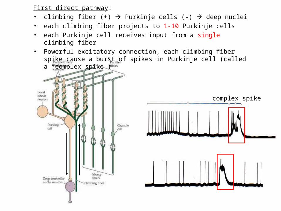
First direct pathway:
• climbing fiber (+) Purkinje cells (-) deep nuclei
• each climbing fiber projects to 1-10 Purkinje cells
• each Purkinje cell receives input from a single climbing fiber
• Powerful excitatory connection, each climbing fiber spike cause a burst of spikes in Purkinje cell (called a “complex spike”)
complex spike

Second direct pathway:
• Mossy fiber (+) granule cells (axon: parallel fibers, +) Purkinje cells (-) deep nuclei
• each parallel fiber projects to thousands of Purkinje cells (high divergence)
• each Purkinje cell receives input from ~200,000 parallel fibers (high convergence)
• Weak excitatory connection, spatiotemporal summation of inputs from many parallel fibers causes a single spike in Purkinje cell (called a “simple spike”)
glomeruli
simple spikes
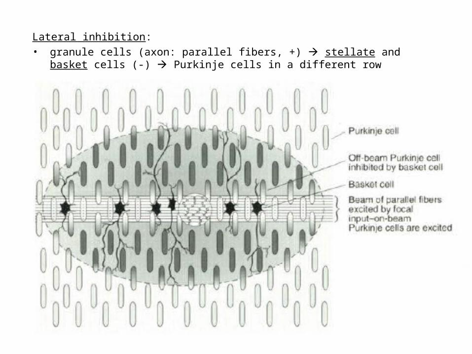
Lateral inhibition:
• granule cells (axon: parallel fibers, +) stellate and basket cells (-) Purkinje cells in a different row

Negative feedback:
• granule cells (axon: parallel fibers, +) Golgi cells (-) granule cells

Vestibulo-Ocular Reflex
-- coordinated response that maintains the eyes on a fixed target when the head is rotated Head rotation → vestibular labyrinth output
→ eye movement in opposite direction → image on the retina unchanged
Adaptation in Vestibulo-Ocular Reflex -- Experiment on reversing L & R visual fields with prism glasses Head rotation → eye movement in the same direction
→ image move further away → native V-O reflex is reduced (in a few days) and eventually reverses direction
* Lesion of vestibulo cerebellum (in monkeys) → no adaptation
Cerebellum Functions - Vestibulo-ocular reflex

Cerebellum Functions - Eye-hand coordination
Dart-throwing Experiment: Wearing eye prism that bends the light path sideways
→ initial throws off to the side → correct throws via adaptation of the motor system after 30 throws
→ prism removal lead to off-target throws toward the opposite side
* Patients with inferior cerebellar peduncle damage – no adaptation

Cerebellum and motor learning
• During learning of a new motor task, subject makes mistakes, but the error reduces with practice
• The standard notion is that the “error signal” causes changes in brain circuits involved in motor control (e.g., cerebellum), thus improving motor performance
Patient H.M., who had bilateral removal of medial temporal lobe and lack of ability to form episodic memory, but motor learning is intact, indicating it is mediated by different brain structure
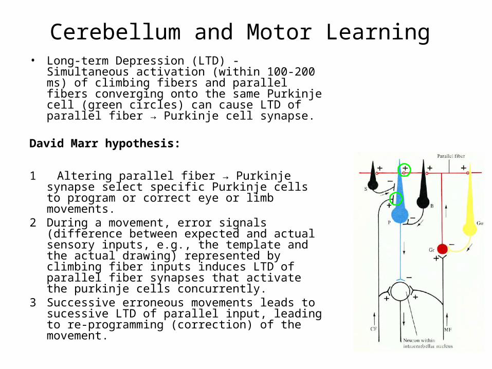
Cerebellum and Motor Learning• Long-term Depression (LTD) - Simultaneous
activation (within 100-200 ms) of climbing fibers and parallel fibers converging onto the same Purkinje cell (green circles) can cause LTD of parallel fiber → Purkinje cell synapse.
David Marr hypothesis:
1 Altering parallel fiber → Purkinje synapse select specific Purkinje cells to program or correct eye or limb movements.
2 During a movement, error signals (difference between expected and actual sensory inputs, e.g., the template and the actual drawing) represented by climbing fiber inputs induces LTD of parallel fiber synapses that activate the purkinje cells concurrently.
3 Successive erroneous movements leads to sucessive LTD of parallel input, leading to re-programming (correction) of the movement.