motor learning and neuroplasticity in humans
TRANSCRIPT

i
Motor learning and neuroplasticity
in humans
by
Dr James TEO Teong Han Institute of Neurology
University College London
A thesis submitted to UCL for a PhD in the Faculty of Science.
June 2009.
Supervisors: Professor John Rothwell
Dr Richard Greenwood
Word Count: 56103 (chapters only)
58621 (preface and chapters only)
ii
Declaration
I, James TEO Teong Han , confirm that the work presented in this thesis
is my own. Where information has been derived from other sources, I
confirm that this has been indicated in the thesis.

iii
Abstract
The central nervous system is plastic, in that the number and strength of synaptic
connections changes over time. In the adult the most important driver of such changes is
experience, in the form of learning and memory. There are thought to be a number of
rules, operating relatively local to each synapse that govern changes in strength and
organisation. Some of these such as Hebbian plasticity or plasticity following repeated
activation of a connection have been studied in detail in animal preparations. However,
recent work with non-invasive methods of transcranial stimulation in human, such as
transcranial magnetic stimulation, has opened the opportunity to study similar effects in
the conscious human brain.
In this thesis I use these methods to explore some of the presumed changes in synaptic
connectivity in the motor cortex during different forms of motor learning. The
experiments only concern learning in the healthy brain; however it seems likely that the
same processes will be relevant to neurorehabilitation and disease of the nervous system.
This thesis explores the link between neuroplasticity and motor learning in humans using
non-invasive brain stimulation, pharmacological agents and psychomotor testing in 6
related studies.
1) Chapter 3 reports initial pharmacological investigations to confirm the idea that
some of the long term effects of TMS are likely to involve LTP-like mechanisms.
The study shows that NMDA agonism can affect the response to a repetitive form
of TMS known as theta burst stimulation (TBS)
2) Following up on the initial evidence for the role of NMDA receptors in the long
term effects of TBS, Chapter 4 explores the possible modulatory effects of
dopaminergic drugs on TBS.
3) Chapter 5 takes the investigations to normal behaviours by examining how the
NMDA dependent plasticity produced by TBS interacts with learning a simple
motor task of rapid thumb abduction. The unexpected results force a careful
examination of the possible mechanisms of motor learning in this task.
4) Chapter 6 expands on these effects by employing a battery of TMS methods as
well as drug agents to examine the role of different intracortical circuits in
ballistic motor learning.
5) Chapter 7 studies the plasticity of intracortical circuits involved in transcallosal
inhibition.
6) Chapter 8 studies the interaction between synaptic plasticity invoked by TBS and
sequence learning.
The studies described in the thesis contribute to understanding of how motor learning and
neuroplasticity interact, and possible strategies to enhance these phenomena for clinical
application.

iv
Acknowledgements
This thesis is dedicated to my wife, Ping – who
kept me focused and writing this thesis even
when I had lost the motivation. She has also
supported my lunatic interests for which I am
grateful for.
It is also not possible to thank enough the
sacrifices that my parents have made to
ensure that I was educated enough to be able
to write this thesis.
And my supervisors, John Rothwell and Richard
Greenwood, who nudged me every so often in
the right direction.

v
Contents
Abstract
Acknowledgement
Contents
Figures and Tables
Abbreviations
Publications in relation to this thesis
iii
iv
v
xiii
xvi
xviii
Chapter 1 – Introduction 1
1.1 Plasticity
1.1.1 Long term potentiation (LTP) / Hebbian plasticity
1.1.1.1 Hebbian features of LTP/ LTD
1.1.1.2 Non-classical LTP
1.1.2 Structural plasticity
1.1.3 Metaplasticity
1.1.4 Homeostatic plasticity
1.1.5 Brain-derived neurotrophic factor (BDNF) and
neuroplasticity
1.2 The study of plasticity in humans
1.2.1 Transcranial magnetic stimulation
1.2.2 Repetitive transcranial magnetic stimulation
1.2.2.1 Theta burst stimulation
1.2.2.2 I-wave interval rTMS
1.2.3 Paired associative stimulation
1.2.4 Transcranial direct current stimulation
4
5
7
7
8
9
11
12
12
13
16
16
18
18
19

vi
1.2.5 Direct cortical stimulation
1.2.6 Similarities between human and animal
neuroplasticity models
1.3 Drugs in the study of plasticity
1.3.1 Noradrenergic drugs
1.3.2 Dopaminergic drugs
1.3.3 Cholinergic drugs
1.3.4 GABA-ergic drugs
1.3.5 Endocannabinoids
1.3.6 Glutamergic drugs
1.4 Motor learning
1.4.1 Motor learning paradigms
1.4.1.1 Sequence learning
1.4.1.2 Ballistic motor learning
1.4.1.3 Visuomotor transformations
1.4.1.4 Force field adaptation
1.4.1.5 Locomotor adaptation
1.4.1.6 Classical conditioning
1.4.1.7 Aimed rapid movements
1.4.2 Explicit learning in motor learning
1.4.3 Adaptation versus skill-learning
1.4.4 Stages of motor learning
1.4.5 Summary of motor learning
1.5 Relationship between plasticity and motor learning
1.5.1 Evidence from animal models
20
20
22
22
23
24
25
26
27
28
29
30
30
31
32
33
33
34
36
36
37
39
39
39
vii
1.5.2 Evidence from humans
1.6 Motor learning and plasticity in disease
1.6.1 Stroke
1.6.1.1 Stroke recovery through neuroplasticity
and motor learning
1.6.1.2 The ‘hemispheric rivalry’ hypothesis
1.6.2 Parkinson’s Disease
1.6.2.1 Levodopa-associated dyskinesias
1.6.3 Cerebellar disease
1.6.4 Dystonia
1.6.5 Huntington’s disease
1.6.6 Alzheimer’s disease
1.6.7 Relevance of motor learning to neurorehabilitation
1.7 Goal of this thesis
41
43
43
44
46
47
49
49
50
51
52
53
54
Chapter 2 – General Methods 56
2.1 Subjects
2.2 Electromyography
2.3 Transcranial magnetic stimulation
2.4 Repetitive transcranial magnetic stimulation
2.5 Behavioural measures
2.6 EMG data analysis
2.7 Statistical analysis
57
57
58
59
60
60
61
Chapter 3 – NMDA agonism and theta burst stimulation 62
viii
3.1 Introduction
3.2 Study design
3.3 Drug
3.4 Results
3.5 Discussion
63
64
65
67
68
Chapter 4 – Theta burst stimulation and neuromodulatory drugs 71
4.1 Introduction
4.2 Amphetamine & levo-dopa
4.2.1 Study Design
4.2.1.1 Drugs
4.2.1.2 Statistical analysis
4.2.2 Results
4.2.3 Discussion
4.3 Nicotine
4.3.1 Study Design
4.3.1.1 Drug
4.3.1.2 Statistical analysis
4.3.2 Results
4.3.3 Discussion
72
72
73
74
74
74
78
79
80
80
81
81
85
Chapter 5 – Theta burst stimulation and ballistic motor learning 86
5.1 Introduction
5.2 Study design
87
87

ix
5.2.1 Ballistic motor learning
5.2.2 Data analysis
5.3 Results
5.4 Discussion in the interlude
5.4.1 The effect of iTBS on motor learning
5.4.2 The effect of the iTBS-nicotine interaction on learning
5.4.3 Trial-by-trial analysis of data
5.5 Modelling the ballistic motor learning task
5.7.1 Implications of the model
5.6 Analysing variability of performance during learning
5.7 Control experiment: TBS on variability of TMS-evoked movements
5.8 Conclusion
88
89
90
95
95
96
97
99
105
107
111
114
Chapter 6 – Intracortical circuits and practice-dependent plasticity 117
6.1 Introduction
6.2 Study design
6.3 TMS measurements
6.4 Motor practice
6.5 Results
6.5.1 Drug-induced changes
6.5.2 Practice
6.5.3 Practice-induced changes
6.5.4 Correlations
6.6 Discussion
6.6.1 Drug induced changes in cortical circuits
118
119
121
123
124
125
128
129
132
134
134

x
6.6.2 Practice-dependent plasticity
6.6.3 Link between SAI and perception variability
6.6.4 Conclusion
135
138
139
Chapter 7 – Intracortical circuits and transcallosal pathways 140
7.1 Introduction
7.2 Intracortical circuits that modulate transcallosal inhibition
7.2.1 Study design
7.2.2 Data analysis
7.2.2.1 Data analysis of iSP
7.2.2.2 Data analyses of IHI
7.2.2.3 Data analyses of cMEP
7.2.2.4 Statistical analysis
7.2.3 Results
7.2.3.1 Demonstration of SICF-like effects on iSP
7.2.3.2 Demonstration of SICF-like effects on IHI
7.2.4 Discussion
7.2.4.1 Site of facilitatory interaction
7.2.4.2 Nature of facilitatory interaction
7.3 Effect of rTMS on transcallosal circuits
7.3.1 Study design
7.3.2 Data analysis
7.3.3 Results
7.3.3.1 SICIcMEP and SICIiSP
7.3.3.2 Effect of rTMS on SICIcMEP and SICIiSP
141
142
142
145
145
146
146
147
147
147
155
160
161
159
163
164
165
166
167
170

xi
7.3.4 Discussion
7.4 Conclusion
172
174
Chapter 8 – Theta burst stimulation and sequence learning 175
8.1 Introduction
8.2 Methods
8.2.1 Participants
8.2.2 Serial Reaction Time (SRT) Task
8.2.3 Theta burst stimulation
8.2.4 SRT task data analysis
8.3 Results
8.3.1 Reaction Times
8.3.2 Variability of Reaction Times
8.3.3 Errors
8.4 Discussion
8.4.1 Methodological differences across studies of the effects of
rTMS and tDCS on SRT learning
8.4.2 Neural basis of motor sequence learning
8.4.3 The role of the M1 and the SMA in sequential learning.
8.4.4 Why doesn’t excitatory TBS produce enhanced learning?
8.4.5 The lack of reaction time improvement in probabilistic
sequence learning
8.5 Conclusion
176
179
179
180
182
184
184
185
191
191
192
192
194
195
196
197
198
Chapter 9 – Conclusion 199
9.1 Summary 200

xii
9.2 Brain stimulation, motor learning and plasticity
9.3 Future possible studies
9.3.1 Studying the relationship between variability and plasticity
9.3.2 Studying the role of motor learning in neurorehabilitation
9.3.3 Studying the role of endocannabinoids in human
neuroplasticity
9.4 Closing statements
201
202
202
203
203
204
Appendix: References 206

xiii
Figure and Tables
Figure/
Table
Description Page
Figure
1.1
Frequency and calcium-dependency of classical LTP/ LTD 6
Figure
1.2
Graphical representation of the Bienenstock-Cooper-Munro
learning rule
10
Figure
1.3
Example of epidural volleys and motor-evoked potentials from a
single TMS pulse
14
Table
1.1
Summary of various paired-pulse TMS measurements 15
Figure
1.4
Pictoral representation of cTBS and iTBS repetitive TMS 17
Figure
3.1
The effect of d-cycloserine or placebo on iTBS repetitive TMS 66
Table
3.1
Baseline measures for d-cycloserine or placebo experimental arms 67
Table
4.1
Baseline measures for amphetamine, levodopa or placebo
experimental arms
75
Figure
4.1
The effect of amphetamine or placebo on iTBS repetitive TMS 76
Figure
4.2
The effect of levodopa or placebo on iTBS repetitive TMS 77
Table
4.2
Baseline measures for nicotine or placebo experimental arms 81
Figure
4.3
The effect of nicotine or placebo on iTBS repetitive TMS 83
Figure
4.4
The effect of nicotine on motor-evoked potentials 84
Figure
5.1
The effect of nicotine and iTBS on repeated peak initial
acceleration of left thumb abduction
91
Table
5.1
Analysis of variance (ANOVA) of ballistic motor learning 93-94
Table
5.2
Analysis of variance (ANOVA) of baseline ballistic performance 94
Figure
5.2
Probability of an individual trial being better than previous trial
98
Figure
5.3
Model of a possible strategy for the ballistic learning task
101-102
Figure
5.4
The interaction of performance variability, perception variability
and learning gain in the ballistic learning model
104-105
Figure
5.5
Example of trial-to-trial measure of performance variability
108

xiv
Figure
5.6
Performance variability during the learning task (group means of
each session)
110-111
Figure
5.7
The effect of iTBS on the variability of TMS-evoked thumb
movements
113-114
Figure
6.1
Study design of the role of GABA-circuits in practice-dependent
plasticity
120
Table
6.1
Subject and TMS parameters at T1 (before drug) 124
Table
6.2
Subject and TMS parameters at T2 (after drug) 125
Table
6.3
Subject and TMS parameters at T3 (after training) 125
Figure
6.2
The effect of drug on TMS measurements 126
Figure
6.3
Motor performance during training 129
Figure
6.4
The effect of training on TMS measurements 130-131
Figure
6.5
Correlation analysis of the drug-induced and practice-induced
changes
133
Figure
7.1
Study design of transcallosal experiments 1a and 1b 142
Figure
7.2
Example of MEPs and ispilateral silent period of two stimuli
delivered at different ISI
148
Figure
7.3
Group means of ipsilateral silent period and contralateral MEP
from single and paired TMS pulses
150-151
Table
7.1
Statistical analysis of experiment 1a 152
Figure
7.4
Correlations between iSP and cMEP 154
Figure
7.5
Group means of interhemispheric inhibition and contralateral MEP
from single and paired TMS pulses
156
Table
7.2
Statistical analysis of experiment 1b 158
Figure
7.6
Correlations between IHI and cMEP 159
Figure
7.7
Study design of experiment 2 to study the effect of rTMS on the
inhibitory intracortical circuits
164
Figure
7.8
Effect on conditioning stimulus on cMEP and iSP 168
Table
7.3
Statistical analysis of effect on conditioning stimulus on cMEP
and iSP
169-170
Table
7.4
Baseline characteristics in experiment 2 170
Figure
7.9
Effect of rTMS on cMEP and iSP 171
xv
Table
7.5
Statistical analysis of the effect of rTMS on cMEP and iSP 172
Table
7.6
Statistical analysis of the effect of rTMS on SICIcMEP with SICIiSP 173
Figure
8.1
Mean RTs across training blocks 185-186
Figure
8.2
Mean RT difference scores by epoch 188-189
xvi
Abbreviations
CTS Corticospinal
TC Transcallosal
CS Conditioning stimulus
TS Test stimulus
iSP Ispilateral silent period
IHI Interhemispheric inhibition
SICF Short-interval intracortical facilitation
SICIcMEP Short-interval intracortical inhibition of the corticospinal pathway
SICIiSP Short-interval intracortical inhibition of the transcallosal pathway
SICI2ms Short-interval intracortical inhibition with 2ms interstimulus interval
SICI3ms Short-interval intracortical inhibition with 3ms interstimulus interval
SICIcomb Short-interval intracortical inhibition averaged for 2ms and 3ms
interstimulus intervals
ICF Intracortical facilitation
SAI Short latency afferent inhibition
LAI Long latency afferent inhibition
cMEP Contralateral motor-evoked potential
MEP Motor-evoked potential
RMT Resting motor threshold
AMT Active motor threshold
MSO Maximum stimulator output
rTMS Repetitive transcranial magnetic stimulation
TMS Transcranial magnetic stimulation
TBS Theta-burst stimulation
iTBS Intermittent theta burst stimulation
cTBS Continuous theta burst stimulation
FDI First dorsal interosseus
APB Abductor pollicus brevis
ADM Adductor digiti minimi
xvii
GABA Gamma-aminobutyric acid
AChR Acetylcholine receptor
PAS Paired associative stimulation
TDCS Transcranial direct current stimulation
ISI Interstimulus interval
IQ Intelligence quotient
SMA Supplementary motor area
DLPFC Dorsolateral prefrontal cortex
M1 Primary motor cortex
SRT Serial reaction time
PD Parkinson’s Disease
RT Reaction time
EMG Electromyography
xviii
Publications in relation to this thesis
The following publications have come from work recorded in this thesis:
• Teo JT, Swayne OB, Rothwell JC. Further evidence for NMDA-dependence of the
after-effects of human theta burst stimulation. Clin Neurophysiol. 2007 Jul;
118(7):1649-51.
I am the lead author of this paper as I contributed substantially in the design,
concept and interpretation of the study. Data from this paper is used in Chapter 3.
• Teo JT, Terranova C, Swayne OB, Greenwood R, Rothwell JC. Practice-dependent
plasticity is limited by different intracortical circuits. Exp Brain Res. 2009
Mar;193(4):555-63.
I am the lead author of this paper as I contributed substantially in the design,
concept and interpretation of the study. Data from this paper is used in Chapter 6.
• Avanzino L, Teo JT, Rothwell JC. Intracortical circuits modulate transcallosal
inhibition in humans. J Physiol. 2007 Aug 15; 583(Pt 1):99-114.
I am one of the shared lead authors of this paper as I contributed substantially in
the design, concept and interpretation of the studies. Data from this paper is used in
Chapter 7.
• Wilkinson L, Teo JT, Obeso I, Rothwell JC, Jahanshahi M. The contribution of the
primary motor cortex is essential for probabilistic implicit sequence learning:
evidence from theta burst magnetic stimulation. J Cogn Neurosci. 2009 Mar 20.
[Epub ahead of print]
I am the second author of this paper although I contributed substantially in the
design, concept and interpretation of the study; especially the use of transcranial
magnetic stimulation. Data from this paper is used in Chapter 8.
Accepted
� Swayne OB, Teo JT, Greenwood R, Rothwell JC. Nicotine modulates the effects of
theta bust stimulation (accepted by Clinical Neurophysiology; 22nd June 2009).
I am the second author of this paper although I contributed substantially in the
conduct of experiments and interpretation of the study. Data from this paper is used
in Chapter 4.
Submitted
� Teo JT, Swayne OB, Cheeran BJ, Greenwood R, Rothwell JC. Human theta burst
stimulation enhances subsequent motor learning while increasing performance
variability (submitted to Cerebral Cortex; 27th June 2009).
I am the lead author of this paper and I contributed substantially in the design,
concept, data collection, modeling design and interpretation of the study. Data from
this paper is used in Chapter 5.

- Chapter 1 -
- 1 -
Chapter 1
Introduction
- Chapter 1 -
- 2 -
The central nervous system has a wide array of functions: receiving sensory input, storing
memories, coordinating motor plans, maintaining posture, and generating consciousness
and higher thought. The nervous system accomplishes this diversity of functions with one
key feature: it can change and adapt. In this way, characteristics can be tuned to the task
at hand and new properties can be acquired. This ability of the nervous system to change
is perplexing as the adult nervous system generates relatively few new cells.
This dilemma was recognised by the great Spanish histologist and neuroscientist,
Santiago Ramón y Cajal (1852-1934) who posited an explanation in his 1894 Croonian
Lecture to the Royal Society in London:
"La gymnastique cérébrale n'est pas susceptible d'améliorer l'organisation
du cerveau en augmentant le nombre de cellules, car, on le sait, les éléments
nerveux ont perdu depuis l’époque embryonnaire la propriété de proliférer;
mais on peut admettre comme une chose très vraisemblable que l’exercice
mental suscite dans les régions cérébrales plus sollicitées un plus grand
développement de l’appareil protoplasmique et du système des collatérales
nerveuses. De la sorte, des associations déjà créées entre certains groupes
de cellules se renforceraient notablement au moyen de la multiplication des
ramilles terminales des appendices protoplasmiques et des collatérales
nerveuses; mais, en outre, des connexions intercellulaires tout à fait
nouvelles pourraient s'établir grâce à la néoformation de collatérales et
d'expansions protoplasmiques."
- Cajal, The Croonian Lecture (1894)
- Chapter 1 -
- 3 -
“Cerebral acrobatics cannot improve the organisation of the brain by
increasing the number of cells because, since their embryological stages,
the elements of the nervous system have lost the ability to multiply
themselves; but it seems very likely that mental exercise engenders greater
expansion of the dendritic apparatus and the system of axonal collaterals.
In this way, connections already establised between certain groups of cells
would be particularly strengthened by the multiplication of the small
terminal branches of the dendritic appendages and axonal collaterals;
moreover, new intercellular connections could be established thanks to the
formation of new collaterals and dendrites”
- English translation
However, Cajal’s prescient proposal – that it is not new cells which are generated but
new pathways and synapses – was overshadowed by his view 20 years later that:
“nerve paths are something fixed, ended, immutable.
Everything may die, nothing may be regenerated”
- Cajal (1913-1914)
And this view of a rigid, static nervous system prevailed for most of the 20th
century. In
the later half of the 20th
century, more evidence began to mount to demonstrate that the
central nervous system does indeed adapt and is mutable even in adulthood; this broad
idea is commonly termed neuroplasticity.
- Chapter 1 -
- 4 -
1.1 Plasticity
Neuroplasticity using the broadest definition is the ability of neurons (or the nervous
system) to rearrange their anatomical and functional connectivity and properties in
response to environmental input. This broad definition encompasses functional,
structural, physiological and molecular changes but the most interesting form of
neuroplasticity is neuroplasticity that obeys Hebbian rules as first described by Daniel
Hebb:
When an axon of cell A is near enough to excite cell B and repeatedly or
persistently takes part in firing it, some growth process or metabolic change
takes place in one or both cells such that A's efficiency, as one of the cells
firing B, is increased.
- Hebb (1949)
This description has often been simplified to the more pithy “neurons that fire together,
wire together”. Hebbian principles form the mathematical basis of neural network models
and provide a principle that governs neuroplasticity, allowing synapses to retain a
memory of previous activity.
The first clues for the molecular basis of how a nervous system can display
neuroplasticity and adapt its motor behaviour was found in the invertebrate sea-slug,
Aplysia californica by Eric Kandel and his group in 1969-1973 (Frazier et al., 1969;
Kupfermann et al., 1970; Castelluci et al., 1970; Pinkser et al., 1973): changes in synaptic
properties were shown to occur after the Aplysia californica had acquired a memory. This
led to the discovery of long-term potentiation (LTP) in the mammalian hippocampus

- Chapter 1 -
- 5 -
1973 by Bliss & Lømo, which provided a molecular mechanism for neuroplasticity which
obeys Hebbian principles.
1.1.1 Long term potentiation (LTP) / Hebbian plasticity
Long term potentiation (LTP) was first described as the long-lasting increase in synaptic
efficacy after tetanic stimulation of the presynaptic neuron (review: Collingridge & Bliss,
1987; Collingridge & Bliss, 1995; Bliss et al., 2004). This long-lasting change is due to
presynaptic neurotransmitter release and postsynaptic receptor expression and the key
trigger is the NMDA glutamate receptor.
The NMDA receptor is a ligand-gated calcium channel. It has a binding site for glutamate
on the extracellular surface which gates the opening of the channel. Additionally, the
channel pore is also blocked by a Mg2+
ion, which has to be first displaced by
depolarisation of the postsynaptic neuron, before the channel is fully open. In this way,
the NMDA receptor acts as a 'coincidence detector' for presynaptic and postsynaptic
depolarisation, and allows LTP to obey Hebbian principles (Bliss et al., 2004).
The transient rise in intracellular Ca2+
concentration serves to activate Ca2+
-dependent
enzymes, Ca2+
/ calmodulin-dependent protein kinase II (CaMKII) and protein kinase C
(PKC). These enzymes phosphorylate various proteins and receptors, and crucially the
cAMP-response-element-binding-protein (CREB) which triggers CREB-dependent gene
expression (Yin & Tully, 1996; Ahmed & Frey, 2004). Presynaptic processes also
mediate LTP, and retrograde messengers such as nitric oxide and endocannabinoids
deliver the message to the presynaptic cell to increase or decrease synaptic vesicle fusion
(Arancio et al., 1996; Kanto et al., 1996; Wilson et al., 2001).

- Chapter 1 -
- 6 -
Long term depression (LTD) is the corrollary of LTP with reduction of synaptic efficacy
after lower frequency repetitive stimulation, and this is also dependent on the NMDA
receptor (Dudek & Bear, 1992; Dudek & Bear, 1993). The determinant for whether LTP
or LTD is induced is the intracellular Ca2+
concentration which binds differentially to C
and N lobes of the calmodulin kinase depending on the rise of Ca2+
(Fig 1.1)
Fig 1.1 Frequency and calcium-dependency of classical LTP/ LTD where
the ordinate represents change in synaptic efficacy, and abscissa can
represent either frequency of induction stimulation or postsynaptic
intracellular calcium.
Neuroplasticity operating on similar molecular pathways has since been discovered with
other more physiologically-realistic forms of experimental stimulation: theta frequency
stimulation (Larson et al., 1986; Larson et al., 1988) and spike-timing-dependent
plasticity, STDP (Markram et al., 1997). It has also been found in different brain regions

- Chapter 1 -
- 7 -
including: striatum (Charpier & Deniau, 1997; Fino et al., 2005) and sensorimotor cortex
(Hess & Donoghue, 1994; Hess et al., 1996), thus verifying its physiological relevance to
the brain.
1.1.1.1 Hebbian features of LTP/ LTD
The key features of LTP/ LTD which make it an attractive molecular mechanism for
Hebbian neuroplasticity are:
(a) Rapid induction: LTP/ LTD can be induced rapidly by one or more brief
tetanic stimuli;
(b) Input specificity: LTP/ LTD once induced occurs only at inputs which have
been stimulated;
(c) Associativity: Weak inputs can produce LTP/ LTD in the presence of strong
inputs depending on precise timing (spike-timing dependency);
(d) Cooperativity: Multiple weak inputs can summate in space and/ or time
(frequency-dependency) to produce LTP/ LTD; and
(e) Long-lasting: The effects are immediate and last several hours.
These characteristics of LTP/ LTD govern neural network and computational models
based on Hebbian principles, and also set a benchmark for assessing other models of
neuroplasticity.
1.1.1.2 Non-classical LTP
There are many other forms of synaptic plasticity that do not conform to the above
classical form of LTP / LTD. One such variant is the synaptic plasticity in the parallel
- Chapter 1 -
- 8 -
fibre and Purkinje cell synapse in the cerebellum (Ito et al., 2002; Jörntell & Hansel,
2006) as described by the Marr-Albus model (Marr, 1969; Albus, 1971). This model is
beyond the scope of this thesis but broadly follows Hebbian rules at the synapse between
the parallel fibre and Purkinje cells with LTD (instead of LTP) occurring when there is
coincident activation of the parallel-fibre-Purkinje cell synapse and climbing fibre
synapse (Ito et al., 1982; Ito et al., 2006).
Other variants of non-classical LTP also exist with LTP at inhibitory synapses in the
developing visual cortex (Komatsu, 1996; Komatsu & Yoshimura, 2000) and non-
NMDA-dependent LTP (Cavus & Teyler, 1998; Grover & Yan, 1999). It is worthwhile
noting though that all these non-classical types of LTP identified still appear to broadly
follow Hebbian rules.
1.1.2 Structural plasticity
The previous forms of neuroplasticity suggest a change in the properties of synapses
between neurons. However, the past decade have provided evidence for the unmasking of
silent synapses (Geinisman et al., 1996; Atwood & Wojtowicz, 1999; Ward et al., 2006;
Itami et al., 2003) and new synapse formation (Geinisman et al., 1991) associated with
LTP-induction indicating structural neuroplasticity after neuronal stimulation. Dendritic
spines and synaptic boutons are extremely dynamic in animals, and changes have been
shown to be associated with experience (Lendvai et al., 2000; Knott et al., 2002;
Trachtenburg et al., 2002) and associative learning (Geinisman et al., 2001) in a number
of brain regions. These changes in dendritic spine morphology and synaptogenesis have
- Chapter 1 -
- 9 -
also been shown to be linked with the induction of LTP (Toni et al., 1999; Engert et al.,
1999), thereby providing a link to Hebbian principles discussed earlier.
Another form of structural plasticity is neurogenesis. While it has been accepted for most
of the 20th
century that no new neurons are formed in the adult brain, this has been shown
to be false. It is now widely accepted that neurogenesis does occur in the adult
hippocampus and the olfactory bulb (Altman & Das, 1967; Eriksson et al., 1998; Bernier
et al., 2002). There is some limited evidence that neurogenesis also occurs in other brain
regions (e.g. neocortex, striatum, amygdala) (Gould et al., 1999; Magavi et al., 2000;
Dayer et al., 2005) although this remains controversial (Rakic et al., 1985; Bhardwarj et
al., 2006). Also how widespread this phenomenon is and whether it participates in
learning and memory remains controversial (Leuner et al., 2006; Gould et al., 2007;
Cameron & Dayer 2008).
1.1.3 Metaplasticity
One consequence of the Hebbian characteristics of LTP / LTD is that it is inherently
unstable. This was first predicted by Bienenstock, Cooper & Munro (1982) after
modelling the development of orientation and binocular selectivity of neurons in the
visual cortex, showing that synapses in a purely Hebbian model would segregate into
maximally saturated synapses via LTP and maximally desaturated synapses via LTD.
They proposed the BCM learning rule: recent high synaptic activity makes LTP harder to
induce and LTD easier to induce and vice versa with recent low synaptic activity
(Bienenstock et al., 1982; Wexler & Stanton, 1993). The mechanics of the BCM rule is
summarised by Fig 1.2.
- Chapter 1 -
- 10 -
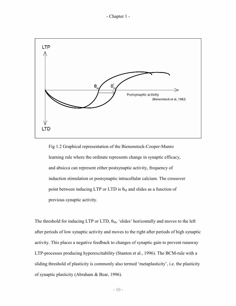
Fig 1.2 Graphical representation of the Bienenstock-Cooper-Munro
learning rule where the ordinate represents change in synaptic efficacy,
and absicca can represent either postsynaptic activity, frequency of
induction stimulation or postsynaptic intracellular calcium. The crossover
point between inducing LTP or LTD is θM and slides as a function of
previous synaptic activity.
The threshold for inducing LTP or LTD, θM, ‘slides’ horizontally and moves to the left
after periods of low synaptic activity and moves to the right after periods of high synaptic
activity. This places a negative feedback to changes of synaptic gain to prevent runaway
LTP-processes producing hyperexcitability (Stanton et al., 1996). The BCM-rule with a
sliding threshold of plasticity is commonly also termed ‘metaplasticity’, i.e. the plasticity
of synaptic plasticity (Abraham & Bear, 1996).

- Chapter 1 -
- 11 -
Experimental evidence to support the BCM rule has been found in the visual cortex
(Kirkwood et al., 1996; Philpot et al., 2001; Philpot et al., 2003) where sensory
deprivation alters NMDA receptor subunit composition such that LTD is easier to induce,
while prior experience changes NMDA receptor subunit composition and reduces LTD.
The BCM-rule has also shown to be valid indirectly in the hippocampus (Whitlock et al.,
2006) and in the motor cortex (Rioult-Pedotti et al., 1998, Rioult-Pedotti et al., 2000;
Harms et al., 2008) where prior experience occludes further LTP induction; this
relationship of plasticity with learning is discussed in greater detail in section 1.5.1.
1.1.4 Homeostatic plasticity
Homeostatic plasticity is a distinct mechanism from metaplasticity but is an indirect
consequence of the BCM rule. If LTP was induced, the θM would slide to the right
making further LTP harder to induce, thus reducing the bidirectionality of synaptic
plasticity. A homeostatic mechanism is thus required to realign the θM back to a
physiological range, and this mechanism is termed homeostatic plasticity.
Homeostatic plasticity is a mechanism by which synaptic efficacy appears to ‘scale’ up
after a period of low synaptic activity (and vice versa) (Turrigiano et al., 1998; Leslie et
al., 2001) and is related to changes in postsynaptic glutamate receptors (Watt et al., 2000;
Wieranga et al., 2005) and postsynaptic ion channels (Misonou et al., 2004). This allows
there to be sufficient range for further bidirectional LTP or LTD to be induced (Burrone
& Murthy, 2003; Rabinovitch & Segev, 2008) while maintaining the relative weights of
the different synaptic inputs to a neuron (Turrigiano et al., 1998). It is important to note

- Chapter 1 -
- 12 -
though that the time scale of homeostatic plasticity is in the order of hours, so it is
unlikely that it is relevant in rapid acquisition or the early phases of plasticity or learning.
1.1.5 Brain-derived neurotrophic factor (BDNF) and neuroplasticity
The neurotrophin, brain-derived neurotrophic factor (BDNF) has been demonstrated to
play a central role in both Hebbian plasticity and this regulatory homeostatic process
(Rutherford et al., 1998; Turrigiano & Nelson, 2000; Leslie et al., 2001; Copi et al.,
2005). BDNF acts on TrkB post-synaptic receptors to produce LTP without requiring
neuronal stimulation (Ying et al., 2002; Messaouodi et al., 2002; Bekinschtein et al.,
2008) and when combined with theta burst stimulation facilitates late phase LTP (Pang et
al., 2004; Kramár et al., 2004). In addition, chronic bath exposure to BDNF blocks the
‘scaling up’ of synaptic activity during a period of low synaptic activity (Rutherford et
al., 1998; Desai et al., 1999).
BDNF makes an attractive candidate for being a regulator of activity-dependent processes
like the BCM rule and homeostatic plasticity as it is released in an activity-dependent
manner by the dendrites of neurons (Zafra et al., 1991; Wetmore et al., 1994; Lindholm et
al., 1994) and there is evidence for synapse-specific release as well (Schinder et al., 2000;
Hartmann et al., 2001; Kojima et al., 2001). A review of the molecular biology of BDNF
can be found in Lu et al., 2003.
1.2 The study of plasticity in humans
The study of neuroplasticity in humans was initially limited to the study of cultured
human neurons or slices from surgical excisions in patients with epilepsy. However when
- Chapter 1 -
- 13 -
Anthony Barker and colleagues developed transcranial magnetic stimulation (Barker et
al., 1985), this technique spurred newer different types of non-invasive stimulation which
allowed the study of neuroplasticity in humans. The mechanism of transcranial magnetic
stimulation (TMS) will be discussed first as the principles are central in understanding
how neuroplasticity in humans is studied.
1.2.1 Transcranial magnetic stimulation
Transcranial magnetic stimulation uses a coil carrying a rapidly changing electrical
current which produces a magnetic field at right angles to the current. If the coil is placed
on the scalp, the magnetic field will in turn induce an electrical current in the underlying
cortex. If the electrical current is perpendicular to neuronal plasma membranes, this can
depolarise the neuron. Thus, a TMS pulse would induce neurons in underlying cortex to
discharge an action potential.
TMS to the primary motor cortex produces a motor-evoked potential (MEP) on surface
electromyography (EMG) in peripheral muscles that are represented by that region of
cortex (Day et al., 1989). An MEP is the summation of the discharge of multiple motor
units, and epidural recordings of the pyramidal tract demonstrate that the discharge down
the pyramidal tract consists of a D wave and several I waves with more I-waves being
recruited with increasing intensity of stimulation (Di Lazzaro et al., 1998; Fig 1.3).

- Chapter 1 -
- 14 -
Fig 1.3 Example of epidural volleys and motor-evoked potentials recorded
after a single TMS pulse over the primary motor cortex (Adapted from Di
Lazzaro et al., 1998)
The prevailing consensus is that the D-wave represents TMS activation of the
corticospinal pyramidal neurons while I-waves represent trans-synaptic activation of the
pyramidal neuron by excitatory interneurons depolarised by the TMS pulse. One curious
feature of I-waves is that successive I-waves are recruited at 1.3-1.5ms periodicity (Di
Lazzaro et al., 1998).
As TMS has a high degree of temporal resolution, paired-pulse techniques and techniques
combining peripheral electrical stimulation has allowed the measurement of a plethora of
intracortical circuits (Table 1.1):

- Chapter 1 -
- 15 -
Measurement Description Reference
Short-interval
intracortical
inhibition (SICI)
Subthreshold conditioning TMS pulse 2-
3ms before test TMS pulse
Kujirai et al., 1993
Intracortical
facilitation (ICF)
Subthreshold conditioning TMS pulse 8-
15ms before test TMS pulse
Kujirai et al., 1993
Long-interval
intracortical
inhibition (LICI)
Suprathreshold conditioning TMS pulse
100-200ms before test TMS pulse
Valls-Solé et al.,
1992; Wassermann
et al., 1996
Short-latency
afferent inhibition
(SAI)
Peripheral conditioning electrical
stimulation 20-24ms before test TMS pulse
Tokimura et al.,
2000
Long-latency
afferent inhibition
(LAI)
Peripheral conditioning electrical
stimulation 50-100ms before test TMS
pulse
Sailer et al., 2002
Interhemispheric
inhibition (IHI)
Suprathreshold conditioning TMS pulse to
contralateral M1 8-40ms before test TMS
pulse
Ferbert et al., 1992
Interhemispheric
facilitation (IHF)
Near suprathreshold conditioning TMS
pulse to contralateral M1 10ms before test
TMS pulse
Mochizuki et al.,
2004; Baumer et
al., 2006
Ipsilateral
Premotor
inhibition
Near subthreshold conditioning TMS pulse
to ipsilateral premotor area 6-8ms before
test TMS pulse
Civardi et al., 2001
Interhemispheric
premotor
inhibition
Near subthreshold conditioning TMS pulse
to contralateral premotor area 8-10ms
before test TMS pulse
Mochizuki et al.,
2004; Baumer et
al., 2006
Posterior parietal
motor inhibition
Near threshold conditioning TMS pulse to
ipsilateral M1 3-10ms before test TMS
pulse
Koch et al., 2007
Table 1.1 Summary of the various paired-pulse measurements as recorded
from TMS pulses delivered to the hand muscles
- Chapter 1 -
- 16 -
Single and paired-pulse TMS allows the measurement of the excitability and
synaptic efficacy of excitatory and inhibitory neurons in the motor cortex thereby
allowing the measurement of neuroplastic changes in vivo.
1.2.2 Repetitive transcranial magnetic stimulation
Repetitive transcranial magnetic stimulation (rTMS) delivers trains of TMS pulses to the
cortex non-invasively, and unlike single and paired-pulse TMS produces effects which
outlast the period of stimulation (Fitzgerald et al., 2006). The chief effect is that it can
modify the subsequent MEP amplitude evoked by a single TMS pulse: low-frequency
(1Hz) rTMS reduces MEP amplitude (Chen et al., 1997) and high-frequency (>5Hz)
rTMS increases MEP amplitude (Pascual-Leone et al., 1994). As these changes are
blocked by an NMDA antagonist (Ziemann et al., 1998), it is thought that the changes in
MEP amplitudes reflect changes in synaptic efficacy of excitatory interneurons synapsing
onto corticospinal pyramidal neurons and this has also been confirmed by epidural
recordings of I-wave changes (Di Lazzaro et al., 2008).
1.2.2.1 Theta burst stimulation
Theta burst stimulation (TBS), is a recent rTMS protocol, which delivers TMS pulses in a
high frequency patterned fashion similar to theta burst stimulation used in animal studies
of LTP / LTD (Huang et al., 2005). Human TBS consists of bursts of subthreshold TMS
pulses delivered at theta-frequency (5Hz). Animal studies have suggested that this is both
similar to physiological bursting patterns and optimal for inducing LTP (Larson et al.,
1986) as the first burst primes the neurons for plastic changes from subsequent bursts
- Chapter 1 -
- 17 -
(Larson & Lynch, 1986; Pacelli et al., 1989). The advantage of this technique is also that
the low intensity of stimulation increases the spatial specificity of the TMS delivered and
the high frequency reduces the duration of stimulation needed. In the original description
of TBS, two forms of TBS were developed: continuous TBS (cTBS) and intermittent
TBS (iTBS).
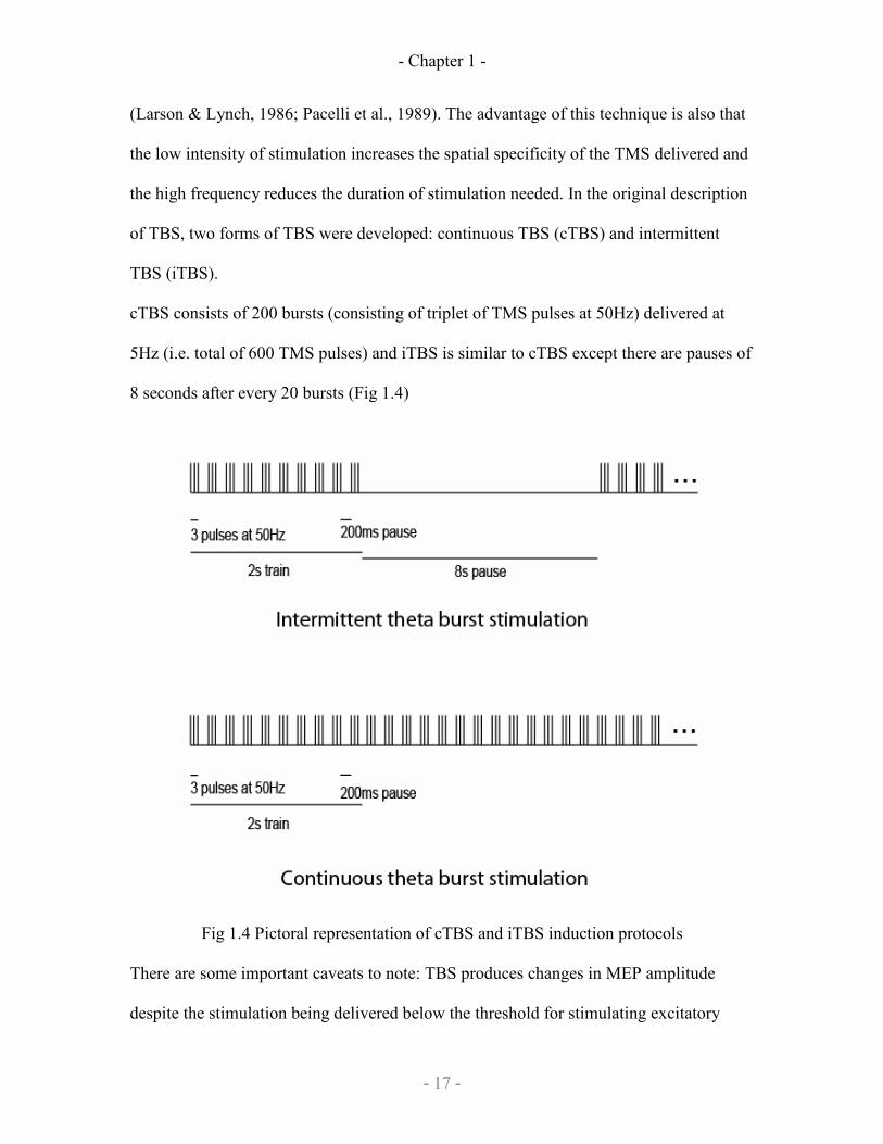
cTBS consists of 200 bursts (consisting of triplet of TMS pulses at 50Hz) delivered at
5Hz (i.e. total of 600 TMS pulses) and iTBS is similar to cTBS except there are pauses of
8 seconds after every 20 bursts (Fig 1.4)
Fig 1.4 Pictoral representation of cTBS and iTBS induction protocols
There are some important caveats to note: TBS produces changes in MEP amplitude
despite the stimulation being delivered below the threshold for stimulating excitatory
- Chapter 1 -
- 18 -
interneurons or pyramidal neurons that produce MEPs. Additionally, just like some other
forms of brain stimulation, the effects on MEPs and intracortical inhibition do not occur
immediately after the induction stimulation but build up to peak about 5-10 minutes later
(Huang et al., 2005). These caveats need to be taken into account when considering the
arguments on the similarity between TBS-induced plasticity and animal models of
Hebbian plasticity.
1.2.2.2 I-wave interval rTMS
I-wave interval rTMS is another newer form of rTMS which uses a patterned delivery of
TMS pulses. Pairs of TMS pulses with an inter-stimulus interval of 1.5ms (I-wave
interval) are delivered at a low frequency; this makes use of the periodicity of I-waves for
coincident summation of presynaptic and postsynaptic action potentials to produce
progressive enlargement of MEP amplitudes (Thickbroom et al., 2006; Benwell et al.,
2006). This is a relatively new technique and the advantage of this technique over other
rTMS protocols remains to be determined.
1.2.3 Paired associative stimulation
Paired associative stimulation is another plasticity-inducing TMS protocol that has
borrowed on principles from animal models of Hebbian plasticity. This uses the
phenomenon of spike-timing-dependent plasticity (Markram et al., 1997): when the
discharge of a presynaptic neuron is followed very shortly after by depolarization of the
postsynaptic neuron, LTP is produced after only a few paired stimuli, compared to
several hundred stimuli with repetitive tetanic stimulation.
- Chapter 1 -
- 19 -
In humans, the same phenomenon is produced by paired associative stimulation, PAS
(Stefan et al., 2000), where a peripheral electrical nerve stimulus is paired with a TMS
pulse to the primary motor cortex of the same body part. The peripheral electrical nerve
stimulus produces an afferent volley to the somatosensory cortex (S1) then to the primary
motor cortex (M1). This afferent impulse to M1 arrives at the same time as the
corticospinal neurons are depolarised by the TMS pulse. The net effect is that after ~90
pairings of electrical and TMS stimulus at about 22ms inter-stimulus interval, MEP
amplitude enlarges in a somatotopic fashion (Stefan et al., 2000).
The phenomenon of spike-timing-dependent LTD also occurs with PAS (Wolters et al.,
2003): when the TMS pulse precedes the arrival of the peripheral electrical pulse (at
inter-stimulus interval of 15ms), MEP amplitudes is reduced.
1.2.4 Transcranial direct current stimulation
Early work done in animals to modulate neuronal resting membrane potentials showed
that weak direct currents (DC) can modulate the firing rates of cortical neurons
(Creutzfeldt et al., 1962; Purpura & McMurtry, 1965). Transcranial direct current
stimulation (TDCS) is a revival of this observation where weak DC current is applied to
the scalp and alters underlying cortical excitability as measured by motor-evoked
potentials (Nitsche & Paulus, 2000). When the anodal electrode is placed over the
primary motor cortex, there is an immediate long-lasting increase in MEP amplitudes and
when the cathodal electrode is placed over the primary motor cortex, there is an
immediate long-lasting decrease in MEP amplitudes. Both these changes are blocked by
an NMDA antagonist, while the voltage-gated sodium-channel blocker, carbamazepine,
- Chapter 1 -
- 20 -
only blocked the MEP-facilitating effects of anodal TDCS (Liebetanz et al., 2002)
suggesting that changes in synaptic efficacy is the underlying mechanism for the change
in MEP amplitudes.
The current consensus is that the weak depolarising current of anodal TDCS shifts the
resting membrane potential of postsynaptic neurons such that postsynaptic neurons
require less synaptic inputs to produce an action potential, thereby biasing the induction
of LTP (Nitsche et al., 2003). The converse applies to the hyperpolarising current of
cathodal TDCS.
1.2.5 Direct cortical stimulation
As yet, there are no studies of plasticity in humans after direct cortical stimulation, due to
technical feasibility and ethical concerns, although some safety studies of cortical
stimulation in stroke patients are encouraging (Brown et al., 2006; Levy et al., 2008).
1.2.6 Similarities between human and animal neuroplasticity models
In humans, motor-evoked potentials (MEPs) are the commonest means of measuring
neuroplasticity as changes in this measure are believed to reflect changes in synaptic
efficacy of excitatory interneurons onto pyramidal neurons (Ziemann et al., 1998).
Neuroplasticity can be induced by various types of non-invasive stimulation, e.g.
repetitive transcranial magnetic stimulation (rTMS), paired associative stimulation (PAS)
and transcranial direct current stimulation (TDCS). Some characteristics that the changes
produced by these non-invasive stimulation paradigms in humans share with animal
models of LTP include:
- Chapter 1 -
- 21 -
1) Blocking NMDA receptors blocks changes in MEP amplitude (NMDA-
dependent) (Ziemann et al., 2001; Stefan et al., 2002; Liebetanz et al., 2002)
2) Repetitive transcranial magnetic stimulation increases or decreases MEP
amplitude depending on frequency of stimulation (frequency-dependent)
(Pascual-Leone et al., 1994; Chen et al., 1997)
3) Precise timing of stimuli can produce changes in MEP amplitude in paired-
associative stimulation (spike-timing dependent) (Wolters et al., 2003)
4) The changes in MEP amplitude have a degree of somatotopy in paired-
associative stimulation (Hebbian plasticity) (Stefan et al., 2000)
5) Consecutive sessions of PAS produces an effect similar to the BCM rule and
metaplasticity (Müller et al., 2007)
6) The effect of BDNF polymorphisms in human plasticity (Cheeran et al., 2008)
All this suggests that paradigms of artificially-induced neuroplasticity in humans are very
similar to animal models of LTP and LTD. A few caveats which may or may not be
significant however should be noted:
1) The changes in MEP in some induction protocols do not always occur
immediately after induction
2) The changes in MEP in most induction protocols last up to an hour at most
(with the exception of transcranial direct current stimulation) although animal
models of homeostatic plasticity occurs in the order of hours
3) High degree of inter-subject and intra-subject variability
4) Little evidence currently exists for newer induction protocols like I-wave
interval rTMS or theta-burst stimulation
- Chapter 1 -
- 22 -
5) Changes in the excitability of corticospinal neurons, rather than just the
synaptic efficacy of excitatory interneurons synapsing onto corticospinal
neurons, can also produce changes in MEP amplitude.
1.3 Drugs in the study of plasticity
The identification of secondary messenger systems involved in classical LTP/ LTD
suggests that neurotransmitters that interact with the same molecular pathways can be
used to study the modulation of Hebbian plasticity. In this section, the action of some
neuropharmacological agents is reviewed.
1.3.1 Noradrenergic drugs
Noradrenergic regulation of LTP in hippocampus (Hopkins & Johnston, 1988) is
complex and is dependent on the type of experimental stimulation provided: theta-burst
LTP was not affected but LTP induced by lower frequency stimulation was facilitated
and induction of LTD was inhibited (Katsuki et al., 1997). These effects in the
hippocampus are mediated by both β-adrenoceptors and α1-adrenoceptors. The role of
noradrenergic input in regulating neuroplasticity in other brain regions however is less
clear, although recently noradrenergic modulation of LTP of inhibitory synapses in the
visual cortex have been described (Yamada et al., 2006).
Neuroplasticity with noradrenergic drugs was considered a likely candidate for
modulating human neuroplasticity with the discovery that amphetamine increased motor
recovery in primate cortical stroke models (Barbay et al., 2006). In humans, amphetamine

- Chapter 1 -
- 23 -
prolonged the effects of TDCS (Nitsche et al., 2004) and noradrenergic agonism
enhanced changes in motor representation after practice (Mientzschel & Ziemann 2006).
1.3.2 Dopaminergic drugs
Dopamine plays a role in hippocampal Hebbian plasticity (Kusuki et al., 1997; Swant &
Wagner, 2005; Granado et al., 2008) and corticostriatal Hebbian plasticity (Centonze et
al., 1999; Pawlak & Kerr, 2008). For corticostriatal plasticity, dopamine appears to be
critical for Hebbian plasticity to occur there (Calebresi et al., 2007; Pawlak & Kerr 2008),
whereas in the hippocampus dopamine only facilitates LTP/ LTD (Li et al., 2003; Lemon
& Manahan-Vaughan, 2006).
Hebbian plasticity in the striatum is complex and involves the interaction of both D1 and
D2 dopamine receptors and glutamate receptors on medium-spiny interneurons. The
prevailing opinion is that LTD at the corticostriatal synapse requires the activation of
both D1 and D2 dopamine receptors (but not NMDA glutamate receptors) (Picconi et al.,
2003; Picconi et al., 2008), while LTP at the corticostriatal synapse is NMDA-dependent
and requires D1 receptor activation but is inhibited by D2 receptor activation. In addition,
chronic administration of levodopa, the precursor molecule to dopamine, which is known
to alter the expression of corticostriatal dopamine receptors, produces a loss of
corticostriatal plasticity (Picconi et al. 2003). More recent work has further expanded the
role of dopamine by showing that dopamine plays a role in ensuring that Hebbian
plasticity remains bidirectional (Shen et al., 2008). Thus, it is likely that the patterned
release of dopamine by nigrostriatal projections plays a role in governing metaplasticity
at the corticostriatal synapse and that disruption of dopamine output in diseases like
- Chapter 1 -
- 24 -
Parkinson’s disease produces abnormal expressions of plasticity (Calebresi et al., 2007;
Shen et al., 2008).
In the neocortex, there are limited studies in animals suggesting a role for dopamine in
facilitating both LTD and LTP (Otani et al., 1998; Otani et al., 2003; Matsuda et al.,
2006), but it is still unclear if it merely modulates Hebbian plasticity as it does in the
hippocampus, or is critical to Hebbian plasticity as in the striatum.
In human studies, LTP-like plasticity is restored by dopamine in Parkinson’s disease
patients (Morgante et al., 2006), while non-specific dopamine receptor activation with
the dopamine agonist pergolide, or enhancement of dopamine release using the dopamine
precursor levodopa, produced varying effects depending on the method of brain
stimulation (Mientzschel & Ziemann 2006; Kuo et al., 2008; Lang et al., 2008).
1.3.3 Cholinergic drugs
The role of acetylcholine in neuroplasticity was first suggested by the role of cholinergic
fibers from the basal forebrain in modulating hippocampal theta rhythm (Teitelbaum et
al., 1975; Auerbach & Segal, 1996) and hippocampal LTP. In the hippocampus,
muscarine depressed LTP in the CA3 region of the hippocampus (Williams & Johnston,
1988), while in the dentate and CA1 region muscarinic receptor agonism facilitated LTP
(Blitzer et al., 1990). Muscarinic receptors also regulate plasticity in the striatum where
cholinergic interneurons synapse onto striatal neurons of the indirect pathway and
regulate LTD there (Wang et al., 2006; Shen et al., 2008).
In the neocortex, muscarinic receptor blockade inhibits LTP in layer II/III synapses in the
primary motor cortex (Hess & Donoghue, 1999) and in the visual cortex (Dringenberg et
- Chapter 1 -
- 25 -
al., 2007). There is also evidence that the activation of nicotinic acetylcholine receptors
can both modulate NMDA-dependent synaptic plasticity (Ji et al., 2001; Couey et al.,
2007) and can also induce LTP independently of the NMDA receptor (Matsuyama et al.,
2000; Yamazaki et al., 2005).
Additionally, cholinergic neurons have direct effects on the firing rates of cortical
inhibitory interneurons, with segregation of nicotinic and muscarinic receptors on distinct
inhibitory interneurons (Xiang et al., 1998; Kruglikov & Rudy, 2008), which some have
suggested plays a role in sensory gating and regulating plasticity during the sleep-wake
cycle (Steriade & Timofeev, 2003; Lee et al., 2005).
In humans, the plasticity of cortical motor representations associated with practice is
blocked by scopolamine (Sawaki et al., 2002) and enhanced by the acetylcholinesterase
inhibitor, tacrine (Mientzschel & Ziemann 2006), and plasticity-inducing stimulation in
humans are also modulated by the acetylcholinesterase inhibitor, rivastigmine (Kuo et al.,
2007).
1.3.4 GABA-ergic drugs
The gating of LTP by GABA inhibition has been appreciated since the early days of the
discovery of LTP (Douglas et al., 1982; Wigström & Gustafsson, 1986; Del Cerro et al.,
1992), but the pharmacological characterization of GABA receptors has allowed for more
detailed study of their role in regulating plasticity. GABA-B autoreceptors activation on
the presynaptic inhibitory interneuron have been shown to promote the induction of LTP
(Davies et al., 1991; Mott & Lewis 1991), and in animals the LTP-induction protocol,
- Chapter 1 -
- 26 -
theta-burst stimulation, is believed to capitalise on this gating phenomenon (Larson &
Lynch, 1986; Pacelli et al., 1989; Hess et al., 1996).
In humans, the after-effects rTMS and PAS are inhibited by lorazepam and diazepam
(Ziemann et al., 1998; Stefan et al., 2002) while the after-effects of anodal TDCS are also
modulated by lorazepam but not cathodal stimulation (Nitsche et al., 2004).
However, animal models of plasticity where activation of GABA-B autoreceptors
facilitated the induction of LTP (Davies et al., 1991; Mott & Lewis 1991) was not
replicated in the human model of neuroplasticity, paired-associative stimulation
(McDonnell et al., 2007). It remains to be determined if this discrepancy also applies to
other human neuroplasticity models.
1.3.5 Endocannabinoids
The discovery of endogenous synthesis of substances that bind to cannabinoid receptors
(Devane et al., 1992) heralded the discovery of a number of endocannabinoids including
anandamide (Devane et al., 1992) and 2-arachidonoyl glycerol (2-AG) (Stella et al.,
1997). Depolarised postsynaptic cells synthesise and release these lipid-soluble molecules
(Devane & Axelrod, 1994) and the localisation of the CB1 cannabinoid receptor to the
axons and presynaptic terminals (Tsou et al., 1992; Mackie et al., 2005; Nyiri et al., 2005)
suggest they act as retrograde messengers in synaptic transmission (Wilson & Nicoll,
2001). Activation of the CB1 receptor decreases presynaptic neurotransmitter release
(Shen et al., 1996) and endocannabinoids are synthesised during periods of postsynaptic
depolarization suggesting an activity-dependent function. The role of endocannabinoids
- Chapter 1 -
- 27 -
in synaptic plasticity is diverse and these compounds are found throughout the nervous
system (for a more comprehensive review: Mackie, 2008).
It is currently widely accepted that endocannabinoids are the mediator of the
phenomenon of depolarisation-induced suppression of inhibition (DSI), where induction
of LTP at a glutamatergic synapse also induces reduction in surrounding GABA-ergic
inhibitory transmission (Pitler & Alger, 1992; Földy et al., 2005), thereby acting as a
metaplastic primer to facilitate further LTP to occur (Chevaleyre et al., 2004; Carlson et
al., 2004). The segregation of endocannabinoid signaling onto only a subset of inhibitory
synapses (Bacci et al., 2004; Galarreta et al., 2008) is also very suggestive of a complex
role played by these molecules.
So far there is no direct evidence for endocannabinoid modulation in human models of
neuroplasticity as there are limited pharmacological agents available for use in humans.
In an animal model of Parkinson’s disease, inhibitors of endocannabinoid degradation
rescue some striatal plasticity and improve motor deficits (Kreitzer & Malenka, 2007)
making the study of endocannabinoids in human neuroplasticity a promising area of
continuing research.
1.3.5 Glutamatergic drugs
The role of AMPA glutamate receptors has become an area of intense interest as it has
been suggested that modulation of the activity of these receptors may promote memory
encoding. Positive modulation of these receptors promote glutamatergic synaptic
transmission by prolonging the opening times of these key glutamatergic receptors (Jin et
al., 2005) and thus promote the hippocampal LTP duration and magnitude (Arai et al.,
- Chapter 1 -
- 28 -
2004), but the scientific literature is limited on these compounds due to commercial
pharmaceutical interest (Cortex Pharmaceuticals and Schering-Plough). The only
compound formally tested in the scientific literature is CX516 and although it had some
effects on short term memory, no proper tests of human models of neuroplasticity or
motor learning have been formed (Wezenberg et al., 2007).
1.4 Motor learning
Conventional classifications of learning and memory distinguish between learning of
explicit memories and learning of implicit memories. Motor learning belongs in the
category of implicit learning where complex information is learnt without the ability to
provide conscious verbal recollection of what has been learned. Perhaps because of this,
there are few universally agreed definitions of motor learning and many have grappled
with defining it and settled with pragmatic definitions:
“Motor learning does not need to be rigidly defined in order to be
effectively studied. Instead it is better thought of as a fuzzy category that
includes skill acquisition, motor adaptation, such as prism adaptation, and
decision making, that is, the ability to select the correct movement in the
proper context. A motor skill is the ability to plan and execute a movement
goal.”
- Krakauer, 2006
Meanwhile, others have opted for mechanistic descriptions rather than actual definitions:
“Motor learning takes many forms, including: (1) learning over
generations that becomes encoded in the genome, is epigenetically
- Chapter 1 -
- 29 -
expressed as instincts and reflexes, and contributes to learned
(conditioned) reflexes; (2) learning new skills to augment your inherited
motor repertoire, and adapting those skills to maintain performance at a
given level; and (3) learning what movements to make and when to make
them.”
- Shadmehr & Wise, 2005
For the purposes of this thesis, it is appropriate to use the broadest definition of motor
learning: a lasting change in motor performance shaped by prior experience. This
encompasses the following definition of learning:
Learning involves changes in behaviour that arise from interaction with
the environment and is distinct from maturation, which involves changes
that occur independent of such interaction.
- Wolpert et al., 2003
A key feature from these definitions and descriptions of motor learning is that it involves
changes in motor performance, but as motor performance (outcome) can be measured in a
number of different ways depending on the goal, intrinsic in the definition is the
recognition that the optimisation of motor performance is both task-specific and goal-
specific. As such, the study of motor learning requires the appreciation of the paradigms
used to study motor learning.
1.4.1 Motor learning paradigms
There are a growing number of motor learning paradigms and some common types are
reviewed as follows:
- Chapter 1 -
- 30 -
1.4.1.1 Sequence learning
Sequence learning or procedural learning was first devised as the serial reaction time task
(SRTT) (Nissen & Bullemer, 1987). Subjects performed a repeating series of button
presses and the reaction times became progressively faster. Then a different series of
button presses was presented and the reaction time would be slower. Thus, the difference
between the repeating sequence and a new random sequence allows a measure of
learning. There are a number of variants that deal with deficiencies of this task using
mixed or probabilistic sequences of button presses, non-spatial colour cues, measurement
of learning in the non-performing hand or using more complex movements.
Functional imaging studies have identified underlying brain networks that are associated
with this task: dorsolateral prefrontal cortex, supplementary motor area and cerebellum.
There are some deficiencies with this paradigm, including the lack of generalisation of
this learning and the ecological validity when used as a clinical test (Muslimovic et al.,
2007). Nonetheless it is one of the most established and widely used paradigm of motor
learning in man.
1.4.1.2 Ballistic motor learning
The classic ballistic motor learning task was devised by Muellbacher et al., 2001, and
showed that voluntary repeated thumb abduction progressively increased peak thumb
acceleration. The plastic changes required for performance improvement in this task are
localised in the primary motor cortex (Muellbacher et al. 2002), and changes in motor
representation are also documented (Classen et al. 1998). Additionally, repeated use
alone is associated with changes in cortical excitability (Muellbacher et al., 2001; Lotze
- Chapter 1 -
- 31 -
et al., 2003; Kaelin-Lang et al., 2005) and functional activation patterns on fMRI (Karni
et al., 1998).
Some have termed this a ‘practice’ or ‘use’-dependent effect rather than learning as it is
intuitively difficult to see the ‘learning’ behind this process. However, from a motor
system’s perspective, the motor system certainly has to acquire new information resulting
in lasting performance improvement over time, by 'learning' the combination of agonist
and antagonist motor units required to produce the optimal thumb peak acceleration. This
suggests that this form of motor learning is a very elementary form of motor learning.
This is discussed in greater detail in Chapter 5 of this thesis.
1.4.1.3 Visuomotor transformations
Visuomotor transformation is a broad category of tasks which involve performing a
motor task with transformation of the visual sensory feedback (e.g. displacements,
rotations, inversions, mirroring and depth distortion), thus combining elements of visual
and proprioceptive sensory learning combined with motor learning. Classic tasks include
mirror drawing (Corkin, 1968) and rotor pursuit (Ammons, 1951; Ammons et al., 1958).
Mirror drawing is self-explanatory but can be difficult to quantify. Rotor pursuit is a
continuous motor task where subjects have to track a predictably rotating target (rotor)
with the hand (Ammons, 1951). Modern versions of the rotor pursuit task use styluses or
computer screens to provide the visual feedback and allow manipulation of visual
feedback independent of proprioceptive feedback.
This group of tasks involve a number of secondary motor areas and sensorimotor
association areas like premotor cortex, supplementary motor area (SMA), inferior frontal,

- Chapter 1 -
- 32 -
dorsolateral prefrontal cortex (DLPFC) and inferior parietal cortex, as well as cerebellum,
basal ganglia and primary motor cortex (Halsband & Lange, 2006), but the heterogeneity
of the visual and sensory feedback used makes it difficult to compare tasks within this
category.
1.4.1.4 Force field adaptation
The force field adaptation task involves manipulating a robot arm in a reaching
movement with the robot arm providing resistance, thereby simulating a force field
(Shadmehr & Mussa-Ivaldi, 1994). As the force field affects the dynamics of the reaching
movement, initially, movement trajectories are grossly distorted but with repeated
movements, reaching trajectories resemble more normal movements in free space. This
experimental paradigm proposes a system whereby the nervous system gradually builds
an internal model of the force field and adapts motor behaviour ‘using an intrinsic
coordinate system of the sensors and actuators’ (Shadmehr & Mussa-Ivaldi, 1994;
Conditt et al., 1997). This paradigm is also related to visuomotor transformations as it
requires the mapping of sensory input to motor commands, but requires that at baseline
there is already an optimised model of performance. Functional imaging shows that hours
after practice, the performance improvement is retained but there is a change in the
activation pattern with more premotor, parietal and cerebellar cortices being recruited
(Shadmehr & Holcomb, 1997; Nezafat et al., 2001). Interventions have also suggested
that this form of learning is not dependent on the primary motor cortex (Baraduc et al.,
2004).

- Chapter 1 -
- 33 -
This task has been extensively studied and complex computational models have been
derived based on experimental data (Shadmehr & Wise, 2005), but this is beyond the
scope of this thesis. This experimental paradigm is well-studied in the field of robotics
and engineering, but the relevance of this task on human motor control is unclear.
1.4.1.5 Locomotor adaptation
Most motor learning paradigms focus on the upper limbs and hands, and there are limited
studies on whether motor learning in the lower limbs or trunk operates on similar
principles. A new motor learning paradigm to correct this upper-limb bias is ‘split-belt
treadmill walking’. In this task, subjects learn to walk on a treadmill with each lower limb
on a different belt such that each lower limb walks at a different rate (Morton & Bastian,
2006). This type of motor learning involves adaptation of the central pattern generators
(CPG) in the spinal cord and their descending control, and has revealed separate
functional networks controlling different walking patterns (Choi & Bastian 2007). As yet,
few research groups have used this experimental paradigm.
1.4.1.6 Classical conditioning
Classical conditioning is a form of motor learning that was discovered serendipitously by
the Russian physician, Ivan Pavlov, while studying gastric and salivary function of dogs
in the 1890s and 1900s for which he won the Nobel Prize in Physiology and Medicine
(Pavlov, 1927). In this work, Pavlov described how a sensory stimulus (conditioned
stimulus, CS) can be paired with a stimulus (unconditioned stimulus, UCS) that provokes
a reflex motor response (unconditioned response, UR), such that after successive pairings

- Chapter 1 -
- 34 -
the conditioned stimulus produces the reflex motor response (conditioned response, CR).
Pavlov also described the concept of extinction where the association between the CS and
UR disappears if the CS is presented repeatedly in the absence of the UCS.
Over the 20th
century, this paradigm has been developed and the standard modern
paradigm in motor learning is the eyeblink classical conditioning experiment
(Gormezano, 1966) which is well-described in a number of mammals (including
humans). Repeated CS (short auditory tone) played at a short delay before the UCS (a
puff of air to the cornea or an electrical stimulus to the supraorbital nerve) eventually
produces CRs: blinks occurring before or in the absence of UCS. Detailed knowledge is
known about the circuits, brain regions and molecular processes involved in this
paradigm with central roles played by pontine structures, the inferior olives, the
cerebellar nuclei (and possibly the cerebellar cortex) (Gerwig et al., 2005; Gerwig et al.,
2007; Wada et al., 2007). As decerebrate animals have no problems acquire this
conditioning (Jirenhed et al., 2007) and the sparing of this form of motor learning in
anterograde amnesia (Clark & Squire 1998), this form of motor learning is considered
‘primitive’ and relatively isolated in brainstem and cerebellar structures. Classical
conditioning paradigms are also used to study other types of learning and memory (e.g.
fear conditioning), but these other forms of learning are not the subject of this thesis.
1.4.1.7 Aimed rapid movements
All aimed rapid movements are governed by the psychomotor principle, Fitts’ Law, first
described by Paul Fitts (Fitts, 1954). When pointing rapidly at a target, there is an inverse
relationship between speed and accuracy, and Fitts’ Law is expressed mathematically as:

- Chapter 1 -
- 35 -
MT = a + b ID
where MT is movement time, ID is index of difficulty, a and b are coefficients.
ID = log2 (2A/W)
where A is the distance from starting point to centre of target,
and W is the width of the target
Fitts’ Law thus demonstrates that there is a speed-accuracy trade-off when performing
rapid pointing actions. The slope coefficient, b, is of particular interest to motor learning
as it roughly translates into the amount that movement time increases for a given unit of
difficult, and repeated practice is associated with a reduction of this coefficient (Kelso,
1984; Schmidt & Less, 2005). Thus some have proposed that a change in the slope (b)
reflects acquisition of skill (Cohen, 2008). Of note, older individuals tend to have a
higher slope (Welford et al., 1969; Goggin & Meeuwsen, 1992; Welsh et al., 2007) and it
is unclear if this is due to central nervous or peripheral mechanical factors. There are
some inherent limits to this paradigm: peripheral mechanical factors affect the slope,
limiting the use of this measure in disease models where peripheral mechanical factors
are affected (e.g. spasticity).
It is also worthwhile noting that some rapid aimed movement tasks combine elements of
visuomotor transformation: for example, ramped increases of force based on visual
feedback (Muellbacher et al., 2001; Ward et al., 2003) clearly comprises visuomotor
transformation but also incorporates a speed-accuracy trade-off if precision is required.

- Chapter 1 -
- 36 -
1.4.2 Explicit learning in motor learning
The discovery that bilateral temporal lobectomy in humans produced anterograde
amnesia (Scoville & Milner, 1957) and the subsequent discovery that the amnesic H.M.
could acquire motor skills (Corkin, 1968) led to the prevailing opinion that there is a
segregation of neuronal circuits for explicit declarative knowledge and implicit motor
skills. This segregation holds true for a number of motor learning paradigms: visuomotor
transformation (Corkin 1968), sequence learning (Reber & Squire 1994; Vandenberghe et
al., 2006) and eyeblink classical conditioning at short interstimulus intervals (Woodruff-
Pak, 1993; Clark & Squire 1998).
This segregation however does not exclude a role for explicit knowledge or simultaneous
explicit learning in modulating implicit motor learning, as there is evidence that in
sequence learning there is an interaction (Reber & Squire, 1998; Boyd & Winstein, 2004;
Vandenberghe et al., 2006; Brown & Robertson 2007). Thus, appreciating the role of
explicit knowledge and motivation is important when designing experimental paradigms
for motor learning e.g. probabilistic sequence learning (Wilkinson & Jahanshahi, 2007;
Vandenberghe et al., 2006; Song et al., 2007).
1.4.3 Adaptation versus skill learning
Shadmehr & Wise (2005) attempted to sub-classify motor learning paradigms and
have provided an important distinction between learning of a new motor skill and
motor adaptation:
(1) Learning a motor skill is expansion of motor repertoire or acquisition of a new
motor program with generalisation to other tasks;
- Chapter 1 -
- 37 -
(2) Adaptation is the retuning of an existing motor skill in altered circumstances to
maintain performance.
From this it is clear then that the sequence learning and classical conditioning paradigms
are paradigms that test learning of a new motor program while force-field adaptation and
locomotor adaptation are testing motor adaptations. It is more difficult to classify some
motor learning paradigms like visuomotor transformations and ballistic motor learning.
Visuomotor transformations appear to resemble adaptation as motor performance is
optimised under a different set of dimensions, but being able to ‘mirror draw’ is clearly a
new motor skill. Ballistic motor learning resembles adaptation although it is clear that a
new motor force vector is being learnt.
Different types of motor learning are obviously dependent on different sensory and motor
systems but it is less clear if they operate under similar molecular mechanisms of
neuroplasticity. Also, it is important to consider that although these motor learning
paradigms dominate the study of motor learning, it is unclear how they relate to more
ecologically realistic human motor learning behaviours like learning how to cycle, how to
play a violin or neurorehabilitation.
1.4.4 Stages of motor learning
Motor learning is also believed to be composed of several stages, with differing systems
involved, first described in 1967 (Fitts & Posner, 1967):
(1) Verbal-cognitive stage: This is the initial stage with a large
cognitive component involving interpretation of instructions and
assessment of the goals of the task. This phase is characterised by
- Chapter 1 -
- 38 -
large errors and extremely variable performance. This phase is also
often termed ‘familiarisation’ and is dependent on attention and
some higher cognition.
(2) Associative stage: This intermediate stage is also known the
‘refining’ phase, and is the characterised by the gradual decrease in
errors and variability, and is believed to be the development of
associations of sensory cues with movements that more closely
achieves the goals (‘sensorimotor mapping’). Most experimental
paradigms test performance in this phase.
(3) Autonomous stage: This final stage is not achieved by all
individuals, and is characterised by increasing motor and cognitive
efficiency with only modest further decreases in variability.
Different stages of motor learning depend on different cognitive or motor ‘modules’ so
the different stages are also likely to be dependent on different brain regions and different
types of neuroplastic responses. This staged time course may also be relevant in the
process of consolidation where memories are become more resistant to disruption over
time. This has been shown to occur in ballistic motor learning which although initially
dependent on the primary motor cortex becomes consolidated after 6 hours (Muellbacher
et al., 2002). Most motor learning paradigms focus on the intermediate associative phase
but clearly occasionally subjects might be performing in a different stage depending on
their prior familiarity. Also for some paradigms (e.g. eyeblink classical conditioning) it is
difficult to even perceive such a staged process. This staged nature of motor learning
- Chapter 1 -
- 39 -
reflects the dynamic nature of motor learning where different processes and cortical
regions play roles at different points of the learning process.
1.4.5 Summary of motor learning
The varied paradigms of motor learning may suggest that there are greater differences
than similarities between the learning of different motor tasks. However, mathematical
relationships between movement characteristics like kinematics, speed, accuracy and
cognitive load, suggest that there are underlying principles in motor behaviour and motor
learning and these similarities may reflect fundamental characteristics of the brain
physiology and network function. Some of these fundamental characteristics are likely to
be dependent on molecular principles of neuroplasticity (e.g. interference, permanence)
while other characteristics are likely to reflect the interaction and differential dependency
between different brain networks (e.g. consolidation, role of explicit learning).
Motor learning is likely to be an emergent phenomenon from the interaction of multiple
brain regions rather than isolated neuroplasticity occuring in synapses in only one cortical
or subcortical brain region. However, it is likely that within these multiple brain regions
neuroplasticity is also what allows the interaction to remain dynamic. Also, the likely
emergent nature of motor learning does not exclude the possibility that molecular and
physiological mechanisms set rules and limits to motor learning. Thus, the relationship
between motor learning and neuroplasticity will be discussed next.
1.5 The relationship between plasticity and motor learning
1.5.1 Evidence from animal models
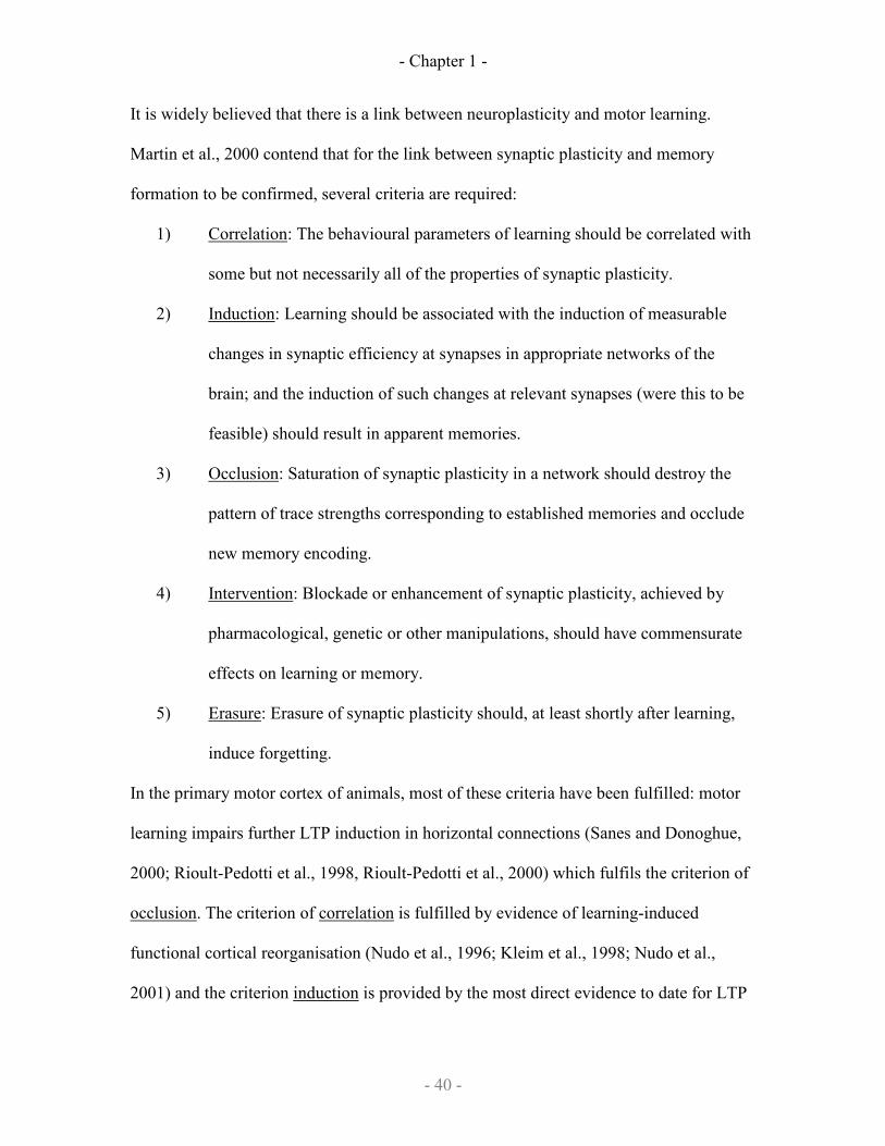
- Chapter 1 -
- 40 -
It is widely believed that there is a link between neuroplasticity and motor learning.
Martin et al., 2000 contend that for the link between synaptic plasticity and memory
formation to be confirmed, several criteria are required:
1) Correlation: The behavioural parameters of learning should be correlated with
some but not necessarily all of the properties of synaptic plasticity.
2) Induction: Learning should be associated with the induction of measurable
changes in synaptic efficiency at synapses in appropriate networks of the
brain; and the induction of such changes at relevant synapses (were this to be
feasible) should result in apparent memories.
3) Occlusion: Saturation of synaptic plasticity in a network should destroy the
pattern of trace strengths corresponding to established memories and occlude
new memory encoding.
4) Intervention: Blockade or enhancement of synaptic plasticity, achieved by
pharmacological, genetic or other manipulations, should have commensurate
effects on learning or memory.
5) Erasure: Erasure of synaptic plasticity should, at least shortly after learning,
induce forgetting.
In the primary motor cortex of animals, most of these criteria have been fulfilled: motor
learning impairs further LTP induction in horizontal connections (Sanes and Donoghue,
2000; Rioult-Pedotti et al., 1998, Rioult-Pedotti et al., 2000) which fulfils the criterion of
occlusion. The criterion of correlation is fulfilled by evidence of learning-induced
functional cortical reorganisation (Nudo et al., 1996; Kleim et al., 1998; Nudo et al.,
2001) and the criterion induction is provided by the most direct evidence to date for LTP

- Chapter 1 -
- 41 -
occurring physiologically in animals when LTP was induced in a spike-timing-dependent
fashion in the primary motor cortex of freely behaving primates, and produced changes in
primate cortical representation of motor movements (Jackson et al. 2006). The evidence
for the criterion intervention is provided only indirectly: concurrent cortical stimulation
with motor training enhanced performance and facilitated re-emergence of cortical maps
(Plautz et al., 2003), improved recovery and structural plasticity can be found with
concurrent cortical stimulation and rehabilitation in rats (Adkins-Muir & Jones, 2003),
and amphetamine has an enhancing effect on motor training and cortical maps (Barbay et
al., 2006). The only criterion left to be fulfilled in animals are erasure. Thus the evidence
for the link between LTP and motor learning in animals is very convincing.
1.5.2 Evidence from humans
The proof of causation between models of human neuroplasticity and human motor
learning is also incomplete. Changes in physiological parameters of excitability are
associated with ballistic motor learning, and this is termed practice-dependent plasticity,
(Classen et al., 1998; Muellbacher et al., 2001; Muellbacher et al., 2002. These changes
in representation are thought to be due to changes in synaptic efficacy probably involving
LTP (Ziemann et al., 2001; Boroojerdi et al., 2001), analogous to animal models of
practice-dependent plasticity (Sanes & Donoghue, 2000; Rioult-Pedotti et al., 1998,
Rioult-Pedotti et al., 2000). These changes are also accompanied by an increase in
functional MRI (fMRI) blood oxygen level-dependent (BOLD) signal indicative of
increased neural activity (Lotze et al., 2003). Changes in representation and BOLD signal
are believed to reflect changes in synaptic efficacy probably involving LTP (Ziemann et

- Chapter 1 -
- 42 -
al., 2001; Boroojerdi et al., 2001b), analogous to animal models (Sanes and Donoghue,
2000; Rioult-Pedotti et al., 1998, Rioult-Pedotti et al., 2000). Thus, there is ample
evidence to fulfill the criterion of intervention.
Inhibition and disinhibition of practice-dependent plasticity by pharmacologically
inhibiting or disinhibiting synaptic transmission and neuroplasticity is associated with
poorer rates of motor learning (Donchin et al., 2002; Ziemann et al., 2006) providing
evidence of correlation.
There is ample evidence for occlusion of motor learning and human neuroplasticity:
Ziemann et al., 2004 have investigated how different paradigms interact with each other
and have shown that prior ballistic motor learning interacts with subsequent artificial
induction of plasticity by rTMS. This suggests that ballistic motor learning and rTMS-
induced plasticity are likely to be interrelated. Stefan et al., 2006 also showed PAS
occlusion after force adaptation motor training. The criterion of erasure has been fulfilled
by evidence that performance improvements from ballistic motor learning are inhibited
when inhibitory 1Hz rTMS is delivered to the primary motor cortex shortly after motor
training (Muellbacher et al., 2002).
In humans, there is only limited evidence for induction. One study showed that sequence
learning could be enhanced by anodal TDCS (Nitsche et al., 2003) but there has been less
success with other paradigms (Agostino et al., 2007). The criterion of induction is
difficult to experimentally implement: artificial induction of neuroplasticity alone is
unlikely to encode useful information as effective motor learning is likely to require the
encoding of information into multiple brain networks. The need for multiple networks is
demonstrated by a study showing how motor practice alters MEPs and sensorimotor
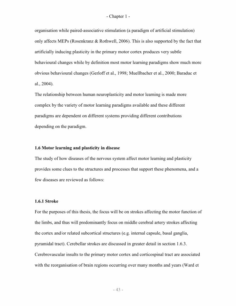
- Chapter 1 -
- 43 -
organisation while paired-associative stimulation (a paradigm of artificial stimulation)
only affects MEPs (Rosenkranz & Rothwell, 2006). This is also supported by the fact that
artificially inducing plasticity in the primary motor cortex produces very subtle
behavioural changes while by definition most motor learning paradigms show much more
obvious behavioural changes (Gerloff et al., 1998; Muellbacher et al., 2000; Baraduc et
al., 2004).
The relationship between human neuroplasticity and motor learning is made more
complex by the variety of motor learning paradigms available and these different
paradigms are dependent on different systems providing different contributions
depending on the paradigm.
1.6 Motor learning and plasticity in disease
The study of how diseases of the nervous system affect motor learning and plasticity
provides some clues to the structures and processes that support these phenomena, and a
few diseases are reviewed as follows:
1.6.1 Stroke
For the purposes of this thesis, the focus will be on strokes affecting the motor function of
the limbs, and thus will predominantly focus on middle cerebral artery strokes affecting
the cortex and/or related subcortical structures (e.g. internal capsule, basal ganglia,
pyramidal tract). Cerebellar strokes are discussed in greater detail in section 1.6.3.
Cerebrovascular insults to the primary motor cortex and corticospinal tract are associated
with the reorganisation of brain regions occurring over many months and years (Ward et
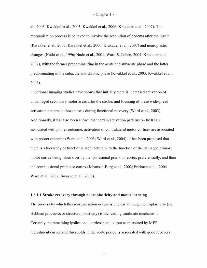
- Chapter 1 -
- 44 -
al., 2003; Kwakkel et al., 2003; Kwakkel et al., 2006; Krakauer et al., 2007). This
reorganisation process is believed to involve the resolution of oedema after the insult
(Kwakkel et al., 2003; Kwakkel et al., 2006; Krakauer et al., 2007) and neuroplastic
changes (Nudo et al., 1996; Nudo et al., 2001; Ward & Cohen, 2004; Krakauer et al.,
2007), with the former predominanting in the acute and subacute phase and the latter
predominating in the subacute and chronic phase (Kwakkel et al., 2003; Kwakkel et al.,
2006).
Functional imaging studies have shown that initially there is increased activation of
undamaged secondary motor areas after the stroke, and focusing of these widespread
activation patterns to fewer areas during functional recovery (Ward et al., 2003).
Additionally, it has also been shown that certain activation patterns on fMRI are
associated with poorer outcome: activation of contralateral motor cortices are associated
with poorer outcome (Ward et al., 2003; Ward et al., 2004). It has been proposed that
there is a hierarchy of functional architecture with the function of the damaged primary
motor cortex being taken over by the ipsilesional premotor cortex preferentially, and then
the contralesional premotor cortex (Johansen-Berg et al., 2002; Fridman et al., 2004
Ward et al., 2007; Swayne et al., 2008).
1.6.1.1 Stroke recovery through neuroplasticity and motor learning
The process by which this reorganisation occurs is unclear although neuroplasticity (i.e.
Hebbian processes or structural plasticity) is the leading candidate mechanism.
Certainly the remaining ipsilesional corticospinal output as measured by MEP
recruitment curves and thresholds in the acute period is associated with good recovery
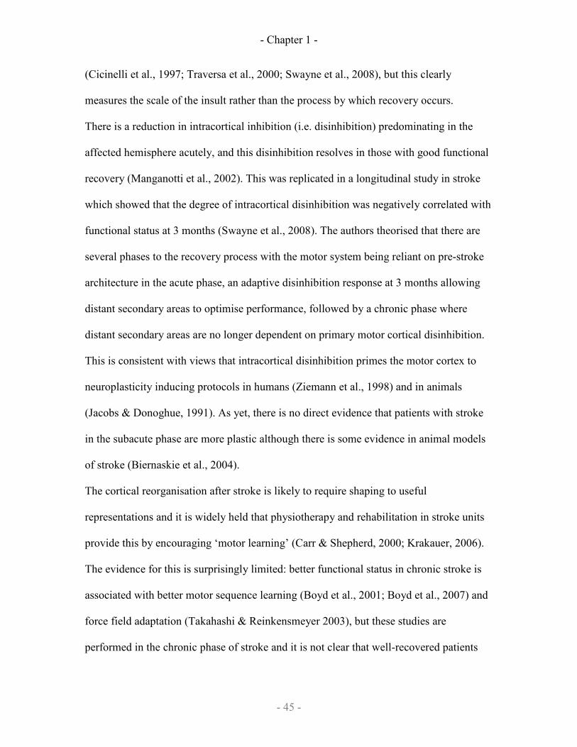
- Chapter 1 -
- 45 -
(Cicinelli et al., 1997; Traversa et al., 2000; Swayne et al., 2008), but this clearly
measures the scale of the insult rather than the process by which recovery occurs.
There is a reduction in intracortical inhibition (i.e. disinhibition) predominating in the
affected hemisphere acutely, and this disinhibition resolves in those with good functional
recovery (Manganotti et al., 2002). This was replicated in a longitudinal study in stroke
which showed that the degree of intracortical disinhibition was negatively correlated with
functional status at 3 months (Swayne et al., 2008). The authors theorised that there are
several phases to the recovery process with the motor system being reliant on pre-stroke
architecture in the acute phase, an adaptive disinhibition response at 3 months allowing
distant secondary areas to optimise performance, followed by a chronic phase where
distant secondary areas are no longer dependent on primary motor cortical disinhibition.
This is consistent with views that intracortical disinhibition primes the motor cortex to
neuroplasticity inducing protocols in humans (Ziemann et al., 1998) and in animals
(Jacobs & Donoghue, 1991). As yet, there is no direct evidence that patients with stroke
in the subacute phase are more plastic although there is some evidence in animal models
of stroke (Biernaskie et al., 2004).
The cortical reorganisation after stroke is likely to require shaping to useful
representations and it is widely held that physiotherapy and rehabilitation in stroke units
provide this by encouraging ‘motor learning’ (Carr & Shepherd, 2000; Krakauer, 2006).
The evidence for this is surprisingly limited: better functional status in chronic stroke is
associated with better motor sequence learning (Boyd et al., 2001; Boyd et al., 2007) and
force field adaptation (Takahashi & Reinkensmeyer 2003), but these studies are
performed in the chronic phase of stroke and it is not clear that well-recovered patients
- Chapter 1 -
- 46 -
have achieved their level of performance through preserved motor learning or if
preserved motor learning is just an epiphenomenon of a well-recovered patient. More
work will need to be done to establish this intuitive link.
1.6.1.2 The ‘hemispheric rivalry’ hypothesis
There is some evidence that the unlesioned primary motor cortex has a negative influence
on motor performance after stroke; this is termed the ‘hemispheric rivalry’ hypothesis.
The first suggestion of this occurred when it was found that while the damaged primary
motor cortex was hypoexcitable after a stroke, the undamaged primary motor cortex was
disinhibited after stroke (Cicinelli et al., 2003; Werhahn et al., 2003; Murase et al., 2004;
Ward & Cohen, 2004). The identification of transcallosal inhibitory pathways suggested
that the undamaged primary motor cortex suppressed the excitability of the damaged
primary motor cortex, thereby providing this negative influence (Murase et al., 2004).
This has prompted many studies on using rTMS or TDCS to suppress the excitability of
the unaffected hemisphere and enhance the excitability of the affected hemisphere
(Takeuchi et al., 2004; Ward & Cohen, 2004; Mansur et al., 2005; Hummel & Cohen,
2005 Kim et al., 2006; Fregni et al., 2006; Baggio et al., 2006; Talelli et al., 2007;
Takeuchi et al., 2008; Nowak et al., 2008). Additionally, Di Lazzaro et al., (2006)
reported an interesting single case of hemichorea secondary to stroke dramatically
responsive to cTBS to the primary motor cortex indicating that neuroplastic interventions
could be tailored to the condition. Whatever the case, if brain stimulation is intended for
clinical use, it is hoped that the increase in excitability might allow motor learning to
occur, but to date no studies have followed patients up long-term.
- Chapter 1 -
- 47 -
The physiotherapy protocol, constraint-induced modified therapy (CIMT) which has
recently been shown to be effective in enhancing stroke recovery (Wolf et al., 2006;
Liepert et al., 2006; Dahl et al., 2008; Gauthier et al., 2008) is thought to be another way
of modulating this rivalry. Restriction of the movements of the unaffected arm is
associated with less disinhibition of SICI in the affected hemisphere (Liepert et al., 2006),
more gray matter volume (Gauthier et al., 2008) and enlarged cortical representations
(Boake et al., 2007), although there is no evidence that transcallosal inhibition mediates
this mechanism as yet.
1.6.2 Parkinson’s Disease
The nigrostriatal degeneration that typifies Parkinson’s disease results in dopaminergic
deficiency in the striatum and thus impaired corticostriatal plasticity (discussed in section
1.3.2), but there is also some evidence of impairments in cortical plasticity.
Cortical plasticity as measured by PAS was impaired in Parkinson’s disease patients
(Ueki et al., 2006) and was restored by levodopa (Morgante et al., 2006). However
another study showed that the MEP changes did occur with Parkinson’s Disease but were
less focal while off dopaminergic medication (Bagnato et al., 2006). The discrepancy
may arise from the fact that these studies were performed on patients exposed to
dopaminergic therapy for some time, and this is known to affect the dopaminergic
receptor expression as well as predispose to dyskinesias. rTMS to the premotor cortex of
newly diagnosed Parkinson’s disease patients produced effects that lasted longer
compared to chronic patients (Buhmann et al., 2004), suggesting that future studies may
have to focus on newly diagnosed patients.

- Chapter 1 -
- 48 -
It is well-established that motor learning is impaired in Parkinson’s Disease: although
patients were able to improve their reaction times in sequence motor learning, they
required greater number of repetitions to acquire the same level of performance as age-
matched controls and this has been demonstrated in a number of studies (Ferraro 1993;
Pascual-Leone et al., 1993; Jackson et al., 1995; Sommer et al., 1999; Brown et al., 2003;
Wilkinson et al., 2007). An intriguing theory is that the dopaminergic deficiency in the
striatum of Parkinson’s Disease patients results in insufficient ‘motivation’ to increase
movement speed (Mazzoni et al., 2007), thus the impairments in sequence motor learning
is related to a lack of a ‘reward’ signal. Nonetheless, the clinical significance of this
impairment is unclear as the impairment was not correlated with functional scales and
only correlated with disability scores (Muslimovic et al., 2007).
These motor learning impairments do not translate to other paradigms: patients with
Parkinson’s Disease have no problems with eyeblink classical conditioning (Sommer et
al., 1999). Insufficient evidence exists for motor learning impairments in other
conditions.
The modulation of cortical excitability and plasticity has also been proposed as a possible
treatment modality for Parkinson’s Disease (Edwards et al., 2008) and numerous trials
have confirmed that there are some modest benefits to be gained from this form of
treatment (Siebner et al., 1999; Khedr et al., 2006; Lomarev et al., 2006; Hamada et al.,
2008).

- Chapter 1 -
- 49 -
1.6.2.1 Levodopa-induced dyskinesias
Chronic administration of levodopa is associated with the development of dyskinesia and
it has been postulated that this is due to altered plasticity from chronic dopaminergic
stimulation (Picconi et al., 2005; Pisani et al., 2005). Evidence that neuroplasticity is
altered by dopaminergic medication is provided by the study of Morgante et al., 2006
which demonstrates that levodopa medication fails to restore plasticity in Parkinson’s
Disease patients with levodopa-induced dyskinesias.
Modulation of neuroplasticity by 5Hz rTMS of the SMA have also been shown to
beneficial (Koch et al., 2005; Brusa et al., 2006; Edwards et al., 2008) and this requires
further larger studies.
1.6.3 Cerebellar disease
Cerebellar disease is associated with impairments of motor coordination and control,
typified by abnormal kinematics in single and multi-joint movements of ballistic or
reaching movements with a prolonged acceleration phase, a delayed deceleration phase
and terminal tremor (Berardelli et al., 1996; Day et al., 1998; Diedrichsen et al., 2007).
The role the cerebellum plays in motor learning though was first demonstrated by
impairments in classical Pavlovian nictitating membrane (eyelid) conditioning in rabbit
(McCormick et al., 1982; McCormick & Thompson, 1984), identifying the dentate-
interpositus nucleus as a vital structure in animals. This has been replicated in humans
and studies of cerebellar stroke patients exhibiting this impairment have suggested the
anterior cerebellar cortex to be a region where this motor learning occurs (Gerwig et al.,
2003, Gerwig et al., 2005, Gerwig et al., 2007).
- Chapter 1 -
- 50 -
Another form of motor learning dependent on the cerebellum is sequence learning.
Patients with cerebellar disease had little to no improvements in reaction time in the hand
ipsilateral to the cerebellar lesion (Pascual-Leone et al., 1993; Molinari et al., 1997;
Gómez-Beldarrain et al., 1998; Torriero et al., 2007).
Clearly plasticity within the cerebellum may be affected by any disease process affecting
the cerebellum. For example, in Fragile X syndrome there is evidence in animals and in
humans of impairments of cerebellar plasticity due to inactivation of the FMR1 gene
(Koekkoek et al., 2005) but neuroplasticity in other brain regions appear to be unaffected
by cerebellar disease with normal cortical plasticity to paired-associative stimulation in
patients with spinocerebellar ataxia 6 (Teo et al., 2008).
1.6.4 Dystonia
Primary dystonia is a disorder of abnormal muscle tone without any other neurological
disorder, and is believed to be related to abnormality in basal ganglia functioning
(Berardelli et al., 2002; Breakefield et al., 2008) and hyperexcitability of cortical and
brainstem interneurons (Sohn et al., 2004; Butefisch et al., 2005; Tisch et al., 2006;
Huang et al., 2006; Tamura et al., 2008).
Plasticity have been found to be abnormal in a number of distonic conditions: abnormal
response to facilitatory and inhibitory PAS in primary hand dystonia lasting longer and
with less somatotopy (Quartarone et al., 2003; Weise et al., 2006), abnormal response to
cTBS in manifesting and non-manifesting carriers of the DYT1 gene (Edwards et al.,
2006), abnormal responses to 1Hz rTMS in primary hand dystonia (Quartarone et al.,
2005; Baumer et al., 2007); and abnormal plasticity in the blink reflex circuit in
- Chapter 1 -
- 51 -
blepharospasm (Quartarone et al., 2006). Additionally, this abnormal plasticity also
extends to body parts unaffected by the dystonia (Quartarone et al., 2008) prompting the
hypothesis that the primary abnormality in dystonia is a problem of plasticity (Classen,
2003; Quartarone et al., 2008).
Primary adult-onset dystonia is associated with rehearsed skilled motor tasks, thereby
producing a proliferation of manifestations (e.g. writer’s cramp, musician’s dystonia,
golfer’s yip), and this is suggestive of a role for motor learning in this condition. Thus it
is surprising that the literature on motor learning in primary dystonia is sparse, and only
motor sequence learning has been reported in these patients. Motor sequence learning is
impaired in carriers of DYT1 gene (Ghilhardi et al., 2003), with increasing dependency
on the cerebellum even in non-manifesting carriers (Carbon et al., 2008). Clearly
additional work will need to be done to explore motor learning in patients with primary
dystonia.
1.6.5. Huntington’s disease
Huntington’s disease is a neurodegenerative disorder characterised by striatal
degeneration related to the huntingtin protein. The striatal degeneration is focused
predominantly on the striatal medium spiny neurons that receive dopaminergic and
glutamatergic input from the cortex. Animal models of Huntington’s disease display
abberent hippocampal and cortical synaptic plasticity (Usdin et al., 1999; Cummings et
al., 2006), which can be restored by BDNF (Lynch et al., 2007). Likewise, human models
of cortical plasticity also demonstrate impairment in responses to PAS (Crupi et al., 2008)
and bursts of high-frequency rTMS (Lorenzano et al., 2006). A pilot study has also
- Chapter 1 -
- 52 -
suggested that 1Hz rTMS reduces severity of chorea in Huntington’s disease for about 30
minutes (Brusa et al., 2005).
Motor learning in Huntington’s disease is limited to demonstrating impairment of
sequence learning (Knopman & Nissen, 1991) which is unsurprising considering the
dependence of this motor learning task on striatal circuits (Wilkinson & Jahanshahi
2007).
1.6.6 Alzheimer’s Disease
Alzheimer’s disease is a diffuse neurodenegerative process which is typified by loss of
declarative and episodic memory, and there is evidence that the beta-amyloid protein
which defines this condition impairs LTP, enhances LTD and reduces dendritic structural
plasticity in the rat hippocampus (Shankar et al., 2008). It is also established that there is
most paradigms of motor learning are spared, e.g. sequence motor learning (Willingham
et al., 1997), mirror drawing (Rouleau et al., 2002) and rotor pursuit (Deweer et al., 1994;
Jacobs et al., 1999). However, cortical measures of excitability and integration
demonstrate impairments in short-latency afferent inhibition (a parameter affected by
reduced muscarinic receptor activation), and a recent study has also demonstrated that
5Hz rTMS fails to facilitate MEPs in patients (Inghilleri et al., 2006). It is certainly
possible that in the early stage of the disease, although there are impairments in motor
cortical plasticity related to diffuse neurodegeneration, motor learning is still relatively
spared and motor learning paradigms are not sensitive enough to detect this. Whatever
the case, insufficient work has been done on Alzheimer’s Disease at this stage to arrive at
any definite conclusions.

- Chapter 1 -
- 53 -
1.6.7 Relevance of motor learning to neurorehabilitation
It is widely held that neurorehabilitation is a learning process; motor recovery from stroke
depends on re-learning lost skills and learning new compensatory movements
(Greenwood 2004; Greenwood & Ward 2007). Certainly it seems intuitive that
neurorehabilitation involves motor learning, as patients practice movements showing
accompanying motor recovery.
Longitudinal studies of stroke suggest that motor recovery proceeds through a series of
stereotypical stages over the first 6 months post-stroke (Kwakkel et al., 2006). Even
despite heterogeneity of stroke severity, a logistic regression model based on functional
state in the 1st week makes a reasonable prediction of recovery at 6 months accounting for
50% (Kwakkel et al., 2003) to 89% (Prabhakaran et al., 2008) of the variance of the
outcome, suggesting that there is a spontaneous process of recovery which is independent
of neurorehabilitation and motor learning, and is likely to be related to restoration of
activity in the ischemic penumbra, resolution of diaschisis and reduction of cerebral
oedema. The remaining variance appears relatively small, offering a pessimistic view of
the benefits of neurorehabilitation.
High intensity neurorehabilitation in the first 6 months after stroke produced more rapid
improvements in the first 6 months but had similar outcomes after 1 year (Sunderland et
al., 1994; Kwakkel et al., 2002). Although at first glance pessimistic, evidence showing
lasting benefit from specialised stroke units early in the condition (Indredavik et al.,
1999) suggests that duration or intensity is not the issue but quality of the
neurorehabilitation.
- Chapter 1 -
- 54 -
Thus the apparent small variance of outcome left accountable after spontaneous recovery
may be due to insufficient knowledge about how to provide high quality
neurorehabilitation that specifically targets the deficits. Thus, neurorehabilitation
therapies based on motor learning principles provide a framework to improve outcomes
and a number of such therapies have been developed: constraint-induced movement
therapy (CIMT) discussed previously (Wolf et al., 2006), EMG-triggered neuromuscular
stimulation (Bolton et al., 2004), arm ability training (Platz et al., 2001), robotic therapy
(Fasoli et al., 2003; Ferraro et al., 2003) and virtual reality interfaces (Holden, 2005).
Certainly the successful trial of CIMT is encouraging and provides a template for how to
translate motor learning theories into clinical applications (Academy of Medical
Sciences, 2004; Cumberland Consensus Statement, 2008).
1.7 Goal of this thesis
The goal of this thesis is to attempt to study the modulation of neuroplasticity, using
pharmacological agents and non-invasive brain stimulation and to describe how this links
with motor learning in humans. The use of pharmacological agents is particularly relevant
clinically as various drugs have been suggested as candidates to enhance
neurorehabilitation (Goldstein, 2000; Goldstein, 2006) and neuroplasticity (Ziemann et
al., 2006). The use of non-invasive brain stimulation to modulate excitability and/ or
plasticity to thereby affect motor learning in patients and normal humans is also attractive
(Nitsche et al., 2003; Takeuchi et al., 2005; Carey et al., 2006; Takeuchi et al., 2008).
- Chapter 1 -
- 55 -
This thesis presents 6 studies of neuroplasticity and motor learning focusing on different
aspects in this wide field:
Chapter 3 will study the effect of NMDA agonism on theta burst stimulation. The
research question was to determine if theta burst rTMS (like other forms of artificially-
induced plasticity) is also dependent on NMDA receptors.
Chapter 4 will study the role of various neurotransmitter systems on theta burst
stimulation. The research question was to determine if the after-effects of theta burst
rTMS could be modulated by various neuromodulators.
Chapter 5 will study the effect of theta burst stimulation on ballistic motor learning. The
research question was to determine of theta burst stimulation had any effect on ballistic
motor learning and to try to elucidate possible mechanisms for this effect.
Chapter 6 will study the role of different intracortical circuits in motor plasticity. The
research question would be to determine which inhibitory intracortical circuits regulate
motor plasticity.
Chapter 7 will study the intracortical circuits that modulate transcallosal inhibition and
their plasticity. The research question was whether transcallosal output has similar
properties to corticospinal output and whether this has a significant influence on the
primary motor cortex.
Chapter 8 will study the effect of theta burst stimulation on sequence learning. The
research question was whether sequence learning could be modulated by theta burst
rTMS and whether any effect was location-specific.

- Chapter 2 -
- 56 -
Chapter 2
General Methods
- Chapter 2 -
- 57 -
The development of transcranial magnetic stimulation (TMS) and repetitive transcranial
magnetic stimulation (rTMS) has allowed the experimental study and manipulation of
cortical excitability and plasticity in humans non-invasively. This chapter briefly
describes the methods used throughout this thesis.
2.1 Subjects
Subjects were recruited for each study in each chapter independently. Written informed
consent was obtained after the subject was provided with an information sheet. The
studies, information sheets and protocols were reviewed and approved by the National
Hospital for Neurology and Neurosurgery (NHNN) and University College London
Hospital (UCLH) research ethics committee. For the use of medicinal compounds in
these studies, The Medicines and Healthcare products Regulatory Authority (MHRA)
confirmed that these experiments did not constitute a clinical trial. All experiments
conformed to the standards set by the Declaration of Helsinki, and subjects were allowed
to drop out of the study at any point.
In total, 2 subjects dropped out of all the studies performed and their data were not
included in the analysis. The reasons for dropping out were: loss of contact as the subject
had left the country, and scalp discomfort from TMS when starting the experiment.
2.2 Electromyography (EMG)
EMG was recorded with silver disc surface electrodes were placed in a tendon-belly
montage. Unless otherwise stated, in most experiments the first dorsal interosseus (FDI)
muscle was used. The negative electrode was placed over the bulk of the FDI muscle and

- Chapter 2 -
- 58 -
the positive electrode over the first metacarpophalangeal joint. The ground electrode was
placed at the dorsum of the wrist.
EMG signals were amplified and filtered (20 Hz to 1 kHz) with a D360 amplifier
(Digitimer Limited, Welwyn Garden City, UK). The signals were sampled at 5000 Hz,
digitised using a laboratory interface (Power1401, Cambridge Electronics Design (CED),
Cambridge, UK) and stored on a personal computer for display and later off-line data
analysis. Each recording epoch at least 400 ms, of which at least 100 ms preceded the
TMS. Trials with muscle activity in the time preceding the TMS was discarded.
2.3 Transcranial magnetic stimulation (TMS)
Transcranial magnetic stimulation was performed with a pair of linked Magstim 2002
Bistim magnetic stimulators or a single Magstim 200 magnetic stimulator (Magstim
Company, Whitland, Dyfed, UK) depending on whether paired-pulse stimulation was
necessary. The magnetic stimuli produced a monophasic pulse with a rise-time of
approximately 100µs, decaying back to zero over approximately 0.8ms. The coil was
placed tangentially to the scalp with the handle pointing postero-laterally at a 45º angle to
the sagittal plane inducing a posterior-anterior current in the brain. This orientation was
chosen based on the findings that the lowest motor threshold is achieved when the
induced electrical current flows approximately perpendicular to the line of the central
sulcus.
We determined the optimal position for activation of the FDI muscles by moving the coil
in 0.5 cm steps around the presumed motor hand area of the motor cortex of both
hemispheres. The sites where stimuli of slightly suprathreshold intensity consistently

- Chapter 2 -
- 59 -
produced the largest MEPs in corresponding FDI muscle (referred to as “motor hot spot”;
M1) were marked with a red marker pen by drawing a crescent line following the anterior
bifurcation of the coil and a straight line indicating the orientation of the coil.
Resting motor threshold (RMT) was defined as the minimum stimulus intensity that
produced a MEP of at least 50 µV in 5 out of 10 consecutive trials. Active motor
threshold (AMT) was determined during voluntary tonic contraction at 5-10%of
maximum voluntary contraction (MVC) and was defined as the minimum intensity which
produced a MEP of at least 200 µV at least three out of five consecutive trials. Motor
thresholds were expressed as a percentage of maximum stimulator output (MSO).
2.4 Repetitive transcranial magnetic stimulation (rTMS)
Focal rTMS was applied over the left M1 with a Magstim Rapid stimulator and a flat
figure-of-eight coil with mean loop diameter of 9 cm. The magnetic stimulus had a
biphasic waveform with a pulse width of approximately 300 µs. The handle of the coil
pointed backwards and laterally at a 45° angle from the midline. The coil was placed
tangentially to the scalp in the same position used for the single TMS. All rTMS
protocols were in accordance with published safety recommendations (Wassermann
1998).
For most studies (Chapter 3, 4, 5 and 8), theta-burst stimulation was used (Huang et al.,
2005). This protocol can consist of either an inhibitory continuous protocol consisting of
a burst of 3 stimuli at 50Hz (a theta burst) repeated every 200ms for a total of 600 stimuli
(inhibitory TBS) or an excitatory intermittent protocol consisting of an 8 second pause

- Chapter 2 -
- 60 -
between every 10 theta bursts (excitatory TBS). Each stimulus was delivered at 80% of
the individual’s active motor threshold over the hand area of the motor cortex.
For chapter 7, conventional repetitive transcranial magnetic stimulation (rTMS) at 5 Hz
was delivered at 90%RMT split into two conditioning trains of 300 stimuli each with a
pause of 1 minute. This protocol was chosen as inhibitory intracortical circuits was the
predominant focus of the study and Quartarone et al. (2005) demonstrated that this
protocol affects the excitability of the inhibitory intracortical circuits without
significantly affecting the corticospinal excitability.
2.5 Behavioural measures
In chapter 5 and 6, motor learning was assessed using a simple ballistic motor task as
previously described (Muellbacher et al., 2001; Muellbacher et al., 2002) while in chapter
8, sequence learning was assessed using a variant of the serial reaction time task (SRTT).
Details of the tasks are described in the relevant chapters.
2.6 EMG data analysis
The primary measure of EMG signal in all studies was peak-to-peak amplitude of MEP
of individual trials. This was measured using a customized script for Signal software.
For paired-pulse and afferent stimulation measurements, peak-to-peak amplitude of
conditioned MEP was normalised against peak-to-peak amplitude of test MEP as
represented as a percentage. This is described in greater detail in Chapter 6.
For analysis of the ipsilateral silent period, the method described by Trompetto et al.
2004 was used. A more complete description is described in Chapter 7.

- Chapter 2 -
- 61 -
2.7 Statistical analysis
All statistical procedures were conducted using the statistical package, SPSS 12.0 (SPSS
for Windows 12.0 Chicago: SPSS; 2004). Numerical data are mean ± SD unless
otherwise stated. Significance for all procedures was set at a level of 0.05. Details of the
various t-tests, analysis of variance (ANOVA) and correlations are described in the
relevant chapters.
In Chapter 5, the circular statistics software Oriana (Oriana for Windows, Kovach
Computing Services, Anglesey, Wales) was used. The Rayleigh test was first performed
on the movement vectors in order to verify that they were not circularly uniform. The
concentration parameter (κ) was derived from the TMS-evoked movement vectors. κ is a
measure of the directionality of the distribution (Fisher, 1993) for which a value of 0
would represent no vector directionality (a distribution resembling a perfect circle), and
thus maximal motor output variability. As κ is a non-linear parameter, it was transformed
with log10 and a mean calculated for graphical representation. The non-parametric
Wilcoxon paired signed ranks test was used to test for significant differences.
- Chapter 3 -
- 62 -
Chapter 3
NMDA agonism and rTMS-induced plasticity
Work described in this chapter has been previously published:
Teo JT, Swayne OB, Rothwell JC. Further evidence for NMDA-dependence of the
after-effects of human theta burst stimulation. Clin Neurophysiol. 2007 Jul;
118(7):1649-51.
- Chapter 3 -
- 63 -
3.1 Introduction
The activation of postsynaptic N-methyl-D-aspartate (NMDA) receptors is one of the
central characteristics of the classical postsynaptic model of LTP and LTD: the
induction protocol (with high-frequency stimulation) activates postsynaptic NMDA
receptors allowing calcium influx which lead to long-term changes in synaptic
strength (MacDermott et al., 1986; Larson et al., 1986; Larson et al., 1988).
Pharmacological blockade of TDCS (Liebetanz et al., 2002) and PAS by the NMDA
antagonist dextromethorphan (Stefan et al., 2002) supports the idea that these two
paradigms are dependent on LTP.
Pharmacological blockade of the after-effects of TBS has very recently been shown
with the NMDA non-competitive antagonist, memantine (Huang et al., 2007). This
provides strong evidence that TBS is an NMDA-dependent phenomenon. The
question thus arises whether pharmacological activation of the NMDA receptors
would alter the effect of TBS. To date, there is only one study which shows an effect
of NMDA agonist activity on non-invasive stimulation. Nitsche et al. have shown that
the partial NMDA receptor agonist D-cycloserine prolongs the duration of the
excitatory effect of anodal TDCS without having any effect on the inhibitory effect of
cathodal TDCS (Nitsche et al., 2004a).
The purpose of this study is to study the effect of the NMDA partial agonist D-
cycloserine on the effects of TBS. We hypothesised that if the effects are mediated in
a straightforward manner by an NMDA-dependent process such as LTP then they
should be enhanced by the drug. D-cycloserine was chosen as it is one of the few

- Chapter 3 -
- 64 -
NMDA partial agonists available for safe use in humans and to allow a comparison
with the effects seen in TDCS (Nitsche et al; 2004a).
3.2 Study design
The study was designed as a double-blind placebo-controlled within-subjects design
and recruited with written informed consent. The studies, information sheets and
protocols were reviewed and approved by the National Hospital for Neurology and
Neurosurgery (NHNN) and University College London Hospital (UCLH) research
ethics committee, and conformed with the Declaration of Helsinki.
6 subjects (2 females, 4 males; 30.5 + 4.4SD years) were recruited. Each subject
participated for 2 sessions and was pseudo-randomised and counterbalanced to
receive either 2x50mg of D-cycloserine capsules or 2x50mg of placebo (ascorbic
acid) capsules at each session. Sessions were at least 1 week apart to avoid carry-over
effects. TBS consisted of bursts containing 3 pulses at 50 Hz at an intensity of 80%
AMT repeated at 200 ms intervals (i.e. at theta-bursts of 5 Hz) and given in an
intermittent theta burst stimulation pattern (iTBS): a 2 s train of TBS repeated every
10 s for 20 repetitions. This pattern is identical to that used by Huang et al (2005) who
showed it to be excitatory, producing increased MEP amplitudes lasting up to 20
minutes. The drug was taken 2 hours before theta-burst stimulation so that it
coincided with peak plasma concentration (van Berckel et al., 1997). 30 minutes
before receiving theta-burst stimulation, the ‘motor hotspot’ was identified and RMT
and AMT were measured. The intensity of stimulation to elicit an MEP amplitude of
approximately 1-mV was set and then a baseline measure of MEPs was recorded
consisting of 15 MEP trials. Immediately after, intermittent TBS (iTBS) was delivered
to the motor hotspot. MEPs were then recorded using the same intensity of
- Chapter 3 -
- 65 -
stimulation as baseline in blocks of 15 trials at various time points after completion of
iTBS: 1, 10, 20, 30 and 60 minutes. Subjects were permitted to leave the laboratory
for a break after the 30 minute measurement.
3.3 Drug
D-cycloserine is a partial agonist at the glycine-binding site of the NMDA receptor.
At low doses (50-250mg) it acts as an agonist, but at higher doses (>500mg) it acts as
an antagonist (Watanabe et al., 1992). Thus, the correct dosing of the drug is essential.
We chose 100mg as the dose for this study as it has been shown in other non-invasive
brain stimulation study to be sufficient to produce a pharmacological agonist effect
(Nitsche et al., 2004a). Two 50mg D-cycloserine capsules were used for the D-
cycloserine arm of the study while two 50mg ascorbic acid capsules were used for the
placebo arm.
Susan Ryan of The Guy’s Hospital Pharmacy Unit (Guy's & St. Thomas' NHS
Foundation Trust, London, SE1 7EH) assisted in preparing and quality-checking the
D-cycloserine. Rima Gupta and Karen Kneller, clinical trials pharmacists at The
National Hospital for Neurology and Neurosurgery, London assisted in dispensing the
drugs to the subjects in a blinded fashion.

- Chapter 3 -
- 66 -
0.0
0.2
0.4
0.6
0.8
1.0
1.2
1.4
Baseline 1 10 20 30 60
Time after rTMS (min)
Peak to peak amplitude of MEP (mV)
Placebo with rTMS
D-cycloserine with rTMS
Fig 3.1: The time course of the motor-evoked potentials (MEP) after
intermittent theta-burst rTMS in the placebo and D-cycloserine arm of
the study. The placebo arm of the study (triangles) shows facilitation of
MEPs after intermittent theta-burst rTMS while the D-cycloserine arm
of the study (squares) show inhibition of MEPs after intermittent theta-
burst rTMS. Error bars represent standard error of mean. Data for 6
subjects are shown.
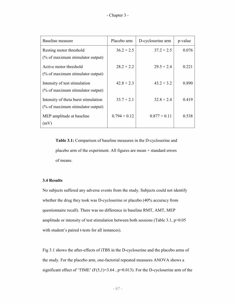
- Chapter 3 -
- 67 -
Baseline measure Placebo arm D-cycloserine arm p-value
Resting motor threshold
(% of maximum stimulator output)
36.2 + 2.5 37.2 + 2.5 0.076
Active motor threshold
(% of maximum stimulator output)
28.2 + 2.2 29.5 + 2.4 0.221
Intensity of test stimulation
(% of maximum stimulator output)
42.8 + 2.3 43.2 + 3.2 0.890
Intensity of theta burst stimulation
(% of maximum stimulator output)
33.7 + 2.1 32.8 + 2.4 0.419
MEP amplitude at baseline
(mV)
0.794 + 0.12 0.877 + 0.11 0.538
Table 3.1: Comparison of baseline measures in the D-cycloserine and
placebo arm of the experiment. All figures are mean + standard errors
of means.
3.4 Results
No subjects suffered any adverse events from the study. Subjects could not identify
whether the drug they took was D-cycloserine or placebo (40% accuracy from
questionnaire recall). There was no difference in baseline RMT, AMT, MEP
amplitude or intensity of test stimulation between both sessions (Table 3.1, p>0.05
with student’s paired t-tests for all instances).
Fig 3.1 shows the after-effects of iTBS in the D-cycloserine and the placebo arms of
the study. For the placebo arm, one-factorial repeated measures ANOVA shows a
significant effect of ‘TIME’ (F(5,1)=3.64 , p=0.013). For the D-cycloserine arm of the

- Chapter 3 -
- 68 -
experiment, one-factorial repeated measures ANOVA for ‘TIME’ shows a significant
effect of ‘TIME’ (F(5,1)=2.65, p=0.047). There were no post-hoc differences from
baseline after Bonferroni correction in either arm of the study. In both instances, the
after-effects of iTBS appear to dissipate by 60 minutes.
Two-factorial repeated measures ANOVA for ‘DRUG’ and ‘TIME’ showed a
significant ‘DRUG’x‘TIME’ interaction (F(1,5)=4.10, p=0.007) with no significant
‘DRUG’ effect (F(1,5)=3.02, p=0.143) or ‘TIME’ effect (F(5,1)=2.22, p=0.139). It is
clear in Figure 3.1 that iTBS has a facilitatory after-effect on MEP amplitudes when
given with placebo, but an inhibitory after-effect when given with D-cycloserine.
3.5 Discussion
This study shows that the effects of iTBS can be modulated by pharmacological
intervention with an NMDA partial agonist, D-cycloserine. Moreover, in the presence
of the drug the after-effects of iTBS were switched from facilitation to inhibition. It is
unlikely that the D-cycloserine dosage is inappropriate for agonist action as it is
identical to that used in a previous study using TDCS (Nitsche et al., 2004a). Also, it
is unlikely that D-cycloserine had an effect on cortical excitability independent of
iTBS, as it has been shown to have no effect on thresholds, MEPs or paired-pulse
paradigms using short-interval intracortical inhibition or intracortical facilitation
(Nitsche et al., 2004a).
One other possible site of action of D-cycloserine is on glycine-A strychnine-sensitive
receptors that are found predominantly in the brainstem and spinal cord. However, D-
cycloserine has a much lower affinity to these receptors (Kd of 90-100µmol/l), in

- Chapter 3 -
- 69 -
comparison to the glycine-B binding site on the NMDA receptor (Kd of 100–
300nmol/l) (D’Souza et al. 1995). Previous studies of the bioavailability and
pharmacokinetics of D-cycloserine show that a 150mg dose achieves 2 hour peak
plasma level of 35µmol/l (van Berckel et al. 1997) and a 250mg dose achieves a 2
hour peak CSF level of 12.8µmol/l (Nair et al. 1956). Thus, at the 100mg dose used in
this study D-cycloserine would not significantly bind to glycine-A receptors but
would be still be sufficient to bind to the glycine-B binding site of the NMDA
receptor.
On the other hand, it is not clear why the excitatory effects of TBS should be reversed
into inhibition by NMDA receptor activation by D-cycloserine. In particular, this
contrasts with the finding in TDCS where the excitatory effects are prolonged by D-
cyloserine (Nitsche et al., 2004a). One possible explanation could lie in the suggestion
(Huang et al., 2004; Huang et al., 2005) that the after-effects of TBS could be due to
two simultaneous excitatory and inhibitory effects with differing time courses and
strengths. It is thus conceivable that these two proposed effects have differing
NMDA-dependency, and are differentially modulated by D-cycloserine. This remains
to be confirmed but it is interesting to note that in the previous study with D-
cycloserine the effects on TDCS were not ‘symmetrical’: D-cycloserine prolonged
excitatory (anodal) TDCS but had no effect on inhibitory (cathodal) TDCS (Nitsche et
al., 2004a).
One possible limitation of this study is the lack of measurement of D-cylocserine
plasma of CSF levels. As D-cycloserine is a partial agonist, the effect is critically
dependent on the bioavailability of the drug; D-cycloserine may be producing agonist

- Chapter 3 -
- 70 -
and antagonist effects on NMDA-receptors in different subjects due to the lack of
control on this bioavailability. This cannot be excluded from this study, although it is
unlikely the drug dose was excessive as Huang et al., 2007 showed that NMDA-
antagonism completely blocks theta burst rTMS rather than altering its after-effects.
In rat sensorimotor cortex, the effects of theta burst stimulation is dependent on
NMDA receptors and the effects can be modulated by calcium channel blockade
(Castro-Alamancos et al., 1995) and this effect can be reversed depending on
extracellular calcium concentration (Barr et al., 1995). This reversal of the effect of
theta burst stimulation has also been observed with theta burst rTMS in humans with
reversal of effects depending on pre-existing cortical state (Gentner et al., 2008) and
immediate activity (Huang et al., 2008). Thus, it is conceivable that D-cycloserine
may have a similar effect on theta burst rTMS in humans by converting LTP induced
by iTBS into impaired excitability (i.e. LTD).
The next step would be to try and modulate the effects of TBS using drugs which act
on known neuromodulators like acetylcholine, dopamine and noradrenaline. This is
the focus of the next chapter.
- Chapter 4 -
- 71 -
Chapter 4
Theta burst stimulation and
neuromodulatory drugs
Work described in this chapter is being prepared submission for publication:
Swayne OB, Teo JT, Cheeran BJ, Greenwood R, Rothwell JC. The interaction between
human theta burst stimulation and nicotine reveals a role for performance variability in
motor learning (preparing for submission to Cerebral Cortex).
- Chapter 4 -
- 72 -
4.1 Introduction
The previous chapter demonstrated that the effect of theta burst stimulation is
modulated by activity at the NMDA receptor, the initial molecular trigger of long term
potentiation as studied in animals.
Long term potentiation is also known to be modulated by a variety of other
neurotransmitters: the noradrenergic system, the dopaminergic system, the cholinergic
system and the GABA-ergic system have all been shown to alter the duration, degree
and persistence of long term potentiation (see section 1.3 in the Introduction). The
mechanism of action varies depending on the neurotransmitter, as different receptors
intervene at different points of the molecular pathway:
As mentioned in Chapter 1, dopaminergic receptors have been suggested to play a role
in modulating striatal and hippocampal plasticity, as well as in homeostatic plasticity
and metaplasticity. Likewise nicotinic acetylcholine receptors and adrenergic receptors
have also been implicated in animal models of plasticity as well.
Thus, based on the findings in the literature of animal experiments with theta burst
stimulation, this chapter will explore the effects of various neurotransmitter systems on
the after-effects of theta burst stimulation: study A explored the noradrenergic and
dopaminergic system using amphetamine and levo-dopa and study B used nicotine to
study the cholinergic system.
4.2 Amphetamine & levodopa
In humans, a comprehensive study of cholinergic, noradrenergic and dopaminergic
systems in practice-dependent plasticity showed that noradrenergic activation enhances
this plasticity transiently while cholinergic and dopaminergic activation enhances
practice-dependent plasticity beyond the period of training (Meintszchel et al., 2006).
- Chapter 4 -
- 73 -
This strongly suggests a role for dopaminergic and cholinergic systems in human motor
system plasticity. The role of dopamine receptors in modulating LTP in the striatum is
well-established in animal models (Centonze et al., 1999; Calebresi et al., 2007), so it is
reasonable to expect that activation of dopaminergic system would enhance rTMS-
induced plasticity if the latter is analogous to LTP.
Activation of noradrenergic and dopaminergic systems also affect cortical excitability
independent of plasticity changes. Amphetamine has been shown to enhance MEP
amplitude (Boroojerdi et al., 2001a; Ziemann, 2004). Thus any study of these two
neuromodulatory systems will have to consider the drug effects on cortical excitability
independent of rTMS-induced plastic changes.
In this study, we aimed to see if rTMS-induced plasticity can be enhanced in the same
way as practice-dependent plasticity was by Meintszchel et al. (2006) using the drugs,
dexamphetamine and L-DOPA in healthy normal subjects.
4.2.1 Study design
A double-blind randomised placebo-controlled crossover trial was conducted. 10
subjects (4 females, 6 males; 31.1 + 5.0SD years) were recruited for this study Subjects
received amphetamine, L-DOPA or placebo before the delivery of rTMS, which was
timed to coincide with the peak plasma concentration of the drug (2.5 hour for
amphetamine, 1 hour for L-DOPA and 2 hour for placebo). After rTMS, MEPs were
then recorded immediately and 5, 10, 20, 30 and 40 minutes after rTMS. Paired-pulse
stimulation with an interstimulus interval of 2ms measuring short-interval intracortical
inhibition (SICI) was also recorded every 5 minutes up to 20 minutes, then less
frequently up to 40 minutes.

- Chapter 4 -
- 74 -
In all cases, MEPs were recorded from the left FDI (TMS pulses on the right primary
motor cortex). The rTMS protocol used was intermittent theta-burst stimulation (iTBS)
which is an excitatory protocol (Huang et al. 2005).
4.2.1.1 Drug
Two Madopar 100/25 capsules (Roche Products Ltd) and two Dexamphetamine 5mg
tablets (Celltech, UCB Pharma Ltd.) were used in the L-DOPA and the
dexamphetamine arms of the study respectively. Two 50mg ascorbic acid mint-
flavoured lozenges were used for the placebo arm of the experiment. Both investigator
and subjects were blinded to the drug.
4.2.1.2 Statistical analysis
A two-factorial analysis of variance (ANOVA) with repeated measures were used with
factors: “DRUG” (Amphetamine. L-DOPA, Placebo) and “TIME” (Baseline, 0min,
10min, 20min, 30min, 40min) for only the 6 subjects who completed all three drug
arms (n=6).
Additionally, as 10 subjects completed the amphetamine and placebo arms, two non-
independent ANOVAs were also performed comparing only the amphetamine or L-
DOPA with the placebo arm. Any significance is corrected by a Bonferroni correction
due to the two non-independent comparisons.
4.2.2 Results
No subjects suffered any adverse events from the study. Subjects were able to identify
accurately when amphetamine was taken (70% accuracy) due to a subjective feeling of
euphoria. However, subjects did not experience any significant side effects with either

- Chapter 4 -
- 75 -
L-DOPA or placebo but was able to identify above chance which drug was taken (L-
DOPA at 67% accuracy, placebo at 60% accuracy).
There was no difference in baseline RMT, AMT, MEP amplitude or intensity of test
stimulation between both sessions (p>0.05 with student’s paired t-tests for all
instances). This is summarised in Table 4.1.
Baseline measure Placebo
arm
Amphetamine
arm
L-DOPA
arm
Intensity of test stimulation
(% of maximum stimulator output)
47.5 + 3.9 45.2 + 3.6 44.8 + 3.1
Intensity of paired-pulse stimulation
(% of maximum stimulator output)
24.5 + 1.60 22.7 + 0.97 24.0 + 1.73
Intensity of theta burst stimulation
(% of maximum stimulator output)
36.4 + 2.4 36.6 + 2.4 34.8 + 2.7
MEP amplitude at baseline
(mV)
1.08 + 0.11 1.07 + 0.13 1.19 + 0.14
Table 4.1: Comparison of baseline measures in the amphetamine, L-
DOPA and placebo arm of the experiment. All figures are mean +
standard errors of means.
Fig 4.1 shows the after-effects of intermittent theta-burst stimulation in the
amphetamine and the placebo arms of the study for 10 subjects, and Fig 4.2 shows the
after-effects of iTBS in the L-DOPA and placebo arms of the study for 6 subjects. The
three factorial ANOVA with all three drug arms and only the 6 subjects which
completed the study
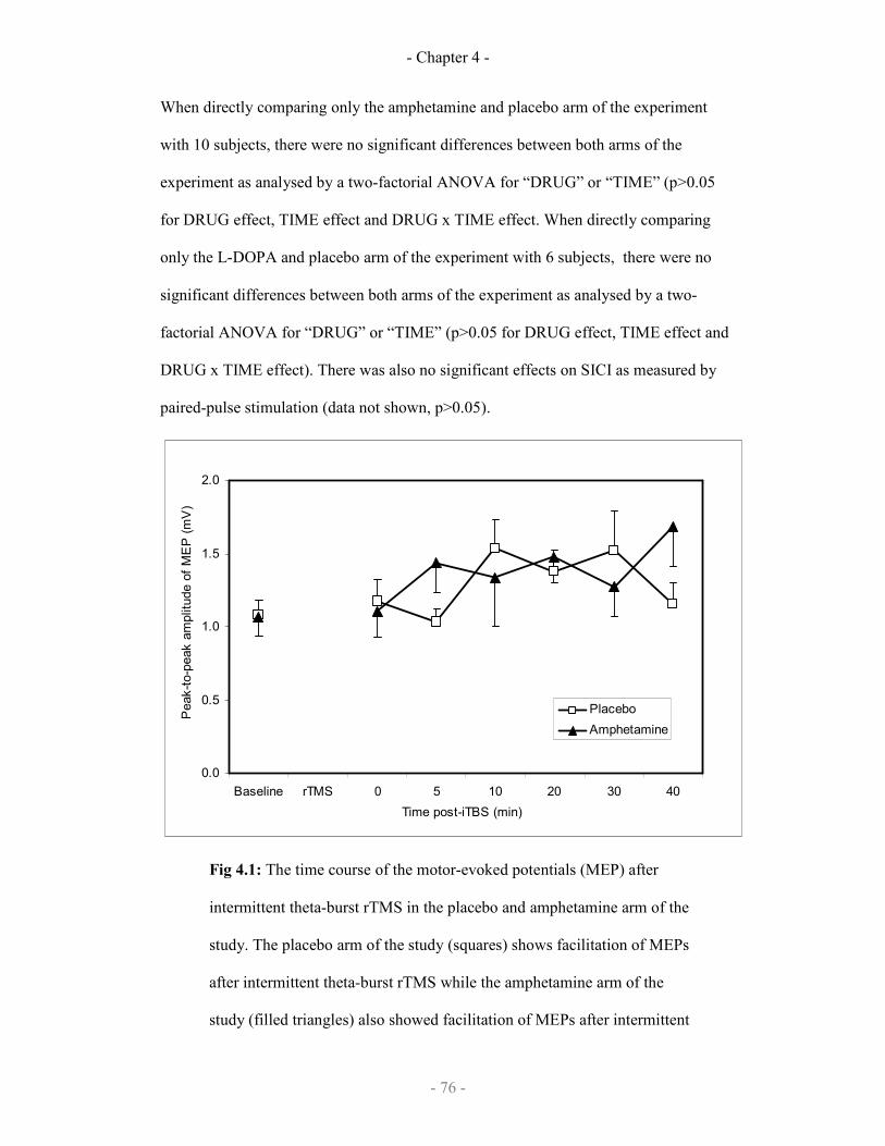
- Chapter 4 -
- 76 -
When directly comparing only the amphetamine and placebo arm of the experiment
with 10 subjects, there were no significant differences between both arms of the
experiment as analysed by a two-factorial ANOVA for “DRUG” or “TIME” (p>0.05
for DRUG effect, TIME effect and DRUG x TIME effect. When directly comparing
only the L-DOPA and placebo arm of the experiment with 6 subjects, there were no
significant differences between both arms of the experiment as analysed by a two-
factorial ANOVA for “DRUG” or “TIME” (p>0.05 for DRUG effect, TIME effect and
DRUG x TIME effect). There was also no significant effects on SICI as measured by
paired-pulse stimulation (data not shown, p>0.05).
0.0
0.5
1.0
1.5
2.0
Baseline rTMS 0 5 10 20 30 40
Time post-iTBS (min)
Peak-to-peak amplitude of MEP (mV)
Placebo
Amphetamine
Fig 4.1: The time course of the motor-evoked potentials (MEP) after
intermittent theta-burst rTMS in the placebo and amphetamine arm of the
study. The placebo arm of the study (squares) shows facilitation of MEPs
after intermittent theta-burst rTMS while the amphetamine arm of the
study (filled triangles) also showed facilitation of MEPs after intermittent
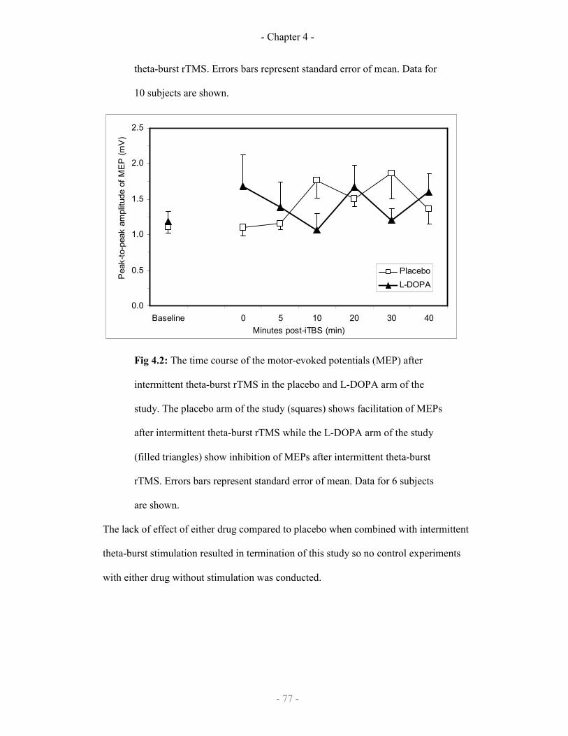
- Chapter 4 -
- 77 -
theta-burst rTMS. Errors bars represent standard error of mean. Data for
10 subjects are shown.
0.0
0.5
1.0
1.5
2.0
2.5
Baseline 0 5 10 20 30 40
Minutes post-iTBS (min)
Peak-to-peak amplitude of MEP (mV)
Placebo
L-DOPA
Fig 4.2: The time course of the motor-evoked potentials (MEP) after
intermittent theta-burst rTMS in the placebo and L-DOPA arm of the
study. The placebo arm of the study (squares) shows facilitation of MEPs
after intermittent theta-burst rTMS while the L-DOPA arm of the study
(filled triangles) show inhibition of MEPs after intermittent theta-burst
rTMS. Errors bars represent standard error of mean. Data for 6 subjects
are shown.
The lack of effect of either drug compared to placebo when combined with intermittent
theta-burst stimulation resulted in termination of this study so no control experiments
with either drug without stimulation was conducted.
- Chapter 4 -
- 78 -
4.2.3 Discussion
There is a lack of effect of amphetamine and L-DOPA compared to placebo on the
after-effects of TBS. There is no good explanation for this since these two drugs are
known to modulate the after-effects of transcranial direct current stimulation (Nitsche et
al., 2004b; Nitsche et al., 2006) and practice-dependent plasticity (Tegenthoff et al.,
2004; Meintzchel et al., 2006). Additionally amphetamine has been shown to modulate
rTMS-induced plasticity in a complex fashion: it suppresses MEP enhancement from
rTMS during ischaemic nerve block despite enhancing MEP during ischaemic nerve
block alone (Ziemann et al., 2002). Thus the lack of effect in study B could be related
to the paradigm used (i.e. theta burst stimulation) which may be similar but not
identical to the paradigms used in previous studies.
However, it should be noted there is a very strong effect of TBS in the placebo arm of
the experiment (Fig 4.1 and Fig 4.2). This strong effect is much more potent than the
effect seen in the original description of TBS (Huang et al., 2005), lasting in excess of
20 minutes. Thus one possible explanation is that the placebo effects are particularly
potent. Certainly some subjects described the expectation of possibly receiving
amphetamine as ‘exciting’. Thus, it is conceivable that this associated anticipation may
have obscured the results of amphetamine since emotional states are known to change
in neurotransmitter levels and cortical excitability as measured by MEPs.
The lack of effect with L-DOPA is also disappointing. L-DOPA has been shown to
affect practice-dependent plasticity most prominently in the elderly as well as in
chronic stroke patients (Floel et al., 2005a & 2005b). The likely explanation for the
absence of effect is probably due to an error in study design: young subjects (24-35
years of age) were used in this study (unlike the above studies) and one would expect
- Chapter 4 -
- 79 -
that there might be a ‘ceiling’ effect in young normal subjects who are not deficient in
dopaminergic transmission.
Thus, no firm conclusions can be drawn from study B about the role of noradrenergic
and dopaminergic systems in rTMS-induced plasticity in particular theta-burst-induced
plasticity. The experimental design will need closer scrutiny in the next experiment,
where the role of nicotinic acetylcholine receptors in rTMS-induced plasticity is
studied.
4.3 Nicotine
Acetylcholine receptors are widely distributed in the central nervous system and are
divided into nicotinic acetylcholine receptors and muscarinic acetylcholine receptors.
Both receptor types are expressed in sensorimotor cortex with variable expression on
different interneuron populations (Sihver et al., 1998; Alkondon et al., 2000). There is
significant evidence for muscarinic acetylcholine receptors playing a role in the cortico-
cortical circuit mediating short afferent inhibition (SAI), as the muscarinic antagonist
scopolamine inhibits SAI (Di Lazzaro et al., 2000) and SAI has been demonstrated to
be deficient in Alzheimer’s disease but not in fronto-temporal dementia (Di Lazzaro et
al., 2004) or mild cognitive impairment (Sakuma et al., 2007). The acetylcholinesterase
inhibitor, taurine, has also been shown to affect intracortical circuits by decreasing
short-interval intracortical inhibition, SICI (Korchounov et al., 2005).
Nicotinic acetylcholine receptors are known to be expressed on interneurones in the
cerebral cortex including the somatosensory and primary motor cortex (Sihver et al,
1998; Alkondon et al., 2000; Xiang et al., 1998; Christophe et al., 2002). In humans,
nicotine while not altering SAI, SICI or MEPs in normal subjects, corrects the
abnormalities in SICI seen in patients with Tourette’s syndrome (Orth et al. 2005). This

- Chapter 4 -
- 80 -
suggests that nicotinic acetylcholine receptors have a role to play in modulating motor
cortex activity despite not being easily detectable in normal subjects using single or
paired-pulse TMS.
Nicotinic acetylcholine receptors are also known to be involved in various animal
models of hippocampal and striatal synaptic plasticity. Nicotine enhances presynaptic
and postsynaptic LTP: nicotine can also enhance the generation of LTP (Ge & Dani,
2005), via presynaptic effects on glutamate release in the ventral tegmentum
(Mansvelder & McGehee, 2000), and nicotine enhances LTP induction primarily
through activation of postsynaptic nAChRs in the hippocampus (Fujii et al., 1999; Ji et
al., 2001). Thus, the purpose of this study is to determine if activating nicotinic
acetylcholine receptors with nicotine can modulate rTMS-induced plasticity in the
primary motor cortex in normal healthy non-smoking subjects.
4.3.1 Study design
A double-blind randomised placebo-controlled crossover trial was conducted with
subjects receiving either nicotine or placebo 1 hour before the delivery of rTMS. 10
subjects (3 females, 7 males; 29.6 + 4.7SD years) were recruited.
After rTMS, MEPs were then recorded every 5 minutes for 40 minutes. Paired-pulse
stimulation with an interstimulus interval of 2ms measuring SICI was also recorded
every 5 minutes up to 20 minutes, then less frequently up to 40 minutes. Additionally,
in a separate control experiment investigating the effects of nicotine independent of
rTMS, subjects received nicotine and had MEPs measured for 40 minutes. Current
smokers and recent ex-smokers, within the past year, were excluded from the study.
- Chapter 4 -
- 81 -
In all cases, MEPs were recorded from the left FDI (TMS pulses on the right primary
motor cortex). The rTMS protocol used was intermittent theta-burst stimulation (iTBS)
which is an excitatory protocol (Huang et al. 2005) lasting about 20 minutes.
4.3.1.1 Drug
Two 2mg mint-flavoured nicotine lozenges were used for the nicotine arm of the
experiment. Lozenges were selected because of the fast pharmacokinetics profile with
rapid absorption (Hukkanen et al., 2005; Russel, 1987; Tobacco Advisory Group,
2000); additionally, swallowing the tablet does not significantly alter the
pharmacokinetics profile of the nicotine (Choi et al., 2003). Two 50mg ascorbic acid
mint-flavoured lozenges were used for the placebo arm of the experiment. To mask the
distinctive taste of the nicotine lozenges subjects also took strong menthol lozenges
(Fisherman’s Friend) before taking either drug and continued to take the menthol
lozenges while they had the drug in the mouth. Both investigator and subjects were
blinded to the drug taken during each session.
4.3.1.2 Statistical analysis
Each drug arm was compared individually with the placebo arm in their respective
studies using a two-factorial analysis of variance (ANOVA) with repeated measures
were used with factors: “DRUG” and “TIME”.
Experimental
arms compared
Factor 1 Factor 2
Nicotine vs.
placebo
“DRUG” “TIME”: Baseline, 0-5min, 10-15min, 20-
25min, 30-35min, 40min
Nicotine-alone vs.
Nicotine-rTMS
“rTMS” “TIME”: Baseline, 0-5min, 10-15min, 20-
25min, 30-35min, 40min
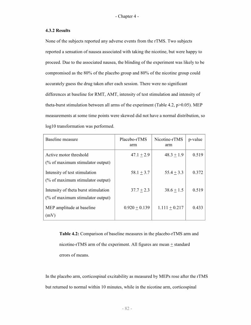
- Chapter 4 -
- 82 -
4.3.2 Results
None of the subjects reported any adverse events from the rTMS. Two subjects
reported a sensation of nausea associated with taking the nicotine, but were happy to
proceed. Due to the associated nausea, the blinding of the experiment was likely to be
compromised as the 80% of the placebo group and 80% of the nicotine group could
accurately guess the drug taken after each session. There were no significant
differences at baseline for RMT, AMT, intensity of test stimulation and intensity of
theta-burst stimulation between all arms of the experiment (Table 4.2, p>0.05). MEP
measurements at some time points were skewed did not have a normal distribution, so
log10 transformation was performed.
Baseline measure Placebo-rTMS
arm
Nicotine-rTMS
arm
p-value
Active motor threshold
(% of maximum stimulator output)
47.1 + 2.9 48.3 + 1.9 0.519
Intensity of test stimulation
(% of maximum stimulator output)
58.1 + 3.7 55.4 + 3.3 0.372
Intensity of theta burst stimulation
(% of maximum stimulator output)
37.7 + 2.3 38.6 + 1.5 0.519
MEP amplitude at baseline
(mV)
0.920 + 0.139 1.111 + 0.217 0.433
Table 4.2: Comparison of baseline measures in the placebo-rTMS arm and
nicotine-rTMS arm of the experiment. All figures are mean + standard
errors of means.
In the placebo arm, corticospinal excitability as measured by MEPs rose after the rTMS
but returned to normal within 10 minutes, while in the nicotine arm, corticospinal
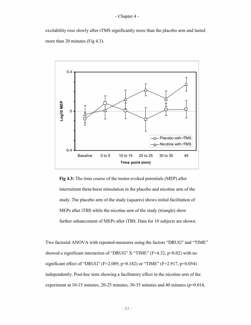
- Chapter 4 -
- 83 -
excitability rose slowly after rTMS significantly more than the placebo arm and lasted
more than 20 minutes (Fig 4.3).
-0.4
0
0.4
Baseline 0 to 5 10 to 15 20 to 25 30 to 35 40
Time point (min)
Log10 MEP
Placebo with rTMS
Nicotine with rTMS
Fig 4.3: The time course of the motor-evoked potentials (MEP) after
intermittent theta-burst stimulation in the placebo and nicotine arm of the
study. The placebo arm of the study (squares) shows initial facilitation of
MEPs after iTBS while the nicotine arm of the study (triangle) show
further enhancement of MEPs after iTBS. Data for 10 subjects are shown.
Two factorial ANOVA with repeated-measures using the factors “DRUG” and “TIME”
showed a significant interaction of “DRUG” X “TIME” (F=4.32, p=0.02) with no
significant effect of “DRUG” (F=2.089, p=0.182) or “TIME” (F=2.917, p=0.054)
independently. Post-hoc tests showing a facilitatory effect in the nicotine arm of the
experiment at 10-15 minutes, 20-25 minutes, 30-35 minutes and 40 minutes (p=0.014,
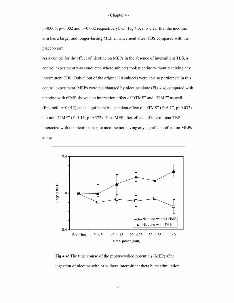
- Chapter 4 -
- 84 -
p=0.006, p=0.002 and p=0.002 respectively). On Fig 4.3, it is clear that the nicotine
arm has a larger and longer-lasting MEP enhancement after iTBS compared with the
placebo arm.
As a control for the effect of nicotine on MEPs in the absence of intermittent TBS, a
control experiment was conducted where subjects took nicotine without receiving any
intermittent TBS. Only 9 out of the original 10 subjects were able to participate in this
control experiment. MEPs were not changed by nicotine alone (Fig 4.4) compared with
nicotine with rTMS showed an interaction effect of “rTMS” and “TIME” as well
(F=4.660; p=0.012) and a significant independent effect of “rTMS” (F=6.77, p=0.032)
but not “TIME” (F=1.11, p=0.372). Thus MEP after-effects of intermittent TBS
interacted with the nicotine despite nicotine not having any significant effect on MEPs
alone.
-0.4
0
0.4
Baseline 0 to 5 10 to 15 20 to 25 30 to 35 40
Time point (min)
Log10 MEP
Nicotine without rTMS
Nicotine with rTMS
Fig 4.4: The time course of the motor-evoked potentials (MEP) after
ingestion of nicotine with or without intermittent theta burst stimulation.
- Chapter 4 -
- 85 -
The rTMS arm of the study (filled triangles) shows facilitation of MEPs
after iTBS and nicotine while the nicotine without rTMS arm of the study
(squares) does not have any significant effect on MEPs. Data for 9 subjects
are shown (1 subject dropped out from one arm).
Paired pulse stimulation of corticospinal excitability to measure SICI did not show any
significant differences before and after rTMS in either experimental arm (data not
shown, p>0.05).
4.3.3 Discussion
The transient increase in MEP amplitudes following iTBS delivered to the motor cortex
was first described in 2005 (Huang et al 2005) and has been widely reproduced (Di
Lazzaro et al 2006; Huang et al 2007a; Huang et al 2007b). With nicotine this increase
was more marked, started later and was more prolonged. Excitability was still increased
at 40 minutes, and it is unclear how long this would have persisted. Nicotine alone did
not alter excitability, consistent with previous reports (Orth et al 2005), suggesting that
the effect resulted from an iTBS-drug interaction. As nicotine up-regulates LTP this
would seem a reasonable candidate mechanism for enhancing the effect of iTBS on
corticospinal excitability.
Thus nicotine has been identified as an agent that can facilitate and prolong the
enhancement of corticospinal excitability by intermittent theta-burst stimulation. It is
therefore hypothesised that using nicotine to enhance the effect of intermittent theta-
burst stimulation on corticospinal excitability might also enhance motor learning. If this
was the case, nicotine and intermittent theta burst stimulation may be an attractive tool
for clinical application in rehabilitation and learning. This is the focus of the next
chapter that studied the effect of nicotine, TBS and motor learning.

- Chapter 5 -
- 86 -
Chapter 5
Theta burst stimulation and
ballistic motor learning
Work described in this chapter has been submitted to:
Swayne OB, Teo JT, Greenwood R, Rothwell JC. Nicotine modulates the effects of
theta bust stimulation (accepted by Clinical Neurophysiology; 22nd June 2009).
Teo JT, Swayne OB, Cheeran BJ, Greenwood R, Rothwell JC. Human theta burst
stimulation enhances subsequent motor learning while increasing performance
variability (submitted to Cerebral Cortex; 27th June 2009).

- Chapter 5 -
- 87 -
5.1 Introduction
From Chapter 4, nicotine has been identified as an agent that can facilitate and
prolong the enhancement of corticospinal excitability of intermittent theta-burst
stimulation. It is therefore hypothesised that using nicotine to enhance the effect of
intermittent theta-burst stimulation on corticospinal excitability might also enhance
motor learning. If this was the case, nicotine and intermittent theta burst stimulation
may be an attractive tool for clinical application in rehabilitation and learning.
A simple ballistic motor task was used to assess motor learning as it is well-
established to be dependent in its early phase on the primary motor cortex
(Muellbacher et al., 2001; Muellbacher et al., 2002). This makes the ballistic motor
learning paradigm more suitable for study compared to other paradigms of motor
learning which have not been shown to involve the primary motor cortex, like force
field adaptation learning (Baraduc et al., 2004) or serial reaction time tasks which
involve learning of abstract sequences which may or may not be dependent on the
primary motor cortex.
5.2 Study Design
In this study, the same nicotine and rTMS protocol as in study C (see Chapter 4) was
reproduced, and the subject was required to perform a motor learning task during the
period of peak MEP enhancement according to study C (10-20 minutes). A new group
of 10 subjects (2 females, 8 males; 29.5 + 4.1SD years) were recruited for this study.
MEP measurements were not made during the motor learning as any measurements
would not be reliable during activity. This study was conducted as a randomised
controlled blinded trial with a cross-over design over the four experimental arms
(placebo and sham-rTMS, placebo and real-rTMS, nicotine and sham-rTMS, and

- Chapter 5 -
- 88 -
nicotine and real-rTMS). The order of the sessions was pseudo-randomised. To
minimise carry-over effects in a motor learning task, intersession gap was kept very
long (mean 31 days, range 14-55 days). Subject and investigator were both blinded to
the drug, and subjects were also blinded to whether real-rTMS or sham-rTMS was
delivered. Current smokers and recent ex-smokers (within the past year) were
excluded from the study.
5.2.1 Ballistic motor learning
Motor learning was assessed using a simple ballistic motor task as previously
described (Muellbacher et al., 2001; Muellbacher et al., 2002). This task was chosen
as it subjectively resembles the efforts made by stroke patients when trying to move a
paretic limb with more force, as opposed to other motor learning tasks which often do
not resemble motor learning as it occurs in a rehabilitative setting.
The left hand was positioned supine on a board with the wrist, metacarpophalangeal
and distal interphalangeal joints fixed with Velcro straps. The thumb was left
unsecured and could abduct and oppose freely. A piezoresistive monoaxial
accelerometer (Model SA-105 vibrometer, Fribourg, Switzerland) was attached on the
lateral aspect of the left thumb proximal phalanx with the maximal vector being
thumb abduction. The accelerometer signal was sampled at 5000Hz and not filtered.
The left (non-dominant) thumb was used in all conditions to minimise ceiling effects
(which may occur in the dominant hand).
Subjects were familiarized with the motor task (to abduct the left thumb as quickly as
possible in the direction of the accelerometer in time with a 0.5Hz audio metronome
to maximize initial peak acceleration using the computer monitor for explicit visual
feedback. The monitor displayed the last three thumb abductions. The investigator

- Chapter 5 -
- 89 -
motivated the subject by providing a target ~10% above the highest acceleration in the
last three thumb abductions. Subjects were allowed ten successful movements for
familiarization before each experiment. Successful movements were defined as rapid
ballistic thumb abduction movements of at least 1g during the first 100ms of
movement. Subjects were instructed to maintain the original thumb position by
ensuring that the accelerometer signal returned to baseline (+0.05g) after each
movement. Subjects performed 6 training blocks separated by rest blocks of 1 minute.
Each training block lasted 1 minute, consisting of 30 training movements.
Unsuccessful training movements were not compensated for unless <27 successful
training movements (<90%) were performed in the training block.
5.2.2 Data analysis
For study D, to exclude baseline differences, data from Block1 of each experimental
arm was also compared in a two-factorial ANOVA with repeated measured using
factors “DRUG” (Nicotine or placebo) and “rTMS” (Sham-rTMS or real-rTMS).
Three factorial ANOVA with repeated-measures was then conducted using the factors
“DRUG”, “rTMS” and “BLOCK”. If there was significant interaction of “DRUG”,
“rTMS” and “BLOCK”, two factorial ANOVAs were done comparing the learning
for all four arms of the experiment as follows:

- Chapter 5 -
- 90 -
Arm v Arm Factor 1 Factor 2
Sham-rTMS
Placebo
v Sham-
rTMS
Nicotine
“DRUG”
(Nicotine, placebo)
“BLOCK”
(Block1 to Block6)
Sham-rTMS
Placebo
v Real-rTMS
Placebo
“rTMS”
(Real-rTMS, sham-rTMS)
“BLOCK”
(Block1 to Block6)
Sham-rTMS
Placebo
v Real-rTMS
Nicotine
“2x INTEVENTION”
(Active, sham)
“BLOCK”
(Block1 to Block6)
Real-rTMS
Placebo
v Real-rTMS
Nicotine
“DRUG”
(Nicotine, placebo)
“BLOCK”
(Block1 to Block6)
Sham-rTMS
Nicotine
v Real-rTMS
Nicotine
“rTMS”
(Real-rTMS, sham-rTMS)
“BLOCK”
(Block1 to Block6)
Sham-rTMS
Nicotine
v Real-rTMS
Placebo
“INTERVENTION”
(Nicotine, real-rTMS)
“BLOCK”
(Block1 to Block6)
5.3 Results
None of the subjects reported any adverse events to the drug or the rTMS. In all arms
of the experiment, subjects improved in their initial peak acceleration over 6 blocks
(Fig 5.1). Firstly, to confirm that learning occurs in this paradigm as previously
reported, a one-way ANOVA with repeated-measures of “BLOCK” on the sham-
rTMS and placebo arm of the experiment was performed and showed a significant
effect of “BLOCK” (F=4.05, p=0.025). Initial peak acceleration improved from mean
of 1.49g to 1.82g (~20%) after 6 blocks of practice (6 minutes of practice or 180
movements) (Fig 5.1). This results reproduce the results of previous studies of motor
learning (Muellbacher et al., 2001; Muellbacher et al., 2002; Baraduc et al., 2004) and
is also comparable with previous studies where 30 minutes of practice (or 900
movements) produced about 60% increase in performance.

- Chapter 5 -
- 91 -
1.0
1.5
2.0
2.5
3.0
iTBS 10-min
rest
Block1 Block2 Block3 Block4 Block5 Block6
Block of practice (min)
L Thumb initial peak acceleration (g)
Sham-Nicotine
TBS-Nicotine
TBS-Placebo
Sham-Placebo
Fig 5.1: Peak initial acceleration of left thumb abduction after drug
and/or stimulation over 6 blocks of 0.5Hz ballistic thumb training.
Each block is separated by 1 min of rest. Four experimental arms are
represented: sham stimulation with placebo (square), sham stimulation
with nicotine (diamond), theta-burst stimulation with placebo
(triangles) and theta-burst stimulation with nicotine (circle). Data for
10 subjects are shown.

- Chapter 5 -
- 92 -
For analysis of all four experimental arms, a three-factorial ANOVA with repeated-
measures using the factors “DRUG”, “rTMS” and “BLOCK” showed a significant
interaction of “DRUG” x “rTMS” x “BLOCK” (F=2.70, p=0.032) (Table 5.1). It is
apparent on the Fig 5.1 that three arms of the experiment have similar rates of motor
learning: sham-rTMS and placebo, sham-rTMS and nicotine, and real-rTMS and
nicotine, while the real-rTMS and placebo had a higher rate of motor learning. This is
confirmed with two-factorial ANOVA comparing real-rTMS and placebo with sham-
rTMS and placebo which showed a significant interaction of “rTMS” x “BLOCK”
(F=3.21, p=0.049) and a two-factorial ANOVA comparing real-rTMS and nicotine
“DRUG” x “BLOCK” (F=2.73, p=0.031). Finally, sham-TBS with nicotine did not
have any significant effect on the rate of motor learning (F=0.50, p=0.648). The
results of the statistical analysis are summarised in Table 5.1.
The intersession gap between subjects were kept very long (mean = 31 days, range
14-55 days) to minimise carry-over effects. Baseline measures at the beginning of
each learning session (Block1) showed no significant interaction of “DRUG”,
“rTMS” or “DRUG” x “rTMS” indicating that there was no significant carryover
effect detected or any significant effect of either intervention (rTMS or drug) on
baseline motor performance (Table 5.2).
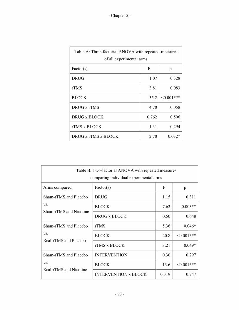
- Chapter 5 -
- 93 -
Table A: Three-factorial ANOVA with repeated-measures
of all experimental arms
Factor(s) F p
DRUG 1.07 0.328
rTMS 3.81 0.083
BLOCK 35.2 <0.001***
DRUG x rTMS 4.70 0.058
DRUG x BLOCK 0.762 0.506
rTMS x BLOCK 1.31 0.294
DRUG x rTMS x BLOCK 2.70 0.032*
Table B: Two-factorial ANOVA with repeated measures
comparing individual experimental arms
Arms compared Factor(s) F p
DRUG 1.15 0.311
BLOCK 7.62 0.003**
Sham-rTMS and Placebo
vs.
Sham-rTMS and Nicotine DRUG x BLOCK 0.50 0.648
rTMS 5.36 0.046*
BLOCK 20.8 <0.001***
Sham-rTMS and Placebo
vs.
Real-rTMS and Placebo rTMS x BLOCK 3.21 0.049*
INTERVENTION 0.30 0.297
BLOCK 13.6 <0.001***
Sham-rTMS and Placebo
vs.
Real-rTMS and Nicotine INTERVENTION x BLOCK 0.319 0.747

- Chapter 5 -
- 94 -
DRUG 0.001 0.975
BLOCK 16.3 <0.001***
Real-rTMS and Placebo
vs.
Real-rTMS and Nicotine DRUG x BLOCK 0.260 0.933
rTMS 3.52 0.094
BLOCK 37.2 <0.001***
Sham-rTMS and Nicotine
vs.
Real-rTMS and Nicotine rTMS x BLOCK 2.73 0.031*
INTERVENTION 3.02 0.116
BLOCK 27.0 <0.001***
Sham-rTMS and Nicotine
vs.
Real-rTMS and Placebo INTERVENTION x BLOCK 1.54 0.235
Table 5.1: Analysis of Variance (ANOVA) of ballistic motor learning.
(A) Three-factorial ANOVA with repeated measures of “DRUG” x
“rTMS” x “BLOCK”; (B) Two-factorial ANOVA with repeated
measures comparing the four different arms of the experiment with each
other. * indicates p<0.05, ** indicates p<0.01 and ***indicates p<0.001.
Table 6: Two-factorial ANOVA with repeated-measures
of baseline measures (Block1 only)
Factor(s) F p
DRUG 0.300 0.597
rTMS 0.885 0.371
DRUG x rTMS 0.607 0.456
Table 5.2: Analysis of Variance (ANOVA) of baseline block1 of all
experimental arms showing no significant carryover effect at baseline or
effect of the intervention on baseline motor performance.
- Chapter 5 -
- 95 -
5.4 Discussion in the interlude
5.4.1 The effect of iTBS on motor learning
iTBS increased the rate at which subjects improved performance of the thumb
abduction task without affecting baseline measures. To our knowledge, this is the first
report in healthy volunteers in which excitatory rTMS enhances subsequent motor
learning. A recent study found that training in a finger abduction task was not affected
by 5 Hz rTMS (Agostino et al 2007) or iTBS (Agostino et al., 2008). However, this
study involved motor performance assessments both before and after rTMS, with
likely consequent carry-over effects. Additionally, performance feedback of peak
acceleration was not provided which is central to enhancing performance. In a group
of stroke patients 10 Hz rTMS enhanced the acquisition of a serial reaction time task
but unlike the present study stimulation was interspersed with task practice, making it
difficult to distinguish improved performance from improved learning (Kim et al
2006).
Improvement in initial peak acceleration involves two processes. First, there must be a
driver to change, such that performance on a trial differs from, and on average is
better than, that of the previous trial. Second, any beneficial changes in output should
be stabilised, perhaps by changes in synaptic connectivity. Indeed, motor learning is
accompanied by LTP within M1 (Rioult-Pedotti et al 2000) that likely involves
synaptic strengthening in selected pathways. iTBS is thought to act at a cortical level
(Di Lazarro et al 2005), and can promote changes in synaptic strength (Huang et al
2005, 2007) that are thought to involve LTP (Huang et al 2007; Teo et al 2007): it
thus seems a possibility that enhanced learning following iTBS may occur via
increased synaptic activity with up-regulation of LTP.

- Chapter 5 -
- 96 -
5.4.2 The effect of the iTBS-nicotine interaction on learning
Nicotine alone did not affect training, but when combined with iTBS it blocked the
positive effects observed in the placebo arm. This is arguably a surprising result.
Nicotinic receptors are expressed widely throughout the brain, including within M1
(Sihver et al 1998; Alkondon et al 2000), and cholinergic modulation of plasticity
protocols has been demonstrated (Kuo et al 2007). Furthermore, pre- or post-synaptic
enhancement of LTP by nicotine is well documented in animal models (Fisher et al
1998; Mansvelder & McGehee 2000; Ji et al 2001; Ge & Dani 2005). One may
therefore have expected nicotine to enhance task acquisition.
Experiments of iTBS on motor learning above and on MEP (Chapter 4) showed that
iTBS and nicotine interacted differently with regard to motor learning and
corticospinal excitability. This apparent dissociation could potentially be explained by
an unpredictable action of nicotine at receptors outside the motor cortex, for example
in the striatum or the cerebellum (Paterson & Nordberg 2000; Gotti & Clementi
2004). In this case, however, one would expect nicotine alone to modulate motor
learning, which was not the case.
One explanation is the BCM model where higher levels of excitability occlude or
impair further learning (but one would expect equivalent levels of learning to TBS
alone). Also others have suggested that this type of sliding threshold plays a limited
role in motor learning (Siebner et al., 2004; Kuo et al., 2008) (Note: these references
refer to the BCM rule as homeostatic plasticity which is a confusion of nomenclature
compared to molecular models of homeostatic plasticity).
- Chapter 5 -
- 97 -
An alternative possibility is that the improvement in performance is not driven by
corticospinal excitability. A presumption of the original hypothesis is that enhanced
corticospinal excitability would drive more performance improvement under Hebbian
principles as synaptic efficacy is enhanced and thus there would be enhanced
encoding of motor memory or a learning rule. We tested if this presumption is correct
by analysing the behavioural data for evidence of acquisition of a learning rule.
5.4.3 Trial-by-trial analysis of data
Trial-by-trial data of each individual subject session was analysed. Within a block, the
probability of an individual trial would be better than the previous trial was calculated
with a simple formula:
P = t(x) / T
where P is the probability of an individual trial being better than the previous trial;
t(x) is the number of trials where performance in xth
trial is greater than (x-1)th
trial;
and T is the total number of trials in the block.
Thus the hypothesis is that if an underlying strategy, learning rule or motor memory
was being acquired, improving performance would be linked to increasing P across
blocks and if TBS was enhancing learning through enhancing corticospinal
excitability, sessions with active iTBS intervention would have a higher P than
sessions with a sham-TBS intervention.
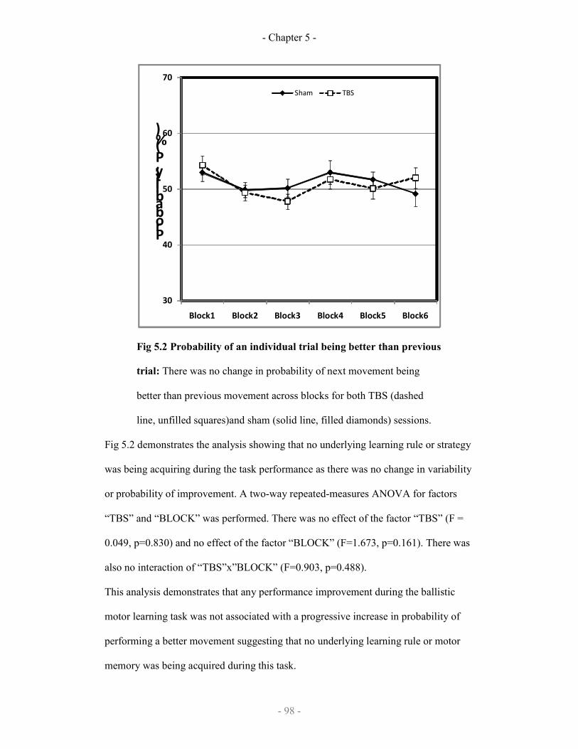
- Chapter 5 -
- 98 -
30
40
50
60
70
Block1 Block2 Block3 Block4 Block5 Block6
Probability, P (%)
Sham TBS
Fig 5.2 Probability of an individual trial being better than previous
trial: There was no change in probability of next movement being
better than previous movement across blocks for both TBS (dashed
line, unfilled squares)and sham (solid line, filled diamonds) sessions.
Fig 5.2 demonstrates the analysis showing that no underlying learning rule or strategy
was being acquiring during the task performance as there was no change in variability
or probability of improvement. A two-way repeated-measures ANOVA for factors
“TBS” and “BLOCK” was performed. There was no effect of the factor “TBS” (F =
0.049, p=0.830) and no effect of the factor “BLOCK” (F=1.673, p=0.161). There was
also no interaction of “TBS”x”BLOCK” (F=0.903, p=0.488).
This analysis demonstrates that any performance improvement during the ballistic
motor learning task was not associated with a progressive increase in probability of
performing a better movement suggesting that no underlying learning rule or motor
memory was being acquired during this task.

- Chapter 5 -
- 99 -
The question then arises as to what mechanism is responsible for enhancing learning
after iTBS. Working from first principles, if no learning rule or motor memory was
being acquired, the only mechanism driving performance change (whether
improvement or deterioration) is intrinsic performance variability. We sought to test
the viability for performance variability as a driver of performance change in this task
by constructing a simple theoretical model of the ballistic motor learning task.
5.5 Modelling the ballistic motor learning task
A simple theoretical model was designed to show how a performance improvement
may be achieved with minimal assumptions about how learning would occur. The aim
of creating such a model was to allow us to investigate the effects of altering
performance variability on the outcome, and thereby to determine whether it is
feasible that such variability may have a positive effect on learning. This model was
based on 2 assumptions:
1) that there is a maximum physically achievable peak acceleration; and
2) that the motor cortex has a fixed repertoire of possible outputs, each coding for
different muscle groups, which can be discharged in parallel.
Maximising the motor output would therefore involve determining the optimum
weighting in which these motor outputs are to be discharged, presumably favouring
task agonists over task antagonists. Thus the system must gradually solve a multi-
dimensional problem using feedback given in one dimension, in the form of visual
feedback from the previous trial. For the sake of our model we reduced the motor
output repertoire to 2 dimensions, represented by 2 orthogonal axes x and y. The
contribution of each axis to the observed motor output was defined by the same
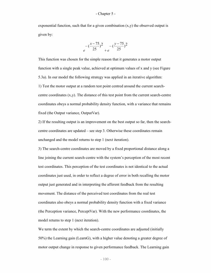
- Chapter 5 -
- 100 -
exponential function, such that for a given combination (x,y) the observed output is
given by:
2)25
75(2)
25
75(
−−
+
−−
y
e
x
e
This function was chosen for the simple reason that it generates a motor output
function with a single peak value, achieved at optimum values of x and y (see Figure
5.3a). In our model the following strategy was applied in an iterative algorithm:
1) Test the motor output at a random test point centred around the current search-
centre coordinates (x,y). The distance of this test point from the current search-centre
coordinates obeys a normal probability density function, with a variance that remains
fixed (the Output variance, OutputVar).
2) If the resulting output is an improvement on the best output so far, then the search-
centre coordinates are updated – see step 3. Otherwise these coordinates remain
unchanged and the model returns to step 1 (next iteration).
3) The search-centre coordinates are moved by a fixed proportional distance along a
line joining the current search-centre with the system’s perception of the most recent
test coordinates. This perception of the test coordinates is not identical to the actual
coordinates just used, in order to reflect a degree of error in both recalling the motor
output just generated and in interpreting the afferent feedback from the resulting
movement. The distance of the perceived test coordinates from the real test
coordinates also obeys a normal probability density function with a fixed variance
(the Perception variance, PerceptVar). With the new performance coordinates, the
model returns to step 1 (next iteration).
We term the extent by which the search-centre coordinates are adjusted (initially
50%) the Learning gain (LearnG), with a higher value denoting a greater degree of
motor output change in response to given performance feedback. The Learning gain

- Chapter 5 -
- 101 -
thus reflects the capacity for plastic change in this model. It may be noted that the
model is simply an iterative implementation of a fixed set of rules – it does not
include the capacity to discover underlying rules that find a solution more efficiently.
This model is represented in flow diagram form in Figure 5.3b. The initial test
coordinates were always (50, 50), and 100 iterations were performed in the course of
each run. The effects of varying either the OutputVar or the PerceptVar were
examined by running the model 20 times at each set of values across a range and
recording the resulting outputs. For each run of the model, the final output achieved
was recorded as the mean of the last 10 trials (out of 100).

- Chapter 5 -
- 102 -
Figure 5.3: Model of a possible strategy for the ballistic learning
task
We designed a simple iterative model by which an improvement in
performance may be achieved in this task. The aim is to identify the
optimum weighting of 2 simultaneously-discharged motor outputs (x
and y), using the available performance feedback in the form of
observed motor output.
(a) The motor output function employed in the model. The observed
motor output (vertical axis) was defined as the sum of the contributions
of the 2 individual motor outputs (x and y), each of which obeyed an
inverse exponential function such that there is a single peak which
represents the maximum possible peak acceleration. In the contour
view (right), the asterisk represents the starting position.
(b) The structure of the model is given in flow diagram form. Random
combinations of x and y are tried: if the resulting output of a given trial
represents an improvement on prior performance then the search centre
is moved in the perceived direction of the new coordinate. There are 2
distinct sources of variance, which remain fixed within a given run of
the model: Output variance reflects the distribution of trials around the
search centre, while Perception variance reflects error in the correct
recollection of the previous trial coordinates.
The effects of changing the OutputVar (with PerceptVar set at 1 and LearnG set at
0.2), the PerceptVar (with OutputVar set at 7 and LearnG set at 0.2) and the effects of
changing the LearnG (with OutputVar set at 7 and PerceptVar set at 1) are shown in

- Chapter 5 -
- 103 -
Figure 5.4. At each setting the final performance depended crucially on the variable
tested.
For OutputVar (Figure 5.4a), low values resulted in poor final performance, with little
improvement across the 100 trials. Increasing OutputVar was initially beneficial, but
beyond an optimal value further increases resulted in impaired performance. For
LearnG (Figure 5.4c), increases resulted in a similar inverse-U-shaped curve with an
optimum value beyond which further increases were detrimental to performance. For
PerceptVar (Figure 5.4b), by contrast, there was no optimal value – increasing this
form of variability resulted in a steady decline in performance. Detailed analysis of
the interaction between these variables demonstrates that this principle holds true for
all values of these variables (Figure 5.4). Thus in this simple model of a learning
strategy we observed a complex interaction between variability and learning, with
divergent effects of the 2 forms of variability tested on final performance. For
OutputVar and LearnG, there is an optimum range where maximal performance gain
occurs while any increase in PerceptVar is detrimental.

- Chapter 5 -
- 104 -
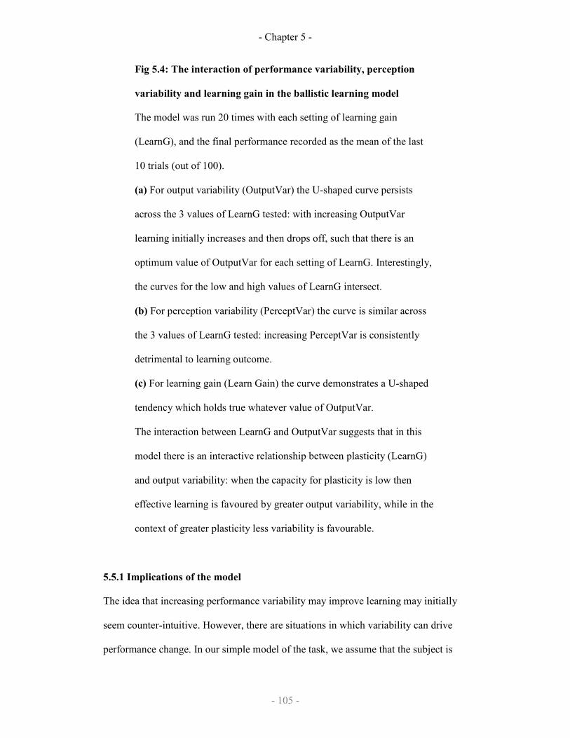
- Chapter 5 -
- 105 -
Fig 5.4: The interaction of performance variability, perception
variability and learning gain in the ballistic learning model
The model was run 20 times with each setting of learning gain
(LearnG), and the final performance recorded as the mean of the last
10 trials (out of 100).
(a) For output variability (OutputVar) the U-shaped curve persists
across the 3 values of LearnG tested: with increasing OutputVar
learning initially increases and then drops off, such that there is an
optimum value of OutputVar for each setting of LearnG. Interestingly,
the curves for the low and high values of LearnG intersect.
(b) For perception variability (PerceptVar) the curve is similar across
the 3 values of LearnG tested: increasing PerceptVar is consistently
detrimental to learning outcome.
(c) For learning gain (Learn Gain) the curve demonstrates a U-shaped
tendency which holds true whatever value of OutputVar.
The interaction between LearnG and OutputVar suggests that in this
model there is an interactive relationship between plasticity (LearnG)
and output variability: when the capacity for plasticity is low then
effective learning is favoured by greater output variability, while in the
context of greater plasticity less variability is favourable.
5.5.1 Implications of the model
The idea that increasing performance variability may improve learning may initially
seem counter-intuitive. However, there are situations in which variability can drive
performance change. In our simple model of the task, we assume that the subject is

- Chapter 5 -
- 106 -
unable to formulate a set of rules to aid improvement from the information available
about the current trial. This contrasts, for example, with sequence learning in which
knowledge of the sequence in itself predicts the optimal output on each trial and is
validated by the fact that there was no improving probability of learning between
blocks within a session.
In the model, the drivers to performance change were random variation in motor
output (OutputVar) and in the memory of the previous trial’s output (PerceptVar). The
effects of altering the two forms of variability were fundamentally different. While
variability in accurate recollection of the previous trial (PerceptVar) was entirely
detrimental effect to final performance, the same was not true for variability in search-
centre coordinates (OutputVar) where an inverted U-shaped curve was observed.
Increasing OutputVar allows the model to try a wider range of combinations, allowing
the system to ‘escape’ a performance plateau and continue improving. This is akin to
a selection process where a degree of diversity allows a gradual evolutionary process
to occur. On the other hand, excessive OutputVar adversely affects the reproducibility
of good movements so that learning suffers. The LearnG determines the extent of
output adjustment made in response to an improvement and so reflects the degree of
plasticity available. A relatively small amount is required for optimal learning, beyond
which improvement declines. Impaired performance at LearnG higher values were
explained here by greater system variability, suggesting a complex interaction
between plasticity and variability. Thus, a highly variable system would benefit from
less plasticity (due to the risk of learning an error) while a less variable system would
benefit from greater plasticity.
These results are obtained from a simple model that shows how the motor system
might operate in the absence of rule-based optimisations. Although an extreme
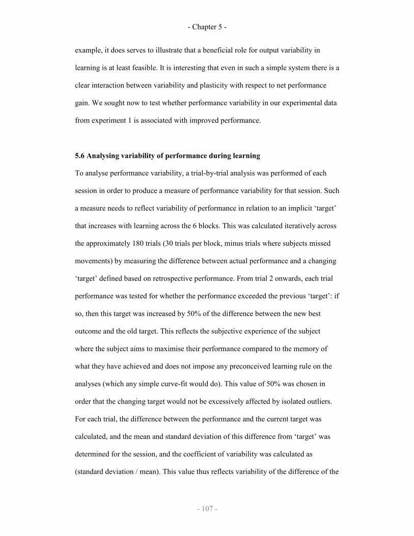
- Chapter 5 -
- 107 -
example, it does serves to illustrate that a beneficial role for output variability in
learning is at least feasible. It is interesting that even in such a simple system there is a
clear interaction between variability and plasticity with respect to net performance
gain. We sought now to test whether performance variability in our experimental data
from experiment 1 is associated with improved performance.
5.6 Analysing variability of performance during learning
To analyse performance variability, a trial-by-trial analysis was performed of each
session in order to produce a measure of performance variability for that session. Such
a measure needs to reflect variability of performance in relation to an implicit ‘target’
that increases with learning across the 6 blocks. This was calculated iteratively across
the approximately 180 trials (30 trials per block, minus trials where subjects missed
movements) by measuring the difference between actual performance and a changing
‘target’ defined based on retrospective performance. From trial 2 onwards, each trial
performance was tested for whether the performance exceeded the previous ‘target’: if
so, then this target was increased by 50% of the difference between the new best
outcome and the old target. This reflects the subjective experience of the subject
where the subject aims to maximise their performance compared to the memory of
what they have achieved and does not impose any preconceived learning rule on the
analyses (which any simple curve-fit would do). This value of 50% was chosen in
order that the changing target would not be excessively affected by isolated outliers.
For each trial, the difference between the performance and the current target was
calculated, and the mean and standard deviation of this difference from ‘target’ was
determined for the session, and the coefficient of variability was calculated as
(standard deviation / mean). This value thus reflects variability of the difference of the
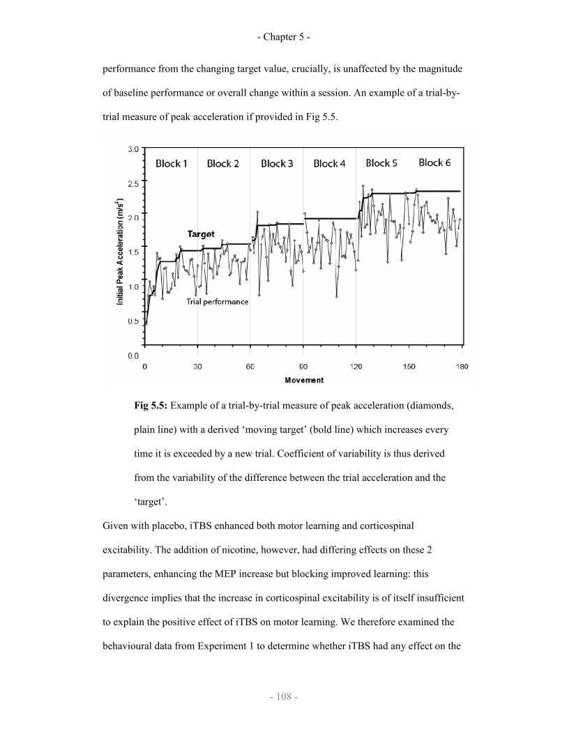
- Chapter 5 -
- 108 -
performance from the changing target value, crucially, is unaffected by the magnitude
of baseline performance or overall change within a session. An example of a trial-by-
trial measure of peak acceleration if provided in Fig 5.5.
Fig 5.5: Example of a trial-by-trial measure of peak acceleration (diamonds,
plain line) with a derived ‘moving target’ (bold line) which increases every
time it is exceeded by a new trial. Coefficient of variability is thus derived
from the variability of the difference between the trial acceleration and the
‘target’.
Given with placebo, iTBS enhanced both motor learning and corticospinal
excitability. The addition of nicotine, however, had differing effects on these 2
parameters, enhancing the MEP increase but blocking improved learning: this
divergence implies that the increase in corticospinal excitability is of itself insufficient
to explain the positive effect of iTBS on motor learning. We therefore examined the
behavioural data from Experiment 1 to determine whether iTBS had any effect on the

- Chapter 5 -
- 109 -
variability with which the motor task was performed. For each session, a measure of
trial-by-trial performance variability was calculated using a method that is
independent of baseline performance or overall improvement. Performance variability
was calculated as variability of the difference from a continuously updated target
derived from previous good performance. Fig 5.5 shows the target calculated for a
single session. This iterative approach made no assumptions about the mechanism a
subject learns, and the coefficient of variability (standard deviation / mean) ensured
that the resulting measure was independent of performance magnitude.
Figure 5.6a demonstrates that iTBS alone enhances the coefficient of variation in
performance which is confirmed on a 2-way ANOVA testing the effects of the factors
‘Stimulation’ and ‘Preparation’, and revealing a significant interaction (F1,9=7.637,
P=0.022), with a significant main effect of Stimulation (F1,9=9.265, P=0.014) but not
of Preparation (F1,9=0.150, P=0.707). This interaction was explained by significantly
greater performance variability after TBS than after sham stimulation in the placebo
sessions (P<0.001) but not in the nicotine sessions (P=0.965). A comparison of the
TBS sessions (nicotine vs placebo) revealed that performance variability was
significantly reduced in the presence of nicotine (P=0.031). The results of this
analysis suggest that iTBS had the effect of increasing performance variability when
the subject had taken placebo, but this increase in performance variability did not
occur if they had taken nicotine: this was the same pattern observed in the effect of
TBS on learning.
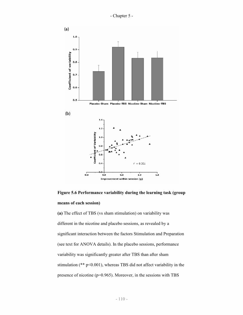
- Chapter 5 -
- 110 -
Figure 5.6 Performance variability during the learning task (group
means of each session)
(a) The effect of TBS (vs sham stimulation) on variability was
different in the nicotine and placebo sessions, as revealed by a
significant interaction between the factors Stimulation and Preparation
(see text for ANOVA details). In the placebo sessions, performance
variability was significantly greater after TBS than after sham
stimulation (** p<0.001), whereas TBS did not affect variability in the
presence of nicotine (p=0.965). Moreover, in the sessions with TBS
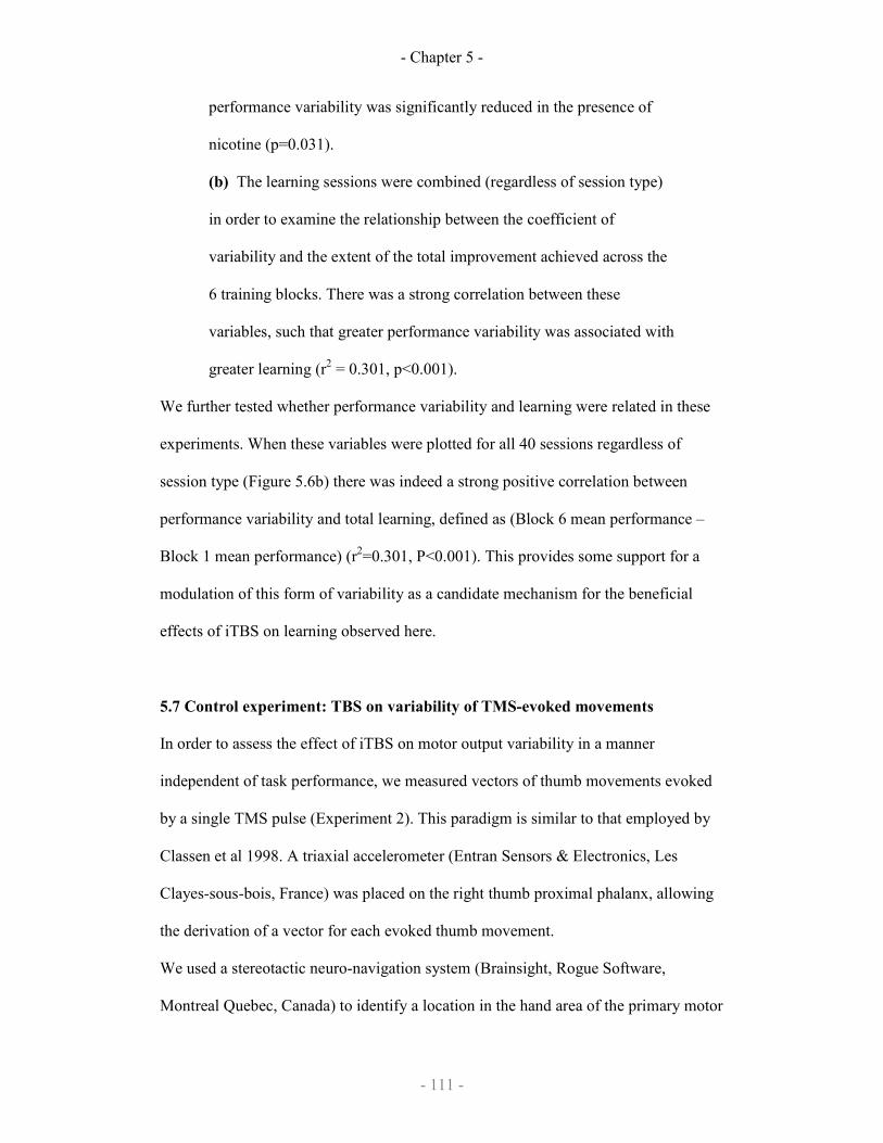
- Chapter 5 -
- 111 -
performance variability was significantly reduced in the presence of
nicotine (p=0.031).
(b) The learning sessions were combined (regardless of session type)
in order to examine the relationship between the coefficient of
variability and the extent of the total improvement achieved across the
6 training blocks. There was a strong correlation between these
variables, such that greater performance variability was associated with
greater learning (r2 = 0.301, p<0.001).
We further tested whether performance variability and learning were related in these
experiments. When these variables were plotted for all 40 sessions regardless of
session type (Figure 5.6b) there was indeed a strong positive correlation between
performance variability and total learning, defined as (Block 6 mean performance –
Block 1 mean performance) (r2=0.301, P<0.001). This provides some support for a
modulation of this form of variability as a candidate mechanism for the beneficial
effects of iTBS on learning observed here.
5.7 Control experiment: TBS on variability of TMS-evoked movements
In order to assess the effect of iTBS on motor output variability in a manner
independent of task performance, we measured vectors of thumb movements evoked
by a single TMS pulse (Experiment 2). This paradigm is similar to that employed by
Classen et al 1998. A triaxial accelerometer (Entran Sensors & Electronics, Les
Clayes-sous-bois, France) was placed on the right thumb proximal phalanx, allowing
the derivation of a vector for each evoked thumb movement.
We used a stereotactic neuro-navigation system (Brainsight, Rogue Software,
Montreal Quebec, Canada) to identify a location in the hand area of the primary motor
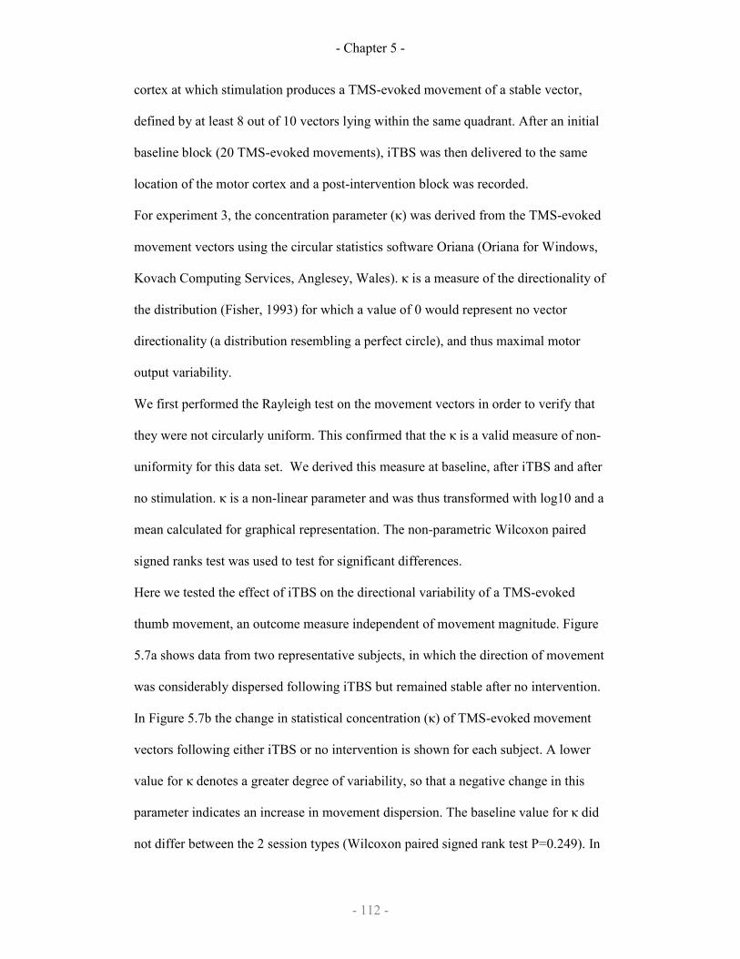
- Chapter 5 -
- 112 -
cortex at which stimulation produces a TMS-evoked movement of a stable vector,
defined by at least 8 out of 10 vectors lying within the same quadrant. After an initial
baseline block (20 TMS-evoked movements), iTBS was then delivered to the same
location of the motor cortex and a post-intervention block was recorded.
For experiment 3, the concentration parameter (κ) was derived from the TMS-evoked
movement vectors using the circular statistics software Oriana (Oriana for Windows,
Kovach Computing Services, Anglesey, Wales). κ is a measure of the directionality of
the distribution (Fisher, 1993) for which a value of 0 would represent no vector
directionality (a distribution resembling a perfect circle), and thus maximal motor
output variability.
We first performed the Rayleigh test on the movement vectors in order to verify that
they were not circularly uniform. This confirmed that the κ is a valid measure of non-
uniformity for this data set. We derived this measure at baseline, after iTBS and after
no stimulation. κ is a non-linear parameter and was thus transformed with log10 and a
mean calculated for graphical representation. The non-parametric Wilcoxon paired
signed ranks test was used to test for significant differences.
Here we tested the effect of iTBS on the directional variability of a TMS-evoked
thumb movement, an outcome measure independent of movement magnitude. Figure
5.7a shows data from two representative subjects, in which the direction of movement
was considerably dispersed following iTBS but remained stable after no intervention.
In Figure 5.7b the change in statistical concentration (κ) of TMS-evoked movement
vectors following either iTBS or no intervention is shown for each subject. A lower
value for κ denotes a greater degree of variability, so that a negative change in this
parameter indicates an increase in movement dispersion. The baseline value for κ did
not differ between the 2 session types (Wilcoxon paired signed rank test P=0.249). In
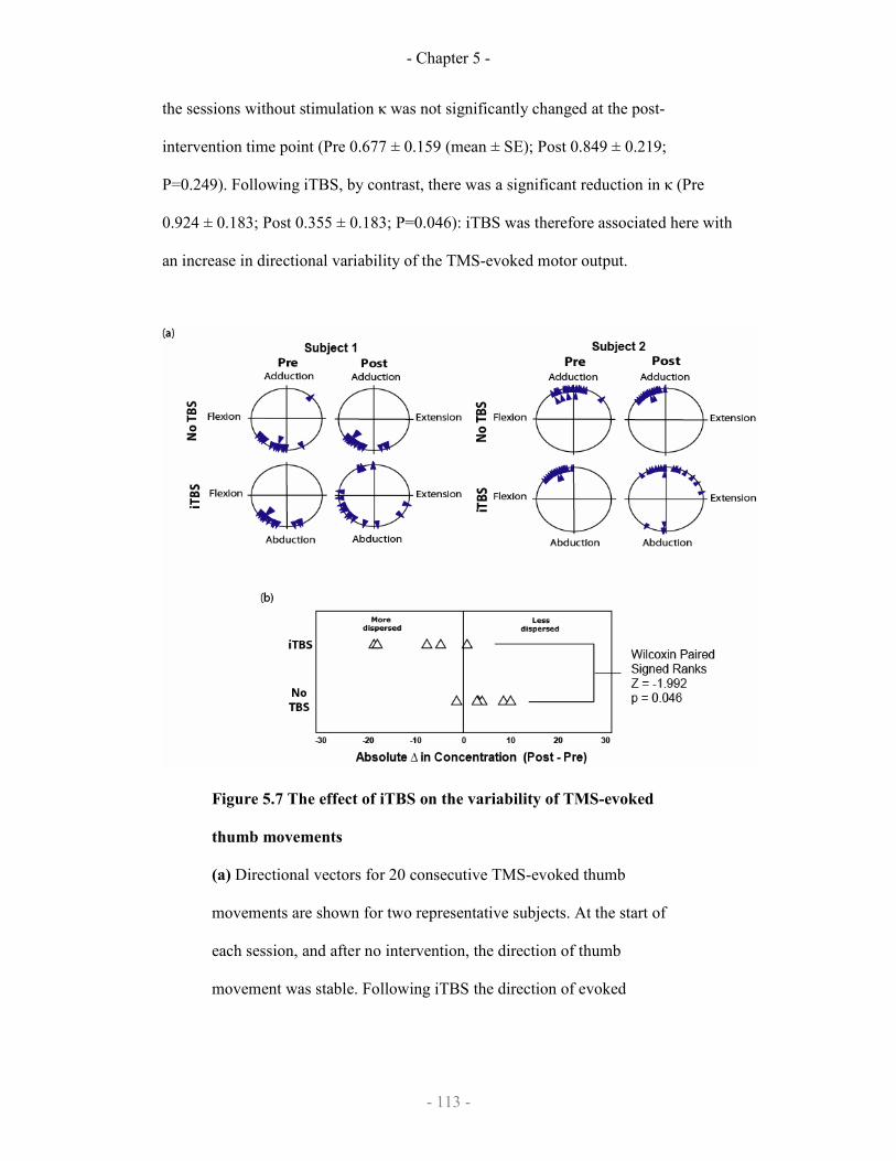
- Chapter 5 -
- 113 -
the sessions without stimulation κ was not significantly changed at the post-
intervention time point (Pre 0.677 ± 0.159 (mean ± SE); Post 0.849 ± 0.219;
P=0.249). Following iTBS, by contrast, there was a significant reduction in κ (Pre
0.924 ± 0.183; Post 0.355 ± 0.183; P=0.046): iTBS was therefore associated here with
an increase in directional variability of the TMS-evoked motor output.
Figure 5.7 The effect of iTBS on the variability of TMS-evoked
thumb movements
(a) Directional vectors for 20 consecutive TMS-evoked thumb
movements are shown for two representative subjects. At the start of
each session, and after no intervention, the direction of thumb
movement was stable. Following iTBS the direction of evoked
- Chapter 5 -
- 114 -
movements became more variable, such that the concentration
parameter (κ) was reduced.
(b) The change in concentration parameter κ following either iTBS or
no intervention is shown for each subject. A negative change in κ
denotes an increase in the variability of TMS-evoked movement
vectors. Baseline values did not differ between the 2 session types
(Wilcoxon paired signed rank test P=0.249). κ was significantly
reduced following iTBS (0.046) but not after no intervention (0.249),
indicating that iTBS increased the variability of TMS-evoked thumb
movements.
5.8 Conclusion
An analysis of performance variability in Experiment 1 revealed a pattern similar to
that observed with learning outcome: iTBS increased variability but this effect was
blocked by nicotine. Moreover, performance variability in a given session was
correlated with the behavioural gain observed. One source of changes in performance
variability may be due to increased variation in the voluntary drive to motor cortex
from distant sites. However, the fact that iTBS increased directional variability in
Experiment 2, in which a TMS pulse evoked motor cortical output directly, suggests
that a major source of change lay within the motor cortex itself. We conclude that
iTBS increases motor cortex output variability independent of voluntary drive.
Variability is known to increase with larger outputs (Jones et al 2002, Hamilton et al
2004). This scaling effect is recognised in sensory input from psychophysical
literature and follows the Weber–Fechner law:

- Chapter 5 -
- 115 -
"In order that the intensity of a sensation may increase in arithmetical
progression, the stimulus must increase in geometrical progression."
(translated from Fechner, 1860)
If a Weber-type phenomenon is involved in the motor learning task, it is conceivable
that the initial performance (or excitability) may produce a geometric increase in
performance or performance variability. Although this is corrected for in the
calculation of coefficient of variation by normalisation with baseline, this correction
for baseline may not fully account for the increase in performance variability. This
could mean that any increased variability may be an epiphenomenon of increased
baseline performance (or excitability). It is telling to note that in the TBS-placebo arm
of the experiment, the mean performance in Block 1 appears to be larger than the
mean performance in other experimental arms, although this is not significant
(p>0.05). Thus, small changes in baseline excitability or performance that this study is
not powered to detect, may produce large changes in performance variability which is
detected in our trial-by-trial analysis. Thus, a causal relationship between motor
performance and variability is not established and further experiments will need to be
done to clarify the statistical correlation.
Nicotine blocked the iTBS-related increase in performance variability, but did not
alter variability on its own. A recent study in humans has suggested that cholinergic
stimulation may increase the signal-to-noise ratio in the motor cortex (Kuo et al
2007). Similarly, nicotine increases the gain in thalamic inputs to the visual cortex
(Disney et al 2007). The present results may be explained in these terms if nicotine
were to reduce variability within the motor cortex, with a consequent negative effect
on learning.
- Chapter 5 -
- 116 -
In the present study we demonstrate enhanced acquisition of a motor task following
iTBS, and blockade of this effect by nicotine. This interaction was not explained by
effects on synaptic plasticity, at least as judged by their influence on corticospinal
excitability. We believe that iTBS may enhance task acquisition by increasing
variability of motor cortical output and thereby driving performance change rather
than by increasing synaptic strength although this causal relationship is not proven.
We used a simple mathematical model to demonstrate that a beneficial effect of
variability on learning is theoretically feasible in a simple task and this model has
similarities to differential stochastic motor learning (Frank et al., 2008; Schöllhorn et
al., 2009) and schema motor learning models (Schmidt et al., 1975) in the movement
science literature.
The potential to enhance the effects of rTMS pharmacologically has obvious clinical
appeal. The positive effect of iTBS alone on subsequent learning may also provide
encouragement in this regard, as it raises the possibility of enhancing the response to
therapy in patients with motor impairment. However, the observed dissociation
between the physiological and behavioural effects of such a combined approach
introduces a note of caution. Incorporating concepts with computational or cognitive
modelling into the study of motor learning may help to shed light on this relationship.
- Chapter 6 -
- 117-
Chapter 6
Intracortical circuits and
practice-dependent plasticity
Work described in this chapter have been published:
Teo JT, Terranova C, Swayne OB, Greenwood R, Rothwell JC. Practice-dependent
plasticity is limited by different intracortical circuits. Exp Brain Res. 2009
Mar;193(4):555-63.
- Chapter 6 -
- 118-
6.1 Introduction
Pharmacological interventions coupled with transcranial magnetic stimulation (TMS)
methods have made it possible to study a number of inhibitory circuits in the human
cerebral cortex and to test how they are involved in particular types of motor behaviour.
The present paper focuses on the role of subtypes of the GABAA receptor in synaptic
plasticity induced when subjects learn a new motor task.
Butefisch et al. (2000) initially showed that if subjects practise an isolated thumb
movement in a particular direction then the amplitude of MEPs evoked in agonist
muscles is larger after practice than before. Since this was blocked by NMDA receptor
antagonists, it was presumed to involve LTP-like changes in the efficacy of glutamatergic
synapses in motor cortex. The authors also found that the effect was blocked by
pretreatment with lorazepam, a non-selective GABAA agonist. The latter was
compatible with reports in the animal literature that emphasised the role of GABA in
regulating motor cortical plasticity (Hess et al. 1996) as well as with other investigations
of synaptic plasticity in humans (Ziemann et al. 1998a; Ziemann et al. 1998b; Ziemann et
al. 2001; Pleger et al. 2003). Taken together these results suggest that LTP-like plasticity
is enhanced when GABA inhibition is reduced. However, there is no information on
whether specific subtypes of receptor are preferentially involved in the effect.
The present experiments examined this question by comparing the effects on practice
dependent synaptic plasticity of the non-selective GABAA agonist lorazepam with the
selective GABAA-alpha1 receptor agonist, zolpidem. We predicted that as both drugs are
GABA agonists then both of them might potentially interfere with plasticity. However, if
- Chapter 6 -
- 119-
the GABAA-alpha1 receptor were not involved then plasticity would be reduced only by
lorazepam whereas it would be unaffected by zolpidem.
Simultaneously to this experiment, we also asked which neural circuits might be most
involved in controlling levels of synaptic plasticity. A number of inhibitory intracortical
circuits have been identified using transcranial magnetic stimulation (TMS): short-
interval intracortical inhibition (SICI), short-interval afferent inhibition (SAI) and long-
interval intracortical inhibition (LICI) (Kujirai et al. 1993; Wassermann et al. 1996;
Tokimura et al. 2000; Sailer et al. 2002). SICI is believed to involve GABAA receptor
neurotransmission (Ziemann 2004; Florian et al. 2008) and LICI is believed to involve
GABAB receptor neurotransmission (McDonnell et al. 2006; Florian et al. 2008). Studies
with the drug zolpidem indicate that the GABAA-alpha1 receptor is associated with the
pathway mediating SAI but not with that mediating SICI (Di Lazzaro et al. 2006; Di
Lazzaro et al. 2007). We argued that if particular pathways are responsible for controlling
plasticity then changes in the amount of plasticity produced by lorazepam or zolpidem
would correlate with their effects on SICI, LICI or SAI.
6.2 Study design
The study was structured as a double-blind randomised controlled cross-over trial with
two drug arms. 2.5mg lorazepam or 10mg zolpidem was prepared by the pharmacy of the
National Hospital for Neurology and Neurosurgery into unmarked containers. A placebo
arm would be easily unblinded and would also not control for the effects of sedation so
no placebo arm was used. Subjects had TMS measurements at three time points: T1, T2

- Chapter 6 -
- 120-
and T3. The allocated drug was given at the end of T1. TMS measurements at T2 occur 2
hours after drug ingestion coinciding with the peak plasma concentration of lorazepam
and zolpidem: lorazepam, 1.5–2.5 hours (Kyriakopoulos et al., 1978); zolpidem, 0.75–2.6
hours (Salva & Costa, 1995). After T2, subjects performed the motor practice task (see
below) and after completion of the task, proceeded to have TMS measurements again at
T3. The study design is summarised in Fig 6.1a.
Fig 6.1: Study design of the role of GABA-circuits in practice-
dependent plasticity (a) Timeline of a single experimental session with
- Chapter 6 -
- 121-
three timepoints: T1, T2 and T3, corresponding to baseline, during drug peak
levels and after task practice; (b) neurophysiological measurements were
made during each timepoint; and (c) task practice consisted of 15 paced
thumb abduction movements repeated over 10 blocks with frequent rest
breaks.
7 healthy subjects were recruited and informed written consent was obtained. All subjects
were right-handed and were not on any medication. For the subjects’ second session, they
received the other drug. The order was randomised and balanced (4 subjects received
lorazepam in the first session). The inter-session interval was 27.7 days (range 10-50
days).
6.3 TMS measurements
Surface EMG was recorded from the left abductor pollicis brevis muscles (APB), the left
first dorsal interossei (FDI) and the left flexor carpi radialis (FCR) with Ag/AgCl
electrodes using a tendon-belly montage. EMG signals were amplified with Digitimer
D360 amplifiers (Digitimer, Welwyn Garden City, UK) with 1000x gain and band-pass
filtered (30-1000Hz for MEP) and sampled at 5kHz using a CED1401 laboratory
interface and Signal software (Cambridge Electronic Design, Cambridge, UK).
Magnetic stimuli were delivered with two Magstim-200 magnetic stimulator (The
Magstim Co., Whitland, UK) connected by a Y-cable. A figure-of-8 coil (diameter 80
mm) was adjusted over the optimal scalp position to evoke an MEP in the right APB with
the coil handle pointed postero-laterally at a 45º angle to the sagittal plane. The resting
motor threshold (RMT) was defined as the lowest intensity capable of inducing at least 5
- Chapter 6 -
- 122-
out of 10 MEPs of >50µV peak-to-peak amplitude. The active motor threshold (AMT)
was defined as the lowest intensity capable of inducing at least 5 out of 10 MEPs of
>200µV peak-to-peak amplitude during an active tonic contraction of thumb APB.
The settings for the various TMS measures are as follows:
1) The corticospinal excitability was measured at rest at 150%RMT over 10 trials.
2) Short-interval intracortical inhibition (SICI) and intracortical facilitation (ICF)
were measured with the test MEP amplitude set at ~1-mV and the conditioning
stimulus set at 80%AMT or 100%AMT. The interstimulus interval was 3ms (for
SICI), 8ms and 15ms (for ICF). 10 trials were recorded for each condition and the
test condition.
3) Long-interval intracortical inhibition (LICI) was measured with the test MEP
amplitude set at ~1-mV and the conditioning stimulus set at 110%RMT or
120%RMT. The interstimulus interval was 100ms (for LICI). 10 trials were
recorded for each condition and the test condition.
4) Short-latency afferent inhibition (SAI) and long-latency afferent inhibition (LAI)
was measured with the test MEP amplitude set at ~1-mV. Electrical stimulation
(200µs pulse width) was delivered to the median nerve using a Digitimer DS7A
Constant Current Stimulator (Digitimer, Welwyn Garden City, Herts, UK) at
twice or thrice sensory threshold. The interstimulus interval was 22ms (for SAI)
and 100ms (for LAI). 10 trials were recorded for each condition and the test
condition.
The order of the various TMS measurements were randomised (Fig 6.1b). In addition to
the TMS measurements, all subjects filled a visual analogue scale (VAS) of arousal
- Chapter 6 -
- 123-
before and after each time point. The visual analogue scale was a 24cm horizontal line
with the left extreme marked ‘Wide awake’ and the right extreme marked ‘Fast asleep’.
6.4 Motor practice
The motor task consists of a paced ballistic thumb abduction task similar to that used in
previous studies (Muellbacher et al. 2002). This motor task has been demonstrated to be
dependent on the primary motor cortex, is associated with changes in MEP amplitude and
movement representation (Classen et al., 1998; Muellbacher et al. 2001) and can be
disrupted in the first hour after motor practice (Muellbacher et al. 2002). In this study, the
subject’s left forearm, hand and fingers were secured in a wooden frame leaving the
thumb free to abduct in the horizontal plane. A piezoresistive monoaxial accelerometer
(Model SA-105 vibrometer, Fribourg, Switzerland) was attached on the lateral aspect of
the left thumb proximal phalanx with the maximal vector being thumb abduction. The
accelerometer signal was sampled at 5000Hz and not filtered.
The task consisted of paced ballistic thumb abduction to a loud auditory tone played at
0.333Hz. 15 thumb abductions were performed per block for 10 blocks, with a 15-second
rest break between blocks (Fig 6.1c). If the subjects missed any movement, they were
required to perform a ‘replacement movement’. In total, subjects practiced 150
movements for 10 minutes. Subjects were motivated with verbal encouragement by a
blinded investigator. Visual feedback of the acceleration from the previous trial was
provided on a computer screen to the subject and the subject was told to attend to the
feedback on the computer screen not to their hand as subjective judgement of thumb
acceleration is often inaccurate.

- Chapter 6 -
- 124-
6.5 Results
Subjects correctly identified the drug taken on 6 out of 14 sessions (42.9%) which is not
above chance (two-tailed Fisher’s exact test, p>0.05). The RMT, AMT, sensory threshold
and conditioning stimulus intensities at baseline, after drug ingestion and after practice
are shown in Table 6.1, 6.2 and 6.3 respectively. There were no statistically significant
differences in RMT, AMT, sensory threshold or test stimulus amplitude between the drug
arms.
Table 6.1 Lorazepam Zolpidem
RMT 39.9 + 6.5% 40.0 + 5.2%
AMT 31.1+ 4.5% 29.1 + 5.7%
Sensory Threshold 0.21 + 0.05mA 0.19 + 0.06mA
SICI/ ICF TS Intensity 49.9 + 8.5% 50.1 + 7.3%
TS Amplitude 0.64 + 0.35mV 0.85 + 0.29mV
LICI TS Intensity 49.9 + 0.61% 50.1 + 6.5%
TS Amplitude 0.61 + 0.4mV 0.79 + 0.27mV
SAI TS Intensity 50.4 + 8.7% 50.1 + 7.0%
TS Amplitude 0.74 + 0.53mV 0.98 + 0.15mV
Table 6.1: Subject and TMS parameters at T1 (baseline) where all % values
represent % maximum stimulator output unless stated otherwise. Ranges
represent standard deviation.
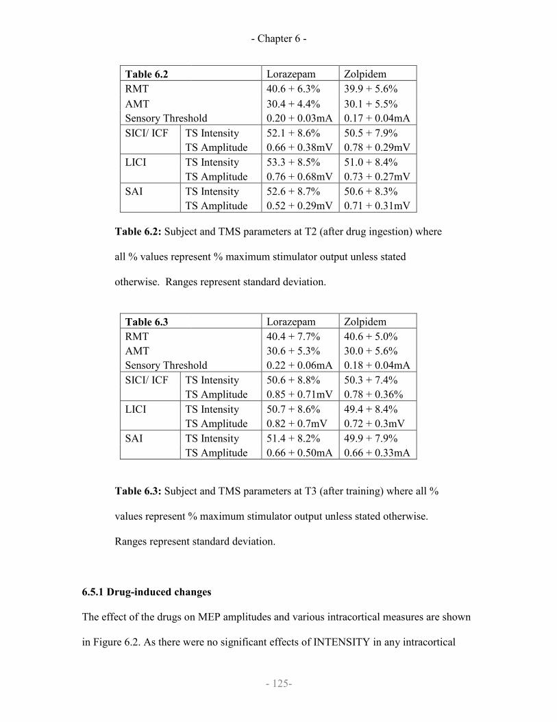
- Chapter 6 -
- 125-
Table 6.2 Lorazepam Zolpidem
RMT 40.6 + 6.3% 39.9 + 5.6%
AMT 30.4 + 4.4% 30.1 + 5.5%
Sensory Threshold 0.20 + 0.03mA 0.17 + 0.04mA
SICI/ ICF TS Intensity 52.1 + 8.6% 50.5 + 7.9%
TS Amplitude 0.66 + 0.38mV 0.78 + 0.29mV
LICI TS Intensity 53.3 + 8.5% 51.0 + 8.4%
TS Amplitude 0.76 + 0.68mV 0.73 + 0.27mV
SAI TS Intensity 52.6 + 8.7% 50.6 + 8.3%
TS Amplitude 0.52 + 0.29mV 0.71 + 0.31mV
Table 6.2: Subject and TMS parameters at T2 (after drug ingestion) where
all % values represent % maximum stimulator output unless stated
otherwise. Ranges represent standard deviation.
Table 6.3 Lorazepam Zolpidem
RMT 40.4 + 7.7% 40.6 + 5.0%
AMT 30.6 + 5.3% 30.0 + 5.6%
Sensory Threshold 0.22 + 0.06mA 0.18 + 0.04mA
SICI/ ICF TS Intensity 50.6 + 8.8% 50.3 + 7.4%
TS Amplitude 0.85 + 0.71mV 0.78 + 0.36%
LICI TS Intensity 50.7 + 8.6% 49.4 + 8.4%
TS Amplitude 0.82 + 0.7mV 0.72 + 0.3mV
SAI TS Intensity 51.4 + 8.2% 49.9 + 7.9%
TS Amplitude 0.66 + 0.50mA 0.66 + 0.33mA
Table 6.3: Subject and TMS parameters at T3 (after training) where all %
values represent % maximum stimulator output unless stated otherwise.
Ranges represent standard deviation.
6.5.1 Drug-induced changes
The effect of the drugs on MEP amplitudes and various intracortical measures are shown
in Figure 6.2. As there were no significant effects of INTENSITY in any intracortical

- Chapter 6 -
- 126-
measures, the graphs represent the mean result of high and low intensity conditioning
stimuli for clarity.
Fig 6.2: The effect of drug on the (a) corticospinal excitability in the APB
muscle; (b) short-interval intracortical inhibition (SICI); (c) short-interval
afferent inhibition (SAI); (d) long-interval intracortical inhibition (LICI);
(e) intracortical facilitation (ICF); and (f) long-interval afferent inhibition
(LAI). Unfilled bars represent the lorazepam sessions and filled bars
represent the zolpidem sessions. Comparisons were by post-hoc student’s

- Chapter 6 -
- 127-
paired t-tests with * represents p<0.05; ** represents p<0.01; *** represents
p<0.001 and n.s. represents p>0.05.
There was no significant difference in MEP amplitude after lorazepam and MEP
amplitude after zolpidem (Fig 6.2a) with two-factorial ANOVA (within-subject factor
TIME and between-subject factor DRUG) showing no effect of TIME (F(1,12)=0.699,
p=0.42), no effect of DRUG (F(1,12)=0.441, p=0.519) and no DRUG x TIME interaction
(F(1,12)=0.004, p=0.950).
As previously reported (Di Lazzaro et al. 2006), lorazepam increased SICI, while
zolpidem did not affect SICI (Fig 6.2b). The difference in effect on SICI of lorazepam
and zolpidem was statistically significant on three-factorial ANOVA (within-subject
factors INTENSITY, TIME, DRUG) with an interaction of TIME x DRUG (F(1,12)=7.227,
p=0.020). Post-hoc testing with Bonferroni correction showed a significant difference
between T1 and T2 for the lorazepam group (p=0.002) but not in the zolpidem group
(p=0.802). Post-hoc testing of the SICI at T2 showed a significant difference between the
lorazepam and zolpidem arms (student’s paired t-test, p=0.041).
For SAI (Fig 6.2c), three-factorial ANOVA (within-subject factor INTENSITY, TIME
and DRUG) showed a significant interaction of TIME x DRUG (F(1,12)=7.23, p=0.02) and
no significant interaction of TIME x DRUG x INTENSITY (F(1,12)=0.206, p=0.658).
Post-hoc testing with Bonferroni correction showed a significant difference between T1
and T2 for the lorazepam group (p=0.018) and for the zolpidem group (p<0.001), but no
significant differences between the two drugs at T1 (p=0.539). Post-hoc testing of the
SAI at T2 did not show any significant difference between the lorazepam and zolpidem
arms (student’s paired t-test, p=0.118).

- Chapter 6 -
- 128-
For LICI (Fig 6.2d), three-factorial ANOVA (within-subject factor INTENSITY, TIME
and DRUG) showed no significant interaction of TIME x DRUG (F(1,12)=0.027, p=0.871)
and TIME x DRUG x INTENSITY (F(1,12)=0.041, p=0.843). For ICF at 15ms (Fig 6.2e),
three-factorial ANOVA (within-subject factor INTENSITY and TIME; between-subject
factor DRUG) showed no significant interaction of TIME x DRUG (F(1,12)=0.834,
p=0.379) or TIME x DRUG x INTENSITY (F(1,12)=0.031, p=0.864). For LAI (Fig 6.2f),
three factorial ANOVA (within-subject factor INTENSITY and TIME; between-subject
factor DRUG) showed no significant interaction of TIME x DRUG (F(1,12)=1.35,
p=0.268) or TIME x DRUG x INTENSITY (F(1,12)=1.07, p=0.322).
In summary after lorazepam, SICI was increased but SAI was decreased while after
zolpidem, SICI was unchanged and SAI was decreased.
6.5.2 Practice
All subjects completed 10 blocks of 15 movements training. Peak acceleration during the
ten minutes is shown in Fig 6.3. On a two-factorial ANOVA of DRUG x BLOCK, there
was an effect of BLOCK (F(1,9)=2.94, p=0.004) indicating a progressively stronger initial
peak acceleration of thumb abduction despite sedation. There appeared to be a fatiguing
effect in the zolpidem group after block 7-10 but there were no significant differences
between drug sessions (F(1,12)=0.583, p=0.460) and there was also no significant
interaction of DRUG x BLOCK (F(1,9)=0.868, p=0.447).

- Chapter 6 -
- 129-
Fig 6.3: Motor performance as represented by initial peak acceleration of
thumb abduction over ten 1-minute blocks of practice. In all cases, the
solid line and squares represent the zolpidem sessions and the dashed line
and triangles represent the lorazepam sessions.
6.5.3 Practice-induced changes
The effect of practice on MEP amplitudes and various intracortical measures are shown
in Fig 6.4. As there were no significant effect of INTENSITY in all intracortical
measures, the graphs represent the mean result of high and low intensity conditioning
stimuli for clarity.
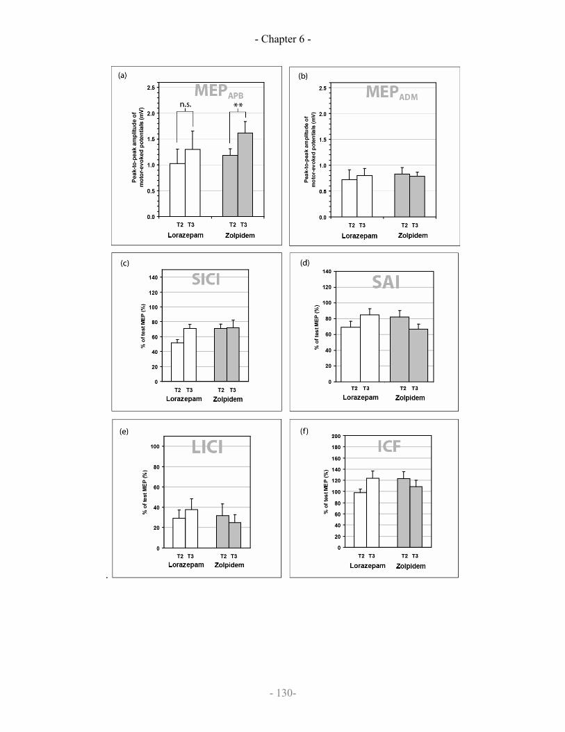
- Chapter 6 -
- 130-
.
- Chapter 6 -
- 131-
Fig 6.4: The effect of training after drug ingestion on (a) corticospinal
excitability in the APB muscle; (b) corticospinal excitability in the
untrained muscle, abductor digiti minimi (ADM); (c) short-interval
intracortical inhibition (SICI); (d) short-interval afferent inhibition (SAI);
(e) long-interval intracortical inhibition (LICI); and (f) intracortical
facilitation (ICF). Unfilled bars represent the lorazepam sessions and filled
bars represent the zolpidem sessions. Comparisons were by post-hoc
paired t-tests with ** represents p<0.01 and n.s. represents p>0.05
The MEP changes after practice are shown in Fig 6.4a and Fig 6.4b and a three-factorial
repeated-measures ANOVA was performed with three within-subject factors (TIME,
MUSCLE and DRUG). This showed an interaction of TIME x MUSCLE x DRUG
(F(1,12)=5.21, p=0.042). Post-hoc testing with Bonferroni correction showed that in the
trained APB muscle, MEP amplitudes enlarged in the zolpidem group (p=0.005) but not
in the lorazepam group (p=0.750). Interestingly, in the untrained ADM muscle of the
zolpidem group, there was no significant increase in MEP amplitude (p=0.634).
For SICI at 3ms (Fig 6.4c), three-factorial repeated-measures ANOVA (within-subject
factor INTENSITY and TIME; between-subject factor DRUG) showed a significant
interaction of DRUG x TIME (F(1,12)=5.40, p=0.039). Post-hoc testing with Bonferroni
correction revealed that in the lorazepam arm, there was a significant difference between
T2 and T3 timepoints (p=0.004) but not in the zolpidem arm (p=0.841). However, there
were baseline differences at T2 between two drug arms (p=0.026) making it likely that
this result is a spurious finding resulting from the lorazepam-enhanced SICI at baseline.

- Chapter 6 -
- 132-
For the intracortical measures of LICI, SAI, LAI and ICF at 15ms, repeated-measures
ANOVA (within-subject factor INTENSITY and TIME; between-subject factor DRUG
in all cases) did not show any significant effect of TIME nor interaction of DRUG x
TIME, TIME x INTENSITY or DRUG x TIME x INTENSITY (p>0.05 in all cases).
This is shown in Fig 6.4d-f.
In summary after zolpidem, MEP amplitude in the APB muscle increased after task
practice, while after lorazepam, there was no increase after task practice. Other
intracortical measures did not show any consistent changes after task practice.
6.5.4 Correlations
The change of SICI from T1 to T2 for the lorazepam sessions (i.e. lorazepam-induced
SICI change) was significantly correlated with the change of MEP amplitude from T3 to
T2 (Spearman's rank correlation, rho =-0.86, p=0.01) indicating that an increase in SICI
was negatively correlated with the amount of plasticity induced. When including data
from the zolpidem experimental arm (i.e. where SICI does not change significantly), the
correlation is maintained (Spearman’s rank correlation, rho =-0.574, p=0.032).
The change of SAI from T2 to T1 (i.e. drug-induced SAI change) was not correlated with
the difference of MEP amplitude from T3 to T2 (Spearman’s rank correlation, rho = -
0.04, p=0.94 for lorazepam sessions and Spearman’s rank correlation, rho = 0.07, p=0.88
for zolpidem sessions). This correlation can be seen on Figure 6.5 where positive values
of SICI on the right represent stronger inhibition while negative values of SAI on the left
represent weaker inhibition.

- Chapter 6 -
- 133-
Fig 6.5: (a) Correlation analysis between lorazepam-induced SICI change
(triangles) and practice-induced MEP change, zolpidem-induced SICI
change (squares) are also shown for comparison; (b) correlation analysis

- Chapter 6 -
- 134-
between zolpidem-induced SAI change (squares) and lorazepam-induced
SAI change (triangles) with practice-induced MEP change.
Surprisingly, there was also a tendency for drug-induced SAI change to correlate with
drug-induced change in the visual analogue scale for the level of sedation (Spearman's
rank correlation, rho=-0.529, p=0.052). There is no correlation between performance in
the task practice and the amount of MEP increase (Spearman's rank correlation, p=0.34
for the zolpidem arm of the experiment).
6.6 Discussion
In summary, this study confirms previous reports that non-specific enhancement of
GABAA transmission with lorazepam blocks practice-induced MEP plasticity (Butefisch
et al. 2000; Ziemann et al. 2001) and that the selective GABAA-alpha1 receptor agonist,
zolpidem reduces SAI but has no effect on SICI (Di Lazzaro et al. 2007). Our novel
finding is that zolpidem does not affect practice-induced MEP plasticity. Together the
data imply that the SAI circuit is not an important controller of practice-induced MEP
plasticity. In contrast, the inhibition of practice-induced MEP plasticity by lorazepam
correlates with the increase of SICI induced by lorazepam. This suggests that the circuits
involving SICI may be important controllers of practice-induced MEP plasticity.
6.6.1 Drug induced changes in cortical circuits
Our results showing a lack of effect of zolpidem on SICI confirm a previous study (Di
Lazzaro et al. 2006). In both studies, the dose of zolpidem was similar, and was
calculated to be specific to the GABAA-alpha1 receptor (Mohler et al. 2002; Mohler et al.
- Chapter 6 -
- 135-
2005). The conclusion is that the effect of lorazepam on SICI is not due to activation of
GABAA-alpha1 receptors. As previously noted (Di Lazzaro et al. 2007), the specificity of
the effect on SICI contrasts with that of the SAI circuit which was affected by both
lorazepam and zolpidem.
The lack of effect on MEP amplitude by lorazepam in this study compared to previous
studies (Boroojerdi et al. 2001; Kimiskidis et al. 2006) can be explained by the low
intensity used to assess corticospinal excitability (~60% MSO) compared to previous
studies (>65% MSO in Kimiskidis et al., 2006). Additionally, Kimiskidis et al., 2006 also
found an effect on cortical silent period (a measure considered analogous to LICI) at
higher intensities. Again, higher intensities were not assessed in this current study, so no
conclusions can be made about these inhibitory circuits or GABAB receptors.
6.6.2 Practice-dependent plasticity
This study confirms previous reports that this practice-dependent plasticity is blocked by
the GABA agonist lorazepam (Butefisch et al. 2000). Such effects have direct parallels in
the animal literature. Direct recordings in the primary motor cortex of rats after motor
training suggest that motor training is associated with LTP of the excitatory synapses
onto pyramidal neurons in layer II/III of the primary motor cortex (Rioult-Pedotti et al.
1998; Rioult-Pedotti et al. 2000). In addition, induction of LTP in the same synapses by
direct electrical stimulation requires GABA activity to be reduced by prior administration
of an antagonist, bicuculline (Hess et al. 1996). Thus, it may be that the enhancement of
GABAergic activity in humans by lorazepam is the principal cause of the reduction in
practice-dependent plasticity (Butefisch et al. 2000).

- Chapter 6 -
- 136-
This conclusion is strengthened by the new data reported here showing that the subjects
in whom lorazepam increased SICI were less likely to show practice-dependent plasticity
of MEPs than those in whom the drug-induced effects on SICI were weak. The fact that
zolpidem had little effect on SICI and little effect on plasticity may even indicate that the
inhibitory connections activated during the SICI paradigm are a primary controller of
practice-dependent MEP changes. In contrast the inhibitory effects produced by the SAI
circuit may be much less relevant, since the changes in SAI were not correlated with
changes in plasticity. It would also be consistent with the fact that administration of
scopolamine, a muscarinic antagonist that reduces SAI (Di Lazzaro et al. 2000) also
reduces (rather than increases) practice-dependent plasticity (Sawaki et al. 2002). Since
scopolamine does not affect SICI, its influence on learning is probably via a different
mechanism to the GABA-related effects discussed here.
One important drawback of this study is the inability to use a true placebo due to the
behavioural effects of these two sedative drugs. Without the placebo arm, it is not
possible to quantify precisely how much of the inhibition of practice-dependent plasticity
is related to drowsiness and how much is related to GABA-ergic agonism as plasticity is
known to be affected by attention (Stefan et al., 2002). Additionally, without a placebo
arm, it is not possible to conclusively state that there is no role to play for GABAA-alpha1
receptors or SAI in practice-dependent plasticity as there might still be a smaller degree
of inhibition of practice-dependent plasticity. Nonetheless, it is possible to conclude that
there is a difference in degree if inhibition of practice-dependent plasticity between SICI
and SAI.
- Chapter 6 -
- 137-
It is interesting to note that although there was no significant MEP enhancement in the
lorazepam arm of the experiment after practice, there was clear improving motor
performance during the practice. Thus, additional factors or mechanisms beyond practice-
dependent plasticity of MEPs are likely to be playing a role in the performance
improvement in the lorazepam arm.
Although this study has shown an association for SICI in practice-dependent plasticity
like previous studies (Ziemann et al., 1998; Ziemann et al., 2002), it is also possible that
the correlation between the practice-dependent plasticity and the drug-induced effect on
SICI may not necessarily be causally linked as lorazepam may be enhancing GABA-ergic
signalling in other independent circuits which have yet to be identified and it is these
circuits that are predominantly inhibiting practice-dependent plasticity. This remains a
possibility although a less parsimonious one.
Finally, why is it that increasing GABAergic signalling in the SICI pathway affects
practice-dependent plasticity to a greater degree than SAI? Any inhibition should reduce
excitability in the cortex, and make it more difficult to produce LTP (Hess et al. 1996;
Glazewski et al. 1998; Steele and Mauk 1999; Casasola et al. 2004). However inhibitory
synapses have different effects depending on the spatial localisation of the synapses on
the pyramidal cell with inhibitory synapses to the perisomatic region being more potent
but less selective than inhibitory synapses to the distal dendrites (Miles et al. 1996, Segev
and Burke 1998; Markram et al. 2004). The predominant expression of zolpidem-
sensitive GABA receptors to the perisomatic region of pyramidal cells (Klausberger et al.
2002; Xiang et al. 2002) and zolpidem-insensitive GABA receptors to the distal dendrites

- Chapter 6 -
- 138-
(Ali and Thomson 2008) suggest that SICI and SAI may synapse at different locations on
the pyramidal cell and thus modulate practice-dependent plasticity to differing degrees.
As muscarinic receptor antagonists also affect SAI (Di Lazzaro et al. 2000), SAI
inhibitory interneurons may be similar to fast-spiking basket cells described in animal
studies which also express GABAA-alpha1 receptor and muscarinic acetylcholine
receptors and synapse onto the perisomatic region of the pyramidal cell (Nusser et al.
1996; Kawaguchi and Kubota 1997; Fritschy et al. 1998; Kubota and Kawaguchi 2000;
Klausberger et al. 2002; Kawaguchi et al. 2006; Ali and Thomson 2008; Ascoli et al.
2008) and thus the perisomatic inhibition by SAI may be less relevant to practice-
dependent plasticity which predominantly occurs in layer II/III synapses in the pyramidal
dendrites (Rioult-Pedotti et al. 1998; Rioult-Pedotti et al. 2000). Thus, the similarities
between SAI and inhibition by fast-spiking basket cells are suggestive, but this still
remains unproven.
6.6.3 Link between SAI and perception variability
It is tempting to speculate about a link between SAI and PerceptVar described in the
model proposed in Chapter 5. PerceptVar represents the accuracy of performance
perception so is likely to incorporate sensory elements including somatosensory afferent
input (as well as visual input) and spatial cognition (e.g. amplitude comparison) as well.
Thus this study’s finding that SAI does not appear to have a significant role in practice-
dependent plasticity is in line with the model demonstrating that PerceptVar is
detrimental to motor learning.
- Chapter 6 -
- 139-
6.6.4 Conclusion
In summary, we demonstrate that practice-dependent plasticity of MEPs is limited to
differing degrees by different GABA-ergic intracortical circuits. The use of GABA-
subunit selective drugs allows for dissection of the physiological functions of various
inhibitory intracortical circuits.
- Chapter 7 -
- 140 -
Chapter 7
Intracortical circuits and
transcallosal pathways
Work described in this chapter have been previously published:
Avanzino L, Teo JT, Rothwell JC. Intracortical circuits modulate transcallosal inhibition in
humans. J Physiol. 2007 Aug 15; 583(Pt 1):99-114.
- Chapter 7 -
- 141 -
7.1 Introduction
The role of intracortical circuits in modulating the activity of corticospinal neurons is well-
established and can be explored using paired-pulse paradigms as in Chapter 6. The paired-
pulse paradigm, short-latency intracortical inhibition (SICI), is believed to represent
inhibitory interneuron input onto corticospinal neurons while another paired-pulse paradigm,
short-interval intracortical facilitation (SICF), occurring at fixed intervals (Tokimura et al
1996; Ziemann et al 1998), probably represents short-interval summation of excitatory I-
wave interneuron input onto pyramidal neurons (Tokimura et al 1996; Di Lazzaro et al 1999;
Ziemann et al 1998).
In addition to the projections along the corticospinal tract (CTS), the primary motor cortex
also sends projections transcallosally to the contralateral primary motor cortex (Wassermann
et al 1991; Ferbert et al 1992; Meyer et al 1995, Gerloff et al 1998). Transcallosal output can
be measured either as a period of silence in ongoing EMG activity (ipsilateral silent period;
iSP) or as an inhibition of the amplitude of the cMEP evoked by a TMS pulse over the M1 of
the contralateral hemisphere (interhemispheric inhibition; IHI). Both these effects have been
shown to be absent in some patients with callosal lesions or agenesis of the corpus callosum,
as such they are believed to involve transcallosal pathways (Rothwell et al 1991; Meyer et al
1995; Meyer et al 1998; Boroojerdi et al 1996).
Recently Trompetto et al 2004 provided evidence that transcallosal output receives a SICI-
like projection suggesting that transcallosal pyramidal neurons are similarly controlled by
inhibitory interneurons as corticospinal pyramidal neurons. This chapter tries to characterise
further the intracortical circuits synapsing onto transcallosal pyramidal neurons, by looking
for a SICF-like effect in transcallosal pathways and determining if these intracortical circuits
are also affected by rTMS.
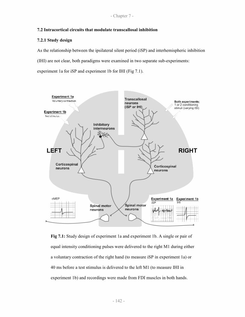
- Chapter 7 -
- 142 -
7.2 Intracortical circuits that modulate transcallosal inhibition
7.2.1 Study design
As the relationship between the ipsilateral silent period (iSP) and interhemispheric inhibition
(IHI) are not clear, both paradigms were examined in two separate sub-experiments:
experiment 1a for iSP and experiment 1b for IHI (Fig 7.1).
Fig 7.1: Study design of experiment 1a and experiment 1b. A single or pair of
equal intensity conditioning pulses were delivered to the right M1 during either
a voluntary contraction of the right hand (to measure iSP in experiment 1a) or
40 ms before a test stimulus is delivered to the left M1 (to measure IHI in
experiment 1b) and recordings were made from FDI muscles in both hands.
LEFT RIGHT

- Chapter 7 -
- 143 -
For experiment 1a, we studied 13 right-handed subjects (5 males and 8 females, mean age
29.2 ± 2.9 years, range 25-39 years). During experiment 1a, the subjects maintained a
contraction of the left FDI at approximately 50% of maximum voluntary contraction (MVC)
while the corresponding right FDI remained at rest. The level of contraction was controlled
by visual feedback of ongoing EMG activity of the left FDI muscle. Single or paired TMS
pulses delivered using a pair of linked Magstim 2002 to the left primary motor cortex were
then delivered at varying interstimulus intervals (ISIs) thereby allowing the simultaneous
assessment of MEPs in the right FDI (from corticospinal stimulation) and iSPs in the left FDI
from transcallosal stimulation.
Paired-pulse stimulation was performed following a paradigm similar to that described by
Tokimura et al. (1996) in order to investigate the excitatory interactions that occur at short
intervals between two stimuli of identical intensity. To study the iSP, the intensity of the
stimuli was set at an intensity that is close to “threshold” for the iSP; for most subjects this
was around 150% AMT for both stimuli. Although the intensity used for both stimuli was
higher than the one used by Tokimura et al. (1996), a recent study by Ilic et al (2002) showed
that this also could produce SICF. Nine interstimulus intervals (ISI) were studied (1.3 ms, 1.5
ms, 2.0 ms, 2.3 ms, 2.5 ms, 3.0 ms, 3.3 ms, 3.5 ms and 4.3 ms). Blocks of 30 trials were
performed consisting of three randomly intermixed conditions (10 trials for each condition:
CS1 given alone and CS1 preceded by CS2 at two different intervals). The three stimulation
conditions (CS1, CS2-CS1 at two different ISI) were tested in a pseudo-randomised order
within each block. An example of one subject’s testing is presented as follows:
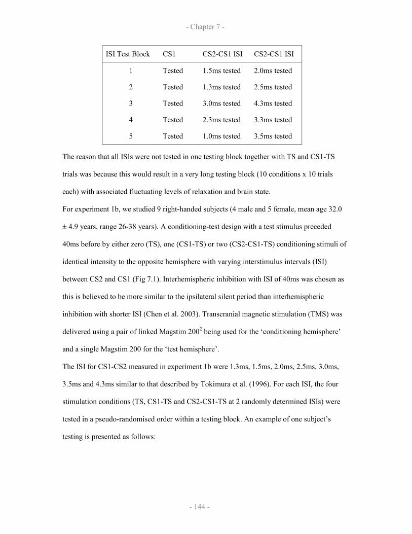
- Chapter 7 -
- 144 -
ISI Test Block CS1 CS2-CS1 ISI CS2-CS1 ISI
1 Tested 1.5ms tested 2.0ms tested
2 Tested 1.3ms tested 2.5ms tested
3 Tested 3.0ms tested 4.3ms tested
4 Tested 2.3ms tested 3.3ms tested
5 Tested 1.0ms tested 3.5ms tested
The reason that all ISIs were not tested in one testing block together with TS and CS1-TS
trials was because this would result in a very long testing block (10 conditions x 10 trials
each) with associated fluctuating levels of relaxation and brain state.
For experiment 1b, we studied 9 right-handed subjects (4 male and 5 female, mean age 32.0
± 4.9 years, range 26-38 years). A conditioning-test design with a test stimulus preceded
40ms before by either zero (TS), one (CS1-TS) or two (CS2-CS1-TS) conditioning stimuli of
identical intensity to the opposite hemisphere with varying interstimulus intervals (ISI)
between CS2 and CS1 (Fig 7.1). Interhemispheric inhibition with ISI of 40ms was chosen as
this is believed to be more similar to the ipsilateral silent period than interhemispheric
inhibition with shorter ISI (Chen et al. 2003). Transcranial magnetic stimulation (TMS) was
delivered using a pair of linked Magstim 2002 being used for the ‘conditioning hemisphere’
and a single Magstim 200 for the ‘test hemisphere’.
The ISI for CS1-CS2 measured in experiment 1b were 1.3ms, 1.5ms, 2.0ms, 2.5ms, 3.0ms,
3.5ms and 4.3ms similar to that described by Tokimura et al. (1996). For each ISI, the four
stimulation conditions (TS, CS1-TS and CS2-CS1-TS at 2 randomly determined ISIs) were
tested in a pseudo-randomised order within a testing block. An example of one subject’s
testing is presented as follows:
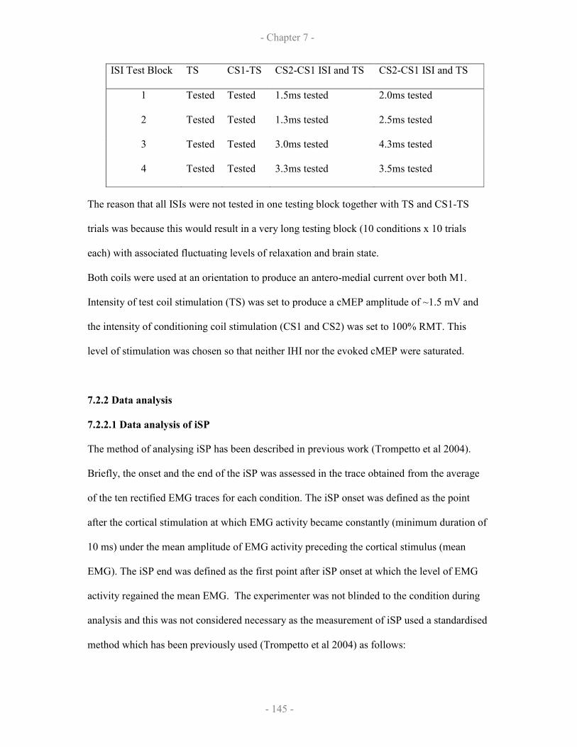
- Chapter 7 -
- 145 -
ISI Test Block TS CS1-TS CS2-CS1 ISI and TS CS2-CS1 ISI and TS
1 Tested Tested 1.5ms tested 2.0ms tested
2 Tested Tested 1.3ms tested 2.5ms tested
3 Tested Tested 3.0ms tested 4.3ms tested
4 Tested Tested 3.3ms tested 3.5ms tested
The reason that all ISIs were not tested in one testing block together with TS and CS1-TS
trials was because this would result in a very long testing block (10 conditions x 10 trials
each) with associated fluctuating levels of relaxation and brain state.
Both coils were used at an orientation to produce an antero-medial current over both M1.
Intensity of test coil stimulation (TS) was set to produce a cMEP amplitude of ~1.5 mV and
the intensity of conditioning coil stimulation (CS1 and CS2) was set to 100% RMT. This
level of stimulation was chosen so that neither IHI nor the evoked cMEP were saturated.
7.2.2 Data analysis
7.2.2.1 Data analysis of iSP
The method of analysing iSP has been described in previous work (Trompetto et al 2004).
Briefly, the onset and the end of the iSP was assessed in the trace obtained from the average
of the ten rectified EMG traces for each condition. The iSP onset was defined as the point
after the cortical stimulation at which EMG activity became constantly (minimum duration of
10 ms) under the mean amplitude of EMG activity preceding the cortical stimulus (mean
EMG). The iSP end was defined as the first point after iSP onset at which the level of EMG
activity regained the mean EMG. The experimenter was not blinded to the condition during
analysis and this was not considered necessary as the measurement of iSP used a standardised
method which has been previously used (Trompetto et al 2004) as follows:
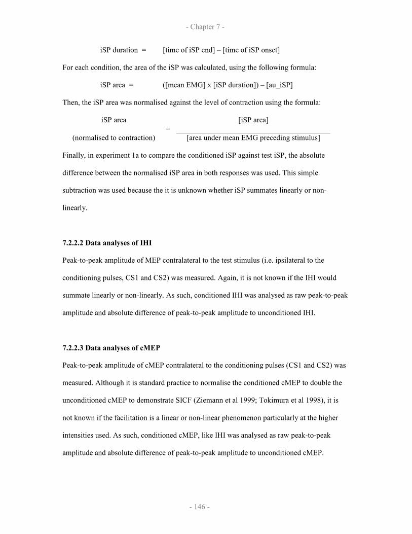
- Chapter 7 -
- 146 -
iSP duration = [time of iSP end] – [time of iSP onset]
For each condition, the area of the iSP was calculated, using the following formula:
iSP area = ([mean EMG] x [iSP duration]) – [au_iSP]
Then, the iSP area was normalised against the level of contraction using the formula:
iSP area [iSP area]
(normalised to contraction)
=
[area under mean EMG preceding stimulus]
Finally, in experiment 1a to compare the conditioned iSP against test iSP, the absolute
difference between the normalised iSP area in both responses was used. This simple
subtraction was used because the it is unknown whether iSP summates linearly or non-
linearly.
7.2.2.2 Data analyses of IHI
Peak-to-peak amplitude of MEP contralateral to the test stimulus (i.e. ipsilateral to the
conditioning pulses, CS1 and CS2) was measured. Again, it is not known if the IHI would
summate linearly or non-linearly. As such, conditioned IHI was analysed as raw peak-to-peak
amplitude and absolute difference of peak-to-peak amplitude to unconditioned IHI.
7.2.2.3 Data analyses of cMEP
Peak-to-peak amplitude of cMEP contralateral to the conditioning pulses (CS1 and CS2) was
measured. Although it is standard practice to normalise the conditioned cMEP to double the
unconditioned cMEP to demonstrate SICF (Ziemann et al 1999; Tokimura et al 1998), it is
not known if the facilitation is a linear or non-linear phenomenon particularly at the higher
intensities used. As such, conditioned cMEP, like IHI was analysed as raw peak-to-peak
amplitude and absolute difference of peak-to-peak amplitude to unconditioned cMEP.
- Chapter 7 -
- 147 -
7.2.2.4 Statistical analysis
For experiment 1a and 1b, two-factorial repeated-measures ANOVAs with factors “CS
MODE” (CS1 and CS2-CS1) and “ISI BLOCK” (1.3ms, 1.5ms, 2.0ms, 2.3ms 2.5ms, 3.0ms,
3.3ms, 3.5ms and 4.3ms) were performed for iSP, IHI and cMEP on the raw peak-to-peak
MEP amplitude or absolute iSP area. One-factorial repeated-measures ANOVA with factor
“ISI BLOCK” (1.3ms, 1.5ms, 2.0ms, 2.3ms 2.5ms, 3.0ms, 3.3ms, 3.5ms and 4.3ms) was also
performed with all the various methods of correction for cMEP, IHI and iSP (i.e. absolute
differences between conditioned and baseline measures or normalised to baseline measure).
For post-hoc comparisons, Tukey least significant difference (LSD) test was used for all the
various methods of correction for cMEP, IHI and iSP.
Correlations between conditioned IHI with the cMEP were calculated by Pearson linear
correlation representing conditioned IHI and conditioned cMEP as absolute difference or
normalised.
Any correlations of conditioned cMEP with conditioned IHI or iSP were performed
comparing equivalently corrected values (i.e. normalised cMEP with normalised IHI,
absolute difference cMEP with absolute difference IHI). Also for correlating the change in
iSP area or duration with the change in the cMEP amplitude, only data from subjects in
which the test iSP was reliably present from the test stimulus.
7.2.3 Results
None of the participants reported any adverse effect during the course of the study.
7.2.3.1 Demonstration of SICF-like effects on iSP
The intensity of the TMS pulses was adjusted in each subject so that CS2 alone produced a
just visible iSP in the contracting FDI muscle. CS1 was applied at the same intensity. The
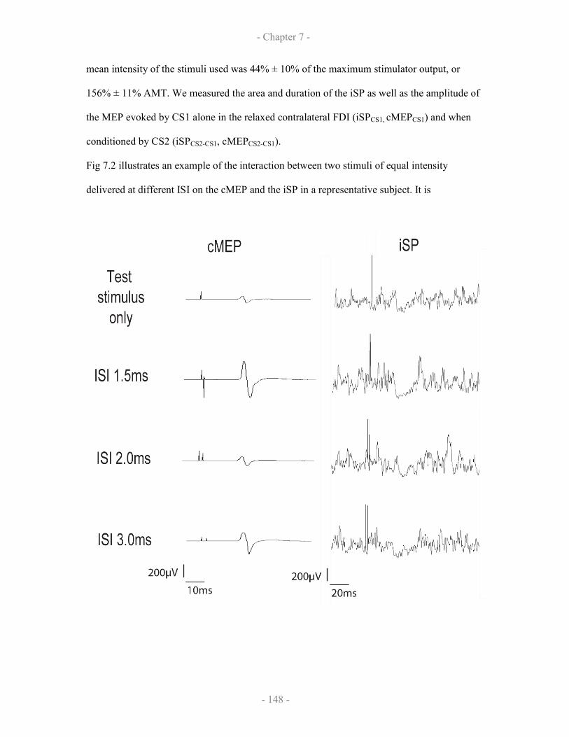
- Chapter 7 -
- 148 -
mean intensity of the stimuli used was 44% ± 10% of the maximum stimulator output, or
156% ± 11% AMT. We measured the area and duration of the iSP as well as the amplitude of
the MEP evoked by CS1 alone in the relaxed contralateral FDI (iSPCS1, cMEPCS1) and when
conditioned by CS2 (iSPCS2-CS1, cMEPCS2-CS1).
Fig 7.2 illustrates an example of the interaction between two stimuli of equal intensity
delivered at different ISI on the cMEP and the iSP in a representative subject. It is
- Chapter 7 -
- 149 -
Fig 7.2: Effect of two stimuli delivered at different ISI ( 1.5, 2 and 3 ms) on the
contralateral MEP in the right relaxed FDI muscle (left panel) and on the iSP in
the left FDI muscle during a contraction equal to almost 50% of the maximal
voluntary contraction (right panel) in experiment 1a. The first line represents
the control response (due to the test stimulus alone) in the contralateral and
ipsilateral muscle, while the other three lines represent the responses due to a
paired stimulation (test stimulus + conditioning stimulus at 1.5, 2.0 and 3.0ms
interstimulus intervals).
clear that at specific intervals for both the cMEP and the iSP, the response to pairs of stimuli
was depended on the interstimulus interval between conditioning stimuli CS2 and CS1.
Due to the difficulty determining the most suitable way to represent conditioned-iSP to the
test-iSP, various methods of analysis were used, as follows:
1) absolute values of conditioned iSP area and test iSP area (Fig 7.3A)
2) absolute values of conditioned iSP duration and test iSP duration (Fig 7.3B)
3) absolute difference between conditioned iSP area and test iSP area (Fig 7.3D)
4) absolute difference between conditioned iSP duration and test iSP duration (Fig 7.3E)
The various methods of analysis were also used for cMEP for consistency, as follows:
1) absolute values of conditioned cMEP and test cMEP (Fig 7.3C)
2) absolute difference between conditioned cMEP and test cMEP (Fig 7.3F)

- Chapter 7 -
- 150 -
(Legend to Figure 7.3 is on next page)

- Chapter 7 -
- 151 -
[Fig 7.3A] Ipsilateral silent period (iSP) area in the left first dorsal interosseus
in response to single or paired transcranial magnetic stimulation to the right
primary motor cortex (dashed lines, test iSP area mean ± s.e.m.; solid line and
triangles, conditioned iSP area, respectively); [B] iSP duration in response to
single or paired transcranial magnetic stimulation in the left first dorsal
interosseus (test iSP duration, dashed lines; conditioned iSP duration, solid line
and triangles, respectively); [C] peak-to-peak MEP amplitude of the right first
dorsal interosseus in response to single or paired stimuli (dashed line, mean
range of CS1; CS2-CS1, solid line and triangles, respectively) measured
simultaneous to [A-B]; [D] absolute difference of the MEP amplitude of the
right first dorsal interosseus with CS2-CS1 stimulation (conditioned cMEP) and
CS1 stimulation (baseline cMEP); [E] absolute difference of the iSP duration of
CS2-CS1 stimulation (conditioned iSP duration) with iSP duration of CS1
stimulation; [F] absolute difference of the MEP amplitude of right first dorsal
interosseus with CS2-CS1 stimulation (conditioned cMEP) with the MEP
amplitude of CS1 stimulation (baseline cMEP). In all graphs, the abscissa
indicates the blocks of interstimulus interval between CS2 and CS1.

- Chapter 7 -
- 152 -
From the above figures, it is clear that no matter how the data are expressed, all three
measures depend on the ISI between CS2 and CS1. In sum, the area and duration of the iSP
as well as the amplitude of the cMEP seem to peak at ISIs 1.5 and 3ms. This was borne out in
the statistical analysis summarised in Table 7.1.
Table 7.1A: Two-way ANOVA with "CS MODE" and "ISI"
iSP area iSP duration cMEP
df F p df F p df F p
"CS MODE" 1 31.127 <0.001 1 22.691 <0.001 1 17.221 <0.001
"ISI" 8 2.869 0.039 8 2.236 0.089 8 3.048 0.035
"CS MODE" x
"ISI" 8 2.685 0.048 8 1.759 0.160 8 7.846 <0.001
Table 7.1B: One-way ANOVA with factor "ISI"
conditioned iSP
area absolute ∆
conditioned iSP
duration absolute ∆
conditioned
cMEP absolute ∆
df 8 8 8
F 2.685 1.759 7.846
p 0.048 0.160 <0.001
Table 7.1C: Post-hoc tests
ISI conditioned iSP area vs test iSP area
1.3ms p=0.001
1.5ms p<0.001
2.0ms n.s.
2.3ms n.s.
2.5ms p=0.006
3.0ms p=0.001
3.3ms p=0.024
3.5ms n.s.
4.3ms p=0.013
Table 7.1 [A] Two-factorial ANOVA for Experiment 1a with factors “CS
MODE” and “ISI” using peak-to-peak MEP amplitude for 11 subjects; [B] One-
factorial ANOVA for Experiment 1a with factor “ISI” comparing absolute
differences of conditioned iSP area, iSP duration or cMEP with baseline test
measures for 11 subjects; [C] Post-hoc Tukey’s t-test comparing raw values of

- Chapter 7 -
- 153 -
conditioned and unconditioned iSP area. n.s. indicates non-significance
(p>0.05).
A two-factor ANOVA of repeated measures on the raw data (Table 7.1 A-C) with “CS
MODE” and “ISI” as main factors revealed a significant interaction between “CS MODE”
and “ISI”. Follow up one-factor ANOVAs with “ISI” as main factor showed significant
effects for iSP area and cMEP amplitude, indicating that these measures depended on the
interval between CS1 and CS2. Post-hoc analysis of the data was conducted to test for
intervals where paired CS2-CS1 conditioning produced significant differences to the effect of
CS1 alone (Table 7.1C). The results varied depending on the method of analysis, but in
general confirmed that maximum effects occurred with ISIs of 1.5 and 3ms. It is not possible
to compare the post-hoc effects of cMEP as there was no consensus on how high intensity
stimulation of the CTS (~150%AMT) would summate (linearly or non-linearly) but it is well
established that whatever the manner of summation, the peaks of facilitatory interaction
would occur at 1.5ms and 3.0ms (Tokimura et al 1996; Ziemann et al 1998).
A correlation analysis was performed to test whether absolute differences of conditioned iSP
area or conditioned iSP duration correlated with the amplitude of the conditioned cMEP over
all ISIs. Only subjects in whom CS2 alone evoked a measurable iSP in every trial were
included in the analysis (n=8 subjects out of 13 subjects). The data showed a significant
linear correlation for conditioned iSP area (expressed as iSPCS2-CS1 – iSPCS1) (Fig 7.4A, r
=0.262, p=0.026, n=72). Linear correlation for the absolute difference in conditioned iSP
duration was not significant (Fig 7.4B, r =0.101, p=0.398, n=72).
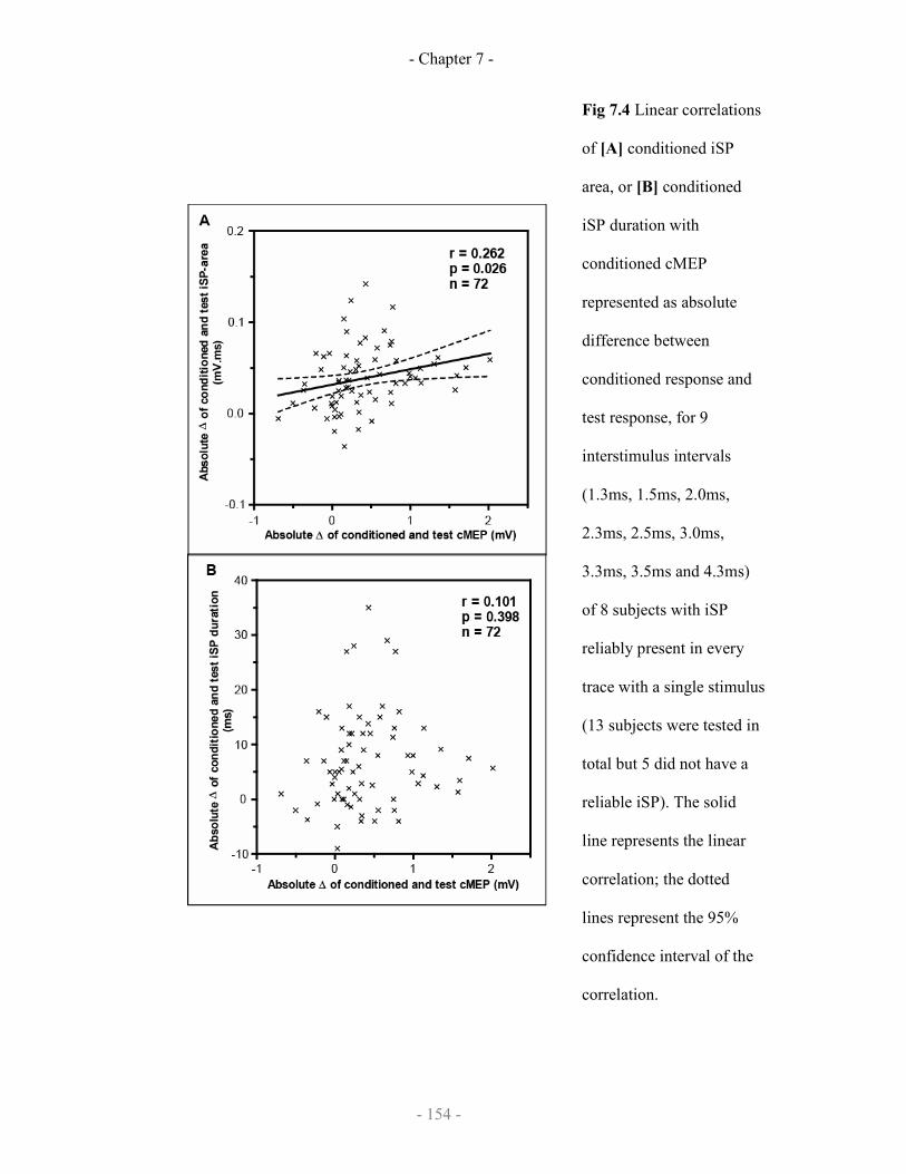
- Chapter 7 -
- 154 -
Fig 7.4 Linear correlations
of [A] conditioned iSP
area, or [B] conditioned
iSP duration with
conditioned cMEP
represented as absolute
difference between
conditioned response and
test response, for 9
interstimulus intervals
(1.3ms, 1.5ms, 2.0ms,
2.3ms, 2.5ms, 3.0ms,
3.3ms, 3.5ms and 4.3ms)
of 8 subjects with iSP
reliably present in every
trace with a single stimulus
(13 subjects were tested in
total but 5 did not have a
reliable iSP). The solid
line represents the linear
correlation; the dotted
lines represent the 95%
confidence interval of the
correlation.
- Chapter 7 -
- 155 -
7.2.3.2 Demonstration of SICF-like effects on IHI
In this experiment, TMS pulses (CS2, CS1) were applied to the right M1 in order to evoke
IHI of test MEPs evoked from the left M1. We measured the amplitude of the contralateral
MEPs from both hemispheres. Stimulus intensities were adjusted so that the test stimulus
(TS) on the left hemisphere evoked a cMEP of about 1mV; the conditioning intensity was
adjusted to give a minimal amount of IHI with CS1 alone. The mean of intensity of TS was
116% ± 5% RMT and the mean of intensity of CS1 and CS2 was 99% ± 6% RMT (or 135%
± 10% AMT). The test stimulus alone produced a cMEP amplitude of 1.30mV ± 0.13mV
which was suppressed by the conditioning stimulus CS1 to 1.12mV ± 0.15mV (85.7% ±
9.7%) of the test stimulus; paired t-test, p<0.01, n=9) (Fig 7.5A).
The principal finding was that the addition of CS2 increased the amount of interhemispheric
inhibition over that seen with CS1 alone at specific intervals. Fig 7.5 shows measures of the
(transcallosally inhibited) cMEP evoked from the right hemisphere (Fig 7.5A and 7.5C), as
well as the cMEP evoked by CS2 and CS1-CS2 from the left hemisphere (Fig 7.5B and
7.5D). At the same intervals as the amount of IHI was maximal, the amplitudes of the cMEPs
evoked by CS2-CS1 were largest. The top two graphs (A and B) plot absolute amplitudes of
MEP, and the bottom two graphs (C and D) shows the data expressed as differences in the
amplitudes of responses with CS1 alone and CS2-CS1 together.

- Chapter 7 -
- 156 -
[Fig 7.5A] Peak-to-peak MEP amplitude of the left first dorsal interosseus in
response to single, paired or triple stimuli (bold line, mean range of TS ± s.e.m.;
dashed line, mean range of CS1-TS; CS2-CS1-TS, solid line and triangles,
respectively); [B] Peak-to-peak MEP amplitude of the right first dorsal
interosseus in response to single or paired stimuli (dashed line, mean range of
CS1; CS2-CS1, solid line and triangles, respectively) measured simultaneous to
[A-C]; [C] Absolute difference of the MEP amplitude of the left first dorsal
interosseus with CS2-CS1-TS stimulation (conditioned IHI) and CS1-TS
stimulation (baseline IHI); [D] Absolute difference of the MEP amplitude of the
right first dorsal interosseus with CS2-CS1 stimulation (conditioned cMEP) and
CS1 stimulation (baseline cMEP). In all graphs, the abscissa indicates the
interstimulus interval between CS2 and CS1.

- Chapter 7 -
- 157 -
Table 7.2 gives the statistical analysis of the results. Two-way repeated measures ANOVA
with factors “CS MODE” and “ISI BLOCK” for MEP amplitude showed a significant “CS
MODE” X “ISI BLOCK” interaction (Table 7.2A, p<0.05). Post hoc one-way ANOVAs with
“ISI BLOCK” as main factor (Table 7.2B) confirmed that no matter which way the data were
analysed, the amount of IHI as well as the amplitude of the cMEP elicited from the right
(CS2-CS1) hemisphere varied with ISI. Paired comparisons to detect at what interval the
largest effects of ISI on IHI was at ISIs of about 1.5 and 3ms (Table 7.2C). Post-hoc tests of
ISI on cMEP were not performed as there was no consensus on how high intensity
stimulation of the CTS (~135%AMT) would summate (linearly or non-linearly) but it is well
established that whatever the manner of summation, the peaks of facilitatory interaction
would occur at 1.5ms and 3.0ms (Tokimura et al 1996; Ziemann et al 1998).
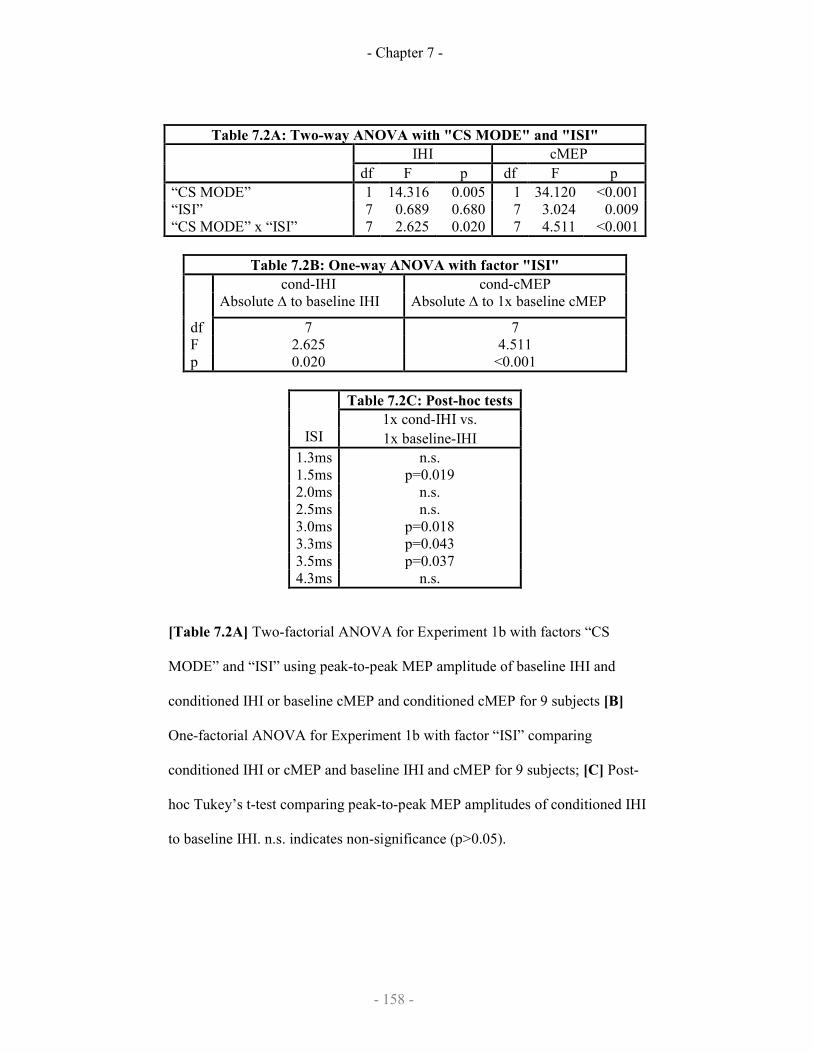
- Chapter 7 -
- 158 -
Table 7.2A: Two-way ANOVA with "CS MODE" and "ISI"
IHI cMEP
df F p df F p
“CS MODE” 1 14.316 0.005 1 34.120 <0.001
“ISI” 7 0.689 0.680 7 3.024 0.009
“CS MODE” x “ISI” 7 2.625 0.020 7 4.511 <0.001
Table 7.2B: One-way ANOVA with factor "ISI"
cond-IHI cond-cMEP
Absolute ∆ to baseline IHI Absolute ∆ to 1x baseline cMEP
df 7 7
F 2.625 4.511
p 0.020 <0.001
Table 7.2C: Post-hoc tests
1x cond-IHI vs.
ISI 1x baseline-IHI
1.3ms n.s.
1.5ms p=0.019
2.0ms n.s.
2.5ms n.s.
3.0ms p=0.018
3.3ms p=0.043
3.5ms p=0.037
4.3ms n.s.
[Table 7.2A] Two-factorial ANOVA for Experiment 1b with factors “CS
MODE” and “ISI” using peak-to-peak MEP amplitude of baseline IHI and
conditioned IHI or baseline cMEP and conditioned cMEP for 9 subjects [B]
One-factorial ANOVA for Experiment 1b with factor “ISI” comparing
conditioned IHI or cMEP and baseline IHI and cMEP for 9 subjects; [C] Post-
hoc Tukey’s t-test comparing peak-to-peak MEP amplitudes of conditioned IHI
to baseline IHI. n.s. indicates non-significance (p>0.05).

- Chapter 7 -
- 159 -
As with the iSP, we next asked whether ISI affected IHI and cMEP in the same way. The
conditioned IHI (CS2-CS1-TS) and the conditioned cMEP (CS2-CS1) showed a significant
negative correlation across the eight interstimulus intervals in all subjects regardless of
method for data analysis (Fig 7.6, p<0.05, n=72).
-0.5 0.0 0.5 1.0 1.5 2.0-2.0
-1.5
-1.0
-0.5
0.0
0.5
1.0
r = -0.274p = 0.02n = 72
Absolute ∆∆∆∆ of conditioned and test cMEPamplitude (mV)
Absolute∆∆ ∆∆ of conditioned and test IHI amplitude
(mV)
[Figure 7.6] Linear correlations of conditioned IHI amplitude with conditioned
cMEP amplitude represented as absolute difference measures respectively for 8
interstimulus intervals (1.3ms, 1.5ms, 2.0ms, 2.5ms, 3.0ms, 3.3ms, 3.5ms and
4.3ms) of all 9 subjects. The solid line represents the linear correlation; the
dotted lines represent the 95% confidence interval of the correlation.

- Chapter 7 -
- 160 -
7.2.4 Discussion
TMS of the motor cortex evokes activity in the corticospinal system that is detected by
measuring the amplitude of MEPs. The excitability of this system is controlled by a number
of intrinsic cortical circuits that have been explored with paired pulse TMS methods. Of
these, the best described are short interval intracortical inhibition (SICI) and short interval
intracortical facilitation (SICF) that are thought to be due to activity in local GABAA
inhibitory interneurones and repetitive facilitatory I-wave input respectively.
In addition to activation of the corticospinal output, motor cortex stimulation can evoke
activity in transcallosal projections. These produce a silent period in the EMG of contracting
ipsilateral muscles (iSP) as well as interhemispheric inhibition (IHI) of MEPs evoked from
the opposite motor cortex. Recently Trompetto et al (2004) showed that transcallosal outputs
from motor cortex receive inhibitory input analogous to corticospinal SICI. We refer to this
as SICIiSP to distinguish it from the usual corticospinal SICI (SICIcMEP). The present
experiments extend these similarities between corticospinal and transcallosal systems by
showing that transcallosal outputs have SICF-like properties. We suggest that the data are
compatible with a model in which the transcallosally projecting pyramidal neurones of
cortical layer III are controlled by circuits similar to those that control layer V pyramidal
neurones of the corticospinal tract. However, it should be noted that there are multiple
interhemispheric effects. We only tested IHI at an ISI of 40ms; IHI at short intervals (8-
12ms) may have a different mechanism (Chen et al 2003). In addition, very low intensity
conditioning pulses evoke a weak interhemispheric facilitation that has another mechanism
(Ugawa et al 1993; Hanajima et al 2001). No firm conclusions can be made about
interneuronal control of these other pathways.
- Chapter 7 -
- 161 -
7.4.1 Site of facilitatory interaction
We investigated transcallosal output with two methods: the ipsilateral silent period (iSP), and
paired pulse interhemispheric inhibition (IHI). As noted in the Introduction, there is good
evidence that both effects are mediated predominantly by pathways running through the
corpus callosum, presumably in the transcallosal axons of layer III pyramidal neurones
(Jacobsen et al 1974; Jones et al 1979).
Experiments 1a and 1b showed that if the intensity of the conditioning stimulus was reduced
to threshold levels for iSP or IHI, pairs of stimuli at intervals of around 1.5 and 3.0 ms
consistently produced much greater transcallosal inhibition than for other intervals. Since this
time course is exactly the same as SICF on cMEP (Tokimura et al 1996; Ziemann et al 1998),
the most parsimonious explanation is that a similar mechanism is involved.
As it is unknown if IHI and iSP summates linearly or nonlinearly, post-hoc tests were done
on absolute data. As such, cautious interpretation of post-hoc tests is necessary. However
whatever the method of analysis, it is clear that the interstimulus intervals where post-hoc
differences are consistently largest are interstimulus intervals of 1.5ms and 3.0ms;
conversely, whatever the method of analysis, the interstimulus intervals where there were no
post-hoc differences in iSP area, iSP duration and IHI were 2.0ms.
Nevertheless whatever the method of data analysis for iSP and IHI, the interstimulus intervals
where post-hoc differences are consistently present in all cases are interstimulus intervals of
1.5ms and 3.0ms. The relationship between SICF in iSP and SICF in IHI with SICF in cMEP
are also confirmed by correlation analysis indicating the presence of a positive linear
correlation for iSP area with cMEP amplitude and a negative linear correlation of IHI with
cMEP amplitude. iSP duration was not correlated with cMEP amplitude, and this probably
reflects that duration of inhibition is not equivalent to degree of inhibition.
- Chapter 7 -
- 162 -
The question arises as to the location of the paired pulse interaction on iSP/IHI. This could
occur either within the stimulated hemisphere, or, since each individual pulse was strong
enough to activate a threshold transcallosal effect, it could also have occurred in the opposite
hemisphere, or even in the spinal cord. In the absence of direct recordings of activity at each
site it is difficult to be certain. However, transcallosal axons are smaller diameter than the
larger corticospinal axons, and therefore will have a longer refractory period after firing an
action potential. Direct measurements show that the absolute refractory period of
corticospinal axons is about 1ms, with the relative refractory period lasting for at least a
further 2ms (Deletis et al 2001; Novak et al 2004). This means that at the first peak of paired
pulse facilitation of iSP/IHI (1.3-1.5ms), transcallosal axons are likely to be still refractory
from the first TMS pulse, and therefore difficult to recruit by a second identical stimulus.
Thus we conclude that at the least the first peak of interaction at 1.5ms is likely to have
occurred within the stimulated hemisphere rather than at any other site.
7.4.2. Nature of facilitatory interaction
I-waves are believed to result from rhythmic excitatory trans-synaptic input of corticospinal
neurons (Amassian & Cracco 1987; Ziemann & Rothwell 2000) the timing of which may be
due to properties of the neurons, synapses or the interneuron network stimulated. Our
experiment indicates that excitatory input onto transcallosal neurons also has the same
rhythmic timing as I-waves on corticospinal neurons. This suggests that both these networks
of excitatory interneurons have similar properties. It is also worthwhile to note that in a
recent paper (Koch et al. 2006), the premotor cortex appears to have a different SICF-like
rhythmicity (0.8ms rather than 1.5ms) which suggests that this rhythmicity is specific to the
primary motor cortex.
- Chapter 7 -
- 163 -
The similar rhythmicity of I-wave interaction may suggest that the corticospinal and
transcallosal neurons share a common excitatory interneuron network. On the other hand, it
may also suggest that I-waves are a shared network property of the excitatory interneuronal
networks rather than a shared network of neurons. It is not possible to exclude either of these
possibilities based on our study.
It is interesting to note the low level of correlation between the corticospinal SICF and the
transcallosal SICF (r2 = 0.069-0.075 depending on the measure used for transcallosal
inhibition). It could be extrapolated from this that the intracortical circuits responsible for
SICF in TC neurons and CTS neurons are only minimally overlapping as mentioned above;
however it could also suggest that SICF (via transcallosal inhibition) has only a minor
inhibitory effect on contralateral corticospinal neuronal excitability. Certainly a recent study
in the rat has also shown that individual deep cortical layer pyramidal cells both receive and
send direct inputs to callosal fibres monosynaptically (Karayannis et al., 2007) suggesting the
latter explanation, but this is unproven.
7.3 Effect of rTMS on transcallosal circuits
As intracortical circuits that synapse onto transcallosal neurons show SICF-like effects and
SICI-like effects (Trompetto et al., 2004), it is likely they behave in a similar manner to
intracortical circuits that synapse onto corticospinal neurons. It has been demonstrated that
intracortical circuits that synapse onto corticospinal neurons can be modulated by repetitive
transcranial magnetic stimulation (rTMS) (Peinnemann et al., Quartarone et al., 2005), but it
is unknown if the intracortical circuits that modulate transcallosal neurons demonstrate
plasticity in response to rTMS. This is the aim of the next study.
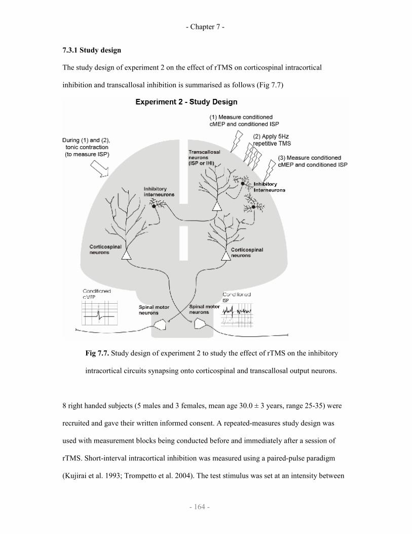
- Chapter 7 -
- 164 -
7.3.1 Study design
The study design of experiment 2 on the effect of rTMS on corticospinal intracortical
inhibition and transcallosal inhibition is summarised as follows (Fig 7.7)
Fig 7.7. Study design of experiment 2 to study the effect of rTMS on the inhibitory
intracortical circuits synapsing onto corticospinal and transcallosal output neurons.
8 right handed subjects (5 males and 3 females, mean age 30.0 ± 3 years, range 25-35) were
recruited and gave their written informed consent. A repeated-measures study design was
used with measurement blocks being conducted before and immediately after a session of
rTMS. Short-interval intracortical inhibition was measured using a paired-pulse paradigm
(Kujirai et al. 1993; Trompetto et al. 2004). The test stimulus was set at an intensity between
- Chapter 7 -
- 165 -
150%-200% AMT where there was a clear iSP recognisable in every trial. In order to
evaluate the threshold of inhibition for both iSP and cMEP various intensities of conditioning
stimulus were measured (70%, 90%, 110%, 130% AMT), starting from 70%AMT that is
known to be at or below threshold for inducing SICI (Kujirai et al 1993; Trompetto et al
2004)
Four blocks (one for every CS intensity) of 30 trials were performed consisting of three
randomly intermixed conditions (10 trials for each condition): TS given alone and TS
preceded by CS at two different intervals (2 ms and 3 ms). The four stimulation conditions of
different CS intensities were tested in a pseudo-randomised order. iSP and cMEP
measurements were carried out in a an identical fashion to experiment 1a with contraction of
the left FDI at approximately 50% of maximum force while maintaining the corresponding
right muscle at rest. The test stimulus intensity was kept constant after rTMS for each subject
as was the order of the testing of the conditioning stimuli intensity.
rTMS was applied over the left M1 with a Magstim Rapid stimulator and a flat figure-of-
eight coil with mean loop diameter of 9 cm. The magnetic stimulus had a biphasic waveform
with a pulse width of approximately 300 µs. We applied two conditioning trains of 5 Hz
rTMS (600 stimuli in total). The intensity of rTMS was set at 90% RMT. We chose this
protocol as Quartarone et al. (2005) demonstrated that it can selectively shape the excitability
of the intracortical circuits without significantly affecting corticospinal excitability.
7.3.2 Data analysis
The measurement of iSP and cMEP was identical to experiment 1a. However, in experiment
2, as the inhibition of iSP was being measured (rather than a summation of iSP), we
normalised the conditioned iSP against the test iSP and expressed the result as a percentage:

- Chapter 7 -
- 166 -
[iSP area in conditioned response] Conditioned iSP (as % of test) =
[iSP area in test response]
For statistical analysis, two-tailed Student’s paired t-test was used to compare the single-
pulse measures (the motor thresholds, iSP, the duration of the iSP and the amplitude of the
cMEP) in the test responses before and after the rTMS.
The effect of the paired pulse stimulation before rTMS was analysed using a two-factorial
analysis of variance (ANOVA) for repeated-measures using the factors “INTENSITY” (70%,
90%, 110%, 130% AMT) and the “MODE OF STIMULATION” (single test-pulse, paired-
pulse with 3ms ISI and paired-pulse with 2ms ISI). The analysis was performed with the raw
data of iSP area and cMEP obtained before the rTMS. If ANOVA showed a significant
effect, we performed post-hoc comparisons using the Tukey least significant difference
(LSD) test to compare directly the experimental conditions. This was to determine which
intensities of conditioning stimulation produced significant effects on SICIiSP and SICIcMEP
compared with baseline.
To analyse the effect of rTMS on paired-pulse stimulation, only intensities where there was
SICIiSP and SICIcMEP at baseline (from above) were analysed using the normalised iSP and
cMEP. A three-factorial ANOVA for repeated-measures was performed using the factors
“INTENSITY” (intensities where there was significant SICIiSP or SICIcMEP at baseline),
“TIME” (before rTMS, after rTMS) and “ISI” (2ms ISI, 3ms ISI).
For all ANOVAs, the Greenhouse-Geisser method was used if necessary to correct for non-
sphericity and post-hoc Tukey (LSD) tests were done for significant results to compare
directly the experimental conditions.
7.3.3 Results
None of the participants reported any adverse effect during the course of the study.

- Chapter 7 -
- 167 -
7.3.3.1 SICIcMEP and SICIiSP
In the “classical” SICI paradigm of Kujirai et al (1993), a subthreshold conditioning stimulus
(CS) is used to suppress the cMEP evoked 2-3 ms later by a suprathreshold test stimulus
(TS); we refer to this as SICIcMEP. Trompetto et al. (2004) recently showed that the CS also
had a similar effect in reducing the area of the iSP evoked in the ipsilateral contracting
muscle by the TS (SICIiSP). We reproduced this finding at baseline pre-rTMS (Fig 7.7, Table
7.3) showing that SICIcMEP and SICIiSP occur at 90% to 130% AMT but not at 70% AMT
(Fig 7.7, Table 7.3A-B). The two-factorial repeated measures ANOVA for
“CONDITIONING INTENSITY BLOCK” and “MODE OF STIMULATION” and the post-
hoc analysis (Table 7.3A-B) clearly indicate that the threshold for inducing SICIcMEP and
SICIiSP during contraction was 90%AMT and above. This lack of conditioning effect at
70%AMT is similar to the original finding where SICIcMEP and SICIiSP were only reliably
found at 80%RMT (Trompetto et al. 2004)
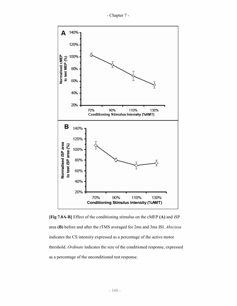
- Chapter 7 -
- 168 -
[Fig 7.8A-B] Effect of the conditioning stimulus on the cMEP (A) and iSP
area (B) before and after the rTMS averaged for 2ms and 3ms ISI. Abscissa
indicates the CS intensity expressed as a percentage of the active motor
threshold. Ordinate indicates the size of the conditioned response, expressed
as a percentage of the unconditioned test response.
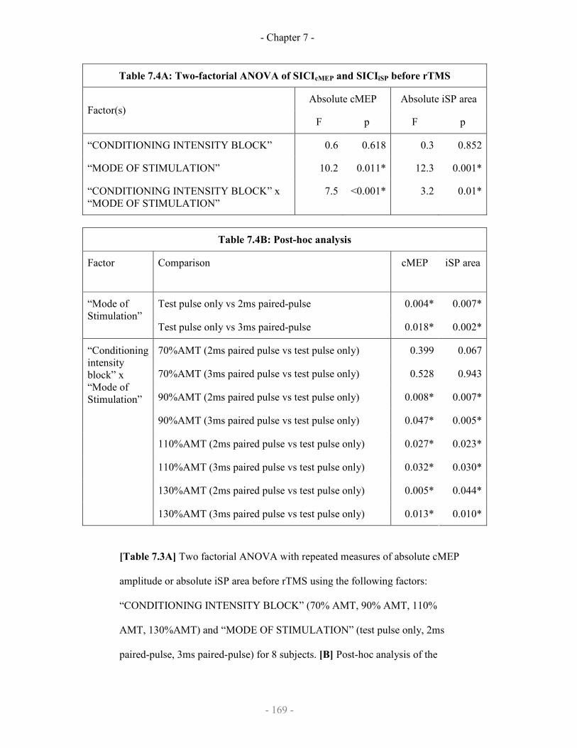
- Chapter 7 -
- 169 -
Table 7.4A: Two-factorial ANOVA of SICIcMEP and SICIiSP before rTMS
Absolute cMEP Absolute iSP area Factor(s)
F p F p
“CONDITIONING INTENSITY BLOCK” 0.6 0.618 0.3 0.852
“MODE OF STIMULATION” 10.2 0.011* 12.3 0.001*
“CONDITIONING INTENSITY BLOCK” x
“MODE OF STIMULATION”
7.5 <0.001* 3.2 0.01*
Table 7.4B: Post-hoc analysis
Factor Comparison
cMEP iSP area
Test pulse only vs 2ms paired-pulse 0.004* 0.007* “Mode of
Stimulation” Test pulse only vs 3ms paired-pulse 0.018* 0.002*
70%AMT (2ms paired pulse vs test pulse only) 0.399 0.067
70%AMT (3ms paired pulse vs test pulse only) 0.528 0.943
90%AMT (2ms paired pulse vs test pulse only) 0.008* 0.007*
90%AMT (3ms paired pulse vs test pulse only) 0.047* 0.005*
110%AMT (2ms paired pulse vs test pulse only) 0.027* 0.023*
110%AMT (3ms paired pulse vs test pulse only) 0.032* 0.030*
130%AMT (2ms paired pulse vs test pulse only) 0.005* 0.044*
“Conditioning
intensity
block” x
“Mode of
Stimulation”
130%AMT (3ms paired pulse vs test pulse only) 0.013* 0.010*
[Table 7.3A] Two factorial ANOVA with repeated measures of absolute cMEP
amplitude or absolute iSP area before rTMS using the following factors:
“CONDITIONING INTENSITY BLOCK” (70% AMT, 90% AMT, 110%
AMT, 130%AMT) and “MODE OF STIMULATION” (test pulse only, 2ms
paired-pulse, 3ms paired-pulse) for 8 subjects. [B] Post-hoc analysis of the

- Chapter 7 -
- 170 -
significant interactions indicating that the conditioning intensity 70% AMT was
significantly different from 90%-130% AMT and was not significantly different
from test-pulse alone. For both tables, * indicates significant effect of at least
p<0.05.
7.3.3.2 Effect of rTMS on SICIcMEP and SICIiSP
Here we ask whether these two effects are influenced in the same way by preconditioning M1
with rTMS at 5 Hz. rTMS was applied at an intensity of 90% RMT, which was equal to 39%
± 5% maximal stimulator output (MSO). The TS intensity was set to evoke a cMEP of
approximately 1mV; this intensity was the same before and after rTMS (63% ± 6.8% MSO
before and 62% ± 7.7% MSO after). Pre-conditioning with rTMS decreased the area of the
iSP evoked by TS alone (p< 0.01) but had no effect on the amplitude of the cMEP (Table
7.4).
Parameter Before
rTMS
After
rTMS
t-test
RMT
(% of maximal stimulator output)
33.1 + 6.1 33.8 + 6.2 n.s.
AMT
(% of maximal stimulator output)
25.3 + 4.9 25.0 + 4.6 n.s.
Mean cMEP amplitude during
ipsilateral contraction (mV)
3.1 + 1.9 3.2 + 1.9 n.s.
Mean iSP area
(mv.ms)
0.16 + 0.07 0.14 + 0.06 p<0.01
iSP duration
(ms)
33.5 + 9.2 30.5 + 10.4 n.s
[Table 7.4] Active and resting motor threshold, resting MEP amplitudes, resting
iSP area and duration of the iSP before and after 600 stimuli of subthreshold
5Hz rTMS to the left M1. RMT: resting motor threshold; AMT: active motor
threshold; n.s.: not significant; each value corresponds to the mean (±SEM) of
nine subjects.
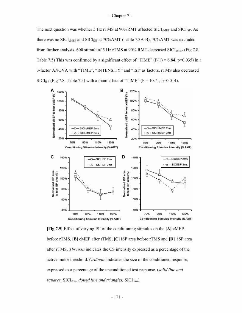
- Chapter 7 -
- 171 -
The next question was whether 5 Hz rTMS at 90%RMT affected SICIcMEP and SICIiSP. As
there was no SICIcMEP and SICIiSP at 70%AMT (Table 7.3A-B), 70%AMT was excluded
from further analysis. 600 stimuli of 5 Hz rTMS at 90% RMT decreased SICIcMEP (Fig 7.8,
Table 7.5) This was confirmed by a significant effect of “TIME” (F(1) = 6.84, p=0.035) in a
3-factor ANOVA with “TIME”, “INTENSITY” and “ISI” as factors. rTMS also decreased
SICIiSP (Fig 7.8, Table 7.5) with a main effect of “TIME” (F = 10.71, p=0.014).
[Fig 7.9] Effect of varying ISI of the conditioning stimulus on the [A] cMEP
before rTMS, [B] cMEP after rTMS, [C] iSP area before rTMS and [D] iSP area
after rTMS. Abscissa indicates the CS intensity expressed as a percentage of the
active motor threshold. Ordinate indicates the size of the conditioned response,
expressed as a percentage of the unconditioned test response. (solid line and
squares, SICI2ms, dotted line and triangles, SICI3ms).
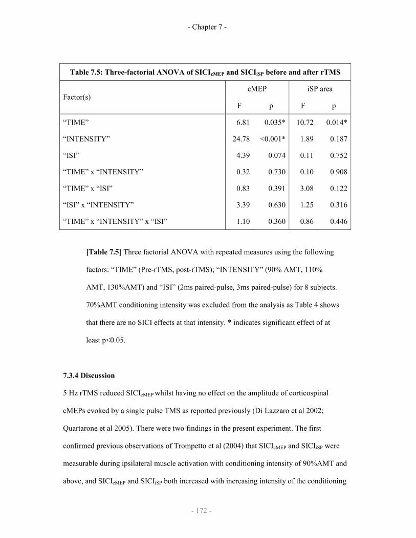
- Chapter 7 -
- 172 -
Table 7.5: Three-factorial ANOVA of SICIcMEP and SICIiSP before and after rTMS
cMEP iSP area Factor(s)
F p F p
“TIME” 6.81 0.035* 10.72 0.014*
“INTENSITY” 24.78 <0.001* 1.89 0.187
“ISI” 4.39 0.074 0.11 0.752
“TIME” x “INTENSITY” 0.32 0.730 0.10 0.908
“TIME” x “ISI” 0.83 0.391 3.08 0.122
“ISI” x “INTENSITY” 3.39 0.630 1.25 0.316
“TIME” x “INTENSITY” x “ISI” 1.10 0.360 0.86 0.446
[Table 7.5] Three factorial ANOVA with repeated measures using the following
factors: “TIME” (Pre-rTMS, post-rTMS); “INTENSITY” (90% AMT, 110%
AMT, 130%AMT) and “ISI” (2ms paired-pulse, 3ms paired-pulse) for 8 subjects.
70%AMT conditioning intensity was excluded from the analysis as Table 4 shows
that there are no SICI effects at that intensity. * indicates significant effect of at
least p<0.05.
7.3.4 Discussion
5 Hz rTMS reduced SICIcMEP whilst having no effect on the amplitude of corticospinal
cMEPs evoked by a single pulse TMS as reported previously (Di Lazzaro et al 2002;
Quartarone et al 2005). There were two findings in the present experiment. The first
confirmed previous observations of Trompetto et al (2004) that SICIcMEP and SICIiSP were
measurable during ipsilateral muscle activation with conditioning intensity of 90%AMT and
above, and SICIcMEP and SICIiSP both increased with increasing intensity of the conditioning
- Chapter 7 -
- 173 -
stimulus over a range from 90-130% AMT (Table 7.4A-B). It may seem surprising that
SICIcMEP could be elicited in view of the ongoing contraction of the opposite FDI that was
required for measurement of the iSP. Muellbacher et al (2000) had reported that SICI was
greatly depressed in such conditions. However, as in experiment 1, the intensity of the test
pulse had been adjusted to evoke a clear iSP. This meant that the contralateral MEP was
larger than the usual 1mV peak-to-peak conventionally used to evaluate SICIcMEP (Kujirai et
al.1993, Fisher et al. 2002, Chen 2004). Larger MEPs are associated with increased SICIcMEP
(Sanger et al. 2001), therefore our experimental protocol may favour demonstrating SICIcMEP.
It is also interesting to note that SICIiSP (but not SICIcMEP) appears to saturate at about
130%AMT (Fig 7.8A, Fig 7.8C and Table 7.6). This is difficult to interpret; it may indicate
that interneurones of SICIcMEP and interneurones of SICIiSP are distinct, but it is important to
stress that at such high intensities, the conditioning stimulus is suprathreshold for producing
both corticospinal volleys and SICF-like phenomena, which complicate the interpretation.
The second finding was that 5 Hz rTMS reduced SICIiSP in the same way as it did SICIcMEP.
We note that although SICIcMEP was less effective after 5Hz rTMS, it was not completely
abolished as reported by Quartarone et al (2005). One possible reason for the discrepancy is
that we found preserved SICIcMEP after rTMS only when we used relatively high conditioning
pulse intensities (110% and 130%AMT) which were not tested in the previous studies.
SICIiSP was reduced by 5Hz rTMS in parallel with SICIcMEP (Table 7.2). Furthermore the
effect was the same at all intensities of the conditioning stimulus. This implies that the
inhibitory interneurons that synapse onto transcallosal neurons are regulated in a similar
manner by rTMS as the inhibitory interneurons that synapse onto the corticospinal cells.
There was one difference between the effect of 5Hz rTMS on cMEP and iSP: it had no effect
on cMEP, whereas there was a small reduction in the depth of iSP. This effect was
unexpected as we had anticipated that high-frequency rTMS would, if anything, increase
- Chapter 7 -
- 174 -
(rather than decrease) the excitability of transcallosal neurons (Cincotta et al 2005). One
possible explanation may relate to the fact that the transcallosal effects between the
hemispheres are not purely inhibitory; there is also a low threshold facilitatory pathway that
is more difficult to study and whose effect is usually masked by the higher threshold and
stronger inhibition (Ugawa et al 1993; Hanajima et al 2001). 5Hz rTMS at 90%RMT may
have preferentially affected the lower threshold transcallosal facilitatory neurons, increasing
their excitability thus producing an apparent reduction in iSP. Whatever the case, the effect of
5Hz rTMS on iSP is small.
7.4 Conclusion
In summary, this data demonstrate that the control of transcallosal connections between the
two hand areas of motor cortex is very similar to that described for the corticospinal outputs
from the same area of cortex. Thus, there is evidence for I-wave facilitatory interaction of
transcallosal projections as well as short interval inhibitory inputs explored by SICI.
Additionally, the population of SICI-like interneurons that control the transcallosal neurons
appear in many ways to behave similarly to those that control corticospinal neurons after
subthreshold 5 Hz rTMS. We conclude that intracortical circuits which synapse onto
transcallosal and corticospinal neurons in layer III and V of the cortex have similar network
properties, and that this may be relevant for effective control of bilateral hand movement.

- Chapter 8 -
- 175 -
Chapter 8
Theta burst stimulation and
sequence learning
Work described in this chapter have published:
Wilkinson L, Teo JT, Obeso I, Rothwell JC, Jahanshahi M. The contribution of the
primary motor cortex is essential for probabilistic implicit sequence learning:
evidence from theta burst magnetic stimulation. J Cogn Neurosci. 2009 Mar 20.
[Epub ahead of print]

- Chapter 8 -
- 176 -
8.1 Introduction
It has been suggested that implicit (unconscious) and explicit (conscious) memory are
separable learning systems e.g. (Squire & Zola, 1996). The implicit system is
believed to be involved in motor skill learning acquired incidentally with practice
(e.g. riding a bicycle, playing golf), whereas the explicit system is considered to play
a role in the acquisition of knowledge in a more intentional way (e.g. remembering
lists of words). Furthermore, it has been proposed that the striatal structures with their
cortical projections support implicit learning whereas the cortico-limbic-diencephalic
structures are the substrate for explicit (conscious) learning e.g. (Cohen & Squire,
1980).
One paradigm that has been developed to study implicit learning in the laboratory is
the serial reaction time (SRT) task (Nissen & Bullemer, 1987). Typically, on each
trial of the SRT task a target appears in one of four locations and participants must
respond as quickly as possible by pressing a corresponding key on a keypad,
participants perform several blocks of trials (e.g. 10 blocks of 100 trials) and reaction
times (RTs) are measured. Unknown to participants, the majority of targets actually
appear in a pre-determined repeating sequence of box locations (e.g. 3-4-2-3-1-2-1-4-
3-2-4-1). The sequence can be presented in either a deterministic or a probabilistic
way. If mean RTs across blocks become faster for the sequence relative to the
random or pseudo-random trials then it can be inferred that participants learned the
trained sequence.
Imaging studies have revealed the functional anatomy of implicit motor sequence
learning and have shown that such learning is associated with activation of the
primary motor cortex supplementary motor area (SMA) and premotor cortex,
dorsolateral prefrontal cortex (DLPFC) and the putamen and caudate e.g. (Grafton et

- Chapter 8 -
- 177 -
al., 1995; Hazeltine et al., 1997; Poldrack et al., 2005; Schendan et al., 2003; Seidler
et al., 2005). However, functional imaging does not reveal whether the contribution
of these various brain regions to implicit motor sequence learning is essential or not.
To address this question the technique of repetitive transcranial magnetic stimulation
(rTMS) to induce ‘virtual lesions’ has been used in several studies. Early studies have
suggested that deterministic SRT learning was impaired by rTMS over the DLPFC
but not by rTMS over the SMA (Pascual-Leone et al., 1996) or the primary motor
cortex (M1) (Pascual-Leone et al., 1999). However, more recent studies on the effect
of stimulation of the primary motor cortex contradict this result. Motor sequence
learning was enhanced by 5Hz rTMS over the primary motor cortex (Kim et al.,
2004), whereas both anodal and cathodal transcranial direct current stimulation
(atDCS, ctDCS) over this area enhanced learning if they were delivered during the
task (Nitsche et al., 2003) and they impaired learning if delivered prior to learning
(Kuo et al., 2008).
Similarly, while rTMS over the SMA was previously shown not to affect SRT
learning (Pascual-Leone et al., 1996), it affected transfer of knowledge to the non-
performing hand (Perez et al., 2007). The role of DLPFC in sequence learning also
appears to be more complex. Although rTMS over the DLPFC delivered during a
deterministic SRT task (Pascual-Leone et al., 1999; Pascual-Leone et al., 1996)
impaired normal learning, this impairment disappeared if there was no spatial
component to the visual cues (Robertson et al., 2001). Furthermore, an impairment of
SRT learning in the left hand of a patient with a focal lesion of the left cerebellum
was shown to be restored by rTMS over both the cerebellum and DLPFC delivered
prior to SRT learning (Torriero et al., 2007).
- Chapter 8 -
- 178 -
This inconsistent pattern of results partly relates to the different methodologies of
delivering brain stimulation. As mentioned previously, some experimenters delivered
stimulation during the SRT task (Nitsche et al., 2003; Pascual-Leone et al., 1999;
Pascual-Leone et al., 1996) or interspersed with the SRT task (Kim et al., 2004). The
delivery of stimulation during performance of the SRT task is compounded by
problems of distraction as rTMS produces a palpable scalp sensation and a loud
‘click’, which may interfere with task performance and learning. Finally, all of the
above studies used a deterministic SRT task which is a less sensitive index of learning
and less likely to foster learning that is truly explicit compared to the probabilistic
SRT task which provides an ‘on line’ index of learning on every block and the
element of noise in the probabilistic sequence blocks explicit knowledge and
promotes implicit learning of the sequence.
Theta burst rTMS (TBS) is a more recent rTMS technique (Huang, Edwards, Rounis,
Bhatia, & Rothwell, 2005) which can be delivered relatively rapidly (< 3 minutes) and
has been shown to produce excitatory (intermittent TBS) or inhibitory (continuous
TBS) effects on cortical excitability lasting for 40 minutes after the stimulation has
ended. This longer lasting stimulation effect allows an ‘offline’ approach with the
participant being able to perform the SRT task undistracted by concurrent stimulation
and unconstrained by the stimulating coil. TBS has been shown to produce plastic
changes in human motor cortex (Huang et al., 2005), but to date the effects of TBS on
learning have not been investigated. In this study we had two objectives. Our first
aim was to assess whether the contribution of the M1, SMA and DLPFC to implicit
sequence learning are essential by applying continuous inhibitory TBS over these
areas immediately before performance of a probabilistic SRT task. We predicted that
if the contribution of these areas to learning is essential, then inhibitory TBS over the
- Chapter 8 -
- 179 -
area would impair subsequent implicit sequence learning. Our second aim was to
assess whether implicit sequence learning could be improved by intermittent
excitatory TBS over M1.
8.2 Methods
8.2.1 Participants
40 right-handed healthy volunteers were recruited, all of whom met the safety criteria
for transcranial magnetic stimulation (Keel, Smith, & Wassermann, 2001). None of
the participants had any neurological disorder or history of psychiatric illness, drug or
alcohol abuse or were on any drug treatments that might influence performance. The
study was approved by the Joint Ethics Committee of the Institute of Neurology and
The National Hospital for Neurology and Neurosurgery. Informed consent was
obtained from all participants. Participants were randomly assigned either to:
a) Sham stimulation group (n = 8, 4 female) aged 22-36 years (M = 27.63,
SD = 4.44).
b) Continuous (Inhibitory) TBS over M1group (n = 8, 5 female) aged 24-37
years (M = 30.63, SD =4.57).
c) Continuous (Inhibitory) TBS over SMA group (n = 8, 4 female) aged 20-
33 years (M = 25.38, SD =4.78).
d) Continuous (Inhibitory) TBS over DLPFC group (n = 8, 5 female) aged
24-38 years (M = 30.38, SD =5.48).
e) Intermittent (Excitatory) TBS over M1group (n = 8, 6 female) aged 22-
36 years (M = 27.63, SD =4.44).
- Chapter 8 -
- 180 -
8.2.2 Serial Reaction Time (SRT) task
In the deterministic SRT task (employed in the majority of SRT studies) this sequence
is repeatedly presented during all blocks with the exception of a single ‘transfer’
block (e.g. block 9) in which different random or pseudo-random trials are introduced.
It is often concluded that knowledge acquired during the deterministic SRT task is
implicit or unconscious however, in some studies participants have been shown to
develop conscious sequence knowledge during supposedly implicit SRT learning e.g.
(Wilkinson & Shanks, 2004). To address this concern, experimenters have attempted
to minimize the chance that SRT learning will be explicit by adopting probabilistic,
rather than deterministic, sequence presentation.
In the probabilistic SRT task (employed here) the sequence is presented such that on
any single trial there is an 85% chance that the target will appear according to the
sequence and a 15% chance that it will appear in a according to an alternate sequence.
Hence, the element of noise during the probabilistic sequence presentation reduces the
chance of participants developing explicit knowledge of the sequence and allows for a
more sensitive online measure of learning across all blocks (rather than just one) by
comparing RTs on probable versus improbable trials.
The probabilistic SRT task was performed immediately after the TBS procedure was
completed. Stimulus presentation, response recording and RT measurement were all
implemented on a PC with a 33 cm color monitor connected to a four-button box.
The four buttons were arranged in a row and will be referred to as 1-4 from left to
right. Stimulus presentation involved four boxes arranged horizontally along the
middle of the computer screen in white against a grey background. The boxes were
26 mm wide and 26 mm high. On each trial of the SRT task, a black X appeared in
the centre of one of the boxes, to which participants had to respond.
- Chapter 8 -
- 181 -
Two second order conditional sequences, SOC1 = 3-1-4-3-2-4-2-1-3-4-1-2 and SOC2
= 4-3-1-2-4-1-3-2-1-4-2-3 were used in the probabilistic SRT task. These sequences
are equated with respect to location frequency (each location occurs three times),
first-order transition frequency (each location is preceded once by each of the other
three locations) and repetitions (no repetitions in either sequence) (Reed & Johnson,
1994). The sequences differed in their second- and higher-order conditional structure.
For approximately half the participants in each condition, SOC1 was the training
sequence and for the remainder it was SOC2. SOCS 1 & 2 and SOCS 3 & 4 are
different but parallel pairs of SOCS. For counter-balancing purposes, for half of the
participants in the implicit sequence learning task, SOCS 1 & 2 were substituted by
SOCS 3 & 4.
In the course of the probabilistic SRT task, the location of the target was specified by
the assigned training sequence with probability .85 and by the alternate sequence with
probability .15. The probabilistic sequences were implemented by using the two most
recent events to select the next event. There was a probability of .85 that the next
target would be the event in the training sequence specified by the last two locations
and a probability of .15 that it would be the event in the alternate sequence specified
by the last two locations. For example, for a given participant trained on SOC1, the
transition 4-1 was followed by a target at location 2 (following the specified sequence
of SOC1) with a probability of .85, and it was followed by a target at location 3
(following the specified sequence of SOC2) with a probability of .15. This algorithm
was applied on each trial and determined the location of the current target simply
based on the two preceding targets.
The probabilistic SRT task comprised 10 blocks, each block with 100 trials during
which participants were exposed to a four-choice SRT task. On each trial,
- Chapter 8 -
- 182 -
participants reacted to the location of the target as quickly as possible by pressing the
corresponding button on the four-button response box. Buttons A, B, C, and D
corresponded to locations 1-4, in that order. Participants were required to respond to
locations 1-4 with the first four fingers respectively of their right hand. Participants
were instructed to respond to the target as fast and as accurately as possible.
Each block began at a random point in the sequence. A trial ended when a participant
pressed the correct key, at which time the target disappeared from the screen. The
next target appeared after a 250 msec interval. Response latencies were measured in
milliseconds from the onset of the target to the completion of a response.1 In total
participants took 16-24 minutes to complete 10 blocks.
8.2.3 Theta burst stimulation
Stimulation was delivered using a Magstim Rapid stimulator (Magstim Co., Dyfed,
UK) connected to a figure-of-eight cased coil with an internal wing diameter of 70
mm, held with the handle pointing posterolaterally. Electromyographic (EMG)
recordings were made using a belly-to-tendon montage from the right first dorsal
interosseous (FDI) muscle. The location of the hand representation in the left
hemisphere was determined, defined as the position at which stimulation produced
optimal muscle evoked potentials (MEPs) in the right FDI. The active motor
threshold (AMT) was assessed during voluntary contraction of the target FDI at
approximately 10% of maximum force, and was defined as the lowest stimulus
intensity required to evoke an MEP of >200 µV in 5 out of 10 trials.
Theta Burst Stimulation was given according to the continuous (cTBS) or intermittent
(iTBS) protocol described by Huang et al. (2005). A theta burst consists of 3 pulses at
- Chapter 8 -
- 183 -
50 Hz, at an intensity of 80% AMT. For the cTBS protocol, theta bursts were given
every 200 ms (i.e. 5 Hz) for a total of 600 pulses (200 theta bursts or 600 pulses) in
the cTBS protocol. The stimulation lasted in total 40 seconds and has been shown to
produce a decrease in corticospinal excitability lasting up to 40 minutes (Huang et al.,
2005). For the iTBS protocol, theta bursts were given every 200ms for 2 seconds (i.e.
10 theta bursts or 30 pulses), followed by a pause of 8 seconds before another 2
seconds of theta bursts. This was repeated 20 times, thereby producing a total of 200
theta bursts or 600 pulses. The stimulation lasted in total of 200 seconds and has been
shown to produce an increase in corticospinal excitability lasting up to 20 minutes
(Huang et al., 2005).
For Sham stimulation, the coil was held rotated 90 degrees over the hand
representation of the motor cortex so that the point of contact with the scalp was
unchanged but the handle pointed vertically upwards.
For continuous (Inhibitory) TBS over M1, cTBS was delivered as described above to
the hand representation of the motor cortex as identified above with the coil handle in
the postero-lateral position.
For continuous (Inhibitory) TBS over SMA, the coil centre was placed over a point
3cm anterior and 0.5 cm to the left of the standard 10-20 electrode position, Cz, with
the coil handle pointed laterally to the left (Matsunaga et al., 2005). At this point,
there was no discernable twitch in the muscles of the leg of the participant.
For continuous (Inhibitory) TBS over DLPFC, the coil centre was placed over a point
5cm anterior to the hand representation of the motor cortex as identified above with
the coil handle in the postero-lateral position (Pascual-Leone et al., 1996).
- Chapter 8 -
- 184 -
For intermittent (Excitatory) TBS over M1, iTBS was delivered as described above to
the hand representation of the motor cortex as identified above with the coil handle in
the postero-lateral position.
8.2.4 SRT task data analysis
For each participant, mean overall RT, mean overall errors, and mean RTs and errors
for both probable and improbable trials at each block were calculated. Any RTs
shorter than 200ms or longer than 3 standard deviations above an individual’s overall
mean RT were excluded from the analysis. The analysis of RT data included trials on
which errors were made because the presence of significantly more error trials in the
improbable data is caused by anticipation (see analysis of error data), therefore, it is
informative and contributes to the developing difference between probable and
improbable RTs across blocks. The standard deviations of RTs for probable and
improbable trials at each block were calculated as a measure of variability of RTs.
In all subsequent analyses: i) RTs or errors for participants trained on one of the two
possible sequences were combined. ii) RTs or errors to the first two targets of each
block were excluded because their locations cannot be predicted. iii) If there was a
violation of the sphericity assumption, Pillai’s multivariate test of significance was
employed (V). Thus, if the Greenhouse-Geisser was less than 1.0, Pillai’s exact F is
reported.
8.3 Results
Participants randomly assigned to the Sham, Inhibitory M1, Inhibitory SMA,
Inhibitory DLPFC or Excitatory M1 groups did not differ in terms of either age [F(4,
39) = 1.69, p >0.05], IQ [F(4, 39) = 1.13, p >0.05] or sex distribution [χ(4) = 2.13, p <
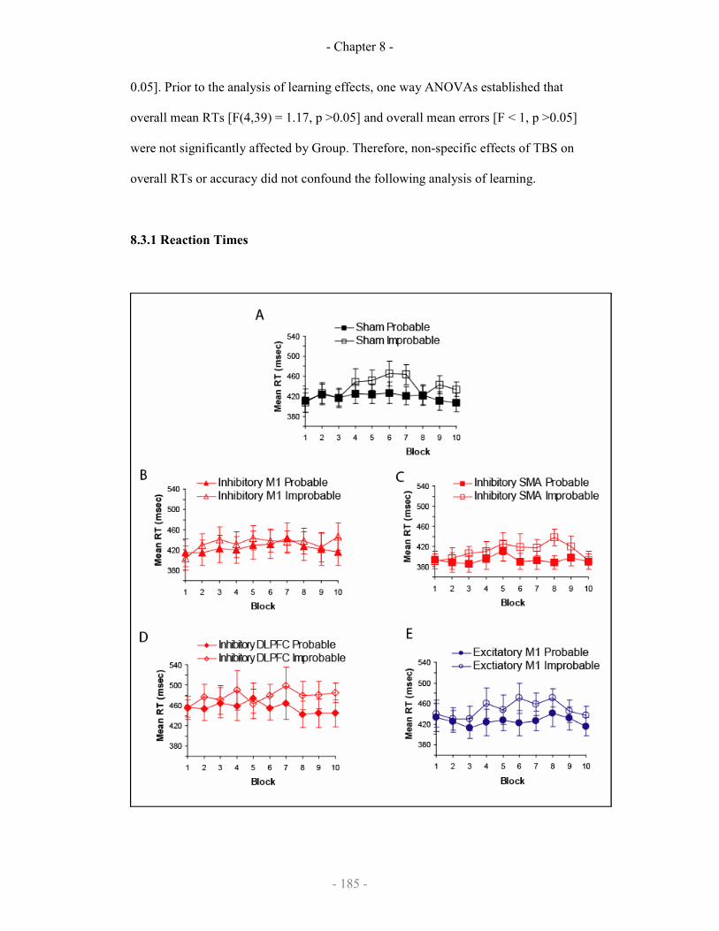
- Chapter 8 -
- 185 -
0.05]. Prior to the analysis of learning effects, one way ANOVAs established that
overall mean RTs [F(4,39) = 1.17, p >0.05] and overall mean errors [F < 1, p >0.05]
were not significantly affected by Group. Therefore, non-specific effects of TBS on
overall RTs or accuracy did not confound the following analysis of learning.
8.3.1 Reaction Times

- Chapter 8 -
- 186 -
Fig 8.1 Mean RTs across training blocks for the implicit sequence
learning task plotted on separate figures for the [A] Sham, [B] Inhibitory
M1, [C] Inhibitory SMA, [D] Inhibitory DLPFC, and [E] Excitatory M1
groups. Probable targets were consistent with the generating sequence
whereas improbable targets were not. Error bars represent standard
errors.
Figures 8.1a-e depict mean RTs obtained over the training phase, plotted separately
for the five groups and for each type of target location, probable or improbable. First,
to establish whether RTs for probable trials changed significantly across blocks in the
five groups, an ANOVA was performed on mean RT for probable trials with Block
(1-10) as a within-subject variable and Group (Sham vs. Inhibitory M1 vs. Inhibitory
SMA vs. Inhibitory DLPFC vs. Excitatory M1) as a between-groups variable. This
analysis revealed a significant main effect of Block [V = 0.50, F(9,27) = 2.95, p =
0.01] because RTs for probable trials significantly changed across blocks. The main
effect of Group [F(4,35) = 1.01] and interaction between Group x Block [F < 1] were
both not significant. For the main effect of Block there was a significant quadratic
trend [F(1,35) = 8.72, p = 0.01] reflecting the fact that across all groups RTs for
probable trials increased across the first couple of blocks, followed by a period of
leveling off, after which they showed a decrease. The eventual speed-up in RTs for
probable trials was seen in all groups and could either be the result of learning the
probable sequence or be due to a non-specific effect of task practice.
Second, to examine whether learning was present in the five groups and to compare
patterns of learning across blocks and in the five groups, an ANOVA was performed
on mean RT with Probability (probable vs. improbable) and Block as within-subject

- Chapter 8 -
- 187 -
variables and Group as a between-groups variable. This analysis revealed a significant
main effect of Probability [F(1,35) = 52.25, p < 0.001] because, overall, probable
targets were performed faster than improbable targets, which is indicative of sequence
learning. There was also a significant main effect of Block [F(5.4,188.3) = 3.97, p =
0.001] and significant interactions between and Probability x Block [F(9,315) = 3.96,
p < 0.001] and Group x Probability x Block [F(9,315) = 1.53, p = 0.03]; showing that
the magnitude of differentiation between RTs for probable and improbable targets
(i.e. extent of learning) changed across blocks -and this pattern differed significantly
between the groups. The main effect of Group [F(4,35) = 1.17 p > 0.05] and
interactions between and Group x Probability and Group x Block were not significant
(Fs < 1, p > 0.05).
In view of the different patterns of learning demonstrated by the five groups across
blocks, composite measures of learning for epochs at the beginning, middle and end
of the training phase were obtained by calculating a difference score (improbable -
probable trials) and comparing the mean difference score across blocks 1-4, 5-7 and
8-10. If learning has occurred, probable trials should be performed faster than
improbable trials, therefore, a positive difference score, which is also significantly
different from zero, is evidence of learning.
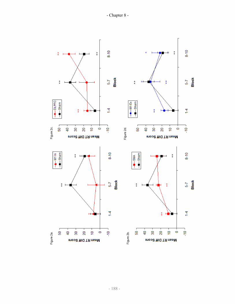
- Chapter 8 -
- 188 -
- Chapter 8 -
- 189 -
Figures 8 2 Mean RT difference scores by epoch for epochs 1-3 of
training blocks plotted on separate figures for the (a) Inhibitory M1, (b)
Inhibitory SMA, (c) Inhibitory DLPFC and (d) Excitatory M1 groups.
All figures are plotted in relation to the Sham groups’ performance. A
positive RT difference score indicates better learning and a double
asterisk indicates scores that were significantly different from zero (2-
tailed), while a single asterisk indicates scores that were significantly
different from zero (1- tailed). Error bars depict standard errors.
Figures 8.2a - d depict the mean of the difference scores for the three training epochs,
plotted separately -relative to the Sham groups’ performance -for each of the TBS
groups. An ANOVA was performed on difference scores with Epoch (1-3) as a
within-participant variable and Group as a between groups variable. This analysis
revealed a significant interaction between Group x Epoch [F(8,70) = 2.61, p = 0.02]
again indicating that the magnitude of RT differences between probable and
improbable targets changed across blocks and between groups. The main effect of
Epoch [F(2,70) = 7.68, p = 0.01] was also significant, whereas the main effect of
Group failed to reach significance[F(4,35) = 1.00].
In light of the significant Group x Epoch interaction and to establish whether learning
occurred at each epoch, in each group, we compared mean difference scores to zero.
For the Sham group (Figures 2a –d), mean difference scores were all negative
however, only the 2nd [t(7) = 5.34, p = 0.001] and 3rd [t(7) = 3.05, p = 0.02] epoch
scores were significantly different from 0 indicating that the Sham group showed

- Chapter 8 -
- 190 -
significant evidence of learning at the middle and towards the end of the training
phase but not at the beginning.
In contrast, for the Inhibitory M1 group (Figure 8.2a), mean difference scores were all
negative; however, none of the epoch scores were significantly different from 0 [1st
t(7) = 1.47, 3rd, t(7) = 1.80] demonstrating that inhibitory TBS of the M1 abolished
probabilistic SRT learning completely across all phases of training.
For the Inhibitory SMA and Excitatory M1 groups (Figure 8.2b and 8.2d), mean
difference scores were all negative and all epoch scores were significantly different
from 0 [Inhibitory SMA, 1st t(7) = 3.08, p = 0.02, 2nd, t(7) = 8.13, p < 0.001, 3rd, t(7)
= 3.20, p = 0.02, Excitatory M1, 1st t(7) = 4.00, p = 0.01, 2nd, t(7) = 2.91, p = 0.02,
3rd, t(7) = 2.08, p = 0.04, 1 -tailed] indicating that these groups learned the sequence
across all epochs.
For the Inhibitory DLPFC group (Figure 8.2c), mean difference scores were all
negative however, only the 1st [t(7) = 3.20, p = 0.02] and 3rd [t(7) = 5.04, p = 0.001]
epoch scores were significantly different from 0 [2nd, t(7) = 1.66] indicating that this
group showed significant evidence of learning at the beginning and towards the end of
learning but not at the middle of the training phase.
Next, for each TBS group we compared mean difference scores at each epoch with
the Sham groups’ performance. This is a non-independent contrast as the Sham group
was used for all comparisons. With a Bonferroni correction, there were no significant
differences in all epochs with the Sham group. (Fig 8.2a – 8.2d). Without a post-hoc
- Chapter 8 -
- 191 -
correction, the mean difference scores of only the inhibitory M1 group (Figure 8.2a)
were significantly different during the second epoch [t(14) = -2.54, p = 0.03]. The
lack of significance with a post-hoc correction does not allow any strong conclusion
about the time-course of the effects.
8.3.2 Variability of Reaction Times
To examine whether the TBS manipulation changed viability of RTs across blocks, an
ANOVA was performed on mean standard deviation of RTs with Probability and
Block as within-subject variables and Group as a between groups variable. This
analysis revealed a significant main effect of Block [V = 0.54, F(9,27) = 3.56, p =
0.01]. The main effect of Group [F(4,35) = 1.12] and all other main effects and
interaction were not significant (all Fs < 1, ps > 0.05).
8.3.3 Errors
Overall mean error rates were as follows:
Sham = 0.04, SD = 0.04,
Inhibitory M1 = 0.04, SD =0 .03,
Inhibitory SMA = 0.07, SD = 0.12,
Inhibitory DLPFC = 0.05, SD = 0.03,
Excitatory M1 = 0.04, SD = 0.03.
To compare the rate of errors across blocks and in the five groups, an ANOVA was
performed on mean error rate with Probability and Block as within-subject variables
and Group as a between groups variable. This analysis revealed a significant main
effect of Probability [F(1,35) = 24.20, p < 0.001] because, overall, more errors were
made for improbable relative to probable targets, this reflects the fact that participants

- Chapter 8 -
- 192 -
were able to develop expectations about the location of the probable target which
caused anticipations and hence errors when the target appeared in the unanticipated
location. The main effect of Block [V = 0.40, F(9,27) = 1.97], interaction between
Probability x Block [V = 0.30, F(9,27) = 1.26] and all other main effects and
interactions were not significant (all Fs < 1, ps > 0.05).
8.4 Discussion
In the present study, we demonstrate, for the first time, that inhibitory TBS delivered
over the M1 impaired subsequent probabilistic implicit sequence learning in healthy
participants. In the Inhibitory M1 group, sequence learning was completely abolished
- relative to chance- and the difference between the Sham Group and the Inhibitory
M1 group was most apparent during the middle stage of the learning phase. In
contrast, inhibitory TBS to the DLPFC, SMA and excitatory TBS to the M1 did not
affect probabilistic implicit sequence learning.
8.4.1 Methodological differences across studies of the effects of rTMS and tDCS
on SRT learning
Our findings with TBS rTMS have similarities but also important differences from
those of Nitsche et al. (2003) and Kuo et al. (2008) with tDCS. The consistent
conclusions across these studies are that while the M1 is implicated in implicit
sequence learning, the DLPFC is not. Furthermore, if TBS/ tDCS is delivered to M1
prior to learning, as was the case here and in Kuo et al.’s study, subsequent learning is
impaired whereas if tDCS is delivered during the task it enhances concurrent learning
(Nitsche et al., 2003).
- Chapter 8 -
- 193 -
The common results from this study with the studies of Nitsche et al. (2003) and Kuo
et al. (Kuo et al., 2008) also stand in contrast to some of the findings of less recent
studies (Pascual-Leone et al., 1999; Pascual-Leone et al., 1996) that did not show an
effect of rTMS over M1 on learning on the SRT task but showed an effect of rTMS
over the DLPFC. Nevertheless, our finding that Inhibitory TBS over the SMA did not
affect SRT learning is consistent with one of these studies (Pascual-Leone et al.,
1996).
There are several possible explanations for the differences in the current results and
those seen previously by Pascual-Leone et al. (1999; 1996) and Robertson et al.
(2001). First, it is possible that the inhibitory rTMS procedures used in previous
studies were not of sufficient intensity/frequency to induce changes in plasticity in the
M1. Second, the possibility remains that in previous studies (Pascual-Leone et al.,
1999; Pascual-Leone et al., 1996) concurrently delivered rTMS modified learning
because of other reasons such as interference with attentional focusing.
Furthermore, one limitation of this study is the lack of stereotaxic co-registration of
the site of the TMS. While M1 localization is quite reliable due to presence of MEPs
in the hand muscles, localization of the SMA and DLPFC is less reliable. For the
SMA, this study used landmarks from previous rTMS studies which produced an
effect (Matsunaga et al., 2005) and here we established that participants did not make
any leg movements during stimulation over the SMA to make sure that stimulation
did not affect the leg motor area (just posterior to the SMA). For the DLPFC, 5cm
from the motor hotspot was used. The lack of stereotaxic co-registration may have
meant that the stimulation was insufficiently specific to the DLPFC and may have
- Chapter 8 -
- 194 -
affected the dorsal premotor cortex. Another possibility is the lower intensity of
stimulation used in TBS may have reduced the potency of the effect. Thus, our failure
to find an effect of Inhibitory TBS over the DLPFC on SRT learning is less
conclusive than the presence of an effect in the M1.
8.4.2 Neural basis of motor sequence learning
Imaging evidence for the contribution of brain regions to implicit sequence learning
during the SRT task is inconsistent. Some studies have demonstrated activation of the
M1, SMA and putamen during the SRT task e.g. (Grafton et al., 1995; Hazeltine et al.,
1997; Seidler et al., 2005) whereas other studies have revealed activation in different
areas including the caudate and prefrontal cortex (PFC) e.g. (Poldrack et al., 2005;
Schendan et al., 2003). Similar to the rTMS and tDCS studies of SRT learning,
differences in patterns of brain activation associated with the SRT task across studies
relate to several important methodological variations. First, most imaging studies
have used deterministic SRT tasks and some have employed a dual task approach
(e.g. tone counting concurrently with the SRT) to block awareness of the repeating
sequence. It is likely that studies differ in the extent to which learning on the SRT
task was truly implicit; with activation of the PFC likely to reflect awareness and
explicit learning of the sequence (Seidler et al., 2005). Second, studies differ in the
extent to which their designs allow successful isolation of brain activity specifically
associated with learning per se rather than performance of sequential movements.
From the results of imaging studies of implicit and explicit sequence learning, it is
possible to suggest that two distinct fronto-striatal circuits are involved. It is plausible
that intentional learning of motor sequences with explicit knowledge activates both
- Chapter 8 -
- 195 -
the associative circuit between the dorsal caudate and the dorsolateral prefrontal
cortex as well as the motor circuit between the putamen and M1, SMA and lateral
premotor cortex. Once performance of such intentionally and explicitly learned
sequences becomes skilled and automatic, then control is passed on to the motor
circuit alone. Incidental sequence learning without explicit knowledge that there is a
repeating sequence also appears to be mediated by the motor circuit which sub serves
skilled performance of motor sequences (Brown, 1999). These proposed substrates of
implicit and explicit sequence learning, combined with the important methodological
differences between TMS studies of sequence learning noted above (degree of explicit
knowledge, extent of training and skilled performance, intensity/frequency of
stimulation) shed some light on the discrepant pattern of findings across studies.
Patients with Parkinson’s disease have impaired sequence learning on the SRT task
(Ferraro et al., 1993; Jackson et al., 1995; Pascual-Leone et al., 1993; Wilkinson &
Jahanshahi, 2007). Furthermore, posteroventral pallidotomy (PVP) which alters basal
ganglia output to M1 and SMA completely abolishes SRT sequence learning in PD
patients, which was present, albeit at an attenuated level pre-operatively (Brown et al.,
2003). These findings on the effects of PD and the further negative impact of PVP on
the SRT, similar to our results from TBS rTMS, further support the role of the M1 in
implicit sequence learning.
8.4.3 The role of the M1 in sequential learning
It has been suggested that the M1 is specifically involved in long term consolidation
and storage of sequential knowledge (Karni et al., 1995; Matsuzaka et al., 2007;
Robertson et al., 2005). For instance, in a study of primates who had already

- Chapter 8 -
- 196 -
completed two years of SRT task learning, Matsuzaka, et al. (2007) identified
differential patterns of neuronal firing in M1during performance of sequential relative
to random trials. Furthermore, Robertson et al. (2005) showed that rTMS to M1,
delivered after SRT learning, disrupted subsequent consolidation of sequential
knowledge. However, in contrast to the view that the role of M1 is restricted to long
term consolidation of sequence learning, Seidler et al. (2005) demonstrated learning-
related activation in M1 during the early encoding phase of the SRT task and, Nitsche
et al. (2003) modified early SRT learning using anodal DCS over M1. Our findings
also demonstrate that M1is directly involved in the initial encoding and acquisition
stage of sequence learning.
The precise nature of M1 involvement in the SRT is unclear. Overall reaction times
were the same in all five groups of participants implying that finger movements
themselves were unaffected by the preceding cTBS. One possibility is that cTBS over
M1 interferes with the short term memory trace of preceding movements that
becomes linked during learning to the most probable subsequent movement. This
might be analogous to the memory trace that could contribute to the “repetition
effect” (Bertelson, 1965; Pashler & Baylis, 1991) where there is a speed advantage
when the same stimulus and response are repeated on two consecutive trials.
However, further experiments would be required to test this fully.
8.4.4 Why didn’t excitatory TBS produce enhanced sequence learning?
Despite our finding that inhibitory TBS over the M1 impaired subsequent SRT
learning, we failed to observe a significant improvement of implicit sequence learning
following excitatory TBS over the M1. It is possible that learning during the SRT

- Chapter 8 -
- 197 -
task is dependent on a more complex process than simply changes in motor cortical
excitability. Interestingly, Nitsche et al. (2003) reported significant improvement of
SRT learning with concurrent excitatory anodal tDCS to M1, whereas excitatory
anodal tDCS over M1 impaired subsequent learning when delivered prior to the SRT
task (Kuo et al., 2008) and there may be similar temporal effects of excitatory TBS on
learning. Furthermore, it is also possible that the between-subjects design used in the
present study to minimize potential transfer effects that can occur with a within-
subjects design reduced the power of detecting such an enhanced learning effect for
the excitatory M1 group.
8.4.5 The lack of reaction time improvement in probabilistic sequence learning
The lack of performance in the reaction time for all groups including sham group
stands in contrast to deterministic sequence learning studies where reaction times
decrease over successive training blocks. In contrast, the reaction time of the probable
sequence is maintained while the reaction time for the improbable sequence
deteriorates (Fig 8.1).
This can be explained by the presence of an improbable sequence competing with the
probable sequence within a motor system with limited resources for movement
preparation. This competition is likely to result in the motor system being unable to be
optimally prepared for the more probable sequence, and so the lack of net reaction
time improvement and instead opts to decrease the performance of the less probable
sequence.
The implication of this paradigm is that the primary motor cortex is more than a
motor output centre but reflects probabilistic estimates of intended motor outputs.
This is in keeping with studies showing that the sensorimotor system operates within
- Chapter 8 -
- 198 -
a probabilistic framework (Körding & Wolpert, 2004; Ma et al., 2007; Faisal &
Wolpert, 2009).
8.5 Conclusion
Here, we have presented evidence that continuous inhibitory TBS over the M1 impair
implicit sequence learning in a probabilistic SRT task. Future studies, to examine
temporal effects and using a within-subject design will further investigate the
potential of intermittent excitatory TBS over M1 to enhance sequence learning in the
SRT task.

- Chapter 9 -
- 199 -
Chapter 9
Conclusion
- Chapter 9 -
- 200 -
9.1 Summary
This thesis has presented 6 studies of neuroplasticity and motor learning focusing on
different aspects in this wide field:
Chapter 3 demonstrated that the NMDA agonism modulates the activity of TBS
providing evidence that this form of non-invasive neuroplasticity in humans is
dependent on molecular models of neuroplasticity.
Chapter 4 demonstrated that the effects of theta burst stimulation can be altered by
various neuromodulatory drugs that affect the dopaminergic or cholinergic system,
with nicotine appearing to enhance the excitatory effects of intermittent TBS.
Chapter 5 studied the interaction of nicotine and theta burst stimulation in ballistic
motor learning and suggests that this can be explained by the effects on motor output
variability rather than enhancement of corticospinal excitability.
Chapter 6 studied the role of various GABA receptor subtypes on practice-dependent
plasticity and showed that GABAA-alpha1 subunits are not involved in inhibiting this
form of plasticity. It also demonstrated that the SICI intracortical circuit is the
predominant inhibitor of practice-dependent plasticity.
Chapter 7 studied the intracortical circuits that modulate transcallosal output and
demonstrated that they display properties similar to intracortical circuits that modulate
corticospinal output.
Chapter 8 demonstrated that probabilistic sequence learning can be impaired by
continuous TBS to the primary motor cortex (M1) or the supplementary motor area
(SMA), supporting the role of these areas in implicit motor learning and action
planning.
- Chapter 9 -
- 201 -
So in summary, this thesis shows the diverse manner that motor learning and
plasticity interacts and provides some experimental data for strategies to modulate
these phenomena.
There are three central messages to the studies of this thesis:
(1) The effects of theta burst rTMS can be modulated pharmacologically by drugs
that modulate on synaptic transmission; this provides support for the idea that
the effects of theta burst rTMS are based on LTP-principles;
(2) Intracortical circuits in the primary motor cortex have specific molecular and
neurophysiological characteristics, and this reflects their specific functional
roles they perform in the primary motor cortex; and
(3) The link between plasticity and motor learning is far more complex than
simplistic models of enhancing LTP to enhance learning, and that modelling
of motor tasks highlights paradigm-specific elements to motor learning to
allow modulation of motor learning.
The failure of a number of drug experiments highlight the importance of good study
design with particular focus on the value of sham-controlled studies and appropriate
placebos.
9.2 Brain stimulation, motor learning and plasticity
Evidence from our study and from other studies (Meintzchel & Ziemann, 2005) that
brain stimulation can enhance neuroplasticity and practice-dependent plasticity has
led to suggestions that it might be possible to enhance motor learning and thus motor
rehabilitation using the same techniques (Ziemann et al., 2006).

- Chapter 9 -
- 202 -
However our study with nicotine and theta burst stimulation in Chapter 5 should act
as a caution against the assumption that by simply enhancing plasticity, motor
learning would be enhanced. Thus, although plasticity may be the underlying
molecular mechanism by which motor learning occurs, other factors (e.g. the
contributions of the various brain regions, the cognitive task structure and peripheral
factors) may be overriding factors in limiting motor learning. Thus, if the goal is to
boost motor learning, it is vital to study motor learning specifically rather than
plasticity alone. Additionally the task-specificity of motor learning means that if the
ultimate goal is to translate this into enhancing motor rehabilitation, it is important to
study what makes some types of motor learning generalisable to other tasks (thus
more likely to be clinically relevant) and what makes other types of motor learning
non-generalisable (less likely to be clinically relevant).
9.3 Future possible studies
Work following on from this study can be broadly divided into:
(i) Studying the relationship between variability and plasticity
(ii) Studying the relationship between motor learning and plasticity
(iii) Studying the generalisation of motor learning
9.3.1 Studying the relationship between variability and plasticity
The relationship between output variability and plasticity was suggested in Chapter 5
with the effect of TBS on directional variability and with the model proposed.
One way of testing model would be to alter the reliability of feedback of movement
performance which would provide a method of altering the perception variability
- Chapter 9 -
- 203 -
(PerceptVar). Based on the model in Chapter 5, the hypothesis is that by increasing te
PerceptVar, the amount of motor learning would be reduced.
The plasticity of some individuals have been found to be impaired: non-manifesting
carriers of the DYT1 gene (Edwards et al., 2006) and carriers of a common
polymorphism BDNF gene (Cheeran et al., 2008). Studying the motor output
variability of such individuals before and after rTMS would be interesting as it would
determine if the increase in motor output variability from rTMS is related to the
effects of rTMS on MEPs.
9.3.2 Studying the role of motor learning in neurorehabilitation
It has been postulated that the recovery from stroke occurs in part via motor learning
(Krakauer et al., 2006), but little is know about the impact of stroke on motor
learning, particularly in the acue and sub-acute phase (when the speed of recovery is
most rapid). There is some evidence that there is a reduction in intracortical inhibition
during the subacute phase of stroke (Swayne et al., 2008) which could act as a driver
for increased motor learning and/or plasticity in the motor cortex. A formal
longitudinal study of motor learning and whether it correlates with longer term
functional outcome is a worthwhile follow-on study. If there is such a correlation, it
provides a strong foundation for assessing motor learning in the acute and sub-acute
stages in stroke to identify those patients who could benefit more rehabilitation.
9.3.3 Studying the role of endocannabinoids in human neuroplasticity
As mentioned in Chapter 1, endocannabinoids have been shown to play an important
role in modulating neuroplasticity in animals. As yet, there have not been any studies
in humans of the roles of endocannabinoids. This may be related to the limited
- Chapter 9 -
- 204 -
repertoire of specific cannabinoid-based licensed agents, but the recent development
of the cannabinoid receptor antagonist, rimonabant, for use in the treatment of obesity
(Scheen et al., 2006), allows the translation of neuroplasticity research in animal
models to humans. Such a study would use human neuroplasticity protocols to assess
plasticity before and after drug ingestion (similar to Chapter 2-4).
9.4 Closing statements
Plasticity in the control of movement is likely to be one of the core functions of the
central nervous system, under intense evolutionary pressure as poor movements are
highly selected against, and the prospect of being able to modulate it in humans non-
invasively could have far-reaching uses and implications. Research tools like TMS
allowing the study of plasticity and motor learning have been only available for the
past 20 years, but the questions to be answered hark back to the dawn of Western
civilization. In his treatise, “On the Motion of Animals”, Aristotle clearly understood
the concept of plasticity in the control of movement:
“In an animal the same part has the power of becoming now larger and
now smaller, and changing its form, as the parts increase by warmth
and again contract by cold and change their quality. This change of
quality is caused by imaginations and sensations and by ideas.
Sensations are obviously a form of change of quality, and imagination
and conception have the same effect as the objects so imagined and
conceived…
Now all these affections involve changes of quality, and with those
changes some parts of the body enlarge, others grow smaller. And it is
not hard to see that a small change occurring at the centre makes great

- Chapter 9 -
- 205 -
and numerous changes at the circumference, just as by shifting the
rudder a hair's breadth you get a wide deviation at the prow. And
further, when by reason of heat or cold or some kindred affection a
change is set up in the region of the heart, even in an imperceptibly
small part of the heart, it produces a vast difference in the periphery of
the body.
- Aristotle, ~350 B.C.
Twenty three centuries later, we are a little closer to understanding how this occurs.
- Appendix -
- 206 -
Appendix:
References
- Appendix -
- 207 -
Abraham WC, Bear MF. Metaplasticity: the plasticity of synaptic plasticity. Trends
Neurosci. 1996 Apr;19(4):126-30.
Academy of Medical Sciences (2004 ). Restoring Neurological Function: putting the
neurosciences to work in neurorehabilitation, Academy of Medical Sciences, London.
Adkins, D. L., P. Campos, D. Quach, M. Borromeo, K. Schallert and T. A. Jones
(2006). "Epidural cortical stimulation enhances motor function after sensorimotor
cortical infarcts in rats." Exp Neurol. 200(2): 356-370.
Adkins, D. L. and T. A. Jones (2005). "D-amphetamine enhances skilled reaching
after ischemic cortical lesions in rats." Neurosci Lett. 380 (3): 214-8.
Adkins-Muir, D. L. and T. A. Jones (2003). "Cortical electrical stimulation combined
with rehabilitative training: enhanced functional recovery and dendritic plasticity
following focal cortical ischemia in rats." Neurol Res 25(8): 780-8.
Agostino, R., E. Iezzi, L. Dinapoli, F. Gilio, A. Conte, F. Mari and A. Berardelli
(2007). "Effects of 5 Hz subthreshold magnetic stimulation of primary motor cortex
on fast finger movements in normal subjects." Exp Brain Res 180(1): 105-11.
Ahmed, T. and J. U. Frey (2005). "Plasticity-specific phosphorylation of CaMKII,
MAP-kinases and CREB during late-LTP in rat hippocampal slices in vitro."
Neuropharmacology 49(4): 477-92.
Aisen, M. L., H. I. Krebs, N. Hogan, F. McDowell and B. T. Volpe (1997). "The
effect of robot-assisted therapy and rehabilitative training on motor recovery
following stroke." Arch Neurol. 54(4): 443-6.
Albus, J. S. (1971). "A theory of cerebellar function." Math Biosci. 10: 25-61.
- Appendix -
- 208 -
Ali, A. B. and A. M. Thomson (2008). "Synaptic alpha 5 subunit-containing GABAA
receptors mediate IPSPs elicited by dendrite-preferring cells in rat neocortex." Cereb
Cortex 18(6): 1260-71.
Altman, J. and G. D. Das (1967). "Postnatal neurogenesis in the guinea-pig." Nature
214(5093): 1098-101.
Ammons, R. B. (1951). "Effect of distribution of practice on rotary pursuit "hits"." J
Exp Psychol 41(1): 17-22.
Ammons, R. B., R. G. Farr, E. Bloch, E. Neumann, M. Dey, R. Marion and C. H.
Ammons (1958). "Long-term retention of perceptualmotor skills." J Exp Psychol
55(4): 318-28.
Arai, A. C., Y. F. Xia and E. Suzuki (2004). "Modulation of AMPA receptor kinetics
differentially influences synaptic plasticity in the hippocampus." Neuroscience
123(4): 1011-24.
Artola, A., S. Bröcher and W. Singer (1990). "Different voltage-dependent thresholds
for inducing long-term depression and long-term potentiation in slices of rat visual
cortex." Nature. 347(6288): 69-72.
Ascoli, G. A., L. Alonso-Nanclares, S. A. Anderson, G. Barrionuevo, R. Benavides-
Piccione, A. Burkhalter, G. Buzsaki, B. Cauli, J. Defelipe, A. Fairen, D. Feldmeyer,
G. Fishell, Y. Fregnac, T. F. Freund, D. Gardner, E. P. Gardner, J. H. Goldberg, M.
Helmstaedter, S. Hestrin, F. Karube, Z. F. Kisvarday, B. Lambolez, D. A. Lewis, O.
Marin, H. Markram, A. Munoz, A. Packer, C. C. Petersen, K. S. Rockland, J. Rossier,
B. Rudy, P. Somogyi, J. F. Staiger, G. Tamas, A. M. Thomson, M. Toledo-Rodriguez,
Y. Wang, D. C. West and R. Yuste (2008). "Petilla terminology: nomenclature of
- Appendix -
- 209 -
features of GABAergic interneurons of the cerebral cortex." Nat Rev Neurosci 9(7):
557-68.
Auerbach, J. M. and M. Segal (1996). "Muscarinic receptors mediating depression
and long-term potentiation in rat hippocampus." J Physiol 492 ( Pt 2): 479-93.
Avanzino, L., J. T. Teo and J. C. Rothwell (2007). "Intracortical circuits modulate
transcallosal inhibition in humans." J Physiol 583(Pt 1): 99-114.
Bacci, A., J. R. Huguenard and D. A. Prince (2004). "Long-lasting self-inhibition of
neocortical interneurons mediated by endocannabinoids." Nature 431(7006): 312-6.
Bacci, A., U. Rudolph, J. R. Huguenard and D. A. Prince (2003 ). "Major differences
in inhibitory synaptic transmission onto two neocortical interneuron subclasses." J
Neurosci. 23(29): 9664-74.
Bagnato, S., R. Agostino, N. Modugno, A. Quartarone and A. Berardelli (2006).
"Plasticity of the motor cortex in Parkinson's disease patients on and off therapy."
Mov Disord 21(5): 639-45.
Baraduc, P., N. Lang, J. C. Rothwell and D. M. Wolpert (2004). "Consolidation of
dynamic motor learning is not disrupted by rTMS of primary motor cortex." Curr Biol
14(3): 252-6.
Barbay, S., E. J. Plautz, K. M. Friel, S. B. Frost, N. Dancause, A. M. Stowe and R. J.
Nudo (2006). "Behavioral and neurophysiological effects of delayed training
following a small ischemic infarct in primary motor cortex of squirrel monkeys." Exp
Brain Res. 169(1): 106-16.
Barbay, S., E. V. Zoubina, N. Dancause, S. B. Frost, I. Eisner-Janowicz, A. M. Stowe,
E. J. Plautz and R. J. Nudo (2006). "A single injection of D-amphetamine facilitates
- Appendix -
- 210 -
improvements in motor training following a focal cortical infarct in squirrel
monkeys." Neurorehabil Neural Repair 20(4): 455-8.
Baron, H., I. G. Epstein, M. G. Mulinos and K. G. Nair (1955). "Absorption,
distribution, and excretion of cycloserine in man." Antibiot Annu 3: 136-40.
Barr DS, Lambert NA, Hoyt KL, Moore SD, Wilson WA. Induction and reversal of
long-term potentiation by low- and high-intensity theta pattern stimulation. J
Neurosci. 1995 Jul;15(7 Pt 2):5402-10.
Baumer, T., C. Demiralay, U. Hidding, R. Bikmullina, R. C. Helmich, S. Wunderlich,
J. Rothwell, J. Liepert, H. R. Siebner and A. Munchau (2007). "Abnormal plasticity of
the sensorimotor cortex to slow repetitive transcranial magnetic stimulation in patients
with writer's cramp." Mov Disord 22(1): 81-90.
Bear, M. F. and R. C. Malenka (1994 ). "Synaptic plasticity: LTP and LTD." Curr
Opin Neurobiol. 4(3): 389-99.
Benwell, N. M., F. L. Mastaglia and G. W. Thickbroom (2006). "Paired-pulse rTMS
at trans-synaptic intervals increases corticomotor excitability and reduces the rate of
force loss during a fatiguing exercise of the hand." Exp Brain Res 175(4): 626-32.
Berardelli, A. and A. Curra (2002). "Pathophysiology and treatment of cranial
dystonia." Mov Disord 17 Suppl 2: S70-4.
Berardelli, A., M. Hallett, J. C. Rothwell, R. Agostino, M. Manfredi, P. D. Thompson
and C. D. Marsden (1996). "Single-joint rapid arm movements in normal subjects and
in patients with motor disorders." Brain 119 ( Pt 2): 661-74.
Bernier, P. J., A. Bedard, J. Vinet, M. Levesque and A. Parent (2002). "Newly
generated neurons in the amygdala and adjoining cortex of adult primates." Proc Natl
Acad Sci U S A 99: 11464–9.
- Appendix -
- 211 -
Bienenstock, E. L., L. N. Cooper and P. W. Munro (1982). "Theory for the
development of neuron selectivity: orientation specificity and binocular interaction in
visual cortex." J Neurosci. 2(1): 32-48.
Biernaskie, J., G. Chernenko and D. Corbett (2004). "Efficacy of rehabilitative
experience declines with time after focal ischemic brain injury." J Neurosci 24(5):
1245-54.
Bliss, T., G. Collingridge and R. Morris (2004). Long-term Potentiation: Enhancing
Neuroscience for 30 Years. Oxford, Oxford University Press.
Bliss, T. and T. Lømo (1973). "Long-lasting potentiation of synaptic transmission in
the dentate area of the anaesthetized rabbit following stimulation of the perforant
path." J. Physiol. (Lond.) 232: 331-56.
Blitzer, R. D., O. Gil and E. M. Landau (1990). "Cholinergic stimulation enhances
long-term potentiation in the CA1 region of rat hippocampus." Neurosci Lett 119(2):
207-10.
Boake, C., E. A. Noser, T. Ro, S. Baraniuk, M. Gaber, R. Johnson, E. T. Salmeron, T.
M. Tran, J. M. Lai, E. Taub, L. A. Moye, J. C. Grotta and H. S. Levin (2007).
"Constraint-induced movement therapy during early stroke rehabilitation."
Neurorehabil Neural Repair 21(1): 14-24.
Boggio, P. S., M. Alonso-Alonso, C. G. Mansur, S. P. Rigonatti, G. Schlaug, A.
Pascual-Leone and F. Fregni (2006). "Hand function improvement with low-
frequency repetitive transcranial magnetic stimulation of the unaffected hemisphere in
a severe case of stroke." Am J Phys Med Rehabil 85(11): 927-30.
- Appendix -
- 212 -
Bolton, D. A., J. H. Cauraugh and H. A. Hausenblas (2004). "Electromyogram-
triggered neuromuscular stimulation and stroke motor recovery of arm/hand
functions: a meta-analysis." J Neurol Sci 223(2): 121-7.
Boroojerdi, B., F. Battaglia, W. Muellbacher and L. G. Cohen (2001). "Mechanisms
influencing stimulus-response properties of the human corticospinal system." Clin
Neurophysiol 112(5): 931-7.
Boroojerdi, B., U. Ziemann, R. Chen, C. M. Butefisch and L. G. Cohen (2001).
"Mechanisms underlying human motor system plasticity." Muscle Nerve 24(5): 602-
13.
Boyd, L. A., B. M. Quaney, P. S. Pohl and C. J. Winstein (2007). "Learning
implicitly: effects of task and severity after stroke." Neurorehabil Neural Repair
21(5): 444-54.
Boyd, L. A. and C. J. Winstein (2001). "Implicit motor-sequence learning in humans
following unilateral stroke: the impact of practice and explicit knowledge." Neurosci
Lett 298(1): 65-9.
Breakefield, X. O., A. J. Blood, Y. Li, M. Hallett, P. I. Hanson and D. G. Standaert
(2008). "The pathophysiological basis of dystonias." Nat Rev Neurosci 9(3): 222-34.
Brown, J. A., H. L. Lutsep, M. Weinand and S. C. Cramer (2006). "Motor cortex
stimulation for the enhancement of recovery from stroke: a prospective, multicenter
safety study." Neurosurgery 58(3): 464-73.
Brown, R. G., M. Jahanshahi, P. Limousin-Dowsey, D. Thomas, N. P. Quinn and J. C.
Rothwell (2003). "Pallidotomy and incidental sequence learning in Parkinson's
disease." Neuroreport 14: 21-4.
- Appendix -
- 213 -
Brusa, L., V. Versace, G. Koch, G. Bernardi, C. Iani, P. Stanzione and D. Centonze
(2005). "Improvement of choreic movements by 1 Hz repetitive transcranial magnetic
stimulation in Huntington's disease patients." Ann Neurol 58(4): 655-6.
Brusa, L., V. Versace, G. Koch, C. Iani, P. Stanzione, G. Bernardi and D. Centonze
(2006). "Low frequency rTMS of the SMA transiently ameliorates peak-dose LID in
Parkinson's disease." Clin Neurophysiol 117(9): 1917-21.
Buhmann, C., A. Gorsler, T. Baumer, U. Hidding, C. Demiralay, K. Hinkelmann, C.
Weiller, H. R. Siebner and A. Munchau (2004). "Abnormal excitability of premotor-
motor connections in de novo Parkinson's disease." Brain 127(Pt 12): 2732-46.
Burrone, J. and V. N. Murthy (2003). "Synaptic gain control and homeostasis, ." Curr.
Opin. Neurobiol. 13: 560-7.
Butefisch, C. M., B. Boroojerdi, R. Chen, F. Battaglia and M. Hallett (2005). "Task-
dependent intracortical inhibition is impaired in focal hand dystonia." Mov Disord
20(5): 545-51.
Butefisch, C. M., B. C. Davis, S. P. Wise, L. Sawaki, L. Kopylev, J. Classen and L. G.
Cohen (2000). "Mechanisms of use-dependent plasticity in the human motor cortex."
Proc Natl Acad Sci U S A 97(7): 3661-5.
Butefisch, C. M., R. Kleiser and R. J. Seitz (2006). "Post-lesional cerebral
reorganisation: evidence from functional neuroimaging and transcranial magnetic
stimulation. ." J Physiol Paris. 99(4-6): 437-54.
Cajal, S. R. (1894). "La fine structure dex centres nerveux, The Croonian Lecture."
Proc. R. Soc. London 55: 444-68.
Cajal, S. R. (1913-14). Degeneration and Regeneration of the Nervous System, .
London, Oxford University Press.

- Appendix -
- 214 -
Cameron, H. A. and A. G. Dayer (2008). "New interneurons in the adult neocortex:
small, sparse, but significant?" Biol Psychiatry. 63(7): 650-5.
Carbon, M., M. F. Ghilardi, M. Argyelan, V. Dhawan, S. B. Bressman and D.
Eidelberg (2008). "Increased cerebellar activation during sequence learning in DYT1
carriers: an equiperformance study." Brain 131(Pt 1): 146-54.
Carey, J. R., F. Fregni and A. Pascual-Leone (2006). "rTMS combined with motor
learning training in healthy subjects." Restor Neurol Neurosci 24(3): 191-9.
Carlson, G., Y. Wang and B. E. Alger (2002). "Endocannabinoids facilitate the
induction of LTP in the hippocampus." Nat Neurosci. 5(8): 723-4.
Carr, J. H. and R. B. Shepherd (2000). A motor learning model for rehabilitation.
Movement science: foundations for physical therapy in rehabilitation. J. H. Carr and
R. B. Shepherd. Rockville, MD, Pro-Ed: 31-91.
Casasola, C., T. Montiel, E. Calixto and S. Brailowsky (2004). "Hyperexcitability
induced by GABA withdrawal facilitates hippocampal long-term potentiation."
Neuroscience 126(1): 163-71.
Castellucci, V., Pinsker H, I. Kupfermann and E. R. Kandel (1970). "Neuronal
mechanisms of habituation and dishabituation of the gill-withdrawal reflex in
Aplysia." Science 167: 1745-8
Castro-Alamancos MA, Donoghue JP, Connors BW. Different forms of synaptic
plasticity in somatosensory and motor areas of the neocortex. J Neurosci. 1995
Jul;15(7 Pt 2):5324-33.
Cavus, I. and T. J. Teyler (1998). "NMDA receptor-independent LTP in basal versus
apical dendrites of CA1 pyramidal cells in rat hippocampal slice." Hippocampus 8(4):
373-9.

- Appendix -
- 215 -
Charpier, S. and J. M. Deniau (1997). "In vivo activity-dependent plasticity at cortico-
striatal connections: evidence for physiological long-term potentiation." Proc Natl
Acad Sci U S A. 94(13): 7036-40.
Cheng, V. Y., L. J. Martin, E. M. Elliott, J. H. Kim, H. T. Mount, F. A. Taverna, J. C.
Roder, J. F. Macdonald, A. Bhambri, N. Collinson, K. A. Wafford and B. A. Orser
(2006). "Alpha5GABAA receptors mediate the amnestic but not sedative-hypnotic
effects of the general anesthetic etomidate." J Neurosci 26(14): 3713-20.
Chevaleyre, V. and P. E. Castillo (2004). "Endocannabinoid-mediated metaplasticity
in the hippocampus." Neuron. 43(6): 871-81.
Cho, K., J. P. Aggleton, M. W. Brown and Z. I. Bashir (2001). "An experimental test
of the role of postsynaptic calcium levels in determining synaptic strength using
perirhinal cortex of rat." J. Physiol. 532(2): 459-66.
Christie, B. R. and W. C. Abraham (1992). "Priming of associative long-term
depression in the dentate gyrus by theta frequency synaptic activity." Neuron 9(1): 79-
84.
Cicinelli, P., P. Pasqualetti, M. Zaccagnini, R. Traversa, M. Oliveri and P. M. Rossini
(2003). "Interhemispheric asymmetries of motor cortex excitability in the postacute
stroke stage: a paired-pulse transcranial magnetic stimulation study." Stroke 34(11):
2653-8.
Cicinelli, P., R. Traversa and P. M. Rossini (1997). "Post-stroke reorganization of
brain motor output to the hand: a 2-4 month follow-up with focal magnetic
transcranial stimulation." Electroencephalogr Clin Neurophysiol 105(6): 438-50.
- Appendix -
- 216 -
Classen J, Liepert J, Wise SP, Hallett M, Cohen LG. Rapid plasticity of human
cortical movement representation induced by practice. J Neurophysiol. 1998
Feb;79(2):1117-23
Classen, J. (2003). "Focal hand dystonia--a disorder of neuroplasticity?" Brain 126(Pt
12): 2571-2.
Cohen, L. (2008). Motor learning and plasticity. Institute of Neurology, London:
Lecture.
Cohen, N. J. and L. R. Squire (1980). "Preserved Learning and Retention of Pattern-
Analyzing Skill in Amnesia - Dissociation of Knowing How and Knowing That."
Science 210: 207-10.
Collingridge, G. L. and T. V. Bliss (1995). "Memories of NMDA receptors and LTP."
Trends Neurosci. 18(2): 54-6.
Collingridge, G. L. and T. V. P. Bliss (1987). "NMDA receptors - their role in long
term potentiation." Trends Neurosci. 10(7): 288-93.
Conditt, M. A., F. Gandolfo and F. A. Mussa-Ivaldi (1997). "The motor system does
not learn the dynamics of the arm by rote memorization of past experience." J
Neurophysiol 78(1): 554-60.
Corkin, S. (1968). "Acquisition of motor skill after bilateral medial temporal-lobe
excision. ." Neuropsychologia 6: 255-65.
Creutzfeldt, O. D., G. H. Fromm and H. Kapp (1962). "Influence of transcortical d-c
currents on cortical neuronal activity." Exp Neurol 5: 436-52.

- Appendix -
- 217 -
Crupi, D., M. F. Ghilardi, C. Mosiello, A. Di Rocco, A. Quartarone and F. Battaglia
(2008). "Cortical and brainstem LTP-like plasticity in Huntington's disease." Brain
Res Bull 75(1): 107-14.
Cumberland Consensus Working Group (2008). "Future of restorative neurosciences
in stroke: driving the translational research pipeline from basic science to
rehabilitation of people after stroke." manuscript submitted.
Cummings, D. M., A. J. Milnerwood, G. M. Dallerac, V. Waights, J. Y. Brown, S. C.
Vatsavayai, M. C. Hirst and K. P. Murphy (2006). "Aberrant cortical synaptic
plasticity and dopaminergic dysfunction in a mouse model of Huntington's disease."
Hum Mol Genet 15(19): 2856-68.
Dahl, A. E., T. Askim, R. Stock, E. Langorgen, S. Lydersen and B. Indredavik (2008).
"Short- and long-term outcome of constraint-induced movement therapy after stroke:
a randomized controlled feasibility trial." Clin Rehabil 22(5): 436-47.
Dancause, N., S. Barbay, S. B. Frost, E. J. Plautz, D. Chen, E. V. Zoubina, A. M.
Stowe and R. J. Nudo (2005). "Extensive cortical rewiring after brain injury. ." J
Neurosci. 25(44): 10167-79.
Davies, C. H., S. J. Starkey, M. F. Pozza and G. L. Collingridge (1991). "GABA
autoreceptors regulate the induction of LTP." Nature 349(6310): 609-11.
Day, B. L., P. D. Thompson, A. E. Harding and C. D. Marsden (1998). "Influence of
vision on upper limb reaching movements in patients with cerebellar ataxia." Brain
121 ( Pt 2): 357-72.
Dayer, A. G., K. M. Cleaver, T. Abouantoun and H. A. Cameron (2005). "New
GABAergic interneurons in the adult neocortex and striatum are generated from
different precursors." J Cell Biol. 168: 415-27.
- Appendix -
- 218 -
Del Cerro, S., M. Jung and G. Lynch (1992). "Benzodiazepines block long-term
potentiation in slices of hippocampus and pinform cortex." Neuroscience 49: 1-6.
Devane, W. A. and J. Axelrod (1994). "Enzymatic synthesis of anandamide, an
endogenous ligand for the cannabinoid receptor, by brain membranes." Proc Natl
Acad Sci U S A 91(14): 6698-701.
Devane, W. A., L. Hanus, A. Breuer, R. G. Pertwee, L. A. Stevenson, G. Griffin, D.
Gibson, A. Mandelbaum, A. Etinger and R. Mechoulam (1992). "Isolation and
structure of a brain constituent that binds to the cannabinoid receptor." Science
258(5090): 1946-9.
Deweer, B., A. M. Ergis, P. Fossati, B. Pillon, F. Boller, Y. Agid and B. Dubois
(1994). "Explicit memory, procedural learning and lexical priming in Alzheimer's
disease." Cortex 30(1): 113-26.
Di Lazzaro, V., M. Dileone, F. Pilato, M. F. Contarino, G. Musumeci, A. R.
Bentivoglio, P. A. Tonali and J. C. Rothwell (2006). "Repetitive transcranial magnetic
stimulation of the motor cortex for hemichorea." J Neurol Neurosurg Psychiatry
77(9): 1095-7.
Di Lazzaro, V., A. Oliviero, M. Meglio, B. Cioni, G. Tamburrini, P. Tonali and J. C.
Rothwell (2000). "Direct demonstration of the effect of lorazepam on the excitability
of the human motor cortex." Clin Neurophysiol 111(5): 794-9.
Di Lazzaro, V., F. Pilato, M. Dileone, P. Profice, F. Ranieri, V. Ricci, P. Bria, P. A.
Tonali and U. Ziemann (2007). "Segregating two inhibitory circuits in human motor
cortex at the level of GABAA receptor subtypes: a TMS study." Clin Neurophysiol
118(10): 2207-14.
- Appendix -
- 219 -
Di Lazzaro, V., F. Pilato, M. Dileone, F. Ranieri, V. Ricci, P. Profice, P. Bria, P. A.
Tonali and U. Ziemann (2006). "GABAA receptor subtype specific enhancement of
inhibition in human motor cortex." J Physiol 575(Pt 3): 721-6.
Di Lazzaro, V., D. Restuccia, A. Oliviero, P. Profice, L. Ferrara, A. Insola, P.
Mazzone, P. Tonali and J. C. Rothwell (1998). "Magnetic transcranial stimulation at
intensities below active motor threshold activates intracortical inhibitory circuits."
Exp Brain Res 119(2): 265-8.
Diedrichsen, J., S. E. Criscimagna-Hemminger and R. Shadmehr (2007).
"Dissociating timing and coordination as functions of the cerebellum." J Neurosci
27(23): 6291-301.
Douglas, R. M., G. V. Goddard and M. Riives (1982). "Inhibitory modulation of long-
term potentiation: evidence for a postsynaptic locus of control." Brain Res 240(2):
259-72.
Doyon, J., D. Gaudreau, R. J. Laforce, M. Castonguay, P. J. Bédard, F. Bédard and J.
P. Bouchard (1997). "Role of the striatum, cerebellum, and frontal lobes in the
learning of a visuomotor sequence." Brain Cogn. 34: 218-45.
Doyon, J., R. J. Laforce, G. Bouchard, D. Gaudreau, J. Roy, M. Poirier, P. J. Bédard,
F. Bédard and J. P. Bouchard (1998). "Role of the striatum, cerebellum and frontal
lobes in the automatization of a repeated visuomotor sequence of movements."
Neuropsychologia 36(7): 625-41.
Doyon, J., A. M. Owen, M. Petrides, V. Sziklas and A. C. Evans (1996). "Functional
anatomy of visuomotor skill learning in human subjects examined with positron
emission tomography." Eur J Neurosci. 8: 637-48.

- Appendix -
- 220 -
Dringenberg, H. C., B. Hamze, A. Wilson, W. Speechley and M. C. Kuo (2007).
"Heterosynaptic facilitation of in vivo thalamocortical long-term potentiation in the
adult rat visual cortex by acetylcholine." Cereb Cortex 17(4): 839-48.
D'Souza, D. C., R. Gil, K. Cassello, K. Morrissey, D. Abi-Saab, J. White, R.
Sturwold, A. Bennett, L. P. Karper, E. Zuzarte, D. S. Charney and J. H. Krystal
(2000). "IV glycine and oral D-cycloserine effects on plasma and CSF amino acids in
healthy humans." Biol Psychiatry 47(5): 450-62.
Dudek, S. M. and M. Bear (1992). "Homosynaptic long-term depression in area CAl
of hippocampus and effects of N-methyl-D-aspartate receptor blockade." Proc. Natl.
Acad. Sci. 89: 4363-7.
Dudek, S. M. and M. F. Bear (1993). "Bidirectional long-term modification of
synaptic effectiveness in the adult and immature hippocampus." J Neurosci 13(7):
2910-8.
Edwards, M. J., Y. Z. Huang, P. Mir, J. C. Rothwell and K. P. Bhatia (2006).
"Abnormalities in motor cortical plasticity differentiate manifesting and
nonmanifesting DYT1 carriers." Mov Disord 21(12): 2181-6.
Edwards, M. J., Y. Z. Huang, N. W. Wood, J. C. Rothwell and K. P. Bhatia (2003).
"Different patterns of electrophysiological deficits in manifesting and non-manifesting
carriers of the DYT1 gene mutation." Brain 126(Pt 9): 2074-80.
Engert, F. and T. Bonhoeffer (1999). "Dendritic spine changes associated with
hippocampal long-term synaptic plasticity." Nature 399(6731): 66-70.
Eriksson, P. S., E. Perfilieva, T. Björk-Eriksson, A. M. Alborn, C. Nordborg, D. A.
Peterson and F. H. Gage (1998). "Neurogenesis in the adult human hippocampus."
Nat Med. 4(11): 1313-7.

- Appendix -
- 221 -
Faisal AA, Wolpert DM. Near optimal combination of sensory and motor
uncertainty in time during a naturalistic perception-action task. J Neurophysiol.
2009 Apr;101(4):1901-12
Fasoli, S. E., H. I. Krebs, J. Stein, W. R. Frontera and N. Hogan (2003). "Effects of
robotic therapy on motor impairment and recovery in chronic stroke. ." Arch Phys
Med Rehabil. 84(4): 477-82.
Fechner, G.T. (1860). Elemente der Psychophysik.
Feeney, D. M., A. Gonzales and W. Law (1982). "Amphetamine, haloperidol and
experience interact to affect rate of recovery after motor cortex injury." Science. 217:
855-57.
Feigin, V. L., C. M. M. Lawes, D. A. Bennett and C. S. Anderson (2003). "Stroke
epidemiology: a review of population-based studies of incidence, prevalence, and
case-fatality in the late 20th Century. ." Lancet Neurology. 2: 43-53.
Ferraro, M., J. J. Palazzolo, J. Krol, H. I. Krebs, N. Hogan and B. T. Volpe (2003).
"Robot-aided sensorimotor arm training improves outcome in patients with chronic
stroke." Neurology. 61(11): 1604-7.
Feydy, A., R. Carlier, A. Roby-Brami, B. Bussel, F. Cazalis, L. Pierot, Y. Burnod and
M. A. Maier (2002). "Longitudinal study of motor recovery after stroke: recruitment
and focusing of brain activation. ." Stroke. 33(6): 1610-7.
Fino, E., J. Glowinski and L. Venance (2005). "Bidirectional activity-dependent
plasticity at corticostriatal synapses." J Neurosci. 25(49): 11279-87.
Fitts, P. M. (1954). "The information capacity of the human motor system in
controlling the amplitude of movement." J Exp Psychol 47(6): 381-91.

- Appendix -
- 222 -
Fitts, P. M. and M. I. Posner (1967). Human performance. . Wokingham, Wadsworth.
Fitzgerald, P. B., S. Fountain and Z. J. Daskalakis (2006). "A comprehensive review
of the effects of rTMS on motor cortical excitability and inhibition. ." Clin
Neurophysiol. 117(12): 2584-96.
Floel, A., F. Hummel, C. Breitenstein, S. Knecht and L. G. Cohen (2005).
"Dopaminergic effects on encoding of a motor memory in chronic stroke." Neurology.
65(3): 472-4.
Florian, J., M. Muller-Dahlhaus, Y. Liu and U. Ziemann (2008). "Inhibitory circuits
and the nature of their interactions in the human motor cortex a pharmacological TMS
study." J Physiol 586(2): 495-514.
Foldy, C., A. Neu, M. V. Jones and I. Soltesz (2006). "Presynaptic, activity-dependent
modulation of cannabinoid type 1 receptor-mediated inhibition of GABA release." J
Neurosci 26(5): 1465-9.
Frank, T. D., Michelbrink, M., Beckmann, H., & Schöllhorn, W. I. (2008). A
quantitative dynamical systems approach to differential learning: Self-organization
principle and order parameter equations. Biological Cybernetics, 98, 19–31.
Fraser, C., M. Power, S. Hamdy, J. Rothwell, D. Hobday, I. Hollander, P. Tyrell, A.
Hobson, S. Williams and D. Thompson (2002). "Driving plasticity in human adult
motor cortex is associated with improved motor function after brain injury. ." Neuron.
34(5): 831-40.
Frazier, W. T., E. R. Kandel, I. Kupfermann, R. Waziri and R. E. Coggeshall (1967).
"Morphological and functional properties of identified neurons in the abdominal
ganglion of Aplysia californica." J. Neurophysiol. 30: 1288-1351.

- Appendix -
- 223 -
Fregni, F., P. S. Boggio, A. C. Valle, R. R. Rocha, J. Duarte, M. J. Ferreira, T.
Wagner, S. Fecteau, S. P. Rigonatti, M. Riberto, S. D. Freedman and A. Pascual-
Leone (2006). "A sham-controlled trial of a 5-day course of repetitive transcranial
magnetic stimulation of the unaffected hemisphere in stroke patients." Stroke 37(8):
2115-22.
Fridman, E. A., T. Hanakawa, M. Chung, F. Hummel, R. C. Leiguarda and L. G.
Cohen (2004). "Reorganization of the human ipsilesional premotor cortex after
stroke." Brain 127(Pt 4): 747-58.
Friel, K. M., A. A. Heddings and R. J. Nudo (2000). "Effects of postlesion experience
on behavioral recovery and neurophysiologic reorganization after cortical injury in
primates." Neurorehabil Neural Repair. 14(3): 187-98.
Fritschy, J. M., O. Weinmann, A. Wenzel and D. Benke (1998). "Synapse-specific
localization of NMDA and GABA(A) receptor subunits revealed by antigen-retrieval
immunohistochemistry." J Comp Neurol 390(2): 194-210.
Frost, S. B., S. Barbay, K. M. Friel, E. J. Plautz and R. J. Nudo (2003).
"Reorganization of remote cortical regions after ischemic brain injury: a potential
substrate for stroke recovery. ." J Neurophysiol. 89(6): 3205-14.
Galarreta, M., F. Erdelyi, G. Szabo and S. Hestrin (2008). "Cannabinoid Sensitivity
and Synaptic Properties of 2 GABAergic Networks in the Neocortex." Cereb Cortex.
Gauthier, L. V., E. Taub, C. Perkins, M. Ortmann, V. W. Mark and G. Uswatte
(2008). "Remodeling the brain: plastic structural brain changes produced by different
motor therapies after stroke." Stroke 39(5): 1520-5.
- Appendix -
- 224 -
Geinisman, Y., R. W. Berry, J. F. Disterhoft, J. M. Power and E. A. Van der Zee
(2001). "Associative Learning Elicits the Formation of Multiple-Synapse Boutons." J.
Neurosci. 21: 5568-5573 21: 5568-73.
Gentner R, Wankerl K, Reinsberger C, Zeller D, Classen J. Depression of human
corticospinal excitability induced by magnetic theta-burst stimulation: evidence of
rapid polarity-reversing metaplasticity. Cereb Cortex. 2008 Sep;18(9):2046-53.
Gerloff, C., K. Bushara, A. Sailer, E. M. Wassermann, R. Chen, T. Matsuoka, D.
Waldvogel, G. F. Wittenberg, K. Ishii, L. G. Cohen and M. Hallett (2006).
"Multimodal imaging of brain reorganization in motor areas of the contralesional
hemisphere of well recovered patients after capsular stroke. ." Brain 129(3): 791-808.
Gerloff, C., B. Corwell, R. Chen, M. Hallett and L. G. Cohen (1998). "The role of the
human motor cortex in the control of complex and simple finger movement
sequences." Brain 121 ( Pt 9): 1695-709.
Ghilardi, M. F., M. Carbon, G. Silvestri, V. Dhawan, M. Tagliati, S. Bressman, C.
Ghez and D. Eidelberg (2003). "Impaired sequence learning in carriers of the DYT1
dystonia mutation." Ann Neurol 54(1): 102-9.
Gladstone, D. J., C. J. Danells, A. Armesto, W. E. McIlroy, W. R. Staines, S. J.
Graham, N. Herrmann, J. P. Szalai and S. E. Black (2006). "Subacute Therapy with
Amphetamine and Rehabilitation for Stroke Study Investigators. Physiotherapy
coupled with dextroamphetamine for ehabilitation after hemiparetic stroke: a
randomized, double-blind, placebo-controlled trial." Stroke 37(1): 179-85.
Glazewski, S., C. Herman, M. McKenna, P. F. Chapman and K. Fox (1998). "Long-
term potentiation in vivo in layers II/III of rat barrel cortex." Neuropharmacology
37(4-5): 581-92.
- Appendix -
- 225 -
Goggin, N. L. and H. J. Meeuwsen (1992). "Age-related differences in the control of
spatial aiming movements." Res Q Exerc Sport 63(4): 366-72.
Goldstein, L. B. and J. N. Davis (1990). "Post-lesion practice and amphetamine-
facilitated recovery of beam-walking in the rat." Restor Neurol Neurosci. 2: 311-14.
Gomez-Beldarrain, M., J. C. Garcia-Monco, B. Rubio and A. Pascual-Leone (1998).
"Effect of focal cerebellar lesions on procedural learning in the serial reaction time
task." Exp Brain Res 120(1): 25-30.
Gonzalez, C. L., O. A. Gharbawie and B. Kolb (2006). "Chronic low-dose
administration of nicotine facilitates recovery and synaptic change after focal
ischemia in rats." Neuropharmacology 50(7): 777-87.
Gould, E. (2007). "How widespread is adult neurogenesis in mammals?" Nat Rev
Neurosci. 8(6): 481-8.
Gould, E., A. J. Reeves, M. S. Graziano and C. G. Gross (1999). "Neurogenesis in the
neocortex of adult primates." Science. 1999 Oct 15;286(5439):548-52 286(5439):
548-52.
Gow, D., J. Rothwell, A. Hobson, D. Thompson and S. Hamdy (2004). "Induction of
long-term plasticity in human swallowing motor cortex following repetitive cortical
stimulation. ." Clin Neurophysiol. 115(5): 1044-51.
Greenwood, R. and N. Ward (2007). Rehabilitation of Stroke. Neurology and Clinical
Neuroscience. A. H. V. Schapira. Philadelphia, , Mosby Elsevier: 645-660.
Greenwood, R. J., Ed. (2002). The Handbook of Neurological Rehabilitation. London,
Psychology Press Ltd.
- Appendix -
- 226 -
Grover, L. M. and C. Yan (1999). "Evidence for involvement of group II/III
metabotropic glutamate receptors in NMDA receptor-independent long-term
potentiation in area CA1 of rat hippocampus." J Neurophysiol. 82(6): 2956-69.
Gulledge, A. T., S. B. Park, Y. Kawaguchi and G. J. Stuart (2007). "Heterogeneity of
phasic cholinergic signaling in neocortical neurons." J Neurophysiol. 97(3): 2215-29.
Hallett, M. (2006). "Pathophysiology of dystonia." J Neural Transm Suppl(70): 485-8.
Halsband, U. and R. K. Lange (2006). "Motor learning in man: a review of functional
and clinical studies." J Physiol Paris. 2006 Jun;99(4-6):414-24 99(4-6): 414-24.
Hamada, M., Y. Ugawa and S. Tsuji (2008). "High-frequency rTMS over the
supplementary motor area for treatment of Parkinson's disease." Mov Disord.
Hamdy S, Rothwell JC, Aziz Q, Singh KD, Thompson DG (1998). "Long-term
reorganization of human motor cortex driven by short-term sensory stimulation." Nat
Neurosci. 1(1): 64-8.
Hamdy S, Aziz Q, Rothwell JC, Power M, Singh KD, Nicholson DA, Tallis RC,
Thompson DG (1998). "Recovery of swallowing after dysphagic stroke relates to
functional reorganization in the intact motor cortex." Gastroenterology 115(5): 1104-
12.
Hamdy, S., J. C. Rothwell, Q. Aziz and D. G. Thompson (2000). "Organization and
reorganization of human swallowing motor cortex: implications for recovery after
stroke." Clin Sci (Lond). 99(2): 151-7.
Harms, K. J., M. S. Rioult-Pedotti, D. R. Carter and A. Dunaevsky (2008). "Transient
spine expansion and learning-induced plasticity in layer 1 primary motor cortex." J
Neurosci 28(22): 5686-90.
- Appendix -
- 227 -
Hebb, D. O. (1949). Organization of Behavior. New York, Wiley.
Hess, G., C. D. Aizenman and J. P. Donoghue (1996). "Conditions for the induction
of long-term potentiation in layer II/III horizontal connections of the rat motor
cortex." J Neurophysiol. 75(5): 1765-78.
Hess, G. and J. P. Donoghue (1994). "Long-term potentiation of horizontal
connections provides a mechanism to reorganize cortical motor maps." J
Neurophysiol. 71(6): 2543-7.
Hicks AU, Hewlett K, Windle V, Chernenko G, Ploughman M, Jolkkonen J, Weiss S,
Corbett D (2007). "Enriched environment enhances transplanted subventricular zone
stem cell migration and functional recovery after stroke." Neuroscience 146: 31-40.
Holden, M. K. (2005). "Virtual environments for motor rehabilitation: review. ."
Cyberpsychol Behav. 8: 187-219.
Hopkins, W. F. and D. Johnston (1988). "Noradrenergic enhancement of long-term
potentiation at mossy fiber synapses in the hippocampus." J Neurophysiol. 59(2): 667-
87.
Hovda DA, Feeney DM (1984). "Amphetamine and experience promote recovery of
locomotor function after unilateral frontal cortex injury in the cat." Brain Res. 298:
358-61.
Huang YZ, Rothwell JC, Edwards MJ, Chen RS. Effect of physiological activityon an
NMDA-dependent form of cortical plasticity in human. Cereb Cortex. 2008
Mar;18(3):563-70.
Huang, Y. Z., R. S. Chen, J. C. Rothwell and H. Y. Wen (2007). "The after-effect of
human theta burst stimulation is NMDA receptor dependent." Clin Neurophysiol
118(5): 1028-32.

- Appendix -
- 228 -
Huang, Y. Z., M. J. Edwards, E. Rounis, K. P. Bhatia and J. C. Rothwell (2005).
"Theta burst stimulation of the human motor cortex." Neuron. 45(2): 201-6.
Huang, Y. Z. and J. C. Rothwell (2004). "The effect of short-duration bursts of high-
frequency, low-intensity transcranial magnetic stimulation on the human motor
cortex." Clin Neurophysiol 115(5): 1069-75.
Huang, Y. Z., I. Trender-Gerhard, M. J. Edwards, P. Mir, J. C. Rothwell and K. P.
Bhatia (2006). "Motor system inhibition in dopa-responsive dystonia and its
modulation by treatment." Neurology 66(7): 1088-90.
Hummel, F., P. Celnik, P. Giraux, A. Floel, W. H. Wu, C. Gerloff and L. G. Cohen
(2005). "Effects of non-invasive cortical stimulation on skilled motor function in
chronic stroke." Brain 128(Pt 3): 490-9.
Hurwitz, B. E., W. D. Dietrich, P. M. McCabe, O. Alonson and B. D. Watson (1991).
"Amphetamine promotes recovery from sensory-motor integration deficit after
thrombotic infarction of the primary somatosensory rat cortex." Stroke 22: 648-54.
Indredavik, B., F. Bakke, S. A. Slordahl, R. Rokseth and L. L. Haheim (1999).
"Stroke unit treatment. 10-year follow-up." Stroke 30(8): 1524-7.
Inghilleri, M., A. Conte, V. Frasca, N. Scaldaferri, F. Gilio, M. Santini, G. Fabbrini,
M. Prencipe and A. Berardelli (2006). "Altered response to rTMS in patients with
Alzheimer's disease." Clin Neurophysiol 117(1): 103-9.
Iriki, A., C. Pavlides, A. Keller and H. Asanuma (1989). "Long-Term Potentiation in
the Motor Cortex." 245(4924): 1385.
Ito, M. (1982). "Experimental verification of Marr-Albus' plasticity assumption for
the cerebellum." Acta Biol Acad Sci Hung 33(2-3): 189-99.
- Appendix -
- 229 -
Ito, M. (2002). "The molecular organization of cerebellar long-term depression." Nat.
Rev. Neurosci. 3: 896-902.
Ito, M. (2006). "Cerebellar circuitry as a neuronal machine." Prog Neurobiol 78(3-5):
272-303.
Jackson, A., J. Mavoori and E. E. Fetz (2006). "Long-term motor cortex plasticity
induced by an electronic neural implant." Nature 444(7115): 56-60.
Jackson, G. M., S. R. Jackson, J. Harrison, L. Henderson and C. Kennard (1995).
"Serial Reaction-Time Learning and Parkinsons-Disease - Evidence for a Procedural
Learning Deficit." Neuropsychologia 33: 577-93.
Jacobs, D. H., J. C. Adair, D. J. Williamson, D. L. Na, M. Gold, A. L. Foundas, J. E.
Shuren, J. E. Cibula and K. M. Heilman (1999). "Apraxia and motor-skill acquisition
in Alzheimer's disease are dissociable." Neuropsychologia 37(7): 875-80.
Jacobs, K. M. and J. P. Donoghue (1991). "Reshaping the cortical motor map by
unmasking latent intracortical connections." Science 251(4996): 944-7.
Ji, D., R. Lape and J. A. Dani (2001). "Timing and location of nicotinic activity
enhances or depresses hippocampal synaptic plasticity." Neuron 31(1): 131-41.
Jin, R., S. Clark, A. M. Weeks, J. T. Dudman, E. Gouaux and K. M. Partin (2005).
"Mechanism of positive allosteric modulators acting on AMPA receptors." J Neurosci
25(39): 9027-36.
Jirenhed, D. A., F. Bengtsson and G. Hesslow (2007). "Acquisition, extinction, and
reacquisition of a cerebellar cortical memory trace." J Neurosci 27(10): 2493-502.
- Appendix -
- 230 -
Johansen-Berg, H., M. F. Rushworth, M. D. Bogdanovic, U. Kischka, S. Wimalaratna
and P. M. Matthews (2002). "The role of ipsilateral premotor cortex in hand
movement after stroke." Proc Natl Acad Sci U S A 99(22): 14518-23.
Jörntell, H. and C. Hansel (2006). "Synaptic memories upside down: bidirectional
plasticity at cerebellar parallel fiber-Purkinje cell synapses." Neuron 52(2): 227-38.
Kaelin-Lang, A., L. Sawaki and L. G. Cohen (2005). "Role of voluntary drive in
encoding an elementary motor memory." J Neurophysiol 93(2): 1099-103.
Karayannis, T., I. Huerta-Ocampo and M. Capogna (2007). "GABAergic and
pyramidal neurons of deep cortical layers directly receive and differently integrate
callosal input." Cereb Cortex 17(5): 1213-26.
Karni, A., G. Meyer, C. Rey-Hipolito, P. Jezzard, M. M. Adams, R. Turner and L. G.
Ungerleider (1998). "The acquisition of skilled motor performance: fast and slow
experience-driven changes in primary motor cortex." Proc Natl Acad Sci U S A 95(3):
861-8.
Katsuki, H., Y. Izumi and C. F. Zorumski (1997). "Noradrenergic regulation of
synaptic plasticity in the hippocampal CA1 region." J Neurophysiol. 77(6): 3013-20.
Kauhanen ML, Korpelainen JT, Hiltunen P, Nieminen P, Sotaniemi KA, Myllylä VV
(2000). "Domains and determinants of quality of life after stroke caused by brain
infarction." Archives of Physical Medicine and Rehabilitation 81: 1541-6.
Kawaguchi, Y., F. Karube and Y. Kubota (2006). "Dendritic branch typing and spine
expression patterns in cortical nonpyramidal cells." Cereb Cortex 16(5): 696-711.
Kawaguchi, Y. and Y. Kubota (1997). "GABAergic cell subtypes and their synaptic
connections in rat frontal cortex." Cereb Cortex 7(6): 476-86.
- Appendix -
- 231 -
Keel JC, Smith MJ, Wassermann EM. (2001). "A safety screening questionnaire for
transcranial magnetic stimulation." Clin Neurophysiol. 112: 720.
Kelso, B. A. (1984). The effects of extended practice on aiming movements in terms
of Fitts' Law., York University.
Kessler, K. R., D. Ruge, T. V. Ilic and U. Ziemann (2005). "Short latency afferent
inhibition and facilitation in patients with writer's cramp." Mov Disord 20(2): 238-42.
Khedr, E. M., J. C. Rothwell, O. A. Shawky, M. A. Ahmed and A. Hamdy (2006).
"Effect of daily repetitive transcranial magnetic stimulation on motor performance in
Parkinson's disease." Mov Disord 21(12): 2201-5.
Kim, Y. H., J. W. Park, M. H. Ko, S. H. Jang and P. K. W. Lee (2004). "Facilitative
effect of high frequency subthreshold repetitive transcranial magnetic stimulation on
complex sequential motor learning in humans." Neurosci Lett. 367: 181-5.
Kimiskidis, V. K., S. Papagiannopoulos, D. A. Kazis, K. Sotirakoglou, G. Vasiliadis,
F. Zara, A. Kazis and K. R. Mills (2006). "Lorazepam-induced effects on silent period
and corticomotor excitability." Exp Brain Res 173(4): 603-11.
Kirkwood, A., M. G. Rioult and M. F. Bear (1996). "Experience-dependent
modification of synaptic plasticity in visual cortex." Nature 381: 526-8.
Klausberger, T., J. D. Roberts and P. Somogyi (2002). "Cell type- and input-specific
differences in the number and subtypes of synaptic GABA(A) receptors in the
hippocampus." J Neurosci 22(7): 2513-21.
Kleim JA, Barbay S, Cooper NR, Hogg TM, Reidel CN, Remple MS, Nudo RJ
(2002). "Motor learning-dependent synaptogenesis is localized to functionally
reorganized motor cortex." Neurobiol Learn Mem. 77(1): 63-77.
- Appendix -
- 232 -
Kleim JA, Bruneau R, VandenBerg P, MacDonald E, Mulrooney R, Pocock D (2003).
"Motor cortex stimulation enhances motor recovery and reduces peri-infarct
dysfunction following ischemic insult." Neurol Res. 25(8): 789-93.
Kleim, J. A., S. Chan, E. Pringle, K. Schallert, V. Procaccio, R. Jimenez and S. C.
Cramer (2006). "BDNF val66met polymorphism is associated with modified
experience-dependent plasticity in human motor cortex." Nat Neurosci 9(6): 735-7.
Knopman, D. and M. J. Nissen (1991). "Procedural learning is impaired in
Huntington's disease: evidence from the serial reaction time task." Neuropsychologia
29(3): 245-54.
Knott, G., C. Quairiaux, C. Genoud and E. Welker (2002). "Formation of Dendritic
Spines with GABAergic Synapses Induced by Whisker Stimulation in Adult Mice."
Neuron 34(2): 265-73.
Koch, G., L. Brusa, C. Caltagirone, A. Peppe, M. Oliveri, P. Stanzione and D.
Centonze (2005). "rTMS of supplementary motor area modulates therapy-induced
dyskinesias in Parkinson disease." Neurology 65(4): 623-5.
Koekkoek, S. K., K. Yamaguchi, B. A. Milojkovic, B. R. Dortland, T. J. Ruigrok, R.
Maex, W. De Graaf, A. E. Smit, F. VanderWerf, C. E. Bakker, R. Willemsen, T.
Ikeda, S. Kakizawa, K. Onodera, D. L. Nelson, E. Mientjes, M. Joosten, E. De
Schutter, B. A. Oostra, M. Ito and C. I. De Zeeuw (2005). "Deletion of FMR1 in
Purkinje cells enhances parallel fiber LTD, enlarges spines, and attenuates cerebellar
eyelid conditioning in Fragile X syndrome." Neuron 47(3): 339-52.
Komatsu, Y. (1996). "GABA-B receptors, monoamine receptors, and postsynaptic
inositol trisphosphate-induced Ca2+ release are involved in the induction of long-term
potentiation at visual cortical inhibitory synapses." J Neurosci. 16: 6342-52.
- Appendix -
- 233 -
Komatsu, Y. and Y. Yoshimura (2000). "Activity-dependent maintenance of long-
term potentiation at visual cortical inhibitory synapses." J Neurosci. 20(20): 7539-46.
Körding KP, Wolpert DM. Bayesian integration in sensorimotor learning. Nature.
2004 Jan 15;427(6971):244-7
Krakauer, J. W. (2006). "Motor learning: its relevance to stroke recovery and
neurorehabilitation." Curr Opin Neurol 19(1): 84-90.
Kramar, E. A., B. Lin, C. Y. Lin, A. C. Arai, C. M. Gall and G. Lynch (2004). "A
novel mechanism for the facilitation of theta-induced long-term potentiation by brain-
derived neurotrophic factor." J Neurosci 24(22): 5151-61.
Kreitzer, A. C. and R. C. Malenka (2007). "Endocannabinoid-mediated rescue of
striatal LTD and motor deficits in Parkinson's disease models." Nature 445(7128):
643-7.
Kruglikov, I. and B. Rudy (2008). "Perisomatic GABA release and thalamocortical
integration onto neocortical excitatory cells are regulated by neuromodulators."
Neuron 58(6): 911-24.
Kubota, Y. and Y. Kawaguchi (2000). "Dependence of GABAergic synaptic areas on
the interneuron type and target size." J Neurosci 20(1): 375-86.
Kujirai, T., M. D. Caramia, J. C. Rothwell, B. L. Day, P. D. Thompson, A. Ferbert, S.
Wroe, P. Asselman and C. D. Marsden (1993). "Corticocortical inhibition in human
motor cortex." J Physiol 471: 501-19.
Kuo, M. F., M. Unger, D. Liebetanz, N. Lang, F. Tergau, W. Paulus and M. A.
Nitsche (2008). "Limited impact of homeostatic plasticity on motor learning in
humans." Neuropsychologia 46(8): 2122-8.

- Appendix -
- 234 -
Kupfermann, I., V. Castellucci, P. H and E. R. Kandel (1970). "Neuronal correlates of
habituation and dishabituation of the gill-withdrawal reflex in Aplysia." Science 167:
1743-5.
Kwakkel, G., B. Kollen and E. Lindeman (2004). "Understanding the pattern of
functional recovery after stroke: facts and theories." Restor Neurol Neurosci 22(3-5):
281-99.
Kwakkel, G., B. Kollen and J. Twisk (2006). "Impact of time on improvement of
outcome after stroke." Stroke 37(9): 2348-53.
Kwakkel, G., B. J. Kollen, J. van der Grond and A. J. Prevo (2003). "Probability of
regaining dexterity in the flaccid upper limb: impact of severity of paresis and time
since onset in acute stroke." Stroke 34(9): 2181-6.
Kwakkel, G., B. J. Kollen and R. C. Wagenaar (2002). "Long term effects of intensity
of upper and lower limb training after stroke: a randomised trial." J Neurol Neurosurg
Psychiatry 72(4): 473-9.
Kyriakopoulos, A. A., D. J. Greenblatt and R. I. Shader (1978). "Clinical
pharmacokinetics of lorazepam: a review." J Clin Psychiatry 39(10 Pt 2): 16-23.
Larson, J. and G. Lynch (1986). "Induction of synaptic potentiation in hippocampus
by patterned stimulation involves two events." Science 232(4753): 985-8.
Larson, J. and G. Lynch (1988). "Role of N-methyl-D-aspartate receptors in the
induction of synaptic potentiation by burst stimulation patterned after the
hippocampal theta-rhythm." Brain Res 441(1-2): 111-8.
Larson, J. and G. Lynch (1989). "Theta pattern stimulation and the induction of LTP:
the sequence in which synapses are stimulated determines the degree to which they
potentiate." Brain Res 489(1): 49-58.

- Appendix -
- 235 -
Larson, J., D. Wong and G. Lynch (1986). "Patterned stimulation at the theta
frequency is optimal for the induction of hippocampal long-term potentiation." Brain
Res 368(2): 347-50.
LeBrasseur NK, Sayers SP, Ouellette MM, Fielding RA (2006). "Muscle impairments
and behavioral factors mediate functional limitations and disability following stroke."
Physical Therapy 86: 1342-50.
Lee, M. G., O. K. Hassani, A. Alonso and B. E. Jones (2005). "Cholinergic basal
forebrain neurons burst with theta during waking and paradoxical sleep." J Neurosci
25(17): 4365-9.
Lendvai, B., E. A. Stern, B. Chen and K. Svoboda (2000). "Experience-dependent
plasticity of dendritic spines in the developing rat barrel cortex in vivo." Nature
404(6780): 876-81.
Leuner, B., E. Gould and T. J. Shors (2006). "Is there a link between adult
neurogenesis and learning?" Hippocampus 16(3): 216-24.
Levy, R., S. Ruland, M. Weinand, D. Lowry, R. Dafer and R. Bakay (2008). "Cortical
stimulation for the rehabilitation of patients with hemiparetic stroke: a multicenter
feasibility study of safety and efficacy." J Neurosurg 108(4): 707-14.
Liebetanz, D., M. A. Nitsche, F. Tergau and W. Paulus (2002). "Pharmacological
approach to the mechanisms of transcranial DC-stimulation-induced after-effects of
human motor cortex excitability." Brain 125(Pt 10): 2238-47.
Liepert, J. (2006). "Motor cortex excitability in stroke before and after constraint-
induced movement therapy." Cogn Behav Neurol 19(1): 41-7.
- Appendix -
- 236 -
Lomarev, M. P., S. Kanchana, W. Bara-Jimenez, M. Iyer, E. M. Wassermann and M.
Hallett (2006). "Placebo-controlled study of rTMS for the treatment of Parkinson's
disease." Mov Disord 21(3): 325-31.
Lorenzano, C., L. Dinapoli, F. Gilio, A. Suppa, S. Bagnato, A. Curra, M. Inghilleri
and A. Berardelli (2006). "Motor cortical excitability studied with repetitive
transcranial magnetic stimulation in patients with Huntington's disease." Clin
Neurophysiol 117(8): 1677-81.
Lotze M, Markert J, Sauseng P, Hoppe J, Plewnia C, Gerloff C (2006). "The role of
multiple contralesional motor areas for complex hand movements after internal
capsular lesion." J Neurosci. 26(22): 6096-102.
Lotze, M., C. Braun, N. Birbaumer, S. Anders and L. G. Cohen (2003). "Motor
learning elicited by voluntary drive." Brain 126(Pt 4): 866-72.
Low, K., F. Crestani, R. Keist, D. Benke, I. Brunig, J. A. Benson, J. M. Fritschy, T.
Rulicke, H. Bluethmann, H. Mohler and U. Rudolph (2000). "Molecular and neuronal
substrate for the selective attenuation of anxiety." Science 290(5489): 131-4.
Luft AR, Waller S, Forrester L, Smith GV, Whitall J, Macko RF, Schulz JB, Hanley
DF (2004). "Lesion location alters brain activation in chronically impaired stroke
survivors." Neuroimage 21(3): 924-35.
Ma WJ, Beck JM, Latham PE, Pouget A. Bayesian inference with probabilistic
population codes. Nat Neurosci. 2006 Nov;9(11):1432-8.
MacDermott, A. B., M. L. Mayer, G. L. Westbrook, S. J. Smith and J. L. Barker
(1986). "NMDA-receptor activation increases cytoplasmic calcium concentration in
cultured spinal cord neurones." Nature 321(6069): 519-22.
- Appendix -
- 237 -
Mackie, K. (2008). "Signaling via CNS cannabinoid receptors." Mol Cell Endocrinol
286(1-2 Suppl 1): S60-5.
Magavi, S. S., B. R. Leavitt and J. D. Macklis (2000). "Induction of neurogenesis in
the neocortex of adult mice." Nature 405: 951-5.
Maldonado MA, Allred RP, Felthauser EL, Jones TA (2008). "Motor Skill Training,
but not Voluntary Exercise, Improves Skilled Reaching After Unilateral Ischemic
Lesions of the Sensorimotor Cortex in Rats." Neurorehabil Neural Repair. 22(3): 250-
61.
Mansur, C. G., F. Fregni, P. S. Boggio, M. Riberto, J. Gallucci-Neto, C. M. Santos, T.
Wagner, S. P. Rigonatti, M. A. Marcolin and A. Pascual-Leone (2005). "A sham
stimulation-controlled trial of rTMS of the unaffected hemisphere in stroke patients."
Neurology 64(10): 1802-4.
Markram, H., J. Lubke, M. Frotscher and B. Sakmann (1997). "Regulation of synaptic
efficacy by coincidence of postsynaptic APs and EPSPs." Science 275(5297): 213-5.
Markram, H., J. Lübke, M. Frotscher and B. Sakmann (1997). "Regulation of synaptic
efficacy by coincidence of postsynaptic APs and EPSPs." Science 275(5297): 213-5.
Markram, H., M. Toledo-Rodriguez, Y. Wang, A. Gupta, G. Silberberg and C. Wu
(2004). "Interneurons of the neocortical inhibitory system." Nat Rev Neurosci 5(10):
793-807.
Marr, D. (1969). "A theory of cerebellar cortex." J Physiol 202(2): 437-70.
Martin PI, Naeser MA, Theoret H, Tormos JM, Nicholas M, Kurland J, Fregni F,
Seekins H, Doron K, Pascual-Leone A (2004). "Transcranial Magnetic Stimulation as
a Complementary Treatment for Aphasia. ." Semin Speech Lang. 25: 181-91.

- Appendix -
- 238 -
Matsunaga, K., A. Maruyama, T. Fujiwara, R. Nakanishi, S. Tsuji and J. C. Rothwell
(2005). "Increased corticospinal excitability after 5 Hz rTMS over the human
supplementary motor area." J Physiol. 562(1): 295-306.
Matsuyama, S., A. Matsumoto, T. Enomoto and T. Nishizaki (2000). "Activation of
nicotinic acetylcholine receptors induces long-term potentiation in vivo in the intact
mouse dentate gyrus." Eur J Neurosci 12(10): 3741-7.
Mazzoni, P., A. Hristova and J. W. Krakauer (2007). "Why don't we move faster?
Parkinson's disease, movement vigor, and implicit motivation." J Neurosci 27(27):
7105-16.
McCormick, D. A., G. A. Clark, D. G. Lavond and R. F. Thompson (1982). "Initial
localization of the memory trace for a basic form of learning." Proc Natl Acad Sci U S
A 79(8): 2731-5.
McCormick, D. A. and R. F. Thompson (1984). "Cerebellum: essential involvement
in the classically conditioned eyelid response." Science 223(4633): 296-9.
McDonnell, M. N., Y. Orekhov and U. Ziemann (2006). "The role of GABA(B)
receptors in intracortical inhibition in the human motor cortex." Exp Brain Res
173(1): 86-93.
McDonnell, M. N., Y. Orekhov and U. Ziemann (2007). "Suppression of LTP-like
plasticity in human motor cortex by the GABAB receptor agonist baclofen." Exp
Brain Res 180(1): 181-6.
Meintzschel, F. and U. Ziemann (2006). "Modification of practice-dependent
plasticity in human motor cortex by neuromodulators." Cereb Cortex 16(8): 1106-15.
Miles, R., K. Toth, A. I. Gulyas, N. Hajos and T. F. Freund (1996). "Differences
between somatic and dendritic inhibition in the hippocampus." Neuron 16(4): 815-23.
- Appendix -
- 239 -
Misonou, H., D. P. Mohapatra, E. W. Park, V. Leung, D. Zhen, K. Misonou, A. E.
Anderson and J. S. Trimmer (2004). "Regulation of ion channel localization and
phosphorylation by neuronal activity." Nat Neurosci. 7(7): 711-8.
Mohler, H., J. M. Fritschy and U. Rudolph (2002). "A new benzodiazepine
pharmacology." J Pharmacol Exp Ther 300(1): 2-8.
Mohler, H., J. M. Fritschy, K. Vogt, F. Crestani and U. Rudolph (2005).
"Pathophysiology and pharmacology of GABA(A) receptors." Handb Exp
Pharmacol(169): 225-47.
Molinari, M., M. G. Leggio, A. Solida, R. Ciorra, S. Misciagna, M. C. Silveri and L.
Petrosini (1997). "Cerebellum and procedural learning: evidence from focal cerebellar
lesions." Brain 120 ( Pt 10): 1753-62.
Morgante, F., A. J. Espay, C. Gunraj, A. E. Lang and R. Chen (2006). "Motor cortex
plasticity in Parkinson's disease and levodopa-induced dyskinesias." Brain 129(Pt 4):
1059-69.
Mott, D. D. and D. V. Lewis (1991). "Facilitation of the induction of long-term
potentiation by GABA B receptors." Science 252: 1718-20.
Muellbacher, W., U. Ziemann, B. Boroojerdi, L. Cohen and M. Hallett (2001). "Role
of the human motor cortex in rapid motor learning." Exp Brain Res 136(4): 431-8.
Muellbacher, W., U. Ziemann, J. Wissel, N. Dang, M. Kofler, S. Facchini, B.
Boroojerdi, W. Poewe and M. Hallett (2002). "Early consolidation in human primary
motor cortex." Nature 415(6872): 640-4.
Muller, J. F., Y. Orekhov, Y. Liu and U. Ziemann (2007). "Homeostatic plasticity in
human motor cortex demonstrated by two consecutive sessions of paired associative
stimulation." Eur J Neurosci 25(11): 3461-8.

- Appendix -
- 240 -
Murase, N., J. Duque, R. Mazzocchio and L. G. Cohen (2004). "Influence of
interhemispheric interactions on motor function in chronic stroke." Ann Neurol 55(3):
400-9.
Nezafat, R., R. Shadmehr and H. H. Holcomb (2001). "Long-term adaptation to
dynamics of reaching movements: a PET study." Exp Brain Res 140(1): 66-76.
Nitsche, M. A., J. Grundey, D. Liebetanz, N. Lang, F. Tergau and W. Paulus (2004).
"Catecholaminergic consolidation of motor cortical neuroplasticity in humans." Cereb
Cortex 14(11): 1240-5.
Nitsche, M. A., W. Jaussi, D. Liebetanz, N. Lang, F. Tergau and W. Paulus (2004).
"Consolidation of human motor cortical neuroplasticity by D-cycloserine."
Neuropsychopharmacology 29(8): 1573-8.
Nitsche, M. A., D. Liebetanz, A. Antal, N. Lang, F. Tergau and W. Paulus (2003).
"Modulation of cortical excitability by weak direct current stimulation--technical,
safety and functional aspects." Suppl Clin Neurophysiol 56: 255-76.
Nitsche, M. A., D. Liebetanz, A. Schlitterlau, U. Henschke, K. Fricke, K. Frommann,
N. Lang, S. Henning, W. Paulus and F. Tergau (2004). "GABAergic modulation of
DC stimulation-induced motor cortex excitability shifts in humans." Eur J Neurosci
19(10): 2720-6.
Nitsche, M. A. and W. Paulus (2000). "Excitability changes induced in the human
motor cortex by weak transcranial direct current stimulation." J Physiol 527 Pt 3: 633-
9.
Nitsche, M. A., A. Roth, M. F. Kuo, A. K. Fischer, D. Liebetanz, N. Lang, F. Tergau
and W. Paulus (2007). "Timing-dependent modulation of associative plasticity by
general network excitability in the human motor cortex." J Neurosci 27(14): 3807-12.
- Appendix -
- 241 -
Nitsche, M. A., A. Schauenburg, N. Lang, D. Liebetanz, C. Exner, W. Paulus and F.
Tergau (2003). "Facilitation of implicit motor learning by weak transcranial direct
current stimulation of the primary motor cortex in the human." J Cogn Neurosci
15(4): 619-26.
Nowak, D. A., C. Grefkes, M. Dafotakis, S. Eickhoff, J. Kust, H. Karbe and G. R.
Fink (2008). "Effects of low-frequency repetitive transcranial magnetic stimulation of
the contralesional primary motor cortex on movement kinematics and neural activity
in subcortical stroke." Arch Neurol 65(6): 741-7.
Nudo, R. J. and G. W. Milliken (1996). "Reorganization of movement representations
in primary motor cortex following focal ischemic infarcts in adult squirrel monkeys."
J Neurophysiol 75(5): 2144-9.
Nudo, R. J., E. J. Plautz and S. B. Frost (2001). "Role of adaptive plasticity in
recovery of function after damage to motor cortex." Muscle Nerve 24(8): 1000-19.
Nudo, R. J., B. M. Wise, F. SiFuentes and G. W. Milliken (1996). "Neural substrates
for the effects of rehabilitative training on motor recovery after ischemic infarct."
Science 272(5269): 1791-4.
Nusser, Z., W. Sieghart, D. Benke, J. M. Fritschy and P. Somogyi (1996).
"Differential synaptic localization of two major gamma-aminobutyric acid type A
receptor alpha subunits on hippocampal pyramidal cells." Proc Natl Acad Sci U S A
93(21): 11939-44.
Nyiri, G., C. Cserep, E. Szabadits, K. Mackie and T. F. Freund (2005). "CB1
cannabinoid receptors are enriched in the perisynaptic annulus and on preterminal
segments of hippocampal GABAergic axons." Neuroscience 136(3): 811-22.
- Appendix -
- 242 -
O'Shea, J., H. Johansen-Berg, D. Trief, S. Gobel and R. M. F. (2007). "Functionally
specific reorganization in human premotor cortex." Neuron 54(3): 479-90.
Pascual-Leone A, Wassermann EM, Grafman J, Hallett M (1996). "The role of the
dorsolateral prefrontal cortex in implicit procedural learning." Exp Brain Res. 107:
479-85.
Pascual-Leone, A., J. Grafman, K. Clark, M. Stewart, S. Massaquoi, J. S. Lou and M.
Hallett (1993). "Procedural learning in Parkinson's disease and cerebellar
degeneration." Ann Neurol 34(4): 594-602.
Pascual-Leone A, T. F., Keenan J, Tormos JM, Hamilton R, Catala MD (1999).
"Transcranial magnetic stimulation and neuroplasticity. ." Neuropsychologia 37: 207-
17.
Perez MA, Tanaka S, Wise SP, Sadato N, Tanabe HC, Willingham DT, Cohen LG
(2007). "Neural substrates of intermanual transfer of a newly acquired motor skill."
Curr Biol. 17(21): 1896-902.
Perez MA, T. S., Wise SP, Sadato N, Tanabe H, Willingham DT, et al. (2007). "The
role of the supplementary motor area in intermanual transfer of procedural learning. ."
Annals of Neurology 62: S45-46.
Philpot, B. D., J. S. Espinosa and M. F. Bear (2003). "Evidence for altered NMDA
receptor function as a basis for metaplasticity in visual cortex." J Neurosci 23(13):
5583-8.
Philpot, B. D., A. K. Sekhar, H. Z. Shouval and M. F. Bear (2001). "Visual
experience and deprivation bidirectionally modify the composition and function of
NMDA receptors in visual cortex." Neuron 29(1): 157-69.
- Appendix -
- 243 -
Picconi, B., A. Pisani, I. Barone, P. Bonsi, D. Centonze, G. Bernardi and P. Calabresi
(2005). "Pathological synaptic plasticity in the striatum: implications for Parkinson's
disease." Neurotoxicology 26(5): 779-83.
Pinsker, H. M., W. A. Hening, T. J. Carew and E. R. Kandel (1973). "Long-term
sensitization of a defensive withdrawal reflex in Aplysia." Science 182: 1039-42.
Pisani, A., D. Centonze, G. Bernardi and P. Calabresi (2005). "Striatal synaptic
plasticity: implications for motor learning and Parkinson's disease." Mov Disord
20(4): 395-402.
Pitler, T. A. and B. E. Alger (1992). "Postsynaptic spike firing reduces synaptic
GABAA responses in hippocampal pyramidal cells." J Neurosci 12(10): 4122-32.
Platz T, Kim IH, Engel U, Pinkowski C, Eickhof C, Kutzner M (2005).
"Amphetamine fails to facilitate motor performance and to enhance motor recovery
among stroke patients with mild arm paresis: interim analysis and termination of a
double blind, randomised, placebo-controlled trial." Restor Neurol Neurosci. 23(5-6):
271-80.
Platz, T., T. Winter, N. Muller, C. Pinkowski, C. Eickhof and K. H. Mauritz (2001).
"Arm ability training for stroke and traumatic brain injury patients with mild arm
paresis: a single-blind, randomized, controlled trial." Arch Phys Med Rehabil 82(7):
961-8.
Plautz E, Nudo R (2005). "Neural plasticity and functional recovery following cortical
ischemic injury." Conf Proc IEEE Eng Med Biol Soc 4: 4145-8.
Plautz EJ, Barbay S, Frost SB, Friel KM, Dancause N, Zoubina EV, Stowe AM,
Quaney BM, Nudo RJ (2003). "Post-infarct cortical plasticity and behavioral recovery
- Appendix -
- 244 -
using concurrent cortical stimulation and rehabilitative training: a feasibility study in
primates. ." Neurol Res. 25(8): 801-10.
Plautz, E. J., S. Barbay, S. B. Frost, K. M. Friel, N. Dancause, E. V. Zoubina, A. M.
Stowe, B. M. Quaney and R. J. Nudo (2003). "Post-infarct cortical plasticity and
behavioral recovery using concurrent cortical stimulation and rehabilitative training: a
feasibility study in primates." Neurol Res 25(8): 801-10.
Pleger, B., P. Schwenkreis, H. R. Dinse, P. Ragert, O. Hoffken, J. P. Malin and M.
Tegenthoff (2003). "Pharmacological suppression of plastic changes in human
primary somatosensory cortex after motor learning." Exp Brain Res 148(4): 525-32.
Poldrack RA, Sabb FW, Foerde K, Tom SM, Asarnow RF, Bookheimer SY,
Knowlton BJ (2005). "The neural correlates of motor skill automaticity." J Neurosci.
25: 5356-64.
Prabhakaran, S., E. Zarahn, C. Riley, A. Speizer, J. Y. Chong, R. M. Lazar, R. S.
Marshall and J. W. Krakauer (2008). "Inter-individual variability in the capacity for
motor recovery after ischemic stroke." Neurorehabil Neural Repair 22(1): 64-71.
Purpura, D. P. and J. G. McMurtry (1965). "INTRACELLULAR ACTIVITIES AND
EVOKED POTENTIAL CHANGES DURING POLARIZATION OF MOTOR
CORTEX." J Neurophysiol 28: 166-85.
Quartarone, A., S. Bagnato, V. Rizzo, H. R. Siebner, V. Dattola, A. Scalfari, F.
Morgante, F. Battaglia, M. Romano and P. Girlanda (2003). "Abnormal associative
plasticity of the human motor cortex in writer's cramp." Brain 126(Pt 12): 2586-96.
Quartarone, A., F. Morgante, A. Sant'angelo, V. Rizzo, S. Bagnato, C. Terranova, H.
R. Siebner, A. Berardelli and P. Girlanda (2008). "Abnormal plasticity of
- Appendix -
- 245 -
sensorimotor circuits extends beyond the affected body part in focal dystonia." J
Neurol Neurosurg Psychiatry 79(9): 985-90.
Quartarone, A., V. Rizzo, S. Bagnato, F. Morgante, A. Sant'Angelo, M. Romano, D.
Crupi, P. Girlanda, J. C. Rothwell and H. R. Siebner (2005). "Homeostatic-like
plasticity of the primary motor hand area is impaired in focal hand dystonia." Brain
128(Pt 8): 1943-50.
Quartarone, A., V. Rizzo and F. Morgante (2008). "Clinical features of dystonia: a
pathophysiological revisitation." Curr Opin Neurol 21(4): 484-90.
Quartarone, A., A. Sant'Angelo, F. Battaglia, S. Bagnato, V. Rizzo, F. Morgante, J. C.
Rothwell, H. R. Siebner and P. Girlanda (2006). "Enhanced long-term potentiation-
like plasticity of the trigeminal blink reflex circuit in blepharospasm." J Neurosci
26(2): 716-21.
Rabinowitch, I. and I. Segev (2008). "Two opposing plasticity mechanisms pulling a
single synapse." Trends Neurosci. 31(8): 377-83.
Rakic, P. (1985). "Limits of neurogenesis in primates." Science 227: 1054-6.
Rauch SL, Whalen PJ, Savage CR, Curran T, Kendrick A, Brown HD, Bush G,
Breiter HC, Rosen BR (1997). "Striatal recruitment during an implicit sequence
learning task as measured by functional magnetic resonance imaging." Human Brain
Mapping 5: 124-32.
Rioult-Pedotti, M. S., D. Friedman and J. P. Donoghue (2000). "Learning-induced
LTP in neocortex." Science 290(5491): 533-6.
Rioult-Pedotti, M. S., D. Friedman, G. Hess and J. P. Donoghue (1998).
"Strengthening of horizontal cortical connections following skill learning." Nat
Neurosci 1(3): 230-4.
- Appendix -
- 246 -
Robertson EM, Tormos JM, Maeda F, Pascual-Leone A (2001). "The role of the
dorsolateral prefrontal cortex during sequence learning is specific for spatial
information." Cerebral Cortex 11: 628-35.
Rosenkranz, K. and J. C. Rothwell (2006). "Differences between the effects of three
plasticity inducing protocols on the organization of the human motor cortex." Eur J
Neurosci 23(3): 822-9.
Rosenkranz, K., A. Williamon and J. C. Rothwell (2007). "Motorcortical excitability
and synaptic plasticity is enhanced in professional musicians." J Neurosci 27(19):
5200-6.
Rouleau, I., D. P. Salmon and M. Vrbancic (2002). "Learning, retention and
generalization of a mirror tracing skill in Alzheimer's disease." J Clin Exp
Neuropsychol 24(2): 239-50.
Rudolph, U., F. Crestani, D. Benke, I. Brunig, J. A. Benson, J. M. Fritschy, J. R.
Martin, H. Bluethmann and H. Mohler (1999). "Benzodiazepine actions mediated by
specific gamma-aminobutyric acid(A) receptor subtypes." Nature 401(6755): 796-
800.
Sailer, A., G. F. Molnar, D. I. Cunic and R. Chen (2002). "Effects of peripheral
sensory input on cortical inhibition in humans." J Physiol 544(Pt 2): 617-29.
Salva, P. and J. Costa (1995). "Clinical pharmacokinetics and pharmacodynamics of
zolpidem. Therapeutic implications." Clin Pharmacokinet 29(3): 142-53.
Sawaki, L., B. Boroojerdi, A. Kaelin-Lang, A. H. Burstein, C. M. Butefisch, L.
Kopylev, B. Davis and L. G. Cohen (2002). "Cholinergic influences on use-dependent
plasticity." J Neurophysiol 87(1): 166-71.

- Appendix -
- 247 -
Schäbitz WR, Berger C, Kollmar R, Seitz M, Tanay E, Kiessling M, Schwab S,
Sommer C (2004). "Effect of brain-derived neurotrophic factor treatment and forced
arm use on functional motor recovery after small cortical ischemia." Stroke 35: 992-7.
Schmidt, RA (1975). A schema theory of discrete motor skill learning. Psychological
Review, 82, 225–260.
Schöllhorn WI, Mayer-Kress G, Newell KM, Michelbrink M. Time scales of
adaptive behavior and motor learning in the presence of stochastic perturbations.
Hum Mov Sci. 2009 Jun;28(3):319-33.
Schmidt, R.A. and Lee, T.D. (2005). Motor Control and Learning, Human Kinetics.
Segev, I. and Burke, R., Eds. (1998). Methods in Neuronal Modeling. Cambridge,
Massachusetts, MIT Press,.
Shadmehr, R. and Holcomb H.H. (1997). "Neural correlates of motor memory
consolidation." Science 277(5327): 821-5.
Shadmehr, R. and Mussa-Ivaldi F.A. (1994). "Adaptive representation of dynamics
during learning of a motor task." J Neurosci 14(5 Pt 2): 3208-24.
Shadmehr, R. and Wise S.P. (2005). The Computational Neurobiology of Reaching
and Pointing: A Foundation for Motor Learning. Boston, MIT Press.
Shankar, G. M., S. Li, T. H. Mehta, A. Garcia-Munoz, N. E. Shepardson, I. Smith, F.
M. Brett, M. A. Farrell, M. J. Rowan, C. A. Lemere, C. M. Regan, D. M. Walsh, B. L.
Sabatini and D. J. Selkoe (2008). "Amyloid-beta protein dimers isolated directly from
Alzheimer's brains impair synaptic plasticity and memory." Nat Med 14(8): 837-42.
- Appendix -
- 248 -
Shanks DR, Wilkinson L, Channon S (2003). "Relationship between priming and
recognition in deterministic and probabilistic sequence learning. ." Journal of
Experimental Psychology-Learning Memory and Cognition 29: 248-61.
Shen, M., T. M. Piser, V. S. Seybold and S. A. Thayer (1996). "Cannabinoid receptor
agonists inhibit glutamatergic synaptic transmission in rat hippocampal cultures." J
Neurosci 16(14): 4322-34.
Siebner, H. R., N. Lang, V. Rizzo, M. A. Nitsche, W. Paulus, R. N. Lemon and J. C.
Rothwell (2004). "Preconditioning of low-frequency repetitive transcranial magnetic
stimulation with transcranial direct current stimulation: evidence for homeostatic
plasticity in the human motor cortex." J Neurosci 24(13): 3379-85.
Siebner, H. R., C. Mentschel, C. Auer and B. Conrad (1999). "Repetitive transcranial
magnetic stimulation has a beneficial effect on bradykinesia in Parkinson's disease."
Neuroreport 10(3): 589-94.
Sivenius, J., K. Pyorala, O. P. Heinonen, J. T. Salonen and P. Riekkinen (1985). "The
significance of intensity of rehabilitation of stroke--a controlled trial." Stroke 16(6):
928-31.
Sohn, Y. H. and M. Hallett (2004). "Disturbed surround inhibition in focal hand
dystonia." Ann Neurol 56(4): 595-9.
Song S, Howard JH Jr, Howard DV (2008). "Perceptual sequence learning in a serial
reaction time task." Exp Brain Res. Epub.
Steele, P. M. and M. D. Mauk (1999). "Inhibitory control of LTP and LTD: stability
of synapse strength." J Neurophysiol 81(4): 1559-66.
- Appendix -
- 249 -
Stefan, K., E. Kunesch, R. Benecke, L. G. Cohen and J. Classen (2002). "Mechanisms
of enhancement of human motor cortex excitability induced by interventional paired
associative stimulation." J Physiol 543(Pt 2): 699-708.
Stefan, K., E. Kunesch, L. G. Cohen, R. Benecke and J. Classen (2000). "Induction of
plasticity in the human motor cortex by paired associative stimulation." Brain 123 Pt
3: 572-84.
Stella, N., P. Schweitzer and D. Piomelli (1997). "A second endogenous cannabinoid
that modulates long-term potentiation." Nature 388(6644): 773-8.
Steriade, M. and I. Timofeev (2003). "Neuronal plasticity in thalamocortical networks
during sleep and waking oscillations." Neuron 37(4): 563-76.
Stinear CM, Barber PA, Smale PR, Coxon JP, Fleming MK, Byblow WD (2007).
"Functional potential in chronic stroke patients depends on corticospinal tract
integrity." Brain 130(1): 170-80.
Sturm JW, Donnan GA, Dewey HM, Macdonell RAL, Gilligan AK, Srikanth V,
Thrift AG (2004). "Determinants of handicap after stroke: the North East Melbourne
Stroke Incidence Study (NEMESIS)." Stroke 35: 2340-5.
Sunderland, A., D. Fletcher, L. Bradley, D. Tinson, R. L. Hewer and D. T. Wade
(1994). "Enhanced physical therapy for arm function after stroke: a one year follow
up study." J Neurol Neurosurg Psychiatry 57(7): 856-8.
Sung NS, Crowley WF Jr, Genel M, Salber P, Sandy L, Sherwood LM, Johnson SB,
Catanese V, Tilson H, Getz K, Larson EL, Scheinberg D, Reece EA, Slavkin H, Dobs
A, Grebb J, Martinez RA, Korn A, Rimoin D (2003). "Central challenges facing the
national clinical research enterprise." JAMA 289: 1278-87.
- Appendix -
- 250 -
Takahashi, C. D. and D. J. Reinkensmeyer (2003). "Hemiparetic stroke impairs
anticipatory control of arm movement." Exp Brain Res 149(2): 131-40.
Takeuchi, N., T. Chuma, Y. Matsuo, I. Watanabe and K. Ikoma (2005). "Repetitive
transcranial magnetic stimulation of contralesional primary motor cortex improves
hand function after stroke." Stroke 36(12): 2681-6.
Takeuchi, N., T. Tada, T. Chuma, Y. Matsuo and K. Ikoma (2007). "Disinhibition of
the premotor cortex contributes to a maladaptive change in the affected hand after
stroke." Stroke 38(5): 1551-6.
Takeuchi, N., T. Tada, M. Toshima, T. Chuma, Y. Matsuo and K. Ikoma (2008).
"Inhibition of the unaffected motor cortex by 1 Hz repetitive transcranical magnetic
stimulation enhances motor performance and training effect of the paretic hand in
patients with chronic stroke." J Rehabil Med 40(4): 298-303.
Talelli, P., B. J. Cheeran, J. T. Teo and J. C. Rothwell (2007). "Pattern-specific role of
the current orientation used to deliver Theta Burst Stimulation." Clin Neurophysiol
118(8): 1815-23.
Talelli, P., R. J. Greenwood and J. C. Rothwell (2007). "Exploring Theta Burst
Stimulation as an intervention to improve motor recovery in chronic stroke." Clin
Neurophysiol 118(2): 333-42.
Tamura, Y., M. Matsuhashi, P. Lin, B. Ou, S. Vorbach, R. Kakigi and M. Hallett
(2008). "Impaired intracortical inhibition in the primary somatosensory cortex in focal
hand dystonia." Mov Disord 23(4): 558-65.
Teitelbaum, H., J. F. Lee and J. N. Johannessen (1975). "Behaviorally evoked
hippocampal theta waves: a cholinergic response." Science 188(4193): 1114-6.

- Appendix -
- 251 -
Teo, J. T., S. A. Schneider, B. J. Cheeran, M. Fernandez-del-Olmo, P. Giunti, J. C.
Rothwell and K. P. Bhatia (2008). "Prolonged cortical silent period but normal
sensorimotor plasticity in spinocerebellar ataxia 6." Mov Disord 23(3): 378-85.
Teo, J. T., O. B. Swayne and J. C. Rothwell (2007). "Further evidence for NMDA-
dependence of the after-effects of human theta burst stimulation." Clin Neurophysiol
118(7): 1649-51.
Thickbroom, G. W., M. L. Byrnes, D. J. Edwards and F. L. Mastaglia (2006).
"Repetitive paired-pulse TMS at I-wave periodicity markedly increases corticospinal
excitability: a new technique for modulating synaptic plasticity." Clin Neurophysiol
117(1): 61-6.
Tisch, S., P. Limousin, J. C. Rothwell, P. Asselman, N. Quinn, M. Jahanshahi, K. P.
Bhatia and M. Hariz (2006). "Changes in blink reflex excitability after globus pallidus
internus stimulation for dystonia." Mov Disord 21(10): 1650-5.
Todman J, Dugard P (2001). Single case and small-n experimental designs: A
practical guide to randomization tests. . Mahwah, NJ, Lawrence Erlbaum.
Tokimura, H., V. Di Lazzaro, Y. Tokimura, A. Oliviero, P. Profice, A. Insola, P.
Mazzone, P. Tonali and J. C. Rothwell (2000). "Short latency inhibition of human
hand motor cortex by somatosensory input from the hand." J Physiol 523 Pt 2: 503-
13.
Toni, N., P. A. Buchs, I. Nikonenko, C. R. Bron and D. Muller (1999). "LTP
promotes formation of multiple spine synapses between a single axon terminal and a
dendrite." Nature 402(6760): 421-5.

- Appendix -
- 252 -
Torriero, S., M. Oliveri, G. Koch, E. Lo Gerfo, S. Salerno, L. Petrosini and C.
Caltagirone (2007). "Cortical networks of procedural learning: evidence from
cerebellar damage." Neuropsychologia 45(6): 1208-14.
Trachtenberg, J. T., B. E. Chen, G. W. Knott, G. Feng, J. R. Sanes, E. Welker and K.
Svoboda (2002). "Long-term in vivo imaging of experience-dependent synaptic
plasticity in adult cortex." Nature 420(6917): 788-94.
Traversa, R., P. Cicinelli, M. Oliveri, M. Giuseppina Palmieri, M. M. Filippi, P.
Pasqualetti and P. M. Rossini (2000). "Neurophysiological follow-up of motor
cortical output in stroke patients." Clin Neurophysiol 111(9): 1695-703.
Tsou, K., S. Brown, M. C. Sanudo-Pena, K. Mackie and J. M. Walker (1998).
"Immunohistochemical distribution of cannabinoid CB1 receptors in the rat central
nervous system." Neuroscience 83(2): 393-411.
Turrigiano, G. G. (1999). "Homeostatic plasticity in neuronal networks: the more
things change, the more they stay the same." Trends Neurosci 22(5): 221-7.
Ueki, Y., T. Mima, M. A. Kotb, H. Sawada, H. Saiki, A. Ikeda, T. Begum, F. Reza, T.
Nagamine and H. Fukuyama (2006). "Altered plasticity of the human motor cortex in
Parkinson's disease." Ann Neurol 59(1): 60-71.
Usdin, M. T., P. F. Shelbourne, R. M. Myers and D. V. Madison (1999). "Impaired
synaptic plasticity in mice carrying the Huntington's disease mutation." Hum Mol
Genet 8(5): 839-46.
van Berckel, B. N., C. Lipsch, S. Timp, C. Gispen-de Wied, H. Wynne, J. M. van Ree
and R. S. Kahn (1997). "Behavioral and neuroendocrine effects of the partial NMDA
agonist D-cycloserine in healthy subjects." Neuropsychopharmacology 16(5): 317-24.
- Appendix -
- 253 -
Wang, Z., L. Kai, M. Day, J. Ronesi, H. H. Yin, J. Ding, T. Tkatch, D. M. Lovinger
and D. J. Surmeier (2006). "Dopaminergic control of corticostriatal long-term
synaptic depression in medium spiny neurons is mediated by cholinergic
interneurons." Neuron 50(3): 443-52.
Ward NS, Brown MM, Thompson AJ, Frackowiak RS (2004). "The influence of time
after stroke on brain activations during a motor task." Ann Neurol. 55(6): 829-34.
Ward NS, Cohen LG (2004). "Mechanisms underlying recovery of motor function
after stroke. ." Arch Neurol. 61(12): 1844-8.
Ward NS, Newton JM, Swayne OB, Lee L, Thompson AJ, Greenwood RJ, Rothwell
JC, Frackowiak RS (2006). "Motor system activation after subcortical stroke depends
on corticospinal system integrity." Brain 129(3): 809-19.
Ward, N. S., M. M. Brown, A. J. Thompson and R. S. Frackowiak (2003). "Neural
correlates of motor recovery after stroke: a longitudinal fMRI study." Brain 126(Pt
11): 2476-96.
Ward, N. S., M. M. Brown, A. J. Thompson and R. S. Frackowiak (2003). "Neural
correlates of outcome after stroke: a cross-sectional fMRI study." Brain 126(Pt 6):
1430-48.
Ward, N. S., M. M. Brown, A. J. Thompson and R. S. Frackowiak (2004). "The
influence of time after stroke on brain activations during a motor task." Ann Neurol
55(6): 829-34.
Ward, N. S. and L. G. Cohen (2004). "Mechanisms underlying recovery of motor
function after stroke." Arch Neurol 61(12): 1844-8.
Ward, N. S., J. M. Newton, O. B. Swayne, L. Lee, R. S. Frackowiak, A. J. Thompson,
R. J. Greenwood and J. C. Rothwell (2007). "The relationship between brain activity

- Appendix -
- 254 -
and peak grip force is modulated by corticospinal system integrity after subcortical
stroke." Eur J Neurosci 25(6): 1865-73.
Wassermann, E. M., J. Grafman, C. Berry, C. Hollnagel, K. Wild, K. Clark and M.
Hallett (1996). "Use and safety of a new repetitive transcranial magnetic stimulator."
Electroencephalogr Clin Neurophysiol 101(5): 412-7.
Wassermann, E. M., A. Samii, B. Mercuri, K. Ikoma, D. Oddo, S. E. Grill and M.
Hallett (1996). "Responses to paired transcranial magnetic stimuli in resting, active,
and recently activated muscles." Exp Brain Res 109(1): 158-63.
Watanabe, Y., T. Himi, H. Saito and K. Abe (1992). "Involvement of glycine site
associated with the NMDA receptor in hippocampal long-term potentiation and
acquisition of spatial memory in rats." Brain Res 582(1): 58-64.
Weise, D., A. Schramm, K. Stefan, A. Wolters, K. Reiners, M. Naumann and J.
Classen (2006). "The two sides of associative plasticity in writer's cramp." Brain
129(Pt 10): 2709-21.
Welford, A. T., A. H. Norris and N. W. Shock (1969). "Speed and accuracy of
movement and their changes with age." Acta Psychol (Amst) 30: 3-15.
Welsh, T. N., L. Higgins and D. Elliott (2007). "Are there age-related differences in
learning to optimize speed, accuracy, and energy expenditure?" Hum Mov Sci 26(6):
892-912.
Werhahn, K. J., A. B. Conforto, N. Kadom, M. Hallett and L. G. Cohen (2003).
"Contribution of the ipsilateral motor cortex to recovery after chronic stroke." Ann
Neurol 54(4): 464-72.
Westwater, H., J. McDowall, R. Siegert, S. Mossman and D. Abernethy (1998).
"Implicit learning in Parkinson's disease: Evidence from a verbal version of the serial

- Appendix -
- 255 -
reaction time task." Journal of Clinical and Experimental Neuropsychology 20: 413-
18.
Wexler, E. M. and P. K. Stanton (1993). "Priming of homosynaptic long-term
depression in hippocampus by previous synaptic activity." Neuroreport 4(5): 591-4.
Wezenberg, E., R. J. Verkes, G. S. Ruigt, W. Hulstijn and B. G. Sabbe (2007). "Acute
effects of the ampakine farampator on memory and information processing in healthy
elderly volunteers." Neuropsychopharmacology 32(6): 1272-83.
Whitlock, J. R., A. J. Heynen, M. G. Shuler and M. F. Bear (2006). "Learning induces
long-term potentiation in the hippocampus." Science 313(5790): 1093-7.
Wigstrom, H. and B. Gustafsson (1986). "Postsynaptic control of hippocampal long-
term potentiation." J Physiol (Paris) 81(4): 228-36.
Wilkinson, L. and M. Jahanshahi (2007). "The striatum and probabilistic implicit
sequence learning." Brain Res. 1137: 117-30.
Williams, S. and D. Johnston (1988). "Muscarinic depression of long-term
potentiation in CA3 hippocampal neurons." Science 242(4875): 84-7.
Willingham, D. B., E. W. Peterson, C. Manning and H. R. Brashear (1997). "Patients
with Alzheimer's disease who cannot perform some motor skills show normal
learning of other motor skills." Neuropsychology 11(2): 261-71.
Wilson, R. I. and R. A. Nicoll (2001). "Endogenous cannabinoids mediate retrograde
signalling at hippocampal synapses." Nature 410(6828): 588-92.
Wolf, S. L., C. J. Winstein, J. P. Miller, E. Taub, G. Uswatte, D. Morris, C. Giuliani,
K. E. Light, D. Nichols-Larsen and E. Investigators (2006). "Effect of constraint-
- Appendix -
- 256 -
induced movement therapy on upper extremity function 3 to 9 months after stroke: the
EXCITE randomized clinical trial." JAMA 296(17): 2095-104.
Wolfe CDA (2000). "The impact of stroke." British Medical Bulletin 56: 275-86.
Xiang, Z., J. R. Huguenard and D. A. Prince (1998). "Cholinergic switching within
neocortical inhibitory networks." Science 281(5379): 985-8.
Xiang, Z., J. R. Huguenard and D. A. Prince (2002). "Synaptic inhibition of pyramidal
cells evoked by different interneuronal subtypes in layer v of rat visual cortex." J
Neurophysiol 88(2): 740-50.
Yamazaki, Y., Y. Jia, N. Hamaue and K. Sumikawa (2005). "Nicotine-induced switch
in the nicotinic cholinergic mechanisms of facilitation of long-term potentiation
induction." Eur J Neurosci 22(4): 845-60.
Yin, J. C. P. and T. Tully (1996). "CREB and the formation of long-term memory."
Curr. Opin. Neurobiol. 6: 264-8.
Ziemann, U., B. Corwell and L. G. Cohen (1998a). "Modulation of plasticity in
human motor cortex after forearm ischemic nerve block." J Neurosci 18(3): 1115-23.
Ziemann, U., M. Hallett and L. G. Cohen (1998b). "Mechanisms of deafferentation-
induced plasticity in human motor cortex." J Neurosci 18(17): 7000-7.
Ziemann, U., W. Muellbacher, M. Hallett and L. G. Cohen (2001). "Modulation of
practice-dependent plasticity in human motor cortex." Brain 124(Pt 6): 1171-81.
Ziemann, U. (2004a). "TMS and drugs." Clin Neurophysiol 115(8): 1717-29.
Ziemann, U., T. V. Ilic, C. Pauli, F. Meintzschel and D. Ruge (2004b). "Learning
modifies subsequent induction of long-term potentiation-like and long-term
depression-like plasticity in human motor cortex." J Neurosci 24(7): 1666-72.

- Appendix -
- 257 -
Ziemann, U., F. Meintzschel, A. Korchounov and T. V. Ilic (2006). "Pharmacological
modulation of plasticity in the human motor cortex." Neurorehabil Neural Repair
20(2): 243-51.