motic vm digital slide system and application software · pdf filemotic vm digital slide...
TRANSCRIPT

Motic VM Digital Slide System And Application Software
Pathology - Pathological Morphology - Teleconferencing - Research - Teaching And Consultation Of A Second Medical Opinion, Using The New Innovative Motic Vm Platform And Software Solutions

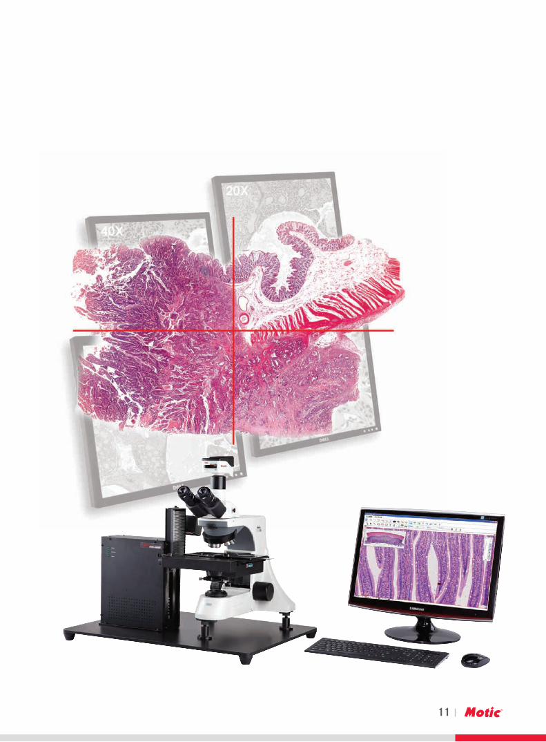
The Motic Vitual Microscope is a integrated and automated Image acquisition and Image processing system.
Through its motorized microscope platform and its automated Image scanning system, is digitizes traditional glass slides and transforms them into a “Whole Slide Image” (WSI).The provided WSI contains all available image information of the slide same as using a traditional Microscope, but in a digital way for Pathologists, for research or medical education.The WSI digital slide has all the features of traditional slide by having the advantage of not restricted from location or time.
The VM digital slide scanning System provides automated image acquisition, storage and management of the digitized slide “WSI” and standardized interfaces to PACS/HIS for hospitals and research groups.
Using Motic VM shared digital pathology information offers a competitive solution for teaching, research, and medical diagnostic.It’s remote pathology information system enables Hospitals and Pathologists in different locations to carry out academic exchanges and Internet conferences
Motic VMDigital Slide Scanning System And Software Applications
2
3
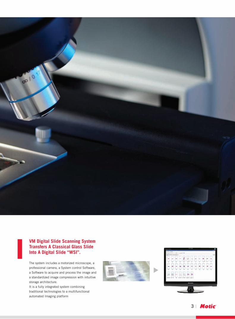
VM Digital Slide Scanning System Transfers A Classical Glass Slide Into A Digital Slide “WSI”.
The system includes a motorized microscope, a
professional camera, a System control Software,
a Software to acquire and process the image and
a standardized image compression with intuitive
storage architecture.
It is a fully integrated system combining
traditional technologies to a multifunctional
automated Imaging platform
4
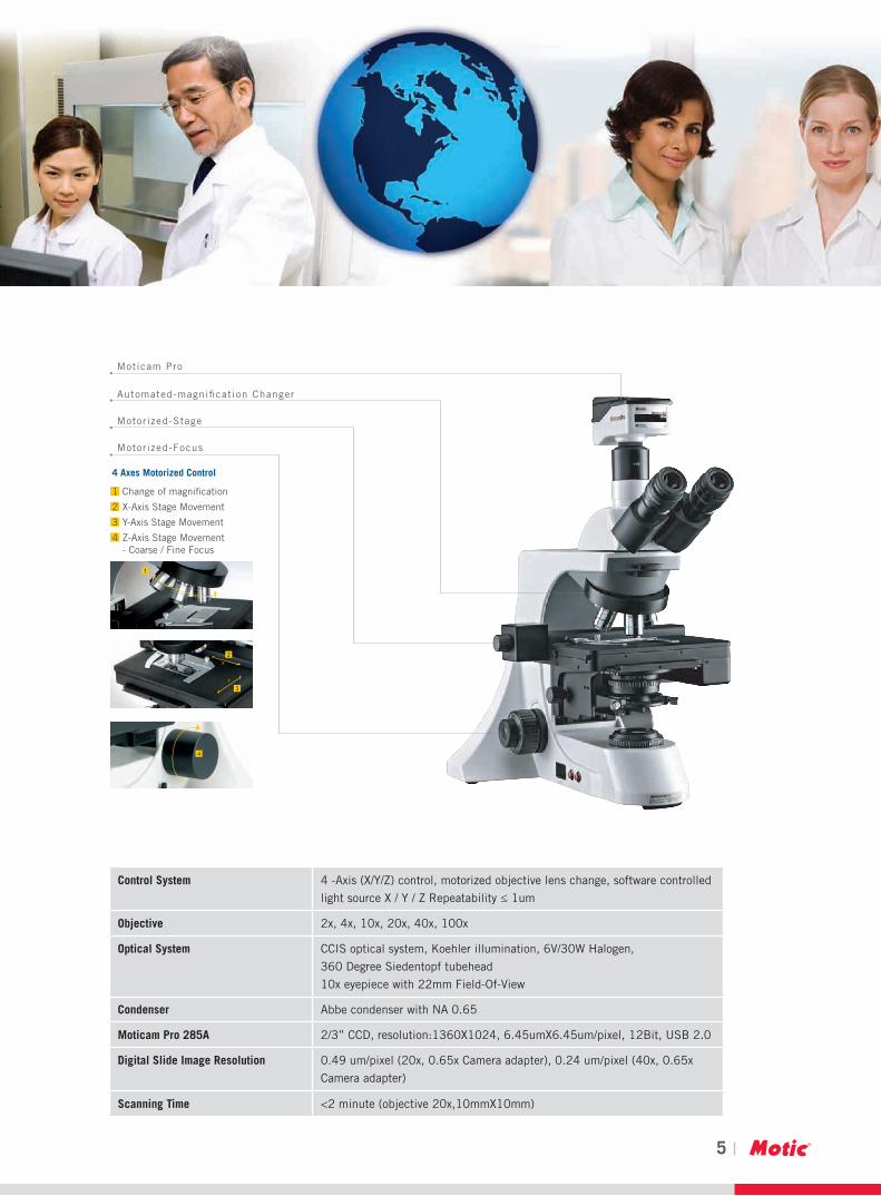
4 Axes Motorized Control
1 Change of magnification
2 X-Axis Stage Movement
3 Y-Axis Stage Movement
4 Z-Axis Stage Movement - Coarse / Fine Focus
Moticam Pro
Automated-magnification Changer
Motorized-Stage
Motorized-Focus
1) Motorized Microscope Platform
The motorized microscope platform, it is using an electronically driven X-Y-Z Axis
System and a automated magnification changer.
Observation the automate image with the Motorized VM Microscope Platform is similar observing the image with traditional microscope.
Changing the magnifications is done with the fully automated system and changing the magnification on the acquired image is done the
software.
This dual use feature also allows directly comparing of the image acquisition and the classical microcopy image.
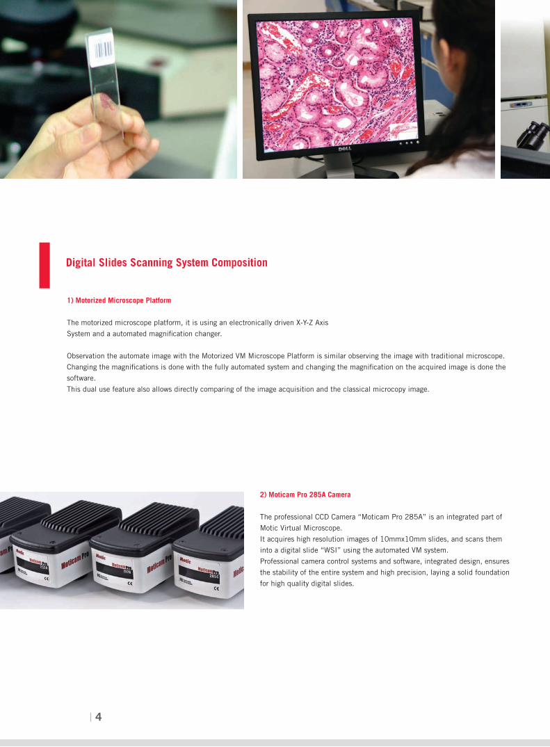
Digital Slides Scanning System Composition
2) Moticam Pro 285A Camera
The professional CCD Camera “Moticam Pro 285A” is an integrated part of
Motic Virtual Microscope.
It acquires high resolution images of 10mmx10mm slides, and scans them
into a digital slide “WSI” using the automated VM system.
Professional camera control systems and software, integrated design, ensures
the stability of the entire system and high precision, laying a solid foundation
for high quality digital slides.
4 Axes Motorized Control
1 Change of magnification
2 X-Axis Stage Movement
3 Y-Axis Stage Movement
4 Z-Axis Stage Movement - Coarse / Fine Focus
Moticam Pro
Automated-magnification Changer
Motorized-Stage
Motorized-Focus
5
4 Axes Motorized Control
1 Change of magnification
2 X-Axis Stage Movement
3 Y-Axis Stage Movement
4 Z-Axis Stage Movement - Coarse / Fine Focus
Moticam Pro
Automated-magnification Changer
Motorized-Stage
Motorized-Focus
Digital Slides Scanning System Composition
Control System 4 -Axis (X/Y/Z) control, motorized objective lens change, software controlled
light source X / Y / Z Repeatability ≤ 1um
Objective 2x, 4x, 10x, 20x, 40x, 100x
Optical System CCIS optical system, Koehler illumination, 6V/30W Halogen,
360 Degree Siedentopf tubehead
10x eyepiece with 22mm Field-Of-View
Condenser Abbe condenser with NA 0.65
Moticam Pro 285A 2/3” CCD, resolution:1360X1024, 6.45umX6.45um/pixel, 12Bit, USB 2.0
Digital Slide Image Resolution 0.49 um/pixel (20x, 0.65x Camera adapter), 0.24 um/pixel (40x, 0.65x
Camera adapter)
Scanning Time <2 minute (objective 20x,10mmX10mm)
6
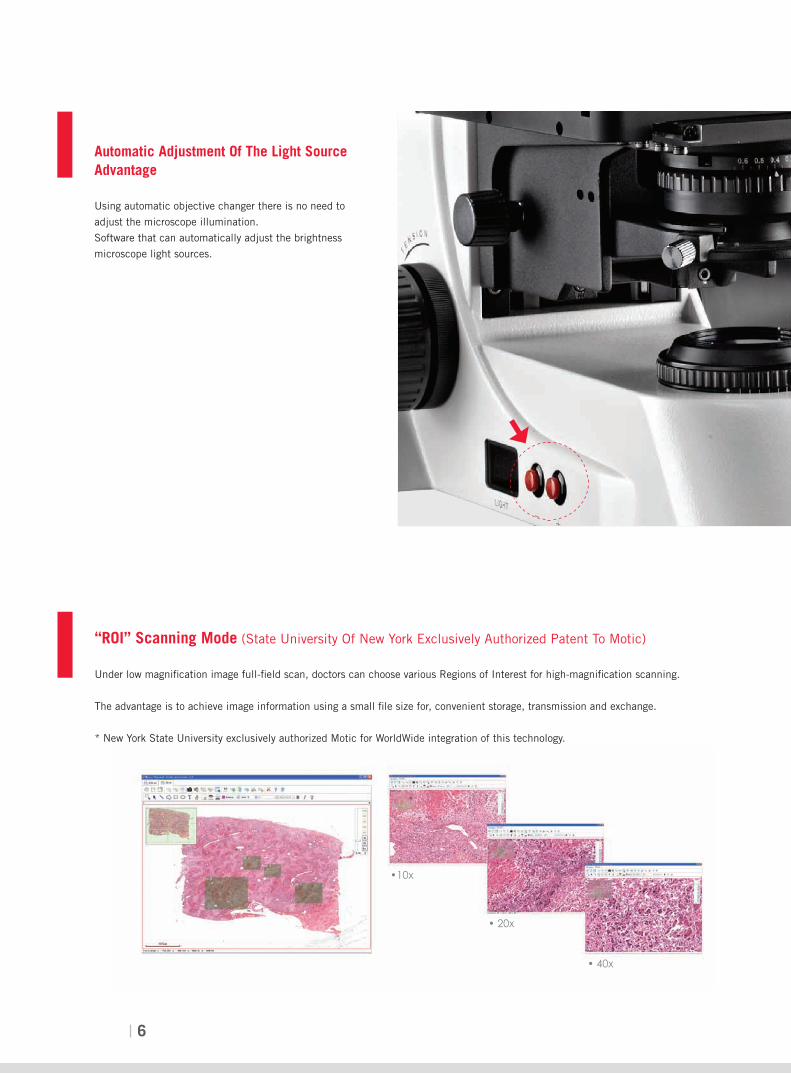
“ROI” Scanning Mode (State University Of New York Exclusively Authorized Patent To Motic)
Under low magnification image full-field scan, doctors can choose various Regions of Interest for high-magnification scanning.
The advantage is to achieve image information using a small file size for, convenient storage, transmission and exchange.
* New York State University exclusively authorized Motic for WorldWide integration of this technology.
Automatic Adjustment Of The Light Source Advantage
Using automatic objective changer there is no need to
adjust the microscope illumination.
Software that can automatically adjust the brightness
microscope light sources.
•10x
• 20x
• 40x
7
Application Of Digital Slides In Teaching / University Education
Such as embryology, pathology, microbiology,
moving.
Botany, parasitology and other figures can be
made to a digital slide.
It is the revolutionary breakthrough in
morphology teaching.
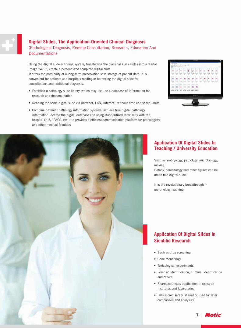
Digital Slides, The Application-Oriented Clinical Diagnosis (Pathological Diagnosis, Remote Consultation, Research, Education And Documentation)
Using the digital slide scanning system, transferring the classical glass slides into a digital
image “WSI”, create a personalized complete digital slide.
It offers the possibility of a long-term preservation save storage of patient data. It is
convenient for patients and hospitals reading or borrowing the digital slide for
consultations and additional diagonsis.
• Establishapathologyslidelibrary,whichmayincludeadatabaseofinformationfor
research and documentation
• Readingthesamedigitalslidevia(intranet,LAN,Internet),withouttimeandspacelimits;
• Combinedifferentpathologyinformationsystems,achievetruedigitalpathology
information. Access the digital database and using standardized Interfaces with the
hospital (HIS / PACS, etc.), to provides a efficient communication platform for pathologists
and other medical faculties
Application Of Digital Slides In Sientific Research
• Suchasdrugscreening
• Genetechnology
• Toxicologicalexperiments
• Forensicidentification,criminalidentification
andothers;
• Pharmaceuticalsapplicationinresearch
institutes and laboratories
• Datastoredsafely,sharedorusedforlater
comparison and analysis’s

8
DSScanner
•Fastandreliableslidescanningthroughanautomaticcontrolofamotorized
stage and several other motorized components (X / Y / Z / objective
conversionetc.);
•Automaticscanningandseamlesslystitchingforaslideindifferent
magnifications and different regions,
•Supportforvariousslidescanningmodes,suchasROIscanning(low
magnification for whole slide + high magnification for more interested sub-
regions), quick scanning, high precision scanning (auto-focus for every field),
multi-focus scanning (automatic fusion of several z levels in each field) etc.,
tofitfordifferentapplications;
•Optionalscanningslideencryption,suchasdongleprotection,password
protectionandotherprotectionmethodstoenhancethesecurityofdigitalslides;
•ROIscanningmode:full-fieldscanatlowmagnificationimage,selectthemultipleregionsofinterestatdifferenthighmagnification
scanning.Thefilestoragespaceissmall,onlyafewMBytes.Itisconvenientforstorageandtransmission;
•DatastorageandexportsupportforJPEGandJPEG2000formats,andsupportformultipleROIcollectionandmassscanning;
•Supportfluorescenceslidescanningand100Xoilimmersionscanning.
•SupportDEMOconfiguration.Demomodecanbeinstalledonanydesktoporlaptopcomputer,andthedemomodeDSScannercan
run without the need of hardware to be convenient for demonstration purpose.
•TheROIscanningmodetechnologyisunderlicenseoftheStateUniversityofNewYork,USA.

9
DSAssisant
• Supportmultipleslidessimultaneouslycomparingandsynchronizingpanforbrowsing;
• Automaticpanadigitalslidewithflexiblespeedsettings,andaddannotationsontheflyduringautomaticpanmovement;
• Shareddigitalslidelibraryformultiplebrowsing;
• Supportannotationswithdifferentfonttypes,colors,fontsizes,charactersetc;
• Variousmeasurementtools,suchaslength,perimeter,area,etc.,andmeasurementscanbelabeled,andsortedbylabelsorviewedby
labelnavigation;
• Annotationswithmultiplelayersandareabletobeselectableandcustomizable;
• SeveralROIbaseddigitalslidemakingmethods;
• Recordingfunctiontorecordscreen,voiceandthewholeprocessofdigitalslidescanningandoperating.Itisagoodtoolforcourseware
making and expertise sharing.

10
DSStore
• ApowerfulVMdigitalslidedatabasemanagementfunctionalityandslideviewingcrossthe
network (e.g. internet or intranet).
• AFrameworkfordigitalslidestorage,digitalslidesort,search,categorize,userandrole
assignment, network access etc.
• NewSilverlighttechnologyfordigitalslidebrowsing.
• Seamlessintegrationandinter-operationwithDSScannerandDSAssistantmodules.
Tele
• Aremote-controlmicroscopesoftwaresystemwhichsupportsbothautomaticmicroscopeand
manual microscope.
• Offersremotecontrolofmicroscopeoperation,allowingdistanceimageviewing,diagnosis
and consultation between different locations. It also provides automated supplementary
functions that improve precision, work efficiency and the control of microscope.
• Supportrapiddiagnosisoffrozenslidethroughtheremote-internetconnection.
• SameplatformasDSScanner,combinedwithpowerfulscanningsoftware.Thepathological
experts can use the automatic microscope platform to remotely control, viewing the digital
slider under the different magnifications, and then give the diagnosis report in timely manner.
• Itisanecessarycommunicationtoolforobtainingexpertsandpathologists’secondopinions
in real-time.
DSConference
• Simultaneouslybrowsingthesameslideandthesamefieldcrosstheinternetwith
multiple users
• AdoptP2Ptechnologyfordigitalslidereal-timecommunications
• Provideasetofcomprehensivetoolsforcustomerstomanage,processesandanalyzesdigital
slide images.
• Modulesincludes:DSAssistant,InternetVMConferencing
11

Motic Instruments (CANADA)130 - 4611 Viking Way. Richmond, BC V6V 2K9 CanadaTel: 1-877-977 4717 Fax: 1-604-303 9043
Motic Deutschland GmbH (GERMANY)Christian-Kremp-Strasse 11, D-35578 Wetzlar, GermanyTel: 49-6441-210 010 Fax: 49-6441-210 0122
Motic Incorporation Ltd. (HONG KONG)Rm 2907-8, Windsor House, 311 Gloucester Road,Causeway Bay, Hong KongTel: 852-2837 0888 Fax: 852-2882 2792
Motic Spain, S.L. (SPAIN)Polígon Industrial Les Corts, Camí del Mig, 11208349 Cabrera de Mar, Barcelona, SpainTel: 34-93-756 6286 Fax: 34-93-756 6287
Motic Incorporation Limited Copyright © 2002-2010.All Rights Reserved .
* CCIS® is a trademark of Motic Incorporation Ltd.
Design Change :The manufacturer reserves the right to make changes in instrument design in accordance with scientific and mechanical progress, without notice and without obligation.
SAP Code: 1300901303771
Updated: 2011.06
EN | ES | FR | DE | IT | PT | RU
www.motic.com
Canada | China | Germany | Spain | USA