morphological varieties and host ranges …aem.asm.org/content/44/2/466.full.pdf · morphological...
TRANSCRIPT

APPLIED AND ENVIRONMENTAL MICROBIOLOGY, Aug. 1982, p. 466-470 Vol. 44, No. 20099-2240/82/080466-05$02.00/0
Morphological Varieties and Host Ranges of Vibrioparahaemolyticus Bacteriophages Isolated from Seawater
TETSURO KOGA, SATORU TOYOSHIMA, AND TOMIO KAWATA*Department ofFood Microbiology, Tokushima University School of Medicine, Tokushima 770, Japan
Received 17 August 1981/Accepted 5 May 1982
Eighteen bacteriophages lytic for Vibrio parahaemolyticus were isolated fromseawater, and their ultrastructure was examined by electron microscopy. Basedon the phage morphology, they were classified in four groups. Group I phagesconsisted of a hexagonal head and a tail with a contractile sheath. All the phagesof the other three groups had a relatively long, noncontractile tail, but there weredifferences in the head structure among these phages. The phages of groups II andIII had a hexagonal head and an elongated polyhedral head, respectively. GroupIV phages exhibited a unique hexagonal head with knoblike projections. Thereappeared to be no correlation between the 0 and K serotypes of V. parahaemolyt-icus strains and the host ranges of the phages. The phages had varying sensitivitiesto heat and organic solvents.
Vibrio parahaemolyticus, a most importantcausative bacterium of food poisoning in Japan,resides in the near-shore marine environment (8,10). The first isolation of bacteriophages specificfor this bacterium was reported in 1966 byNakanishi et al. (9). The three different phagesdescribed were isolated from seawater, humanfeces, and a lysogenic strain of the bacterium.Later, Baross et al. (2-4) isolated numbers of V.parahaemolyticus phages from marine samples.However, until now no information has beenavailable about the morphology of these phagesexcept for one phage that is structurally similarto the T phages of Escherichia coli (11). We haveattempted to isolate phages for the vibrio toinvestigate the ultrastructure of the phages. Thestructure of a novel phage, VP3, exhibitingknoblike projections around the head, was re-cently reported (7). This paper describes themorphological variety and host ranges of 18 V.parahaemolyticus phages isolated from seawa-ter. Based on the morphological characteristics,these phages are divided into four groups, andtheir sensitivities to heat and organic solventsare examined.
MATERIALS AND METHODS
Bacterial strains and growth. Ten K serotype pilotstrains and four other strains of V. parahaemolyticuswere used in this study (Tables 1 and 2). In addition,four K serotype pilot strains (K-29, K-41, K-46, andK-56), which are not listed in the tables, were used. Allthe strains, except strain 3283-61, which has been keptin our laboratory for many years, were supplied by theResearch Institute for Microbial Diseases, Osaka Uni-versity. For isolation and propagation of phages, 3%NaCl broth (9) was routinely used. Bacteria weregrown in the broth at 30°C with gentle shaking. Solid
medium was composed of 3% NaCl broth containing1.5 and 0.6% agar for the bottom and top agar layers,respectively, for phage assay.
Isolation of bacteriophages. Samples of coastal sea-water were harvested near Tokushima during thesummer months. Seawater samples were mixed withequal volumes of double-strength 3% NaCl broth. Theenrichment mixtures were incubated with log-phasecultures of host strains and incubated further at 30°Cfor 5 h. Single-plaque isolation from the enrichmentculture was performed repeatedly according to themethod of Adams (1) as described previously (7).Phage assay. Phage titers were determined as
plaque-forming units (PFU) by the agar layer method(1) as described earlier (7). After incubation overnightat 30°C, the efficiency of plating was estimated relativeto that on the host strain which produced the highestphage titer for each phage. The host range was deter-mined by qualitative spot tests on lawns of differentstrains by using phage stocks containing about 108PFU/ml.
Preparation of phage stock. Phage stock was pre-pared as described previously (7, 12). Phages werepropagated in the log-phase culture of the host strain in3% NaCl broth by incubation at 30°C for 5 h. Afterunlysed cells were removed from the culture by cen-trifugation at 3,000 x g for 10 min, phages in thesupernatant were precipitated by adding 10% (wt/vol)polyethylene glycol 6000 and 1 M NaCl and centrifug-ing at 8,000 x g for 10 min. The pellet composed ofphage particles was resuspended in AAM solution (7)containing 1% ammonium acetate and 0.01 MgCl2, andthe resulting phage stock was stored in the cold.
Sensitivity of phages to heat. Samples (1 ml) of phagepreparation containing about 108 PFU/ml in 3% NaClbroth were maintained at desired temperatures for 30min, then immediately cooled in ice water and assayedfor phage titers. The temperature at which a decreasein phage titer of more than 90% occurred was scored.
Sensitivity of phages to organic solvents. Phage sensi-tivity to the organic solvents toluene, ethyl ether, and
466
on May 15, 2018 by guest
http://aem.asm
.org/D
ownloaded from

MORPHOLOGY OF V. PARAHAEMOLYTICUS PHAGES 467
chloroform was examined by the method of Carvalhoand Vary (6), except that 3% NaCl broth was used forthe preparation of phage lysate and for phage assay.Samples (1 ml) of diluted phage lysate containingabout 2 x 10⁵ PFU/ml in 3% NaCl broth were mixedwith each solvent (0.2 ml) by stirring gently with aThermo-mixer (Theromics Co., Ltd., Tokyo) for 15 s.After incubation at 30°C for 30 min, the aqueous phaseof the mixtures was assayed for viability of phages.
Electron microscopy. Phage stock was fixed with 5%Formalin, washed with distilled water, and then nega-tively stained with 1% ammonium molybdate in 0.1 Mammonium acetate buffer (pH 7.2) as described previ-ously (12). Samples were examined in a Hitachi HU-11E electron microscope with an accelerating voltageof 75 kV.
RESULTS AND DISCUSSIONMorphological grouping of phages. Bacterio-
phages exhibit a great diversity in their morphol-ogy. Bradley (5) proposed six basic morphologi-cal types, A through F. So far there has been nomorphological classification of V. parahaemo-lyticus phages. In the present study, we isolated18 phages for the vibrio from coastal seawaterduring the summer months. The electron micro-graphs of some typical phages are shown in Fig.1 and 2. We divided the phages into four differ-ent groups, I through IV, as judged by theirmorphological variety. The 10 group I phagesdiffered from each other in dimensions but weresimilar in having a hexagonal head and a tail witha contractile sheath (Fig. la-c). Therefore, thisgroup belongs to type A according to the classifi-cation of Bradley (5). Their heads varied from109 by 103 nm (VP10) to 65 by 60 nm (VP2, Fig.lb) in dimension, and the tail contained a baseplate with several pins and tail fibers. Somephages of this group had a base plate with largepins (Fig. lc).
Eight phages, other than group I phages, werecomposed of a hexagonal or polyhedral head anda long, noncontractile tail, so they belong toBradley's type B (5). These phages were dividedinto three groups, II, III, and IV, based on thehead morphology (Fig. 2). Group II phages had ahexagonal head with a long, flexible tail (Fig. 2aand b). The dimensions of the head ranged from79 by 65 to 92 by 89 nm. Group III phagesexhibited an elongated polyhedral head and along, flexible tail (Fig. 2c and d). The dimensionsof the head varied from 111 by 73 nm to 91 by 53nm. The distal ends of their tails tended toadhere to each other. Group IV phages consist-ed of a hexagonal head about 78 by 74 to 78 nmand a flexible tail with cross striations (Fig. 2e).The appearance of the capsid covered with theknoblike projections was most conspicuous,since the detailed structure of phage VP3 hasbeen described (7). The knoblike particles,about 7 to 8 nm in diameter, had a hollow about
0
O9 0
0 .
r0.
O 2X3 GO
2:.33C)
=0
OC
OC )
~ 5
o3_.0s
0.
P:C)
fD00)=GO__C5
C)
~0_.0)GO
,1z
C)
C)C)
0-
ON~ ~~
x x
0
|- -} k
O O0
N W
III
Dw X0 w
O X.o0000 0
X X--- ts,
0 000I I I
ia> Xi0
o.
0
II
0
oo1
x xI
w w
I o0 0
0. ~7 &.
o II,I xa)
o-o-C) 0
oo
x.
w
~ool=-) I..." w t~A so 00ON .- O
Ix . x
W w
00
x.,x-
0
p.
C)
0
l0laa
010z
wr
Pz0)
CDCA0
0
CD0.la
p
UQ
CA
0.0)
,ItCeaZ-02.,
x0
0
-_4
c
I
VOL. 44, 1982
on May 15, 2018 by guest
http://aem.asm
.org/D
ownloaded from
468 KOGA, TOYOSHIMA, AND KAWATA
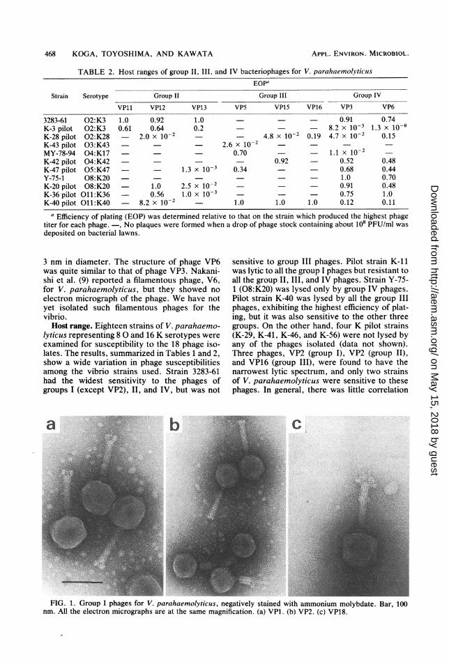
TABLE 2. Host ranges of group II, III, and IV bacteriophages for V. parahaemolyticusEOP"
Strain Serotype Group II Group III Group IV
VPll VP12 VP13 VP5 VP15 VP16 VP3 VP6
3283-61 02:K3 1.0 0.92 1.0 - - 0.91 0.74K-3 pilot 02:K3 0.61 0.64 0.2 - 8.2 x 10⁻³ 1.3 x 10⁻⁸K-28 pilot 02:K28 - 2.0 x 10⁻² 4.8 x 10⁻² 0.19 4.7 x 10⁻² 0.15K-43 pilot 03:K43 2.6 x 10⁻²MY-78-94 04:K17 0.70 1.1 X 10⁻²K-42 pilot 04:K42 0.92 - 0.52 0.48K-47 pilot 05:K47 - 1.3 x 10⁻³ 0.34 - 0.68 0.44Y-75-1 08:K20 - - 1.0 0.70K-20 pilot 08:K20 1.0 2.5 x 10⁻² 0.91 0.48K-36 pilot O11:K36 0.56 1.0 x 10⁻³ 0.75 1.0K-40 pilot O11:K40 8.2 x 10⁻² 1.0 1.0 1.0 0.12 0.11
a Efficiency of plating (EOP) was determined relative to that on the strain which produced the highest phagetiter for each phage. -, No plaques were formed when a drop of phage stock containing about 108 PFU/ml wasdeposited on bacterial lawns.
3 nm in diameter. The structure of phage VP6was quite similar to that of phage VP3. Nakani-shi et al. (9) reported a filamentous phage, V6,for V. parahaemolyticus, but they showed noelectron micrograph of the phage. We have notyet isolated such filamentous phages for thevibrio.Host range. Eighteen strains of V. parahaemo-
lyticus representing 8 0 and 16 K serotypes wereexamined for susceptibility to the 18 phage iso-lates. The results, summarized in Tables 1 and 2,show a wide variation in phage susceptibilitiesamong the vibrio strains used. Strain 3283-61had the widest sensitivity to the phages ofgroups I (except VP2), II, and IV, but was not
sensitive to group III phages. Pilot strain K-11was lytic to all the group I phages but resistant toall the group II, III, and IV phages. Strain Y-75-1 (08:K20) was lysed only by group IV phages.Pilot strain K-40 was lysed by all the group IIIphages, exhibiting the highest efficiency of plat-ing, but it was also sensitive to the other threegroups. On the other hand, four K pilot strains(K-29, K-41, K-46, and K-56) were not lysed byany of the phages isolated (data not shown).Three phages, VP2 (group I), VP2 (group II),and VP16 (group III), were found to have thenarrowest lytic spectrum, and only two strainsof V. parahaemolyticus were sensitive to thesephages. In general, there was little correlation
1,I"
FIG. 1. Group I phages for V. parahaemolyticus, negatively stained with ammonium molybdate. Bar, 100nm. All the electron micrographs are at the same magnification. (a) VP1. (b) VP2. (c) VP18.
APPL. ENVIRON. MICROBIOL.
on May 15, 2018 by guest
http://aem.asm
.org/D
ownloaded from

MORPHOLOGY OF V. PARAHAEMOLYTICUS PHAGES 469
FIG. 2. Group II, III, and IV phages for V. parahaemolyticus, negatively stained with ammonium molybdate.Bar, 100 nm. Group II phages: (a) VP11; (b) VP12. Group III phages: (c) VP5; (d) VP16. Group IV phage: (e)VP6.
between the 0 and K serotypes of V. parahae-molyticus and the host ranges of the phageisolates. Baross et al. (4) also reported no corre-lation between the serotypes of the vibrio andphage lytic spectra.
Sensitivity to heat and organic solvents. All thephages ofgroup I were almost totally inactivatedwhen exposed to 55°C for 30 min. Some of thephages (VP4, VP9, VP17, and VP19) were veryheat sensitive, and they lost lytic activity at45°C. Group II, III, and IV phages were general-ly more heat resistant than group I phages, andthey were inactivated at 60°C for 30 min. Naka-
nishi et al. (9) reported that a filamentous phagefor V. parahaemolyticus was completely resist-ant to heating at 60°C for 80 min. None of thephages examined in this study was filamentous,and all the phages were sensitive to heating at60°C for 30 min.Most of the phages were more sensitive to
ethyl ether and chloroform than to toluene.Group I phages exhibited a wide variation insensitivity to the organic solvents. Group II(except VP13), III, and IV phages were moresensitive to chloroform than to ether. PhageVP13 was considerably resistant to all the organ-
VOL . 44, 1982
on May 15, 2018 by guest
http://aem.asm
.org/D
ownloaded from

470 KOGA, TOYOSHIMA, AND KAWATA
ic solvents tested. Thus, the phages for V.parahaemolyticus had varying sensitivities toorganic solvents.
ACKNOWLEDGMENTS
We are indebted to T. Miwatani and Y. Takeda, theResearch Institute for Microbial Diseases, Osaka University,for kindly supplying the bacterial strains.
LITERATURE CITED
1. Adams, M. H. 1959. Bacteriophages. Interscience Pub-lishers, Inc., New York.
2. Baross, J. A., and J. Liston. 1968. Isolation of Vibrioparahaemolyticus from the Northwest Pacific. Nature(London) 217:1263-1264.
3. Baross, J. A., J. Liston, and R. Y. Morita. 1978. Incidenceof Vibrio parahaemolyticus bacteriophages and other Vib-rio bacteriophages in marine samples. Appl. Environ.Microbiol. 36:492-499.
4. Baross, J. A., J. Liston, and R. Y. Morita. 1978. Ecologi-cal relationship between Vibrio parahaemolyticus andagar-digesting vibrios as evidenced by bacteriophage sus-ceptibility patterns. Appl. Environ. Microbiol. 36:500-505.
APPL. ENVIRON. MICROBIOL.
5. Bradley, D. E. 1967. Ultrastructure of bacteriophages andbacteriocins. Bacteriol. Rev. 31:230-314.
6. Carvalho, P. M., and J. C. Vary. 1977. Isolation andcharacterization of a Bacillus megateriium QM B1551bacteriophages. J. Gen. Virol. 36:547-550.
7. Koga, T., and T. Kawata. 1981. Structure of a novelbacteriophage VP3 for Vibrio parahaemolyticus. Micro-biol. Immunol. 25:737-740.
8. Miwatani, T., and Y. Takeda. 1976. Vibr-io parahaetnolyti-cus. A causative bacterium of food poisoning. SaikonPublishing Co., Ltd., Tokyo.
9. Nakanishi, H., Y. Iida, K. Maeshima, T. Teramoto, Y.Hosaka, and M. Ozaki. 1966. Isolation and properties ofbacteriophages of Vibrio parahaemolyticus. Biken J.9:149-157.
10. Sakazaki, R., S. Iwanami, and H. Fukumi. 1963. Studieson the enteropathogenic, facultatively halophilic bacteria,Vibrio parahaemolyticus. I. Morphological, cultural andbiochemical properties and its taxonomical position. Jpn.J. Med. Sci. Biol. 16:161-188.
11. Sklarow, S. S., R. R. Colwell, G. B. Chapman, and S. F.Zane. 1973. Characteristics of a Vibrio parahaemnolyticiusbacteriophage isolated from Atlantic coast sediment. Can.J. Microbiol. 19:1519-1520.
12. Takumi, K., T. Kinouchi, and T. Kawata. 1980. Isolationof two inducible bacteriophages from Clostridiiurn botiuli-nium type A 190L. FEMS Microbiol. Lett. 9:23-27.
on May 15, 2018 by guest
http://aem.asm
.org/D
ownloaded from