morphological characteristics of the plantar
TRANSCRIPT
RESEARCH Open Access
Morphological characteristics of the plantarcalcaneocuboid ligamentsMutsuaki Edama1,2* , Tomoya Takabayashi1, Hirotake Yokota1, Ryo Hrabayashi1, Chie Sekine1 and Ikuo Kageyama2
Abstract
Background: The aim of this study was to clarify the differences in morphological features between the longplantar ligament (LPL) and the short plantar ligament (SPL).
Methods: This investigation examined 50 legs from 25 Japanese cadavers. The LPL and SPL of each leg wereclassified into one of three types based on the shape and number of fiber bundles. Then, fiber bundle length, fiberbundle width, and fiber bundle thickness were measured.
Results: The LPL was rectangular in shape (Type I) in 12%, hourglass shape (Type II) in 62%, and triangular in shape(Type III) in 26%. The SPL was a single fiber bundle (Type I-a) in 26%, a surface fiber bundle and a deep fiber bundle(Type I-b) in 60%, and a surface fiber bundle (medial and lateral) and a deep fiber bundle (Type II) in 14%.Regarding the morphological characteristics, there were no significant differences among the types in the LPL, butthere were differences between types and between surface and deep fiber bundles in the SPL.
Conclusions: For the LPL, the hourglass shape is the most common type. However, there appeared to be nofunctional difference due to the difference in the shape of the LPL, since there were no significant differencesamong the types in the LPL. For the SPL, there were types of single, double and triple fiber bundles; there may befunctional differences based on the number of fiber bundles and between superficial and deep fibers.
Keywords: Long plantar ligament, Short plantar ligament, Calcaneocuboid joint
BackgroundIsolated injuries involving the calcaneocuboid joint arerare and frequently overlooked. Indeed, these injuriesare a cause of symptoms after an inversion injury of thefoot, and the resulting clinical manifestations may mimica lateral ankle ligament injury, which is more common[1–4]. These injuries predominantly affect young and ac-tive persons and are of significant economic importance[1, 2]. Furthermore, patients with injuries involving thecalcaneocuboid joint require different treatment ap-proaches than those with the more frequently occurringlateral ankle ligament injury [1]. With a greater
understanding of the morphological characteristics ofthe ligament, an accurate and timely diagnosis can bemade, preventing late sequelae from developing and ob-viating complicated surgical procedures.The calcaneocuboid joint is formed by the quadrilat-
eral facets of the calcaneus and cuboid bones and thecapsule, which is reinforced by ligaments. The four liga-ments connecting the calcaneus and cuboid are: themedial calcaneocuboid ligament, a component of the bi-furcate ligament; the dorsolateral calcaneocuboid liga-ment; the plantar calcaneocuboid ligament, or shortplantar ligament; and the long plantar ligament [5, 6].These ligaments play a major role in supporting themedial and lateral longitudinal arches [7–9]. However,morphological characteristics and the functional roles ofthe plantar calcaneocuboid ligaments have not been fullyconsidered.
© The Author(s). 2021 Open Access This article is licensed under a Creative Commons Attribution 4.0 International License,which permits use, sharing, adaptation, distribution and reproduction in any medium or format, as long as you giveappropriate credit to the original author(s) and the source, provide a link to the Creative Commons licence, and indicate ifchanges were made. The images or other third party material in this article are included in the article's Creative Commonslicence, unless indicated otherwise in a credit line to the material. If material is not included in the article's Creative Commonslicence and your intended use is not permitted by statutory regulation or exceeds the permitted use, you will need to obtainpermission directly from the copyright holder. To view a copy of this licence, visit http://creativecommons.org/licenses/by/4.0/.The Creative Commons Public Domain Dedication waiver (http://creativecommons.org/publicdomain/zero/1.0/) applies to thedata made available in this article, unless otherwise stated in a credit line to the data.
* Correspondence: [email protected] for Human Movement and Medical Sciences, Niigata University ofHealth and Welfare, Shimami-cho 1398, Kita-ku, Niigata City 950-3198, Japan2Department of Anatomy, School of Life Dentistry at Niigata, Nippon DentalUniversity, Niigata, Japan
Edama et al. Journal of Foot and Ankle Research (2021) 14:3 https://doi.org/10.1186/s13047-020-00443-7

Previous studies [5, 6, 10] referred to the plantar calca-neocuboid ligament that was then subdivided into thelong plantar ligament (LPL) and the short plantar liga-ment (SPL); the SPL is also known as the plantar calca-neocuboid ligament. It is generally agreed that the LPLattaches posteriorly to the inferior surface of the calca-neus between the posterior and anterior tubercles [5]. Ithas been reported that the superficial fibers insert intothe bases of the second to fourth metatarsals (MTs) andnot the distal cuboid. Previous studies suggested that thesuperficial fibers insert variably to the metatarsal bone[6, 11]. One study [6] reported that, in all 59 specimensexamined, the LPL had an hourglass shape, and struc-tural variations were observed in the LPL in 20.3% (12ft), in the form of medial twisting fibers in 11.8% (7 ft),lateral twisting fibers in 3.3% (2 ft), and additional bandsin 5% (3 ft). In another study of 10 ft, the shape of theLPL was hourglass to rectangular in 100% [10]. The fol-lowing morphological data have been reported: length28.5 ± 10.5 mm [5], 44.2 ± 4.4 mm [6], 38.9 ± 2.1 (lateral)mm [10], and 61.4 ± 24.4 (medial) mm [10]; and width10.7 ± 2.8 mm [5], 10.9 ± 2.8 mm [6], and 12.2 ± 1.1 mm[10].The SPL passes anteromedially as a widening band
from the anterior tubercle of the calcaneus to attach tothe plantar surface of the cuboid posterior to the ridgefor the tendon of peroneus longus, and it is often de-scribed as blending with and reinforcing the calcaneocu-boid joint capsule [5, 6, 10]. Some displayed superficialand deep bands, and in 23 ft (39%) of the 59 specimens,the deep band had a distinct attachment to the calcaneus[6]. The overall shape and arrangement of the SPL wastriangular in 21 ft (35%), rectangular in 34 ft (59%), andtrapezoidal in 4 ft (6%). In addition, an extra band orbands were also observed in 19 ft (32%), in two of which
the extra bands were also twisted [6]. In another study,every SPL had at least two bands: superficial and deep[10]. A rectangular or slightly triangular superficial bandwas seen in 6 ft (60%), and in the remaining 4 ft (40%), itwas triangular with small separate bundles [10]. The fol-lowing morphological data were reported: length 18.2 ±4.3 mm [5], 19.4 ± 3.6 mm [6], and 21.0 ± 1.9 mm [10];width 10.3 ± 4.4 mm [6], 11.2 ± 0.8 mm [10], and 12.2 ±3.3 mm [5]; and thickness 4.8 ± 0.3 mm [10]. Thus, thereare few morphological reports of the plantar calcaneocu-boid ligaments, and there is no consensus.Therefore, the aim of this study was to clarify the dif-
ferences in morphological features between the LPL andthe SPL.
Materials and methodsCadaversThis investigation examined 50 legs from 25 Japanesecadavers (mean age at death, 78 ± 12 years; 28 sides frommen, 22 from women; 25 right sides, 25 left sides) thathad been switched to alcohol after placement in 10% for-malin. None showed signs of previous major surgeryaround the foot or ankle or any relevant deformities, andthere was no obvious degeneration in all specimens. Thisstudy was approved by the Ethics Committee of our in-stitution (18071).
MethodsThe procedures for dissection of the LPL and SPL aredescribed below. All dissections were performed by thesame examiner. Isolated specimens of the leg were cre-ated by transection 10 cm above the ankle. The skin,subcutaneous tissue, and crural fascia were removed,and the peroneus longus and brevis and the tibialis pos-terior were carefully dissected and inspected. The
Fig. 1 Dissection procedure for the long plantar ligament: right foot, plantar view 1: First metatarsal, 2: second metatarsal, 3: third metatarsal, 4:fourth metatarsal, 5: base of the fifth metatarsal, 6: calcaneus, 7: long plantar ligament, 8: short plantar ligament, 9: blended fiber bundle of thelong plantar ligament, 10: medial cuneiform–metatarsal 2 & 3 plantar ligament, 11: posterior tibialis, 12: spring ligament (plantar calcaneonavicularligament), 13: peroneus longus tendon
Edama et al. Journal of Foot and Ankle Research (2021) 14:3 Page 2 of 6

plantar aponeurosis was meticulously dissected from theflexor digitorum brevis and removed. The first, second,and third plantar layers (flexor digitorum brevis, flexordigitorum longus, flexor hallucis longus, lumbricals,plantar quadratus, abductor hallucis, abductor digitiminimi) were then dissected and excised. Upon reachingthe fourth layer, the peroneus longus tendon was onceagain identified behind the lateral malleolus and thenfollowed into the plantar aspect of the foot. In all speci-mens, the LPL formed the roof of the peroneus longuscanal, blending with the canal before inserting on themetatarsal bases. To clarify the ligaments, the plantarmuscles were dissected and excised (Fig. 1). The LPLand SPL of each leg were then classified into one of thethree types based on the morphological characteristicsand the number of fiber bundles.Fiber bundle length, fiber bundle width, and fiber bun-
dle thickness were measured for the LPL and the SPL.Fiber bundle length, fiber bundle width, and fiber bundlethickness were measured in the central portions of theLPL and SPL using calipers (Digital Caliper, Shinwa, Nii-gata, Japan). All measurements were made by the sameexaminer, with each site measured three times, and themean value and standard deviation were then calculated.
Statistical analysisStatistical analyses were performed using SPSS (version24.0, SPSS Japan Inc., Tokyo, Japan). The chi-squaredtest was used for comparisons between men and womenand between right and left sides in the classifications
based on differences in the type. Comparisons of fiberbundle length, fiber bundle width, and fiber bundlethickness in each type were made with an unpaired t-test, one-way repeated measures analysis of variance(ANOVA), and Bonferroni’s method. Comparisons offiber bundle length, fiber bundle width, and fiber bundlethickness among types were made with a paired t-test.The level of significance was 5%.
ResultsClassification of the long plantar ligamentUsing the classification based on differences in theshape, there were three types: Type I, Type II, and TypeIII. The types were as follows: Type I, the LPL was arectangular shape; Type II, the LPL was an hourglassshape; and Type III, the LPL was a triangular shape.Type I was seen in 6 ft (12%), Type II in 31 ft (62%), andType III in 13 ft (26%) (Fig. 2).There were no significant differences between males
and females and between left and right sides (Table 1).
Fig. 2 Classification of the long plantar ligament. Type I: the LPL has a rectangular shape. Type II: the LPL has an hourglass shape. Type III: the LPLhas a triangular shape. 1: calcaneus, 2: long plantar ligament, 3: short plantar ligament, L: lateral, M: medial
Table 1 Comparison of classifications of the long plantarligament by sex and laterality
Type I Type II Type III
Male (n = 28) 3 (6) 19 (38) 6 (12)
Female (n = 22) 3 (6) 12 (24) 7 (14)
Right (n = 25) 3 (6) 16 (32) 6 (12)
Left (n = 25) 3 (6) 15 (30) 7 (14)
Values are reported as number of specimens (%)
Edama et al. Journal of Foot and Ankle Research (2021) 14:3 Page 3 of 6

Classification of the short plantar ligamentUsing the classification based on differences in the num-ber of fiber bundles, there were three types: Type I-a, TypeI-b, and Type II. The types were as follows: Type I-a, theSPL was a single (superficial) fiber bundle; Type I-b, theSPL was a surface fiber bundle and a deep fiber bundle;Type II, the SPL was a surface fiber bundle (medial andlateral) and a deep fiber bundle. Type I-a was seen in 13 ft(26%), Type I-b in 30 ft (60%), and Type II in 7 ft (14%)(Fig. 3).
Every SPL was rectangular in shape. The deep fiberband that reinforced the calcaneocuboid joint capsulewas identified with a distinct attachment to the calca-neus and showed a more oblique orientation.There were no significant differences between males
and females and between left and right sides (Table 2).
Morphological characteristics of the long plantarligament (Table 3)There were no significant differences among the types inthe LPL.
Fig. 3 Classification of the short plantar ligament. Type I-a: the SPL is a superficial (single) fiber bundle. Type I-b: the SPL is a superficial fiberbundle and a deep fiber bundle. Type II: the SPL is a superficial fiber bundle (medial and lateral) and a deep fiber bundle. 1: calcaneus, 2: longplantar ligament, 3: the superficial fiber bundle of the short plantar ligament, 4: the deep fiber bundle of the short plantar ligament, 5: superficialfiber bundle (lateral) of the short plantar ligament, 6: superficial fiber bundle (medial) of the short plantar ligament, 7: cuboid, L: lateral, M: medial,Red area: the footprint of the long plantar ligament at the calcaneus and cuboid. Blue area: the footprint of the superficial fiber bundle of theshort plantar ligament at the calcaneus and cuboid. Yellow area: the footprint of the deep fiber bundle of the short plantar ligament at thecalcaneus and cuboid
Table 2 Comparison of classifications of the short plantarligament by sex and left and right sides
Type I-a Type I-b Type II
Male (n = 28) 8 (16) 16 (32) 4 (8)
Female (n = 22) 5 (10) 14 (28) 3 (6)
Right (n = 25) 7 (14) 14 (28) 4 (8)
Left (n = 25) 6 (12) 16 (32) 3 (6)
Values are reported as number of specimens (%)
Table 3 Morphological characteristics of the long plantarligament
Type I Type II Type III
Length (mm) 47.4 ± 3.8 44.3 ± 5.9 47.9 ± 5.9
Width (mm) 14.7 ± 1.8 14.2 ± 2.4 13.7 ± 2.6
Depth (mm) 1.7 ± 0.3 2.0 ± 0.5 2.1 ± 0.6
Values are means ± standard deviation
Edama et al. Journal of Foot and Ankle Research (2021) 14:3 Page 4 of 6
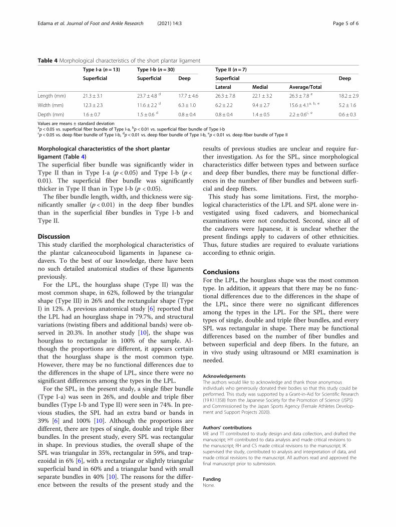
Morphological characteristics of the short plantarligament (Table 4)The superficial fiber bundle was significantly wider inType II than in Type I-a (p < 0.05) and Type I-b (p <0.01). The superficial fiber bundle was significantlythicker in Type II than in Type I-b (p < 0.05).The fiber bundle length, width, and thickness were sig-
nificantly smaller (p < 0.01) in the deep fiber bundlesthan in the superficial fiber bundles in Type I-b andType II.
DiscussionThis study clarified the morphological characteristics ofthe plantar calcaneocuboid ligaments in Japanese ca-davers. To the best of our knowledge, there have beenno such detailed anatomical studies of these ligamentspreviously.For the LPL, the hourglass shape (Type II) was the
most common shape, in 62%, followed by the triangularshape (Type III) in 26% and the rectangular shape (TypeI) in 12%. A previous anatomical study [6] reported thatthe LPL had an hourglass shape in 79.7%, and structuralvariations (twisting fibers and additional bands) were ob-served in 20.3%. In another study [10], the shape washourglass to rectangular in 100% of the sample. Al-though the proportions are different, it appears certainthat the hourglass shape is the most common type.However, there may be no functional differences due tothe differences in the shape of LPL, since there were nosignificant differences among the types in the LPL.For the SPL, in the present study, a single fiber bundle
(Type I-a) was seen in 26%, and double and triple fiberbundles (Type I-b and Type II) were seen in 74%. In pre-vious studies, the SPL had an extra band or bands in39% [6] and 100% [10]. Although the proportions aredifferent, there are types of single, double and triple fiberbundles. In the present study, every SPL was rectangularin shape. In previous studies, the overall shape of theSPL was triangular in 35%, rectangular in 59%, and trap-ezoidal in 6% [6], with a rectangular or slightly triangularsuperficial band in 60% and a triangular band with smallseparate bundles in 40% [10]. The reasons for the differ-ence between the results of the present study and the
results of previous studies are unclear and require fur-ther investigation. As for the SPL, since morphologicalcharacteristics differ between types and between surfaceand deep fiber bundles, there may be functional differ-ences in the number of fiber bundles and between surfi-cial and deep fibers.This study has some limitations. First, the morpho-
logical characteristics of the LPL and SPL alone were in-vestigated using fixed cadavers, and biomechanicalexaminations were not conducted. Second, since all ofthe cadavers were Japanese, it is unclear whether thepresent findings apply to cadavers of other ethnicities.Thus, future studies are required to evaluate variationsaccording to ethnic origin.
ConclusionsFor the LPL, the hourglass shape was the most commontype. In addition, it appears that there may be no func-tional differences due to the differences in the shape ofthe LPL, since there were no significant differencesamong the types in the LPL. For the SPL, there weretypes of single, double and triple fiber bundles, and everySPL was rectangular in shape. There may be functionaldifferences based on the number of fiber bundles andbetween superficial and deep fibers. In the future, anin vivo study using ultrasound or MRI examination isneeded.
AcknowledgementsThe authors would like to acknowledge and thank those anonymousindividuals who generously donated their bodies so that this study could beperformed. This study was supported by a Grant-in-Aid for Scientific Research(19 K11358) from the Japanese Society for the Promotion of Science (JSPS)and Commissioned by the Japan Sports Agency (Female Athletes Develop-ment and Support Projects 2020).
Authors’ contributionsME and TT contributed to study design and data collection, and drafted themanuscript; HY contributed to data analysis and made critical revisions tothe manuscript; RH and CS made critical revisions to the manuscript; IKsupervised the study, contributed to analysis and interpretation of data, andmade critical revisions to the manuscript. All authors read and approved thefinal manuscript prior to submission.
FundingNone.
Table 4 Morphological characteristics of the short plantar ligament
Type I-a (n = 13) Type I-b (n = 30) Type II (n = 7)
Superficial Superficial Deep Superficial Deep
Lateral Medial Average/Total
Length (mm) 21.3 ± 3.1 23.7 ± 4.8 d 17.7 ± 4.6 26.3 ± 7.8 22.1 ± 3.2 26.3 ± 7.8 e 18.2 ± 2.9
Width (mm) 12.3 ± 2.3 11.6 ± 2.2 d 6.3 ± 1.0 6.2 ± 2.2 9.4 ± 2.7 15.6 ± 4.1a, b, e 5.2 ± 1.6
Depth (mm) 1.6 ± 0.7 1.5 ± 0.6 d 0.8 ± 0.4 0.8 ± 0.4 1.4 ± 0.5 2.2 ± 0.6c, e 0.6 ± 0.3
Values are means ± standard deviationap < 0.05 vs. superficial fiber bundle of Type I-a, bp < 0.01 vs. superficial fiber bundle of Type I-bcp < 0.05 vs. deep fiber bundle of Type I-b, dp < 0.01 vs. deep fiber bundle of Type I-b, ep < 0.01 vs. deep fiber bundle of Type II
Edama et al. Journal of Foot and Ankle Research (2021) 14:3 Page 5 of 6
Availability of data and materialsThe data that support the findings of this study are available from thecorresponding author upon reasonable request.
Ethics approval and consent to participateInformed consent was obtained from the families of all subjects. This studywas approved by the ethics committee of the Niigata University of Healthand Welfare, Niigata, Japan.
Consent for publicationNot applicable.
Competing interestsThe authors declare that they have no competing interests.
Received: 2 November 2020 Accepted: 15 December 2020
References1. Lohrer H, Arentz S. Calcaneocuboid joint instability: a novel operative
technique for anatomic reconstruction. Foot Ankle Int. 2004;25:349–56.2. Andermahr J, Helling HJ, Maintz D, Mönig S, Koebke J, Rehm KE. The injury
of the calcaneocuboid ligaments. Foot Ankle Int. 2000;21:379–84.3. Leland RH, Marymont JV, Trevino SG, Varner KE, Noble PC. Calcaneocuboid
stability: a clinical and anatomic study. Foot Ankle Int. 2001;22:880–4.4. Jahss MH, Kay BS. An anatomic study of the anterior superior process of the
os calcis and its clinical application. Foot Ankle. 1983;3:268–81.5. Sarrafian SK. Syndesmology. Sarrafian's anatomy of the foot and ankle. 3rd
ed. Philadelphia: Lippincott Williams & Wilkin; 2011. p. 204–6.6. Ward KA, Soames RW. Morphology of the plantar calcaneocuboid
ligaments. Foot Ankle Int. 1997;18:649–53.7. Dinucci KR, Christensen JC, Dinucci KA. Biomechanical consequences of
lateral column lengthening of the calcaneus: part I. long plantar ligamentstrain. J Foot Ankle Surg. 2004;43:10–5.
8. Otis JC, Deland JT, Kenneally S. Medial arch strain after lateral columnlengthening: an in vitro study. Foot Ankle Int. 1999;20:797–802.
9. Huang CK, Kitaoka HB, An KN, Chao EY. Biomechanical evaluation oflongitudinal arch stability. Foot Ankle. 1993;14:353–7.
10. Melão L, Canella C, Weber M, Negrão P, Trudell D, Resnick D. Ligaments ofthe transverse tarsal joint complex: MRI-anatomic correlation in cadavers.AJR Am J Roentgenol. 2009;193:662–71.
11. Hiramoto Y. Variation of the long plantar ligament in Japanese. OkajimasFolia Anat Jpn. 1984;60:401–8.
Publisher’s NoteSpringer Nature remains neutral with regard to jurisdictional claims inpublished maps and institutional affiliations.
Edama et al. Journal of Foot and Ankle Research (2021) 14:3 Page 6 of 6