morphological analysis of the resin–dentin interface in cavities prepared with er,cr:ysgg laser or...
TRANSCRIPT
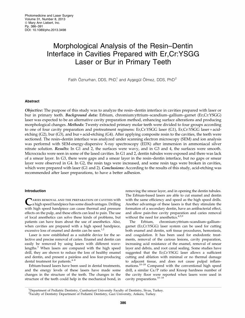
Morphological Analysis of the Resin–DentinInterface in Cavities Prepared with Er,Cr:YSGG
Laser or Bur in Primary Teeth
Fatih Oznurhan, DDS, PhD,1 and Aysxegul Olmez, DDS, PhD2
Abstract
Objective: The purpose of this study was to analyze the resin–dentin interface in cavities prepared with laser orbur in primary teeth. Background data: Erbium, chromium:yttrium–scandium–gallium–garnet (Er,Cr:YSGG)laser was expected to be an alternative cavity preparation method, enhancing surface alterations and producingmorphological changes. Methods: Twenty extracted primary molar teeth were divided to four groups accordingto one of four cavity preparation and pretreatment regimens: Er,Cr:YSGG laser (G1), Er,Cr:YSGG laser + acid-etching (G2), bur (G3), and bur + acid-etching (G4). After applying composite resin to the cavities, the teeth weresectioned. The resin–dentin interface was analyzed under scanning electron microscopy (SEM) and ion analysiswas performd with SEM-energy-dispersive X-ray spectroscopy (EDX) after immersion in ammoniacal silvernitrate solution. Results: In G1 and 2, the surfaces were wavy, and in G3 and 4, the surfaces were smooth.Microcracks were seen in some of the lased cavities. In G1 and 2, dentin tubules were exposed and there was lackof a smear layer. In G3, there were gaps and a smear layer in the resin–dentin interface, but no gaps or smearlayer were observed in G4. In G2, the resin tags were increased, and some resin tags were broken in cavities,which were prepared with laser (G1 and 2). Conclusions: According to the results of this study, acid-etching wasrecommended after laser preparations, to have a better adhesion.
Introduction
Caries removal and the preparation of cavities witha high speed handpiece has some disadvantages. Drilling
with high speed handpiece can cause thermal and pressureeffects on the pulp, and these effects can lead to pain. The useof local anesthetics can solve these kinds of problems, butpatients can have fears about the use of anesthetics. Also,when cavities are prepared with a high speed handpiece,excessive loss of enamel and dentin can be seen.1,2
Laser is now established as a suitable device for the se-lective and precise removal of caries. Enamel and dentin caneasily be removed by using lasers with different wave-lengths.3 When lasers are compared with the high speeddrill, they are shown to reduce the loss of healthy enameland dentin, and present a painless and less fear-producingdental treatment for patients.4–8
Erbium-based lasers have been used in dental treatments,and the energy levels of these lasers have made somechanges in the structure of the teeth. The changes in thestructure of the teeth could help in the mechanical bond, in
removing the smear layer, and in opening the dentin tubules.The Erbium-based lasers are able to cut enamel and dentinwith the same efficiency and speed as the high speed drills.Another advantage of these lasers is that they stimulate theformation of a secondary dentin, have an antibacterial effect,and allow pain-free cavity preparation and caries removalwithout the need for anesthetics.4,5,9
The Erbium, chromium:yttrium–scandium–gallium–garnet (Er,Cr:YSGG) laser system can be used for cuttingboth enamel and dentin, soft tissue procedures, hemostasis,and coagulation. It has been used for endodontic treat-ments, removal of the carious lesions, cavity preparation,increasing acid resistance of the enamel, removal of smearlayer and debris, and root canal sealing. Some studies havesuggested that the Er,Cr:YSGG laser allows a sufficientcutting and ablation with minimal or no thermal damageto adjacent tissue, and does not cause pulpal inflam-mations.10–18 Compared with the conventional high speeddrill, a similar Ca/P ratio and Knoop hardness number ofthe cavity floor were reported when lasers were used incavity preparations.10–18
1Department of Pediatric Dentistry, Cumhuriyet University Faculty of Dentistry, Sivas, Turkey.2Faculty of Dentistry Department of Pediatric Dentistry, Gazi University, Ankara, Turkey.
Photomedicine and Laser SurgeryVolume 31, Number 8, 2013ª Mary Ann Liebert, Inc.Pp. 386–391DOI: 10.1089/pho.2013.3498
386

To date, there have been many investigations aiming toexamine the morphology of the resin–dentin interface ofvarious adhesive systems on flat dentin surfaces or cavitiesprepared with high speed drills in permanent teeth. How-ever, a small number of studies have been made to determinethe quality of Er,Cr:YSGG laser-prepared dentin surfaces forcomposite resin bonding in primary teeth. Therefore, thepurpose of this study was to analyze and compare the resin–dentin interface in cavities prepared with Er,Cr:YSGG laseror bur in primary teeth. The tested null hypothesis was thatthere was no difference between Er,Cr:YSGG and bur prep-aration in terms of interfacial micromorphology, with orwithout acid-etching procedure.
Materials and Methods
Extracted human primary molars (n = 20) were used in thisstudy. The teeth were stored in distillated water and usedwithin 1 month.
Cavity preparations
Ten teeth were selected randomly and class V cavities(3 · 2 · 1.5 mm) were prepared with Er,Cr:YSGG laser (Wa-terlase MD, Biolase Technology Inc., San Clemente, CA) onbuccal and palatinal/lingual surfaces (Fig. 1A). The poweroutput was set at 6.0 W (85–90% air and 80–85% water) forenamel and 3.5 W (70% air and 65% water) for dentinepreparation. Therefore, 10 primary molar teeth were usedand 20 cavities were obtained. Laser was used in a defocusedmode with a working distance of 1.5–2 mm.
The remaining 10 teeth were used for the bur-preparationgroup; class V cavities were prepared with diamond bur(Shofu Inc., Kyoto, Japan) as in the laser group. Then, allpalatinal/lingual cavities were acid etched, rinsed, andgently dried. Acid etching procedure was not performed onbuccal cavities. Therefore, four groups were made.
Group 1 (G1): Er,Cr:YSGG laserGroup 2 (G2): Er,Cr:YSGG laser and acid etching
FIG. 1. (A) Buccal and palatinal/lin-gual cavities. (B) Section types of theteeth (mesiodistally [1], buccolingually[2]). (C) Black arrow shows the ana-lyzed area under scanning electronmicroscopy (SEM).
FIG. 2. Rugged surface andmicrocracks were observed inlaser cavities ( · 250). CR,composite resin; D, dentin.Black arrow shows a micro-crack.
ANALYSIS OF THE RESIN–DENTIN INTERFACE 387

Group 3 (G3): BurGroup 4 (G4): Bur and acid etching
Adhesive system and bonding procedures
Single Bond (AdperTM 3M ESPE, St. Paul, MN) was ap-plied to the cavities and light-cured with Hilux Ultra DentalCuring Light (600 mW cm2, 450–520 nm, Benlioglu, Turkey),then a composite resin (FiltekTM Z250, 3M ESPE, St. Paul,MN) were placed to the cavities according to the manufac-turer’s instructions. The composites were light cured for20 sec and stored in distilled water for 24 h. The teeth werepolished with flexible polishing disks, embedded into acrylicresin. All teeth was sectioned in the x and y direction with aslow speed saw (Mecatome T201 Presi, France) under watercooling to obtain 1 · 1 mm sticks from the center of the cav-ities (Fig. 1A–C). The sticks were immersed in ammoniacal
silver nitrate solution for 24 h in a dark chamber. At the endof 24 h, the sticks were washed under water for 5 min andplaced in photodeveloping solution for 8 h. The sticks werewashed with running water for 5 min and stored in distilledwater for 1 month at room temperature.
Scanning electron microscopy/ energy dispersiveX-ray spectroscopy preparation (SEM-EDX)
After being embedded into acrylic resin, the sticks werepolished with wet silicon carbide papers and finished with adiamond paste. After being conditioned with 5% phosphoricacid for 5 sec, the sticks were immersed in ethanol solutionfor 10 sec, coated with a thin layer of gold, and analyzed inSEM ( Jeol 6060, Japan).
Results
In G1 and G2, the surfaces were wavy (Fig. 2), whereas thesurfaces were smooth in G3 and G4 (Fig. 3). Microcrackswere observed in some of the lased cavities (Fig. 2). In G1
FIG. 3. A smooth surface was observed in a bur cavity( · 250). CR, composite resin; D, dentin.
FIG. 4. Note the exposed tubules. No smear or gap wereobserved ( · 2000). D, dentin; CR, composite resin.
FIG. 5. Gaps and smear layer were observed in G3( · 2000). D, dentin; CR, composite resin.
FIG. 6. No gaps or smear layer were seen in G4 ( · 3000). D,dentin; CR, composite resin.
388 OZNURHAN AND OLMEZ

and 2, the dentin tubules were exposed, and the lack ofsmear layer and gaps was noticeable in the resin–dentin in-terface (Fig. 4). In G3, there were gaps and a smear layer (Fig.5), but in G4, there were no smear layer or gaps (Fig. 6).
In G2, there were increased resin tags (Fig. 7). In G1, therewas a thinner hybrid layer, and in some parts, the absence ofthe hybrid layer was observed. The hybrid layer was thickerin G2 than in G4. Some resin tags were broken in cavities thatwere prepared with laser (with or without acid etching) (Fig.8). In the acid-etched groups (2 and 4), the silver ions were
seen not only in the hybrid layer but also in dentin tubules(Fig. 9).
Discussion
The null hypothesis was rejected. There were differencesamong the groups. Er,Cr:YSGG and bur preparation createddifferences in interfacial micromorphology regardless of acidetching procedure.
The elapsed time for cavity preparations in the use of thelaser was longer than for the high speed drill, which corre-sponded to other studies.17,19
The mechanism of Er,Cr:YSGG laser’s ablation effect wasattributed to the violent microexpansion of water droplets afterefficiently absorbing the laser energy, subsequently forminghydrokinetic forces that could quickly ablate the dental hardtissues.20 The reasons for the energy parameters that wereused in this study were based on case studies. When dentinsurfaces were irradiated > 3.5 W (i.e., 4 or 4.5 W) microcrackswere seen under SEM and a frequency of 20 Hz was set pre-fabricated and could not be changed, which were the reasonsfor selecting 3.5 W and 20 Hz energy parameters. Second, theroughest surface and the highest ablation efficiency were seenat the 80% air pressure level under SEM, and flattened surfaceswere seen in other air pressure levels; therefore, 80% airpressure level was selected. Third, maximal water pressurelevel was selected to avoid charred or carbonated dentin sur-faces. A scaly, irregular and rugged appearance of dentin wasobserved in G1 and G2 (Fig. 2). Absence of smear layer andexposed dentinal tubules were seen, and melting or carbon-ation was not seen in the selected energy parameters underSEM. The peritubular dentin protruding from the surroundingintertubular dentin was possibly the result of the higher min-eral content and the lower water content of peritubular den-tin.18,21,22 Widened dentin tubule orifices were seen when acidwas applied after Er,Cr:YSGG laser irradiation, because of re-moving the mineral content of the dentin. Flat surfaces wereseen after acid etching.18,23
Previous research has shown that Er,Cr:YSGG laser is aneffective device for cutting dental hard tissues,3,4,7,14,15,17,20
and that the dentin surfaces prepared by Erbium-based la-sers are similar in micromorphology.13 These surfaces were
FIG. 7. Increased resin tags were noticeable in laser andacid etch group ( · 3000).
FIG. 8. Some of resin tags were broken in G1 and 2. D,dentin; CR, composite resin.
FIG. 9. Ag ions were in dentin tubules. D, dentin; H, hybridlayer; DT, dentin tubule; CR, composite resin.
ANALYSIS OF THE RESIN–DENTIN INTERFACE 389

rough, irregular, and there is a lack of smear layer. Inagreement with the authors, there was no smear layer, andopened dentin tubules were observed in the lased cavities;the resin–dentin interface was rugged in the laser groupsbecause of the microexplosions produced by the laser.18,24,25
The irradiation effect of the laser was affected by variousfactors, such as variations in the energy level, frequency, andtime of application, all of which might have caused micro-cracks in the dentin surfaces.26
In G3, gaps and a smear layer were seen. However, nogaps or smear layer was observed in G4, possibly related toacid etching.
When single bond was applied to lased cavities, wider,shorter, funnel-shaped resin tags were seen, and they weregreater in number when compared with the bur groups.
In the present study, gap formation was observed in thelaser groups, and the reason for these gaps could be a resultof collagen alteration. Benazzato and Stefani27 reported thatwhen air–water spray was used after laser ablation, it de-natured dentinal collagen fibers in deep regions of dentin,and modified dentinal collagen structurally in the inter-tubular area. However, in the first portion of the dentinaltubules, collagen seemed to indicate a possible hybrid layerformation. On the other hand, De Munck et al.28 observedthat the adhesives bonded significantly less effectively tolased than to bur-cut enamel/dentin. The authors stated thatthe laser irradiation could inhibit hybridization, and hadnegative effects on the dentin/ adhesive systems interface.Acid etching hampers the hybridization of dentin becauseapplying acid after laser irradiation inhibits the provision ofsufficient exposure of collagen fibers.24,29–33
Conclusions
In the present study, laser and bur cavities wereperformed in primary teeth. Based on the findings of thisresearch, it may be concluded that acid etching was re-commended after laser preparations, to have better adhesion.
Author Disclosure Statement
No competing financial interests exist.
References
1. Hosoya, Y., Shinkawa, H., and Marshall, G.W. (2005). In-fluence of Carisolv on resin adhesion for two different ad-hesive systems to sound human primary dentin and youngpermanent dentin. J. Dent. 33, 283–291.
2. Nor, J.E., Feigal, R.J., Dennison, J.B., and Edwards, C.A.(1996). Dentin bonding: SEM comparison of the resin–dentininterface in primary and permanent teeth. J. Dent. Res. 75,1396–1403.
3. Marraccini, T.M., Wigdor, H.A.., Walsh J.T., Jr., Stabholtz A.,and Zezell, D.M. (2005). Morphological evaluation of enameland dentin irradiated with 9.6lm CO2 and 2.94lm Er:YAGlasers. Laser Phys. Lett. 11, 551–555 (onlinelibrary.wiley.com).
4. Jacboson, B., Berger, J., Kravitz, R., and Ko, J. (2004). Laserpediatric Class II composites utilizing no anesthesia. J. Clin.Pediatr. Dent. 28, 99–101.
5. Jacboson, B., Berger, J., Kravitz, R., and Patel, P. (2003). Laserpediatric crowns performed without anesthesia: a contem-porary technique. J. Clin. Pediatr. Dent. 28, 11–2.
6. de FZ Lizarelli, R., Moriyama, L.T., and Bagnato, V.S. (2002).Ablation rate and micromorphological aspects withNd:YAG picosecond pulsed laser on primary teeth. LasersSurg. Med. 31, 177–185.
7. Hadley, J., Young, D.A., Eversole, L.R., and Gornbein, J.A.(2000). A laser-powered hydrokinetic system for cariesremoval and cavity preparation. J. Am. Dent. Assoc. 131,777–785.
8. Yamada, Y., Hossain, M., Suzuki, N., Kinoshita, J.I.,Nakamura, Y., and Matsumoto, K. (2001). Removal of cari-ous dentin by Er:YAG laser irradiation with and withoutcarisolv. J. Clin. Laser Med. Surg. 19, 127–131.
9. Araujo, R.M., Eduardo, C.P., Duarte Junior, S.L., Araujo,M.A., and Loffredo, L.C. (2001). Microleakage and nano-leakage: influence of laser in cavity preparation and dentinpretreatment. J. Clin. Laser Med. Surg. 19, 325–332.
10. Toomarian, L., Fekrazad, R., Sharifi, D., Baghaei, M., Rahi-mi, H., and Eslami, B. (2008). Histopathological evaluationof pulpotomy with Er,Cr:YSGG laser vs formocresol. LasersMed. Sci. 23, 443–450.
11. Hibst, R., and Keller, U. (1989). Experimental studies of theapplication of the Er:YAG laser on dental hard substances: I.Measurement of the ablation rate. Lasers Surg. Med. 9, 338–344.
12. Keller, U., and Hibst R. (1989). Experimental studies of theapplication of the Er:YAG laser on dental hard substances:II. Light microscopic and SEM investigations. Lasers Surg.Med. 9, 345–351.
13. Harashima, T., Kinoshita, J., Kimura, Y., Brugnera, A.,Zanin, F., Pecora, J.D., and Matsumoto, K. (2005). Morpho-logical comparative study on ablation of dental hard tissuesat cavity preparation by Er:YAG and Er,Cr:YSGG lasers.Photomed. Laser Surg. 23, 52–55.
14. Wang, X., Zhang, C., and Matsumoto, K. (2005). In vivostudy of the healing processes that occur in the jaws ofrabbits following perforation by an Er,Cr:YSGG laser. LasersMed. Sci. 20, 21–27.
15. Eversole, L.R., Rizoiu, I., and Kimmel, A.I. (1997). Pulpalresponse to cavity preparation by an erbium, chromium:YSGG laser-powered hydrokinetic system. J. Am. Dent.Assoc. 128, 1099–1106.
16. Matsuoka, E., Jayawardena, J.A., and Matsumoto, K. (2005).Morphological study of the Er,Cr:YSGG laser for root canalpreparation in mandibular incisors with curved root canals.Photomed. Laser Surg. 23, 480–484.
17. Hossain, M., Nakamura, Y., Tamaki, Y., Yamada, Y., Mur-akami, Y., and Matsumoto, K. (2003) Atomic analysis andknoop hardness measurement of the cavity floor preparedby Er,Cr:YSGG laser irradiation in vitro. J. Oral Rehabil. 30,515–521.
18. Lee, B.S., Lin, P.Y., Chen, M.H., Hsieh, T.T., Lin, C.P.,Lai, J.Y., and Lan, W.H. (2007). Tensile bond strength ofEr,Cr:YSGG laser-irradiated human dentin and analysis ofdentin–resin interface. Dent. Mater. 23, 570–578.
19. Aoki, A., Ishikawa, I., Yamada, T., Otsuki, M., Watanabe, H.,Tagami, J., Ando, Y., and Yamamoto, H. (1998). Comparisonbetween Er:YAG laser and conventional technique for rootcaries treatment in vitro. J. Dent. Res. 77, 1404–1414.
20. Dederich, D.N., and Bushick, R.D. (2004). Lasers in dentist-ry: separating science from hype. J. Am. Dent. Assoc. 135,204–212; quiz 229.
21. Lee, B.S., Lin, C.P., Hung, Y.L., and Lan, W.H. (2004).Structural changes of Er:YAG laser–irradiated human den-tin. Photomed. Laser Surg. 22, 330–344.
390 OZNURHAN AND OLMEZ

22. Lee, B.S., Lin, C.P., Lin, F.H., and Lan, W.H. (2002). Ultra-structural changes of human dentin after irradiation byNd:YAG laser. Lasers Surg. Med. 30, 246–252.
23. Fried, D., Ashouri, N., Breunig, T., and Shori, R. (2002).Mechanism of water augmentation during IR laser ablationof dental enamel. Lasers Surg. Med. 31, 186–193.
24. Aranha, A.C., De Paula Eduardo, C., Gutknecht, N., Mar-ques, M.M., Ramalho, K.M., and Apel, C. (2007). Analysis ofthe interfacial micromorphology of adhesive systems incavities prepared with Er,Cr:YSGG, Er:YAG laser and bur.Microsc. Res. Tech. 70, 745–751.
25. Ekworapoj, P., Sidhu, S.K., McCabe, J.F. (2007). Effect ofdifferent power parameters of Er,Cr:YSGG laser on humandentine. Lasers Med. Sci. 22, 175–82.
26. Delfino, C.S. Souza-Zaroni, W.C., Corona, S.A.M., Pecora,J.D., Palma–Dibb, R.G. (2006). Effect of Er:YAG laser energyon the morphology of enamel/adhesive system interface.Appl. Surface Sci. 252, 8476–8481 (www.journals.elsevier.com).
27. Benazzato, P., and Stefani, A. (2003). The effect of ER:YAGlaser treatment on dentin collagen: an SEM investigation. J.Oral Laser Appl. 3, 79–81 (www.quintpub.com).
28. De Munck, J., Van Meerbeek, B., Yudhira, R., Lambrechts, P.,and Vanherle, G. (2002). Micro-tensile bond strength of twoadhesives to Erbium:YAG-lased vs. bur-cut enamel anddentin. Eur. J. Oral Sci. 110, 322–329.
29. Sassi, J.F., Chimello, D.T., Borsatto, M.C., Corona, S.A., Pe-cora, J.D., Palma–Dibb, R.G. (2004). Comparative study ofthe dentin/adhesive systems interface after treatment with
Er:YAG laser and acid etching using scanning electron mi-croscope. Lasers Surg. Med. 34, 385–390.
30. Barceleiro Mde, O., de Mello, J.B., de Mello, G.S., Dias, K.R.,de Miranda, M.S., and Sampaio Filho, H.R. (2005). Hybridlayer thickness and morphology: the influence of cavitypreparation with Er:YAG laser. Oper. Dent. 30, 304–310.
31. Bertrand, M.F., Hessleyer, D., Muller–Bolla, M., Nammour,S., and Rocca, J.P. (2004). Scanning electron microscopicevaluation of resin–dentin interface after Er:YAG laserpreparation. Lasers Surg. Med. 35, 51–57.
32. Dunn, W.J., Davis, J.T., and Bush, A.C. (2005). Shearbond strength and SEM evaluation of composite bonded toEr:YAG laser-prepared dentin and enamel. Dent. Mater. 21,616–624.
33. Malta, C.M., Pelino, J.E.P., de Andrade, M.F., and Lizarelli,R.F.Z. (2008). Bond strength of an adhesive system irradi-ated with Nd:YAG laser in dentin treated with Er:YAG laser.Laser Phys. Lett. 5, 144–150 (www.onlinelibrary.wiley.com).
Address correspondence to:Fatih Oznurhan
Department of Pediatric DentistryCumhuriyet University Faculty of Dentistry
Anabilim Dalı 58140Kampus/ Sivas
Turkey
E-mail: [email protected]
ANALYSIS OF THE RESIN–DENTIN INTERFACE 391