monosynaptic connections between pairs and of l5a ... · monosynaptic connections between pairs of...
TRANSCRIPT
Monosynaptic Connections between Pairsof L5A Pyramidal Neurons in Columns ofJuvenile Rat Somatosensory Cortex
Andreas Frick1, Dirk Feldmeyer1,2, Moritz Helmstaedter1 and
Bert Sakmann1
1Abteilung Zellphysiologie, Max-Planck-Institut fur
Medizinische Forschung, D-69120 Heidelberg, Germany and2Department of Medicine, Research Centre Juelich, Institute
of Neuroscience and Biophysics INB-3, D-52425 Juelich,
Germany
Layer 5 (L5) of somatosensory cortex is a major gateway forprojections to intra- and subcortical brain regions. This layer isfurther divided into 5A and 5B characterized by relatively separateafferent and efferent connections. Little is known about theorganization of connections within L5A of neocortical columns.We therefore used paired recordings to probe the anatomy andphysiology of monosynaptic connections between L5A pyramidalneurons within the barrel columns of somatosensory cortex inacute slices of ~3-week-old rats. Post hoc reconstruction andcalculation of the axodendritic overlap of pre- and postsynapticneurons, together with identification of putative synaptic contacts(3.5 per connection), indicated a preferred innervation domain inthe proximal dendritic region. Synaptic transmission was reliable(failure rate <2%) and had a low variability (coefficient of variationof 0.3). Unitary excitatory postsynaptic potential (EPSP) amplitudesvaried 30-fold with a mean of 1.2 mV and displayed depression overa wide range of frequencies (2--100 Hz) during bursts of presynapticfiring. A single L5A pyramidal neuron was estimated to target ~270other pyramidal neurons within the same layer of its home barrelcolumn, suggesting a mechanism of feed-forward excitation bywhich synchronized single action potentials are efficiently trans-mitted within L5A of juvenile cortex.
Keywords: barrel cortex, cortical connectivity, layer 5A, short-termdynamics, synaptic transmission
Introduction
Cortical columns are structural and functional units that link
cellular and higher functions of the brain and are common to all
areas of the mammalian neocortex (Nelson 2002; Douglas and
Martin 2004). One of the cardinal problems in cortical physiol-
ogy is to elucidate the cellular connectivity within cortical
columns and their functional organization depending on task
and region. To obtain this knowledge, one needs to identify the
cortical cell types involved and to establish the wiring patterns
and the properties of the synaptic connections between them.
In rodents, the region of primary somatosensory cortex that
processes whisker-related information comprises cortical col-
umns representing predominantly individual whiskers. These
columns are called barrel columns and include the cortical area
above and below layer 4 (L4) barrels from pia to white matter
(Woolsey and van der Loos 1970).
Layer 5 (L5) of the barrel cortex receives inputs from several
subcortical regions and all cortical layers and, in turn, con-
stitutes a major output to intra- and subcortical targets (Wise
and Jones 1977; Killackey et al. 1989; Bernardo, McCasland, and
Woolsey 1990; Bernardo, McCasland, Woolsey, and Strominger
1990; Koralek et al. 1990; Ito 1992; Hoeflinger et al. 1995;
Gottlieb and Keller 1997). The division of this layer into
sublayers 5A and 5B is based on histological and functional
differences in the morphology of pyramidal neurons and the
afferent and efferent connections (Wise and Jones 1977; Zilles
andWree 1995; Ahissar et al. 2001; Manns et al. 2004; Larsen and
Callaway 2006). Receptive fields (RFs) for whisker-evoked
responses, for instance, are narrower for L5A pyramidal neurons
than for L5B pyramidal neurons as revealed by in vivo recordings
(Manns et al. 2004). Tactile sensory information from thalamus
reaches L5A pyramidal neurons along 2 parallel projections:
from ventral posteromedial thalamic nucleus (VPM, lemniscal
pathway) via L4 (Feldmeyer et al. 2005; Schubert et al. 2006)
and from posterior thalamic nucleus (POm, paralemniscal
pathway) (Koralek et al. 1988; Chmielowska et al. 1989, Lu
and Lin 1993; Kim and Ebner 1999; Ahissar and Kleinfeld 2003;
Bureau et al. 2006). This convergence of lemniscal and
paralemniscal pathways enables L5A pyramidal neurons to
integrate different aspects of whisker-related information at
an early stage of cortical signal processing. In turn, L5A
pyramidal neurons project to the caudate nucleus and several
intracortical areas including secondary somatosensory and
motor cortices (Donoghue and Parham 1983; Chmielowska
et al. 1989; Koralek et al. 1990; Mercier et al. 1990; Lu and Lin
1993; Alloway et al. 1999, 2004; Hoffer et al. 2005).
This study is part of an effort to elucidate the stream of
excitation within and across the different layers of a neocortical
column in response to a brief whisker deflection. To our
knowledge, this is the first study of cellular connectivity within
the microcircuits of L5A. We describe the existence of mono-
synaptic connections between slender-tufted L5A pyramidal
neurons and correlate synaptic physiology and anatomical
properties for this connection. Based on these data, an estimate
for the functional connectivity within the local L5A micro-
circuits of a whisker-related barrel column is provided. Our
results suggest that the physiology and anatomy of these
connections may enable a network of slender-tufted L5A
pyramidal neurons to contribute to intralayer feed-forward
excitation.
Methods
Preparation of Slice and Extracellular SolutionsWistar rats (18--20 days old) were anesthetized using isoflurane,
decapitated, and coronal or thalamocortical slices (350 lm thick)
were prepared from the whisker-related ‘‘barrel field’’ of the somato-
sensory cortex. Experimental procedures were approved by the Animal
Research Committee of the Max Planck Society and complied with the
guidelines laid out in the EU directive on animal welfare. Brain slices
were incubated in an extracellular solution containing (in mM) the
following: 125 NaCl, 25 NaHCO3, 2.5 KCl, 1.25 NaH2PO4, 6 MgCl2,
1 CaCl2, 3 myo-inositol, 2 Na-pyruvate, 0.4 ascorbic acid, and 25 glucose.
Cerebral Cortex February 2008;18:397--406
doi:10.1093/cercor/bhm074
Advance Access publication June 4, 2007
� The Author 2007. Published by Oxford University Press. All rights reserved.
For permissions, please e-mail: [email protected]
at Generalverw
altung der Max-Planck-G
esellschaft on July 23, 2014http://cercor.oxfordjournals.org/
Dow
nloaded from
The extracellular solution used for recording contained 125 mM NaCl,
25 mM NaHCO3, 2.5 mM KCl, 1.25 mM NaH2PO4, 1 mM MgCl2, 2 mM
CaCl2, and 25 mM glucose and was saturated with 95% O2/5% CO2
(pH 7.4). All recordings were made at 32--35 �C. Where specified, one or
more of the following drugs was added to the bathing solution: D,L-2-
Amino-5-phosphonovaleric acid (50 lM) and 2,3-Dioxo-6-nitro-1,2,3,4-
tetrahydrobenzo[f]quinoxaline-7-sulfonamide (3--5 lM).
Cell Identification and ElectrophysiologyThe whisker-related barrel field in L4 of the somatosensory cortex was
detectable at low magnification (2.53) under bright-field illumination
(Fig. 1A). Neurons were visualized employing differential interference
contrast microscopy using a Zeiss Axioskop I microscope fitted with
a 603/0.90 numerical aperture water-immersion objective (Olympus,
Hamburg, Germany). Recording pipettes (4--6 MX) were pulled from
borosilicate glass and filled with the following solution (in mM): 135
K-gluconate, 10 4-(2-hydroxyethyl)-1-piperazineethanesulfonic acid,
10 phosphocreatine-Na, 4 KCl, 4 ATP-Mg, and 0.3 guanosine triphos-
phate, pH 7.2 (adjusted with KOH). Biocytin (1.5--2.5 mg/mL, Sigma,
Munich, Germany) was included in the recording solution to allow post
hoc staining and morphological reconstruction of the neurons. Mono-
synaptic connections were established by probing presynaptic partners
using the ‘‘loose-seal’’ technique while recording from the postsynaptic
neuron in whole-cell configuration (Feldmeyer et al. 1999). In short, in
the loose-seal configuration, the injection of brief (2.5--5 ms) and large
(7--10 nA) current pulses triggers action potentials (APs), evoking EPSPs
in target neurons. The projecting neuron was then repatched using the
whole-cell configuration. Signals were recorded using Axoclamp-2B and
Axopatch 200B amplifiers (Axon Instruments, Union City, CA), low-
pass filtered at 3 kHz, and sampled at 10--50 kHz. Traces were acquired
and analyzed using commercial software (Igor Pro; WaveMetrics, Lake
Oswego, OR) with in-house algorithms. To quantify short-term dynam-
ics of synaptic transmission, we triggered bursts of 3--5 APs at interspike
intervals (ISIs) ranging from 10 to 500 ms in the projecting neuron and
calculated the paired-pulse ratio (PPR) of the EPSP amplitudes (EPSPX/
EPSP1, X denotes the position of the EPSP during a burst). In order to
prevent false results (for instance due to response failures), we first
averaged the amplitudes for EPSP1, EPSP2, EPSP3, and EPSP5 and then
calculated the PPR values. Group data are expressed as mean ± standard
deviation unless otherwise stated, and statistical significance was
calculated using nonparametric statistical tests (Mann--Whitney test).
StainingAfter recording, biocytin-filled neurons were processed using standard
procedures described previously (Feldmeyer et al. 2005). Slices were
fixed at 4 �C for at least 24 h in phosphate-buffered saline containing
4% paraformaldehyde and then incubated in 0.1% Triton X-100 solution
containing avidin-biotinylated horseradish peroxidase (ABC-Elite;
Camon, Wiesbaden, Germany). Subsequently, 3,3-diaminobenzidine
was used as reactive chromogen until axons and dendrites were clearly
visible (after 2--4 min). To enhance staining contrast, slices were
occasionally postfixed in 0.5% OsO4 for 30--40 min before mounting
on slides and embedding using Moviol (Clariant, Sulzbach, Germany).
HistologyNeurons were reconstructed using Neurolucida software (MicroBright-
Field, Colchester, VT) using an Olympus Optical (Hamburg, Germany)
BX50 microscope equipped with a 1003 objective (Lubke et al. 2003).
No corrections for shrinkage were made. The reconstructions provided
the basis for quantification of 1) the location of the neurons with
respect to the barrels, 2) the number and location of putative synaptic
contacts between pairs, 3) the density of axonal boutons, and 4) the
axonal and dendritic arborization in the different layers of the cortical
columns. Axonal boutons and putative synaptic contacts were identified
under the light microscope at a final magnification of 10003 (1003 oil
immersion lens and 103 eyepiece). Putative synaptic contacts were
defined as close appositions of presynaptic axonal boutons and post-
synaptic spines in the same focal plane (see Fig. 3). To calculate the
density of axonal boutons for an estimate of anatomical connectivity, six
~50-lm sections of axons within the innervation domain were selected
from 3 different presynaptic neurons each. The estimation of the total
number of boutons in a particular layer and column was calculated by
multiplying the measured density by the total length of axons for that
corresponding region.
Innervation DomainsCalculation of axonal and dendritic density maps and innervation
domains has been described in detail elsewhere (Lubke et al. 2003).
Reconstructions of neuronal morphologies were aligned in the plane of
the slice with respect to the home barrel center (barrels were identified
in low-power bright-field micrographs from the acute brain slices). The
dendritic and axonal path length was integrated in 50-lm voxels in the
plane of the slice, yielding a 2D map of ‘‘length density.’’ The raw density
maps were then spatially low-pass filtered by 2D convolution with
a Gaussian kernel (r =50 lm), and continuous 2D density functions
were constructed using bicubic interpolation in custom-made software
package ‘‘Rembrandt I’’ programmed in IGOR Pro (WaveMetrics). To
calculate the putative ‘‘innervation domain’’ between pairs of L5A
pyramidal neurons, these axonal and dendritic length density maps
were multiplied in each voxel; ‘‘80% domains’’ were given as isodensity
contours containing 80% of the total path length.
Results
Dual whole-cell recordings were made from 27 synaptically
coupled pairs of L5A pyramidal neurons. L5A pyramidal neurons
were distinguished from L5B pyramidal neurons in living brain
slices based on 1) their laminar localization within the band
between L4 and L5B (Fig. 1A) and 2) their neuronal morphol-
ogy, which is characterized by a relatively small cell body and
a slender apical dendrite (Fig. 1B) (Feldmeyer et al. 2005;
Schubert et al. 2006). We also determined the biophysical
properties of the selected neurons, including their current--
voltage relation, AP firing properties, and input resistance (Rin).
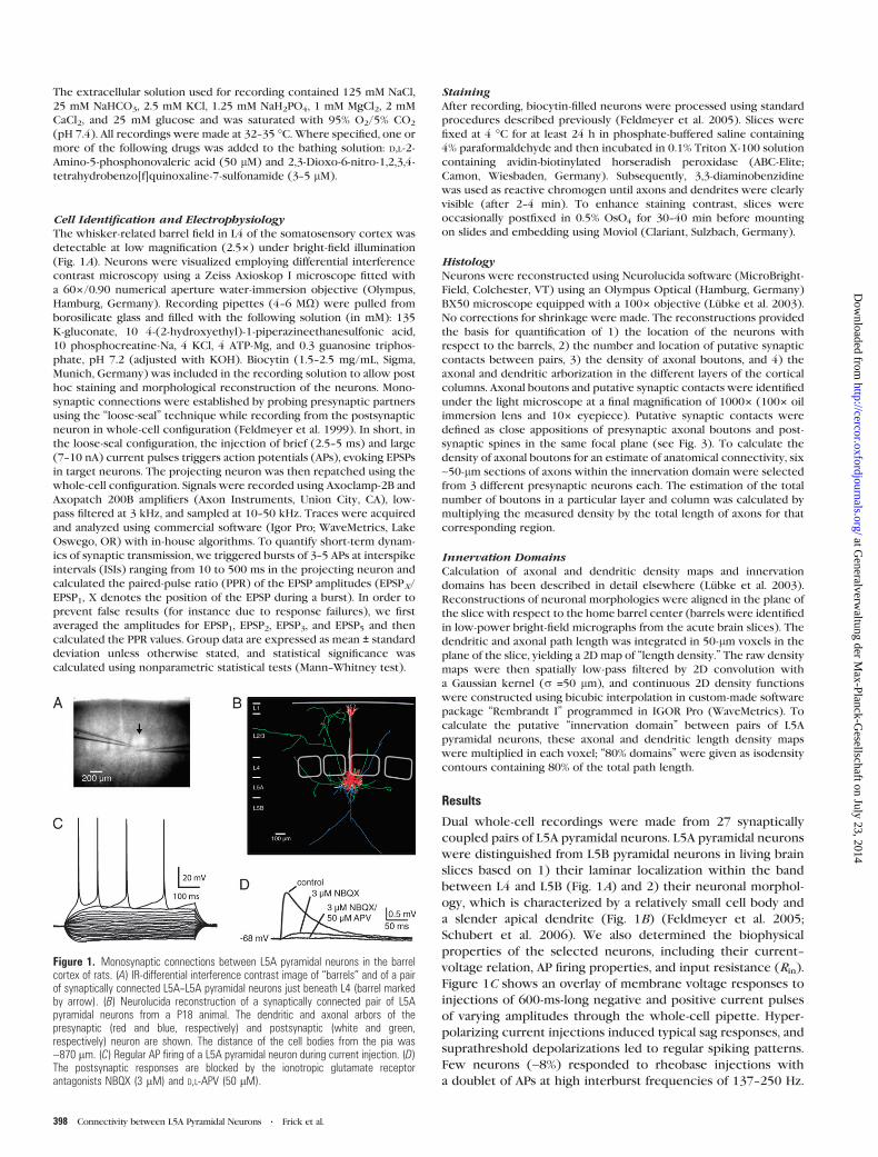
Figure 1C shows an overlay of membrane voltage responses to
injections of 600-ms-long negative and positive current pulses
of varying amplitudes through the whole-cell pipette. Hyper-
polarizing current injections induced typical sag responses, and
suprathreshold depolarizations led to regular spiking patterns.
Few neurons (~8%) responded to rheobase injections with
a doublet of APs at high interburst frequencies of 137--250 Hz.
Figure 1. Monosynaptic connections between L5A pyramidal neurons in the barrelcortex of rats. (A) IR-differential interference contrast image of ‘‘barrels’’ and of a pairof synaptically connected L5A--L5A pyramidal neurons just beneath L4 (barrel markedby arrow). (B) Neurolucida reconstruction of a synaptically connected pair of L5Apyramidal neurons from a P18 animal. The dendritic and axonal arbors of thepresynaptic (red and blue, respectively) and postsynaptic (white and green,respectively) neuron are shown. The distance of the cell bodies from the pia was~870 lm. (C) Regular AP firing of a L5A pyramidal neuron during current injection. (D)The postsynaptic responses are blocked by the ionotropic glutamate receptorantagonists NBQX (3 lM) and D,L-APV (50 lM).
398 Connectivity between L5A Pyramidal Neurons d Frick et al.
at Generalverw
altung der Max-Planck-G
esellschaft on July 23, 2014http://cercor.oxfordjournals.org/
Dow
nloaded from
The input resistance averaged 90 ± 31 MX (n = 31), ranging
from 53 to 173 MX (Table 2).
Characterization of Monosynaptic Connections
To characterize the physiological properties of monosynaptic
connections between L5A pyramidal neurons, we measured
peak amplitude, latency, time course, and failure rate of unitary
EPSPs. Unitary EPSPs were evoked in the target neuron by
triggering single or bursts of 3--5 APs in the projecting
neuron using intracellular current injection. This stimulation
pattern was repeated 40--200 times at rates of 0.05--0.1 Hz
(every 10--20 s), at which there was no obvious rundown of the
EPSP amplitude (see Fig. 6B). The majority (23 out of 27) of
L5A--L5A connections was unidirectional and only a small
percentage (~15%) bidirectional. Unitary EPSPs were mediated
by ionotropic glutamate receptors and completely blocked
by bath application of the a-Amino-3-hydroxy-5-Methyl-4-
isoxazolephropionic acid and N-Methyl-D-aspartic acid recep-
tor blockers NBQX (3--5 lM) and D,L-APV (50 lM), respectively
(n = 4, Fig. 1D).
Axonal and Dendritic Arborization
The morphological reconstruction of a pair of synaptically
connected L5A pyramidal neurons is shown in Figure 1B. In
this and the following figures, dendrites and axons of pre-
synaptic neurons are depicted in red and blue, respectively, and
dendrites and axons of postsynaptic neurons in white and
green, respectively. To quantify the geometry of their axonal
and dendritic arbors and for identification of putative synaptic
contacts between them, we carried out a detailed morpholog-
ical reconstruction of 6 pairs of connected L5A pyramidal
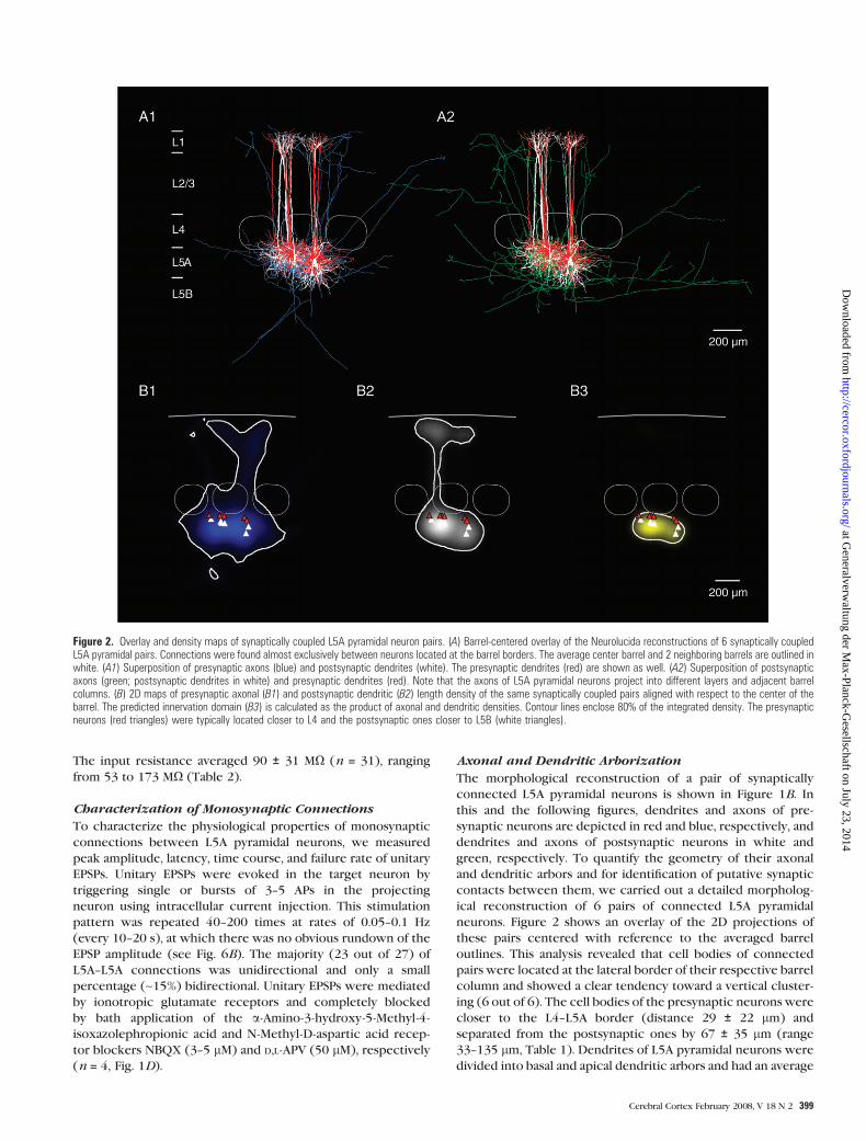
neurons. Figure 2 shows an overlay of the 2D projections of
these pairs centered with reference to the averaged barrel
outlines. This analysis revealed that cell bodies of connected
pairs were located at the lateral border of their respective barrel
column and showed a clear tendency toward a vertical cluster-
ing (6 out of 6). The cell bodies of the presynaptic neurons were
closer to the L4--L5A border (distance 29 ± 22 lm) and
separated from the postsynaptic ones by 67 ± 35 lm (range
33--135 lm, Table 1). Dendrites of L5A pyramidal neurons were
divided into basal and apical dendritic arbors and had an average
Figure 2. Overlay and density maps of synaptically coupled L5A pyramidal neuron pairs. (A) Barrel-centered overlay of the Neurolucida reconstructions of 6 synaptically coupledL5A pyramidal pairs. Connections were found almost exclusively between neurons located at the barrel borders. The average center barrel and 2 neighboring barrels are outlined inwhite. (A1) Superposition of presynaptic axons (blue) and postsynaptic dendrites (white). The presynaptic dendrites (red) are shown as well. (A2) Superposition of postsynapticaxons (green; postsynaptic dendrites in white) and presynaptic dendrites (red). Note that the axons of L5A pyramidal neurons project into different layers and adjacent barrelcolumns. (B) 2D maps of presynaptic axonal (B1) and postsynaptic dendritic (B2) length density of the same synaptically coupled pairs aligned with respect to the center of thebarrel. The predicted innervation domain (B3) is calculated as the product of axonal and dendritic densities. Contour lines enclose 80% of the integrated density. The presynapticneurons (red triangles) were typically located closer to L4 and the postsynaptic ones closer to L5B (white triangles).
Cerebral Cortex February 2008, V 18 N 2 399
at Generalverw
altung der Max-Planck-G
esellschaft on July 23, 2014http://cercor.oxfordjournals.org/
Dow
nloaded from

total length of 6568 ± 1410 lm per neuron (100%, n = 12,
Table 1). Seventy percent of the dendrites were confined to the
home barrel column, increasing to 90% when the neighboring
septa were included. The average total length of the basal
dendritic tree (number of basal dendrites: 5.8 ± 1.4) was 3400 ±1172 lm (51% of all dendrites), extended laterally to about the
width of a barrel (300--400 lm), and was largely confined to L5
and lower L4. The apical dendritic arbor (3168 ± 792 lm, 49% of
all dendrites) spanned 918 ± 49 lm from cell body to pia (range
864--1011 lm, Table 1) and was further subdivided into main,
oblique, and tuft dendrites. Structurally, the main apical
dendrite was slender, giving rise to few (4.4 ± 1.7) oblique
dendrites (1405 ± 718 lm, 21% of all dendrites) mainly
proximally to the soma in L5A and lower L4, and extending
into a small tuft largely confined to L1 (559 ± 297 lm, 9%). The
axons of L5A pyramidal neurons (total length 7819 ± 2491 lm)
projected vertically toward layer 1 and the white matter, as well
as horizontally into neighboring cortical columns within layers
5, 4, and 2/3 (Fig. 2A1,A2). The axonal domain of the pre-
synaptic neurons was very dense in the vicinity of the post-
synaptic cell bodies suggesting a high local connectivity there
(see below).
Innervation Domain and Putative Synaptic Contacts
To assess the target region where L5A pyramidal neurons might
form synaptic contacts with each other (the innervation
domain), we computed the overlap of presynaptic axons and
postsynaptic dendrites (Fig. 2A). For this analysis, 2D maps were
computed from the 3D reconstructions of the respective axonal
(Fig. 2B1) and dendritic (Fig. 2B2) length densities, and the
predicted innervation domain was then calculated as the
product of these densities. Figure 2B3 shows the contour lines
delimiting 80% of the integrated density. These data suggest
that synaptic contacts are mainly located on the basal dendrites
and to a lesser degree on the proximal oblique dendrites and
that the target regions are essentially columnar. A better
estimate for the location and number of putative synaptic
contacts can be achieved by scanning for close approximations
of axons and dendrites under light microscopy at high (10003)
magnification (Fig. 3). For 6 reconstructed L5A pyramidal
neuron pairs, the mean number of putative synaptic contacts
per monosynaptic connection was 3.5 ± 1.8 ranging from 1 to 6
(Fig. 4A, Table 2). The geometric distance of these putative
Table 1Morphological characteristics of rat L5A pyramidal neurons
Cell identity Distance (lm) Dendritic length (lm) No. of primary dendrities
To barrel Pre-post To pia Total Basal Apical Basal Oblique
Main Oblique Tuft in L1 Total
1_pre 19 33 898 6437 3572 1053 1428 385 2865 4.0 3.01_post 52 931 4917 1951 1304 590 1071 2966 5.0 5.02_pre 71 55 931 6925 4077 979 966 902 2848 6.0 6.02_post 126 920 5240 2586 947 802 904 2654 6.0 6.03_pre 27 63 936 5400 3930 1002 407 62 1471 5.0 1.03_post 90 999 8096 3974 1143 2587 392 4122 8.0 2.04_pre 24 135 876 8946 6011 1250 1044 640 2935 6.0 4.04_post 159 1011 7472 3692 1685 1697 418 3780 7.0 6.05_pre 26 65 869 5709 2832 794 1331 752 2877 4.0 4.05_post 90 933 5796 2207 1275 1882 431 3588 8.0 4.06_pre 9 54 864 5288 1920 1442 1462 485 3368 4.0 6.06_post 63 918 8587 4047 1587 2669 284 4540 6.0 6.0Mean 63 67 918 6568 3400 1203 1405 559 3168 5.8 4.4Standard deviation 47 35 49 1410 1172 267 718 297 792 1.4 1.7Median 57 59 919 6116 3632 1196 1379 448 2950 60.0 4.5Minimum 9 33 864 4917 1920 794 407 62 1471 4.0 1.0Maximum 159 135 1011 8946 6011 1665 2669 1071 4540 8.0 6.0% Total dend length 100 51 19 21 9 46
Figure 3. Putative synaptic contacts of a monosynaptic L5A--L5A connection. (A)Biocytin staining of a L5A--L5A pyramidal neuron pair from a P18 animal. Photograph ofa synaptically connected pair. Barrels are indicated by dashed lines. For thisconnection, 5 putative synaptic contacts were identified by light microscopy.(B1--B5) Higher magnification of these putative contacts. Images were improvedusing deconvolution algorithms.
400 Connectivity between L5A Pyramidal Neurons d Frick et al.
at Generalverw
altung der Max-Planck-G
esellschaft on July 23, 2014http://cercor.oxfordjournals.org/
Dow
nloaded from
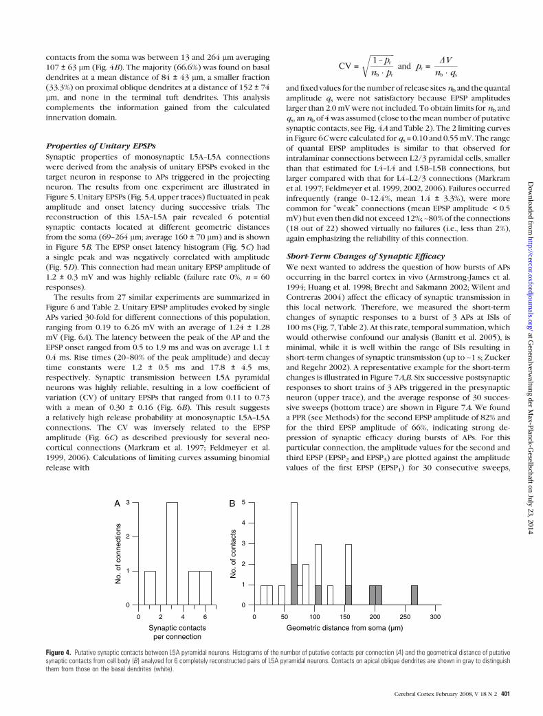
contacts from the soma was between 13 and 264 lm averaging
107 ± 63 lm (Fig. 4B). The majority (66.6%) was found on basal
dendrites at a mean distance of 84 ± 43 lm, a smaller fraction
(33.3%) on proximal oblique dendrites at a distance of 152 ± 74
lm, and none in the terminal tuft dendrites. This analysis
complements the information gained from the calculated
innervation domain.
Properties of Unitary EPSPs
Synaptic properties of monosynaptic L5A--L5A connections
were derived from the analysis of unitary EPSPs evoked in the
target neuron in response to APs triggered in the projecting
neuron. The results from one experiment are illustrated in
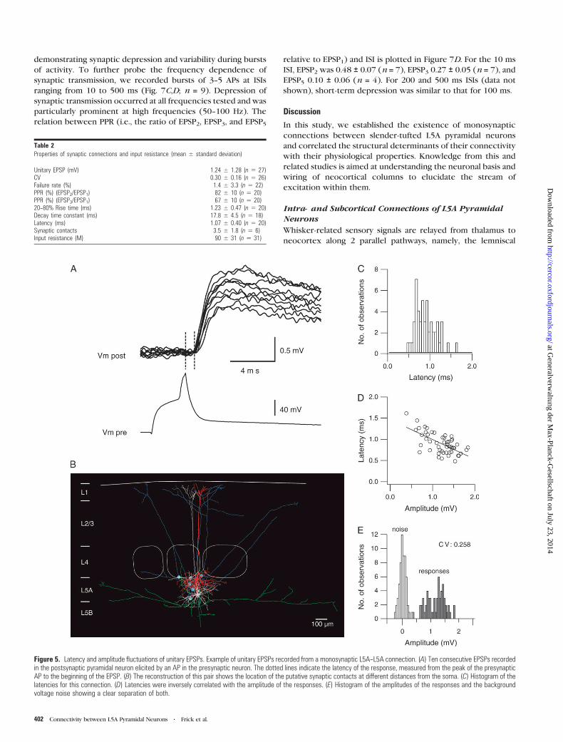
Figure 5. Unitary EPSPs (Fig. 5A, upper traces) fluctuated in peak
amplitude and onset latency during successive trials. The
reconstruction of this L5A--L5A pair revealed 6 potential
synaptic contacts located at different geometric distances
from the soma (69--264 lm; average 160 ± 70 lm) and is shown
in Figure 5B. The EPSP onset latency histogram (Fig. 5C) had
a single peak and was negatively correlated with amplitude
(Fig. 5D). This connection had mean unitary EPSP amplitude of
1.2 ± 0.3 mV and was highly reliable (failure rate 0%, n = 60
responses).
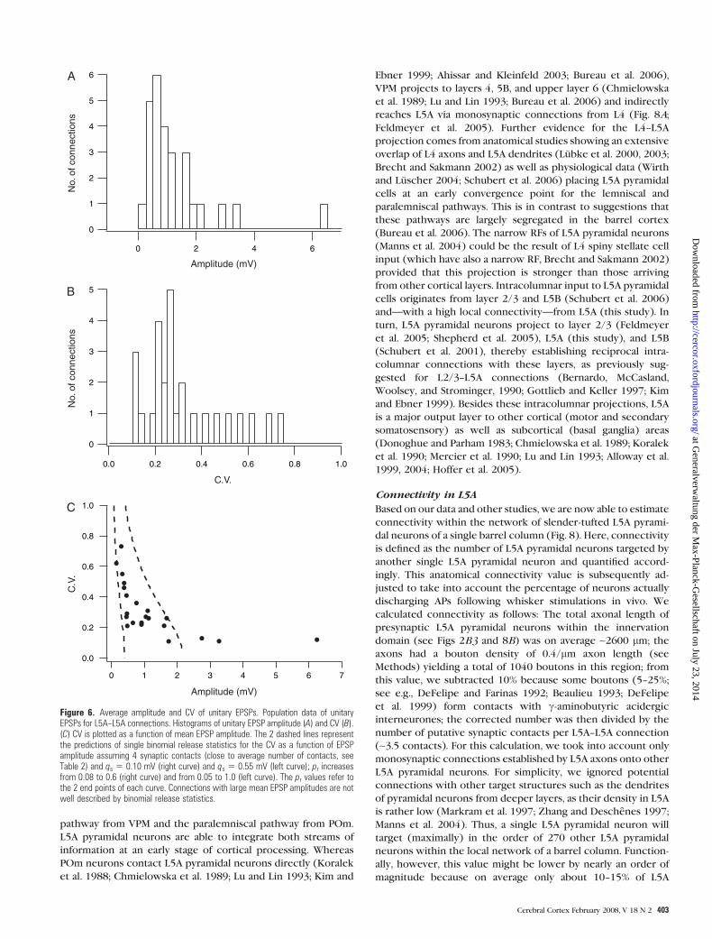
The results from 27 similar experiments are summarized in
Figure 6 and Table 2. Unitary EPSP amplitudes evoked by single
APs varied 30-fold for different connections of this population,
ranging from 0.19 to 6.26 mV with an average of 1.24 ± 1.28
mV (Fig. 6A). The latency between the peak of the AP and the
EPSP onset ranged from 0.5 to 1.9 ms and was on average 1.1 ±0.4 ms. Rise times (20--80% of the peak amplitude) and decay
time constants were 1.2 ± 0.5 ms and 17.8 ± 4.5 ms,
respectively. Synaptic transmission between L5A pyramidal
neurons was highly reliable, resulting in a low coefficient of
variation (CV) of unitary EPSPs that ranged from 0.11 to 0.73
with a mean of 0.30 ± 0.16 (Fig. 6B). This result suggests
a relatively high release probability at monosynaptic L5A--L5A
connections. The CV was inversely related to the EPSP
amplitude (Fig. 6C) as described previously for several neo-
cortical connections (Markram et al. 1997; Feldmeyer et al.
1999, 2006). Calculations of limiting curves assuming binomial
release with
CV =
ffiffiffiffiffiffiffiffiffiffiffiffiffi1 – pr
nb � pr
rand pr =
DVnb � qs
andfixed values for the number of release sitesnb and the quantal
amplitude qs were not satisfactory because EPSP amplitudes
larger than 2.0 mVwere not included. To obtain limits for nb and
qs, annb of 4was assumed (close to themean number of putative
synaptic contacts, see Fig. 4A and Table 2). The 2 limiting curves
in Figure 6Cwere calculated for qs = 0.10 and 0.55mV. The range
of quantal EPSP amplitudes is similar to that observed for
intralaminar connections between L2/3 pyramidal cells, smaller
than that estimated for L4--L4 and L5B--L5B connections, but
larger compared with that for L4--L2/3 connections (Markram
et al. 1997; Feldmeyer et al. 1999, 2002, 2006). Failures occurred
infrequently (range 0--12.4%, mean 1.4 ± 3.3%), were more
common for ‘‘weak’’ connections (mean EPSP amplitude < 0.5
mV) but even then did not exceed 12%; ~80% of the connections
(18 out of 22) showed virtually no failures (i.e., less than 2%),
again emphasizing the reliability of this connection.
Short-Term Changes of Synaptic Efficacy
We next wanted to address the question of how bursts of APs
occurring in the barrel cortex in vivo (Armstrong-James et al.
1994; Huang et al. 1998; Brecht and Sakmann 2002; Wilent and
Contreras 2004) affect the efficacy of synaptic transmission in
this local network. Therefore, we measured the short-term
changes of synaptic responses to a burst of 3 APs at ISIs of
100 ms (Fig. 7, Table 2). At this rate, temporal summation, which
would otherwise confound our analysis (Banitt et al. 2005), is
minimal, while it is well within the range of ISIs resulting in
short-term changes of synaptic transmission (up to ~1 s; Zucker
and Regehr 2002). A representative example for the short-term
changes is illustrated in Figure 7A,B. Six successive postsynaptic
responses to short trains of 3 APs triggered in the presynaptic
neuron (upper trace), and the average response of 30 succes-
sive sweeps (bottom trace) are shown in Figure 7A. We found
a PPR (see Methods) for the second EPSP amplitude of 82% and
for the third EPSP amplitude of 66%, indicating strong de-
pression of synaptic efficacy during bursts of APs. For this
particular connection, the amplitude values for the second and
third EPSP (EPSP2 and EPSP3) are plotted against the amplitude
values of the first EPSP (EPSP1) for 30 consecutive sweeps,
A B
No.
of c
onne
ctio
ns
Synaptic contactsper connection
3
2
1
0
6420
No.
of c
onta
cts
Geometric distance from soma (µm)
5
4
3
2
1
0
300250200150100500
Figure 4. Putative synaptic contacts between L5A pyramidal neurons. Histograms of the number of putative contacts per connection (A) and the geometrical distance of putativesynaptic contacts from cell body (B) analyzed for 6 completely reconstructed pairs of L5A pyramidal neurons. Contacts on apical oblique dendrites are shown in gray to distinguishthem from those on the basal dendrites (white).
Cerebral Cortex February 2008, V 18 N 2 401
at Generalverw
altung der Max-Planck-G
esellschaft on July 23, 2014http://cercor.oxfordjournals.org/
Dow
nloaded from
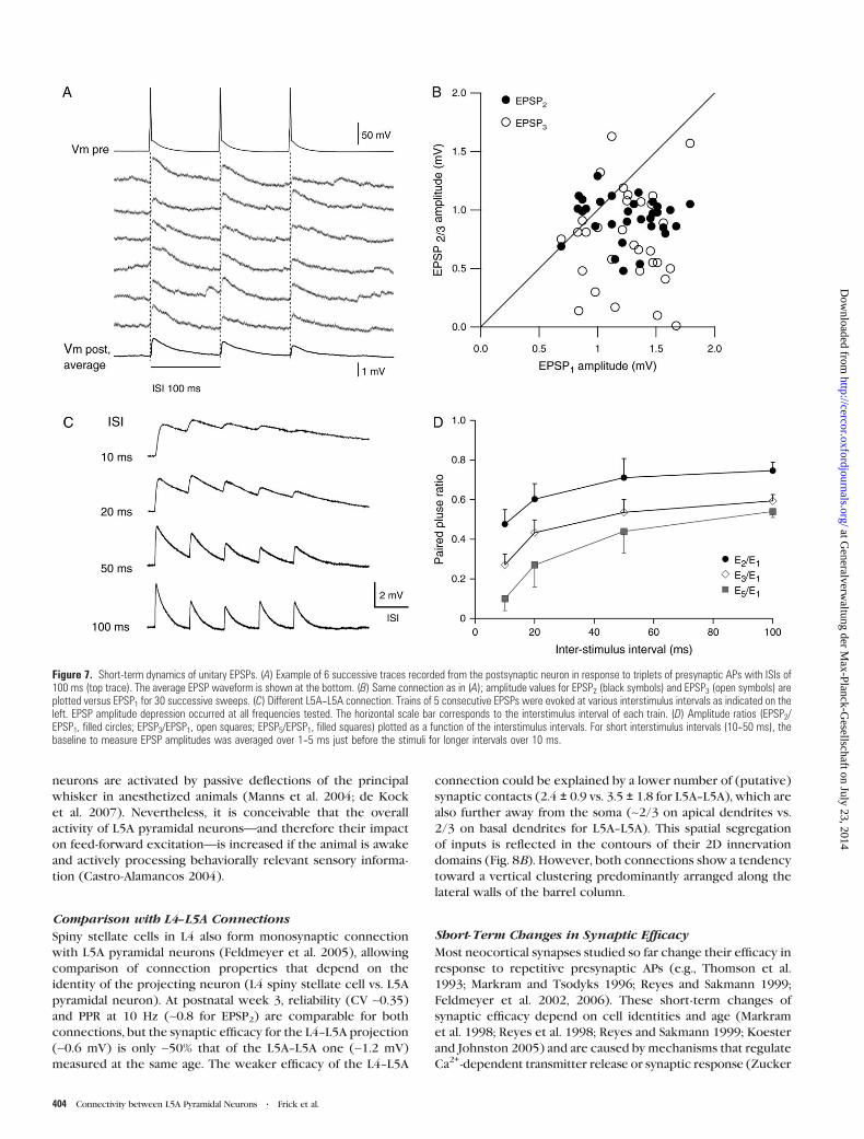
demonstrating synaptic depression and variability during bursts
of activity. To further probe the frequency dependence of
synaptic transmission, we recorded bursts of 3--5 APs at ISIs
ranging from 10 to 500 ms (Fig. 7C,D; n = 9). Depression of
synaptic transmission occurred at all frequencies tested and was
particularly prominent at high frequencies (50--100 Hz). The
relation between PPR (i.e., the ratio of EPSP2, EPSP3, and EPSP5
relative to EPSP1) and ISI is plotted in Figure 7D. For the 10 ms
ISI, EPSP2 was 0.48 ± 0.07 (n = 7), EPSP3 0.27 ± 0.05 (n = 7), and
EPSP5 0.10 ± 0.06 (n = 4). For 200 and 500 ms ISIs (data not
shown), short-term depression was similar to that for 100 ms.
Discussion
In this study, we established the existence of monosynaptic
connections between slender-tufted L5A pyramidal neurons
and correlated the structural determinants of their connectivity
with their physiological properties. Knowledge from this and
related studies is aimed at understanding the neuronal basis and
wiring of neocortical columns to elucidate the stream of
excitation within them.
Intra- and Subcortical Connections of L5A PyramidalNeurons
Whisker-related sensory signals are relayed from thalamus to
neocortex along 2 parallel pathways, namely, the lemniscal
Table 2Properties of synaptic connections and input resistance (mean ± standard deviation)
Unitary EPSP (mV) 1.24 ± 1.28 (n 5 27)CV 0.30 ± 0.16 (n 5 26)Failure rate (%) 1.4 ± 3.3 (n 5 22)PPR (%) (EPSP2/EPSP1) 82 ± 10 (n 5 20)PPR (%) (EPSP3/EPSP1) 67 ± 10 (n 5 20)20--80% Rise time (ms) 1.23 ± 0.47 (n 5 20)Decay time constant (ms) 17.8 ± 4.5 (n 5 18)Latency (ms) 1.07 ± 0.40 (n 5 20)Synaptic contacts 3.5 ± 1.8 (n 5 6)Input resistance (M) 90 ± 31 (n 5 31)
Figure 5. Latency and amplitude fluctuations of unitary EPSPs. Example of unitary EPSPs recorded from a monosynaptic L5A--L5A connection. (A) Ten consecutive EPSPs recordedin the postsynaptic pyramidal neuron elicited by an AP in the presynaptic neuron. The dotted lines indicate the latency of the response, measured from the peak of the presynapticAP to the beginning of the EPSP. (B) The reconstruction of this pair shows the location of the putative synaptic contacts at different distances from the soma. (C) Histogram of thelatencies for this connection. (D) Latencies were inversely correlated with the amplitude of the responses. (E) Histogram of the amplitudes of the responses and the backgroundvoltage noise showing a clear separation of both.
402 Connectivity between L5A Pyramidal Neurons d Frick et al.
at Generalverw
altung der Max-Planck-G
esellschaft on July 23, 2014http://cercor.oxfordjournals.org/
Dow
nloaded from
pathway from VPM and the paralemniscal pathway from POm.
L5A pyramidal neurons are able to integrate both streams of
information at an early stage of cortical processing. Whereas
POm neurons contact L5A pyramidal neurons directly (Koralek
et al. 1988; Chmielowska et al. 1989; Lu and Lin 1993; Kim and
Ebner 1999; Ahissar and Kleinfeld 2003; Bureau et al. 2006),
VPM projects to layers 4, 5B, and upper layer 6 (Chmielowska
et al. 1989; Lu and Lin 1993; Bureau et al. 2006) and indirectly
reaches L5A via monosynaptic connections from L4 (Fig. 8A;
Feldmeyer et al. 2005). Further evidence for the L4--L5A
projection comes from anatomical studies showing an extensive
overlap of L4 axons and L5A dendrites (Lubke et al. 2000, 2003;
Brecht and Sakmann 2002) as well as physiological data (Wirth
and Luscher 2004; Schubert et al. 2006) placing L5A pyramidal
cells at an early convergence point for the lemniscal and
paralemniscal pathways. This is in contrast to suggestions that
these pathways are largely segregated in the barrel cortex
(Bureau et al. 2006). The narrow RFs of L5A pyramidal neurons
(Manns et al. 2004) could be the result of L4 spiny stellate cell
input (which have also a narrow RF, Brecht and Sakmann 2002)
provided that this projection is stronger than those arriving
from other cortical layers. Intracolumnar input to L5A pyramidal
cells originates from layer 2/3 and L5B (Schubert et al. 2006)
and—with a high local connectivity—from L5A (this study). In
turn, L5A pyramidal neurons project to layer 2/3 (Feldmeyer
et al. 2005; Shepherd et al. 2005), L5A (this study), and L5B
(Schubert et al. 2001), thereby establishing reciprocal intra-
columnar connections with these layers, as previously sug-
gested for L2/3--L5A connections (Bernardo, McCasland,
Woolsey, and Strominger, 1990; Gottlieb and Keller 1997; Kim
and Ebner 1999). Besides these intracolumnar projections, L5A
is a major output layer to other cortical (motor and secondary
somatosensory) as well as subcortical (basal ganglia) areas
(Donoghue and Parham 1983; Chmielowska et al. 1989; Koralek
et al. 1990; Mercier et al. 1990; Lu and Lin 1993; Alloway et al.
1999, 2004; Hoffer et al. 2005).
Connectivity in L5A
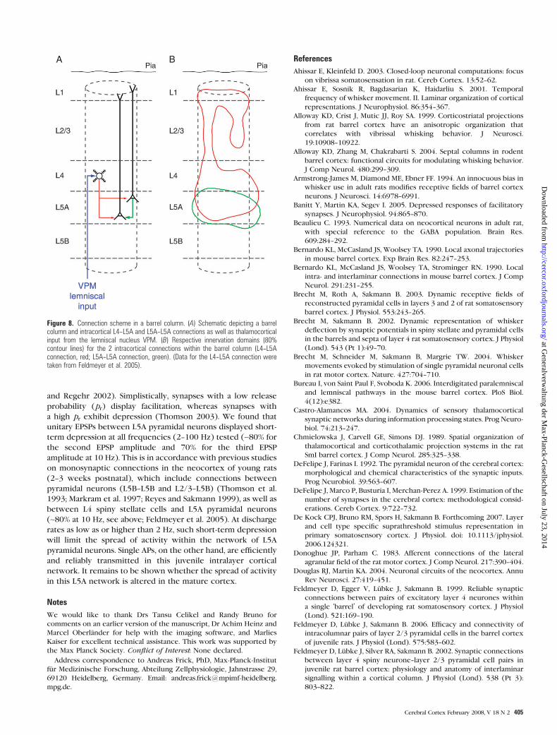
Based on our data and other studies, we are now able to estimate
connectivity within the network of slender-tufted L5A pyrami-
dal neurons of a single barrel column (Fig. 8). Here, connectivity
is defined as the number of L5A pyramidal neurons targeted by
another single L5A pyramidal neuron and quantified accord-
ingly. This anatomical connectivity value is subsequently ad-
justed to take into account the percentage of neurons actually
discharging APs following whisker stimulations in vivo. We
calculated connectivity as follows: The total axonal length of
presynaptic L5A pyramidal neurons within the innervation
domain (see Figs 2B3 and 8B) was on average ~2600 lm; the
axons had a bouton density of 0.4/lm axon length (see
Methods) yielding a total of 1040 boutons in this region; from
this value, we subtracted 10% because some boutons (5--25%;
see e.g., DeFelipe and Farinas 1992; Beaulieu 1993; DeFelipe
et al. 1999) form contacts with c-aminobutyric acidergic
interneurones; the corrected number was then divided by the
number of putative synaptic contacts per L5A--L5A connection
(~3.5 contacts). For this calculation, we took into account only
monosynaptic connections established by L5A axons onto other
L5A pyramidal neurons. For simplicity, we ignored potential
connections with other target structures such as the dendrites
of pyramidal neurons from deeper layers, as their density in L5A
is rather low (Markram et al. 1997; Zhang and Deschenes 1997;
Manns et al. 2004). Thus, a single L5A pyramidal neuron will
target (maximally) in the order of 270 other L5A pyramidal
neurons within the local network of a barrel column. Function-
ally, however, this value might be lower by nearly an order of
magnitude because on average only about 10--15% of L5A
A
B 5
4
3
2
1
0
1.00.80.60.40.20.0
C.V.
No.
of c
onne
ctio
ns
C
Amplitude (mV)
0.2
0.4
0.6
0.8
0.0
1.0
0 1 2 3 4 5 6 7
C.V
.
Amplitude (mV)
No.
of c
onne
ctio
ns6
5
4
3
2
1
0
6420
Figure 6. Average amplitude and CV of unitary EPSPs. Population data of unitaryEPSPs for L5A--L5A connections. Histograms of unitary EPSP amplitude (A) and CV (B).(C) CV is plotted as a function of mean EPSP amplitude. The 2 dashed lines representthe predictions of single binomial release statistics for the CV as a function of EPSPamplitude assuming 4 synaptic contacts (close to average number of contacts, seeTable 2) and qs 5 0.10 mV (right curve) and qs 5 0.55 mV (left curve); pr increasesfrom 0.08 to 0.6 (right curve) and from 0.05 to 1.0 (left curve). The pr values refer tothe 2 end points of each curve. Connections with large mean EPSP amplitudes are notwell described by binomial release statistics.
Cerebral Cortex February 2008, V 18 N 2 403
at Generalverw
altung der Max-Planck-G
esellschaft on July 23, 2014http://cercor.oxfordjournals.org/
Dow
nloaded from
neurons are activated by passive deflections of the principal
whisker in anesthetized animals (Manns et al. 2004; de Kock
et al. 2007). Nevertheless, it is conceivable that the overall
activity of L5A pyramidal neurons—and therefore their impact
on feed-forward excitation—is increased if the animal is awake
and actively processing behaviorally relevant sensory informa-
tion (Castro-Alamancos 2004).
Comparison with L4--L5A Connections
Spiny stellate cells in L4 also form monosynaptic connection
with L5A pyramidal neurons (Feldmeyer et al. 2005), allowing
comparison of connection properties that depend on the
identity of the projecting neuron (L4 spiny stellate cell vs. L5A
pyramidal neuron). At postnatal week 3, reliability (CV ~0.35)and PPR at 10 Hz (~0.8 for EPSP2) are comparable for both
connections, but the synaptic efficacy for the L4--L5A projection
(~0.6 mV) is only ~50% that of the L5A--L5A one (~1.2 mV)
measured at the same age. The weaker efficacy of the L4--L5A
connection could be explained by a lower number of (putative)
synaptic contacts (2.4 ± 0.9 vs. 3.5 ± 1.8 for L5A--L5A), which are
also further away from the soma (~2/3 on apical dendrites vs.
2/3 on basal dendrites for L5A--L5A). This spatial segregation
of inputs is reflected in the contours of their 2D innervation
domains (Fig. 8B). However, both connections show a tendency
toward a vertical clustering predominantly arranged along the
lateral walls of the barrel column.
Short-Term Changes in Synaptic Efficacy
Most neocortical synapses studied so far change their efficacy in
response to repetitive presynaptic APs (e.g., Thomson et al.
1993; Markram and Tsodyks 1996; Reyes and Sakmann 1999;
Feldmeyer et al. 2002, 2006). These short-term changes of
synaptic efficacy depend on cell identities and age (Markram
et al. 1998; Reyes et al. 1998; Reyes and Sakmann 1999; Koester
and Johnston 2005) and are caused bymechanisms that regulate
Ca2+-dependent transmitter release or synaptic response (Zucker
Figure 7. Short-term dynamics of unitary EPSPs. (A) Example of 6 successive traces recorded from the postsynaptic neuron in response to triplets of presynaptic APs with ISIs of100 ms (top trace). The average EPSP waveform is shown at the bottom. (B) Same connection as in (A); amplitude values for EPSP2 (black symbols) and EPSP3 (open symbols) areplotted versus EPSP1 for 30 successive sweeps. (C) Different L5A--L5A connection. Trains of 5 consecutive EPSPs were evoked at various interstimulus intervals as indicated on theleft. EPSP amplitude depression occurred at all frequencies tested. The horizontal scale bar corresponds to the interstimulus interval of each train. (D) Amplitude ratios (EPSP2/EPSP1, filled circles; EPSP3/EPSP1, open squares; EPSP5/EPSP1, filled squares) plotted as a function of the interstimulus intervals. For short interstimulus intervals (10--50 ms), thebaseline to measure EPSP amplitudes was averaged over 1--5 ms just before the stimuli for longer intervals over 10 ms.
404 Connectivity between L5A Pyramidal Neurons d Frick et al.
at Generalverw
altung der Max-Planck-G
esellschaft on July 23, 2014http://cercor.oxfordjournals.org/
Dow
nloaded from
and Regehr 2002). Simplistically, synapses with a low release
probability (pr) display facilitation, whereas synapses with
a high pr exhibit depression (Thomson 2003). We found that
unitary EPSPs between L5A pyramidal neurons displayed short-
term depression at all frequencies (2--100 Hz) tested (~80% for
the second EPSP amplitude and 70% for the third EPSP
amplitude at 10 Hz). This is in accordance with previous studies
on monosynaptic connections in the neocortex of young rats
(2--3 weeks postnatal), which include connections between
pyramidal neurons (L5B--L5B and L2/3--L5B) (Thomson et al.
1993; Markram et al. 1997; Reyes and Sakmann 1999), as well as
between L4 spiny stellate cells and L5A pyramidal neurons
(~80% at 10 Hz, see above; Feldmeyer et al. 2005). At discharge
rates as low as or higher than 2 Hz, such short-term depression
will limit the spread of activity within the network of L5A
pyramidal neurons. Single APs, on the other hand, are efficiently
and reliably transmitted in this juvenile intralayer cortical
network. It remains to be shown whether the spread of activity
in this L5A network is altered in the mature cortex.
Notes
We would like to thank Drs Tansu Celikel and Randy Bruno for
comments on an earlier version of the manuscript, Dr Achim Heinz and
Marcel Oberlander for help with the imaging software, and Marlies
Kaiser for excellent technical assistance. This work was supported by
the Max Planck Society. Conflict of Interest: None declared.
Address correspondence to Andreas Frick, PhD, Max-Planck-Institut
fur Medizinische Forschung, Abteilung Zellphysiologie, Jahnstrasse 29,
69120 Heidelberg, Germany. Email: andreas.frick@mpimf-heidelberg.
mpg.de.
References
Ahissar E, Kleinfeld D. 2003. Closed-loop neuronal computations: focus
on vibrissa somatosensation in rat. Cereb Cortex. 13:52--62.
Ahissar E, Sosnik R, Bagdasarian K, Haidarliu S. 2001. Temporal
frequency of whisker movement. II. Laminar organization of cortical
representations. J Neurophysiol. 86:354--367.
Alloway KD, Crist J, Mutic JJ, Roy SA. 1999. Corticostriatal projections
from rat barrel cortex have an anisotropic organization that
correlates with vibrissal whisking behavior. J Neurosci.
19:10908--10922.
Alloway KD, Zhang M, Chakrabarti S. 2004. Septal columns in rodent
barrel cortex: functional circuits for modulating whisking behavior.
J Comp Neurol. 480:299--309.
Armstrong-James M, Diamond ME, Ebner FF. 1994. An innocuous bias in
whisker use in adult rats modifies receptive fields of barrel cortex
neurons. J Neurosci. 14:6978--6991.
Banitt Y, Martin KA, Segev I. 2005. Depressed responses of facilitatory
synapses. J Neurophysiol. 94:865--870.
Beaulieu C. 1993. Numerical data on neocortical neurons in adult rat,
with special reference to the GABA population. Brain Res.
609:284--292.
Bernardo KL, McCasland JS, Woolsey TA. 1990. Local axonal trajectories
in mouse barrel cortex. Exp Brain Res. 82:247--253.
Bernardo KL, McCasland JS, Woolsey TA, Strominger RN. 1990. Local
intra- and interlaminar connections in mouse barrel cortex. J Comp
Neurol. 291:231--255.
Brecht M, Roth A, Sakmann B. 2003. Dynamic receptive fields of
reconstructed pyramidal cells in layers 3 and 2 of rat somatosensory
barrel cortex. J Physiol. 553:243--265.
Brecht M, Sakmann B. 2002. Dynamic representation of whisker
deflection by synaptic potentials in spiny stellate and pyramidal cells
in the barrels and septa of layer 4 rat somatosensory cortex. J Physiol
(Lond). 543 (Pt 1):49--70.
Brecht M, Schneider M, Sakmann B, Margrie TW. 2004. Whisker
movements evoked by stimulation of single pyramidal neuronal cells
in rat motor cortex. Nature. 427:704--710.
Bureau I, von Saint Paul F, Svoboda K. 2006. Interdigitated paralemniscal
and lemniscal pathways in the mouse barrel cortex. PloS Biol.
4(12):e382.
Castro-Alamancos MA. 2004. Dynamics of sensory thalamocortical
synaptic networks during information processing states. Prog Neuro-
biol. 74:213--247.
Chmielowska J, Carvell GE, Simons DJ. 1989. Spatial organization of
thalamocortical and corticothalamic projection systems in the rat
SmI barrel cortex. J Comp Neurol. 285:325--338.
DeFelipe J, Farinas I. 1992. The pyramidal neuron of the cerebral cortex:
morphological and chemical characteristics of the synaptic inputs.
Prog Neurobiol. 39:563--607.
DeFelipe J, Marco P, Busturia I, Merchan-Perez A. 1999. Estimation of the
number of synapses in the cerebral cortex: methodological consid-
erations. Cereb Cortex. 9:722--732.
De Kock CPJ, Bruno RM, Spors H, Sakmann B. Forthcoming 2007. Layer
and cell type specific suprathreshold stimulus representation in
primary somatosensory cortex. J Physiol. doi: 10.1113/jphysiol.
2006.124321.
Donoghue JP, Parham C. 1983. Afferent connections of the lateral
agranular field of the rat motor cortex. J Comp Neurol. 217:390--404.
Douglas RJ, Martin KA. 2004. Neuronal circuits of the neocortex. Annu
Rev Neurosci. 27:419--451.
Feldmeyer D, Egger V, Lubke J, Sakmann B. 1999. Reliable synaptic
connections between pairs of excitatory layer 4 neurones within
a single ‘barrel’ of developing rat somatosensory cortex. J Physiol
(Lond). 521:169--190.
Feldmeyer D, Lubke J, Sakmann B. 2006. Efficacy and connectivity of
intracolumnar pairs of layer 2/3 pyramidal cells in the barrel cortex
of juvenile rats. J Physiol (Lond). 575:583--602.
Feldmeyer D, Lubke J, Silver RA, Sakmann B. 2002. Synaptic connections
between layer 4 spiny neurone--layer 2/3 pyramidal cell pairs in
juvenile rat barrel cortex: physiology and anatomy of interlaminar
signalling within a cortical column. J Physiol (Lond). 538 (Pt 3):
803--822.
Figure 8. Connection scheme in a barrel column. (A) Schematic depicting a barrelcolumn and intracortical L4--L5A and L5A--L5A connections as well as thalamocorticalinput from the lemniscal nucleus VPM. (B) Respective innervation domains (80%contour lines) for the 2 intracortical connections within the barrel column (L4--L5Aconnection, red; L5A--L5A connection, green). (Data for the L4--L5A connection weretaken from Feldmeyer et al. 2005).
Cerebral Cortex February 2008, V 18 N 2 405
at Generalverw
altung der Max-Planck-G
esellschaft on July 23, 2014http://cercor.oxfordjournals.org/
Dow
nloaded from

Feldmeyer D, Roth A, Sakmann B. 2005. Monosynaptic connections
between pairs of spiny stellate cells in layer 4 and pyramidal cells in
layer 5A indicate that lemniscal and paralemniscal afferent pathways
converge in the infragranular somatosensory cortex. J Neurosci.
25:3423--3431.
Gottlieb JP, Keller A. 1997. Intrinsic circuitry and physiological
properties of pyramidal neurons in rat barrel cortex. Exp Brain
Res. 115:47--60.
Hoeflinger BF, Bennett-Clarke CA, Chiaia NL, Killackey HP, Rhoades RW.
1995. Patterning of local intracortical projections within the
vibrissae representation of rat primary somatosensory cortex.
J Comp Neurol. 354:551--563.
Hoffer ZS, Arantes HB, Roth RL, Alloway KD. 2005. Functional circuits
mediating sensorimotor integration: quantitative comparisons of
projections from rodent barrel cortex to primary motor cortex,
neostriatum, superior colliculus, and the pons. J Comp Neurol.
488:82--100.
Huang W, Armstrong-James M, Rema V, Diamond ME, Ebner FF. 1998.
Contribution of supragranular layers to sensory processing and
plasticity in adult rat barrel cortex. J Neurophysiol. 80:3261--3271.
Ito M. 1992. Simultaneous visualization of cortical barrels and horserad-
ish peroxidase-injected layer 5b vibrissa neurones in the rat.
J Physiol. 454:247--265.
Killackey HP, Koralek KA, Chiaia NL, Rhodes RW. 1989. Laminar and
areal differences in the origin of the subcortical projection neurons
of the rat somatosensory cortex. J Comp Neurol. 282:428--445.
Kim U, Ebner FF. 1999. Barrels and septa: separate circuits in rat barrels
field cortex. J Comp Neurol. 408:489--505.
Koester HJ, Johnston D. 2005. Target cell-dependent normalization of
transmitter release at neocortical synapses. Science. 308:863--866.
Koralek KA, Jensen KF, Killackey HP. 1988. Evidence for two comple-
mentary patterns of thalamic input to the rat somatosensory cortex.
Brain Res. 463:346--351.
Koralek KA, Olavarria J, Killackey HP. 1990. Areal and laminar organi-
zation of corticocortical projections in the rat somatosensory
cortex. J Comp Neurol. 299:133--150.
Larsen DD, Callaway EM. 2006. Development of layer-specific axonal
arborizations in mouse primary somatosensory cortex. J Comp
Neurol. 494:398--414.
Lu SM, Lin RC. 1993. Thalamic afferents of the rat barrel cortex: a light-
and electron-microscopic study using Phaseolus vulgaris leucoag-
glutinin as an anterograde tracer. Somatosens Mot Res. 10:1--16.
Lubke J, Egger V, Sakmann B, Feldmeyer D. 2000. Columnar organization
of dendrites and axons of single and synaptically coupled excitatory
spiny neurons in layer 4 of the rat barrel cortex. J Neurosci.
20:5300--5311.
Lubke J, Roth A, Feldmeyer D, Sakmann B. 2003. Morphometric analysis
of the columnar innervation domain of neurons connecting layer
4 and layer 2/3 of juvenile rat barrel cortex. Cereb Cortex.
13:1051--1063.
Manns ID, Sakmann B, Brecht M. 2004. Sub- and suprathreshold re-
ceptive field properties of pyramidal neurones in layers 5A and 5B
of rat somatosensory barrel cortex. J Physiol (Lond). 556:601--622.
Markram H, Lubke J, Frotscher M, Roth A, Sakmann B. 1997. Physiology
and anatomy of synaptic connections between thick tufted pyrami-
dal neurons in the developing rat neocortex. J Physiol (Lond).
500:409--440.
Markram H, Tsodyks M. 1996. Redistribution of synaptic efficacy:
a mechanism to generate infinite synaptic input diversity from
a homogeneous population of neurons without changing absolute
synaptic efficacies. J Physiol (Paris). 90:229--232.
Markram H, Wang Y, Tsodyks M. 1998. Differential signalling via the
same axon of neocortical pyramidal neurons. Proc Natl Acad Sci USA.
95:5323--5328.
Mercier BE, Legg CR, Glickstein M. 1990. Basal ganglia and cerebellum
receive different somatosensory information in rats. Proc Natl Acad
Sci USA. 87:4388--4392.
Nelson S. 2002. Cortical microcircuits: diverse or canonical? Neuron.
36:19--27.
Reyes A, Lujan R, Rozov A, Burnashev N, Somogyi P, Sakmann B. 1998.
Target-cell-specific facilitation and depression in neocortical cir-
cuits. Nat Neurosci. 1:279--285.
Reyes A, Sakmann B. 1999. Developmental switch in the short-term
modification of unitary EPSPs evoked in layer 2/3 and layer 5
pyramidal neurons of rat neocortex. J Neurosci. 19:3827--3835.
Schubert D, Kotter R, Luhmann HJ, Staiger JF. 2006. Morphology,
electrophysiology and functional input connectivity of pyramidal
neurons characterizes a genuine layer Va in the primary somatosen-
sory cortex. Cereb Cortex. 16:223--236.
Schubert D, Staiger JF, Cho N, Kotter R, Zilles K, Luhmann HJ. 2001.
Layer specific intracolumnar and transcolumnar functional connec-
tivity of layer V pyramidal cells in rat barrel cortex. J Neurosci.
21:3580--3592.
Shepherd MG, Stepanyants A, Bureau I, Chklovskii D, Svoboda K. 2005.
Geometric and functional organization of cortical circuits. Nat
Neurosci. 8:782--790.
Thomson AM. 2003. Presynaptic frequency- and pattern-dependent
filtering. J Comput Neurosci. 15:159--202.
Thomson AM, Deuchars J, West DC. 1993. Large, deep layer pyramid-
pyramid single axon EPSPs in slices of rat motor cortex display paired
pulse and frequency-dependent depression, mediated presynapti-
cally and self-facilitation, mediated postsynaptically. J Neurophysiol.
70:2354--2369.
Wilent WB, Contreras D. 2004. Synaptic responses to whisker deflec-
tions in rat barrel cortex as a function of cortical layer and stimulus
intensity. J Neurosci. 24:3985--3998.
Wirth C, Luscher HR. 2004. Spatiotemporal evolution of excitation and
inhibition in the rat barrel cortex investigated with multielectrode
arrays. J Neurophysiol. 91:1635--1647.
Wise SP, Jones EG. 1977. Cells of origin and terminal distribution of
descending projections of the rat somatic sensory cortex. J Comp
Neurol. 175:129--157.
Woolsey TA, van der Loos H. 1970. The structural organization of layer
IV in the somatosensory region (SI) of mouse cerebral cortex. The
description of a cortical field composed of discrete cytoarchitec-
tonic units. Brain Res. 17:205--242.
Zhang Z-W, Deschenes M. 1997. Intracortical axonal projections of
lamina VI cells of the primary somatosensory cortex in the rat:
a single-cell labeling study. J Neurosci. 17:6365--6379.
Zilles K, Wree A. 1995. Cortex: areal and laminar structure. In: Paxinos G,
editor. The rat nervous system. New York: Academic Press.
p. 649--685.
Zucker RS, Regehr WG. 2002. Short-term synaptic plasticity. Annu Rev
Physiol. 64:355--405.
406 Connectivity between L5A Pyramidal Neurons d Frick et al.
at Generalverw
altung der Max-Planck-G
esellschaft on July 23, 2014http://cercor.oxfordjournals.org/
Dow
nloaded from