monograph of cercosporoid fungi from laos
TRANSCRIPT

Current Research in Environmental & Applied Mycology Doi 10.5943/cream/3/1/2
34
Monograph of Cercosporoid fungi from Laos
Phengsintham P1,2, Chukeatirote E 1, McKenzie EHC3, Hyde
KD1 and Braun U4
1School of Science, Mae Fah Luang University, Chiang Rai 57100, Thailand 2Biology Department, Faculty of Sciences, National University of Laos 3Landcare Research, Private Bag 92170, Auckland, New Zealand 4Martin-Luther-Universität, Institut für Biologie, Bereich Geobotanik und Botanischer Garten, Herbarium, Neuwerk 21
06099 Halle/S. Germany
Phengsintham P, Chukeatirote E, McKenzie EHC, Hyde KD, Braun U 2013 – Monograph of
Cercosporoid fungi from Laos. Current Research in Environmental & Applied Mycology 3(1), 34–
158, doi 10.5943/cream/3/1/2
The Lao People’s Democratic Republic (Lao PDR) or Laos is a landlocked country. During a study
of cercosporoid fungi in Laos, 113 species were identified including 108 species of true
cercosporoid fungi; Cercospora (41 species), Passalora (10), Pseudocercospora (49), and
Zasmidium (8). Five species of morphological similar fungi we also found; Cladosporium (1
species), Periconiella (1), Pseudocercosporella (1), Scolecostigmina (1), and Spiropes (1). Sixteen
new taxa were established namely, Cercospora duranticola, C. senecionis-walkeri, Passalora
dipterocarpi, P. helicteris-viscidae, Pseudocercospora getoniae, P. mannanorensis var.
paucifasciculata, P. micromeli, P. tectoniae, P. wenlandiphila, Zasmidium aporosae, Z. dalbergiae,
Z. jasminicola, Z. meynae-laxiflorae, Z. micromeli, Z. suregadae, Z. pavettae. Eighty-seven species
are described in full and illustrated, and another 26 species are only listed since they have been
previously recorded from Laos.
Key words – Asia – Cercospora – Cercosporoid fungus – monograph
Article Information
Received 29 January 2013
Accepted 20 March 2013
Published online 25 June 2013
*Corresponding author: Kevin D. Hyde – e-mail – [email protected]
Introduction
The Lao People’s Democratic Republic
(Lao PDR) or Laos is a landlocked country
with a total area of 236,800 km2, much of
which is forested and mountainous. The
country is divided into seventeen provinces.
With a population of about 6 million in 2011,
the Lao PDR is the second least populated
country in Association of South-East Asia
Nations (ASEAN) with the lowest population
density of about 25 persons per square
kilometer (Laos 2011).
Lao PDR is considered to be globally
important for biodiversity conservation due to
its relatively high forest cover and high diver-
sity of flora and fauna. Approximately 41.5 %
of Lao PDR is covered with forest which
contains an estimated 8,000–11,000 species of
flowering plants. The country’s fauna includes
166 reported species of reptiles and amphi-
bians, at least 700 bird species, 90 known
species of bats and at least 100 species of large
mammals (MAF and STEA 2003), but only
201 fungal species (Phengsintham et al. 2012).

Current Research in Environmental & Applied Mycology Doi 10.5943/cream/3/1/2
35
The fungi of Laos are poorly known,
although they have been studied since 1959.
Vidal (1959), a French botanist, published a
checklist of plant species of Laos which
included 33 species of Lao fungi. Almost all
names of fungi are local names, but include
some scientific names. Overseas collaboration
was carried with the Mushroom Research
Centre, Papae, Chiang Mai, Thailand and
School of Science, Mae Fah Luang University
(MFU), Chiang Rai, Thailand and other
institutions to document macro- and
microfungi. Phengsintham & Hyde (2003a)
prepared a list of 60 fungi from Laos, and
published on 20 ascomycetes on palms
(Phengsintham & Hyde 2003b). Fungi from
Laos were updated to include 201 species
(Phengsintham et al. 2012).
Sample collection
Leaves of plants with leaf spots or other
lesions were collected during the course of
field trips in Laos. Photos of symptoms,
including the fungal colonies or fruit bodies
were taken. The specimens were collected from
12 provinces (Fig. 1).
Fig. 1 – Collection sites. 1 Louang Namtha 2
Bokeo 3 Oudomxay 4 Loungprabang 5
Sayabouri 6 Huaphanh 7 Xiangkhouang 8
Vientiane Province 9 Vientiane Capital 10
Bolikhamsay 11 Khammoune 12 Savannakhet
Examination of fungal structures
Macroscopic characters were observed
using a stereoscope to check (1) lesions (shape,
size, colour, margin), and (2) details of
colonies/caespituli (e.g., amphigenous,
epiphyllous, punctiform, postulate,
inconspicuous, effuse, loose, dense, colour,
etc.).
Microscopic examination, measure-
ment, description, and presentation of drawings
follow standard procedures outlined by Braun
(1995). In the illustrations, thin-walled
structures are depicted by a single line, thick-
walled ones by double lines, and stippling is
used to accentuate shape and pigmentation.
Measurements and microscopic study
Where sufficient material was
available, 30 measurements of each
morphological character were carried out and
the average estimated by using the formula:
(x M
n µm),
Notes – m = is size of each
components, n = is number of components
The characters described and/or
measured are mycelia (internal, external),
hyphae (branched or not, width, septation,
colour, wall thin/thick, smooth/verruculose),
stromata (location, e.g., substomatal,
intraepidermal; shape, size, colour; cells,
angular or rounded in outline, size, wall
thick/thin), conidiophores (formation,
solitary/fasciculate/sporodochial, arising from
internal/external hyphae/stromata,
erumpent/through stomata; shape; size;
septation; colour; wall, thin/thick,
smooth/verrucuose), conidiogenous cells
(integrated, terminal/intercalary; length, shape,
e.g., cylindrical/geniculate/sinuous),
conidiogenous loci [scars] (shape, size,
thickened, darkened/pigmented or unthickend
or inconspicuous, etc.), and conidia (formation,
solitary/catenate; shape; size; septation; colour;
wall, thin/thick, smooth/verruculose, apex;
base; hila, size, thickened/unthickened,
pigmented or not).
Current Research in Environmental & Applied Mycology Doi 10.5943/cream/3/1/2
36
Identification of fungi
The concept of Crous & Braun (2003)
for classification of Cercospora and
morphologically similar cercosporoid genera
was followed. The species of cercosporoid
hyphomycetes were determined on the basis of
the currently relevant taxonomic publications,
especially the monograph of Cercospora by
Chupp (1954), and the works of Deighton
(1967, 1983), Ellis (1971, 1976), Hsieh & Goh
(1990), Guo & Hsieh (1995), etc.
Single spore isolation
Conidia were picked directly from the
substrate using fine forceps or a needle. The
conidia were placed in sterilized water and
agitated in order to provide a spore suspension
(Choi et al. 1999).
The suspension was prepared on
sterilized glass slides. Sixteen squares were
marked on the bottom of a water agar plate and
the prepared spore suspension was then
transferred with a sterilised pipette, onto the
surface of the water agar plate, above each of
the drawn squares. Alternatively about six
drops of the suspension were pipetted onto the
centre of the agar plate and this was carefully
shaken to spread the suspension. The plates
were incubated at 25oC for 12–24 hours. Once
the conidia had germinated, a sterilised glass
needle was used to pick up a small piece of
agar containing a spore. If the conidia did not
germinate after 12 hours, then the plates were
sealed with film and examined periodically.
Ten germinated spores are transferred and
distributed evenly onto two PDA plate and
incubated at 25oC until their colony diameters
were about 1 to 2 cm. A small piece of
mycelium with agar was then cut out and
transferred to a fresh PDA plate.
Isolates of single spores were deposited
in the culture collection at Herbarium, Biology
Department, School of Science, Mae Fah
Luang University, Biotech Center, Bangkok
and CBS.
Herbarium specimens
Dried specimens were prepared and
stored in the herbaria of the Mae Fah Luang
University, Chiang Rai, Thailand and the
Biology Department, Faculty of Science,
National University of Laos. Duplicates are
preserved in the herbarium of the Institute of
Biology, Geobotany and Botanical Garden,
Halle (Saale), Germany (HAL).
Results
By integrating the morphological and
molecular characters, 113 cercosporoid species
were identified including 108 species of true
cercosporoid fungi Cercospora (41 species),
Passalora (10), Pseudocercospora (49),
Zasmidium (8) and five species of
morphological similar fungi Cladosporium (1
species), Periconiela (1), Pseudocercosporella
(1), Scolecostigmina (1), Spirops (1). Sixteen
new taxa are described: Cercospora
duranticola, C. senecionis-walkeri, Passalora
dipterocarpi, P. helicteris-viscidae,
Pseudocercospora getoniae, P. mannanorensis
var. paucifasciculata, P. micromeli, P.
tectoniae, P. wendlandiphila, Zasmidium
aporosae, Z. dalbergiae, Z. jasminicola, Z.
meynae-laxiflorae, Z. micromeli, Z. suregadae,
Z. pavettae.
Eighty-seven species are described in
full and illustrated, and another 26 species are
only listed because they have been previously
recorded from Laos (Table 1).
Key to true cercosporoid genera
This key contains only the true
cercosporoid genera discussed and treated in
this work (Crous & Braun 2003). The key to
identify species, alphabetically arranged by
host families, are based on models of Chupp
(1954), Ellis (1971, 1976), Deighton (1967,
1973, 1976, 1979), Hsieh & Goh (1990), and
Guo & Hsieh (1995).
1. Conidiogenous loci inconspicuous or
subdenticulate, but always unthickened and not
darkened or subconspicuous, i.e. unthickened,
but somewhat refractive or rarely very slightly
darkened or only outer rim slightly darkened or
refractive (visible as minute rings)……………
…………………………….Pseudocercospora

Current Research in Environmental & Applied Mycology Doi 10.5943/cream/3/1/2
37
Table 1 Cercosporoid fungi found in Laos
Ser# Fungus Hosts Host family Laos
I. True cercosporoid fungi
1 Cercospora achyranthis Achyranthes aspera Amaranthaceae New record
2 Cercospora alocasiae Alocasia macrorrhiza Araceae New record
3 Cercospora apii Byttneria andamensis Malvaceae New record,
new host
4 Cercospora artemisiae Artemisia caudata Asteraceae New record
5 Cercospora asparagi Asparagus officinalis Asparagaceae New record
6 Cercospora begoniae Begonia inflata Begoniaceae New record
7 Cercospora bidentis Bidens pilosa Asteraceae New record
8 Cercospora brassicicola Brassica integrifolia Brassicaceae New record
9 Cercospora cannabis Cannabis sativa Cannabaceae New record 10 Cercospora capsicigena Capsicum annuum Solanaceae New record
11 Cercospora cocciniae Coccinia grandis Cucurbitaceae New record
12 Cercospora duranticola Duranta repens Verbenaceae New species
13 Cercospora erechtitis Erechtites
valerianifolius
Asteraceae New record
14 Cercospora hyptidicola Hyptis suaveolens Lamiaceae New record
15 Cercospora ipomoeae Ipomoea involucrata,
I. aquatica
Convolvulaceae New record
16 Cercospora meliicola Chukrasia tabularis Meliaceae New record
17 Cercospora nasturtii Nasturtium officinale Brassicaceae New record
18 Cercospora nicotianae Nicotiana tabacum Solanaceae New record 19 Cercospora paederiicola Paederia scandens Rubiaceae New record
20 Cercospora physalidis Physalis angulata Solanaceae New record
21 Cercospra ricinella Ricinus communis Euphorbiaceae New record
22 Cercospora senecionis-
walkeri
Senecio walkeri Asteraceae New species
23 Cercospora sp. Oroxylum indicum Bignoniaceae New record
24 Cercospora stahlianthi Stahlianthus thorelii Zingiberaceae New record
25 Cercospora taccae Tacca integrifolia Taccaceae New record
26 Cercospora trewiae Trewia nudiflora Euphorbiaceae New record
27 Cercospora volkameriae Clerodendrum schmidtii Lamiaceae New record
28 Cercospora zinniae Zinnia elegans Asteraceae New record
29 Passalora aenea Cassia siamea Fabaceae New record 30 Passalora bougainvilleae Bougainvillea
spectabilis
Nyctaginaceae New record
31 Passalora capsicicola Capsicum annuum Solanaceae New record
32 Passalora dipterocarpi Dipterocarpus alatus Dipterocarpaceae New species
33 Passalora erytrinae Erythrina stricta Fabaceae New record
34 Passalora haldinae Haldina cordifolia Rubiaceae New record
35 Passalora helicteris-
viscidae
Helicteres viscida Malvaceae New species
36 Passalora henningsii Manihot esculenta Euphorbiaceae New record
37 Passalora perfoliati Chromolaena sp. Asteraceae New record
38 Passalora tithoniae Tithonia diversifolia Asteraceae New record 39 Pseudocercospora alangii Alangium kurzii Cornaceae New record
40 Pseudocercospora
baliospermi
Baliospermum
montanum
Euphorbiaceae New record
41 Pseudocercospora
buddlejae
Buddleja asiatica Scrophulariaceae New record
42 Pseudocercospora
catappae
Terminalia alata Combretaceae New record
43 Pseudocercospora
cotizensis
Crotalaria uncinella
subsp. elliptica
Fabaceae New record
44 Pseudocercospora
duabangae
Duabanga grandiflora Lythraceae New record
45 Pseudocercospora eupatorii–formasani
Chromolaena odorata Asteraceae New record
46 Pseudocercospora formasana Lantana camara Verbenaceae New record

Current Research in Environmental & Applied Mycology Doi 10.5943/cream/3/1/2
38
Ser# Fungus Hosts Host family Laos
47 Pseudocercospora
fuligena
Lycopersicon
esculentum
Solanaceae New record
48 Pseudocercospora
getoniae
Getonia floribunda Combretaceae New species
49 Pseudocercospora
gmelinae
Gmelina arborea Lamiaceae New record
50 Pseudocercospora
holarrhenae
Holarrhena curtisii Apocynaceae New record
51 Pseudocercospora
jussiaeae
Ludwigia prostrata Onagraceae New record
52 Pseudocercospora
lythracearum
Lagerstroemia
macrocarpa
Lythraceae New record
53 Pseudocercospora
macarangae
Macaranga denticulate Euphorbiaceae New record
54 Pseudocercospora maesae Maesa ramentacea Primulaceae New record
55 Pseudocercospora
mannanorensis var.
paucifasciculata
Microcos paniculata Tiliaceae New variety
56 Pseudocercospora
melochiae
Melochia corchorifolia Malvaceae New record
57 Pseudocercospora
micromeli
Micromelum hirsutum Rutaceae New species
58 Pseudocercospora musae Musa paradisiaca Musaceae 59 Pseudocercospora
nigricans
Cassia occidentalis Fabaceae New record
60 Pseudocercospora
ocimicola
Ocimum tenuiflorum Lamiaceae New record
61 Pseudocercospora
paraguayensis
Eucalyptus
sp.
Myrtaceae New record
62 Pseudocercospora piperis Piper lolot Piperaceae New record
63 Pseudocercospora
polygonicola
Polygonum pulchrum Polygonaceae New record
64 Pseudocercospora
puerariicola
Pueraria phaseoloides Fabaceae New record
65 Pseudocercospora sphaerellae-eugeniae
Syzygium cumini Myrtaceae New record
66 Pseudocercospora stahlii Passiflora foetida Passifloraceae New record
67 Pseudocercospora
stizolobii
Vigna unguiculata
subsp. unguiculata
Fabaceae New record
68 Pseudocercospora
tabernaemontanae
Tabernaemontana
coronaria
Apocynaceae New record
69 Pseudocercospora
tectonae
Tectona grandis Verbenaceae New species
70 Pseudocercospora
tetramelis
Tetrameles nudiflora Tetramelaceae New record
71 Pseudocercospora tiliacorae
Tiliacora triandra Menispermaceae New record
72 Pseudocercospora
trichophila var. punctata
Solonum undatum Solanaceae New record
73 Pseudocercospora
wendlandiphila
Weldlandia thorelii Rubiaceae New species
74 Pseudocercospora
wrightiae
Wrightia pubescens Apocynaceae New record
75 Zasmidium aporosae Aporosa villosa Euphorbiaceae New species
76 Zasmidium dalbergiae Dalbergia cultrata Leguminosae New species
77 Zasmidium jasminicola Jasminum undulatum Oleaceae New species
78 Zasmidium meynae-
laxiflorae
Meyna pubescens Rubiaceae Comb.nov.
79 Zasmidium micromeli Micromelum hirsutum Rutaceae New species
80 Zasmidium pavettae Pavetta indica Rubiaceae New species
81 Zasmidium sp. Spondias pinnata Anacardiaceae New species

Current Research in Environmental & Applied Mycology Doi 10.5943/cream/3/1/2
39
Ser# Fungus Hosts Host family Laos
82 Zasmidium suregadae Suregada multiflora Euphorbiaceae New species
II. Morphological similar fungi
83 Cladosporium colocasiae Colocasia esculenta Araceae New record
84 Periconiella lygodii Lygodium polystachyum Lygodiaceae New record
85 Pseudocercosporella
bakeri
Impomoea aquatica Convolvulaceae New record
86 Scolecostigmina
mangiferae
Mangifera indica Anacardiaceae New record
87 Spiropes clavatus Mangifera indica Anacardiaceae New record
III Additional List
88 Cercospora canescens Lablab purpureus subsp. bengalensis
Fabaceae New record
89 Cercospora citrulina Luffa cylindrica Cucurbitaceae New record
90 Cercospora coffeicola Coffea arabica Rubiaceae New record
91 Cercospora crotalariae Crotalaria uncinella
subsp. elliptica
Fabaceae New record
92 Cercospora diplaziicola Diplazium esculentum Woodsiaceae New record
93 Cercospora erythrinicola Erythrina stricta Fabaceae New record
94 Cercospora nilghirensis Conyza bonariensis Asteraceae New record
95 Cercospora papayae Carica papaya Caricaceae New record
96 Cercospora petersii Smilax chinensis Smilacaceae New record
97 Cercospora sambuci Sambucus sp. Caprifoliaceae New record 98 Cercospora scrophulariae Scrophularia sp. Scrophulariaceae New record
99 Cercospora sonchi Taraxacum officinale Asteraceae New record
100 Cercospora tridacis-
procumbentis
Tridax procumbens Asteraceae New record
101 Pseudocercospora
centrosematicola
Centrosema pubescens Fabaceae New record
102 Pseudocercospora cycleae Cyclea peltata Menispermaceae New record
103 Pseudocercospora
ecdysantherae
Ecdysanthera rosea Apocynaceae New record
104 Pseudocercospora
giranensis
Glochidion eriocarpum Euphorbiaceae New record
105 Pseudocercospora ixorae Ixora stricta Rubiaceae New record 106 Pseudocercospora
malloticola
Mallotus thorelii Euphorbiaceae New record
107 Pseudocercospora namae Hydrolea zeylanica Hydroleaceae New record
108 Pseudocercospora
olacicola
Olax scandens Olacaceae New record
109 Pseudocercospora puderi Rosa chinensis Rosaceae New record
110 Pseudocercospora punicae Punica granatum Lythraceae New record
111 Pseudocercospora
sarcocephali
Sarcocephalus cordatus Rubiaceae New record
112 Pseudocercospora
scopariicola
Scoparia dulcis Plantanginaceae New record
113 Pseudocercospora
tremicola
Trema orientalis Cannabaceae New record
1. Condiogenous loci conspicuous i.e. thickened and darkened throughout only with a minute
central pore...........................................................................................................................................2
2. With verruculose superficial secondary mycelium; conidia amero- to scolecosporous, mostly
verruculose............................................................................................................................Zasmidium
2. If secondary mycelium present, hyphae smooth or almost so..........................................................3
3. Conidia hyaline or subhyaline, scolecosporous, acicular, obclavate-cylindrical, filiform, usually
pluriseptate................................................................................................................…......Cercospora
3. Conidia pigmented or, if subhyaline, conidia none scolecosporous, ellipsoid-ovoid, short
cylindrical, fusoid and only few septa.......................................Passalora

Current Research in Environmental & Applied Mycology Doi 10.5943/cream/3/1/2
40
Genus Cercospora
Amaranthaceae
Single species, on Achyranthes..................................................................Cercospora achyranthis (1)
Araceae
Single species, on Alocasia...........................................................................Cercospora alocasiae (2)
Asparagaceae
Single species, on Asparagus.........................................................................Cercospora asparagi (5)
Asteraceae = Compositae
On Artemisia; stromata 15–30 µm in diam.; conidiophores 34–85 × 4–5 µm, unbranched,
geniculate; conidia 25–49 × 2–4 µm, 1–4-septate.......................................Cercospora artemisiae (4)
On Bidens; stromata 10–20 µm in diam.; conidiophores 25–117 × 4–7 µm, unbranched, geniculate;
conidia 31–62 × 2–3 µm, 2–6-septate..............................................................Cercospora bidentis (7)
On Erechtites; stromata 15–30 µm in diam.; conidiophores 4–35 × 5–8 µm, unbranched, geniculate;
conidia 28–83 × 2–3 µm, 3–7-septate..........................................................Cercospora erechtitis (13)
On Senecio; stromata 10–25 µm in diam.; conidiophores 67–170 × 5–6 µm, unbranched, geniculate;
conidia 17–82 × 4–7 µm, 0–8-septate...........................................Cercospora senecionis-walkeri (22)
On Zinnia; stromata 10–20 µm in diam.; conidiophores 32–95 × 4–6 µm, unbranched, geniculate;
conidia 30–102 × 2–3 µm, 3–9-septate...........................................................Cercospora zinniae (28)
Begoniaceae
Single species, on Begonia..........................................................................Cercospora begoniae (6)
Bignoniaceae
Single species, on Oroxylum...............................................................................Cercospora sp. (23)
Brassicaceae
On Brassica; stromata 10–30 µm in diam.; conidiophores 15–232 × 4–6 µm, unbranched,
geniculate; conidia 30–288 × 1.5–5 µm, 2–20-septate.............................Cercospora brassicicola (8)
On Nasturtium; stromata up to 15 µm in diam.; conidiophores 20–134 × 4–6 µm, unbranched,
geniculate; conidia 22–75 × 3–4 µm, 1–7-septate.....................................Cercospora nasturtii (17)
Cannabaceae
Single species, on Cannabis…...................................................................Cercospora cannabis (9)
Convolvulaceae
Single species, on Ipomoea…................................................................... Cercospora ipomoeae (15)
Cucurbitaceae
Single species; on Coccinia.....................................................................Cercospora cocciniae (11)
Euphorbiaceae
On Ricinus; stromata 15–20 µm in diam.; conidiophores 15–105 × 4–6 µm, unbranched, geniculate;
conidia 32–98 × 3–4 µm, 3–11-septate........................................................Cercospora ricinella (21)
On Trewia; stromata 10–72 µm in diam.; conidiophores 35–215 × 4–7 µm, unbranched, geniculate;
conidia 74–135 × 3–5 µm, 4–11-septate........................................................Cercospora trewiae (26)

Current Research in Environmental & Applied Mycology Doi 10.5943/cream/3/1/2
41
Lamiaceae
Single species, on Hyptis........................................................................Cercospora hyptidicola (14)
Malvaceae
Single species, on Byttneria..................................................................................Cercospora apii (3)
Meliaceae
Single species, on Chukrasia....................................................................Cercospora meliicola (16)
Rubiaceae
Single species, on Paederia....................................................................Cercospora paederiicola (19)
Solanaceae
On Capsicum; stromata 10–30 µm in diam.; conidiophores 21–63 × 4–6 µm, unbranched, not
geniculate; conidia 49–70 × 3–4 µm, 4–5-septate..................................Cercospora capsicigena (10)
On Nicotiana; stromata 10–33 µm in diam.; conidiophores 20–395 × 4–7 µm, unbranched,
geniculate; conidia 94–267 × 3–4 µm, 5–8-septae....................................Cercospora nicotianae (18)
On Physalis; stromata 15–32 µm in diam.; conidiophores 10–56 × 3–6 µm, unbranched, geniculate;
conidia 52–59 × 3–4 µm, 3–4-septate.......................................................Cercospora physalidis (20)
Taccaceae
Single species, on Tacca..................................................................................Cercospora taccae (25)
Verbenaceae
On Clerodendron; stromata 19–45 µm in diam.; conidiophores 12–148 × 4–5 µm, unbranched,
geniculate; conidia 47–145 × 2–3 µm, 3–16-septate..............................Cercospora volkameriae (27)
On Duranta; stromata 17–52 µm in diam.; conidiophores 17–35 × 4–5 µm, unbranched, not
geniculate; conidia 24–144 × 2–5 µm, 10–16-septate.............................Cercospora duranticola (12)
Zingiberaceae
Single species, on Stahlianthus..................................................................Cercospora stahlianthi (24)
Genus Passalora
Asteraceae
On Chromolaena; stromata 10–45 µm in diam.; conidiophores 15–150 × 3–6 µm, unbranched,
geniculate; conidia 8–57 × 3–6 µm, 3–6-septate...........................................Passalora perfoliati (37)
On Tithonia; stromata 35–50 µm in diam.; conidiophores 14–144 × 3–5 µm, unbranched, not
geniculate; conidia 17–75 × 3–6 µm, 0–3-septate..........................................Passalora tithoniae (38)
Dipterocarpaceae
Single species, on Dipterocarpus..............................................................Passalora dipterocarpi (32)
Euphorbiaceae
Single species, on Manihot...........................................................................Passalora henningsii (36)
Fabaceae
On Cassia; stromata 10–35 µm in diam.; conidiophores 15–140 × 3–5 µm, unbranched, not
geniculate; conidia 19–53 × 4–6 µm, 2–5-septate...............................................Passalora aenea (29)
On Erythrina; stromata 17–40 µm in diam.; conidiophores 18–54 × 4–6 µm, unbranched, not
geniculate; conidia 43–58 × 4–5 µm, 0–3-septate........................................Passalora erythrinae (33)

Current Research in Environmental & Applied Mycology Doi 10.5943/cream/3/1/2
42
Nystaginaceae
Single species, on Bougainvillea..........................................................Passalora bougainvilleae (30)
Rubiaceae
Single species; on Haldina.............................................................................Passalora haldinae (34)
Solanaceae
Single species; on Capsicum......................................................................Passalora capsicicola (31)
Sterculiaceae
Single species; on Helicteres........................................................Passalora helicteris-viscidae (35)
Genus Pseudocercospora
Alangiaceae
Single species, on Alangium.................................................................Pseudocercospora alangii (39)
Apocynaceae
On Holarrhena; stromata 20–40 µm in diam.; conidiophores 23–37 × 4–6 µm, unbranched,
geniculate; conidia 27–86 × 2–4 µm, 2–7-septate.......................Pseudocercospora holarrhenae (50)
On Tabernaemontana; stromata 15–70 µm in diam.; conidiophores 7–17 × 2–5 µm, unbranched,
rarely geniculate; conidia 15–77 × 2–4 µm, 0–5-septate...Pseudocercospora tabernaemontanae (68)
On Wrightia; stromata 20–42 µm in diam.; conidiophores 10–30 × 3–5 µm, unbranched, not
geniculate; conidia 28–107 × 3–6 µm, 1–7-septate..........................Pseudocercospora wrightiae (74)
Asteraceae
Single species, on Chromolaena......................................Pseudocercospora eupatorii-formosani (45)
Combretaceae
On Getonia; stromata 10–30 µm in diam.; conidiophores 20–99 × 4–5 µm, unbranched, geniculate;
conidia 50–70 × 2–4 µm, 3–8-septate...............................................Pseudocercospora getoniae (48)
On Terminalia; stromata 20–55 µm in diam.; conidiophores 12–25 × 3–5 µm, branched, geniculate;
conidia 51–80 × 3–4 µm, 4–12-septate.............................................Pseudocercospora catappae (42)
Datiscaceae
Single species, on Tetrameles..........................................................Pseudocercospora tetramelis (70)
Euphorbiaceae On Baliospermum; stromata 15–35 µm in diam.; conidiophores 16–160 × 2–5 µm, unbranched,
geniculate; conidia 15–101 × 3–5 µm, 1–8-septate......................Pseudocercospora baliospermi (40)
On Macaranga; stromata 35–45 µm in diam.; conidiophores 30–210 × 4–5 µm, unbranched,
geniculate; conidia 22–58 × 3–4 µm, 1–5-septate.......................Pseudocercospora macarangae (53)
Fabaceae
On Cassia; stromata 10–40 µm in diam.; conidiophores 15–69 × 3–5 µm, branched, geniculate;
conidia 40–53 × 2.5–4 µm, 0–5-septate............................................Pseudocercospora nigricans (59)
On Crotalaria; stromata 8–30 µm in diam.; conidiophores 13–60 × 3–6 µm, branched, geniculate;
conidia 28–85 × 3–5 µm, 0–8-septate.............................................Pseudocercospora cotizensis (43)
On Mucuna; stromata 20–50 µm in diam.; conidiophores 50–118 × 3–4 µm, unbranched,
geniculate; conidia 47–64 × 4–5 µm, 1–7-septate.........................…Pseudocercospora stizolobii (67)
On Pueraria; stromata 10–40 µm in diam.; conidiophores 9–30 × 3–5 µm, branched, geniculate;
conidia 6–80 × 2–3 µm, 1–6-septate...........................................Pseudocercospora puerariicola (64)

Current Research in Environmental & Applied Mycology Doi 10.5943/cream/3/1/2
43
Lamiaceae
Single species, on Ocimum..............................................................Pseudocercospora ocimicola (60)
Lythraceae
On Duabanga; stromata 4–65 µm in diam.; conidiophores 8–34 × 2–5 µm, unbranched, geniculate;
conidia 18–61 × 2–3 µm, 1–7-septate............................................Pseudocercospora duabangae (44)
On Lagerstroemia; stromata 8–49 µm in diam.; conidiophores 5–16 × 3–5 µm, unbranched, not
geniculate; conidia 36–52 × 2–3 µm, 3–5-septate.......................Pseudocercospora lythracearum(52)
Malvaceae
Single species, on Melochia............................................................Pseudocercospora melochiae (56)
Menispermaceae
Single species, on Tiliacora............................................................Pseudocercospora tiliacorae (71)
Musaceae
Single species, on Musa........................................................................Pseudocercospora musae (58)
Myrsinaceae
Single species, on Maesa.....................................................................Pseudocercospora maesae (54)
Myrtaceae
On Eucalyptus; stromata 14–20 µm in diam.; conidiophores 10–53 × 3–4 µm, branched, geniculate;
conidia 18–25 × 2–4 µm, 3–4-septate......................................Pseudocercospora paraguayensis (61)
On Syzygium; stromata 8–40 µm in diam.; conidiophores 8–24 × 3–6 µm, unbranched, geniculate;
conidia 5–78 × 2–3 µm, 0–6-septate.............................Pseudocercospora sphaerellae-eugeniae (65)
Onagraceae
Single species, on Ludwigia..........................................................Pseudocercospora jussiaeae (51)
Passifloraceae
Single species, on Passiflora..............................................................Pseudocercospora stahlii (66)
Piperaceae
Single species, on Piper........................................................................Pseudocercospora piperis (62)
Polygonaceae
Single species, on Polygonum.....................................................Pseudocercospora polygonicola (63)
Rubiaceae
Single species, on Wendlandia................................................Pseudocercospora wendlandiphila (73)
Rutaceae
Single species, on Micromelum.......................................................Pseudocercospora micromeli (57)
Scrophulariaceae
Single species, on Buddleja...........................................................Pseudocercospora buddlejae (41)
Solanaceae
On Solanum; stromata 9–40 µm in diam.; conidiophores 6–50 × 3–4 µm, unbranched, geniculate;
conidia 30–60 × 3–5 µm, 1–6-septate......................Pseudocercospora trichophila var. punctata (72)

Current Research in Environmental & Applied Mycology Doi 10.5943/cream/3/1/2
44
On Lycopersicon; stromata 15–25 µm in diam.; conidiophores 8–31 × 4–5 µm, unbranched,
geniculate; conidia 21–76 × 2.5–4 µm, 1–6-septate...........................Pseudocercospora fuligena (50)
Tiliaceae
Single species, on Microcos...................Pseudocercospora mananorensis var. paucifasciculata (57)
Verbenaceae
On Gmelina; stromata 4–10 µm in diam.; conidiophores 27–70 × 5–7 µm, unbranched, geniculate;
conidia 10–40 × 4–6 µm, 1–10-septate.............................................Pseudocercospora gmelinae (51)
On Lantana; stromata 15–45 µm in diam.;conidiophores 23–30 × 3–5 µm, unbranched, geniculate;
conidia 32–98 × 3–4 µm, 3–10-septate..........................................Pseudocercospora formosana (49)
On Tectona; stromata 25–40 µm in diam.; conidiophores 5–20 × 3–4 µm, unbranched, not
geniculate; conidia 35–63 × 3–4 µm, 2–5-septate..............................Pseudocercospora tectonae (69)
Genus Zasmidium
Anacardiaceae
Single species, on Spondias.....................................................................................Zasmidium sp. (81)
Euphorbiaceae
On Aporosa; stromata absent; conidiophores 6–83 × 3–4 µm; conidia 5–39 × 2–3 µm, 0–3-
septate............................................................................................................Zasmidium aporosae (75)
On Suregada; stromata 35–70 µm in diam.; conidiophores 34–110 × 3–4 µm; conidia 46–153 × 2–4
µm, 1–10-septate.........................................................................................Zasmidium suregadae (82)
Fabaceae
Single species, on Dalbergia......................................................................Zasmidium dalbergiae (76)
Oleaceae
Single species, on Jasminum.................................................................Zasmidium jasminicola (77)
Rubiaceae
On Meyna; stromata 10–40 µm in diam.; conidiophores 14–98 × 3–4 µm; conidia 4–87 × 2–4 µm,
0–6-septate.....................................................................................Zasmidium meynae-laxiflorae (78)
On Pavetta; stromata up to 23 µm in diam.; conidiophores 12–34 × 3–4 µm; conidia 5–65 × 2–4
µm, 0–5-septate.............................................................................................Zasmidium pavettae (80)
Rutaceae
Single species, on Micromelum................................................................Zasmidium micromeli (79)
Key to morphologically similar genera
Leaf-inhabiting, dematiaceous, hypho-
mycetous genera with thalloblastic to
holoblastic conidiogenesis, which are not
Mycosphaerella anamorphs, can be considered
to be “cercosporoid s. lat.” Such genera have
sometimes been confused with true
cercosporoids, and determinations of and
differentiations between the genera concerned
are often difficult for non-specialists (Crous &
Braun 2003). The present key is based on the
keys to cercosporoid and morphologically
similar genera of Crous & Braun (2003) and
Ellis (1971, 1976) and has been adapted. The
key to identify particular species are
alphabically arranged by host families. They
are based on models of Chupp (1954), Ellis
(1971, 1976), Deighton (1967, 1973, 1976,
1979), Hsieh & Goh (1990), and Guo & Hsieh
(1995).

Current Research in Environmental & Applied Mycology Doi 10.5943/cream/3/1/2
45
1. Conidiophores long, forming distinct synnemata; conidiogenous loci conspicuous, thickened,
darkened or distinctly denticulate; conidia often thick-walled, septate or distoseptate;
hyperparasitic on Meliolaceae.............................................................................................Spiropes
1. Conidiophores not forming distinct synnemata; conidiogenous cells (i.e. conidiophores
reduced to conidiogenous cells) solitary, arising from superficial hyphae, or conidiophores
loosely to densely fasciculate or forming sporodochia; not hyperparasitic on Meliolaceae
..........................................................................................................................................................2
2. Conidiophores and conidia colourless; conidiogenous loci inconspicuous, neither thickened
nor darkened; conidia scolecosporous.............................................................Pseudocercosporella
2. Conidiophores and conidia pigmented........................................................................................3
3. Conidiogenous cells percurrent, with annellations, conidiogenous loci neither thickened nor
darkened..................................................................................................................Scolecostigmina
3. Conidiogenous cells sympodial, without annellations; conidiogenous loci conspicuous,
thickened and darkened...................................................................................................................4
4. Conidiophores usually unbranched, in any case without branched head (apical part);
conidiogenous loci protuberant, coronate, i.e. with a central converx part (dome) surrounded by a
raised periclinal rim; conidia in long, often branched acropatal chains.....................Cladosporium
4. Conidiophores long, composed of erect long stalks and more or less branched heads, usually
strongly branched; conidial scars either unthickened and not darkened or thickened and
darkened, but not coronate...............................................................................................................5
5. Conidial scars conspicuous, thickened and darkened-refractive; conidia solitary and
scolecosporous (in the Laos species).............................................................................Periconiella
Genus Cladosporium
Araceae
On Colocasia; stromata absent; conidiophores 28–165 × 3–9 µm; conidia 5–11 × 2–6 µm, 0–3-
septate................................................................................................Cladosporium colocasiae (83)
Genus Periconiella
Schizaeaceae
Single species, on Lygodium..................................................................…Periconiella lygodii (84)
Genus Pseudocercosporella
Convolvulaceae
Single species, on Ipomoea...........................................................Pseudocercosporella bakeri (85)
Genus Scolecostigmina
Anacardiaceae
Single species, on Mangifera........................................................Scolecostigmina mangiferae (86)
Genus Spiropes
Anacardiaceae
Single species, on Mangifera.........................................................................Spiropes clavatus (87)

Current Research in Environmental & Applied Mycology Doi 10.5943/cream/3/1/2
46
True cercosporoids
(1) Cercospora achyranthis Syd. & P. Syd.,
Ann. Mycol. 7: 171, 1909. Figs 2–3.
Leaf spots round, 1–5 mm diam., pale
brown to dark brown in the centre, and with
medium brown to purple-brown margin.
Caespituli amphigenous, scattered, dark brown.
Mycelium internal; hyphae branched, 3–5 µm
wide ( x = 4.5 µm, n = 5), septate, constricted
at the septa, distance between septa 5–10 µm
( x = 8 µm, n = 5), brownish or green-hyaline,
wall 0.3–0.5 µm wide ( x = 0.38 µm, n = 5),
smooth, forming plate-like plectenchymatous
stromatic hyphal aggregations. Stromata
developed, oval to ellipsoidal, substomatal, 12–
25 µm diam. ( x = 20.71 µm, n = 7), brown,
composed of swollen hyphal cells, subglobose,
rounded to angular in outline, 5–8 µm wide ( x
= 5.67 µm, n = 26), brown to dark brown, wall
0.5–1 µm wide ( x = 0.86 µm, n = 26), smooth.
Conidiophores fasciculate, arising from
stromata (3–12 per fascicle), emerging through
stomata, unbranched, straight to curved,
cylindrical, 26–145 × 4–7 µm ( x = 74.8 × 5.19
µm, n = 16), 0–4-septate, distance between
septa 8–54 µm ( x = 29.7 µm, n = 27), medium
brown, paler at the apex, wall 0.5–1 µm wide
( x = 0.75 µm, n = 16), smooth, 0–2 times
geniculate. Conidiogenous cells integrated,
terminal, cylindrical, 23–54 × 3–5 µm ( x =
37.3 × 3.8 µm, n = 10), pale brown;
conidiogenous loci conspicuous, subcircular,
2–3 µm wide ( x = 2.62 µm, n = 30), dark
brown, wall 0.5–0.8 µm thick ( x = 0.68 µm, n
= 30). Conidia solitary, acicular, straight to
curved, 44–194 × 3–5 µm ( x = 95.27 × 3.73
µm, n = 30), 3–16-septate, hyaline, thin-
walled, smooth, tip acute, base truncate to
obconically truncate, hila thickened and
darkened, 1.5–3 µm wide ( x = 2.5 µm, n =
30), wall of the hila 0.5–0.8 µm (x = 0.51 µm,
n = 30) thick.
Colonies on PDA after 3 weeks at 25°C
grey, 4.5–5 mm diam., spreading surface
ridged, smooth, brown; hyphae 1–12 µm wide
( x = 4.3 µm, n = 30), septate, constricted at the
septa, distance between septa 8–26 µm ( x =
13.08 µm, n = 30), brown to subhyaline, wall
0.3–1 µm wide ( x = 0.66 µm, n = 30), smooth.
Conidia not formed in culture.
Hosts – Achyranthes aspera L., A.
bidentata Blume and A. japonica (Miq.) Nakai
(Amaranthaceae).
Distribution – Asia: China, India,
Japan, Korea, Laos, Taiwan; North America
and West Indies: Domican Republic, Puerto
Rico.
Material examined – Vientiane Capital,
Xaysetha District, Non Kho Village, on leaves
of Achyranthes aspera, 11 May 2006, P.
Phengsintham (P43); Xaythany District, Dong
Dok Village, on leaves of A. aspera, 9 June
2007, P. Phengsintham (P283); Dong Dok
Village, on leaves of A. aspera, 12 August
2007, P. Phengsintham (P298); Loungprabang
Province, Lak 10 Village, on leaves of A.
aspera, 7 June 2006, P. Phengsintham (P66).
Notes – The collections from Laos are
similar to those described by Chupp (1954)
[conidiophores fasciculate, 20–80 × 4–6 µm,
pale olivaceous-brown; conidia 40–150 × 3–5
µm].
Literature – Saccardo (1913: 1429),
Chupp (1954: 30), Vasudeva (1963: 31), Shin
& Kim (2001: 24), Crous & Braun (2003: 42).
(2) Cercospora alocasiae Goh & W.H. Hsieh,
Trans. Mycol. Soc. Republ. China 2: 86–87,
1987. Figs 4–5.
Cercospora alocasiae Sawada,
Taiwan Agric. Rev. 38: 693, 1942 (nom.
inval.).
Leaf spots small to fairly large,
suborbicular to irregular, 2–25 mm in diam.,
grey-brown in the centre, and with dark brown
margin. Caespituli amphigenous, scattered,
white brown. Mycelium internal; hyphae
branched, 2–4 µm wide ( x = 3 µm, n = 7),
septate, constricted at the septa, distance
between septa 7–15 µm wide (x = 10.5 µm, n
= 7), brownish or green-hyaline, wall 0.3–0.5
µm wide ( x = 0.46 µm, n = 7), smooth,
forming plate-like plectenchymatous stromatic
hyphal aggregations. Stromata developed,
small to medium-sized, globular to
subglobular, substomatal and intraepidermal,
17–32 µm in diam. ( x = 22.8 µm, n = 7), dark
brown to black in mass, composed of swollen
hyphal cells, subglobose, rounded to angular in
outline, 5–10 µm wide ( x = 7.9 µm, n = 7),
brown to dark brown, wall 0.5–0.8 µm wide (x

Current Research in Environmental & Applied Mycology Doi 10.5943/cream/3/1/2
47
= 0.59 µm, n = 7), smooth. Conidiophores
fasciculate, arising from stromata (2–7 per
fascicle), emerging through stomata,
unbranched, straight to curved, cylindrical, 10–
88 × 4–6 µm ( x = 43.4 × 5.2 µm, n = 13), 0–3-
septate, distance between septa 7–37 µm ( x =
17.4 µm, n = 17), medium brown, paler at the
apex, wall 0.5–0.8 µm wide ( x = 0.63 µm, n =
17), smooth, 0–2-times geniculate.
Conidiogenous cells terminal, cylindrical, 20–
37 × 4–6 µm ( x = 27.3 × 5 µm, n = 8), pale
brown; conidiogenous loci conspicuous,
subcircular, 1.5–2 µm wide ( x = 1.75 µm, n =
8), wall 0.5–0.8 µm thick ( x = 0.57 µm, n =
8), thickened and darkened. Conidia solitary,
acicular to obclavate, straight to curved, 57–
108 × 2–3 µm ( x = 79 × 2.6 µm, n = 10), 6–
11-septate, hyaline to subhyaline, thin-walled,
0.3 µm ( x = 0.3 µm, n = 10), smooth, tip
acute, base truncate to obconically truncate;
hila thickened and darkened, 0.5–2 µm wide
( x = 1.33 µm, n = 10), wall of the hila 0.3–0.5
µm ( x = 0.36 µm, n = 8) thick.
Colonies on PDA after 3 weeks at 25
°C grey, 10–20 mm diam., surface ridged and
smooth, mycelium light brown-violet.
Hosts – Alocasia indica (Lour.) Spach,
A. macrorrhiza (L.) G. Don, A. odora (Lindl.)
K. Koch, Alocasia sp., Pistia stratioites L.
(Araceae).
Distribution – Asia: China, India,
Japan, Laos, Myanmar, Nepal, Taiwan,
Thailand; North America and West Indies:
Cuba; South America: Venezuela.
Material examined – Vientiane
Province, Home District, Pha En Village,
mixed deciduous forest, on leaves of Alocasia
macrorrhiza, 18 November 2009, P.
Phengsintham (P464); Phongsali Province,
Phongsali District, Phon Hin Village, mixed
deciduous forest, on leaves of A. macrorrhiza,
22 June 2010, P. Phengsintham (P598).
(3) Cercospora apii Fresen., Beitr. Mykol.
3:91, 1863. Figs 6–7 s. lat. (sensu Crous &
Braun 2003).
Cercospora penicillata var. apii
Fuckel, Hedwigia 2: 132, 1863.
Leaf spots subcircular to irregular, 1–3
mm diam., brown to dark brown in the centre,
margin yellowish. Caespituli amphigenous,
scattered, dark brown. Mycelium internal;
hyphae branched, 2–5 µm wide (x = 3 µm, n =
11), septate, constricted at the septa, distance
between septa 4–12 µm (x = 7.2 µm, n = 11),
brownish or green hyaline, wall 0.3–0.8 µm
wide ( x = 0.50 µm, n = 11), smooth, forming
plate like plectenchymatous stromatic hyphal
aggregations. Stromata well developed, oval to
ellipsoidal, 10–30 µm diam. ( x = 20 µm, n =
13), brown, substomatal, intraepidermal,
composed of swollen hyphal cells, subglobose,
rounded and angular in outline, 3–7 µm wide
(x = 6 µm, n = 14), brown to dark brown, wall
0.5–1 µm wide ( x = 0.9 µm, n = 15).
Conidiophores formed singly or fasciculate,
arising from stromata (1–7 per fascicle),
emerging through stomata, unbranched,
straight to curved, cylindrical, 14–81 × 4–6 µm
( x = 45.3 × 4,7 µm, n = 30), 0–3-septate,
distance between septa 10–35 µm long ( x =
21.1 µm, n = 30), medium brown, paler at the
apex, wall 0.5–0.8 µm wide ( x = 0.79 µm, n =
30), smooth, 0–1-times geniculate, width
uniform; conidiogenous cells integrated,
terminal or intercalary, cylindrical, 10–35 × 3–
5 µm, ( x = 22.9 × 4.46 µm, n = 28), pale
brown; conidiogenous loci conspicuous,
subcircular, 2–3 µm wide ( x = 0.63 µm, n =
30), dark brown, wall 0.6–0.8 µm thick ( x =
0.70 µm, n = 30). Conidia solitary, acicular,
straight to curved, 9–154 × 2–7 µm (x = 108.5
Fig. 2 – Cercospora achyranthis from
Achyranthes aspera: 1–2. Conidiophores. 3.
Stroma with attached conidiophores. 4–5.
Conidiophores. 6–9. Conidia. Bar = 10 µm.

Current Research in Environmental & Applied Mycology Doi 10.5943/cream/3/1/2
48
Fig. 3 – Cercospora achyranthis on
Achyranthes asspera: 1–2. Lesions on host
leaves (1. upper surface, 2. lower surface). 3.
Caespituli. 4. Stroma. 5. Internal hyphae. 6–8.
Conidiophores. 9–12. Conidia. 13. Culture.
Bars 4–12 = 10 µm, 13 = 10 mm.
Fig. 4 – Cercospora alocasiae on Alocasia
macrorrhiza from leaf spots: 1. Stroma with
attached conidiophores. 2–3. Conidiophores. 4–
7. Conidia. Bar = 10 µm.
Fig. 5 – Cercospora alocasiae on Alocasia
macrorrhiza from leaf spots: 1–2. Lesions on
host leaves (1. upper surface, 2. lower surface).
3. Stroma. 4–5. Stromata with attached
conidiophores. 6–10. Conidia. 11. Culture. Bars
3–10 = 10 µm, 11 = 10 mm.
×4.7 µm, n = 6), 5–19-septate, hyaline, thin
walled 0.25–0.3 µm wide, smooth, tip acute,
base truncate to obconically truncate, hila
thickened and darkened, wall 2–3 µm wide,
wall of the hila 0.5 µm thick.
Colonies on PDA after 3 weeks at 25°C
grey, 25 mm diam., spreading surface ridged
and smooth, mycelium brown, hyphae 2–9 µm
wide ( x = 3.5 µm, n = 30), septate, constricted
at the septa, distance between septa 10–37 µm
(x = 20.2 µm, n = 30), brown to hyaline, wall
0.3–0.8 µm wide ( x = 0.6 µm, n = 30), smooth.
Conidia not formed in culture.
Hosts – On a wide range of hosts of
many genera belonging to numerous unrelated
families.
Distribution – worldwide.
Material examined: Vientiane Capital,
Xaythany District, Nong Viengkham Village,
fallow forest, on leaves of Byttneria
andamensis Kurz, fallow forest, 22 April 2006,
P. Phengsintham (P18); ibid., Xaythany
District, Houay Den Muang Village, fallow
forest, on leaves of B. andamensis, 12
September 2006, P. Phengsintham (P167).

Current Research in Environmental & Applied Mycology Doi 10.5943/cream/3/1/2
49
Notes – This is the first record of a
cercosporoid hyphomycete on a host of the
genus Byttneria. This taxon is morphologically
indistinguishable from Cercospora apii s. lat.
(C. apii complex) as defined and circumscribed
by Crous & Braun (2003). Within this complex,
the morphology and cultures are not sufficient
to indicate if taxa on new hosts are different
species or new hosts for the species. Biological
data (inoculation experiments) and/or molecular
sequence analyses are necessary. The whole
taxonomy and biology within this complex is
complicated. Therefore, we follow the advice of
Crous & Braun (2003) to simply assign such
collections to C. apii s. lat.
Literature – Chupp (1954: 568), Ellis
(1971: 276–278), Crous & Braun (2003: 388).
Fig. 6 – Cercospora apii on Byttneria
andamensis from leaf spots: 1. Stroma with
attached conidiophores. 2–5. Conidiophores.
6–9. Conidia. Bar = 10 µm.
6–9. Conidia. Bar = 10 µm.
(4) Cercospora artemisiae Y. L. Guo & Y.
Jiang, Mycosystema 19: 445, 2000. Figs 8–9.
Leaf spots on cladodes and branches
small oval to elliptic in shape, 0.5–2 mm
diam., pale grey to dingy grey-violet in the
centre, and with a fairly wide reddish brown
margin. Caespituli amphigenous, scattered,
dark brown. Mycelium internal; hyphae
branched, 3–4 µm wide ( x = 3.5 µm, n = 7),
septate, constricted at the septa, distance betw-
Fig. 7 – Cercospora apii on Byttneria
andamensis from leaf spots: 1–2. Lesions on
host leaves (1. upper surface, 2. lower surface).
3. Stroma. 4–7. Stromata with attached
conidiophores. 8. Internal mycelium. 9–13.
Conidia (12. Base of conidium). 14. Culture.
Bars 3–13 = 10 µm, 14 = 10 mm.
een septa 5–10 µm ( x = 8.25 µm, n = 7),
brownish or green-hyaline, wall 0.5–0.8 µm
wide ( x = 0.57 µm, n = 7), smooth, forming
plate-like plectenchymatous stromatic hyphal
aggregations. Stromata oval to ellipsoidal,
substomatal, 15–30 µm diam. ( x = 22.5 µm, n =
9), brown, composed of swollen hyphal cells,
subglobose, rounded and angular in outline, 4–8
µm wide ( x = 6.2 µm, n = 9), brown to dark
brown, wall 0.5–0.8 µm wide ( x = 0.57 µm, n =
9), smooth. Conidiophores solitary or
fasciculate, arising from stromata (3–11 per
fascicle), emerging through stomata,
unbranched, straight to curved, cylindrical, 34–
85 × 4–5 µm ( x = 63.8 × 4.25 µm, n = 13), 1–4-
septate, distance between septa 10–28 µm (x =
16.4 µm, n = 30), medium brown, paler at the
apex, wall 0.5–0.8 µm wide ( x = 0.57 µm, n =
30), smooth, 0–1-times geniculate, width
uniform; conidiogenous cells integrated,
terminal, cylindrical, 15–28 × 3–4 µm (x = 20.3

Current Research in Environmental & Applied Mycology Doi 10.5943/cream/3/1/2
50
× 3.67 µm, n = 9), pale brown; conidiogenous
loci conspicuous, subcircular, 0.7–2 µm wide
( x = 1.23 µm, n = 25), dark brown, wall 0.5–
0.8 µm thick ( x = 0.6 µm, n = 25). Conidia
solitary, acicular, straight to curved, 25–49 × 2–
4 µm ( x = 37.55 × 3 µm, n = 9), 1–4-septate,
hyaline, thin–walled, smooth, tip subotuse to
acute, base truncate, hila thickened and
darkened, 0.7–2 µm wide ( x = 1.56 µm, n = 9),
wall of the hila 0.25–0.3 µm ( x = 0.28 µm, n =
9) thick.
Hosts – Artemisia caudata Michx., A.
lactiflora Wall. ex DC. (Asteraceae).
Distribution – Asia: China, Laos,
Thailand.
Material examined – Phongsali
Province, Phongsali District, Phon Hin Village,
on leaves of Artemisia caudata, 24 June 2010,
P. Phengsintham (P597).
Notes – This species belongs to the
Cercospora apii (s. lat.) complex (Crous &
Braun 2003).
Literature – Crous & Braun (2003: 67).
Fig. 8 – Cercospora artemisiae from
Artemisia caudata: 1. Stroma with attached
conidiophores. 2–3. Conidiophores. 4–6.
Conidia. Bar = 10 µm.
Fig. 9 – Cercospora artemisiae from Artemisia
caudata: 1. Stroma with attached
conidiophores. 2–4. Conidiophores. 5–6.
Conidia. Bars = 10 µm.
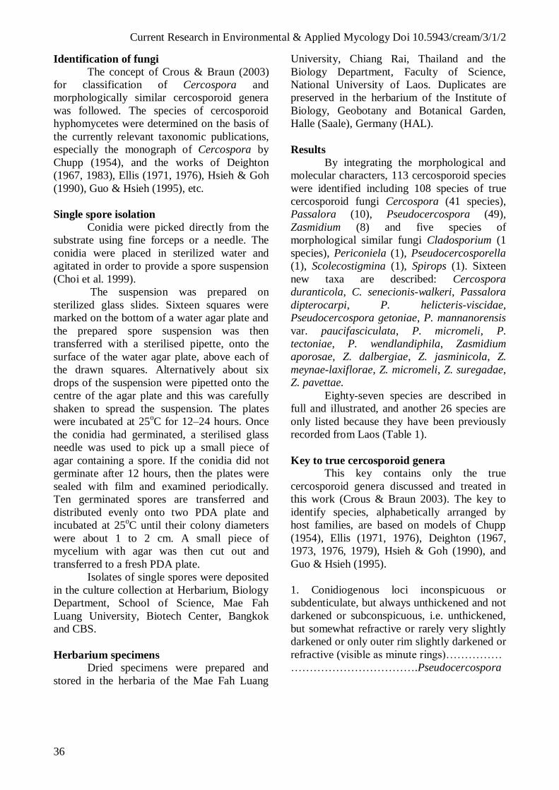
(5) Cercospora asparagi Sacc., Michelia 1: 88,
1877. Figs 10–11.
= Cercospora caulicola G. Winter. J.
Mycol. 1: 125, 1885.
= Cercosporina asparagicola Speg.,
Anal. Mus. Buenos Aires 20: 424, 1910.
Cercosporina asparagicola (Speg.)
Vassiljevsky, in Vassiljevsky & Karakulin,
Fungi imperfecti parasitici. 1. Hyphomycetes:
296, 1937.
Leaf spots on cladodes and branches,
small oval to elliptic in shape, 0.5–2 mm diam.,
pale tan to dingy grey in the centre, and with a
fairly wide reddish brown margin. Caespituli
amphigenous, scattered, dark brown. Mycelium
internal. Stromata well-developed, oval to
ellipsoidal, substomatal, up to 32 µm diam.,
brown, composed of swollen hyphal cells,
subglobose, rounded and angular in outline, 5–
13 µm wide ( x = 8.8 µm, n = 13),
Current Research in Environmental & Applied Mycology Doi 10.5943/cream/3/1/2
51
brown to dark brown, wall 0.5–0.8 µm wide
( x = 0.6 µm, n = 13), smooth. Conidiophores
solitary or fasciculate, arising from stromata (2–
15 per fascicle), emerging through stomata,
unbranched, straight to curved, cylindrical, 28–
63 × 4–6 µm ( x = 43.5 × 4.56 µm, n = 10), 1–3-
septate, distance between septa 4–36 µm ( x =
15 µm, n = 17), medium brown, paler at the
apex, wall 0.4–0.5 µm wide ( x = 0.49 µm, n =
10), smooth, 0–1-times geniculate, width
uniform; conidiogenous cells integrated,
terminal, cylindrical, 10–32 × 4–5 µm ( x = 21.4
× 4.5 µm, n = 11), pale brown; conidiogenous
loci conspicuous, subcircular, 1.5–3 µm wide
( x = 2.5 µm, n = 25), dark brown, wall 0.3–0.5
µm thick ( x = 0.4 µm, n = 25). Conidia solitary,
acicular, straight to curved, 54–112 × 4–5 µm
( x = 80 × 4 µm, n = 5), 1–8-septate, hyaline,
thin-walled, smooth, tip subotuse to acute, base
truncate to obconically truncate, hila thickened
and darkened, 2–3 µm wide ( x = 2.5 µm, n =
5), wall of the hila 0.3–0.5 µm ( x = 0.4 µm, n =
5) thick.
Hosts – Asparagus officinalis L., A.
plumosus (Baker) Oberm. (Asparagaceae).
Distribution: Africa: Ghana, Kenya,
Malawi, South Africa, Zimbabwe; Asia:
Brunei, Cambodia, China, Hong Kong, India,
Israel, Japan, Korea, Laos, Malysia, Nepal,
Pakistan, Taiwan, Thailand; Europe: Italy,
Ukraine, Serbia; North America and West
Indies: Cuba, USA (CA, FL, HI, IL, NC, NE).
Oceania: Solomon Islands; South America:
Argentina, Brazil, Colombia.
Material examined – Vientiane Capital,
Xaythany District, Xay Village, garden, on
leaves of Asparagus officinalis, 30 June 2006,
P. Phengsintham (P57).
Notes – Crous & Braun (2003)
considered Cercospora asparagi close to or
identical with C. apii s. lat. In the Laos
collection conidiophores were fasciculate, 28–
63 × 4–6 µm and conidia are 54–112 × 4–5 µm,
which is similar to those reported by Ellis
(1976) [conidiophores 40–150 × 3–8 µm and
conidia 80–130 × 4–5 µm], Hsieh & Goh
(1990) [conidiophores 30–170 × 4–7 µm and
conidia 35–130 × 2.5–5 µm] and Chupp (1954).
Literature – Saccado (1886: 477),
Chupp (1954: 343), Ellis (1976: 270), Hieh &
Goh (1990: 208), Crous & Braun (2003: 68).
Fig. 10 – Cercospora asparagi from Asparagus
officinalis: 1. Stroma with attached
conidiophores. 2–3. Conidiophores. 4–5.
Conidia. Bar = 10 µm.
Fig. 11 – Cercospora asparagi from Asparagus
officinalis: 1–2. Stroma with attached
conidiophores. 3. Conidium. Bars = 10 µm.
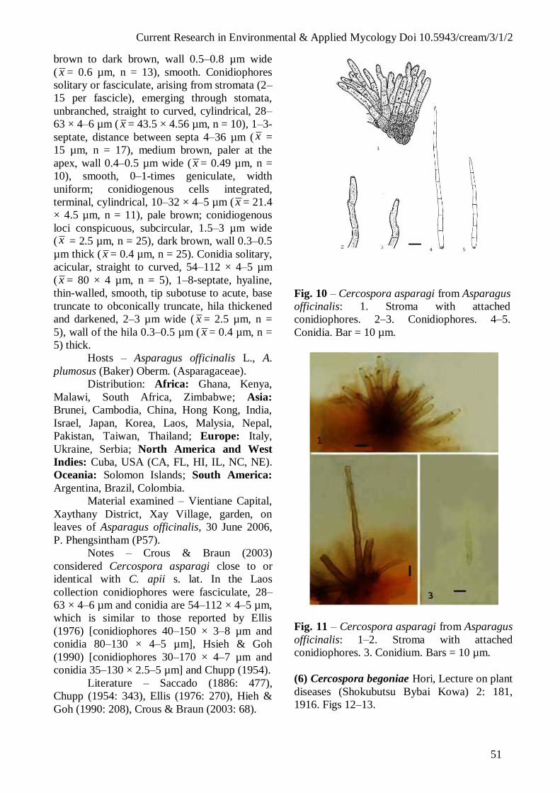
(6) Cercospora begoniae Hori, Lecture on plant
diseases (Shokubutsu Bybai Kowa) 2: 181,
1916. Figs 12–13.

Current Research in Environmental & Applied Mycology Doi 10.5943/cream/3/1/2
52
(= Cercospora apii s. lat.)
Leaf spots small to fairly large,
suborbicular to irregular, 1–4 mm in diam.,
grey-brown in the centre, and with dark brown
margin. Caespituli amphigenous, scattered, dark
brown. Mycelium internal, inconspicuous.
Stromata developed, small to medium-sized,
globular to subglobular, substomatal and
intraepidermal, 10–15 µm in diam. ( x = 12.5
µm, n = 5), dark brown to black in mass,
composed of swollen hyphal cells, subglobose,
rounded to angular in outline, 5–8 µm wide ( x
= 6.7 µm, n = 11), brown to dark brown, wall
0.5–0.8 µm wide ( x = 0.7 µm, n = 11), smooth.
Conidiophores fasciculate, arising from
stromata (2–3 per fascicle), emerging through
stomata, unbranched, straight to curved,
cylindrical, 55–93 × 4–5 µm ( x = 65.3 × 4.4
µm, n = 11), 2–4-septate, distance between
septa 9–36 µm ( x = 18 µm, n = 30), medium
brown, paler at the apex, wall 0.5–0.8 µm wide
( x = 0.59 µm, n = 30), smooth, 0–3-times
geniculate. Conidiogenous cells terminal,
cylindrical, 17–36 × 3.5–4 µm ( x = 28.3 × 3.83
µm, n = 7), pale brown; conidiogenous loci
conspicuous, subcircular, 2–3 µm wide ( x =
2.6 µm, n = 7), wall 0.5–0.8 µm thick ( x = 0.62
µm, n = 7), thickened and darkened. Conidia
solitary, acicular to obclavate, straight to
curved, 57–150 × 2–3 µm (x = 103.5 × 2.5 µm,
n = 8), 7–12-septate, hyaline to subhyaline,
thin-walled 0.3–0.5 µm ( x = 0.35 µm, n = 8),
smooth, tip acute, base truncate to obconically
truncate; hila thickened and darkened 1.5–2 µm
wide ( x = 1.75 µm, n = 8), wall of the hila 0.3–
0.5 µm ( x = 0.37 µm, n = 8) thick.
Hosts – Begonia argenteo-guttata M.
Lemoine, B. evansiana C. Andrews, B. inflata
Clarke, B. palmata D. Don, B. rex Putz., B. rex-
culturum hybrid, B. semperflorens Link & Otto,
Begonia sp. (Begoniaceae).
Distribution – Africa: Zimbabwe; Asia:
Brunei, China, India, Japan, Laos, Malaysia,
Taiwan, Thailand; Europe: Poland; North
America and West Indies: USA (FL).
Material examined – Xiangkhouang
Province, Paek District, Phonsavane Village, on
leaves of Begonia inflata, 3 January 2010, P.
Phengsintham (P517).
Notes – The collection from Laos agrees
with the description of Cercospora begoniae by
Chupp (1954) and Hsieh & Goh (1990)
[conidiophores 20–200 × 3–5 µm and conidia
50–300 × 2–3.5 µm].
Literature – Chupp (1954: 79), Katsuki
(1965: 14), Hsieh & Goh (1990: 42), Crous &
Braun (2003: 78).
Fig. 12 – Cercospora begoniae on Begonia
inflata from leaf spots: 1. Stroma with
attached conidiophores. 2–4. Conidiophores.
5. Conidium. Bar = 10 µm.
(7) Cercospora bidentis Tharp, Mycologia 9:
108, 1917. Figs 14–15.
= Cercospora bidentis Marchal &
Stayaert, Bull. Soc. Roy. Bot. Belgiques 61:
167, 1954.
= Cercospora bidentis-pilosae
Sawada, Rep. Gov. Agric. Res. Inst. Taiwan
85: 98, 1943, nom. inval.
Leaf spots orbicular to irregular, 2–10
mm diam., dark brown to black in the centre,
and with brown to dark brown margin.
Current Research in Environmental & Applied Mycology Doi 10.5943/cream/3/1/2
53
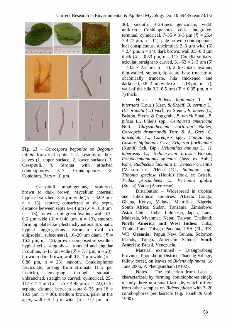
Fig. 13 – Cercospora begoniae on Begonia
inflata from leaf spots: 1–2. Lesions on host
leaves (1. upper surface, 2. lower surface). 3.
Caespituli. 4. Stroma with attached
conidiophores. 5–7. Conidiophores. 8.
Conidium. Bars = 10 µm.
Caespituli amphigenous, scattered,
brown to dark brown. Mycelium internal;
hyphae branched, 3–5 µm wide ( x = 3.69 µm,
n = 13), septate, constricted at the septa,
distance between septa 6–14 µm ( x = 10.8 µm,
n = 13), brownish or green-hyaline, wall 0.3–
0.5 µm wide ( x = 0.46 µm, n = 13), smooth,
forming plate-like plectenchymatous stromatic
hyphal aggregations. Stromata oval to
ellipsoidal, substomatal, 10–20 µm diam. ( x =
16.5 µm, n = 15), brown, composed of swollen
hyphal cells, subglobose, rounded and angular
in outline, 5–11 µm wide ( x = 7.7 µm, n = 23),
brown to dark brown, wall 0.5–1 µm wide ( x =
0.88 µm, n = 23), smooth. Conidiophores
fasciculate, arising from stromata (1–2 per
fascicle), emerging through stomata,
unbranched, straight to curved, cylindrical, 25–
117 × 4–7 µm ( x = 75 × 4.95 µm, n = 22), 0–5-
septate, distance between septa 8–35 µm ( x =
19.9 µm, n = 30), medium brown, paler at the
apex, wall 0.5–1 µm wide ( x = 0.7 µm, n =
30), smooth, 0–2-times geniculate, width
uniform. Conidiogenous cells integrated,
terminal, cylindrical, 7–35 × 3–5 µm (x = 25.4
× 4.27 µm, n = 11), pale brown; conidiogenous
loci conspicuous, subcircular, 2–3 µm wide ( x
= 2.4 µm, n = 14), dark brown, wall 0.5–0.8 µm
thick ( x = 0.53 µm, n = 11). Conidia solitary,
acicular, straight to curved, 31–62 × 2–3 µm ( x
= 43.8 × 2.2 µm, n = 7), 2–6-septate, hyaline,
thin-walled, smooth, tip acute, base truncate to
obconically truncate, hila thickened and
darkened, 0.8–2 µm wide ( x = 1.18 µm, n = 7),
wall of the hila 0.3–0.5 µm ( x = 0.35 µm, n =
7) thick.
Hosts – Bidens bipinnata L., B.
biternata (Lour.) Merr. & Sherff, B. cernua L.,
B. coronata (L.) Fisch. ex Steud., B. laevis (L.)
Britton, Sterns & Poggenb., B. nashii Small, B.
pilosa L., Bidens spp., Centaurea americana
Nutt., Chrysanthemum hortorum Bailey,
Coreopsis drummondii Torr. & A. Grey, C.
lanceolata L., Coreopsis spp., Conyza sp.,
Cosmos bipinnatus Cav., Erigeron floribundus
(Kunth) Sch. Bip., Helianthus annuus L., H.
tuberosus L., Helichrysum brassii Brenan,
Pseudelephantopus spicatus (Juss. ex Aubl.)
Rohr, Rudbeckia laciniata L., Senecio cruentus
(Masson ex L'Hér.) DC., Solidago spp..
Tithonia speciosa (Hook.) Hook. ex Griseb.,
Tridax procumbens L., Vernonia glabra
(Steetz) Vatke (Asteraceae).
Distribution – Widespread in tropical
and subtropical countries. Africa: Congo,
Ghana, Kenya, Malawi, Mauritius, Nigeria,
South Africa, Sudan, Tanzania, Zimbabwe;
Asia: China, India, Indonesia, Japan, Laos,
Malaysia, Myanmar, Nepal, Taiwan, Thailand;
North America and West Indies: Cuba,
Trinidad and Tobago Panama, USA (FL, TX,
WI); Oceania: Papua New Guinea, Solomon
Islands, Tonga, American Samoa; South
America: Brazil, Venezuela.
Material examined – Luangprabang
Province, Phoukhoun District, Phadeng Village,
fallow forest, on leaves of Bidens bipinnata, 18
June 2006, P. Phengsintham (P102).
Notes – The collection from Laos is
characterized by forming conidiophores singly
or only three in a small fascicle, which differs
from other samples on Bidens pilosa with 3–20
conidiophores per fascicle (e.g. Hsieh & Goh
1990).

Current Research in Environmental & Applied Mycology Doi 10.5943/cream/3/1/2
54
Literature – Saccado (1931: 871; 1972:
1369), Chupp (1954: 123–124), Katsuki
(1965: 20), Vasudeva (1963: 50), Ellis (1976:
250), Hsieh & Goh (1990: 62).
Fig. 14 – Cercospora bidentis on Bidens
pilosa: 1–3. Conidiophores. 4–6. Conidia.
Bar = 10 µm.
(8) Cercospora brassicicola Henn., Bot. Jahrb.
Syst. 37: 166, 1905. Figs 16–17.
= Cercospora brassicae-campestris
Rangel, Arq. Mus. Nac., Rio de Janeiro 18: 16,
1917.
Cercosporina brassicae-campestris
(Rangel) Sacc., Syll. Fung. 25: 899, 1931.
= Cercospora brassicae-junceae
Sawada (Brassicae-yunceae), Special Publ.
Coll. Agric. Natl. Taiwan Univ. 8: 212, 1959
(nom. nud.).
= Cercospora bloxami auct. sensu E.
Young, Mycologia 8: 43, 1916.
Leaf spot circular to angular or a long
the margin of the leaves, 1–25 mm diam., pale
green or pale brown to dark brown or black in
the center, and with pale green or yellowish
margin. Caespituli amphigenous, dense, grey.
Fig. 15 – Cercospora bidentis on Bidens pilosa
from leaf spots: 1. Lesions on host leaves (1.
upper surface, 2. lower surface). 3. Internal
mycelium. 4–5. Stromata with attached
conidiophores. 6–10. Conidia. Bars 1 = 10 mm,
3–10 = 10 µm.
Mycelium internal; hyphae branched, 1–4 µm
wide ( x = 3.1 µm, n = 30), septate, constricted
at the septa, distance between septa 5–15 µm
( x = 8.17 µm, n = 30), subhyaline or
olivaceous brown, wall 0.5–0.9 µm wide (x =
0.61 µm, n = 23), smooth. Stromata
substomatal, intraepidermal, oval, ellipsoidal,
15–30 µm diam. ( x = 20.86 µm, n = 8), dark
brown, stromatal cells oval, angular, obclavate,
3–9 µm diam. ( x = 6.47 µm, n = 30), dark
brown, wall 0.5–1 µm wide ( x = 0.7 µm, n =
30). Conidiophores fasciculate, arising from
stromata (2–20 per fascicle), emerging through
stomata, cylindrical, 15–232 × 4–6 µm ( x =
109.4 × 5 µm, n = 30), 1–8-septate, distance
between septa 9–58 µm ( x = 25 µm, n = 30),
pale olivaceous to medium brown, oldest ones
uniform in colour and width, wall 0.5–1 µm
wide ( x = 0.8 µm, n = 30), smooth, 1–2 times
geniculate; conidiogenous cells terminal, 13–68
× 4–6 µm ( x = 33.27 × 4.7 µm, n = 30);
conidiogenous loci conspicuous, subcircular in
outline, planate, 2–4 µm wide ( x = 2.83 µm, n
= 30), thickened, darkened. Conidia solitary,
acicular, curved or undulate, 30–288 × 1.5–5
µm ( x = 120 × 3.52 µm, n = 30), 3–20-septate,
Current Research in Environmental & Applied Mycology Doi 10.5943/cream/3/1/2
55
hyaline, wall 0.25–0.5 µm wide ( x = 0.27 µm,
n = 30), smooth, tip acute, base truncate to
obconically truncate, hila 1–4 µm wide ( x =
2.47 µm, n = 30), wall of the hila 0.25–0.5 µm
thick ( x = 0.27 µm, n = 30), thickened and
darkened.
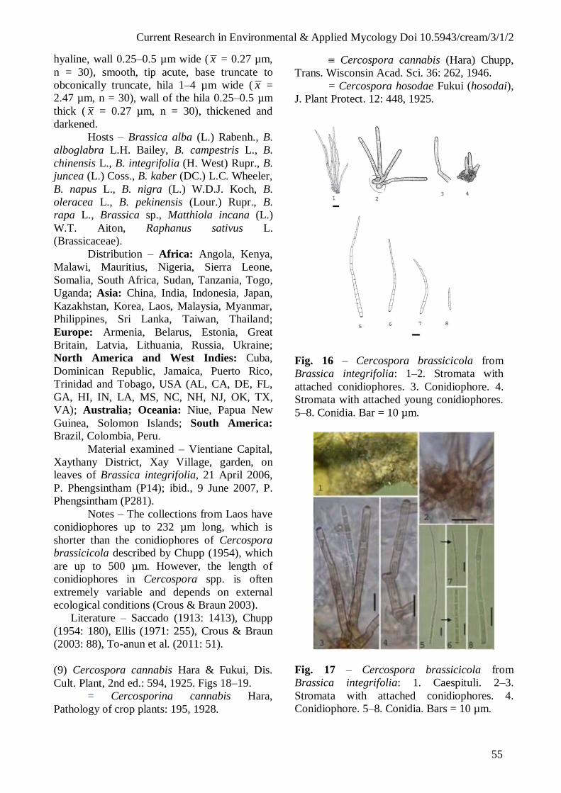
Hosts – Brassica alba (L.) Rabenh., B.
alboglabra L.H. Bailey, B. campestris L., B.
chinensis L., B. integrifolia (H. West) Rupr., B.
juncea (L.) Coss., B. kaber (DC.) L.C. Wheeler,
B. napus L., B. nigra (L.) W.D.J. Koch, B.
oleracea L., B. pekinensis (Lour.) Rupr., B.
rapa L., Brassica sp., Matthiola incana (L.)
W.T. Aiton, Raphanus sativus L.
(Brassicaceae).
Distribution – Africa: Angola, Kenya,
Malawi, Mauritius, Nigeria, Sierra Leone,
Somalia, South Africa, Sudan, Tanzania, Togo,
Uganda; Asia: China, India, Indonesia, Japan,
Kazakhstan, Korea, Laos, Malaysia, Myanmar,
Philippines, Sri Lanka, Taiwan, Thailand;
Europe: Armenia, Belarus, Estonia, Great
Britain, Latvia, Lithuania, Russia, Ukraine;
North America and West Indies: Cuba,
Dominican Republic, Jamaica, Puerto Rico,
Trinidad and Tobago, USA (AL, CA, DE, FL,
GA, HI, IN, LA, MS, NC, NH, NJ, OK, TX,
VA); Australia; Oceania: Niue, Papua New
Guinea, Solomon Islands; South America:
Brazil, Colombia, Peru.
Material examined – Vientiane Capital,
Xaythany District, Xay Village, garden, on
leaves of Brassica integrifolia, 21 April 2006,
P. Phengsintham (P14); ibid., 9 June 2007, P.
Phengsintham (P281).
Notes – The collections from Laos have
conidiophores up to 232 µm long, which is
shorter than the conidiophores of Cercospora
brassicicola described by Chupp (1954), which
are up to 500 µm. However, the length of
conidiophores in Cercospora spp. is often
extremely variable and depends on external
ecological conditions (Crous & Braun 2003).
Literature – Saccado (1913: 1413), Chupp
(1954: 180), Ellis (1971: 255), Crous & Braun
(2003: 88), To-anun et al. (2011: 51).
(9) Cercospora cannabis Hara & Fukui, Dis.
Cult. Plant, 2nd ed.: 594, 1925. Figs 18–19.
= Cercosporina cannabis Hara,
Pathology of crop plants: 195, 1928.
Cercospora cannabis (Hara) Chupp,
Trans. Wisconsin Acad. Sci. 36: 262, 1946.
= Cercospora hosodae Fukui (hosodai),
J. Plant Protect. 12: 448, 1925.
Fig. 16 – Cercospora brassicicola from
Brassica integrifolia: 1–2. Stromata with
attached conidiophores. 3. Conidiophore. 4.
Stromata with attached young conidiophores.
5–8. Conidia. Bar = 10 µm.
Fig. 17 – Cercospora brassicicola from
Brassica integrifolia: 1. Caespituli. 2–3.
Stromata with attached conidiophores. 4.
Conidiophore. 5–8. Conidia. Bars = 10 µm.

Current Research in Environmental & Applied Mycology Doi 10.5943/cream/3/1/2
56
Leaf spots circular to angular or a long
the margin of the leaves, 1–15 mm diam., pale
green or pale brown to dark brown or black in
the center, and with pale green or yellowish
margin. Caespituli amphigenous, dense, grey.
Mycelium internal; hyphae branched, 2–3 µm
wide ( x = 2.66 µm, n = 10), septate, constricted
at the septa, distance between septa 5–10 µm
( x = 7.33 µm, n = 10), subhyaline or
olivaceous brown, wall 0.3–0.5 µm wide ( x =
0.36 µm, n = 10), smooth. Stromata
substomatal, intraepidermal, oval, ellipsoidal,
12–22 µm diam. ( x = 20.5 µm, n = 5), dark
brown, stromatal cells oval, angular, obclavate,
4–6 µm diam. ( x = 5.2 µm, n = 7), dark brown,
wall 0.5–0.8 µm wide ( x = 0.62 µm, n = 7).
Conidiophores solitary or fasciculate, arising
from stromata (2–4 per fascicle), emerging
through stomata, cylindrical, 12–105 × 3–5 µm
( x = 50.6 × 4 µm, n = 9), 0–7-septate, distance
between septa 8–22 µm ( x = 14.8 µm, n = 12),
pale olivaceous to medium brown, oldest ones
uniform in colour and width, wall 0.5–0.8 µm
wide ( x = 0.6 µm, n = 12), smooth, 1–2 times
geniculate; conidiogenous cells terminal, 12–22
× 4–6 µm ( x = 16.22 × 4.7 µm, n = 6);
conidiogenous loci conspicuous, subcircular in
outline, planate, 2–3.5 µm wide ( x = 3.63 µm,
n = 6), thickened, darkened. Conidia solitary,
acicular, curved or undulate, 83–125 × 2.5–3
µm ( x = 106.33 × 2.8 µm, n = 7), 6–9-septate,
hyaline, wall 0.25–0.3 µm wide ( x = 0.28 µm,
n = 7), smooth, tip acute to subacute, base
truncate to obconically truncate, hila 1.5–2.5
µm wide ( x = 2 µm, n = 7), wall of the hila
0.25–0.3 µm thick ( x = 0.27 µm, n = 7),
thickened and darkened.
Hosts – Cannabis sativa L., Humulus
lupulus L. (Cannabaceae).
Distribution – Asia: China, India, Japan,
Nepal, Laos; North America and West Indies:
USA (MO, WI); South America: Colombia.
Material examined – Khammoune
Province, Nakai District, Nahao Village,
garden, on leaves of Cannabis sativa, 20 July
2011, P. Phengsintham (P646).
Notes – The collection from Laos has
conidiophores up to 125 µm long, which is
longer than the conidiophores of Cercospora
cannabis described by Chupp (1954)
[conidiophores 10–100 × 3.5–5.5 µm and
conidia 20–90 × 2–4 µm].
Literature – Chupp (1954: 394),
Vasudeva (1963: 64), Katsuki (1965: 47).
Fig. 18 – Cercospora cannabis on
Cannabis sativa: 1. Stroma with attached
conidiophores. 2–3. Conidiophores. 4.
Stroma with attached conidiophores 5–6.
Conidia. Bar = 10 µm.
Fig. 19 – Cercospora cannabis on Cannabis
sativa: 1–3. Stroma with attached
conidiophores. 4–5. Conidiophores. 6. Conidia.
Bars = 10 µm.

Current Research in Environmental & Applied Mycology Doi 10.5943/cream/3/1/2
57
(10) Cercospora capsicigena Bhartiya, R,
Dubey & S.K. Singh, Indian Phytopathol. 5:
149, 2000. Figs 20–21.
(= Cercospora apii s. lat.)
Leaf spots suborbicular to irregular, 2–5
mm in diam., grey-brown in the centre, and
with dark brown margin. Caespituli
amphigenous, scattered, whitish or grey.
Mycelium internal; hyphae branched, 2–3 µm
wide ( x = 2.25 µm, n = 9), septate, constricted
at the septa, distance between septa 4–9 µm ( x
= 7 µm, n = 9), brownish or green-hyaline, wall
0.3–0.5 µm wide ( x = 0.45 µm, n = 9), smooth,
forming plate-like plectenchymatous stromatic
hyphal aggregations. Stromata developed, small
to medium-sized, globular to subglobular,
substomatal and intraepidermal, 18–30 µm in
diam. ( x = 24 µm, n = 4), dark brown to black
in mass, composed of swollen hyphal cells,
subglobose, rounded to angular in outline, 4–7
µm wide ( x = 6 µm, n = 30), brown to dark
brown, wall 0.5–0.8 µm wide ( x = 0.54 µm, n
= 30), smooth. Conidiophores solitary or
fasciculate, arising from stromata (2–7 per
fascicle), emerging through stomata,
unbranched, straight to curved, cylindrical, 21–
63 × 4–6 µm ( x = 39.8 × 5 µm, n = 15), 1–3-
septate, not geniculate, distance between septa
5–34 µm ( x = 14.4 µm, n = 30), medium
brown, paler at the apex, wall 0.5–0.8 µm wide
( x = 0.6 µm, n = 30), smooth. Conidiogenous
cells terminal, cylindrical, 13–34 × 4–5 µm ( x
= 21.3 × 4.14 µm, n = 7), pale brown;
conidiogenous loci conspicuous, subcircular,
1.5–3 µm wide ( x = 2.07 µm, n = 5), wall 0.5–
0.8 µm thick ( x = 0.7 µm, n = 5), thickened
and darkened. Conidia solitary, acicular to
obclavate, straight to curved, 49–70 × 3–4 µm
( x = 54.75 × 3.25 µm, n = 11), 4–5-septate,
hyaline to subhyaline, thin-walled 0.3–0.5 µm
( x = 0.35 µm, n = 11), smooth, tip acute, base
truncate to obconically truncate; hila thickened
and darkened, 1.5–3 µm wide ( x = 2.12 µm, n
= 11), wall of the hila 0.3–0.5 µm ( x = 0.35
µm, n = 11) thick.
Colonies on PDA after 3 weeks at 25°C
with grey-brown mycelium, reaching 6–8 mm
diam.
Hosts – Capsicum annuum L.
(Solanaceae).
Distribution – Asia: India, Laos,
Thailand.
Material examined – Bolikhamsay
Province, Lak 20 District, Nongsong Village,
garden, on leaves of Capsicum annuum, 17 July
2008, P. Phengsintham (P325); Oudomxay
Province, Houn District, C. annuum, 6 October
2008, P. Phengsintham (P380). GenBank
accession no (ITS, KC677884; LSU,
KC677918).
Notes – The collections from Laos are
close to C. apii complex (Crous & Braun,
2003).
Literature – Crous & Braun (2003: 103).
Fig. 20 – Cercospora capsicigena on Capsicum
annuum from leaf spots: 1. Stroma with attached
conidiophores. 2–3. Conidiophores. 4–7.
Conidia. Bar = 10 µm.
(11) Cercospora cocciniae Munjal, Lall &
Chona, Indian Phytopathol. 12: 86, 1959. Figs
22–23.
Leaf spots oval to elliptic in shape, 1–6
mm diam. ( x = 4 µm, n = 11), white-grey in the
centre, and with a white-greyish margin.
Caespituli amphigenous, scattered, dark brown.
Mycelium internal, inconspicuous. Stromata
oval to ellipsoidal, substomatal, 15–35 µm diam.
( x = 24.1 µm, n = 18), brown, composed of
swollen hyphal cells, subglobose, angular in out-

Current Research in Environmental & Applied Mycology Doi 10.5943/cream/3/1/2
58
Fig. 21 – Cercospora capsicigena on Capsicum
annuum from leaf spots: 1–2. Lesions on host
leaf (1. upper surface. 2. lower surface). 3.
Caespituli. 4. Stroma with attached
conidiophores. 5–6. Conidiophores. 7–10.
Conidia. 11. Culture. Bars 1–2 = 10 mm, 4–10
= 10 µm.
line, 3–7 µm wide ( x = 5.1 µm, n = 30), brown
to dark brown, wall 0.5–1 µm wide ( x = 0.79
µm, n = 25), smooth. Conidiophores solitary or
fasciculate, arising from stromata (2–17 per
fascicle), emerging through stomata,
unbranched, straight to curved, cylindrical, 7–
92 × 4–7 µm ( x = 32 × 5 µm, n = 26), 0–4-
septate, distance between septa 7–35 µm ( x =
16.28 µm, n = 21), medium brown, paler at the
apex, wall 0.8–1 µm wide ( x = 0.82 µm, n =
26), smooth, 0–1-times geniculate, width
uniform. Conidiogenous cells integrated,
terminal, cylindrical, 10–24 × 3–5 µm ( x = 17
× 4.21 µm, n = 17), pale brown; conidiogenous
loci conspicuous, subcircular, 1.5–3 µm wide
( x = 2.45 µm, n = 30), dark brown, wall 0.8–1
µm thick ( x = 0.84 µm, n = 30). Conidia
solitary, acicular, straight to curved, 24–180 ×
3–7 µm (x = 53.61 × 3.92 µm, n = 26), 3–13-
septate, hyaline, thin-walled, smooth, tip
subotuse to acute, base truncate to obconically
truncate, hila thickened and darkened, 1.5–3 µm
wide ( x = 2.21 µm, n = 19), wall of the hila
0.5–1 µm ( x = 0.81 µm, n = 19) thick.
Hosts – Coccinia indica Wight & Arn.,
Momordica charantia L. (Cucurbitaceae).
Distribution – Asia: Brunei, India, Laos,
Pakistan, Thailand.
Material examined – Vientiane Capital,
Xaythany District, Xay Village, on leaves of
Coccinia indica, Xay Village, 28 June 2006, P.
Phengsintham (P106); ibid., on leaves of C.
indica, 28 December 2008, P. Phengsintham
(P392).
Notes – Crous & Braun (2003)
classified this species as morphologically
distinct from C. apii s. str. by having obclavate
conidia. The Laos collections have distinctly
obclavate conidia, with long obconically
truncate bases. The conidiophores and conidia
are similar to those reported from Brunei
(Braun & Sivapalan 1999).
Literature – Chupp (1954: 65), Hsieh &
Goh (1990: 98), Braun & Sivapalan (1999),
Crous & Braun (2003: 129).
Fig. 22 – Cercospora cocciniae on Coccinia
indica: 1. Stroma with attached conidiophores.
2–4. Conidiophores. 5–8. Conidia. Bars = 10
µm.

Current Research in Environmental & Applied Mycology Doi 10.5943/cream/3/1/2
59
Fig. 23 – Cercospora cocciniae on Coccinia
indica from leaf spots: 1–2. Lesions on host
leaves (1. upper surface, 2. lower surface). 3.
Stromata with Conidiophore. 4–5.
Conidiophores. 6–8. Conidia. 9. Based of
conidia. Bars 1–2 = 10 mm, 3–9 = 10 µm.
(12) Cercospora duranticola sp. nov.
Figs 24–25.
MycoBank, MB 801726.
Diagnosis – Differs from the
Cercospora apii s. lat. complex in having
uniformly short conidiophores arising from
well-developed stromata and acicular to
narrowly obclavate conidia with truncate to
distinctly obconically truncate base.
Leaf spots circular or angular, 1–4 mm
diam., grayish brown to brown in the centre,
and with brown to dark brown margin.
Caespituli amphigenous, scattered, yellow to
brown. Mycelium internal; hyphae branched, 2–
4 µm wide ( x = 3 µm, n = 6), septate,
constricted at the septa, distance between septa
5–10 µm ( x = 7.1 µm, n = 6), brownish or
green-hyaline, wall 0.3–0.5 µm wide ( x = 0.36
µm, n = 6), smooth, forming plate-like
plectenchymatous stromatic hyphal
aggregations. Stromata well-developed, oval to
ellipsoidal, substomatal, 17–52 µm diam. ( x =
30.3 µm, n = 8), brown, composed of swollen
hyphal cells, subglobose, rounded and angular
in outline, 5–10 µm wide ( x = 6.6 µm, n = 27),
brown to dark brown, wall 0.5–1 µm wide ( x =
0.67 µm, n = 27), smooth. Conidiophores
fasciculate, arising from stromata (1–11 per
fascicle), emerging through stomata,
unbranched, straight to curved, cylindrical to
distinctly geniculate, uniformly short, 17–35 ×
4–5 µm ( x = 23.1 × 4.6 µm, n = 11), 0–2-
septate, distance between septa 5–25 µm ( x =
15.9 µm, n = 14), medium brown, paler at the
apex, wall 0.3–0.5 µm wide ( x = 0.47 µm, n =
14), smooth, 0–2-times geniculate, pale and
more narrow towards the tip. Conidiogenous
cells integrated, terminal, cylindrical, 8–25 × 3–
5 µm ( x = 17.3 × 4.28 µm, n = 9), pale brown;
conidiogenous loci conspicuous, subcircular, 1–
3 µm wide ( x = 1.73 µm, n = 26), dark brown,
wall 0.3–0.8 µm thick ( x = 0.31 µm, n = 26).
Conidia solitary, acicular to narrowly obclavate,
straight to curved, 24–144 × 2–4 µm ( x = 52 ×
3.35 µm, n = 10), 3–13-septate, hyaline, thin-
walled, smooth, tip acute, base truncate to
obconically truncate, hila thickened and
darkened, 0.5–2 µm wide ( x = 1.64 µm, n =
10), wall of the hila 0.3–0.5 µm (x = 0.32 µm,
n = 10) thick.
Hosts – Duranta repens L.
(Verbenaceae).
Distribution – Asia: Laos.
Material examined – Xiengkhouang
Province, Phonsavanh District, Phonsavanh
Village, urban area, on leaves of Duranta
repens, 3 January 2010, P. Phengsintham
(P515, MFLU12-2197, holotype). GenBank
accession no (ITS, KC677885).
Notes – Cercospora durantae Chupp &
A.S. Mull. is an invalid name. The type material
was re-examined and the conidiogenous loci
and hila at the base of conidia found to be
unthickened and not darkened; the species was
validated as Pseudocercospora durantae Pons,
U. Braun & Crous (in Crous & Braun 2003).
The collection from Laos is quite distinct and
represents a true Cercospora s. str. different
from C. apii s. lat. (sensu Crous & Braun 2003)
by having uniformly short conidiophores
arising from well-developed stromata and
acicular to narrowly obclavate conidia with
truncate to distinctly obconically truncate base.
Cercospora on Duranta in Laos warrants to be
considered a separate species.

Current Research in Environmental & Applied Mycology Doi 10.5943/cream/3/1/2
60
Fig. 24 – Cercospora duranticola on Duranta
repens: 1–2. Stroma with attached
conidiophores. 3. Conidiophore. 4–10.
Conidia. Bar = 10 µm.
Fig. 25 – Cercospora duranticola on Duranta
repens from leaf spots: 1. Lesions on host
leaves (1. upper surface, 2. lower surface). 3.
Stroma with attached conidiophores. 4–7.
Conidia. Bars 1 = 10 mm, 3–7 = 10 µm.
(13) Cercospora erechtitis G.F. Atk., J. Elisha
Mitchell Sci. Soc. 8: 66, 1892. Figs 26–27.
= Cercospora erechtiticola Sawada,
nom. nud. (Chupp 1954: 134).
Leaf spots circular or suborbicular, 1–12
mm diam., greyish brown to dark brown in the
centre, and brown to dark brown margin.
Caespituli amphigenous, scattered, dark brown.
Mycelium internal; hyphae branched, 2–4 µm
wide ( x = 3 µm, n = 30), septate, constricted at
the septa, distance between septa 5–13 µm (x =
9.37 µm, n = 30), brownish or green-hyaline,
wall 0.3–0.5 µm wide ( x = 0.37 µm, n = 30),
smooth, forming plate-like plectenchymatous
stromatic hyphal aggregations. Stromata well
developed, oval to ellipsoidal, substomatal, 15–
30 µm diam. ( x = 20 µm, n = 4), brown,
composed of swollen hyphal cells, subglobose,
rounded and angular in outline, 5–14 µm wide
(x = 9.23 µm, n = 30), brown to dark brown,
wall 0.8–1 µm wide ( x = 0.89 µm, n = 30),
smooth. Conidiophores solitary or fasciculate,
arising from stromata (2–5 per fascicle),
emerging through stomata, unbranched, straight
to curved, cylindrical, 4–35 × 5–8 µm ( x =
24.1 × 5.68 µm, n = 19), 0–1-septate, distance
between septa 7–22 µm ( x = 15.2 µm, n = 19),
medium brown, paler at the apex, wall 0.5–1
µm wide ( x = 0.65 µm, n = 19), smooth, 0–1-
times geniculate, width uniform. Conidiogenous
cells integrated, terminal, cylindrical, 12–25 ×
5–6 µm ( x = 19.1 × 5.5 µm, n = 14), pale
brown; conidiogenous loci conspicuous,
subcircular, 1.5–3 µm wide (x = 2.11 µm, n =
19), dark brown, wall 0.5–0.8 µm thick ( x =
0.6 µm, n = 19). Conidia solitary, acicular,
straight to curved, 28–83 × 2–3 µm (x = 62 ×
2.67 µm, n = 5), 3–7-septate, hyaline, thin-
walled, smooth, tip acute, base truncate to
obconically truncate, hila thickened and
darkened, 2 µm wide ( x = 2 µm, n = 5), wall of
the hila 0.3–0.5 µm ( x = 0.37 µm, n = 5) thick.
Hosts – Erechtites hieraciifolius (L.)
Raf. ex DC., E. valerianifolius (Link ex
Spreng.) DC., Hieracium aurantiacum L.,
Ligularia sp. (Asteraceae).
Distribution – Asia: Laos, Taiwan.
South America: Colombia.
Material examined – Vientiane Capital,
Xaythany District, Xay Village, garden, on
leaves of Erechtites valerianifolius, 8 May
2006, P. Phengsintham (P35).

Current Research in Environmental & Applied Mycology Doi 10.5943/cream/3/1/2
61
Notes – Morphologically Cercospora
erechtitis is indistinguishable from C. apii s. lat.
(Crous & Braun 2003). However, inoculation
results (biological specialization) and molecular
sequence analyses are not available, and hence,
the taxonomic status of this species remains
unclear. The conidiophores and conidia of the
Cercospora from Laos (conidiophores 4–35 ×
5–8 µm, conidia 28–83 × 2–3 µm) are shorter
than C. erechtitis described in Hsieh & Goh
(1990) [conidiophores 30–300 × 4–6 µm and
conidia 40–120 × 2–4.5 µm]. The length of
conidiophores and conidia in Cercospora
species is, however, often variable, depending
on age and external conditions (Crous & Braun
2003).
Literature – Chupp (1954: 134), Hsieh
& Goh (1990: 65), Crous & Braun (2003: 35).
Fig. 26 – Cercospora erechtitis from
Erechtites valerianifolius: 1. Stroma with
attached conidiophores. 2. Stromatal cells
with attached conidiophore. 3–6. Conidia.
Bar = 10 µm.
Fig. 27 – Cercospora erechtitis on Erechtites
valerianifolius: 1. Lesions on host leaf (upper
surface). 2. Internal hyphae. 3–5. Stromata with
attached conidiophores. 6. Stromatal cells with
attached conidiophore. 7. Conidium. 8. Hilum
of conidium. Bars 1 = 10 mm, 2–8 = 10 µm.
(14) Cercospora hyptidicola R.K.Srivast., N.
Srivast. & A.K. Srivast., Proc. Natl. Acad. Sci.
India, Section B, Biol. Sci., 64: 111, 1994. Figs
28–29.
(= Cercospora apii s. lat.)
Leaf spots indistinct to large rusty
brown blotches, 1–8 mm diam., reddish brown
to grey-brown in the centre, with yellowish to
pale brown margin. Caespituli amphigenous,
scattered, dark brown. Mycelium internal;
hyphae branched, 2–4 µm wide (x = 3.7 µm, n
= 6), septate, constricted at the septa, distance
between septa 5–14 µm (x = 8.17 µm, n = 6),
brownish or green-hyaline, wall 0.25–0.3 µm
wide ( x = 0.29 µm, n = 6), smooth, forming
plate like plectenchymatous stromatic hyphal
aggregations. Stromata oval to ellipsoidal,
substomatal, 13–24 µm diam. ( x = 17.5 µm, n
= 4), brown, composed of swollen hyphal cells,
subglobose, rounded and angular in outline, 5–
10 µm wide ( x = 6.21 µm, n = 14), brown to
dark brown, wall 0.8–1 µm wide ( x = 0.87 µm,
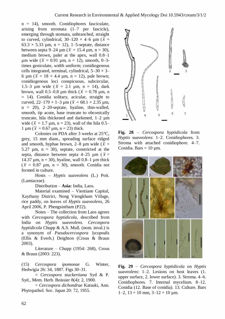
Current Research in Environmental & Applied Mycology Doi 10.5943/cream/3/1/2
62
n = 14), smooth. Conidiophores fasciculate,
arising from stromata (1–7 per fascicle),
emerging through stomata, unbranched, straight
to curved, cylindrical, 30–120 × 4–6 µm ( x =
63.3 × 5.33 µm, n = 12), 1–5-septate, distance
between septa 9–24 µm ( x = 15.4 µm, n = 30),
medium brown, paler at the apex, wall 0.8–1
µm wide ( x = 0.91 µm, n = 12), smooth, 0–3-
times geniculate, width uniform; conidiogenous
cells integrated, terminal, cylindrical, 5–30 × 3–
6 µm ( x = 18 × 4.4 µm, n = 12), pale brown;
conidiogenous loci conspicuous, subcircular,
1.5–3 µm wide ( x = 2.1 µm, n = 14), dark
brown, wall 0.5–0.8 µm thick ( x = 0.78 µm, n
= 14). Conidia solitary, acicular, straight to
curved, 22–170 × 1–3 µm (x = 68.1 × 2.35 µm,
n = 20), 2–20-septate, hyaline, thin-walled,
smooth, tip acute, base truncate to obconically
truncate, hila thickened and darkened, 1–2 µm
wide ( x = 1.7 µm, n = 23), wall of the hila 0.5–
1 µm ( x = 0.67 µm, n = 23) thick.
Colonies on PDA after 3 weeks at 25°C,
grey, 15 mm diam., spreading surface ridged
and smooth, hyphae brown, 2–8 µm wide ( x =
5.27 µm, n = 30), septate, constricted at the
septa, distance between septa 4–25 µm ( x =
14.37 µm, n = 30), hyaline, wall 0.8–1 µm thick
( x = 0.87 µm, n = 30), smooth. Conidia not
formed in culture.
Hosts – Hyptis suaveolens (L.) Poit.
(Lamiaceae).
Distribution – Asia: India, Laos.
Material examined – Vientiane Capital,
Xaythany District, Nong Viengkham Village,
rice paddy, on leaves of Hyptis suaveolens, 26
April 2006, P. Phengsintham (P22).
Notes – The collection from Laos agrees
with Cercospora hyptidicola, described from
India on Hyptis suaveolens. Cercospora
hyptidicola Chupp & A.S. Mull. (nom. inval.) is
a synonym of Pseudocercospora lycopodis
(Ellis & Everh.) Deighton (Crous & Braun
2003).
Literature – Chupp (1954: 268), Crous
& Braun (2003: 223).
(15) Cercospora ipomoeae G. Winter,
Hedwigia 26: 34, 1887. Figs 30–31.
= Cercospora stuckertiana Syd & P.
Syd., Mem. Herb. Boissier 8(4): 2, 1900.
= Cercospora dichondrae Katsuki, Ann.
Phytopathol. Soc. Japan 20: 72, 1955.
Fig. 28 – Cercospora hyptidicola from
Hyptis suaveolens: 1–2. Conidiophores. 3.
Stroma with attached conidiophore. 4–7.
Conidia. Bars = 10 µm.
Fig. 29 – Cercospora hyptidicola on Hyptis
suaveolens: 1–2. Lesions on host leaves (1.
upper surface, 2. lower surface). 3. Stroma. 4–6.
Conidiophores. 7. Internal mycelium. 8–12.
Conidia (12. Base of conidia). 13. Culture. Bars
1–2, 13 = 10 mm, 3–12 = 10 µm.

Current Research in Environmental & Applied Mycology Doi 10.5943/cream/3/1/2
63
Leaf spots small to fairly large,
suborbicular to irregular, 0.5–3 mm in diam.,
grey-brown in the centre, and with dark brown
margin. Caespituli amphigenous, scattered, dark
brown. Mycelium internal; hyphae branched, 1–
4 µm wide ( x = 2.97 µm, n = 30), septate,
constricted at the septa, distance between septa
4–21 µm ( x = 8.5 µm, n = 30), brownish or
green-hyaline, wall 0.25–0.5 µm wide ( x = 0.34
µm, n = 30), smooth, forming plate-like
plectenchymatous stromatic hyphal
aggregations. Stromata developed, small to
medium-sized, globular to subglobular,
substomatal and intraepidermal, 13–32 µm in
diam. ( x = 20.1 µm, n = 10), dark brown to
black in mass, composed of swollen hyphal
cells, subglobose, rounded to angular in outline,
4–10 µm wide ( x = 6.2 µm, n = 30), brown to
dark brown, wall 0.5–1 µm wide ( x = 0.9 µm, n
= 30), smooth. Conidiophores fasciculate,
arising from stromata (1–6 per fascicle),
emerging through stomata, unbranched, straight
to curved, cylindrical, 20–70 × 4–6 µm ( x =
36.1 × 4.8 µm, n = 20), 1–5-septate, distance
between septa 7–33 µm ( x = 15.8 µm, n = 30),
medium brown, paler at the apex, wall 0.5–1
µm wide ( x = 0.63 µm, n = 20), smooth, 0–2-
times geniculate. Conidiogenous cells
integrated, terminal, cylindrical, 8–28 × 4–5 µm
( x = 19.4 × 4.08 µm, n = 12), pale brown;
conidiogenous loci conspicuous, subcircular,
1.5–3 µm wide (x = 2.07 µm, n = 30), wall 0.5–
0.8 µm thick ( x = 0.53 µm, n = 30), thickened
and darkened. Conidia solitary, acicular to
obclavate, straight to curved, 12–152 × 2–5 µm
( x = 59.7 × 3.03 µm, n = 30), 0–16-septate,
hyaline to subhyaline, thin-walled 0.25–0.3 µm
( x = 0.25 µm, n = 30), smooth; tip acute; base
truncate to obconically truncate, hila thickened
and darkened, 1–3 µm wide ( x = 1.95 µm, n =
30), wall of the hila 0.3–0.8 µm ( x = 0.51 µm, n
= 30) thick.
Hosts – Argyreia tiliifolia (Desr.)
Wight, Convolvulus arvensis L., Dichondra
repens J.R. Forst. & G. Forst., Hewittia bicolor
Wight & Arn., Hewittia sp., Ipomoea
acuminata Ruiz & Pav., I. alba L., I. aquatica
Forssk., I. armata Roem. & Schult., I. asarifolia
(Desr.) Roem. & Schult., I. batatas (L.) Lam., I.
biloba Forssk., I. bona-nox L., I. cairica (L.)
Sweet, I. carnea Jacq., I. clarensis Alain, I.
coccinea L., I. cordofana Choisy, I. cymosa
(Desr.) Roem. & Schult., I. eriocarpa R. Br., I.
fistulosa Mart. ex Choisy, I. forsteri A. Grey ex
Hillebr., I. hederacea Jacq., I. hildebrandtii
Vatke, I. indica (Burm.) Merr., I. involucrata P.
Beauv., I. kentrocarpa Hochst. ex A. Rich., I.
lacunosa L., I. leari Knight ex Paxton, I.
longicuspis Meisn., I. nil (L.) Roth, I.
pandurata (L.) G. Mey., I. pes-caprae (L.) R.
Br., I. pestigridis L., I. pes-caprae subsp.
brasiliensis (L.) Ooststr., I. purpurea (L.) Roth,
I. quamoclit L., I. ramonii Choisy, I. reptans
Poir., I. sepiaria Koenig ex Roxb., I. triloba L.,
I. turpethum (L.) R. Br., I. villosa Ruiz & Pav.,
Jacquemontia tamnifolia (L.) Griseb.,
Merremia chryseides (Ker Gawl.) Hallier f., M.
emarginata (Burm. f.) Hallier f., M. umbellata
(L.) Hallier f., Operculina sp.
(Convolvulaceae).
Distribution – Africa: Ivory Coast,
Kenya, Mauritius, Sierra Leone, Sudan,
Tanzania; Asia: Brunei, China, Hong Kong,
India, Indonesia, Japan, Korea, Laos, Malaysia,
Myanmar, Pakistan, Taiwan, Thailand;
Europe: Italy; North America and West
Indies: Antigua and Barbuda, Barbados, Costa
Rica, Cuba, Guam, Jamaica, Kansas, Panama,
Puerto Rico, USA (AL, GA, FL, HI, IL, IN,
KS, MO, NC, NE, NJ, OH, TX, VA);
Australia; Oceania: America Samoa, Cook
Islands, Kiribati, Marshall Islands, New
Caledonia, New Zealand, Fiji, Samoa, Solomon
Islands, Papua New Guinea, Vanuatu; South
America: Argentina, Brazil, Venezuela.
Material examined – Vientiane Capital,
Xaythany District, Dong Makhai Village, rice
paddy, on leaves of Ipomoea involucrata, 5
May 2006, P. Phengsintham (P34);
Xiangkhouang Province, Paek District,
Phonsavan Village, rice paddy, on leaves of I.
aquatica, 3 January 2010, P. Phengsintham
(P514).
Notes – The collections from Laos agree
well with C. ipomoeae as circumscribed by
Chupp (1954), Hsieh & Goh (1990) and other
authors. C. ipomoeae is part of the C. apii
complex (Crous & Braun 2003) from which it is
morphologically barely distinguishable.
Literature – Chupp (1954: 171), Ellis
(1976: 253), Hsieh & Goh (1990: 89–90),
Crous & Braun (2003: 228).

Current Research in Environmental & Applied Mycology Doi 10.5943/cream/3/1/2
64
Fig. 30 – Cercospora ipomoeae on Ipomoea
involucrata from leaf spots: 1. Conidiophore. 2–
3. Stromata with attached conidiophores. 4.
Conidiophore. 5–10. Conidia. Bar = 10 µm.
Fig. 31 – Cercospora ipomoeae on Ipomoea
involucrata from leaf spots: 1. Lesions on host
leaf (lower surface). 2. Caispituli. 3. Stromatal
cells. 4. Internal mycelia. 5–8. Stromata with
attached conidiophores. 9–12. Conidia. Bars 1 =
10 mm, 3–12 = 10 µm.
(16) Cercospora meliicola Speg., Annales
Mus. Nac. Buenos Aires 20: 440, 1910. Figs
32–33.
Leaf spots small to fairly large,
suborbicular to irregular, 0.5–2 mm in diam.,
grey-brown in the centre, and with dark brown
margin. Caespituli amphigenous, scattered, dark
brown. Mycelium internal, inconspicious.
Stromata developed, small to medium-sized,
globular to subglobular, substomatal and
intraepidermal, 10–34 µm in diam. ( x = 24.7
µm, n = 5), dark brown to black in mass,
composed of swollen hyphal cells, subglobose,
rounded to angular in outline, 5–10 µm wide ( x
= 8.5 µm, n = 7), brown to dark brown, wall
0.5–1 µm wide ( x = 0.7 µm, n = 7), smooth.
Conidiophores solitary or fasciculate, arising
from stromata (2–16 per fascicle), emerging
through stomata, unbranched, straight to
curved, cylindrical, 10–402 × 4–5 µm (x = 200
× 4.4 µm, n = 13), 0–15-septate, distance
between septa 7–40 µm (x = 24 µm, n = 12),
medium brown, paler at the apex, wall 0.5–0.8
µm wide ( x = 0.65 µm, n = 12), smooth,
geniculate. Conidiogenous cells terminal,
cylindrical, 13–30 × 4–5 µm (x = 19.4 × 4 µm,
n = 12), pale brown; conidiogenous loci
conspicuous, subcircular, 3–4 µm wide ( x =
3.25 µm, n = 4), wall 0.5–0.8 µm thick ( x =
0.56 µm, n = 12), thickened and darkened.
Conidia solitary, acicular to obclavate, straight
to curved, 83–105 × 3–4 µm (x = 94 × 3.5 µm,
n = 5), 5–14-septate, hyaline to subhyaline,
thin-walled 0.25–0.3 µm ( x = 0.28 µm, n = 5),
smooth, tip acute, base truncate to obconically
truncate; hila thickened and darkened, 2–3 µm
wide ( x = 2.5 µm, n = 5), wall of the hila 0.3–
0.35 µm ( x = 0.32 µm, n = 5) thick.
Colonies on PDA after 3 weeks at 25°C
are brown-grey mycelium, reaching 25–30 mm
diam.
Hosts – Azadirachta indica A. Juss.,
Chukrasia tabularis A. Juss., Melia azadirachta
L., Toona sinensis (A. Juss.) M. Roem.
(Meliaceae).
Distribution – Asia: China, India, Laos;
North America and West Indies: Panama;
South America: Argentina.
Material examined – Khammoune
Province, Yommalad District, Nahao Village,
mixed deciduous forest, on leaves of Chukrasia
tabularis, 6 March 2010, P. Phengsintham

Current Research in Environmental & Applied Mycology Doi 10.5943/cream/3/1/2
65
(P581). GenBank accession no (LSU,
KC677920).
Notes – The collection from Laos agrees
well with C. apii s. lat. as circumscribed by
Crous & Braun (2003) [conidiophores 50–250 ×
4.5–7 µm and conidia 40–200 × 2–4.4 µm].
Literature – Saccado (1913: 639–640),
Chupp (1954: 385).
Fig. 32 – Cercospora meliicola on Chukrasia
from leaf spots: 1. Stroma with attached
conidiophores. 2–3. Conidiophores. 4–5.
Conidia. Bar = 10 µm.
(17) Cercospora nasturtii Pass. Hedwigia 16:
124, 1877. Figs 34–35.
Leaf spots orbicular in shape, 1–7 mm
diam., pale tan to dingy grey in the centre, and
with a dark grey or brown margin. Caespituli
amphigenous, scattered, dark brown. Mycelium
internal. Hyphae branched, 2–5 µm wide ( x =
3.5 µm, n = 6); septate, constricted at the septa,
distance between septa 8–14 µm long ( x = 8.5
µm, n = 6); brown to subhyaline; wall 0.3–0.5
µm ( x = 0.43 µm, n = 6), smooth. Stromata
developed, oval to ellipsoidal, substomatal, up
to 15 µm diam. wide, brown, composed of
swollen hyphal cells, subglobose, rounded and
angular in outline, 5–10 µm wide ( x = 7.14
µm, n = 7), brown to dark brown, wall 0.8–1
µm wide ( x = 0.84 µm, n = 7), smooth.
Fig. 33 – Cercospora meliicola on Chukrasia
tabularis from leaf spots: 1 Caespituli. 2.
Stroma. 3–6. Conidiophores. 7–9. Conidia. 10.
Culture. Bars 2–9 = 10 µm, 10 = 10 mm.
Conidiophores solitary or fasciculate,
arising from stromata (2–12 per fascicle),
emerging through stomata, unbranched, straight
to curved, cylindrical, 20–134 × 4–5 µm (x =
64.4 × 4.9 µm, n = 15), 1–9-septate, distance
between septa 3–22 µm ( x = 12.4 µm, n = 30),
medium brown, paler at the apex, wall 0.5–0.8
µm wide ( x = 0.56 µm, n = 15), smooth, 0–2-
times geniculate, width uniform. Conidiogenous
cells integrated, terminal, cylindrical, 14–55 ×
4–5 µm ( x = 27.4 × 4.5 µm, n = 9), pale
brown; conidiogenous loci conspicuous,
subcircular, 2–3 µm wide ( x = 2.42 µm, n = 9),
dark brown, wall 0.8–1 µm thick (x = 0.81 µm,
n = 9). Conidia solitary, acicular, straight to
curved, 22–75 × 3–4 µm (x = 36.4 × 3.7 µm, n
= 10), 1–7-septate, hyaline, thin-walled,
smooth, tip subotuse to acute, base truncate to
obconically truncate, hila thickened and
darkened, 2–3 µm wide ( x = 2.5 µm, n = 12),
wall of the hila 0.5–1 µm ( x = 0.77 µm, n = 12)
thick.
Hosts – Arabis glabra (L.) Bernh., A.
pendula L., Barbarea orthoceras Ledeb.,
Berteroa incana (L.) DC., Cardamine flexuosa
With., Descurainia pinnata (Walter) Britton,
Lepidium sativum L., Nasturtium officinale
W.T. Aiton, Rorippa amphibia (L.) Besser, R.
indica (L.) L.H. Bailey, R. islandica (Oeder ex

Current Research in Environmental & Applied Mycology Doi 10.5943/cream/3/1/2
66
Murray) Borbás, R. montana (Wall. ex Hook. f.
& Thomson) Small, R. palustris (L.) Besser, R.
sinapis (Burm. f.) Ohwi & H. Hara, R. sinuata
(Nutt.) Hitchc., R. sylvestris (L.) Besser,
Rorippa sp., Sisymbrium officinale (L.) Scop.,
Stanleya pinnata (Pursh) Britton
(Brassicaceae).
Distribution – Africa: Angola,
Mauritius, Morocco, Sao Tome Principe; Asia:
Bhutan, China, Hong Kong, India, Japan,
Korea, Kirghizia, Laos, Taiwan, Thailand;
Europe: Armenia, Azerbaijan, Estonia,
Germany, Great Britan, Italy, Latvia, Lithuania,
Romania, Russia, Slovakia, Slovenia, Ukraine;
North America and West Indies: Canada,
Cuba, Dominican Republic, Mexico, Panama,
USA (CA, CO, DE, FL, HI, IN, KS, NH, OK,
TX, WI, WV); Oceania: New Caledonia, New
Zealand, Papua New Guinea, Vanuatu; South
America: Brazil.
Material examined – Vientiane Capital,
Xaythany District, Dong Dok Village, garden,
on leaves of Nasturtium officinale, 2 February
2007, P. Phengsintham (P220); Xiangkhouang
Province, Kham District, Napa Village, on
leaves of N. officinale, 3 January 2010, P.
Phengsintham (P503). GenBank accession no
(ITS, KC731559; LSU, KC731563).
Notes – In the Lao collections the
conidiophores are 20–134 × 4–5 µm and the
conidia are 22–75 × 3–4 µm, which is similar to
those reported by Hsieh & Goh (1990)
[conidiophores 35–90 × 4–6 µm and conidia
20–85 × 3–4.5 µm] and Chupp (1954)
[conidiophores 20–100(–150) × 4–6.5 µm and
conidia 20–85(–125) × 4–5(–6) µm].
Literature – Saccardo (1886: 533),
Chupp (1954: 183), Katsuki (1965: 26), Hieh &
Goh (1990: 95), Shin & Kim (2001: 89), Crous
& Braun (2003: 289).
(18) Cercospora nicotianae Ellis & Everh.,
Proc. Acad. Sci. Philadelphia 45: 170, 1893.
Figs 36–37.
(= Cercospora apii s. lat.)
Leaf spots small to fairly large,
suborbicular to irregular, 0.5–3 mm in diam.,
grey-brown in the centre, and with dark brown
margin. Caespituli amphigenous, scattered, dark
brown. Mycelium internal; hyphae branched, 3–
6 µm wide ( x = 4.2 µm, n = 15), septate,
constricted at the septa, distance between septa
Fig. 34 – Cercospora nasturtii from
Nasturtium officinale: 1. Stroma with attached
conidiophores. 2–4. Conidiophores. 5–9.
Conidia. Bars = 10 µm.
Fig. 35 – Cercospora nasturtii from Nasturtium
officinale: 1. Internal mycelia. 2. Stroma with
attached conidiophores. 3–6. Conidia. Bars = 10
µm.
7–15 µm ( x = 10.73 µm, n = 15), brownish
or green-hyaline, wall 0.5–0.8 µm wide (x =
0.52 µm, n = 15), smooth, forming plate-like
plectenchymatous stromatic hyphal
aggregations. Stromata developed, small to

Current Research in Environmental & Applied Mycology Doi 10.5943/cream/3/1/2
67
medium-sized, globular to subglobular,
substomatal and intraepidermal, 10–33 µm in
diam. ( x = 22.7 µm, n = 7), dark brown to
black in mass, composed of swollen hyphal
cells, subglobose, rounded to angular in
outline, 6–15 µm wide ( x = 7.9 µm, n = 30),
brown to dark brown, wall 0.5–1 µm wide ( x
= 0.66 µm, n = 30), smooth. Conidiophores
fasciculate, arising from stromata (1–8 per
fascicle), emerging through stomata,
unbranched, straight to curved, cylindrical,
20–395 × 4–7 µm ( x = 237 × 5.54 µm, n =
13), 1–18-septate, distance between septa 9–
40 µm ( x = 24.3 µm, n = 30), medium
brown, paler at the apex, wall 0.5–0.8 µm
wide ( x = 0.69 µm, n = 30), smooth, 0–2-
times geniculate. Conidiogenous cells
terminal, cylindrical, 9–40 × 3–4 µm ( x = 24
× 3.5 µm, n = 8), pale brown; conidiogenous
loci conspicuous, subcircular, 3–4 µm wide
( x = 3.25 µm, n = 4), wall 0.5–0.8 µm thick
( x = 0.57 µm, n = 4), thickened and
darkened. Conidia solitary, acicular to
obclavate, straight to curved, 94–267 × 3–4
µm ( x = 149.6 × 3.6 µm, n = 5), 5–8-septate,
hyaline to subhyaline, thin-walled 0.3 µm ( x
= 0.3 µm, n = 5), smooth, tip acute, base
truncate to obconically truncate; hila
thickened and darkened, 2–3 µm wide ( x =
2.5 µm, n = 5), wall of the hila 0.3–0.5 µm
( x = 0.46 µm, n = 5) thick.
Hosts – Nicotiana tabacum L. and
other species of this genus (Solanaceae)
Distribution – Worldwide.
Material examined – Khammoune
Province, Yommalad District, Nahao Village,
garden, on leaves of Nicotiana tabacum, 6
March 2010, P. Phengsintham (P583).
Notes – The collection from Laos
agrees well with C. apii as circumscribed by
Crous & Braun (2003). C. nicotianae is part
of the C. apii complex from which it is
morphologically barely distinguishable. C.
nicotianicola J.M. Yen (Yen & Lim 1980) is
distinguished from C. nicotianae by its small
whitish leaf spots, long conidiophores
strongly geniculate in the upper half and long
acicular to filiform conidia, up to 480 µm in
length.
Literature – Yen & Lim (1980: 163),
Crous & Braun (2003).
Fig. 36 – Cercospora nicotianae on Nicotiana
tabacum from leaf spots: 1. Stroma with
attached conidiophores. 2–4. Conidia. Bar = 10
µm.
Fig. 37 – Cercospora nicotianae on Nicotiana
tabacum from leaf spots: 1. Lesions on host leaf
(lower surface). 2. Internal mycelium. 3–4.
Stromata with attached conidiophores. 5–6.
Conidiophores. 7–9. Conidia. Bars 1 = 10 mm,
2–9 = 10 µm.

Current Research in Environmental & Applied Mycology Doi 10.5943/cream/3/1/2
68
(19) Cercospora paederiicola Y.L. Guo,
Mycosystema 4: 119, 1991. Figs 38–39.
Leaf spots subcircular to irregular, 1–8
mm diam., pale brown to greyish brown in the
center, and with dark brown margin. Caespituli
amphigenous, scattered, dark brown. Mycelium
internal; hyphae branched, 2–7 µm wide ( x =
3.76 µm, n = 30), septate, constricted at the
septa, distance between septa 5–16 µm ( x =
9.69 µm, n = 17), brownish or green-hyaline,
wall 0.3–0.8 µm wide ( x = 0.52 µm, n = 30),
smooth, forming plate-like plectenchymatous
stromatic hyphal aggregations. Stromata well-
developed, oval to ellipsoidal, 15–60 µm diam.
( x = 27 µm, n = 8), brown, substomatal,
composed of swollen hyphal cells, subglobose,
rounded to angular in outline, 5–10 µm wide ( x
= 6 µm, n = 12), brown to dark brown, wall
0.5–0.8 µm wide ( x = 0.53 µm, n = 12).
Conidiophores fasciculate, arising from
stromata (2–7 per fascicle), emerging through
stomata, unbranched, straight to curved,
cylindrical, 20–126 × 4–6 µm ( x = 52.53 × 5
µm, n = 19), 1–4-septate, distance between
septa 15–32 µm ( x = 23.65 µm, n = 20),
medium brown, paler at the apex, wall 0.5–0.8
µm wide ( x = 0.69 µm, n = 30), smooth, 0–2
times geniculate, width uniform; conidiogenous
cells integrated, terminal or intercalary,
cylindrical, 15–30 × 4–6 µm, pale brown;
conidiogenous loci conspicuous, subcircular, 2–
3 µm wide ( x = 2.53 µm, n = 15), dark brown,
wall 0.8–1 µm wide ( x = 0.84 µm, n = 15).
Conidia solitary, cylindrical or obclavate-
cylindrical when short, fully developed long
conidia always acicular, straight to curved, 25–
157 × 1.5–3 µm (x = 85.86 × 2.7 µm, n = 8),
6–19-septate, hyaline, thin-walled, smooth, tip
acute, base truncate to obconically truncate, hila
1.5–3 µm wide ( x = 2.21 µm, n = 7), wall 0.3–
0.5 µm ( x = 0.41 µm, n = 8), thickened and
darkened.
Colonies on PDA after 3 weeks at 25°C
grey, 12 mm diam., spreading surface ridged
and smooth, mycelium brown, hyphae 2–7 µm
wide ( x = 3.6 µm, n = 30), septate, constricted
at the septa, distance between septa 4–17 µm
long ( x = 8.9 µm, n = 30), wall 0.44 µm,
smooth. Conidia not formed in culture.
Host – Paederia scandens (Lour.) Merr.
(Rubiaceae).
Distribution – Asia: China, Laos.
Material examined – Vientiane Capital,
Xaythany District, Dong Makkhai Village,
mixed deciduous forest, on leaves of Paederia
scandens, 29 March 2006, P. Phengsintham
(P01).
Notes – The collection from Laos differs
from the Chinese type material of Cercospora
paederiicola in having well-developed
stromata, up to 60 µm diam. (versus small or
almost lacking stromata, reduced to a few
brown cells). Morphologically this species
belongs to the Cercospora apii complex,
characterized by having long, brown
conidiophores and long, acicular, pluriseptate,
hyaline conidia (Crous & Braun 2003).
Literature – Crous & Braun (2003: 35).
Fig. 38 – Cercospora paederiicola on
Paederia scandens from leaf spots: 1.
Stroma with attached conidiophores. 2–4.
Conidiophores. 5–8. Conidia. Bar = 10 µm.
(20) Cercospora physalidis Ellis, Amer.
Naturalist 16: 810, 1882. Figs 40–41.
Cercosporina physalidis (Ellis)
Miura, South Manch, Railway Co. Agric.
Rep. 27: 525, 1928.
= Cercospora physalidicola Ellis &
Barthol., Erythea 4: 28, 1896.

Current Research in Environmental & Applied Mycology Doi 10.5943/cream/3/1/2
69
Fig. 39 – Cercospora paederiicola on Paederia
scandens from leaf spots/lesions: 1–2. Lesions
on host leaves (1. upper surface, 2. lower
surface). 3. Caespituli. 4. Stroma with attached
young conidiophores. 5. Internal mycelium. 6–
8. Conidiophores. 9–11. Conidia (10. base of
conidium, 11. apex of conidium). 13. Culture.
Bars 1–2, 13 = 10 mm, 3–12 = 10 µm.
= Cercospora physalidicola Speg.,
Anales Mus. Nac. Buenos Aires II, 3: 342, 1899
(nom. illeg.), homonym of C. physalidicola
Ellis & Barthol., 1896.
= Cercosporina physalidicola Speg.,
Anales Mus. Nac. Hist. Nat. Buenos Aires 20:
425, 1910.
Leaf spots circular or suborbicular, 1–10
mm diam., greyish brown to medium brown in
the centre, and with medium brown to dark
brown margin. Caespituli amphigenous,
scattered, dark brown. Mycelium internal;
hyphae branched, 3–6 µm wide ( x = 4.5 µm, n
= 10), septate, constricted at the septa, distance
between septa 6–14 µm ( x = 10.3 µm, n = 10),
brownish or green-hyaline, wall 0.25–0.8 µm
wide ( x = 0.48 µm, n = 10), smooth, forming
plate-like plectenchymatous stromatic hyphal
aggregations. Stromata well-developed, oval to
ellipsoidal, substomatal, 15–32 µm diam. ( x =
23 µm, n = 15), brown, composed of swollen
hyphal cells, subglobose, rounded to angular in
outline, 4–12 µm wide (x = 7.83 µm, n = 30),
brown to dark brown, wall 0.5–1 µm wide ( x =
0.72 µm, n = 30), smooth. Conidiophores
solitary or fasciculate, arising from stromata (2–
8 per fascicle), emerging through stomata,
unbranched, straight to curved, cylindrical, 10–
56 × 3–6 µm ( x = 38 × 5.73 µm, n = 22), 0–2-
septate, distance between septa 7–35 µm ( x =
19.7 µm, n = 22), medium brown, paler at the
apex, wall 0.5–0.8 µm wide ( x = 0.51 µm, n =
22), smooth; conidiogenous cells integrated,
terminal, cylindrical, 17–35 × 4–6 µm (x = 21.8
× 4.62 µm, n = 14), pale brown; conidiogenous
loci conspicuous, subcircular, 2–2.5 µm wide
(x = 2.03 µm, n = 17), dark brown, wall 0.5–1
µm thick ( x = 0.79 µm, n = 17). Conidia
solitary, acicular, straight to curved, 52–59 × 3–
4 µm ( x = 55.5 × 3.4 µm, n = 6), 3–4-septate,
hyaline, thin-walled, smooth, tip acute, base
truncate to obconically truncate, hila thickened
and darkened, 2 µm wide ( x = 2 µm, n = 6),
wall of the hila 0.5–0.8 µm ( x = 0.65 µm, n =
6) thick.
Hosts – Physalis alkekengi L., P.
angulata L., P. franchetii Mast., P. heterophylla
Nees, P. hybrida Vilm., P. lanceolata Michx.,
P. lobata Torr., P. longifolia Nutt., P. minima
L., P. mollis Nutt., P. parviflora Lag., P.
pubescens L., P. subglabrata Mack. & Bush, P.
variabilis Fries, P. violacea Carrière, P.
virginica A. Grey, P. viscosa L., Physalis sp.
(Solanaceae).
Distribution – Africa: Congo, Ethiopia,
Gabon, Gambia, Ghana, Guinea, Kenya, Libya,
Malawi, Mauritius, Morocco, Nigeria, Samalia,
Sourth Africa, Seychelles, Sierra Leone, Sudan,
Tanzania, Togo, Uganda, Zambia, and
Zimbabwe; Asia: Afghanistan, Bangladesh,
Bhutan, Brunei, Cambodia, China, Guam, Hong
Kong, India, Indonesia, Iraq, Japan, Jordan,
Laos, Korea, Malaysia, Myanmar, Nepal,
Pakistan, Philippines, Singapore, Sri Lanka,
Taiwan, Thailand, Yemen; Europe: Armenia,
Bulgaria, Cyprus, Georgia, Germany, Romania,
Russia, Spain, Switzerland, Ukraine; North
America and West Indies: Barbados, Cuba,
Dominican Republic, French Antilles, El
Salvador, Guatemala, Haiti, Jamaica, Mexico,
Panama, Puerto Rico, Trinidad and Tobago,
Virgin islands, USA (CA, CO, Eastern states,
FL, GA, HI, IA, IN, KS, KY, MI, NE, OK, TX,
WI); Australia; Oceania: American Samoa,

Current Research in Environmental & Applied Mycology Doi 10.5943/cream/3/1/2
70
Fiji, Micronesia, New Caledonia, New Zealand,
Palau, Papua New Guinea, Samoa, Solomon
Islands, Tonga, Vanuatu, Wallis and Futuna;
South America: Argentina, Bolivia, Brazil,
Colombia, Surinam, Venezuela.
Material examined – Vientiane Capital,
Sisathanak District, Had Donchanh, garden, on
leaves of Physalis angulata, 3 May 2006, P.
Phengsintham (P33).
Notes – The collection from Laos has
conidiophores (10–56 × 3–6 µm) and conidia
(52–59 × 3–4 µm) with a similar size as
Cercospora physalidis. They are, however,
rather short, probably due to a relatively
immature stage in which the fungus has been
collected. The taxonomy of Cercospora on
Physalis spp. and other hosts of the Solanaceae
is still confused and unclear. Various
Cercospora species described from different
solanaceous hosts are morphologically
indistinguishable, so that Braun & Melnik
(1997) merged them in a single compound
species under the oldest epithet ‘physalidis’.
Since C. physalidis emend. U. Braun & Melnik
is morphologically also indistinguishable from
C. apii, Crous & Braun (2003) reduced the
whole C. physalidis complex to synonym with
C. apii s. lat. However, detailed inoculation
experiments and molecular sequence analyses
of this complex of fungi are not yet available.
Hence, we prefer to use the traditional name.
Literature – Saccado (1886: 450),
Chupp (1954: 533), Ellis (1976: 286), Braun &
Melnik (1997), Crous & Braun (2003: 321).
(21) Cercospora ricinella Sacc. & Berl., Atti
Reale Ist. Ven. Sci. Lett. Art. 6, Ser. 3: 721,
1885. Figs 42–43.
Cercosporina ricinella (Sacc. & Berl.)
Speg., Anales Mus. Nac. Hist. Nat. Buenos
Aires 20: 429, 1910.
= Cercospora albido-maculans G.
Winter, Hedwigia 24: 202. 1885; also in J.
Mycol. 1: 124, 1885.
= Cercospora ricini Speg., Anales Mus.
Nac. Hist. Nat. Buenos Aires Ser. 2. 3: 343,
1899.
Leaf spots small, suborbicular to
irregular, 0.5–2 mm in diam., grey-brown in the
centre, and with dark brown margin. Caespituli
amphigenous, scattered, dark brown. Mycelium
internal, inconspicuous. Stromata developed,
Fig. 40 – Cercospora physalidis from Physalis
angulata: 1. Stroma with attached
conidiophores. 2–4. Conidiophores. 5–6.
Conidia. Bar = 10 µm.
Fig. 41 – Cercospora physalidis on Physalis
angulata: 1. Lesion on host leaf (upper surface).
2. Internal hyphae. 3–5. Stromata with attached
conidiophores. 6. Conidiophore. 7–8. Conidia.
Bars 1 = 10 mm, 2–8 = 10 µm.

Current Research in Environmental & Applied Mycology Doi 10.5943/cream/3/1/2
71
small to medium-sized, globular to subglobular,
substomatal and intraepidermal, 15–20 µm in
diam. ( x = 17.5 µm, n = 5), dark brown to
black in mass, composed of swollen hyphal
cells, subglobose, rounded to angular in outline,
3–7 µm wide ( x = 5.3 µm, n = 9), brown to
dark brown, wall 0.5–0.8 µm wide ( x = 0.58
µm, n = 9), smooth. Conidiophores fasciculate,
arising from stromata (2–11 per fascicle),
emerging through stomata, unbranched, straight
to curved, cylindrical, 15–105 × 4–6 µm ( x =
46.4 × 4.94 µm, n = 13), 1–3-septate, distance
between septa 5–51 µm ( x = 20.3 µm, n = 30),
medium brown, paler at the apex, wall 0.5–0.8
µm wide ( x = 0.63 µm, n = 30), smooth, 0–2-
times geniculate. Conidiogenous cells terminal,
cylindrical, 10–28 × 4–6 µm ( x = 17.4 × 4.8
µm, n = 8), pale brown; conidiogenous loci
conspicuous, subcircular, 1.5–3 µm wide ( x =
2.12 µm, n = 8), wall 0.5–0.8 µm thick ( x =
0.65 µm, n = 8), thickened and darkened.
Conidia solitary, acicular to obclavate, straight
to curved, 32–98 × 3–4 µm ( x = 54.8 × 3.2 µm,
n = 5), 3–11-septate, hyaline to subhyaline,
thin-walled 0.25–0.3 µm ( x = 0.29 µm, n = 5),
smooth, tip acute, base truncate to obconically
truncate; hila thickened and darkened 3–4 µm
wide ( x = 3.2 µm, n = 5), wall of the hila 0.25–
0.3 µm ( x = 0.28 µm, n = 5) thick.
Hosts – Ricinus communis L.
(Euphorbiaceae).
Distribution – Africa: Angola, Egypt,
Ethiopia, Ghana, Gulf States, Kenya, Malawi,
Mauritius, Morocco, Mozambique, Nigeria,
Sierra Leone, Somalia, South Africa, Sudan,
Tanzania, Togo, Uganda, Zimbabwe; Asia:
Bangladesh, Cambodia, China, India,
Indonesia, Japan, Kazakhstan, Korea, Laos,
Malaysia, Myanmar, Nepal, Pakistan,
Philippines, Sri Lanka, Taiwan, Thailand;
Europe: Bulgaria, Georgia, Russia, Ukraine;
North America and West Indies: Barbados,
Cuba, Dominican Republic, El Salvador,
Guatemala, Haiti, Jamaica, Panama, Puerto
Rico, Trinidad and Tobago, USA (CA, FL,
MO); Australia; Oceania: French Polynesia,
New Caledonia, Vanuatu; South America:
Argentina, Brazil, Colombia, Venezuela.
Material examined – Phongsali
Province, Phongsali District, Hathin Village, on
leaves of Ricinus communis, riverbank, 23 June
2010, P. Phengsintham (P594); Vientiane
Capital, Had Donechan, Mekong riverbank, on
leaves of R. communis, 27 July 2010, P.
Phengsintham (P606).
Notes – A true Cercospora s. str. close
to or identical with C. apii s. lat. (Crous &
Braun 2003). The collections from Laos agree
with C. ricinella as circumscribed by Chupp
(1954) [conidiophores 10–90 × 4.5–5 µm and
conidia 15–120 × 2.5–4.5 µm], and Hsieh &
Goh (1990) [conidiophores 10–90 × 4–6 µm
and conidia 15–120 × 2.5–5 µm].
Literature – Chupp (1964: 229),
Vasudeva (1963: 174), Katsuki (1965: 32), Ellis
(1976: 259), Hsieh & Goh (1990: 112), Shin &
Kim (2001: 97), Cous & Braun (2003: 255).
Fig. 42 – Cercospora ricinella on Ricinus
communis from leaf spots: 1. Stroma with
attached conidiophores. 2–4.
Conidiophores. 5. Conidia. Bar = 10 µm.
(22) Cercospora senecionis-walkeri
Phengsintham; Chukeatirote, McKenzie,
K.D. Hyde & U. Braun, Pl. Pathol. &
Quarantine 2(1): 70–71, 2012. Figs 44–45.
Leaf spots circular to slightly
irregular, 2–3 mm diam., at first dark
green, later becoming brown to dark brown
in the centre, dark brown margin.
Caespituli amphigenous, conspicuous,
scattered, dark brown. Mycelium internal,
internal hyphae branched, 3–4 µm wide (x

Current Research in Environmental & Applied Mycology Doi 10.5943/cream/3/1/2
72
Fig. 43 – Cercospora ricinella on Ricinus
communis from leaf spots: 1. Lesions and
Caespituli on host leaf (lower surface). 2.
Internal mycelium. 3. Stroma with attached
conidiophores. 4–6. Conidiophores. 7.
Conidiophore with attached conidium. 8.
Conidium. 9. Apex of conidium. Bars = 10 µm.
=3.5 µm, n = 7), septate, constricted at the
septa, distance between septa 12–14 µm ( x =
13 µm, n = 7), subhyaline, wall 0.3 µm wide ( x
= 0.3 µm, n = 7), smooth. Stromata
substomatal, intraepidermal, ellipsoidal,
lenticular 10–25 µm diam. ( x = 18.8 µm, n =
6), brown, stromatal cells 4–10 µm diam. ( x =
6 µm, n = 20), wall 0.5–1 µm wide ( x = 0.78
µm, n = 20), smooth. Conidiophores single or
fasciculate, arising from stromata (1–8 per
fascicle), 0–5 geniculate, cylindrical, straight to
curved, 67–170 × 5–6 µm ( x = 114 × 5.53 µm,
n = 20), 0–8-septate, distance between septa
10–32 µm ( x = 21 µm, n = 30), pale brown or
olivaceous brown; wall 0.5–1 µm wide ( x =
0.82 µm, n = 30), smooth. Conidiogenous cells
intergrated, terminal, cylindrical, tapering to the
apex, 14–30 × 4–5 µm ( x = 21,8 × 4.5 µm, n =
9); conidiogenous loci (scars) conspicuous,
thickened and darkened, 2–4 µm wide ( x =
3.07 µm, n = 30), wall of the loci 0.5–1 µm ( x
= 0,54 µm, n = 30); thick. Conidia solitary,
clavate, cylindrical-clavate, obclavate, straight
to curved, 17–82 × 4–7 µm (x = 50.66 × 5.66
µm, n = 10), 0–8-sepate, slightly constricted at
the septa, subhyaline or olivaceous-brown,
smooth, wall 0.3–0.5 µm thick ( x = 0.37 µm, n
= 10), apex subobtuse, based truncate, hila 2–3
µm wide ( x = 2.2 µm, n = 10), wall of the hila
0.5 µm wide ( x = 0.5 µm, n = 10), darkened.
Hosts – Senecio aureus L., S. aureus
var. balamitea Torr. & Grey, S. walkeri Arn.
(Asteraceae).
Distribution – Asia: China, Laos,
Thailand.
Material examined – Bokeo Province,
Houay Xay District, Phimonsine Village, on
leaves of Senecio walkeri, 20 February 2010, P.
Phengsintham (P567, MFLU 12-2478,
holotype). GenBank accession no (ITS,
KC677887; LSU, KC677921).
Notes – Several Cercospora species
have been described from Senecio spp., but all
are distinct from this species. C. senecionis
Ellis & Everh. was reduced to synonym with C.
jacquiniana Thüm. by Chupp (1954). However,
based on a re-examination of type material,
Braun (in Braun & Mel’nik 1997) showed that
C. senecionis represents a distinct true species
of Cercospora with acicular conidia, similar to
those of C. apii s. lat., but 80–200 µm long and
above all 3–6 µm wide. C. jacquiniana is
similar to C. senecionis-walkeri with regard to
its conidial shape and pigmentation, but has
much shorter conidiophores and shorter conidia,
usually only 1–3-sepate, which are hyaline to
faintly pigmented. Therefore, this species was
reallocated to Passalora by Braun (in Braun &
Mel’nik 1997). The Indian C. senecionis-
grahamii Thirum. & Govindu (Thirumalachar
& Govindu 1962) on Senecio grahamii Benth.
is close to C. senecionis, but differs in having
acicular to obclavate conidia, only 3–4 µm
wide. Collections on Senecio walkeri from Laos
and Thailand were originally referred to as C.
senecionicola Davis (Phengsintham et al.
2012), but further examinations and a detailed
comparison with the latter species showed that
the Asian collections are not conspecific with
this North American species, which is
morphologically easily distinguishable from C.
senecionis-walkeri by its narrower acicular-
subcylindrical conidia, only 2–3.5 µm wide

Current Research in Environmental & Applied Mycology Doi 10.5943/cream/3/1/2
73
(Chupp 1954). The South American Passalora
senecionicola U. Braun & Delhey (Braun et al.
2006) on Senecio bonariensis in Argentina is
morphologically very close to C. senecionis-
walkeri but characterized by having quite
distinct lesions, larger stromata, up to 60 µm
diam. and short conidia that are cylindrical. P.
senecionicola was assigned to Passalora due to
subhyaline to pale olivaceous conidia, but it is
possible that this species belongs in Cercospora
which may be suggested by the phylogenetic
position of C. senecionis-walkeri. Despite
having almost hyaline to somewhat pigmented
conidia, the latter species clusters within the
Cercospora clade in an isolated, basal position
adjacent to C. zeae-maydis Tehon & E.Y.
Daniels and C. zeina Crous & U. Braun
(Groenewald et al. 2012).
Literature – Chupp (1954: 22),
Thirumalachar & Govindu (1962: 244–288),
Ellis (1971: 279–284), Braun & Mel’nik
(1997), Crous & Braun (2003: 21), Braun et al.
(2006).
Fig. 44 – Cercospora senecionis-walkeri on
Senecio walker from leaf spots: 1. Stroma
with attached conidiophores. 2. Conidiophore.
3–4. Conidia. Bar = 10 µm.
Fig. 45 – Cercospora senecionis-walkeri on
Senecio walker from leaf spots/lesions: 1–2.
Lesions on host leaves (1. upper surface, 2.
lower surface). 3. Caespituli. 4. Internal
mycelium. 5–7. Conidiophores. 8–10. Conidia.
Bars 1–2 = 10 mm, 4–10 = 10 µm.
(23) Cercospora sp. (C. apii s. lat.). Figs 46–47.
Leaf spots angular to irregular, 1–8 mm
diam., grey-brown in the centre, and with dark
brown margin. Caespituli amphigenous,
scattered, dark brown. Mycelium internal;
hyphae branched, 2–4 µm wide ( x = 3.5 µm, n
= 28), septate, constricted at the septa, distance
between septa 6–14 µm (x = 7.8 µm, n = 20),
brownish or green-hyaline, wall 0.3–0.5 µm
wide ( x = 0.43 µm, n = 28), smooth, forming
plate-like plectenchymatous stromatic hyphal
aggregations. Stromata developed, small to
medium-sized, globular to subglobular,
substomatal and intraepidermal, 8–52 µm diam.
(x = 36.6 µm, n = 5), dark brown to black in
mass, composed of swollen hyphal cells,
subglobose, rounded to angular in outline, 5–12
µm wide ( x = 8.13 µm, n = 30), brown to dark
brown, wall 0.5–1 µm wide ( x = 0.91 µm, n =
30), smooth. Conidiophores fasciculate, arising
from stromata (1–4 per fascicle), emerging
through stomata, unbranched, straight to

Current Research in Environmental & Applied Mycology Doi 10.5943/cream/3/1/2
74
curved, cylindrical, 33–80 × 5–7 µm (x = 51.9
× 5.27 µm, n = 11), 1–3-septate, distance
between septa 8–31 µm ( x = 19 µm, n = 20),
medium brown, paler at the apex, wall 0.5–1
µm wide ( x = 0.83 µm, n = 11), smooth, 0–1-
times geniculate; conidiogenous cells
integrated, terminal, cylindrical, 16–31 × 4–5
µm ( x = 22.8 × 4.67 µm, n = 6), pale brown;
conidiogenous loci conspicuous, subcircular, 2–
3 µm wide, wall 0.5–0.8 µm thick ( x = 0.56
µm, n = 4), thickened and darkened. Conidia
solitary, acicular, straight to curved, 43–185 × 2
µm ( x = 81.25 × 2 µm, n = 4), 3–12-septate,
hyaline to subhyaline, thin-walled, smooth, tip
acute, base truncate to obconically truncate, hila
thickened and darkened, 1–1.5 µm wide, wall of
the hila 0.5 µm thick.
Colonies on PDA after 3 weeks at 25°C
spreading surface ridged, white-grey in the
centre, brown margin, reaching 10–25 mm
diam., hyphae 1–6 µm wide ( x = 3.9 µm, n =
30), septate, constricted at the septa, distance
between septa 8–16 µm ( x = 12 µm, n = 30),
brownish to subhyaline, wall 0.3–0.8 µm thick
( x = 0.51 µm, n = 30), smooth or finely
verruculose. Conidia not formed in the culture.
Hosts – Oroxylum indicum (L.) Kurz
(Bignoniaceae).
Distribution – Asia: Laos.
Material examined – Vientiane Capital,
Xaythany District, Houay Daenmeuang Village,
fallow forest, on leaves of Oroxylum indicum,
26 April 2006, P. Phengsintham (P32).
Notes – Pseudocercospora oroxyli
(A.K. Kar & M. Mandal) Deighton (
Cercospora oroxyli A.K. Kar & M. Mandal,
1969) described from India differs from the
specimen collected in Laos in having
inconspicuous, unthickened conidiogenous loci.
The cercosporoid fungus on Oroxylum indicum
from Laos agrees well with Cercospora apii s.
lat. (Crous & Braun 2003) [conidiophores 10–
450 × 2–8 µm and conidia 10–380 × 1.5–5.5
µm].
Literature – Crous & Braun (2003: key).
(24) Cercospora stahlianthi Z.D. Jiang & P.K.
Chi, in Chi, Fungal diseases of medicinal plants
in Guangdong province: 162, 1994. Figs 48–49.
Leaf spots circular to irregular, 1–5 mm
diam., pale brown to dark brown in the centre,
Fig. 46 – Cercospora sp. on Oroxylum
indicum from leaf spots: 1. Stroma with
attached conidiophores. 2–3. Conidiophores.
4–7. Conidia. Bar = 10 µm
Fig. 47 – Cercospora sp. on Oroxylum indicum
from leaf spots: 1. Lesions on host leaf (lower
surface). 2. Internal mycelium. 3–4. Stromata
with attached conidiophores. 5–6.
Conidiophores. 7–8. Conidia. 9. Culture. Bars
2–8 = 10 µm, 9 = 10 mm.

Current Research in Environmental & Applied Mycology Doi 10.5943/cream/3/1/2
75
and with yellowish to medium brown margin.
Caespituli amphigenous, scattered, dark brown.
Mycelium internal; hyphae branched, 2–6 µm
wide ( x = 3.8 µm, n = 30),septate, constricted
at the septa, distance between septa 6–18 µm
( x = 10.07 µm, n = 30), brownish, wall
approximately 0.3–0.5 µm wide ( x = 0.46 µm,
n = 30), smooth, forming plate-like
plectenchymatous stromatic hyphal
aggregations. Stromata well-developed, oval to
ellipsoidal, substomatal, 8–40 µm diam. ( x =
24.14 µm, n = 7), brown, composed of swollen
hyphal cells, subglobose, rounded to angular in
outline, 5–17 µm wide ( x = 8.3 µm, n = 30),
brown to dark brown, wall 0.5–1 µm wide ( x =
0.81 µm, n = 30), smooth. Conidiophores
fasciculate, arising from stromata (2–15 per
fascicle), emerging through stomata,
unbranched, straight to curved, cylindrical, 20–
240 × 4–6 µm (x = 78.2 × 4.83 µm, n = 30), 1–
6-septate, distance between septa 6–65 µm ( x =
27.5 µm, n = 30), medium brown, paler at the
apex, wall 0.5–1 µm wide ( x = 0.77 µm, n =
30), smooth, 0–2 times geniculate.
Conidiogenous cells integrated, terminal,
cylindrical, 15–65 × 4–5 µm (x = 34.6 × 4.35
µm, n = 17), pale brown; conidiogenous loci
conspicuous, subcircular, 2–4 µm wide ( x =
2.83 µm, n = 30), dark brown, wall 0.5–0.8 µm
thick ( x = 0.51 µm, n = 30). Conidia solitary,
acicular, straight to curved, 30–240 × 2–4 µm
( x = 83.37 × 3.73 µm, n = 30), 2–23-septate,
hyaline, thin-walled, smooth, tip acute, base
truncate to obconically truncate, hila thickened
and darkened, 1.5–3 µm wide ( x = 2.28 µm, n
= 30), wall of the hila 0.3–0.5 µm ( x = 0.49
µm, n = 30) thick.
Hosts – Stahlianthus involucratus (King
ex Baker) Craib and S. thorelii Gagnep.
(Zingiberaceae).
Distribution – Asia: China, Laos.
Material examined – Vientiane Capital,
Xaythani District, Dong Makhai village, dry
dipterocarp forest, on leaves of Stahlianthus
thorelii, 20 May 2006, P. Phengsintham (P51);
Xaythani District, Dongdok Village, on leaves
of S. thorelii, 12 August 2006, P. Phengsintham
(P141).
Notes – The collections from Laos have
a size of conidiophores and conidia close to that
of C. apii s. lat.
Literature – Crous & Braun (2003: 386).
Fig. 48 – Cercospora stahlianthi on
Stahlianthus thorelii: 1–2. Stromata with
attached conidiophores. 3–5. Conidia. Bars = 10
µm.
Fig. 49 – Cercospora stahlianthi on
Stahlianthus thorelii: 1. Lesions on host leaf
(upper surface). 2. stroma. 3. Internal hyphae.
4–5. Stromata with attached conidiophores. 6–
7. Conidiophores. 8–11. Conidia. Bars 1 = 10
mm, 2–11 = 10 µm.

Current Research in Environmental & Applied Mycology Doi 10.5943/cream/3/1/2
76
(25) Cercospora taccae (Syd. & P. Syd.)
Chupp. A monograph of the fungus genus
Cercospora 560, 1954. Figs 50–51.
Cercosporina taccae Syd & P. Syd.,
Ann. Mycol. 11: 406, 1913.
Leaf spots small to fairly large,
suborbicular to irregular, 1–22 mm in diam.,
dark brown in the centre, and with dark brown
margin. Caespituli amphigenous, scattered, dark
brown. Mycelium internal; hyphae branched, 3–
5 µm wide ( x = 3.5 µm, n = 7), septate,
constricted at the septa, distance between septa
10–15 µm ( x = 11.25 µm, n = 7), brownish or
green-hyaline, wall 0.5–0.8 µm wide ( x = 0.57
µm, n = 7), smooth, forming plate-like
plectenchymatous stromatic hyphal
aggregations. Stromata developed, small to
medium-sized, globular to subglobular,
substomatal and intraepidermal, 15–35 µm in
diam. ( x = 21.7 µm, n = 10), dark brown to
black in mass, composed of swollen hyphal
cells, subglobose, rounded to angular in outline,
5–11 µm wide ( x = 7.8 µm, n = 18), brown to
dark brown, wall 0.5–1 µm wide ( x = 0.83 µm,
n = 18), smooth. Conidiophores fasciculate,
arising from stromata (1–13 per fascicle),
emerging through stomata, unbranched, straight
to curved, cylindrical, 35–179 × 4–6 µm ( x =
98 × 4 µm, n = 11), 1–10-septate, distance
between septa 8–38 µm ( x = 18 µm, n = 24),
medium brown, paler at the apex, wall 0.5–0.8
µm wide ( x = 0.66 µm, n = 24), smooth,
geniculate. Conidiogenous cells integrated,
terminal, cylindrical, 8–30 × 4–5 µm ( x = 18.8
× 4.5 µm, n = 6), pale brown; conidiogenous
loci conspicuous, subcircular, 1.5–3 µm wide
( x = 2.5 µm, n = 15), wall 0.5–0.8 µm thick ( x
= 0.6 µm, n = 15), thickened and darkened.
Conidia solitary, acicular to obclavate, straight
to curved, 73–195 × 3–6 µm ( x = 115.25 ×
4.75 µm, n = 7), 9–20-septate, hyaline to
subhyaline, thin-walled 0.25–0.3 µm ( x = 0.29
µm, n = 9), smooth, tip acute, base truncate to
obconically truncate; hila thickened and
darkened 1–2 µm wide ( x = 1.75 µm, n = 9),
wall of the hila 0.25–0.3 µm ( x = 0.28 µm, n =
9) thick.
Hosts – Tacca chantrieri André, T.
cristata Jack, T. integrifolia Ker Gawl., T.
involucrata Schumach. & Thonn., T.
leontopetaloides (L.) Kuntze, T. macrantha H.
Limpr., T. palmata Blume, T. pinnatifida J.R.
Forst. & G. Forst., Tacca sp. (Taccaceae).
Distribution – Africa: Gabon, Malawi,
Sierra Leone; Asia: India, Indonesia, Laos,
Malaysia, Phillipines, Thailand; Australia;
Oceania: American Samoa, Cook Islands, Fiji,
Kiribati, Micronesia, Niue, Palau, Papua New
Guinea, Samoa, Tonga, Tuvalu, Wallis and
Futuna.
Material examined – Luangprabang
province, Xiengngeun District, Lak 10 village,
on leaves of Tacca integrifoia, 7 February
2007, P. Phengsintham (P244).
Notes – A true Cercospora s. str. close
to or identical with Cercospora apii s. lat.
(Crous & Braun 2003). The collection from
Laos agrees with C. taccae as circumscribed by
Chupp (1954) [conidiophores 25–75 × 4–6 µm,
conidia 50–150 × 2–4 µm]
Literature – Chupp (1954: 560), Crous
& Braun (2003: 395).
Fig. 50 – Cercospora taccae on Tacca
integrifolia: 1. Stroma with attached
conidiophores. 3–4. Conidiophores. 5–7.
Conidia. Bar = 10 µm.

Current Research in Environmental & Applied Mycology Doi 10.5943/cream/3/1/2
77
Fig. 51 – Cercospora taccae on Tacca
integrifolia from leaf spots: 1–2. Lesions on
host leaves (1. upper surface, 2. lower surface).
3. Caespituli. 4. Stroma with attached
conidiophores. 5–6. Conidiophores. 7–8.
Conidia. Bars 1–2 = 10 mm, 4–8 = 10 µm.
(26) Cercospora trewiae A.K. Kar & M. Madal,
Trans. Brit. Mycol. Soc. 53: 347, 1969.
Figs 52–53.
Leaf spots small to fairly large,
suborbicular to irregular, 0.5–10 mm in diam.,
grey-brown in the centre, and with dark brown
margin. Caespituli amphigenous, scattered, dark
brown. Mycelium internal; hyphae branched, 3–
6 µm wide ( x = 4.2 µm, n = 5), septate,
constricted at the septa, distance between septa
8–10 µm ( x = 8.8 µm, n = 5), brownish or
green-hyaline, wall 0.3–0.5 µm wide ( x = 0.46
µm, n = 5), smooth, forming plate-like
plectenchymatous stromatic hyphal
aggregations. Stromata developed, small to
medium-sized, globular to subglobular,
substomatal and intraepidermal, 10–72 µm in
diam. ( x = 39.2 µm, n = 9), dark brown to
black in mass, composed of swollen hyphal
cells, subglobose, rounded to angular in outline,
5–13 µm wide ( x = 8.2 µm, n = 21), brown to
dark brown, wall 0.5–0.8 µm wide ( x = 0.64
µm, n = 21), smooth. Conidiophores
fasciculate, arising from stromata (2–23 per
fascicle), emerging through stomata,
unbranched, straight to curved, cylindrical, 35–
215 × 4–7 µm ( x = 130 × 5.33 µm, n = 9), 1–9-
septate, distance between septa 8–35 µm ( x =
20 µm, n = 30), medium brown, paler at the
apex, wall 0.5–0.8 µm wide ( x = 0.59 µm, n =
30), smooth, 1–2-times geniculate.
Conidiogenous cells terminal, cylindrical, 15–
38 × 4–5 µm ( x = 27.1 × 4.57 µm, n = 7), pale
brown; conidiogenous loci conspicuous,
subcircular, 2–3 µm wide ( x = 2.33 µm, n = 7),
wall 0.5–0.8 µm thick ( x = 0.6 µm, n = 7),
thickened and darkened. Conidia solitary,
acicular to obclavate, straight to curved, 74–135
× 3–5 µm ( x = 108 × 3.75 µm, n = 8), 4–11-
septate, hyaline to subhyaline, thin-walled 0.3–
0.5 µm ( x = 0.33 µm, n = 8), smooth, tip acute,
base truncate to obconically truncate; hila
thickened and darkened 1.5–2 µm wide ( x =
1.9 µm, n = 5), wall of the hila 0.3–0.5 µm ( x =
0.34 µm, n = 5) thick.
Colonies on PDA after 3 weeks at 25 °C
grey, 10–15 mm diam., grey green in colour in
the centre and dark green margin, smooth,
hyphae brown.
Hosts – Trewia nudiflora L.
(Euphorbiaceae)
Distribution – Asia: India, Laos.
Material examined – Khammoune
Province, Yommalad District, Nahao Village,
Riverbank, on leaves of Trewia nudiflora, 6
March 2010, P. Phengsintham (P580);
Phongsali Province, Phongsali District, Hathin
Village, riverbank, on leaves of T. nudiflora, 23
June 2010, P. Phengsintham (P595).
Notes – A true Cercospora s. str.
distinct from C. apii s. lat. (Crous & Braun
2003).
Literature – Kar & Madal (1969: 347),
Crous & Braun (2003: 409).
(27) Cercospora volkameriae Speg., Revista
Mus. La Plata 15: 47, 1908. Figs 54–55.
Leaf spots angular to irregular, 1–8 mm
diam., reddish brown to grey brown in the
center, margin yellowish to pale brown.
Caespituli amphigenous, scattered, dark brown.
Mycelium internal; hyphae branched, 2–6 µm
wide ( x = 4.1 µm, n = 20), septate, constricted
at the septa, distance between septa 6–14 µm
( x = 7.8 µm, n = 20), brownish or green-
hyaline, wall 0.5–1 µm wide ( x = 0.70 µm, n =
20), smooth, forming plate-like
plectenchymatous stromatic hyphal
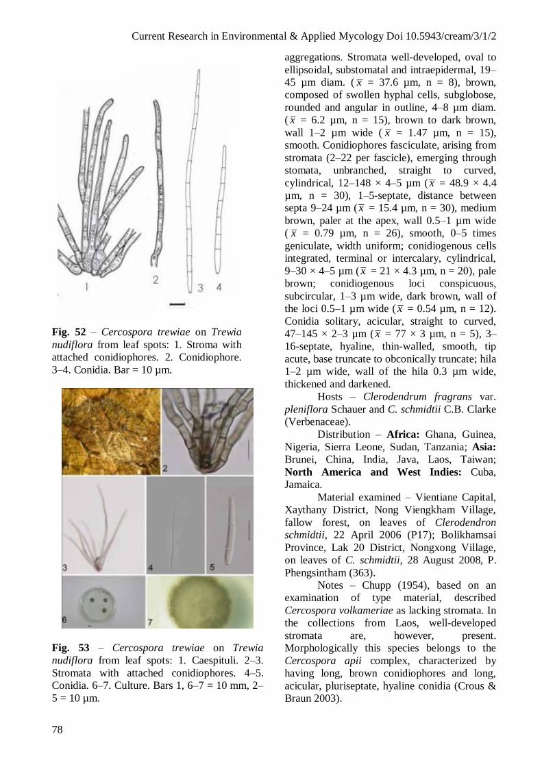
Current Research in Environmental & Applied Mycology Doi 10.5943/cream/3/1/2
78
Fig. 52 – Cercospora trewiae on Trewia
nudiflora from leaf spots: 1. Stroma with
attached conidiophores. 2. Conidiophore.
3–4. Conidia. Bar = 10 µm.
Fig. 53 – Cercospora trewiae on Trewia
nudiflora from leaf spots: 1. Caespituli. 2–3.
Stromata with attached conidiophores. 4–5.
Conidia. 6–7. Culture. Bars 1, 6–7 = 10 mm, 2–
5 = 10 µm.
aggregations. Stromata well-developed, oval to
ellipsoidal, substomatal and intraepidermal, 19–
45 µm diam. ( x = 37.6 µm, n = 8), brown,
composed of swollen hyphal cells, subglobose,
rounded and angular in outline, 4–8 µm diam.
( x = 6.2 µm, n = 15), brown to dark brown,
wall 1–2 µm wide ( x = 1.47 µm, n = 15),
smooth. Conidiophores fasciculate, arising from
stromata (2–22 per fascicle), emerging through
stomata, unbranched, straight to curved,
cylindrical, 12–148 × 4–5 µm (x = 48.9 × 4.4
µm, n = 30), 1–5-septate, distance between
septa 9–24 µm (x = 15.4 µm, n = 30), medium
brown, paler at the apex, wall 0.5–1 µm wide
( x = 0.79 µm, n = 26), smooth, 0–5 times
geniculate, width uniform; conidiogenous cells
integrated, terminal or intercalary, cylindrical,
9–30 × 4–5 µm (x = 21 × 4.3 µm, n = 20), pale
brown; conidiogenous loci conspicuous,
subcircular, 1–3 µm wide, dark brown, wall of
the loci 0.5–1 µm wide ( x = 0.54 µm, n = 12).
Conidia solitary, acicular, straight to curved,
47–145 × 2–3 µm (x = 77 × 3 µm, n = 5), 3–
16-septate, hyaline, thin-walled, smooth, tip
acute, base truncate to obconically truncate; hila
1–2 µm wide, wall of the hila 0.3 µm wide,
thickened and darkened.
Hosts – Clerodendrum fragrans var.
pleniflora Schauer and C. schmidtii C.B. Clarke
(Verbenaceae).
Distribution – Africa: Ghana, Guinea,
Nigeria, Sierra Leone, Sudan, Tanzania; Asia:
Brunei, China, India, Java, Laos, Taiwan;
North America and West Indies: Cuba,
Jamaica.
Material examined – Vientiane Capital,
Xaythany District, Nong Viengkham Village,
fallow forest, on leaves of Clerodendron
schmidtii, 22 April 2006 (P17); Bolikhamsai
Province, Lak 20 District, Nongxong Village,
on leaves of C. schmidtii, 28 August 2008, P.
Phengsintham (363).
Notes – Chupp (1954), based on an
examination of type material, described
Cercospora volkameriae as lacking stromata. In
the collections from Laos, well-developed
stromata are, however, present.
Morphologically this species belongs to the
Cercospora apii complex, characterized by
having long, brown conidiophores and long,
acicular, pluriseptate, hyaline conidia (Crous &
Braun 2003).

Current Research in Environmental & Applied Mycology Doi 10.5943/cream/3/1/2
79
Fig. 54 – Cercospora volkameriae from
Clerodendron schmidtii: 1–4. Conidiophores.
5. Stroma with attached conidiophores. 6–10.
Conidia. Bar = 10 µm.
Fig. 55 – Cercospora volkameriae on
Clerodendron schmidtii: 1–2. Lesions on host
leaves (1. upper surface, 2. lower surface). 3.
Stromata. 4–5. Stromata with attached
conidiophores. 6–10. Conidiophores. 11–12.
Conidia. 13. Base of conidium. Bars 1–2 = 10
mm, 3–13 = 10 µm.
Literature – Saccardo (1913:1424),
Chupp (1954: 597), Vasudeva (1963: 212), Ellis
(1976: 292), Hsieh & Goh (1990: 346), Braun
(2000: 74), Shin & Kim (2001: 107), Crous &
Braun (2003: 428).
(28) Cercospora zinniae Ellis & G. Martin, J.
Mycol. 1: 20, 1885. Figs 56–57.
= Cercospora atricincta Heald & F.A.
Wolf, Mycologia 3: 14, 1911.
= Cercosporina zinniae Takah. & Yosh.,
Pl. Protect. Tokyo 7: 17, 1953.
(= Cercospora apii s. lat.).
Leaf spots angular to irregular, 1–3 mm
diam., reddish brown to grey- brown in the
centre, with yellowish to pale brown margin.
Caespituli amphigenous, scattered, dark brown.
Mycelium internal; hyphae branched, 2–4 µm
wide ( x = 3 µm, n = 9), septate, constricted at
the septa, distance between septa 5–10 µm (x =
9 µm, n = 9), brownish or green-hyaline, wall
0.3–0.5 µm wide ( x = 0.49 µm, n = 9), smooth,
forming plate-like plectenchymatous stromatic
hyphal aggregations. Stromata lacking to well
developed, oval to ellipsoidal, substomatal, 10–
20 µm diam. ( x = 16.3 µm, n = 4), brown,
composed of swollen hyphal cells, subglobose,
rounded and angular in outline, 5–10 µm wide
( x = 5.5 µm, n = 30), brown to dark brown,
wall 0.5–0.8 µm wide ( x = 0.62 µm, n = 30),
smooth. Conidiophores solitary or fasciculate,
arising from stromata (2–10 per fascicle),
emerging through stomata, unbranched, straight
to curved, cylindrical, 32–95 × 4–6 µm ( x =
77.6 × 5.14 µm, n = 7), 1–7-septate, distance
between septa 12–25 µm ( x = 16.6 µm, n =
18), medium brown, paler at the apex, wall 0.5–
0.8 µm wide ( x = 0.7 µm, n = 18), smooth, 0–1
times geniculate, width uniform. Conidiogenous
cells integrated, terminal, cylindrical, 14–25 ×
4–5 µm ( x = 19 × 4.4 µm, n = 12), pale brown;
conidiogenous loci conspicuous, subcircular, 1–
3 µm wide ( x = 2 µm, n = 10), dark brown,
wall 0.5–0.8 µm thick ( x = 0.56 µm, n = 10).
Conidia solitary, acicular, straight to curved,
30–102 × 2–3 µm ( x = 72 × 2.4 µm, n = 12),
3–9-septate, hyaline, thin-walled, smooth, tip
acute, base truncate to obconically truncate, hila
thickened and darkened, 1.5–3 µm wide ( x =
2.2 µm, n = 12), wall of the hila 0.3–0.5 µm (x
= 0.34 µm, n = 12) thick.

Current Research in Environmental & Applied Mycology Doi 10.5943/cream/3/1/2
80
Hosts – Cosmos sp., Zinnia elegans
Jack., Z. multiflora L., Z. pauciflora L., Z.
peruviana L., Z. violacea Cav., Zinnia sp.
(Asteraceae).
Distribution – Africa: Ghana, Malawi,
Mauritius, Nigeria, Tanzania, Togo, South
Africa, Sudan, Uganda, Zambia, Zimbabwe;
Asia: Bangladesh, Bhutan, Brunei, China,
Hong Kong, India, Indonesia, Laos, Malaysia,
Japan, Korea, Myanmar, Nepal, Pakistan,
Philippines, Singapore, Taiwan; Europe:
Lithuania; North America and West Indies:
Cuba, Dominican Republic, El Salvador,
Guatemala, Haiti, Jamaica, Mexico, Panama,
Puerto Rico, Trinidad & Tobago, USA (AI, CO,
FL, IN, KS, MS, NC, PA, SC, SE states, TX);
Oceania: American Samoa, Cook Islands, Fiji,
Guam, Micronesia, New Caledonia, Papua New
Guinea, Samoa, Solomon Islands, Tonga,
Tuvalu, Vanuatu, Virgin Islands; South
America: Brazil, Colombia, Venezuela.
Material examined – Luang Prabang
Province, Nan District, Phonhin Village, on
leaves of Zinnia elegans, July 2006, P.
Phengsintham (P82).
Notes – The collection from Laos agrees
with the description of Cercospora zinniae
published by Chupp (1954).
Literature – Saccardo (1886, 443; 1931,
873), Chupp (1954: 186), Vasudeva (1963:
216), Katsuki (1965: 24), Ellis (1976: 252),
Shin & Kim (201: 111), Crous & Braun (2003:
434).
Fig. 56 – Cercospora zinniae from Zinnia
elegans: 1. Stroma with attached
conidiophore. 2–4. Conidiophores. 5–8.
Conidia. Bar = 10 µm.
Fig. 57 – Cercospora zinniae from Zinnia
elegans: 1–2. Lesions on host leaves (1. upper
surface, 2. lower surface). 3. Caespituli. 4–7.
Conidiophores. 8–12. Conidia. Bars 1–2 = 10
mm, 4–12 = 10 µm.
(29) Passalora aenea (Cif.) U. Braun & Crous,
in Crous & Braun, CBS Biodiversity Series 1:
46, 2003. Figs 58–59.
Berteromyces aeneus Cif., Sydowia 8:
267, 1954.
= Cercospora cassiae Henn., Bull.
Herb. Boissier 1: 121, 1893, non Passalora
cassiae Syd., 1939.
Cercosporidium cassiae (Henn.)
Deighton, Mycol. Pap. 112: 66, 1967.
Phaeoisariopsis cassiae (Henn.) Arx,
Proc. K. Ned. Akad. Wet., C 86: 43, 1983.
Passalora cassiae (Henn.) Poonam
Srivast, J. Living World 1: 114, 1994 (nom.
inval. et illeg.).
Passalora cassiae (Henn.) U. Braun,
Mikol. Fitopatol. 30: 6, 1996 (nom. illeg.).
= Cercospora cassiicola Roum, Fungi
sel. exs. No 4486, 1888.
Leaf spots subcircular to irregular,
zonate spots, 1–5 mm diam., brown or dark
brown in the centre, and with dark brown
margin. Caespituli hypophyllous, scattered.
Mycelium internal, inconspicuous. Stromata
oval or ellipsoidal, 10–35 µm diam. ( x = 23.72
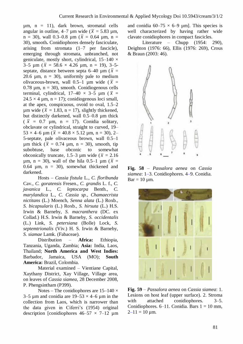
Current Research in Environmental & Applied Mycology Doi 10.5943/cream/3/1/2
81
µm, n = 11), dark brown, stromatal cells
angular in outline, 4–7 µm wide ( x = 5.83 µm,
n = 30), wall 0.3–0.8 µm ( x = 0.64 µm, n =
30), smooth. Conidiophores densely fasciculate,
arising from stromata (1–7 per fascicle),
emerging through stromata, unbranched, not
geniculate, mostly short, cylindrical, 15–140 ×
3–5 µm ( x = 58.6 × 4.26 µm, n = 19), 3–5-
septate, distance between septa 6–40 µm ( x =
20.6 µm, n = 30), uniformly pale to medium
olivaceous-brown, wall 0.5–1 µm wide ( x =
0.78 µm, n = 30), smooth. Conidiogenous cells
terminal, cylindrical, 17–40 × 3–5 µm ( x =
24.5 × 4 µm, n = 17); conidiogenous loci small,
at the apex, conspicuous, ovoid to oval, 1.5–2
µm wide ( x = 1.83, n = 17), slightly thickened,
but distinctly darkened, wall 0.5–0.8 µm thick
( x = 0.7 µm, n = 17). Conidia solitary,
obclavate or cylindrical, straight to curved, 19–
53 × 4–6 µm ( x = 40.8 × 5.12 µm, n = 30), 2–
5-septate, pale olivaceous brown, wall 0.5–1
µm thick ( x = 0.74 µm, n = 30), smooth, tip
subobtuse, base obconic to somewhat
obconically truncate, 1.5–3 µm wide ( x = 2.16
µm, n = 30), wall of the hila 0.5–1 µm ( x =
0.64 µm, n = 30), somewhat thickened and
darkened.
Hosts – Cassia fistula L., C. floribunda
Cav., C. goratensis Fresen., C. grandis L. f., C.
javanica L., C. leptocarpa Benth., C.
marylandica L., C. Cassia sp., Chamaecrista
nictitans (L.) Moench, Senna alata (L.) Roxb.,
S. bicapsularis (L.) Roxb., S. hirsuta (L.) H.S.
Irwin & Barneby, S. macranthera (DC. ex
Collad.) H.S. Irwin & Barneby, S. occidentalis
(L.) Link, S. petersiana (Bolle) Lock, S.
septemtrionalis (Viv.) H. S. Irwin & Barneby,
S. siamae Lamk. (Fabaceae).
Distribution – Africa: Ethiopia,
Tanzania, Uganda, Zambia; Asia: India, Laos,
Thailand; North America and West Indies:
Barbador, Jamaica, USA (MO); South
America: Brazil, Colombia.
Material examined – Vientiane Capital,
Xaythany District, Xay Village, Village area,
on leaves of Cassia siamea, 28 December 2008,
P. Phengsintham (P399).
Notes – The conidiophores are 15–140 ×
3–5 µm and conidia are 19–53 × 4–6 µm in the
collection from Laos, which is narrower than
the data given in Ciferri’s (1954) original
description [conidiophores 46–57 × 7–12 µm
and conidia 60–75 × 6–9 µm]. This species is
well characterized by having rather wide
clavate conidiophores in compact fascicles.
Literature – Chupp (1954: 290),
Deighton (1976: 66), Ellis (1976: 269), Crous
& Braun (2003: 46).
Fig. 58 – Passalora aenea on Cassia
siamea: 1–3. Conidiophores. 4–9. Conidia.
Bar = 10 µm.
Fig. 59 – Passalora aenea on Cassia siamea: 1.
Lesions on host leaf (upper surface). 2. Stroma
with attached conidiophores. 3–5.
Conidiophores. 6–11. Conidia. Bars 1 = 10 mm,
2–11 = 10 µm.

Current Research in Environmental & Applied Mycology Doi 10.5943/cream/3/1/2
82
(30) Passalora bougainvilleae (Munt.-Cvetk.)
R.F. Castañeda & U. Braun, Cryptog. Bot. 2:
291, 1991. Figs 60–61.
Cercospora bougainvilleae Munt.-
Cvetk., Revista Argent. Agron. 24: 84, 1957
Cercosporidium bouganvilleae
(Munt.-Cvetk.) Sobers & C.P. Seymour, Proc.
Florida State Hort. Soc. 81: 398 ‘1968’, 1969.
Leaf spots circular to irregular, 1–5 mm
diam., pale brown or white in the centre, and
with dark brown margin. Caespituli
hypophyllous, inconspicuous. Mycelium
internal; hyphae branched, 2–3 µm wide,
septate, constricted at the septa, distance
between septa 11–13 µm, subhyaline, wall
smooth. Stromata oval or ellipsoidal, 15–45 µm
diam. ( x = 28 µm, n = 9), dark brown,
stromatal cells angular in outline, 5–10 µm
wide ( x = 7.67 µm, n = 12), wall 0.8–1 µm ( x = 0.95 µm, n = 12), smooth. Conidiophores
fasciculate, arising from stromata (14–24 per
fascicle), emerging through stromata, not
branched, not geniculate, mostly short,
cylindrical, 12–63 × 4–6 µm ( x = 31.87 × 4.97
µm, n = 30), 0–2-septate, distance between
septa 8–16 µm ( x = 11 µm, n = 6), uniformly
pale to medium olivaceous-brown, wall 0.5–0.7
µm wide ( x = 0.57 µm, n = 9), smooth.
Conidiogenous cells integrated, terminal,
cylindrical, 12–35 × 5–6 µm ( x = 20 × 5 µm, n
= 8); conidiogenous loci small, at the apex,
conspicuous, ovoid to oval, 1–3 µm ( x = 2 µm,
n = 21), slightly thickened, but distinctly
darkened, wall 0.5–0.9 µm wide ( x = 0.68 µm,
n = 21). Conidia solitary, obclavate or
cylindrical, straight to curved, 6–65 × 4.5–6 µm
( x = 43 × 5.87 µm, n = 30), 2–4-septate, pale
olivaceous brown, wall 0.5–1 µm wide ( x =
0.78 µm, n = 30), smooth, tip subobtuse, base
obconic, hila 1–3 µm wide ( x = 1.9 µm, n = 6),
wall 0.5–1 µm ( x = 0.7 µm, n = 6), thickened
and darkened.
Colonies on PDA after 3 weeks at 25°C
with white-grey in the centre, and dark grey at
the margin, reaching 4 mm diam., hyphae 2–8
µm wide ( x = 4.23 µm, n = 30), septate,
constricted at the septa, distances between septa
7–10 µm ( x = 13.4 µm, n = 30), hyaline or
brownish, wall 0.3–1 µm ( x = 0.65 µm, n =
10), smooth. Conidia not formed in culture.
Hosts – Bougainvillea glabra Choisy, B.
spectabilis Willd. (Nyctaginaceae).
Distribution – Asia: Brunei, China,
India, Indonesia, Japan, Laos, Thailand; North
America and West Indies: Cuba, El Salvador,
Jamaica, USA (FL, HI); South America:
Argentina, Brazil, Venezuela.
Material examined – Vientiane Capital,
Xaythany District, Dong Dok Village, on leaves
of Bougainvillea spectabilis, 15 April 2006, P.
Phengsintham (P05); Vientiane Province, Home
District, Houay Xay Village, on leaves of B.
spectabilis, 28 May 2009, P. Phengsintham
(P411).
Notes – On account of conspicuous,
slightly thickened and somewhat darkened
conidiogenous loci and conidial hila, the
collections from Laos clearly belong to
Passalora bougainvilleae. In the collections
from Laos, the conidia are quite smooth-walled,
differing from Ellis (1976) who described
minutely verruculose conidia. However, conidia
of this species are at first smooth and may turn
asperulate with age.
Literature – Ellis (1976: 297), Crous &
Braun (2003: 86).
Fig. 60 – Passalora bougainvilleae on
Bougainvillea spectabilis: 1. Stroma. 2.
Stroma with Conidiophore. 3. Conidiophore.
4. Conidiophore with attached young
conidium. 5–9. Conidia. Bar = 10 µm.

Current Research in Environmental & Applied Mycology Doi 10.5943/cream/3/1/2
83
Fig. 61 – Passalora bougainvilleae from
Bougainvillea spectabilis from leaf spots: 1–2.
Lesions on host leaves (1. upper surface, 2.
lower surface). 3. Stroma with attached
conidiophores. 4–5. Conidiophores. 6.
Conidiophore with attached Conidium. 7–10.
Conidia. 11–12. Culture. Bars 1–2, 11 = 10
mm, 3–10 = 10 µm.
(31) Passalora capsicicola (Vassiljevsky) U.
Braun & F.O. Freire, Cryptog. Mycol. 23: 299,
2002. Figs 62–63.
Cercospora capsicicola Vassiljevsky,
in Vassiljevsky & Karakulin, Fungi imperfecti
parasitici, 1, Hyphomycetes: 344, 1937.
Phaeoramularia capsicicola
(Vassiljevsky) Deighton, in Ellis, More
Dermatiaceous Hyphomycetes: 323, 1976.
Phaeoramularia capsicicola
(Vassiljevsky) Deighton, Trans. Brit. Mycol.
Soc. 67: 140, 1976.
= Cercospora capsici E.J. Marchal &
Stayaert, Bull. Soc. Roy. Bot. Belgique 61: 167,
1929.
Cladosporium capsici Kovatch., Z.
Pflanzenkr. 48: 321, 1938.
= Cercospora capsici Unamuno, Bol.
Soc. Esp. Hist. Nat. 32: 161, 1932.
= Cercospora unamunoi Castell.,
Revista Agric. Subtrop. Trop. 42: 20, 1948.
= Phaeoramularia unamunoi (Castell.)
Munt.-Cvetk., Lilloa. 30: 183, 1960.
Leaf spots circular to slightly irregular,
1–8 mm diam., dark brown in the centre, dark
brown to yellowish margin. Caespituli
amphigenous, inconspicuous. Mycelium
internal; internal hyphae branched, 2–3 µm
wide (x = 2.5 µm, n = 8), septate, constricted at
the septa, distances between septa 5–18 µm ( x
= 9.75 µm, n = 8), subhyaline, wall 0.3–0.5 µm
wide ( x = 0.4 µm, n = 8), smooth. Stromata
substomatal, intraepidermal, ellipsoidal, 17–50
µm diam. ( x = 36.3 µm, n = 9), brown,
stromatal cells 5–9 µm diam. ( x = 7 µm, n =
10), wall 0.5–0.8 µm wide ( x = 0.53 µm, n =
10), smooth. Conidiophores fasciculate, arising
from stromata (1–5 per fascicle), or born on
external mycelium, unbranched, 0–1 geniculate,
cylindrical, straight to curved, 13–34 × 4–6 µm
( x = 23.3 × 5 µm, n = 10), 0–1-septate,
distance between septa 5–25 µm (x = 14 µm, n
= 14), pale brown or olivaceous-brown; wall
0.5–0.8 µm wide ( x = 0.52 µm, n = 14),
smooth. Conidiogenous cells integrated,
terminal, cylindrical, tapering to the apex, 10–
20 × 4–5 µm ( x = 16.7 × 4.5 µm, n = 6);
conidiogenous loci (scars) small, slightly
thickened and darkened, 1–2 µm diam. ( x =
1.42 µm, n = 6); wall of the loci 0.5–0.8 µm
thick ( x = 0.5 µm, n = 7). Conidia solitary or
catenate, cylindrical or narrowly obclavate,
straight to curved, 14–92 × 3–6 µm (x = 44.07
× 4.28 µm, n = 14), 0–9-septate, slightly
constricted at the septa, subhyaline or
olivaceous-brown, wall 0.3–0.5 µm thick (x =
0.47 µm, n = 14); apex subobtuse, 1.5–2 µm
wide (x = 1.58 µm, n = 6), wall of the hila 0.3–
0.5 µm wide ( x = 0.46 µm, n = 6), darkened;
based long obconically truncate, hila 1.5–2 µm
wide (x = 1.9 µm, n = 14), wall of the hila 0.3–
0.5 µm wide ( x = 0.48 µm, n = 14), darkened.
Colonies on PDA after 3 weeks at 25°C
white to grey in the centre, margin dark grey,
reaching 3–6 mm diam., hyphae 1–4(–5) µm
wide ( x = 1.9 µm, n = 30), septate, distance
between septa 5–19 µm ( x = 12.37 µm, n =
30), primary mycelium brownish, but the
second and following ones hyaline, wall
smooth. Conidia not formed in culture.
Hosts – Capsicum annuum L., C.
frutescens L., C. grossum L., Capsicum sp.
(Solanaceae).

Current Research in Environmental & Applied Mycology Doi 10.5943/cream/3/1/2
84
Distribution – Africa: Congo, Ethiopia,
Ghana, Kenya, Malawi, Mauritius, Morocco,
Nigeria, Senegal, Sierra Leone, Somalia, Sudan,
Tanzania, Uganda, Zambia, Zimbabwe; Asia:
Cambodia, China, India, Indonesia, Jordan,
Laos, Malaysia, Myanmar, Nepal, Thailand,
Yemen; Europe: Romania; North America
and West Indies: French Antilles, Jamaica,
Panama, USA (CA, FL, GA, TX); South
America: Argentina, Brazil.
Material examined – Xiengkhouang
Province, Paek District, Phonsavan Village,
garden, on leaves of Capsicum annuum, 3
January 2010, P. Phengsintham (P513).
Notes – The collection from Laos is
similar to the description of Passalora
capsicicola in Ellis (1976) [conidiophores up to
70 × 3–5 µm and conidia 17–80 × 3–5 µm].
Literature – Saccardo (1971: 1336),
Chupp (1954: 552), Vadsudeva (1963: 206),
Ellis (1976: 323), Crous & Braun (2003: 103).
Fig. 62 – Passalora capsicicola on Capsicum
annuum: 1. Stroma with attached conidiophores.
2–3. Conidiophore. 4–9. Conidia. Bar = 10 µm.
Fig. 63 – Passalora capsicicola on Capsicum
annuum on host leaf: 1. Lesions on host leaf
(upper surface). 3. Caespituli. 4. Stroma with
attached conidiophores. 5–9. Conidia. Bars 1–2
= 10 mm, 4–9 = 10 µm.
(32) Passalora dipterocarpi Phengsintham,
Chukeatirote, Abdelsalam, K.D. Hyde & U.
Braun, Cryptog.. Mycol. 31(2): 167, 2010. Figs
64–65.
Leaf spots circular to slightly irregular,
2–7 mm diam., at first reddish, later becoming
dark brown in the centre, the oldest leaf spots
having a grey to reddish margin. Caespituli
amphigenous, inconspicuous. Mycelium
internal and external. Internal hyphae branched,
1–3 µm wide ( x = 2 µm, n = 17), septate,
constricted at the septa, distances between septa
4–10 µm ( x = 6.47 µm, n = 17), subhyaline,
wall 0.5–0.8 µm wide ( x = 0.63 µm, n = 17),
smooth; external hyphae, branched, 2–5 µm
wide (x = 2.60 µm, n = 30), septate, constricted
at the septa, distance between septa 4–19 µm
(x = 10.1 µm, n = 30), brownish to dark brown,
wall 0.3–0.9 µm wide ( x = 0.63 µm, n = 30),
smooth. Stromata substomatal, intraepidermal,

Current Research in Environmental & Applied Mycology Doi 10.5943/cream/3/1/2
85
ellipsoidal, 15–45 µm diam. ( x = 27.27 µm, n =
15) brown, stromatal cells 3–7 µm diam. ( x =
4.9 µm, n = 30), wall 0.5–0.8 µm wide ( x =
0.69 µm, n = 30), smooth. Conidiophores single
or fasciculate, arising from stromata (1–12 per
fascicle), or born on external mycelium,
unbranched or branched, 0–1 geniculate,
cylindrical, straight to curved, (9–)14–48(–67)
× (2–)3–4 µm ( x = 27 × 3.3 µm, n = 30), 1–9-
septate, distance between septa 4–10 µm ( x =
6.7 µm, n = 30), pale brown or olivaceous-
brown; wall 0.3–0.9 µm wide ( x = 0.70 µm, n
= 25), smooth. Conidiogenous cells intergratae,
terminal, cylindrical, tapering to the apex, 5–10
× 2–4 µm ( x = 6,55 × 2.95 µm, n = 22);
conidiogenous loci (scars) small, thickened and
slightly darkened, 1–2 µm diam., wall of the
loci approximately 0.8 µm thick. Conidia
solitary or caternate, cylindrical or narrowly
obclavate, straight to curved, 4–30(–36) × (1–
)2–5 µm ( x = 15.33 × 2.71 µm, n = 30), 0–5-
sepate, slightly constricted at the septa,
subhyaline or olivaceous brown, wall 0.25–1
µm thick ( x = 0.57 µm, n = 30), apex
subobtuse, based long obconically truncate, hila
0.8–2 µm wide ( x = 1.2 µm, n = 14), wall of
the hila 0.6–1 µm wide ( x = 0.86 µm, n = 14),
darkened.
Colonies on PDA after 3 weeks at 25°C
white–grey in the centre, margin dark grey,
reaching 3–6 mm diam., hyphae 1–5 µm wide
( x = 1.9 µm, n = 30), septate, distances
between septa 5–19 µm ( x = 12.37 µm, n =
30), primary mycelium brownish, but the
second and following ones hyaline, wall
smooth. Conidia not formed in culture.
Hosts – Dipterocarpus alatus Roxb., D.
obtusifolius Teijsm. ex Miq.
(Dipterocarpaceae).
Distribution – Asia: Laos.
Material examined – Vientiane Capital,
Xaythany District, Xay Village, on leaves of
Dipterocarpus alatus, rice paddy area, 19 April
2006, P. Phengsintham (P11, MFLU12-2198,
holotype); same locality and collector, on
leaves of D. obtusifolius, 8 August 2006, P.
Phengsintham (P129, paratype).
Notes – Several cercosporoid
hyphomycete species are known from hosts of
the Dipterocarpaceae, but all of them have been
described from Shorea spp. and belong in the
genera Pseudocercospora (conidiogenous loci
inconspicuous, unthickened, not darkened) and
Stenella (verruculose superficial mycelium),
viz., Pseudocercospora shoreae-robustae U.
Braun (1995) [ Pseudocercosporella shoreae
A.N. Rai, B. Rai & Kamal], Stenella shoreae
M.K. Khan & Kamal (Khan et al. 1995) and S.
shoreicola Crous & U. Braun (2003) [
Cercospora shoreae Thirum. & Chupp,
Pseudocercospora shoreae (Thirum. & Chupp)
Deighton, Stenella shoreae (Thirum. & Chupp)
Crous & U. Braun, non M.K. Khan & Kamal
1995]. The new species on Dipterocarpus spp.
from Laos belongs, however, in Passalora,
characterized by having smooth mycelium,
conspicuous conidiogenous loci and pigmented
conidia.
Literature – Braun (1995), Khan et al.
(1995), Crous & Braun (2003).
Fig. 64 – Passalora dipterocarpi on
Dipterocarpus alatus: 1. Stroma with attached
conidiophores. 2. Stroma with attached
conidiophores and conidia. 3. External hypha
with attached young conidiophores. 5.
Conidiophore. 6–17. Conidia. Bar = 10 µm.

Current Research in Environmental & Applied Mycology Doi 10.5943/cream/3/1/2
86
Fig. 65 – Passalora dipterocarpi on
Dipterocarpus alatus from leaf spots: 1.
Lesions on host leaf (upper surface). 2. Stroma
with attached conidiophoes. 3–7.
Conidiophores. 8–10. Conidia. 11. Culture.
Bars 1, 11 = 10 mm, 3–6 = 10 µm.
(33) Passalora erythrinae (Ellis & Everh.) U.
Braun & Crous, CBS Biodiversity Series 1:
176, 2003. Figs 66–67.
Cercospora erythrinae Ellis & Everh.,
J. Mycol. 3: 18, 1888.
Leaf spots subcircular to irregular,
zonate spots, 1–3 mm diam., pale brown or dark
brown in the centre, and with yellow-brown
margin. Caespituli hypophyllous, scattered.
Mycelium internal, inconspicuous. Stromata
oval or ellipsoidal, up to 40 µm diam., dark
brown. Conidiophores densely fasciculate,
arising from stromata (up to 17 per fascicle),
emerging through stromata, unbranched, not
geniculate, mostly short, cylindrical, 18–54 ×
4–6 µm ( x = 36.67 × 4.67 µm, n = 5), 0–1-
septate, distance between septa 5–19 µm ( x =
10.5 µm, n = 6), uniformly pale to medium
olivaceous-brown, wall 0.5–0.8 µm wide ( x =
0.60 µm, n = 5), smooth. Conidiogenous cells
terminal, cylindrical, 13 × 5 µm; conidiogenous
loci small, at the apex, conspicuous, ovoid to
oval, 2 µm wide, slightly thickened, but
distinctly darkened, wall 0.8–1 µm thick ( x =
0.9 µm, n = 5). Conidia solitary, obclavate or
cylindrical, straight to curved, 43–58 × 4–5 µm
(x = 50.29 × 4.86 µm, n = 8), 0–3-septate, pale
olivaceous brown, wall 0.5–0.8 µm thick ( x =
0.6 µm, n = 8), smooth, tip subobtuse, base
obconic, 2–3 µm wide (x = 2.2 µm, n = 8), wall
of the hila 0.8–1 µm ( x = 0.9 µm, n = 8),
somewhat thickened and darkened.
Hosts – Erythrina crista-galli L. and E.
stricta Roxb. (Fabaceae).
Distribution – Asia: Laos; North
America: USA (FL, LA, TX).
Material examined – Vientiane Capital,
Xaythany District, Xay Village, garden, on
leaves of Erythrina stricta, 27 April 2006, P.
Phengsintham (P27).
Notes – This species is characterized by
having conspicuous conidiogenous loci and
narrowly obclavate, subhyaline to pale
olivaceous conidia (Crous & Braun 2003). The
Laos collection is similar to the description of
Passalora erythrinae in Ellis (1976)
[conidiophores 30–50 × 3–5 µm and conidia
22–55× 3–3.5 µm, 2–5-septate].
Literature – Chupp (1954: 305), Ellis (1976:
269), Crous & Braun (2003: 176).
Fig. 66 – Passalora erytrinae on Erytrina
stricta: 1–3. Conidiophores. 4–9. Conidia. Bar
= 10 µm.
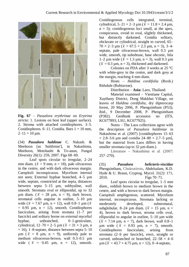
Current Research in Environmental & Applied Mycology Doi 10.5943/cream/3/1/2
87
Fig. 67 – Passalora erythrinae on Erytrina
stricta: 1. Lesions on host leaf (upper surface).
2. Stroma with attached conidiophores. 3–5.
Conidiophores. 6–11. Conidia. Bars 1 = 10 mm,
2–11 = 10 µm.
(34) Passalora haldinae C. Nakash. &
Meeboon [as 'haldiniae'], in Nakashima,
Meeboon, Motohashi & To-anun, Fungal
Diversity 26(1): 259, 2007. Figs 68–69.
Leaf spots circular to irregular, 2–24
mm diam. (x = 9 mm, n = 18), pale olivaceous
in the centre, and with dark olivaceous margin.
Caespituli inconspicuous. Mycelium internal
not seen; External hyphae branched, 4–5 µm
wide, septate, constricted at the septa, distances
between septa 5–15 µm, subhyaline, wall
smooth. Stromata oval or ellipsoidal, up to 32
µm diam. ( x = 28 µm, n = 9), dark brown,
stromatal cells angular in outline, 5–10 µm
wide ( x = 7.67 µm, n = 12), wall 0.8–1 µm ( x
= 0.95 µm, n = 12), smooth. Conidiophores
fasciculate, arising from stomata (1–7 per
fascicle) and solitary borne on external mycelial
hyphae, unbranched, 0–1-geniculate,
cylindrical, 12–70 × 2–5 µm ( x = 31 × 3 µm, n
= 16), 1–8-septate, distance between septa 5–10
µm ( x = 8 µm, n = 9), uniformly pale to
medium olivaceous-brown, wall 0.3–0.5 µm
wide ( x = 0.45 µm, n = 12), smooth.
Conidiogenous cells integrated, terminal,
cylindrical, 5–21 × 2–3 µm ( x = 11.8 × 2.4 µm,
n = 5); conidiogenous loci small, at the apex,
conspicuous, ovoid to oval, slightly thickened,
but distinctly darkened. Conidia solitary,
obclavate or cylindrical, straight to curved, 65–
70 × 2–3 µm (x = 67.5 × 2.3 µm, n = 3), 3–4-
septate, pale olivaceous-brown, wall 0.3 µm
wide, smooth, tip subobtuse, base obconic, hila
1–2 µm wide ( x = 1.3 µm, n = 3), wall 0.3 µm
(x = 0.3 µm, n = 3), thickened and darkened.
Colonies on PDA after 3 weeks at 25 °C
with white-grey in the centre, and dark grey at
the margin, reaching 4 mm diam.
Hosts – Haldina cordifolia (Roxb.)
Ridsdale (Rubiaceae).
Distribution – Asia: Laos, Thailand.
Material examined – Vientiane Capital,
Xaythany District, Dong Makkhai Village, on
leaves of Haldina cordifolia, dry dipterocarp
forest, 20 May 2006, P. Phengsintham (P53);
ibid., 9 December 2008, P. Phengsintham
(P382). GenBank accession no (ITS,
KC677893; LSU, KC677925).
Notes – The Laos collections agree with
the description of Passalora haldiniae in
Nakashima et al. (2007) [conidiophores 15–63
× 2.8–3.6 µm and conidia 24–80 × 2.7–5 µm],
but the material from Laos differs in having
smaller stromata (up to 32 µm diam.).
Literature – Nakashima et al. (2007:
257–270).
(35) Passalora helicteris-viscidae
Phengsintham, Chukeatirote, Abdelsalam, K.D.
Hyde & U. Braun, Cryptog. Mycol. 31(2): 171,
2010. Figs 70–71.
Leaf spots circular to irregular, 1–5 mm
diam., reddish brown to medium brown in the
centre, and with a brown to dark brown margin.
Caespituli amphigenous, scattered. Mycelium
internal, inconspicuous. Stromata lacking or
moderately developed, substomatal,
subglobular, 8–24 µm diam. ( x = 15 µm, n =
4), brown to dark brown, stroma cells oval,
ellipsoidal to angular in outline, 5–10 µm wide
(x = 7.14 µm, n = 7), dark brown, wall 0.8–1
µm wide ( x = 0.93 µm, n = 7), smooth.
Conidiophores fasciculate, arising from
stromata (2–6 per fascicle), erect, straight or
curved, unbranched or branched, 22–58 × 4–6
µm ( x = 43.7 × 4.75 µm, n = 12), 0–4-septate,
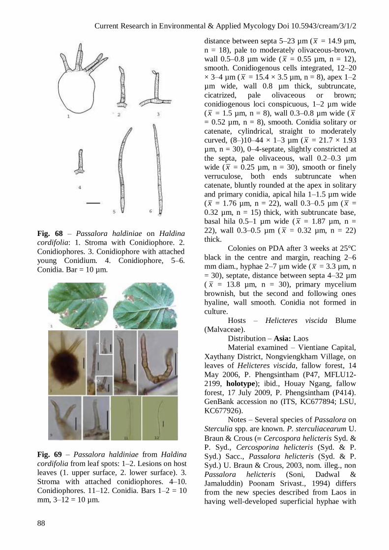
Current Research in Environmental & Applied Mycology Doi 10.5943/cream/3/1/2
88
Fig. 68 – Passalora haldiniae on Haldina
cordifolia: 1. Stroma with Conidiophore. 2.
Conidiophores. 3. Conidiophore with attached
young Conidium. 4. Conidiophore, 5–6.
Conidia. Bar = 10 µm.
Fig. 69 – Passalora haldiniae from Haldina
cordifolia from leaf spots: 1–2. Lesions on host
leaves (1. upper surface, 2. lower surface). 3.
Stroma with attached conidiophores. 4–10.
Conidiophores. 11–12. Conidia. Bars 1–2 = 10
mm, 3–12 = 10 µm.
distance between septa 5–23 µm ( x = 14.9 µm,
n = 18), pale to moderately olivaceous-brown,
wall 0.5–0.8 µm wide ( x = 0.55 µm, n = 12),
smooth. Conidiogenous cells integrated, 12–20
× 3–4 µm ( x = 15.4 × 3.5 µm, n = 8), apex 1–2
µm wide, wall 0.8 µm thick, subtruncate,
cicatrized, pale olivaceous or brown;
conidiogenous loci conspicuous, 1–2 µm wide
(x = 1.5 µm, n = 8), wall 0.3–0.8 µm wide ( x
= 0.52 µm, n = 8), smooth. Conidia solitary or
catenate, cylindrical, straight to moderately
curved, (8–)10–44 × 1–3 µm ( x = 21.7 × 1.93
µm, n = 30), 0–4-septate, slightly constricted at
the septa, pale olivaceous, wall 0.2–0.3 µm
wide ( x = 0.25 µm, n = 30), smooth or finely
verruculose, both ends subtruncate when
catenate, bluntly rounded at the apex in solitary
and primary conidia, apical hila 1–1.5 µm wide
(x = 1.76 µm, n = 22), wall 0.3–0.5 µm ( x =
0.32 µm, n = 15) thick, with subtruncate base,
basal hila 0.5–1 µm wide ( x = 1.87 µm, n =
22), wall 0.3–0.5 µm ( x = 0.32 µm, n = 22)
thick.
Colonies on PDA after 3 weeks at 25°C
black in the centre and margin, reaching 2–6
mm diam., hyphae 2–7 µm wide (x = 3.3 µm, n
= 30), septate, distance between septa 4–32 µm
( x = 13.8 µm, n = 30), primary mycelium
brownish, but the second and following ones
hyaline, wall smooth. Conidia not formed in
culture.
Hosts – Helicteres viscida Blume
(Malvaceae).
Distribution – Asia: Laos
Material examined – Vientiane Capital,
Xaythany District, Nongviengkham Village, on
leaves of Helicteres viscida, fallow forest, 14
May 2006, P. Phengsintham (P47, MFLU12-
2199, holotype); ibid., Houay Ngang, fallow
forest, 17 July 2009, P. Phengsintham (P414).
GenBank accession no (ITS, KC677894; LSU,
KC677926).
Notes – Several species of Passalora on
Sterculia spp. are known. P. sterculiacearum U.
Braun & Crous ( Cercospora helicteris Syd. &
P. Syd., Cercosporina helicteris (Syd. & P.
Syd.) Sacc., Passalora helicteris (Syd. & P.
Syd.) U. Braun & Crous, 2003, nom. illeg., non
Passalora helicteris (Soni, Dadwal &
Jamaluddin) Poonam Srivast., 1994) differs
from the new species described from Laos in
having well-developed superficial hyphae with

Current Research in Environmental & Applied Mycology Doi 10.5943/cream/3/1/2
89
solitary narrower conidiophores
(mycovellosiella-like) and longer subhyaline
conidia, up to 120 µm in length, with up to six
septa (Chupp 1954, Crous & Braun 2003,
Braun & Crous 2007). Passalora meridiniana
(Chupp) U. Braun & Crous is a phaeoramularia-
like species with densely fasciculate
conidiophores in coremioid conidiomata and
much longer conidia, up to 125 µm in length
(Chupp 1954, Crous & Braun 2003). Passalora
helicteris is easily distinguishable from the new
species by its much longer conidiophores, up to
450 µm, and much wider conidia, 20–50 × 7.5–
12.5 µm, formed singly (Soni et al. 1984, Crous
& Braun 2003).
Literature – Crous & Braun (2003: key).
Fig. 70 – Passalora helicteris-viscidae on
Helicteres viscida: 1. Stroma with attached
conidiophores. 2–7. Conidia. Bar = 10 µm.
(36) Passalora henningsii ( llesch.) R.F.
Casta eda & U. Braun, Cryptog. Bot. 1(1): 46,
1989. Figs 72–73.
Cercospora henningsii Allesch., in
Engler, Pflanzenwelt Ostafrikas, Teil C: 35,
1895.
Cercospora henningsii (Allesch.)
Deighton, in Ellis, More demateaceous
hyphomycetes: 295, 1976.
Passalora henningsii (Allesch.)
Poonam Srivast., J. Living World 1: 116, 1994.
= Cercospora cassavae Ellis & Everh.,
Bull. Torrey Bot. Club. 22: 438, 1895.
= Cercospora manihotis Henn.,
Hedwigia 41: 18, 1902.
Fig. 71 – Passalora helicteris-viscidae on
Helicteres viscida from leaf spots: 1–2. Lesions
on host leaves (1. upper surface, 2. lower
surface). 3. Caespituli. 4–5. Stroma with
attached conidiophores and conidium. 6.
Conidiophore; 6–9. Conidia.10. Culture. Bars
1–2, 10 = 10 mm, 4–9 = 10 µm.
= Cercospora cearae Petch, Ann. Roy.
Bot. Gard. Peradeniya 3: 10, 1910.
= Septogloeum manihotis Zimm.,
Centralbl. Bakteriol., Abt. 2, 8: 218, 1912.
= Helminthosporium manihotis Rangle,
Arch. Jard. Bot. Rio de Janeiro 2: 71, 1902.
Leaf spots circular to irregular, 4–15
mm diam., pale brown or dark brown.
Caespituli amphigenous, scattered. Mycelium
internal; hyphae branched, 3–6 µm wide (x = 4
µm, n = 9), septate, constricted at the septa,
distance between septa 5–20 µm ( x = 10.33
µm, n = 9), subhyaline, wall smooth. Stromata
oval or ellipsoidal, 17–40 µm diam. ( x = 28.36
µm, n = 11), dark brown, stroma cells angular
in outline, 3–8 µm wide ( x = 5.17 µm, n = 30),
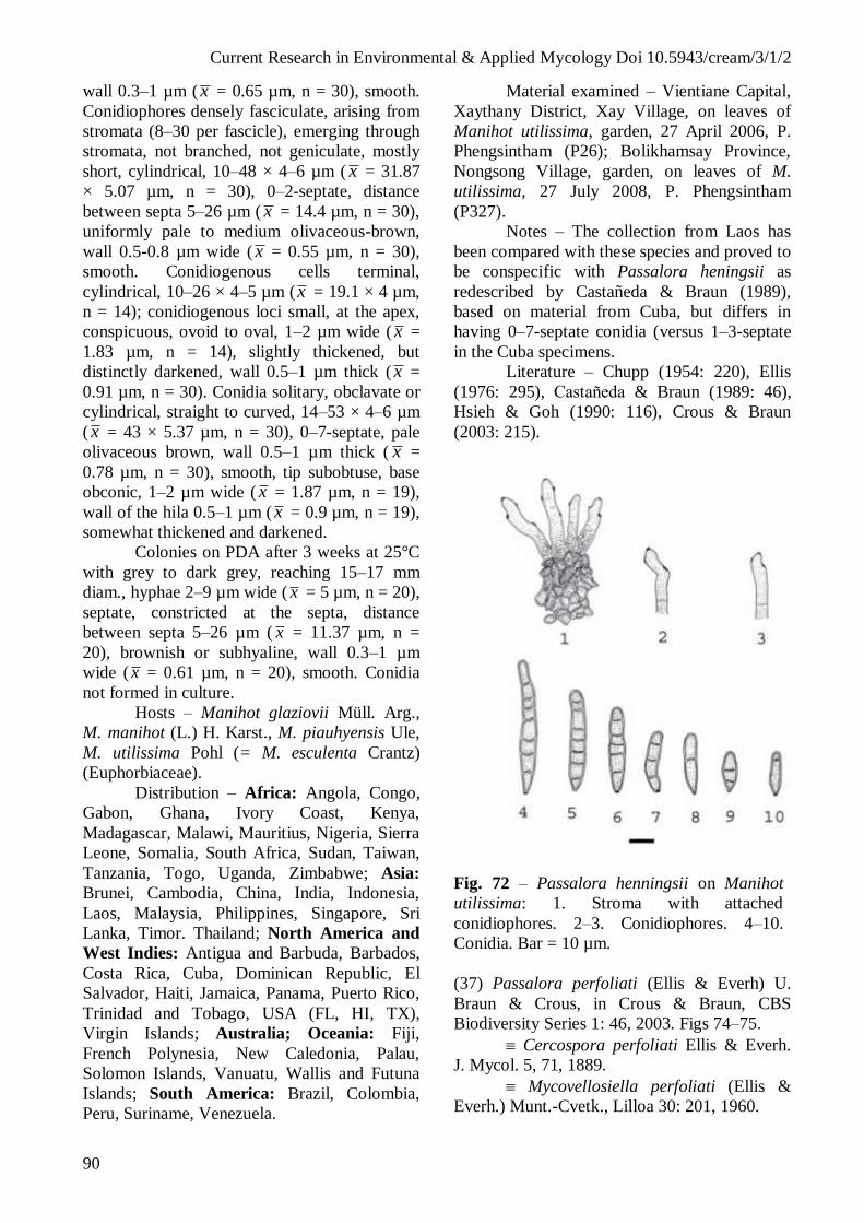
Current Research in Environmental & Applied Mycology Doi 10.5943/cream/3/1/2
90
wall 0.3–1 µm ( x = 0.65 µm, n = 30), smooth.
Conidiophores densely fasciculate, arising from
stromata (8–30 per fascicle), emerging through
stromata, not branched, not geniculate, mostly
short, cylindrical, 10–48 × 4–6 µm ( x = 31.87
× 5.07 µm, n = 30), 0–2-septate, distance
between septa 5–26 µm ( x = 14.4 µm, n = 30),
uniformly pale to medium olivaceous-brown,
wall 0.5-0.8 µm wide ( x = 0.55 µm, n = 30),
smooth. Conidiogenous cells terminal,
cylindrical, 10–26 × 4–5 µm (x = 19.1 × 4 µm,
n = 14); conidiogenous loci small, at the apex,
conspicuous, ovoid to oval, 1–2 µm wide ( x =
1.83 µm, n = 14), slightly thickened, but
distinctly darkened, wall 0.5–1 µm thick ( x =
0.91 µm, n = 30). Conidia solitary, obclavate or
cylindrical, straight to curved, 14–53 × 4–6 µm
( x = 43 × 5.37 µm, n = 30), 0–7-septate, pale
olivaceous brown, wall 0.5–1 µm thick ( x =
0.78 µm, n = 30), smooth, tip subobtuse, base
obconic, 1–2 µm wide ( x = 1.87 µm, n = 19),
wall of the hila 0.5–1 µm ( x = 0.9 µm, n = 19),
somewhat thickened and darkened.
Colonies on PDA after 3 weeks at 25°C
with grey to dark grey, reaching 15–17 mm
diam., hyphae 2–9 µm wide ( x = 5 µm, n = 20),
septate, constricted at the septa, distance
between septa 5–26 µm ( x = 11.37 µm, n =
20), brownish or subhyaline, wall 0.3–1 µm
wide ( x = 0.61 µm, n = 20), smooth. Conidia
not formed in culture.
Hosts – Manihot glaziovii Müll. Arg.,
M. manihot (L.) H. Karst., M. piauhyensis Ule,
M. utilissima Pohl (= M. esculenta Crantz)
(Euphorbiaceae).
Distribution – Africa: Angola, Congo,
Gabon, Ghana, Ivory Coast, Kenya,
Madagascar, Malawi, Mauritius, Nigeria, Sierra
Leone, Somalia, South Africa, Sudan, Taiwan,
Tanzania, Togo, Uganda, Zimbabwe; Asia:
Brunei, Cambodia, China, India, Indonesia,
Laos, Malaysia, Philippines, Singapore, Sri
Lanka, Timor. Thailand; North America and
West Indies: Antigua and Barbuda, Barbados,
Costa Rica, Cuba, Dominican Republic, El
Salvador, Haiti, Jamaica, Panama, Puerto Rico,
Trinidad and Tobago, USA (FL, HI, TX),
Virgin Islands; Australia; Oceania: Fiji,
French Polynesia, New Caledonia, Palau,
Solomon Islands, Vanuatu, Wallis and Futuna
Islands; South America: Brazil, Colombia,
Peru, Suriname, Venezuela.
Material examined – Vientiane Capital,
Xaythany District, Xay Village, on leaves of
Manihot utilissima, garden, 27 April 2006, P.
Phengsintham (P26); Bolikhamsay Province,
Nongsong Village, garden, on leaves of M.
utilissima, 27 July 2008, P. Phengsintham
(P327).
Notes – The collection from Laos has
been compared with these species and proved to
be conspecific with Passalora heningsii as
redescribed by Castañeda & Braun (1989),
based on material from Cuba, but differs in
having 0–7-septate conidia (versus 1–3-septate
in the Cuba specimens.
Literature – Chupp (1954: 220), Ellis
(1976: 295), Casta eda & Braun (1989: 46),
Hsieh & Goh (1990: 116), Crous & Braun
(2003: 215).
Fig. 72 – Passalora henningsii on Manihot
utilissima: 1. Stroma with attached
conidiophores. 2–3. Conidiophores. 4–10.
Conidia. Bar = 10 µm.
(37) Passalora perfoliati (Ellis & Everh) U.
Braun & Crous, in Crous & Braun, CBS
Biodiversity Series 1: 46, 2003. Figs 74–75.
Cercospora perfoliati Ellis & Everh.
J. Mycol. 5, 71, 1889.
Mycovellosiella perfoliati (Ellis &
Everh.) Munt.-Cvetk., Lilloa 30: 201, 1960.
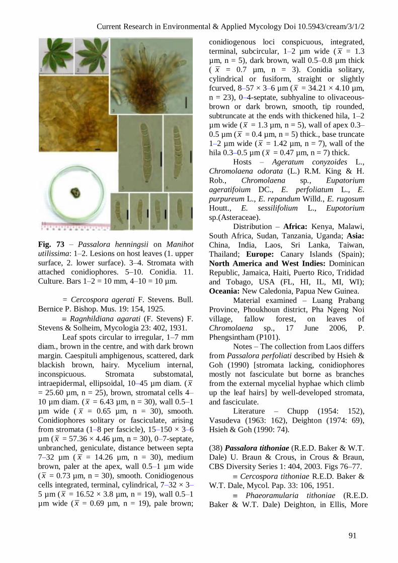
Current Research in Environmental & Applied Mycology Doi 10.5943/cream/3/1/2
91
Fig. 73 – Passalora henningsii on Manihot
utilissima: 1–2. Lesions on host leaves (1. upper
surface, 2. lower surface). 3–4. Stromata with
attached conidiophores. 5–10. Conidia. 11.
Culture. Bars 1–2 = 10 mm, 4–10 = 10 µm.
= Cercospora agerati F. Stevens. Bull.
Bernice P. Bishop. Mus. 19: 154, 1925.
Ragnhildiana agarati (F. Stevens) F.
Stevens & Solheim, Mycologia 23: 402, 1931.
Leaf spots circular to irregular, 1–7 mm
diam., brown in the centre, and with dark brown
margin. Caespituli amphigenous, scattered, dark
blackish brown, hairy. Mycelium internal,
inconspicuous. Stromata substomatal,
intraepidermal, ellipsoidal, 10–45 µm diam. ( x
= 25.60 µm, n = 25), brown, stromatal cells 4–
10 µm diam. ( x = 6.43 µm, n = 30), wall 0.5–1
µm wide ( x = 0.65 µm, n = 30), smooth.
Conidiophores solitary or fasciculate, arising
from stromata (1–8 per fascicle), 15–150 × 3–6
µm ( x = 57.36 × 4.46 µm, n = 30), 0–7-septate,
unbranched, geniculate, distance between septa
7–32 µm ( x = 14.26 µm, n = 30), medium
brown, paler at the apex, wall 0.5–1 µm wide
( x = 0.73 µm, n = 30), smooth. Conidiogenous
cells integrated, terminal, cylindrical, 7–32 × 3–
5 µm ( x = 16.52 × 3.8 µm, n = 19), wall 0.5–1
µm wide ( x = 0.69 µm, n = 19), pale brown;
conidiogenous loci conspicuous, integrated,
terminal, subcircular, 1–2 µm wide ( x = 1.3
µm, n = 5), dark brown, wall 0.5–0.8 µm thick
( x = 0.7 µm, n = 3). Conidia solitary,
cylindrical or fusiform, straight or slightly
fcurved, 8–57 × 3–6 µm (x = 34.21 × 4.10 µm,
n = 23), 0–4-septate, subhyaline to olivaceous-
brown or dark brown, smooth, tip rounded,
subtruncate at the ends with thickened hila, 1–2
µm wide ( x = 1.3 µm, n = 5), wall of apex 0.3–
0.5 µm ( x = 0.4 µm, n = 5) thick., base truncate
1–2 µm wide ( x = 1.42 µm, n = 7), wall of the
hila 0.3–0.5 µm ( x = 0.47 µm, n = 7) thick.
Hosts – Ageratum conyzoides L.,
Chromolaena odorata (L.) R.M. King & H.
Rob., Chromolaena sp., Eupatorium
ageratifoium DC., E. perfoliatum L., E.
purpureum L., E. repandum Willd., E. rugosum
Houtt., E. sessilifolium L., Eupotorium
sp.(Asteraceae).
Distribution – Africa: Kenya, Malawi,
South Africa, Sudan, Tanzania, Uganda; Asia:
China, India, Laos, Sri Lanka, Taiwan,
Thailand; Europe: Canary Islands (Spain);
North America and West Indies: Dominican
Republic, Jamaica, Haiti, Puerto Rico, Trididad
and Tobago, USA (FL, HI, IL, MI, WI);
Oceania: New Caledonia, Papua New Guinea.
Material examined – Luang Prabang
Province, Phoukhoun district, Pha Ngeng Noi
village, fallow forest, on leaves of
Chromolaena sp., 17 June 2006, P.
Phengsintham (P101).
Notes – The collection from Laos differs
from Passalora perfoliati described by Hsieh &
Goh (1990) [stromata lacking, conidiophores
mostly not fasciculate but borne as branches
from the external mycelial hyphae which climb
up the leaf hairs] by well-developed stromata,
and fasciculate.
Literature – Chupp (1954: 152),
Vasudeva (1963: 162), Deighton (1974: 69),
Hsieh & Goh (1990: 74).
(38) Passalora tithoniae (R.E.D. Baker & W.T.
Dale) U. Braun & Crous, in Crous & Braun,
CBS Diversity Series 1: 404, 2003. Figs 76–77.
Cercospora tithoniae R.E.D. Baker &
W.T. Dale, Mycol. Pap. 33: 106, 1951.
Phaeoramularia tithoniae (R.E.D.
Baker & W.T. Dale) Deighton, in Ellis, More

Current Research in Environmental & Applied Mycology Doi 10.5943/cream/3/1/2
92
Fig. 74 – Passalora perfoliati on Chromolaena
sp. from leaf spots. 1. Stromata with attached
conidiophores, 2–4. Conidiophores, 5–10.
Conidia. Bar = 10 µm.
Fig. 75 – Passalora perfoliati on Chromolaena
sp. from leaf spots. 1–2. Lesions on host leaves
(1. upper surface, 2. lower surface), 3.
Caespituli, 4–5. Stromata with attached
conidiophores and young conidia, 6–7.
Conidiophores, 8–12. Conidia. Bars 1–2 = 10
mm, 4–12 = 10 µm.
dematiaceous hyphomycetes: 319, 1976.
= Cercospora tithoniae Chidd.,
Mycopathol. Mycol. Appl. 17: 80, 1962.
= Cercospora tithoniicola J.M. Yen.,
Rev. Mycol. 31: 144, 1936.
Leaf spots circular to irregular, 1–15
mm diam., brown to dark brown in the centre,
and with a brown margin. Caespituli
amphigenous, scattered. Mycelium internal,
conspicuous; internal hyphae branched, 2–3 µm
wide ( x = 2.75 µm, n = 5), septate, constricted
at the septa, distance between septa 6–12 µm
(x = 9 µm, n = 5), brownish, subhyaline, wall
0.3–0.5 µm wide ( x = 0.45 µm, n = 5), smooth,
forming plate-like plectenchymatous stromatic
hyphal aggregations. Stromata developed,
substomatal, subglobular, 33–50 µm diam. (x =
42.4 µm, n = 5), brown to dark brown, stroma
cells oval, ellipsoidal to angular in outline, 5–14
µm wide (x = 8 µm, n = 24), dark brown, wall
0.5–0.8 µm wide ( x = 0.7 µm, n = 24), smooth.
Conidiophores fasciculate, arising from
stromata (8–36 per fascicle), erect, straight or
curved, unbranched, not geniculate, 14–144 ×
3–5 µm ( x = 61.7 × 3.33 µm, n = 12), 1–5-
septate, distance between septa 5–23 µm (x =
15.9 µm, n = 20), pale to moderately
olivaceous-brown, wall 0.5–0.8 µm wide ( x =
0.55 µm, n = 20), smooth. Conidiogenous cells
integrated, subtruncate, cicatrized, 9–23 × 3–4
µm ( x = 17.1 × 3.1 µm, n = 9), pale olivaceous
or brown; conidiogenous loci conspicuous, 1–2
µm wide ( x = 1.7 µm, n = 5), wall 0.5–0.8 µm
wide ( x = 0.56 µm, n = 5), smooth. Conidia
solitary or catenate, cylindrical, straight to
moderately curved, 17–75 × 3–6 µm (x = 32.23
× 3.46 µm, n = 20), 0–3-septate, slightly
constricted at the septa, pale olivaceous, wall
0.3–0.5 µm wide ( x = 0.33 µm, n = 20),
smooth or finely verruculose, both ends
subtruncate when catenate, bluntly rounded at
the apex in solitary and primary conidia, apical
hila 1.5–2 µm wide ( x = 1.54 µm, n = 10), wall
0.3–0.5 µm ( x = 0.36 µm, n = 10) thick, with
subtruncate base; basal hila 1.5–2 µm wide ( x
= 1.55 µm, n = 20), wall 0.3–0.5 µm (x = 0.35
µm, n = 20) thick.
Colonies on PDA after 3 weeks at 25°C
with grey to dark grey, reaching 10–18 mm
diam., hyphae 2–5 µm wide ( x = 53.53 µm, n =
26), septate, constricted at the septa, distance
between septa 6–20 µm ( x = 15.46 µm, n =

Current Research in Environmental & Applied Mycology Doi 10.5943/cream/3/1/2
93
26), brownish or subhyaline, wall 0.3–0.8 µm
wide ( x = 0.60 µm, n = 26), smooth. Conidia
solitary or catenate, cylindrical, straight to
moderately curved, 9–35 × 3–5 µm ( x = 20.72
× 3.72 µm, n = 11), 0–2-septate, slightly
constricted at the septa, pale olivaceous, wall
0.3–0.5 µm wide ( x = 0.46 µm, n = 11) thick;
apical hila 1–2 µm wide (x = 1.66 µm, n = 5),
wall 0.3–0.5 µm ( x = 0.43 µm, n = 5) thick,
with subtruncate base; basal hila 0.5–2 µm wide
( x = 1.63 µm, n = 11), wall 0.3–0.5 µm ( x =
0.42 µm, n = 11) thick.
Hosts – Tithonia diversifolia (Hemsl.)
A. Grey, T. speciosa (Hook.) Hook. ex Griseb.,
T. tagetiflora Lam., Viguera dentata (Cav.)
Spreng. (Asteraceae).
Distribution – Africa: Ivory Coast,
Mauritius; Asia: Hong Kong, India, Laos,
Singapore, Taiwan; North America and West
Indies: Barbados, Cuba, Trinidad and Tobago.
Material examined – Luangnamtha
Province, Luangnamtha District, Chaleunsouk
Village, fallow forest, on leaves of Tithonia
diversifolia, 20 February 2010, P.
Phengsintham (P572). GenBank accession no
(ITS, KC677895; LSU, KC677927).
Notes – The collection from Laos agrees
with the description of Passolora tithoniae in
Hsieh & Goh (1990) [conidiophores 30–90 × 3–
4 µm and conidia 30–50 × 3.5–5 µm].
Literature – Chupp (1954: 162), Ellis
(1976: 319), Hsieh & Goh (1990: 76), Crous &
Braun (2003: 404).
(39) Pseudocercospora alangii Y.L. Guo &
X.L. Liu, Mycosystema 2: 226, 1989. Figs 78–
79.
= Cercospora alangii M. Madal, Indian
J. Mycol. Res. 16: 311, 1978.
Leaf spots circular, 1–5 mm in diam.,
with brown to dark brown and brown margin.
Caespituli amphigenous, but chiefly
hypophyllous. Mycelium internal; hyphae
branched, 2–3 µm wide ( x = 2.5 µm, n = 5),
septate, constricted at the septa, distance
between septa 10–14 µm (x = 12 µm, n = 5),
subhyaline or hyaline, wall 0.3–0.5 µm wide ( x
= 0.4 µm, n = 5), smooth. Stromata developed,
substomatal, subglobular, 12–46 µm wide ( x =
29 µm, n = 5), brown to dark brown, stromatal
cells oval, ellipsoidal to angular in outline, 2–6
µm wide ( x = 3.8 µm, n = 30), dark brown,
Fig. 76 – Passalora tithoniae on Tithonia
diversifolia: 1. Stroma with attached
conidiophores. 2–7. Conidia. Bars = 10 µm.
Fig. 77 – Passalora tithoniae on Tithonia
diversifolia from leaf spots: 1. Caespituli. 2.
Internal mycelium. 3–4. Stromata with attached
conidiophores. 5. Apex of conidiophores. 6–11.
Conidia. Bars 2 –11 = 10 µm.

Current Research in Environmental & Applied Mycology Doi 10.5943/cream/3/1/2
94
wall 0.3–0.5 µm wide ( x = 0.35 µm, n = 30),
smooth. Conidiophores fasciculate, arising from
stromata (4–15 per fascicle), erect, straight or
curved, unbranched, geniculate, 6–19 × 2–4 µm
( x = 10.1 × 2.88 µm, n = 30), 0–1-septate,
distance between septa 4–14 µm ( x = 10 µm, n
= 30), pale to moderately olivaceous-brown,
paler and narrower towards the apex, wall 0.3–
0.5 µm wide ( x = 0.45 µm, n = 30), smooth.
Conidiogenous cells integrated, acute, 6–14 ×
2–4 µm ( x = 9.25 × 2.94 µm, n = 15), pale
olivaceous or brown; conidiogenous loci
inconspicuous. Conidia solitary, obclavate,
straight to moderately curved, 40–71 × 2–3 µm
( x = 60.4 × 2.6 µm, n = 15), 5–7-septate,
slightly constricted at the septa, pale olivaceous,
wall 0.3–0.5 µm wide ( x = 0.38 µm, n = 15),
smooth, obtuse at the apex, with long
obconically truncate base.
Colonies on PDA after 3 weeks at 25°C
dark grey in the centre and grey margin,
spreading surface ridged and smooth, 13–125
mm diam.
Hosts – Alangium kurzii Craib, A.
salviifolium (L. f.) Wangerin (Cornaceae, incl.
Alangiaceae).
Distribution – Asia: China, India, Laos.
Material examined: Phongsali Province,
Phongsali District, Hathin Village, Nam Ou
riverbank, on leaves of Alangium kurzii, 23
June 2010, P. Phengsintham (P596).
Notes – The collection from Laos agrees
with the description of Pseudocercospora
alangii published by Guo et al. (1998).
Literature – Guo et al. (1998: 17–18),
Crous & Braun (2003: 49).
(40) Pseudocercospora baliospermi (S.
Chowdry) Deighton, Mycol. Pap. 140: 139,
1976. Figs 80–81.
Cercospora baliospermi S. Chowdry,
Lloydia 24: 94, 1961.
= Cercospora baliospermi Pavgi & U.P.
Singh, Mycopathol. Mycol. Appl. 27: 90, 1965.
Leaf spots circular to irregular, 1–6 mm
diam., brown to dark brown in the centre, with a
brown to dark brown margin. Caespituli
amphigenous, scattered. Mycelium internal,
inconspicuous. Stromata developed,
substomatal, subglobular, 15–35 µm diam. ( x =
23 µm, n = 5), brown to dark brown, stromatal
cells oval, ellipsoidal to angular in outline, 5–7
Fig. 78 – Pseudocercospora alangii on
Alangium kurzii: 1. Stroma with attached
conidiophores. 2–7. Conidia. Bar = 10 µm.
Fig. 79 – Pseudocercospora alangii on
Alangium kurzii from leaf spots: 1. Lesions on
host leaf with caespituli. 2–3. Stromata with
attached conidiophores. 4–8. Conidia. 9–10.
Cultures. Bars 2–5 = 10 µm, 9 = 10 mm.

Current Research in Environmental & Applied Mycology Doi 10.5943/cream/3/1/2
95
µm wide ( x = 6.3 µm, n = 30), dark brown,
wall 0.5–0.8 µm wide ( x = 0.57 µm, n = 30),
smooth. Conidiophores solitary or fasciculate,
arising from stromata (5–9 per fascicle), erect,
straight or curved, unbranched, geniculate, 16–
60 × 2–5 µm ( x = 44.2 × 4.4 µm, n = 15), 2–5-
septate, distance between septa 5–16 µm ( x =
10.6 µm, n = 30), pale to moderately
olivaceous-brown, wall 0.5–0.8 µm wide ( x =
0.55 µm, n = 30), smooth. Conidiogenous cells
integrated, subtruncate, cicatrized, 10–17 × 4–5
µm ( x = 13.5 × 4.25 µm, n = 9), pale
olivaceous or brown; conidiogenous loci
inconspicuous. Conidia solitary, obclavate,
straight to moderately curved, 15–101 × 3–5
µm ( x = 51.1 × 3.77 µm, n = 30), 1–8-septate,
slightly constricted at the septa, pale olivaceous,
wall 0.3–0.5 µm wide ( x = 0.45 µm, n = 30),
smooth, bluntly rounded at the apex, with
subtruncate base, basal hila 1.5–2 µm wide ( x
= 1.78 µm, n = 30), wall 0.3–0.5 µm (x = 0.47
µm, n = 30) thick.
Hosts – Baliospermum montanum
(Willd.) Müll. Arg. (Euphorbiaceae).
Distribution – Asia: India, Laos,
Myanmar.
Material examined – Xiengkhouang
Province, Kham District, Napa Village, Fallow
forest, on leaves of Baliospermum montanum, 3
January 2010, P. Phengsintham (P504);
Vientiane Province, Thalad, fallow forest, on
leaves of B. montanum, April 2010, P.
Phengsintham (P548).
Notes – The collection from Laos agrees
well with the description in Deighton (1976).
Literature – Deighton (1976: 139),
Crous & Braun (2003: 755).
(41) Pseudocercospora buddlejae (W. Yamam)
Goh & W.H. Hsieh, Trans. Mycol. Soc. Republ.
China 2: 114, 1987. Figs 82–83.
Cercospora buddlejae W. Yamam.,
Trans. Nat. Hist. Soc. Formosa 26: 279, 1936.
= Pseudocercospora buddlejae
(Yamam.) X.J. Liu & Y.L. Guo, Mycosystema.
2: 230, 1989.
Leaf spots subcircular to irregular, 1–8
mm diam., at first greyish, later becoming
brown to dark brown in the center, brown to
dark brown at the margin. Caespituli
amphigenous, conspicuous. Mycelium internal,
inconspicuous. Stromata oval to ellipsoidal,
Fig. 80 – Pseudocercospora baliospermi on
Baliospermum montanum: 1. Stroma with
attached conidiophores. 2. Conidiophore. 3–7.
Conidia. Bar = 10 µm.
Fig. 81 – Pseudocercospora baliospermi on
Baliospermum montanum from leaf spots: 1–2.
Stromata with attached conidiophores. 4–7.
Conidia. Bar = 10 µm.

Current Research in Environmental & Applied Mycology Doi 10.5943/cream/3/1/2
96
20–45 µm diam. ( x = 32.2 µm, n = 5), brown to
dark brown, stromatal cells oval, ellipsoidal and
angular, 4–10 µm wide ( x = 7.5 µm, n = 30),
dark brown, wall 0.5–1 µm wide ( x = 0.7 µm,
n = 30), smooth. Conidiophores fasciculate,
arising from stromata (16–31 per fascicle),
unbranched, geniculate, 25–315 × 3–5 µm ( x =
103 × 4 µm, n = 15), 1–15-septate, slightly
constricted at the septa, distance between septa
11–25 µm long ( x = 19.8 µm, n = 30),
uniformly pale to medium brown, much paler
and more narrow toward the tip, wall 0.3–0.8
µm ( x = 0.55 µm, n = 30), smooth.
Conidiogenous cells terminal, 10–17 × 2–4 µm
( x = 13.4 × 2.88 µm, n = 14), apex obtuse,
conidiogenous loci inconspicuous, unthickened,
not darkened. Conidia solitary, obclavate,
straight to slightly curved, 33–53 × 2–3 µm ( x
= 46 × 2.76 µm, n = 15), 3–4-septate, pale
olivaceous-brown, wall 0.3–0.5 µm wide ( x =
0.45 µm, n = 15), smooth, tip subacute, base
obconically truncate, hila 1–2 µm wide ( x =
1.75 µm, n = 15).
Hosts – Buddleja asiatica Lour., B.
curviflora Hook. & Arn., B. davidii Franch., B.
lindleyana Fortune, B. madagascariensis Lam.,
Buddleja sp. (Scrophulariaceae).
Distribution – Asia: China, India,
Indonesia, Laos, Japan, Philippines, Taiwan.
Material examined – Luangnamtha
Province, Luangnamtha District, Chaleunsouk
Village, fallow forest, on leaves of Buddleja
asiatica, 19 February 2010, P. Phengsintham
(P560).
Notes – The collection from Laos differs
from the description of this species in Hsieh &
Goh (1990) in having larger stromata (20–45
µm diam.) and larger conidiophore fascicles
(16–31 per fascicle) [versus stromata small, and
conidiophores 2–12 per fascicle]. The
conidiophores are 35–315 × 4–5 µm (versus
40–120 × 3.5–6 µm) and the conidia are 33–53
× 3–4 µm (versus 25–75 × 3.5–5 µm).
Literature – Chupp. (1954: 358), Hsieh
& Goh (1990: 210), Crous & Braun (2003: 90).
(42) Pseudocercospora catappae (Henn.) X.J.
Liu & Y. L. Guo, Mycosystema 2: 230. 1989.
Figs 84–85.
Cercospora catalparum Henn., Bot.
Jahrb. Syst. 34: 56, 1905.
Fig. 82 – Pseudocercospora buddlejae on
Buddleja asiatica: 1. Stroma with attached
conidiophores. 2. Conidiophore. 3–6.
Conidia. Bar = 10 µm.
= Pseudocercospora catappae Goh &
W.H. Hsieh, in Hsieh & Goh, Cercospora
and similar fungi from Taiwan: 57, 1990.
= Ramularia catappae Racib, Paras.
Algen und Pilze Javas II, Batavia: 41, 1900.
= Cercospora terminaliae Sawada,
Taiwan Agric. Rev. 38: 701, 1942.
Leaf spots subcircular to irregular, 1–
14 mm diam., brown to dark brown in the
centre, yellowish at the margin. Caespituli
hypophyllous, conspicuous. Mycelium
internal, inconspicuous. Stromata oval to
ellipsoidal, 20–55 µm diam. ( x = 38.5 µm, n
= 9), brown to dark brown, stromatal cells
oval, ellipsoidal and angular, 5–9 µm wide
(x = 6 µm, n = 13), dark brown, wall 0.5–0.8
µm wide ( x = 0.53 µm, n = 13), smooth.
Conidiophores fasciculate, arising from
stromata (7–18 per fascicle), unbranched, 0–

Current Research in Environmental & Applied Mycology Doi 10.5943/cream/3/1/2
97
Fig. 83 – Pseudocercospora buddlejae on
Buddleja asiatica from leaf spots: 1–2. Lesions
on host leaves (1. upper surface and 2. lower
surface). 3. Caespituli. 4–5. Stromata with
attached conidiophores. 6. Apices of
conidiophores. 7–10. Conidia. Bars 1–2 = 10
mm, 4–10 = 10 µm.
1-geniculate, 12–25 × 3–5 µm ( x = 18.5 × 4
µm, n = 11), 0–2-septate, slightly constricted
at the septa, distance between septa 5–14 µm
long ( x = 8.25 µm, n = 17), uniformly pale to
medium brown, much paler and more narrow
toward the tip, wall 0.3–0.5 µm ( x = 0.48
µm, n = 17), smooth. Conidiogenous cells
terminal, 8–14 × 3–4 µm ( x = 10.1 × 3.29
µm, n = 11), obtuse, conidiogenous loci
inconspicuous, unthickened, not darkened.
Conidia solitary, obclavate, straight to
slightly curved, 51–80 × 3–4 µm ( x = 65 ×
3.7 µm, n = 7), 4–12-septate, pale
olivaceous-brown, wall 0.3–0.5 µm wide ( x
= 0.33 µm, n = 7), smooth, tip subacute, base
obconically truncate, hila 1–2 µm wide ( x =
1.6 µm, n = 5).
Hosts – Terminalia arjuna (Roxb.)
Wight & Arn., T. catappa L., T. chebula
Retz., T. crenulata Roth., T. tomentosa Wight
& Arn., Terminalia sp. (Combretaceae).
Distribution – Africa: Guinea.
Tanzania; Asia: China, India, Indonesia,
Laos. Myanmar, Taiwan; North America
and West Indies: Cuba, Dominican
Republic, Panama; Oceania: America
Samoa, Fiji, Micronesia, New Caledonia,
Papua New Guinea, Samoa, Solomon
Islands.
Material examined – Vientiane
Capital, Xaythany District, Dongmakhai
Village, dry dipterocarp forest, on leaves of
Terminalia tomentosa, 4 February 2010, P.
Phengsintham (P543).
Notes – In the Laos specimen the
conidiophores are 12–25 × 3–5 µm and the
conidia are 51–80 × 3–4 µm, which is similar
to those described in Hsieh & Goh (1990)
[conidiophores 20–80 × 3.5–5 µm, conidia
35–145 × 3–5 µm].
Literature – Chupp. (1954: 114), Hsieh
& Goh (1990: 57), Guo & Hsieh (1995: 58),
Crous & Braun (2003: 111).
Fig. 84 – Pseudocercospora catappae on
Terminalia tomentosa: 1. Stroma with
attached conidiophores. 2–4. Conidiophores.
5–10. Conidia. Bar = 10 µm.

Current Research in Environmental & Applied Mycology Doi 10.5943/cream/3/1/2
98
Fig. 85 – Pseudocercospora catappae on
Terminalia tomentosa from leaf spots: 1–2.
Lesions on host leaves (1. upper surface and 2.
lower surface). 3. Caespituli. 4–5. Stromata
with attached conidiophores. 6.7.
Conidiophores. 8–10. Conidia. Bars 1–2 = 10
mm, 4–10 = 10 µm.
(43) Pseudocercospora cotizensis (A.S. Mull.
& Chupp) Deighton, Mycol. Pap. 140: 142,
1976. Figs 86–87.
Cercospora cotizensis A.S. Mull. &
Chupp, Ceiba 1: 173, 1950.
= Cercospora crotalariae Syd., Ann.
Mycol. 28: 208, 1930 (nom.illeg.), homonym of
C. crotalariae Sacc., 1906.
= Cercospora crotalariae Sawada, J.
Taihoku Soc. Agric. 7: 118, 1942 (nom. illeg.),
homonym of C. crotalariae, 1930.
Leaf spots subcircular, 1–12 mm diam.,
at first yellowish, later becoming brown, dingy
grey to pale tan, yellowish margin. Caespituli
hypophyllous. Mycelium internal; hyphae
branched, 1–4 µm wide ( x = 2.56 µm, n = 16),
septate, constricted at the septa, distance
between septa 5–18 µm ( x = 10.5 µm, n = 16),
brownish, subhyaline, wall 0.3–0.8 µm wide ( x
= 0.53 µm, n = 8), smooth, forming plate-like
plectenchymatous stromatic hyphal
aggregations. Stromata oval to ellipsoidal, 8–30
µm diam. ( x = 19.13 µm, n = 30), brown to
dark brown, stromatal cells oval, ellipsoidal and
angular, 4–8 µm wide ( x = 5.67 µm, n = 30),
dark brown, wall 0.8–1 µm wide ( x = 0.95 µm,
n = 30), smooth. Conidiophores fasciculate,
arising from stromata (2–8 per fascicle),
slightly geniculate, unbranched or branched,
13–60 × 3–6 µm ( x = 32.27 × 4.45 µm, n =
30), 1–4-septate, constricted at the septa,
distance between septa 5–14 µm long ( x = 9.77
µm, n = 30), uniformly pale to medium brown,
much paler and more narrow toward the tip,
wall 0.5–1 µm (x = 0.68 µm, n = 21), smooth.
Conidiogenous cells terminal, 7–15 × 3–5 µm
( x = 12.83 × 4.8 µm, n = 8), apex obtuse;
conidiogenous loci inconspicuous, unthickened,
not darkened. Conidia solitary, obclavate,
straight to slightly curved, 28–85 × 3–5 µm (x
= 50.6 × 3.44 µm, n = 30), 0–8-septate, pale
olivaceous-brown, wall 0.5–0.8 µm wide (x =
0.63 µm, n = 30), smooth; apex subacute; base
obconically truncate, hila 1–2 µm wide ( x =
0.62 µm, n = 30).
Colonies on PDA after 3 weeks at 25°C
black-grey mycelium, reaching 2–3 mm diam.;
hyphae 2–10 µm wide (x = 4.33 µm, n = 30),
septate, constricted at the septa, distance
between septa 7–22 µm ( x = 13.57 µm, n =
30), brownish or subhyaline, wall 0.25–1 µm
wide (x = 0.64 µm, n = 30), smooth. Conidia
not formed in culture.
Hosts – Crotalaria anagyroides Kunth,
C. greensis Guill. & Perr., C. incana L., C.
jucea L., C. mucronata Desv., C. pallida
Aiton., C. retusa L., C. spectabilis Roth, C.
sericea Willd., C. striata DC., C. uncinella
subsp. elliptica (Roxb.) Polhill., C. verrucosa L.
(Fabaceae).
Distribution – Africa: Guinea. Asia:
China, Hong Kong, India, Laos, Malaysia,
Philippines, Sabah, Singapore, Sri Lanka,
Taiwan; North America and West Indies:
Cuba, Guatemala, Puerto Rico, USA (FL),
Virgin Islands; Oceania: Cook Island,
Micronesia, New Caledonia, Niue, Papua New
Guinea, Samoa, Solomon Islands; South
America: Venezuela.
Material examined – Vientiane Capital,
Xaythany District, Xay Village, rice paddy, on
leaves of Crotalaria uncinella subsp. elliptica,
15 April 2006, P. Phengsintham (P06); ibid., 28
August 2008, P. Phengsintham (P412).

Current Research in Environmental & Applied Mycology Doi 10.5943/cream/3/1/2
99
Notes – The collections from Laos have
well developed stromata more so than
Pseudocercospora cotizensis as circumscribed
by Hsieh & Goh (1990) [conidiophores 15–75 ×
3–5.6 µm and conidia 20–80 × 3–5 µm].
Literature – Chupp (1954: 297), Hsieh
& Goh (1990: 184–185), Crous & Braun (2003:
141).
Fig. 86 – Pseudocercospora cotizensis on
Crotalaria uncinella subsp. elliptica: 1.
Stroma with attached conidiophores. 2.
Conidiophore with attached young conidium.
3. Stroma with attached conidiophores. 4–5.
Conidiophores. 6–12. Conidia. Bar = 10 µm.
(44) Pseudocercospora duabangae M.D.
Mehrotra & R.K. Verma. Mycol. Res. 95:
116, 1991. Figs 88–89.
Leaf spots subcircular to irregular, 1–
4 mm diam., at first yellowish, later
becoming brown, dingy grey to pale tan,
brown to dark brown at the margin.
Caespituli hypophyllous, conspicuous.
Mycelium internal; hyphae branched, 2–5
µm wide ( x = 2.96 µm, n = 30), septate,
constricted at the septa, distances between
septa 8–25 µm ( x = 14 µm, n = 30),
brownish, subhyaline, wall 0.3–0.5 µm wide
( x = 0.35 µm, n = 30), smooth, forming
plate-like plectenchymatous stromatic hyphal
aggregations. Stromata oval to ellipsoidal, 4–
65 µm diam. ( x = 37.9 µm, n = 30), brown
to dark brown, stroma cells oval, ellipsoidal
to angular, 3–7 µm wide ( x = 4.4 µm, n =
30), dark brown, wall 0.5–0.8 µm wide ( x =
0.63 µm, n = 30), smooth. Conidiophores
fasciculate, arising from stromata (2–45 per
fascicle), geniculate, unbranched, 8–34 × 2–5
µm (x = 18.1 × 3.7 µm, n = 15), 0–3-septate,
slightly constricted at the septa, distance
between septa 5–14 µm long (x = 8.37 µm, n
= 30), uniformly pale to medium brown,
much paler and narrower towards the tip,
wall 0.3–0.5 µm ( x = 0.47 µm, n = 30),
smooth. Conidiogenous cells terminal, 8–14
× 3–4 µm (x = 10.1 × 3.43 µm, n = 14), apex
obtuse, conidiogenous loci inconspicuous,
unthickened, not darkened. Conidia solitary,
obclavate, straight to slightly curved, 18–61
× 2–3 µm ( x = 38.4 × 2.76 µm, n = 15), 1–7-
septate, pale olivaceous-brown, wall 0.3–0.5
µm wide ( x = 0.31 µm, n = 15), smooth, tip
subacute, base obconically truncate, hila 1–2
µm wide ( x = 1.61 µm, n = 9).
Fig. 87 – Pseudocercospora cotizensis on
Crotalaria uncinella subsp. elliptica from leaf
spots: 1–2 Lesions on host leaves (1. upper
surface and 2. lower surface). 3–5.
Conidiophores. 6–9. Conidia. 10. Culture. Bars
1–2, 10 = 10 mm, 3–9 = 10 µm.

Current Research in Environmental & Applied Mycology Doi 10.5943/cream/3/1/2
100
Colonies on PDA after 3 weeks at 25°C
with dark grey mycelium, reaching 6–8 mm
diam., hyphae 2–5 µm wide ( x = 2.96 µm, n =
30), septate, constricted at the septa, distance
between septa 7–25 µm ( x = 14.96 µm, n =
30), brownish or subhyaline, wall 0.3–0.5 µm
wide ( x = 0.35 µm, n = 30), smooth. Conidia
not formed in culture.
Hosts – Duabanga grandiflora (Roxb.
ex DC.) Walp. (Lythraceae).
Distribution – Asia: India, Laos,
Thailand.
Material examined – Vientiane
Province, Home District, Pha En Village, on
leaves of Duabanga grandiflora, 18 November
2009, P. Phengsintham (P465); Xiangkhouang
Province, Phoukood District, Namchad Village,
on leaves of D. grandiflora, 3 January 2010, P.
Phengsintham (P511). GenBank accession no
(ITS, KC677899; LSU, KC677929).
Notes – Duabanga has previously been
placed in the Duabangaceae or Sonneratiaceae.
However, according to new phylogenetic
results, Duabanga is now placed in the
Lythraceae (see “ ngiosperm Phylogeny
Webside” of Missouri Botanical Garden).
Among Pseudocercospora spp. on other hosts
of the Lythraceae, P. duabangae is
morphologically comparable with P.
lagerstroemiae-subcostatae (Sawada) Goh &
W.H. Hsieh (conidiophores up to 60 µm long,
conidia cylindrical to obclavate-cylindrical) and
P. lythracearum (Heald & F.A. Wolf) X.J. Liu
& Y.L. Guo (caespituli amphigenous,
conidiophores uniformly olivaceous or pale
olivaceous-brown, conidia up to 90 × 4 µm)
[Chupp 1954, Hsieh & Goh 1990, Guo & Hsieh
1995]. Other species on hosts of the Lythraceae
are quite distinct, e.g. P. lagerstroemiigena Goh
& W.H. Hsieh (with superficial hyphae and
solitary conidiophores, conidiophores narrower,
only 2–3 µm wide), P. woodfordiae X.J. Liu &
Y.L. Guo (conidiophores up to 260 µm long,
often subsynnematous, conidia 4–6.5 µm wide)
or P. woodfordiigena U. Braun & Crous
(stromata very large, 60–120 µm diam.,
conidiophores up to 65 µm long, conidia
narrowly linear, up to 130 × 2–3.5 µm) [Chupp
1954, Hsieh & Goh 1990, Guo & Hsieh 1995,
Crous & Braun 2003].
Literature – Chupp 1954, Hsieh & Goh
1990, Merhostra & Verma (1991, 1163–1168),
Guo & Hsieh 1995, Crous & Braun (2003).
Fig. 88 – Pseudocercospora duabangae on
Duabanga grandiflora: 1. Stroma with attached
conidiophores. 2–3. Conidiophores. 4–9. Conidia.
Bar = 10 µm.
Fig. 89 – Pseudocercospora duabangae on
Duabanga grandiflora from leaf spots: 1–2.
Lesions on host leaves (1. upper surface and 2.
lower surface). 3. Caespituli. 4–5. Stromata
with attached conidiophores. 6.7.
Conidiophores. 8–11. Conidia. 12. Culture.
Bars 1–2, 12 = 10 mm, 4–11 = 10 µm.

Current Research in Environmental & Applied Mycology Doi 10.5943/cream/3/1/2
101
(45) Pseudocercospora eupatorii-formosani U.
Braun & Bagyan., Sydowia 51: 8, 1999. Figs
90–91.
Cercospora eupatorii-formosani
Sawada, Rep. Gov. Agric. Res. Inst. Taiwan 86:
169, 1943 (nom. inval., Art. 36.1).
Pseudocercospora eupatorii-
formosani (Sawada) J.M. Yen, in Yen & Lim,
Gard. Bull. Singapore 33: 175, 1980 (comb.
inval.).
Pseudocercospora eupatorii-
formosani (Sawada ex Y.L. Guo & W.H.
Hsieh) J.M. Yen ex Y.L. Guo & W.H. Hsieh,
The genus Pseudocercospora in China: 67,
1995 (nom. inval., Art. 37.1).
Leaf spots angular to irregular, 1–5 mm
diam., at first yellowish, later becoming dark
brown or black, and with yellowish margin.
Caespituli amphigenous, inconspicuous.
Mycelium internal, sparsely developed; hyphae
small, branched, intercellular, 2–3.5 µm wide,
septate, constricted at the septa, distance
between septa 4–8 µm, hyaline to subhyaline,
thin-walled 0.3–0.5 µm wide, smooth, forming
plate-like plectenchymatous structures; external
hyphae lacking. Stromata well-developed,
substomatal, oval, ellipsoidal, 15–40 µm wide
( x = 29.93 µm, n = 14), brown to dark brown,
stomatal cells oval, angular to obclavate in
outline, 3–8 µm wide ( x = 4.75 µm, n = 16),
wall 0.6–1 µm wide ( x = 0.74 µm, n = 16),
smooth. Conidiophores single or fasciculate,
arising from stromata (1–16 per fascicle),
emerging through stomata, nearly straight or
cylindrical to moderately geniculate-sinuous,
simple, unbranched, 4–22 × 2–4 µm ( x = 45.45
× 2. 45 µm, n = 30), aseptate, uniformly pale to
medium brown, or much paler and more
narrower toward the tip, thin-walled 0.50 µm,
smooth. Conidiogenous cells terminal, 6–22 ×
2–3 µm, nearly straight or cylindrical to
moderately geniculate-sinuous; conidiogenous
loci inconspicuous or subdenticalate,
unthickened, and not darkened. Conidia formed
singly, narrowly obclavate, straight to slightly
curved, 15–71 × 2–4 µm ( x = 45.45 × 2. 45
µm, n = 30), 1–5-septate, pale olivaceous-
brown, wall 0.5–0.8 µm ( x = 0.68 µm, n = 13),
smooth, apex (tip) subacute, base truncate, hila
1.33 µm wide, wall 0.73 µm wide, unthickened
and not darkened.
Colonies on PDA after 3 weeks at 25°C
with grey mycelium, reaching 6 mm diam.,
hyphae constricted at the septa, width of hyphae
variable, hyphae of the primary mycelium
wider than those of secondary and all following
mycelia, 1–6 µm wide ( x = 2.69 µm, n = 30),
distance between septa 4–23 µm (x = 13 µm, n
= 30), thin-walled 0.3–0.8 µm ( x = 0.53 µm, n
= 30), hyaline or brownish, smooth. Conidia not
formed in culture.
Hosts – Ageratina adenophora
(Spreng.) R.M. King & H. Rob., Chromolaena
odorata (L.) King & Robinson., Eupatorium
ayapana Vent., E. formosanum Hayata,
Eupatorium sp. (Asteraceae).
Distribution – Africa: Ivory Coast;
Asia: Brunei, Cambodia, China, India,
Indonesia, Laos, Nepal, Malaysia, Taiwan;
North America and West Indies: Cuba;
Australia; Oceania: New Zealand; South
America: Brazil.
Material examined – Vientiane Capital,
Xaythany District, Dong Makhai Village,
fallow forest, on leaf of Chromolaena odorata,
19 April 2006, P. Phengsintham (P09).
GenBank accession no (ITS, KC677900; LSU,
KC677930).
Notes – The determination of the
collection from Laos was difficult due to
problems to distinguish Pseudocercospora
eupatorii (Peck) U. Braun & R.F. Castañeda
and P. eupatorii-formosani. The two species are
morphologically very similar. The true P.
eupatorii is only known from North America
(U. Braun, in litt.). Based on its type material, it
is characterized by having short, broad
conidiophores, about 5–30 × 3–8 µm, and
consistently lacking superficial hypha. The
collection from Cuba on Eupatorium sp., which
was referred to as P. eupatorii by Braun &
Castañeda (1991), rather belongs to P.
eupatorii-formosani. The latter species differs
from P. eupatorii in having short but much
narrower conidiophores, ca. 2–4.5 µm wide.
Furthermore, superficial hyphae with solitary
conidiophores are often present in vivo, but
they can also be absent. Hsieh & Goh (1990),
based on type material of P. eupatorii-
formosani, as well as Yen & Lim (1980), based
on material from Malaysia on Chromolaena
odorata, did not find any superficial mycelium,

Current Research in Environmental & Applied Mycology Doi 10.5943/cream/3/1/2
102
whereas Guo & Hsieh (1995) described and
illustrated superficial hyphae with solitary
conidiophores. U. Braun (in litt.) examined
material on Chromolaena odorata and
Eupatorium spp. from Brunei, Cuba and India,
occasionally also without, but mostly with
superficial hyphae. All collections from Asia
have narrow conidiophores and seem to belong
to P. eupatorii-formosani. The collection from
Laos agrees well with the descriptions of Yen &
Lim (1980) as well as Hsieh & Goh (1990).
Pseudocercospora eupatorii and P. eupatorii-
formosani are tentatively maintained as two
different species. The true affinity of the two
species and a possible identity can only be
proven on the basis of inoculation experiments
or molecular sequence analyses.
Literature – Chupp (1954: 135), Braun
& Castañeda (1991: 293), Guo & Hsieh (1995:
67–68), Hsieh & Goh (1990: 84), Yen & Lim
(1980: 175–176), Crous & Braun (2003: 179).
Fig. 90 – Pseudocercospora eupatorii-
formosani on Chromolaena odorata: 1–6.
Conidiophores. 7–11. Conidia. Bar = 10 µm.
(46) Pseudocercospora formosana (W.
Yamam.) Deighton, Mycol. Pap. 140: 144,
1976. Figs 92–93.
Cercospora formosana W. Yamam., J.
Soc. Trop. Agric. 6: 600, 1934.
= Cercospora lantanae-aculeatae J.M.
Yen, Rev. Mycol. 31: 124, 1996.
Pseudocercospora lantanae-aculeatae
(J.M. Yen) J.M. Yen, Bull. Trimestriel Soc.
Mycol. France 96:33, 1980.
= Cercospora lantanae-camarae J.M. Yen & Gilles,
Cah. Maboke 9: 106, (1971) 1973.
= Mycovellosiella lantanae var.
verbenacearum Bhalla, S.K. Singh & A.K.
Srivast., Austral. Syst. Bot. 12: 369, 1999.
Leaf spots irregular, 1–2.5 mm diam.,
brown-purple to dark brown in the centre, and
with pale to purplish-brown margin. Caespituli
hypophillous, scaterred, brown. Mycelium
internal; hyphae branched, 3–5 µm wide ( x =
4.16 µm, n = 12), septate, constricted at the
septa, distance between septa 7–20 µm ( x =
11.15 µm, n = 12), subhyaline or hyaline, wall
approximately 0.5–0.8 µm wide ( x = 0.52 µm,
n = 12), smooth. Stromata developed,
substomatal, subglobular, 15–45 µm diam. ( x =
27.5 µm, n = 7), brown to dark brown, stromatal
cells oval, ellipsoidal to angular in outline, 3–5
Fig. 91 – Pseudocercospora eupatorii-
formosani on Chromolaena odorata from leaf
spots: 1–2 Lesions on host leaves (1. upper
surface and 2. lower surface). 3–4. Stroma with
attached conidiophores. 5–6. Conidiophores. 7–
11. Conidia. 12. Culture. Bars 1–2, 12 = 10
mm, 3–11 = 10 µm.
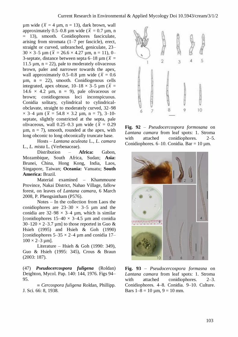
Current Research in Environmental & Applied Mycology Doi 10.5943/cream/3/1/2
103
µm wide ( x = 4 µm, n = 13), dark brown, wall
approximately 0.5–0.8 µm wide ( x = 0.7 µm, n
= 13), smooth. Conidiophores fasciculate,
arising from stromata (1–7 per fascicle), erect,
straight or curved, unbranched, geniculate, 23–
30 × 3–5 µm ( x = 26.6 × 4.27 µm, n = 11), 0–
3-septate, distance between septa 6–18 µm ( x =
11.5 µm, n = 22), pale to moderately olivaceous
brown, paler and narrower towards the apex,
wall approximately 0.5–0.8 µm wide ( x = 0.6
µm, n = 22), smooth. Conidiogenous cells
integrated, apex obtuse, 10–18 × 3–5 µm ( x =
14.6 × 4.2 µm, n = 9), pale olivaceous or
brown; conidiogenous loci inconspicuous.
Conidia solitary, cylindrical to cylindrical-
obclavate, straight to moderately curved, 32–98
× 3–4 µm ( x = 54.8 × 3.2 µm, n = 7), 3–10-
septate, slightly constricted at the septa, pale
olivaceous, wall 0.25–0.3 µm wide ( x = 0.29
µm, n = 7), smooth, rounded at the apex, with
long obconic to long obconically truncate base.
Hosts – Lantana aculeata L., L. camara
L., L. mista L. (Verbenaceae).
Distribution – Africa: Gabon,
Mozambique, South Africa, Sudan; Asia:
Brunei, China, Hong Kong, India, Laos,
Singapore, Taiwan; Oceania: Vanuatu; South
America: Brazil.
Material examined – Khammoune
Province, Nakai District, Nahao Village, fallow
forest, on leaves of Lantana camara, 6 March
2008, P. Phengsintham (P576).
Notes – In the collection from Laos the
conidiophores are 23–30 × 3–5 µm and the
conidia are 32–98 × 3–4 µm, which is similar
[conidiophores 15–40 × 3–4.5 µm and conidia
30–120 × 2–3.7 µm] to those reported in Guo &
Hsieh (1995) and Hsieh & Goh (1990)
[conidiophores 5–35 × 2–4 µm and conidia 17–
100 × 2–3 µm].
Literature – Hsieh & Goh (1990: 349),
Guo & Hsieh (1995: 345), Crous & Braun
(2003: 187).
(47) Pseudocercospora fuligena (Roldan)
Deighton, Mycol. Pap. 140: 144, 1976. Figs 94–
95.
Cercospora fuligena Roldan, Phillipp.
J. Sci. 66: 8, 1938.
Fig. 92 – Pseudocercospora formosana on
Lantana camara from leaf spots: 1. Stroma
with attached conidiophores. 2–5.
Conidiophores. 6–10. Conidia. Bar = 10 µm.
Fig. 93 – Pseudocercospora formasna on
Lantana camara from leaf spots: 1. Stroma
with attached conidiophores. 2–3.
Conidiophores. 4–8. Conidia. 9–10. Culture.
Bars 1–8 = 10 µm, 9 = 10 mm.
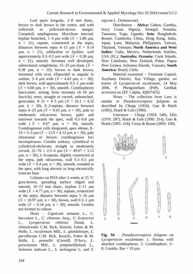
Current Research in Environmental & Applied Mycology Doi 10.5943/cream/3/1/2
104
Leaf spots irregular, 2–8 mm diam.,
brown to dark brown in the centre, and with
yellowish to yellowish-brown margin.
Caespituli amphigenous. Mycelium internal;
hyphae branched, 1–4 µm wide ( x = 2.86 µm,
n = 21), septate, constricted at the septa,
distances between septa 4–15 µm ( x = 9.14
µm, n = 21), subhyaline or hyaline, wall
approximately 0.3–0.5 µm wide ( x = 0.38 µm,
n = 21), smooth. Stromata well developed,
substomatal, subglobular, 15–25 µm diam. ( x =
0.38 µm, n = 10), brown to dark brown,
stromatal cells oval, ellipsoidal to angular in
outline, 3–6 µm wide ( x = 4.43 µm, n = 30),
dark brown, wall approximately 0.5–1 µm wide
( x = 0.68 µm, n = 30), smooth. Conidiophores
fasciculate, arising from stromata (4–18 per
fascicle), erect, straight or curved, unbranched,
geniculate, 8–31 × 4–5 µm ( x = 16.1 × 4.32
µm, n = 30), 0–2-septate, distance between
septa 4–23 µm ( x = 9.43 µm, n = 30), pale to
moderately olivaceous brown, paler and
narrower towards the apex, wall 0.5–0.8 µm
wide ( x = 0.67 µm, n = 30), smooth.
Conidiogenous cells integrated, apex obtuse, 8–
19 × 3–5 µm ( x = 12.9 × 4.12 µm, n = 30), pale
olivaceous or brown; conidiogenous loci
inconspicuous. Conidia solitary, cylindrical to
cylindrical-obclavate, straight to moderately
curved, 21–76 × 2.5–4 µm ( x = 49.67 × 3.12
µm, n = 30), 1–6-septate, slightly constricted at
the septa, pale olivaceous, wall 0.3–0.5 µm
wide ( x = 0.4 µm, n = 30), smooth, rounded at
the apex, with long obconic to long obconically
truncate base.
Colonies on PDA after 3 weeks at 25 °C
grey-brown, spreading surface ridged and
smooth, 10–13 mm diam., hyphae 2–11 µm
wide ( x = 4.77 µm, n = 30), septate, constricted
at the septa, distance between septa 5–16 µm
( x = 10.97 µm, n = 30), brown, wall 0.3–1 µm
wide ( x = 0.54 µm, n = 30), smooth. Conidia
not formed in culture.
Hosts – Capsicum annuum L., C.
baccatum L., C. chinense Jacq., C. frutescens
L., Lycopersicon chilense Dunal, L.
chmielewskii C.M. Rick, Kesicki, Fobes & M.
Holle, L. esculentum Mill., L. glandulosum, L.
parviflorum C.M. Rick, Kesicki, Fobes & M.
Holle, L. pennellii (Correll) D'Arcy, L.
peruvianum Mill., L. pimpinellifolium L.,
Solanum indicum L., S. melongena L. and S.
nigrum L. (Solanaceae).
Distribution – Africa: Gabon, Gambia,
Ivory Coast, Nigeria, Senegal, Somalia,
Tanzania, Togo, Uganda; Asia: Bangladesh,
Brunei, Cambodia, China, Hong Kong, India,
Japan, Laos, Malaysia, Philippines, Taiwan,
Thailand, Vietnam; North America and West
Indies: Cuba, Mexico, Netherlands Antilles,
USA (FL); Australia; Oceania: Cook Islands,
New Caledonia, New Zealand, Palau, Papua
New Guinea, Solomon Islands, Vanuatu; South
America: Brazil, Chile.
Material examined – Vientiane Capital,
Xaythany District, Xay Village, garden, on
leaves of Lycopersicon esculentum, 14 May
2006, P. Phengsintham (P49). GenBak
accession no (EF 1 alpha, JQ837455).
Notes – The collection from Laos is
similar to Pseudocercospora fuligena as
described by Chupp (1954), Guo & Hsieh
(1995), Hsieh & Goh (1990).
Literature – Chupp (1954: 540), Ellis
(1976: 287), Hsieh & Goh (1990: 314), Guo &
Hsieh (1995: 318), Crous & Braun (2003: 190).
Fig. 94 – Pseudocercospora fuligena on
Lycopersicon esculentum: 1. Stroma with
attached conidiophores. 2. Conidiophore. 3–
8. Conidia. Bar = 10 µm.

Current Research in Environmental & Applied Mycology Doi 10.5943/cream/3/1/2
105
Fig. 95 – Pseudocercospora fuligena on
Lycopersicon esculentum from leaf spots: 1–2.
Lesions on host leaves (1. upper surface, 2.
lower surface). 3–4. Stromata with attached
conidiophores. 5–8. Conidia. 9. Culture. Bars
1–2, 9 = 10 mm, 3–8 = 10 µm.
(48) Pseudocercospora getoniae sp. nov. Figs
96–97.
MycoBank, MB 801727
Morphologically close to
Pseudocercospora combretigena, but easily
distinguishable by the formation of
conspicuous, definite leaf spots, hypophyllous
caespituli and by its smaller stromata, 10–30
µm diam., pluriseptate conidiophores (with up
to 8 septa) and obclavate conidia.
Leaf spots subcircular to irregular, 1–10
mm diam., at first yellowish, later becoming
brown to dark brown in the center, brown to
dark brown at the margin. Caespituli
hypophyllous, conspicuous. Mycelium internal;
hyphae branched, 4–8 µm wide ( x = 5.33 µm,
n = 11), septate, constricted at the septa,
distance between septa 10–15 µm ( x = 10.17
µm, n = 11), brownish, subhyaline, wall 0.3–0.5
µm wide ( x = 0.46 µm, n = 11), smooth,
forming plate-like plectenchymatous stromatic
hyphal aggregations. Stromata oval to
ellipsoidal, 10–30 µm diam. ( x = 23.3 µm, n =
5), brown to dark brown, stromatal cells oval,
ellipsoidal and angular, 5–11 µm wide (x = 7.3
µm, n = 30), dark brown, wall 0.3–0.5 µm wide
(x = 0.48 µm, n = 30), smooth. Conidiophores
fasciculate, arising from stromata (1–14 per
fascicle), unbranched, geniculate, 26–99 × 4–5
µm ( x = 72 × 4.75 µm, n = 15), 1–8-septate,
slightly constricted at the septa, distance
between septa 5–22 µm long (x = 11.3 µm, n =
30), uniformly pale to medium brown, much
paler and more narrow toward the tip, wall 0.3–
0.5 µm ( x = 0.49 µm, n = 30), smooth.
Conidiogenous cells terminal, 15–22 × 3–4 µm
( x = 18.5 × 3.5 µm, n = 15), obtuse,
conidiogenous loci inconspicuous, unthickened,
not darkened. Conidia solitary, obclavate,
straight to slightly curved, 50–70 × 2–4 µm (x
= 52 × 3 µm, n = 15), 3–8-septate, pale
olivaceous-brown, wall 0.3–0.5 µm wide (x =
0.33 µm, n = 15), smooth, tip subacute, base
obconically truncate, hilla 1–2 µm wide ( x =
1.66 µm, n = 15).
Hosts – Getonia floribunda Roxb. [
Calycopteris floribunda (Roxb.) Lam. ex Poir.]
(Combretaceae).
Distribution – Asia: Laos.
Material examined – Vientiane Capital,
Xaythany District, Dongmakhai Village, dry
dipterocarp forest, on leaves of Getonia
floribunda, 4 February 2010, P. Phengsintham
(P545, MFLU 12-2201, Holotype).
Notes – Pseudocercospora getoniae
resembles P. combretigena (Braun 2001), but
the latter species does not form any definite leaf
spots, epiphyllous caespituli, larger stromata,
30–50 µm diam., 0–2(–6)-septate
conidiophores, often reduced to conidiogenous
cells, and obclavate-cylindrical conidia. The
genus Getonia is phylogenetically clearly
distinct from Combretum (Tan et al. 2002).
Literature – Crous & Braun (2003: key).
(49) Pseudocercospora gmelinae (J.M. Yen &
Gilles) J.M. Yen, Bull. Trimestriel Soc. Mycol.
France 94: 383, 1979. Figs 98–99.
= Cercospora gmelinae J.M. Yen &
Gilles, Bull. Trimestriel Soc. Mycol. France 91:
99, 1975.
Leaf spots circular, 1–8 mm in diam.,
with brown to dark brown and brown margin.
Caespituli amphigenous, but chiefly
hypophyllous. Mycelium internal; hyphae
branched, 2–3 µm wide ( x = 2.25 µm, n = 6),
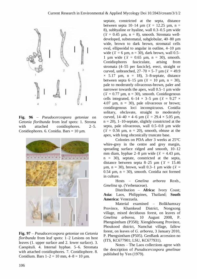
Current Research in Environmental & Applied Mycology Doi 10.5943/cream/3/1/2
106
Fig. 96 – Pseudocercospora getoniae on
Getonia floribunda from leaf spots: 1. Stroma
with attached conidiophores. 2–5.
Conidiophores. 6. Conidia. Bars = 10 µm.
Fig. 97 – Pseudocercospora getoniae on Getonia
floribunda from leaf spots: 1–2 Lesions on host
leaves (1. upper surface and 2. lower surface). 3.
Caespituli. 4. Internal hyphae. 5–6. Stromata
with attached conidiophores. 7. Conidiophore. 8.
Conidium. Bars 1–2 = 10 mm, 4–8 = 10 µm.
septate, constricted at the septa, distance
between septa 10–14 µm ( x = 12.25 µm, n =
8), subhyaline or hyaline, wall 0.3–0.5 µm wide
(x = 0.45 µm, n = 8), smooth. Stromata well-
developed, substomatal, subglobular, 40–80 µm
wide, brown to dark brown, stromatal cells
oval, ellipsoidal to angular in outline, 4–10 µm
wide (x = 6 µm, n = 30), dark brown, wall 0.5–
1 µm wide ( x = 0.65 µm, n = 30), smooth.
Conidiophores fasciculate, arising from
stromata (4–55 per fascicle), erect, straight or
curved, unbranched, 27–70 × 5–7 µm (x = 49.9
× 5.17 µm, n = 18), 3–8-septate, distance
between septa 6–15 µm (x = 10 µm, n = 30),
pale to moderately olivaceous-brown, paler and
narrower towards the apex, wall 0.5–1 µm wide
(x = 0.77 µm, n = 30), smooth. Conidiogenous
cells integrated, 6–14 × 3–5 µm ( x = 9.27 ×
4.07 µm, n = 30), pale olivaceous or brown;
conidiogenous loci inconspicuous. Conidia
solitary, obclavate, straight to moderately
curved, 14–40 × 4–6 µm (x = 29.4 × 5.05 µm,
n = 20), 1–10-septate, slightly constricted at the
septa, pale olivaceous, wall 0.5–0.8 µm wide
(x = 0.56 µm, n = 20), smooth, obtuse at the
apex, with long obconically truncate base.
Colonies on PDA after 3 weeks at 25°C
whire-grey in the centre and grey margin,
spreading surface ridged and smooth, 10–12
mm diam, hyphae 2–8 µm wide (x = 4.43 µm,
n = 30), septate, constricted at the septa,
distance between septa 8–25 µm ( x = 15.46
µm, n = 30), brown, wall 0.3–1 µm wide (x =
0.54 µm, n = 30), smooth. Conidia not formed
in culture.
Hosts – Gmelina arborea Roxb.,
Gmelina sp. (Verbenaceae).
Distribution – Africa: Ivory Coast;
Asia: Laos, Philippines, Thailand; South
America: Venezuela.
Material examined – Bolikhamxay
Province, Khamkeud District, Nongxong
village, mixed deciduous forest, on leaves of
Gmelina arborea, 10 August 2008, P.
Phengsintham (P358); Xiengkhouang Province,
Phoukood district, Namchat village, fallow
forest, on leaves of G. arborea, 3 January 2010,
P. Phengsintham (P505). GenBank accession no
(ITS, KC677901; LSU, KC677931).
Notes – The Laos collections agree with
the description of Pseudocercospora gmelinae
published by Yen (1979).

Current Research in Environmental & Applied Mycology Doi 10.5943/cream/3/1/2
107
Fig. 98 – Pseudocercospora gmelinae on
Gmelina arborea: 1. Stroma with attached
conidiophores. 2. Conidiophore. 3–7. Conidia.
Bar = 10 µm.
Fig. 99 – Pseudocercospora gmelinae on
Gmelina arborea from leaf spots: 1–2. Lesions
on host leaves (1. upper surface, 2. lower
surface). 3–4. Stromata with attached
conidiophores. 5–6. Conidiophores. 7–10.
Conidia. Bars 1–2 = 10 mm, 3–10 = 10 µm.
Literature – Index of Fungi 4 (1981:
603), Yen (1979: 383).
(50) Pseudocercospora holarrhenae (Thirum.
& Chupp) Deighton, Mycol. Pap. 140: 145,
1976. Figs 100–101.
Cercospora ho1arrhenae Thirum. &
Chupp, Mycologia 40: 355, 1948.
= Pseudocercosporella ho1arrhenae
A.N. Ray, B. Ray & Kamal, Mycol. Res. 97:
28, 1991.
Leaf spots subcircular to irregular, 2–15
mm diam., at first brown yellowish, later
becoming brown-grey in the center, brown to
dark brown at the margin. Caespituli
hypophyllous, conspicuous. Mycelium internal;
hyphae branched, 2–3 µm wide ( x = 2.28 µm,
n = 10), septate, constricted at the septa,
distance between septa 7–15 µm ( x = 8.7 µm, n
= 7), brownish, subhyaline, wall approximately
0.3–0.5 µm wide ( x = 0.37 µm, n = 7), smooth,
forming plate-like plectenchymatous stromatic
hyphal aggregations. Stromata oval to
ellipsoidal, 20–40 µm diam. ( x = 30.9 µm, n =
5), brown to dark brown, stromatal cells oval,
ellipsoidal and angular, 5–7 µm wide (x = 5.9
µm, n = 22), dark brown, wall 0.5–0.8 µm wide
(x = 0.64 µm, n = 22), smooth. Conidiophores
fasciculate, arising from stromata (6–19 per
fascicle), geniculate, unbranched, 23–37 × 4–6
µm ( x = 30.6 × 4.8 µm, n = 9), 1–3-septate,
slightly constricted at the septa, distance
between septa 5–18 µm long (x = 10.5 µm, n =
25), uniformly pale to medium brown, much
paler and more narrow toward the tip, wall 0.5–
0.8 µm ( x = 0.57 µm, n = 25), smooth.
Conidiogenous cells terminal, 9–18 × 3–5 µm
( x = 13.5 × 4 µm, n = 10), apex obtuse,
conidiogenous loci inconspicuous, unthickened,
not darkened. Conidia solitary, obclavate,
straight to slightly curved, 27–86 × 2–4 µm (x
= 60.61.4 × 3.15 µm, n = 13), 2–7-septate, pale
olivaceous-brown, wall 0.3–0.5 µm wide (x =
0.32 µm, n = 13), smooth, tip subacute, base
obconically truncate, hilla 1–2 µm wide ( x =
1.61 µm, n = 9).
Hosts – Holarrhena antidysenterica (L.)
Wall. ex A. DC., H. curtisii King & Gamble
(Apocynaceae).
Distribution – Asia: India, Laos.
Material examined – Vientiane
Province, Phonhong District, Thalad Village,
fallow forest, on leaves of Holarrhena curtisii,
4 February 2010, P. Phengsintham (P540).

Current Research in Environmental & Applied Mycology Doi 10.5943/cream/3/1/2
108
GenBank accession no (ITS, KC677902; LSU,
KC677932).
Notes – In the sample from Laos the
conidiophores are 23–37 × 4–6 µm and the
conidia are 27–86 × 2–4 µm, which is similar to
those described by Chupp (1954)
[conidiophores 10–40 × 2–4 µm and conidia
20–75 × 2–4 µm].
Literature – Chupp (1954: 47),
Vasudeva (1963: 121), Braun (1995: 193).
Fig. 100 – Pseudocercospora ho1arrhenae on
Holarrhena curtisii: 1. Stroma with attached
conidiophores. 2. Conidiophore. 3–6.
Conidia. Bar = 10 µm.
(51) Pseudocercospora jussiaeae (G.F. Atk.)
Deighton, Mycol. Pap. 140: 146, 1976. Figs
102–103.
Cercospora jussiaeae G.F. Atk., J.
Elisha Mitchell Sci. Soc. 8: 50, 1892.
Cercospora ludwigiae G.F. Atk., J.
Elisha Mitchell Sci. Soc. 8: 58, 1892.
Leaf spots circular or irregular, 1–7
mm diam., dingy grey to brown or reddish
brown to bright red in the center, and with
dark brown margin. Caespituli amphigenous,
inconspicuous. Mycelium internal; internal
hyphae 1–4 µm wide ( x = 2.26 µm, n = 23),
Fig. 101 – Pseudocercospora holarrhenae on
Holarrhena curtisii from leaf spots: 1–2.
Lesions on host leaves (1. upper surface and 2.
lower surface). 3. Caespituli. 4–5. Stromata
with attached conidiophores. 6–10. Conidia.
Bars 1–2 = 10 mm, 4–10 = 10 µm.
septate, 5–12 µm (x = 7.70 µm, n = 23), pale
brownish to subhyaline, wall 0.2–0.5 µm wide
(x = 0.25 µm, n = 20), smooth, forming plate-
like plectenchymatous stromatic hyphal
aggregations. Stromata well-developed,
ellipsoidal, 11–25 µm diam. (x = 15.84 µm, n =
25), dark brown, stromatal cell 3–5 µm wide ( x
= 3.57 µm, n = 30), wall 0.5–1 µm wide (x =
0.6 µm, n = 30), smooth. Conidiophores
fasciculate, arising from stromata (1–12 per
fascicle), cylindrical, erect, straight or curved,
branched, 5–47 × 2–6 µm (x = 29.75 × 3 µm, n
= 28), 0–3-septate, distance between septa 5–25
µm (x = 11 µm, n = 13), pale to medium brown
near the base, upper half pale in colour, wall
0.4–0.6 µm wide ( x = 0.48 µm, n = 13),
smooth, small rounded to subconic tips.
Conidiogenous cells terminal, 3–15 µm,
cylindrical and pale brown or greenish;
conidiogenous loci inconspicuous, unthickened,
not darkened, only outer rim slightly darkened
and visible as minute rings up to 1 µm. Conidia
solitary, narrowly obclavate, variously curved,

Current Research in Environmental & Applied Mycology Doi 10.5943/cream/3/1/2
109
some conidia have an occasionally with lateral
branches, 4–92 × 2–4 µm ( x = 39 × 3 µm, n =
30), 1–8-septate, subhyaline to pale olivaceous,
wall 0.3–0.8 µm wide ( x = 0.59 µm, n = 30),
smooth; apex subacute; base long obconically
truncate, 1–2 µm wide ( x = 1.78 µm, n = 9),
wall 0.5–1 µm wide ( x = 0.73 µm, n = 9),
thickened and darkened.
Colonies on PDA after three weeks at
25 °C with spreading mycelium, surface ridged,
white-grey in the centre and grey margin,
reaching 10–27 mm diam., hyphae 1–5 µm ( x
= 2.7 µm, n = 30), septate, distance between
septa 5–27 µm ( x = 15.36 µm, n = 30), wall
thin, 0.5–0.8 µm ( x = 0.58 µm, n = 12),
constricted at the septa, hyaline or brownish,
smooth. Conidia not formed in culture.
Hosts – Ludwigia adscendens (L.) Hara,
L. octovalvis (Jacq.) P.H. Raven, L. prostrata
Roxb. (Onagraceae).
Distribution – Asia: Japan, Laos;
Oceania: American Samoa.
Material examined – Vientiane Capital,
Xaythany District, Xay Village, rice paddy, on
leaves of Ludwigia prostrata, 2 April 2006, P.
Phengsintham (P3); on leaves of Ludwigia
adscendens, 11 July 2009, P. Phengsintham
(P427).
Notes – Ludwigia prostrata is the type
host of Pseudocercospora yoshinagiana
(Chupp) U. Braun & Crous, but the collection
from Laos has much shorter conidiophores and
conidia than those the latter species described
from Japan (Chupp 1954) [conidiophores 20–
125 × 3–4 µm and conidia 30–100 × 3–4 µm].
However, the fungus collected in Laos agrees
well with P. jussiaeae, which is widespread on
numerous Jussiaea and Ludwigia species.
Literature – Chupp (1954: 423), Crous
& Braun (2003: 43).
(52) Pseudocercospora lythracearum (Heald &
F.A. Wolf) X.J. Liu & Y.L. Guo, Acta Mycol.
Sin. 11: 294, 1992. Figs 104–105.
Cercospora lythracearum Heald &
F.A. Wolf, Mycologia 3: 18, 1911.
Cercosporina lythracearum (Heald &
F.A. Wolf) Sacc., Syll. Fung. 25: 909, 1931.
= Cercospora lagerstroemiae Syd. & P.
Syd., Ann. Mycol. 12: 203, 1914.
Fig. 102 – Pseudocercospora jussiaeae on
Ludwigia prostrata: 1–3. Conidiophores. 4.
Stroma with attached conidiophores. 5–9.
Conidia. Bars = 10 µm.
Fig. 103 – Pseudocercospora jussiaeae on
Ludwigia prostrata: 1–2 Lesions on host
leaves (1. upper surface and 2. lower
surface). 3. Stroma. 4–7. Stromata with
attached conidiophores. 8–16. Conidia. 17.
Culture. Bars 1–2, 17 = 10 mm, 3–16 µm.

Current Research in Environmental & Applied Mycology Doi 10.5943/cream/3/1/2
110
= Cercospora lagerstroemiae-
subcostatae Sawada, Taiwan Agric. Res. Inst.
Rept. 51: 129, 1931.
Pseudocercospora lagerstroemiae-
subcostatae (Sawada) Goh & W.H. Hsieh, in
Hsieh & Goh, Cercospora and similar fungi
from Taiwan: 212, 1990.
= Cercospora lagerstroemiicola
Sawada, Taiwan Agric. Res. Inst. Rept. 85: 112,
1943.
Leaf spots subcircular to irregular, 2–12
mm diam., at first brown, and than grey-brown
in the centre, brown to dark brown at the
margin. Caespituli amphigenous, conspicuous,
scattered, grey. Mycelium internal,
inconspicuous. Stromata oval to ellipsoidal, 8–
49 µm diam. ( x = 27.27 µm, n = 5), brown to
dark brown, stromatal cells oval, ellipsoidal to
angular, 4–7 µm wide ( x = 5.52 µm, n = 10),
dark brown, wall 0.5–0.8 µm wide ( x = 0.65
µm, n = 10), smooth. Conidiophores fasciculate
or solitary, arising from stromata (1–4 per
fascicle), not geniculate, unbranched, 5–16 × 3–
5 µm ( x = 11.17 × 3.64 µm, n = 7), 0–1-
septate, slightly constricted at the septa,
distance between septa 2–13 µm long ( x = 6.96
µm, n = 6), uniformly pale to medium brown,
much paler and narrower towards the tip, wall
0.3–0.5 µm ( x = 0.36 µm, n = 6), smooth.
Conidiogenous cells terminal, 5–13 × 3–5 µm
( x = 8.97 × 3.67 µm, n = 6), apex obtuse,
conidiogenous loci inconspicuous, unthickened,
not darkened. Conidia solitary, obclavate,
straight to slightly curved, 36–52 × 2–3 µm ( x
= 45.24 × 2.22 µm, n = 10), 3–5-septate, pale
olivaceous-brown, wall 0.3–0.5 µm wide ( x =
0.36 µm, n = 10), smooth, tip subacute, base
obconically truncate, hila 1–2 µm wide ( x =
1.18 µm, n = 10).
Hosts – Lagerstroemia flos-reginae
Retz., L. indica L., L. macrocarpa Wall., L.
parviflora Roxb., L. speciosa (L.) Pers., L.
subcostata Koehne (Lythraceae).
Distribution – Africa: Mauritius,
Uganda; Asia: Brunei, China, Hong Kong,
India, Japan, Korea, Myanmar, Philippines,
Taiwan, Thailand; Europe: Bulgaria; North
America and West Indies: Dominican
Republic, Panama, Puerto Rico, Trinidad and
Tobago, USA (FL, TX), Virgin Islands;
Oceania: Papua New Guinea.
Material examined – Vientiane Capital
Province, Xaythany District, Xay Village, on
leaves of Lagerstroemia macrocarpa, 20
August 2011, P. Phengsintham (P659).
GenBank accession no (ITS, KC677904).
Notes – The collection from Laos differs
to the description of Pseudocercospora
published by Hsieh & Goh (1990) in having
short conidiophores and conidia.
Literature – Saccardo (1931: 883, 909),
Chupp (1954: 361), Vasudeva (1963: 138),
Katsuki (1965: 44), Hsieh & Goh (1990: 212),
Guo & Hsieh (1995: 189–190), Shin & Kim
(2001: 204), Crous & Braun (2003: 259).
Fig. 104 – Pseudocercospora lythracearum on
Lagerstroemia macrocarpa: 1. Stroma with
attached conidiophores. 2–3. Conidiophores.
4–7. Conidia. Bar = 10 µm.
(53) Pseudocercospora macarangae (Syd. & P.
Syd.) Deighton, Mycol. Pap. 140: 47, 1976.
Figs 106–107.
Cercospora macarangae Syd. & P.
Syd., Ann. Mycol. 12: 575, 1914.
Cercospora macarangae J.M. Yen &
Lim, Bull. Trimestriel Soc. Mycol. France. 86:
749, 1971.
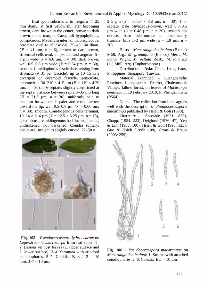
Current Research in Environmental & Applied Mycology Doi 10.5943/cream/3/1/2
111
Leaf spots subcircular to irregular, 1–25
mm diam., at first yellowish, later becoming
brown, dark brown in the center, brown to dark
brown at the margin. Caespituli hypophyllous,
conspicuous. Mycelium internal, inconspicuous.
Stromata oval to ellipsoidal, 35–45 µm diam.
( x = 41 µm, n = 6), brown to dark brown,
stromatal cells oval, ellipsoidal and angular, 5–
9 µm wide ( x = 6.6 µm, n = 30), dark brown,
wall 0.5–0.8 µm wide ( x = 0.54 µm, n = 30),
smooth. Conidiophores fasciculate, arising from
stromata (9–31 per fascicle), up to 10–15 in a
divergent or coremoid fascicle, geniculate,
unbranched, 30–210 × 4–5 µm ( x = 119 × 4.29
µm, n = 30), 1–6-septate, slightly constricted at
the septa, distance between septa 8–35 µm long
( x = 21.6 µm, n = 30), uniformly pale to
medium brown, much paler and more narrow
toward the tip, wall 0.5–0.8 µm ( x = 0.66 µm,
n = 30), smooth. Conidiogenous cells terminal,
10–14 × 3–4 µm ( x = 12.5 × 3.25 µm, n = 15),
apex obtuse, conidiogenous loci inconspicuous,
unthickened, not darkened. Conidia solitary,
obclavate, straight to slightly curved, 22–58 ×
Fig. 105 – Pseudocercospora lythracearum on
Lagerstroemia macrocarpa from leaf spots: 1–
2. Lesions on host leaves (1. upper surface and
2. lower surface). 3–4. Stromata with attached
conidiophores. 5–7. Conidia. Bars 1–2 = 10
mm, 3–7 = 10 µm.
3–5 µm ( x = 35.54 × 3.8 µm, n = 30), 1–5-
septate, pale olivaceous-brown, wall 0.3–0.5
µm wide ( x = 0.48 µm, n = 30), smooth, tip
obtuse, base subtruncate or obconically
truncate, hilla 1–2 µm wide ( x = 1.8 µm, n =
30).
Hosts – Macaranga denticulata (Blume)
Müll. Arg., M. grandifolia (Blanco) Merr., M.
indica Wight, M. peltata Roxb., M. tanarius
(L.) Müll. Arg. (Euphorbiaceae).
Distribution – Asia: China, India, Laos,
Philippines, Singapore, Taiwan.
Material examined – Luangnamtha
Province, Luangnamtha District, Chaleunsouk
Village, fallow forest, on leaves of Macaranga
denticulata, 19 February 2010, P. Phengsintham
(P564).
Notes – The collection from Laos agrees
well with the description of Pseudocercospora
macaranga published by Hsieh & Goh (1990).
Literature – Saccardo (1931: 876),
Chupp. (1954: 223), Deighton (1976: 47), Yen
& Lim (1980: 180), Hsieh & Goh (1990: 123),
Guo & Hsieh (1995: 108), Crous & Braun
(2003: 259).
Fig. 106 – Pseudocercospora macarangae on
Macaranga denticulata: 1. Stroma with attached
conidiophores. 2–6. Conidia. Bar = 10 µm.

Current Research in Environmental & Applied Mycology Doi 10.5943/cream/3/1/2
112
Fig. 107 – Pseudocercospora macarangae on
Macaranga denticulata from leaf spots: 1–2.
Lesions on host leaves (1. upper surface. 2.
lower surface). 3. Caespituli. 4–5. Stromata with
attached conidiophores. 6. Apices of
conidiophores. 7–10. Conidia. Bars 1–2 = 10
mm, 4–10 = 10 µm.
(54) Pseudocercospora maesae (Hansf.) X.J.
Liu & Y.L. Guo, Acta Mycol. Sin. 11: 295,
1992. Figs 108–109.
Cercospora maesae Hansf., Proc.
Linn. Soc. London 1942–1943: 53, 1943.
Leaf spots subcircular to irregular, 1–34
mm diam., at first yellowish, later becoming
brown to dark brown in the center, brown to
brown-yellowish at the margin. Caespituli
amphigenous, conspicuous. Mycelium internal,
inconspicuous. Stromata oval to ellipsoidal, 15–
56 µm diam., brown to dark brown, stromatal
cells ellipsoidal and angular, 4–10 µm wide ( x
= 6 µm, n = 9), dark brown, wall 0.5–0.8 µm
wide ( x = 67 µm, n = 9), smooth.
Conidiophores fasciculate, arising from
stromata (up to 16 per fascicle), geniculate,
unbranched, 25–46 × 4–6 µm ( x = 35.9 × 4.86
µm, n = 7), 2–4-septate, slightly constricted at
the septa, distance between septa 5–18 µm long
( x = 11.3 µm, n = 13), uniformly pale to
medium brown, much paler and more narrow
toward the tip, wall 0.5–0.8 µm ( x = 0.64 µm,
n = 13), smooth. Conidiogenous cells terminal,
8–13 × 4–5 µm (x = 9.67 × 4.83 µm, n = 6),
apex obtuse, conidiogenous loci inconspicuous,
unthickened, not darkened. Conidia solitary,
obclavate, straight to slightly curved, 70–95 ×
3–3.5 µm ( x = 81.4 × 3.1 µm, n = 5), 6–9-
septate, pale olivaceous-brown, wall 0.3–0.5
µm wide ( x = 0.34 µm, n = 5), smooth, tip
subacute, base obconically truncate, hilla 1.5–2
µm wide ( x = 1.75 µm, n = 5).
Colonies on PDA after 3 weeks at 25°C
dark grey mycelium, reaching 6–8 mm diam.,
hyphae 2–5 µm wide ( x = 3.53 µm, n = 15),
septate, constricted at the septa, distance
between septa 8–18 µm ( x = 12.46 µm, n =
15), brownish or subhyaline, wall 0.3–0.5 µm
wide (x = 0.42 µm, n = 15), smooth. Conidia
not formed in culture.
Hosts – Maesa hupehensis Rehd., M.
indica (Roxb.) DC., M. lanceolata Forssk., M.
ramentacea (Roxb.) A. DC., Myrsine africana
L. (Myrsinaceae).
Distribution – Africa: Uganda; Asia:
China, India, Laos; North America and West
Indies: Cuba, USA (FL).
Material examined – Luangnamtha
Province, Luangnamtha District, Chaleunsouk
Village, fallow forest, on leaves of Maesa
ramentacea, 20 February 2010. P.
Phengsintham (P575).
Notes – The collection from Laos agrees
with the description of Pseudocercospora
maesae in Guo & Goh (1995) [conidiophores
4–40 × 3–5 µm and conidia 25–100 × 3–5 µm]
Literature – Chupp. (1954: 404), Guo &
Hsieh (1995: 221), Crous & Braun (2003: 263).
(55) Pseudocercospora mannanorensis
Bagyan., U. Braun & Jagad. var.
paucifasciculata Phengsintham, E.
Chukeatirote, Abdelsalam, K.D. Hyde & U.
Braun, Crypt. Mycol. 31(2): 175, 2010. Figs
110–111.
Leaf spots suborbicular to angular, 1–3
mm diam., grey brown to medium brown in the
centre, and with brown to dark brown margin.
Caespituli amphigenous, scattered. Mycelium
internal, inconspicuous. Stromata developed,
substomatal, subglobular, 15–23 µm diam. ( x =
18.5 µm, n = 4), brown to dark brown, stroma
cells oval, ellipsoidal to angular in outline, 3–6

Current Research in Environmental & Applied Mycology Doi 10.5943/cream/3/1/2
113
Fig. 108 – Pseudocercospora maesae on
Maesa ramentacea: 1. Stroma with
attached conidiophores. 2–3.
Conidiophores. 4–7. Conidia. Bar = 10 µm.
Fig. 109 – Pseudocercospora maesae on
Maesa ramentacea from leaf spots: 1–2.
Lesions on host leaves (1. upper surface
and 2. lower surface). 3. Caespituli. 4–5.
Stromata with attached conidiophores. 6–9.
Conidia. 10–11. Culture. Bars: 1–2, 10 =
10 mm, 4–9 = 10 µm, 10. = 10 mm.
µm wide ( x = 4.53 µm, n = 17), dark brown,
wall 0.3–0.5 µm wide ( x = 0.39 µm, n = 17),
smooth. Conidiophores fasciculate, arising from
stromata (5–10 per fascicle), erect, straight or
curved, unbranched, aseptate, i.e. conidiophores
reduced to conidiogenous cells. Conidiogenous
cells 6–10 × 1.5–3 µm (x = 7.75 × 2.13 µm, n
= 8), aseptate, wall 0.3–0.5 µm wide ( x = 0.33
µm, n = 8), smooth, apex obtuse, pale
olivaceous or brown; conidiogenous loci
inconspicuous. Conidia solitary, subcylindrical
to narrowly obclavate, straight to moderately
curved, 27–69 × 1–3 µm (x = 49.38 × 1.95 µm,
n = 13), 1–5–septate, occasionally slightly
constricted at the septa, pale olivaceous, wall
0.2–0.3 µm wide ( x = 0.28 µm, n = 13),
smooth, subacute to obtuse at the apex, base
obconically truncate, 1–1.5 µm wide ( x = 1.3
µm, n = 5), wall 0.2–0.3 µm (x = 0.28 µm, n =
5) thick.
Colonies on PDA after 3 weeks at 25°C
dark brown in the centre and brown-green
margin, reaching 4–10 mm diam.; hyphae 2–5
µm wide ( x = 3.6 µm, n = 30), septate, distance
between septa 7–40 µm ( x = 19.21 µm, n =
30), primary mycelium brownish, but the
second and following ones hyaline, wall smooth
or verruculose. Conidia not formed in culture.
Hosts – Microcos paniculata L. (
Grewia microcos L.) (Tiliaceae).
Distribution – Asia: Laos.
Material examined – Vientiane Capital,
Xaythany District, Nong Viengkham Village,
on leaves of Microcos paniculata, 13 May
2006, P. Phengsintham (P45, MFLU 12-2202,
holotype); ibid., 20 December 2009, P.
Phengsintham (P488).
Notes – The collection from Laos is
similar to Pseudocercospora mannanorensis
described by Bagyanarayana et al. (1995) on
Grewia sp. from India, but can only tentatively
be assigned to this species since obvious
differences in the size and length of
conidiophores are evident. Pseudocercospora
mannanorensis has much longer, 0–2-septate,
subhyaline, pale greenish to olivaceous
conidiophores, 15–50 × 1.5–3 µm, arranged in
dense, very rich fascicles (up to more than 100).
The conidia are solitary, subcylindrical to
narrowly obclavate, straight to somewhat
curved, 40–80 × 2–4 µm, subhyaline to pale
greenish or olivaceous, i.e. they agree well the

Current Research in Environmental & Applied Mycology Doi 10.5943/cream/3/1/2
114
fungus on M. paniculata. The status of the
fungus from Laos is, however, uncertain. The
features of the lesions and conidia agree well
with P. mannanorensis, but the conidiophores
are much shorter and only formed in small
fascicles. It is unclear if two distinct species are
involved or if these differences have been
caused by the host plant or a possible
immaturity of the sample. Additional
collections are necessary to prove the
consistency of the conidiophore characters on
Microcos paniculata, but due to the obvious
differences we prefer to introduce a new variety
for this fungus.
Literature – Bagyanarayana et al.
(1995), Crous & Braun 2003 (key).
Fig. 110 – Pseudocercospora mannanorensis
var. paucifasciculata on Microcos paniculata: 1.
Stroma with attached conidiophores. 2.
Conidiophore. 3–8. Conidia. Bar = 10 µm.
(56) Pseudocercospora melochiae (Henn.)
Deighton, Mycol. Pap. 140: 147, 1976. Figs
112–113.
Cercospora melochiae Henn.,
Hedwigia 43: 395, 1904.
= Cercospora guineensis J. Kranz,
Sydowia 19: 76, (1965) 1966.
Leaf spots oval or irregular, 1–3 mm
diam., with brown to dark brown and yellowish
margin. Caespituli amphigenous, inconspicuous.
Mycelium internal and external. Internal hyphae
branched, 1–4 µm wide ( x = 2.7 µm, n = 7),
septate, constricted at the septa, distance
between septa 4–10 µm (x = 6.43 µm, n = 7),
subhyaline or hyaline, wall 0.2–0.3 µm wide ( x
= 0.26 µm, n = 7), smooth; external hyphae
branched, 1–3 µm wide ( x = 1.8 µm, n = 12),
septate, constricted at the septa, distance
between septa 6–19 µm ( x = 11.7 µm, n = 12),
brown, wall 0.25–0.3 µm wide (x = 0.26 µm, n
= 7), smooth. Stromata developed, substomatal,
subglobose, 12–26 µm diam., brown to dark
brown, stromatal cells oval, ellipsoidal to
angular in outline, 3–6 µm wide ( x = 4.87 µm, n
= 30), dark brown, wall 0.5–1 µm wide ( x =
0.66 µm, n = 30), smooth. Conidiophores
fasciculate, arising from stromata (3–17 per
fascicle), and secondary conidiophores borne
terminally and laterally on external mycelial
hyphae, very variable in length even when
formed on a single hypha , erect, straight or
curved, unbranched, obtuse, 8–25 × 3–4 µm (x
Fig. 111 – Pseudocercospora mannanorensis
var. paucifasciculata on Microcos paniculata:
1–2. Lesions on host leaves (1. upper surface, 2.
lower surface). 3. Caespituli. 4. Stromata with
attached conidiophores. 5. Stroma. 6–8. Conidia.
9. Culture. Bars 1–2, 9 = 10 mm, 4–8 = 10 µm.

Current Research in Environmental & Applied Mycology Doi 10.5943/cream/3/1/2
115
= 17 × 3.77 µm, n = 30), 0–1-septate, distance
between septa 5–16 µm ( x = 11 µm, n = 14),
pale to medium brown, wall 0.57 µm wide,
smooth. Conidiogenous cells integrated, obtuse,
12–16 × 3–4 µm, pale olivaceous or brown;
conidiogenous loci inconspicuous. Conidia
solitary, subcylindrical, slightly obclavate-
cylindrical, or sometimes slightly clavate-
cylindrical, almost straight or slightly to
strongly curved, 49–132 × 3–5 µm ( x = 87.84
× 4.44 µm, n = 30), 5–11-septate, slightly
constricted at the septa, pale olivaceous, wall
0.5–0.8 µm thick ( x = 0.51 µm, n = 30),
smooth, apex subacute or broadly rounded, base
long obconically truncate or obconic.
Hosts – Melochia corchorifolia L., M.
lupulina Sw., M. melissifolia Benth., M.
odorata L. f., Waltheria american L. and W.
indica L. (Malvaceae, incl. Sterculiaceae).
Distribution – Africa: Gabon, Ghana,
Guinea, Sierra Leone, Sudan; Asia: India, Laos;
North America and West Indies: Dominican
Republic, El Salvador, Jamaica, USA (GA);
Oceania: Papua New Guinea; South America:
Brazil, Columbia,
Material examined – Vientiane Capital,
Xaythany District, Xay Village, rice paddy, on
leaves of Melochia corchorifolia, 3 May 2006,
P. Phengsintham (P30).
Notes – The collection from Laos agrees
well with the description of Pseudocercospora
melochiae published by Chupp (1954)
[conidiophores pale to medium in colour, 3–5
µm in width, conidia 40–150 × 2–4.5 µm].
Literature – Chupp (1954: 556–557),
Crous & Braun (2003: 271)
(57) Pseudocercospora micromeli sp. nov.
Figs 114–115.
MycoBank, MB 801728
Morphologically somewhat similar to
Pseudocercospora glycosmidis but leaf spots
not vein-limited, stromata much larger (30–60
µm), conidiophores in larger fasciles (up to 27),
shorter (7–18 µm) and superficial hyphae with
solitary conidiophores lacking.
Leaf spots subcircular to irregular, 2–10
mm diam., at first yellowish, than becomes
brown to dark brow in the centre, brown to
brown yellowish at the margin. Caespituli
amphigemous, conspicuous. Mycelium internal,
inconspicuous.
Fig. 112 – Pseudocercospora melochiae on
Melochia corchorifolia: 1. Stroma with attached
conidiophores. 2–5. Conidiophores. 6. External
mycelium with attached conidiophore. 7–10.
Conidia. Bar = 10 µm.
Fig. 113 – Pseudocercospora melochiae on
Melochia corchorifolia from leaf spots: 1–2.
Lesions on host leaves (1. upper surface, 2.
lower surface). 3. Internal mycelium. 4–5.
Stromata with attached conidiophores. 6.
Conidiophore. 7. External mycelium with
attached young conidiophore. 8–13. Conidia.
Bars 1–2 = 10 mm, 3–13 = 10 µm.

Current Research in Environmental & Applied Mycology Doi 10.5943/cream/3/1/2
116
Stromata oval to ellipsoidal, 30–60 µm diam.,
brown to dark brown, stromatal cells oval,
ellipsoidal to angular, 3–7 µm wide ( x = 5.2
µm, n = 12), dark brown, wall 0.3–0.5 µm wide
( x = 0.38 µm, n = 12), smooth. Conidiophores
fasciculate, arising from stromata (up to 27 per
fascicle), unbranched, not geniculate, 7–18 × 3–
4 µm ( x = 12.5 × 3.5 µm, n = 10), 0–2-septate,
slightly constricted at the septa, distance
between septa 7–10 µm long ( x = 8.33 µm, n =
15), uniformly pale to medium brown, much
paler and narrower towards the tip, wall 0.5–0.8
µm ( x = 0.6 µm, n = 15), smooth.
Conidiogenous cells terminal, 7–8 × 3–3.5 µm
( x = 7.5 × 3.25 µm, n = 6), apex obtuse,
conidiogenous loci inconspicuous, unthickened,
not darkened. Conidia solitary, obclavate,
straight to slightly curved, 27–64 × 2–3 µm ( x =
44.6 × 2.5 µm, n = 15), 1–4-septate, pale
olivaceous-brown, wall 0.3–0.5 µm wide ( x =
0.37 µm, n = 15), smooth, tip subacute, base
obconically truncate, hila 0.5–1 µm wide ( x =
0.8 µm, n = 15).
Colonies on PDA after 3 weeks at 25°C
with dark green mycelium, reaching 4–7 mm
diam.
Hosts – Micromelum hirsutum Oliv.
(Rutaceae).
Distribution – Asia: Laos.
Material examined – Khammoune
Province, Nakai District, Nahao Village, fallow
forest, on leaves of Micromelum hirsutum, 3
June 2010, P. Phengsintham (P582, MFLU12-
2203, Holotype).
Notes – The Indian Pseudocercospora
glycomidis is somewhat similar, above all its
conidia, but this species forms vein-limited leaf
spots, has smaller stromata (–20 µm), small
fascicles of longer conidiophores (1–6, up to
42.5 µm long) and forms superficial hyphae
with solitary conidiophores (Mandal 1978).
Literature – Crous & Braun (2003: key).
(58) Pseudocercospora musae (Zimm.)
Deighton, Mycol. Pap. 140: 148, 1976. Figs
116–117.
Cercospora musae Zimm., Centralbl.
Bacteriol., Abt. 2, 8: 219, 1902.
= Cercospora musae Massee, Bull.
Misc. Inform. 28: 159, 1914.
Fig. 114 – Pseudocercospora micromeli on
Micromelum hirsutum: 1. Stroma with
attached conidiophores. 2–4. Conidiophores.
5–8. Conidia. Bar = 10 µm.
Fig. 115 – Pseudocercospora micromeli on
Micromelum hirsutum from leaf spots: 1–2.
Lesions on host leaves (1. upper surface and 2.
lower surface). 3. Caespituli. 4. Stromata. 5.
Stroma with attached conidiophores. 6.
Conidiophore. 7–10. Conidia. 11. Culture.
Bars: 1–2, 11 = 10 mm, 4–10 = 10 µm.
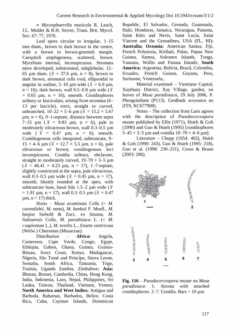
Current Research in Environmental & Applied Mycology Doi 10.5943/cream/3/1/2
117
Mycosphaerella musicola R. Leach,
J.L. Mulder & R.H. Stover, Trans. Brit. Mycol.
Soc. 67: 77, 1976.
Leaf spots circular to irregular, 1–15
mm diam., brown to dark brown in the centre,
with a brown to brown-greenish margin.
Caespituli amphigenous, scattered, brown.
Mycelium internal, inconspicuous. Stromata
more developed, substomatal, subglobular, 12–
65 µm diam. ( x = 37.6 µm, n = 8), brown to
dark brown, stromatal cells oval, ellipsoidal to
angular in outline, 5–10 µm wide ( x = 6.9 µm,
n = 16), dark brown, wall 0.5–0.8 µm wide ( x
= 0.65 µm, n = 16), smooth. Conidiophores
solitary or fasciculate, arising from stromata (6–
13 per fascicle), erect, straight or curved,
unbranched, 10–22 × 5–6 µm ( x = 15.2 × 5.67
µm, n = 6), 0–1-septate, distance between septa
7–15 µm ( x = 9.83 µm, n = 6), pale to
moderately olivaceous-brown, wall 0.3–0.5 µm
wide ( x = 0.47 µm, n = 6), smooth.
Conidiogenous cells integrated, subtruncate, 9–
15 × 4–6 µm ( x = 12.7 × 5.5 µm, n = 6), pale
olivaceous or brown; conidiogenous loci
inconspicuous. Conidia solitary, obclavate,
straight to moderately curved, 19–70 × 3–5 µm
( x = 46.41 × 4.23 µm, n = 17), 1–7-septate,
slightly constricted at the septa, pale olivaceous,
wall 0.3–0.5 µm wide ( x = 0.45 µm, n = 17),
smooth, bluntly rounded at the apex, with
subtruncate base, basal hila 1.5–2 µm wide ( x
= 1.91 µm, n = 17), wall 0.3–0.5 µm (x = 0.47
µm, n = 17) thick.
Hosts – Musa acuminata Colla (= M.
cavendishii, M. nana), M. banksii F. Muell., M.
basjoo Siebold & Zucc. ex Iinuma, M.
liukiuensis Colla, M. paradisiaca L. (= M.
sapientum L.), M. textilis L., Ensete ventricosa
(Welw.) Cheesman (Musaceae).
Distribution – Africa: Angola,
Cameroon, Cape Verde, Congo, Egypt,
Ethiopia, Gabon, Ghana, Guinea, Guinea-
Bissau, Ivory Coast, Kenya, Madagascar,
Nigeria, São Tomé and Príncipe, Sierra Leone,
Somalia, South Africa, Tanzania, Togo,
Tunisia, Uganda Zambia, Zimbabwe; Asia:
Bhutan, Brunei, Cambodia, China, Hong Kong,
India, Indonesia, Laos, Nepal, Philippines, Sri
Lanka, Taiwan, Thailand, Vietnam, Yemen;
North America and West Indies: Antigua and
Barbuda, Bahamas, Barbados, Belize, Costa
Rica, Cuba, Cayman Islands, Dominican
Republic, El Salvador, Grenada, Guatemala,
Haiti, Honduras, Jamaica, Nicaragua, Panama,
Saint Kitts and Nevis, Saint Lucia, Saint
Vincent and the Grenadines, USA (FL, HI);
Australia; Oceania: American Samoa, Fiji,
French Polynesia, Kiribati, Palau, Papua New
Guinea, Samoa, Solomon Islands, Tonga,
Vanuatu, Wallis and Futuna Islands; South
America: Argentina, Bolivia, Brazil, Colombia,
Ecuador, French Guiana, Guyana, Peru,
Suriname, Venezuela,.
Material examined – Vientiane Capital,
Xaythany District, Xay Village, garden, on
leaves of Musa paradisiaca, 29 July 2006, P.
Phengsintham (P113). GenBank accession no
(ITS, KC677908).
Notes – The collection from Laos agrees
with the description of Pseudocercospora
musae published by Ellis (1971), Hsieh & Goh
(1990) and Guo & Hsieh (1995) [conidiophores
5–45 × 3–5 µm and conidia 10–70 × 4–6 µm].
Literature – Chupp (1954: 402), Hsieh
& Goh (1990: 243), Guo & Hsieh (1995: 218),
Guo et al. (1998: 230–231), Crous & Braun
(2003: 286).
Fig. 116 – Pseudocercospora musae on Musa
paradisiaca: 1. Stroma with attached
conidiophores. 2–7. Conidia. Bars = 10 µm.

Current Research in Environmental & Applied Mycology Doi 10.5943/cream/3/1/2
118
Fig. 117 – Pseudocercospora musae on Musa
paradisiaca from leaf spots: 1. Lesions on host
leaf (upper surface). 2–3. Stromata with
attached conidiophores. 4–7. Conidia. Bars 1 =
10 mm, 2–7 = 10 µm.
(59) Pseudocercospora nigricans (Cooke)
Deighton, Mycol. Pap.140: 149, 1976. Figs
118–119.
= Cercospora nigricans Cooke,
Grevillea 12: 30, 1883.
Leaf spots circular to irregular, 2–6 mm
diam., pale brown or dark brown in the centre,
and with yellow margin. Caespituli
amphigenous, inconspicuous. Mycelium
internal. Stromata oval or ellipsoidal, 10–40 µm
diam. ( x = 16.3 µm, n = 10), dark brown,
stromatal cells angular in outline, 3–8 µm wide
( x = 4.5 µm, n = 30), wall 0.5–1 µm ( x = 0.77
µm, n = 30), smooth. Conidiophores
fasciculate, arising from stromata (3–10 per
fascicle), emerging through stomata, branched,
one time geniculate, cylindrical, 15–69 × 3–5
µm ( x = 43.6 × 4.33 µm, n = 30), 0–6-septate,
distance between septa 5–19 µm ( x = 12.3 µm,
n = 30), uniformly pale to medium olivaceous–
brown, wall 0.5–1 µm wide ( x = 0.65 µm, n =
24), smooth. Conidiogenous cells integrated,
terminal, cylindrical, 10–28 × 3–4 µm ( x =
20.17 × 3.4 µm, n = 29); conidiogenous loci
small, at the lateral and apex, conspicuous,
slightly thickened, but distinctly darkened, 1.5–
2 µm, wall 0.5–0.8 µm. Conidia solitary,
obclavate or cylindrical, entire and verruculose,
straight to curved, 10–53 × 2.5–4 µm (x = 30.4
× 3.2 µm, n = 26), 0–5-septate, pale olivaceous
brown, wall 0.3–0.5 µm wide (x = 0.44 µm, n
= 23), smooth, tip subobtuse, base obconically
truncate, hila 1–2 µm wide (x = 1.68 µm, n =
8), wall 0.5–1 µm ( x = 0.78 µm, n = 8),
thickened and darkened.
Hosts – Cassia acutifolia Delile, C.
auriculata L., C. fasciculata Michx., C. fistula
L., C. grandis L. f., C. renigera Wall., C. torosa
Cav., Cassia sp., Chamaecrista nictitans (L.)
Moench, Macroptilium lathyroides (L.) Urb.,
Senna alata (L.) Roxb., S. marilandica (L.)
Link, S. obtusifolia (L.) H.S. Irwin & Barneby,
S. occidentalis (L.) Link, S. sophera (L.) Roxb.,
S. tora (L.) Roxb. (Fabaceae).
Distribution – Africa: Gabon,
Mauritius, Sudan, Tanzania; Asia: China, India,
Japan, Laos, Malaysia, Myanmar, Pakistan,
Philippines, Sabah, Taiwan; North America
and West Indies: Cuba, Puerto Rico, Trinidad
& Tobago, USA (AI, FL, LA, MO, MS, SC,
TX, WI, WV), Virgin Islands; Oceania:
Kiribati, Solomon Islands, Tonga, Vanuatu;
South America: Brazil, Colombia.
Material examined – Vientiane Capital,
Xaythany District, Dong Dok Village, on leaves
of Cassia occidentalis, 25 July 2006, P.
Phengsintham (P108); Dong Dok village, on
leaves of C. occidentalis, 9 December 2008, P.
Phengsintham (P382).
Notes – The identification of the Laos
collections is rather difficult as they do not
agree in all details with any of the numerous
Pseudocercospora species described on Cassia
and Senna, but can be tentatively assigned to
Pseudocercospora nigricans, although they
differ from this species, as keyed out in Braun
(1989), in having somewhat larger stromata and
narrower, sometimes verruculose conidia [10–
53 × 2.5–4 µm ( x = 30.4 × 3.2 µm, n = 26),
versus (15–)20–65(–80) × 3.5–6 µm].
Pseudocercospora cassia-occidentalis (J.M.
Yen) J.M. Yen, considered to be a possible
synonym of P. nigricans in Braun (1989) but
treated as distinct taxon in Crous & Braun
(2003), is distinguished by much longer
conidiophores and conidia (Hsieh & Goh,
1990).

Current Research in Environmental & Applied Mycology Doi 10.5943/cream/3/1/2
119
Literature – Saccardo (1886: 462; 1892:
644; 1931: 879), Chupp (1954: 321), Ellis
(1976: 267), Hsieh & Goh (1990: 195), Guo &
Hsieh (1995: 172), Crous & Braun (2003: 291).
Fig. 118 – Pseudocercospora nigricans on
Senna occidentalis: 1. Stroma with
Conidiophores. 2–3. Conidiophores. 4–9.
Conidia. Bar = 5 µm.
(60) Pseudocercospora ocimicola (Petr. & Cif.)
Deighton, Mycol. Pap. 140: 149, 1976. Figs
120–121.
Cercospora ocimicola Petr. & Cif.,
Ann. Mycol. 30: 324, 1932.
= Cercospora ocimi Sawada, Unknown
(nom. nud).
Leaf spots subcircular to irregular, 1–4
mm diam., at first yellowish, later becoming
brown, dingy grey to pale tan, brown to dark
brown at the margin. Caespituli hypophyllous,
conspicuous. Mycelium internal; hyphae
branched, 3–5 µm wide ( x = 3.6 µm, n = 5),
septate, constricted at the septa, distance
between septa 7–19 µm ( x = 10.6 µm, n = 5),
brownish, subhyaline, wall 0.3–0.5 µm wide ( x
= 0.46 µm, n = 5), smooth, forming plate-like
plectenchymatous stromatic hyphal
aggregations. Stromata oval to ellipsoidal, 20–
23 µm diam., brown to dark brown, stromatal
cells oval, ellipsoidal and angular, 5–9 µm wide
( x = 6 µm, n = 8), dark brown, wall 0.5 µm
wide, smooth. Conidiophores fasciculate,
arising from stromata (1–4 per fascicle),
geniculate, unbranched, 8–24 × 3–5 µm ( x =
16.1 × 4.06 µm, n = 8), 0–2–septate, slightly
constricted at the septa, distance between septa
5–10 µm long ( x = 8.5 µm, n = 10), uniformly
pale to medium brown, much paler and more
narrow toward the tip, wall 0.3–0.5 µm ( x =
0.47 µm, n = 10), smooth. Conidiogenous cells
terminal, 8–10 × 3–4 µm (x = 10.3 × 3.67 µm,
n = 8), apex obtuse, conidiogenous loci
inconspicuous, unthickened, not darkened.
Conidia solitary, obclavate, straight to slightly
curved, 28–82 × 3–4 µm (x = 51.8 × 3.4 µm, n
= 8), 2–7–septate, pale olivaceous-brown, wall
0.3–0.5 µm wide ( x = 0.46 µm, n = 8), smooth,
tip subacute, base obnically truncate, hilla 1.5–
2.5 µm wide ( x = 2.12 µm, n = 8).
Fig. 119 – Pseudocercospora nigricans from
Senna occidentalis from leaf spots: 1–2.
Lesions on host leaves (1. upper surface, 2.
lower surface). 3–4. Stromata with attached
conidiophores. 5. Conidiophore. 6–11. Conidia.
Bars 1–2 = 10 mm, 3–11 = 10 µm.

Current Research in Environmental & Applied Mycology Doi 10.5943/cream/3/1/2
120
Hosts – Marsypianthes chamaedrys
(Vahl) Kuntze, Ocimum americanum L., O.
basilicum L., O. micranthum Willd., O.
sanctum L., Ocimum sp. (Lamiaceae).
Distribution – Asia: China, India, Laos,
Taiwan; North America and West indies:
Cuba, Dominican Republic; Oceania: Fiji, New
Zealand, Vanuatu; South America: Brazil.
Material examined – Vientiane Capital,
Xaythany District, Xay Village, on leaves of
Ocimum tenuiflorum, 4 February 2010, P.
Phengsintham (P541).
Notes – In the Laos collection the
conidiophores are 8–24 × 3–5 µm and the
conidia are 28–82 × 3–4 µm, which is similar to
those described in Hsieh & Goh (1990)
[conidiophores 10–30 × 3–5 µm, conidia 25–70
× 3–4 µm].
Literature – Chupp (1954: 270), Hsieh
& Goh (1990: 155), Guo & Hsieh (1995: 130),
Guo et al. (1998: 144), Crous & Braun (2003:
295).
Fig. 120 – Pseudocercospora ocimicola
on Ocimum tenuiflorum: 1. Stroma with
attached conidiophores. 2–5. Conidia. Bar
= 10 µm.
Fig. 121 – Pseudocercospora ocimicola on
Ocimum tenuiflorum from leaf spots: 1–2.
Lesions on host leaves (1. upper surface and 2.
lower surface). 3. Stroma with attached
conidiophores. 4–5. Conidiophores. 6–10.
Conidia. 11. Culture. Bars 1–2, 10 = 10 mm, 3–
9 = 10 µm.
(61) Pseudocercospora paraguayensis (Tak.
Kobay.) Crous, Mycotaxon 57: 270, 1996. Figs
122–123.
Cercosporina paraguayensis Tak.
Kobay., Trans. Mycol. Soc. Japan 23: 263,
1984.
= Pseudocercospora eucalypti Goh &
W.H. Hsieh, Cercospora and similar fungi from
Taiwan: 244, 1990.
Leaf spots suborbicular to angular, 2–15
mm diam., grey-brown to medium brown in the
centre, and with brown to dark brown margin.
Caespituli amphigenous, scattered. Mycelium
internal, inconspicuous. Stromata developed,
substomatal, subglobular, 14–20 µm diam. (x =
17 µm, n = 5), brown to dark brown, stroma
cells oval, ellipsoidal to angular in outline, 3–5
µm wide ( x = 4 µm, n = 30), dark brown, wall
0.5–0.8 µm wide ( x = 0.6 µm, n = 30), smooth.
Conidiophores fasciculate, arising from
stromata (4–7 per fascicle), erect, straight or

Current Research in Environmental & Applied Mycology Doi 10.5943/cream/3/1/2
121
curved, branched, geniculate, 10–53 × 3–4 µm
( x = 24.2 × 3.67 µm, n = 15), 0–4-septate,
distance between septa 5–10 µm ( x = 7.6 µm, n
= 20), pale to moderately olivaceous-brown,
wall 0.5–0.8 µm wide ( x = 0.68 µm, n = 20),
smooth. Conidiogenous cells 4–10 × 3–4 µm
( x = 7.67 × 3.33 µm, n = 5), aseptate, wall 0.5–
0.8 µm wide ( x = 0.6 µm, n = 5), smooth, apex
obtuse, pale olivaceous or brown;
conidiogenous loci inconspicuous. Conidia
solitary, subcylindrical to narrowly obclavate,
straight to moderately curved, 18–25 × 2–4 µm
( x = 23 × 2.75 µm, n = 15), 3–4-septate,
occasionally slightly constricted at the septa,
pale olivaceous, wall 0.3–0.5 µm wide ( x =
0.45 µm, n = 15), smooth, subacute to obtuse at
the apex, with obconically truncate base, 1–1.5
µm wide ( x = 1.12 µm, n = 5), wall 0.3–0.5 µm
( x = 0.35 µm, n = 5) thick.
Hosts – Eucalyptus globulus Labill., E.
nitens H. Deane & Maiden, Eucalyptus sp.
(Myrtaceae).
Distribution – Asia: China, Israel, Laos,
Taiwan, Thailand; South America: Brazil,
Paraguay.
Material examined – Vientiane Capital,
Xaythany District, Nong Viengkham Village,
on leaves of Eucalyptus sp., 23 December 2008,
P. Phengsintham (P405).
Notes – The collection from Laos agrees
well with the description of Pseudocercospora
paraguayensis published by Guo & Hsieh
(1995) and Meeboon (2009), but shorter than P.
paraguayensis described by Meeboon (2009)
[conidiophores 27–43 × 2.5–5 µm, 12–20 in a
densely fasciculate, 1–4-septate; conidia 31.5–
60 × 2–4 µm, 6–16-septate].
Literature – Guo & Hsieh (1995: 221),
Crous & Braun (2003: 307), Meeboon (2009).
(62) Pseudocercospora piperis (Pat.) Deighton,
Mycol. Pap. 140: 150, 1976. Figs 124–125.
Cercospora piperis Pat., Bull. Soc.,
Mycol. France 11: 233, 1895.
= Cercospora piperis Ellis & Everh.,
Annual Rep. Missouri Bot. Gard. 9: 119, 1898.
Cercospora pipericola Sacc. & P.
Syd., Syll. Fungi 16: 1073, 1902.
= Cercospora portoricensis Earle,
Muhlenbergia 1: 15, 1901.
Fig. 122 – Pseudocercospora
paraguayensis on Eucalyptus sp.: 1. Stroma
with attached conidiophores. 2–3.
Conidiophores. 4–9. Conidia. Bar = 10 µm.
Fig. 123 – Pseudocercospora
paraguayensis on Eucalyptus sp.: 1–2.
Lesions on host leaves (1. upper surface, 2.
lower surface). 3. Caespituli. 4–5. Stromata
with attached conidiophores. 6–10. Conidia.
Bars 1–2, 9 = 10 mm, 4–10 = 10 µm.

Current Research in Environmental & Applied Mycology Doi 10.5943/cream/3/1/2
122
Leaf spots circular to irregular, 2–10
mm diam., brown to dark brown in the centre,
with a brown to brown-yellowish margin.
Caespituli hypophyllous, scattered. Mycelium
internal; hyphae branched, 3–4 µm wide ( x =
3.2 µm, n = 5), septate, constricted at the septa,
distance between septa 6–15 µm ( x = 9 µm, n
= 5), brown to subhyaline, wall smooth.
Stromata more developed, substomatal,
subglobular, 15–52 µm diam. ( x = 27.5 µm, n
= 10), brown to dark brown, stromatal cells
oval, ellipsoidal to angular in outline, 5–8 µm
wide ( x = 6.3 µm, n = 30), dark brown, wall
approximately 0.5–0.8 µm wide ( x = 0.57 µm,
n = 30), smooth. Conidiophores solitary or
fasciculate, arising from stromata (4–8 per
fascicle), erect, straight or curved, unbranched,
10–75 × 3–5 µm ( x = 27.1 × 4.57 µm, n = 15),
0–3-septate, distance between septa 5–12 µm
( x = 8.6 µm, n = 30), pale to moderately
olivaceous-brown, wall 0.5–0.8 µm wide ( x =
0.56 µm, n = 30), smooth. Conidiogenous cells
integrated, subtruncate, cicatrized, 5–12 × 3–5
µm ( x = 8 × 4 µm, n = 15), pale olivaceous or
brown; conidiogenous loci inconspicuous,
smooth. Conidia solitary, obclavate, straight to
moderately curved, 5–14 × 3–5 µm ( x = 9.8 ×
4 µm, n = 20), 2–5–septate, slightly constricted
at the septa, pale olivaceous, wall 0.3–0.5 µm
wide ( x = 0.42 µm, n = 20), smooth, bluntly
rounded at the apex, with subtruncate base,
basal hila 1–1.5 µm wide ( x = 1.2 µm, n = 20),
wall 0.3–0.5 µm ( x = 0.46 µm, n = 20) thick.
Hosts – Piper aduncum L., P. auritum
Kunth, P. dilatatum Rich., P. hispidum Sw., P.
jamaicens C. DC., P. lolot C. DC., P. longum
L., P. marginatum Acq., P. nigrum L., P.
peltatum L., P. tuberculatum Jacq., P.
umbellatum L. (Piperaceae).
Distribution – Asia: Laos; North
America and West Indies: Cuba, Jamaica,
Puerto Rico, Dominican Republic, Trinidad;
South America: Venezuela.
Material examined – Xiengkhouang
Province, Kham District, Napa Village, on
leaves of Piper lolot, 3 January 2010, P.
Phengsintham (P516).
Notes – The collection from Laos agrees
with the description of Pseudocercospora
piperis (≡ Cercospora piperis) in Ellis (1976)
[conidiophores 50–120 × 4–6 µm and conidia
Fig. 124 – Pseudocercospora piperis on
piper lolot from leaf spots: 1. Stroma with
attached conidiophores. 2. Conidiophores.
3–6. Conidia. Bar = 10 µm.
Fig. 125 Pseudocercospora piperis on Piper
lolot from leaf spots: 1. Upper surface. 2.
Stroma. 3. Internal mycelium. 4. Stroma with
attached conidiophores. 5–7. Conidia. 8.
Culture. Bars 1, 8 = 10 mm, 2–7 = 10 µm.

Current Research in Environmental & Applied Mycology Doi 10.5943/cream/3/1/2
123
45–100 × 4–6 µm], but it has conidiophores and
conidia that are shorter.
Literature – Saccardo (1902: 1073;
1906: 609), Chupp (1954: 442), Ellis (1976:
279), Crous & Braun (2003: 325).
(63) Pseudocercospora polygonicola (A.K. Kar
& M. Mandal) Deighton, Trans. Brit. Mycol.
Soc. 88: 388, 1987. Figs 126–127.
Cercospora polygonicola A.K. Kar &
M. Mandal, Trans. Brit. Mycol. Soc. 53: 354,
1969.
Cercospora polygoni Sawada, Rep.
Gov. Agric. Res. Inst. Taiwan 85: 119, 1943.
Leaf spots subcircular to irregular, 1–5
mm diam., at first yellowish, later becoming
brown in the centre, brown at the margin.
Caespituli amphigenous, conspicuous.
Mycelium internal, inconspicuous. Stromata
oval to ellipsoidal, 13.8–27 µm diam., brown to
dark brown, stromatal cells oval, ellipsoidal and
angular, 4–7 µm wide ( x = 6 µm, n = 30), dark
brown, wall 0.5–0.8 µm wide ( x = 0.68 µm, n
= 30), smooth. Conidiophores fasciculate,
arising from stromata (2–25 per fascicle),
geniculate, unbranched, 33–55 × 3–4.2 µm ( x =
41.9 × 3.3 µm, n = 30), 1–2-septate, slightly
constricted at the septa, distance between septa
7–27.6 µm long ( x = 15.4 µm, n = 30),
uniformly pale to medium brown, much paler
and more narrow toward the tip, wall 0.5–0.8
µm ( x = 0.58 µm, n = 30), smooth.
Conidiogenous cells terminal, 18–27 × 3–3.6
µm ( x = 24 × 3.4 µm, n = 14), obtuse,
conidiogenous loci inconspicuous, unthickened,
not darkened. Conidia solitary, obclavate,
straight to slightly curved, 50–87 × 3.6–4.2 µm
( x = 61.65 × 3.75 µm, n = 15), 3–5-septate,
pale olivaceous-brown, wall 0.3–0.5 µm wide
( x = 0.45 µm, n = 15), smooth, tip subacute,
base truncate, hilla 0.5–2 µm wide ( x = 1.2 µm,
n = 15).
Hosts – Polygonum barbatum L., P.
hydropiper L., P. pulchrum Blume
(Polygonaceae).
Distribution – Asia: China, India, Laos,
Taiwan.
Material examined – Oudomxay
Province, Ngoi District, Ngoi Village, on leaves
of Polygonum pulchrum, 24 June 2010, P.
Phengsintham (P599). GenBank accession no
(LSU, KC677936).
Notes – The collection from Laos agrees
with the description of Pseudocercospora
polygonicola published by Hiseh & Goh (1990)
[conidiophores 25–40 × 3–4 µm and conidia
40–100 × 3–4.5 µm], but the collection from
Laos has slightly geniculate conidiophores.
Literature – Hsieh & Goh (1990: 270),
Guo & Hsieh (1995: 255), Crous & Braun
(2003: 332).
Fig. 126 – Pseudocercospora polygonicola on
Polygonum pulchrum: 1. Stroma with attached
conidiophores. 2–4. Conidiophores. 5–8.
Conidia. Bar = 10 µm.
(64) Pseudocercospora puerariicola (W.
Yamam.) Deighton, Mycol. Pap. 140: 151,
1976. Figs 128–129.
Cercospora puerariicola W.
Yamam., Trans. Sapporo Nat. Hist. Soc. 13:
142, 1934.
Leaf spots circular to irregular, 1–6
mm diam., grey-brown to medium brown in
the centre, and with brown to dark-brown
margin. Caespituli amphigenous, scattered.
Mycelium internal; hyphae branched, 2–4 µm
wide ( x = 2.93 µm, n = 14), septate,
constricted at the septa, distance between septa

Current Research in Environmental & Applied Mycology Doi 10.5943/cream/3/1/2
124
Fig. 127 – Pseudocercospora polygonicola on
Polygonum pulchrum from leaf spots: 1.
Lesions on host leaf (lower surface). 2. Stroma
with attached conidiophores. 3. Conidiophore.
4–9. Conidia. Bars = 10 µm.
4–14 µm (x = 9.14 µm, n = 14), subhyaline or
hyaline, wall 0.3–0.8 µm wide ( x = 0.5 µm, n =
14), smooth. Stromata well developed,
substomatal, subglobular, 10–40 µm diam. ( x =
23.57 µm, n = 10), brown to dark brown,
stromatal cells oval, ellipsoidal to angular in
outline, 4–10 µm wide ( x = 5.6 µm, n = 30),
dark brown, wall 0.5–1 µm wide ( x = 0.78 µm,
n = 30), smooth. Conidiophores fasciculate,
arising from stromata (4–30 per fascicle), erect,
straight or curved, branched, 9–30 × 3–5 µm ( x
= 20 × 3.8 µm, n = 30), 0–3-septate, distance
between septa 5–20 µm ( x = 9.43 µm, n = 30),
pale to moderately olivaceous brown, paler and
narrower towards the apex, wall 0.5–0.8 µm
wide ( x = 0.51 µm, n = 30), smooth.
Conidiogenous cells integrated, obtuse to
subacute, 6–20 × 3–4 µm ( x = 11.8 × 3.4 µm, n
= 30), pale olivaceous or brown; conidiogenous
loci inconspicuous. Conidia solitary, cylindrical
to cylindrical-obclavate, straight to moderately
curved, 6–82 × 2–3 µm ( x = 53.97 × 2.85 µm,
n = 30), 1–6-septate, slightly constricted at the
septa, pale olivaceous, wall 0.3–0.5 µm wide
( x = 0.47 µm, n = 30), smooth, rounded to
obtuse at the apex, with long obconic to
truncate base, 1–2 µm wde ( x = 1.87 µm, n =
30), wall 0.3–0.5 µm ( x = 0.43 µm, n = 30)
thick.
Colonies on PDA after 3 weeks at 25 °C
grey-brown, spreading surface ridged and
smooth, 3–5 mm diam., hyphae 1–2 µm wide
(x = 1.53 µm, n = 30), septate, constricted at
the septa, distance between septa 6–18 µm (x =
10.8 µm, n = 30), subhyaline to greenish brown,
wall 0.2–0.25 µm wide ( x = 0.22 µm, n = 30),
smooth. Conidia not formed in culture.
Hosts – Pueraria javanica (Benth.)
Benth., P. lobata (Willd.) Ohwi, P.
phaseoloides (Roxb.) Benth., P. thunbergiana
Benth, P. tonkinensis Gagnep. (Fabaceae).
Distribution – Asia: Cambodia, China,
Hong Kong, Indonesia, Japan, Korea, Laos,
Malaysia, Philippines, Singapore, Taiwan;
North America: USA (AL, FL, GA, MS, NC).
Material examined – Vientiane Capital,
Xaythany District, Nakhae Village, on leaves of
Pueraria phaseoloides, 11 May 2006, P.
Phengsintham (P44). GenBank accession no
(ITS, KC731556; LSU, KC731562).
Notes – The collection from Laos is
similar to the description of Pseudocercospora
puerariicola published by Hsieh & Goh (1990)
[conidiophores 20–70 × 3–4.5 µm and conidia
20–64 × 3–4.5 µm].
Literature – Chupp (1954: 327), Hsieh
& Goh (1990: 199), Guo & Hsieh (1995: 178),
Crous & Braun (2003: 341).
(65) Pseudocercospora sphaerellae-eugeniae
(Sacc.) Crous, Alfenas & R.W. Barreto,
Mycotaxon 64: 425, 1997 Figs 130–131.
Cercosporina sphaerellae-eugeniae
Sacc., Syll. Fung. 25: 912, 1931.
= Cercospora eugeniae Sawada, Rep.
Gov. Agric. Res. Inst. Taiwan 85: 104, 1943.
= Cercospora eugeniae Chupp, Bol.
Soc. Brasil. Argron. 8: 25, 1945.
= Cercospora eugeniae Chupp, A
monograph of the fungus genus Cercospora:
406, 1954.
= Cercospora eugeniae Puckdeedindan,
Techn. Bull. Depart. Agric. (Bangkok) 7: 6,
1966.
= Cercospora eugeniae Chantarasrikul
& Puckdeedinnan, Tech. Doc. Pl. Protect.

Current Research in Environmental & Applied Mycology Doi 10.5943/cream/3/1/2
125
Committee SE Asia Pacific Region: 72:
15, 1969.
Fig. 128 – Pseudocercospora puerariicola on
Pueraria phaseoloides: 1–2. Stromata with
attached conidiophores. 3–4. Conidiophores. 5–
9. Conidia. Bar = 10 µm.
Fig. 129 – Pseudocercospora puerariicola on
Pueraria phaseoloides from leaf spots: 1.
Lesions on host leaf (upper surface). 2. Internal
hyphae. 3–4. Stromata with attached
conidiophores. 5. Conidiophore. 6–8. Conidia.
9. Culture. Bars 1, 9 = 10 mm, 2–8 = 10 µm.
Leaf spots circular to irregular, 2–10
mm diam., red or dark red in the centre, and
with reddish margin. Caespituli amphigenous.
Mycelium internal, inconspicuous. Stromata
oval or ellipsoidal, 8–40 µm diam. ( x = 25 µm,
n = 11), dark brown, stromatal cells angular in
outline, 2–4 µm wide ( x = 3 µm, n = 14), wall
0.3–0.5 µm ( x = 0.38 µm, n = 14), smooth.
Conidiophores fasciculate, arising from
stromata (1–5 per fascicle), emerging through
stomata, unbranched, geniculate, mostly short,
cylindrical, 8–24 × 3–6 µm (x = 14.8 × 4 µm, n
= 13), 0–2-septate, distance between septa 4–10
µm ( x = 7.4 µm, n = 9), uniformly pale to
medium olivaceous-brown, wall 0.3–0.8 µm
wide ( x = 0.37 µm, n = 8), smooth.
Conidiogenous cells integrated, terminal,
cylindrical, 4–10 × 2–4 µm (x = 7.5 × 3 µm, n
= 4); conidiogenous loci small, at the apex,
conspicuous, ovoid to oval, 1–2 µm ( x = 1.5
µm, n = 5), slightly thickened, but distinctly
darkened, wall 0.5 µm wide ( x = 0.5 µm, n =
5). Conidia solitary, obclavate or cylindrical,
straight to curved, 5–78 × 2–3 µm ( x = 30.87 ×
2.35 µm, n = 30), 0–6-septate, pale olivaceous
brown, wall 0.3–0.5 µm wide (x = 0.35 µm, n
= 12), smooth, tip subobtuse, base obconically
truncate, hila 0.5–1.5 µm wide ( x = 1.08 µm, n
= 8), wall 0.3–0.6 µm ( x = 0.53 µm, n = 8),
unthickened and not darkened.
Colonies on PDA after 3 weeks at 25 °C
with dark green in the centre, and green-grey to
dark green at the margin, reaching 4 mm diam.
Hosts – Eugenia jambolana Lam., E.
jambos L., E. javanica Lam., E. uniflora L.,
Syzygium cumini (L.) Skeels, S. samarangense
Merr. & Perry (Myrtaceae).
Distribution – Asia: China, India, Iran,
Laos, Taiwan, Thailand; North America and
West Indies: Bermuda, Panama, USA (FL);
South America: Brazil,
Material examined – Vientiane Capital,
Xaythany District, Nakhae Village, dry
dipterocarp forest, on leaves of Syzygium
cuminii, 29 June 2006, P. Phengsintham (P111);
ibid., Nakhae Village, 9 December 2008, P.
Phengsintham (P383). GenBank accession no
(ITS, KC731558).
Notes – The collections from Laos differ
to the description of Pseudocercospora
sphaerellae-eugeniae in Hsieh & Goh and
Chupp (1954) by the dark red colour of leaf
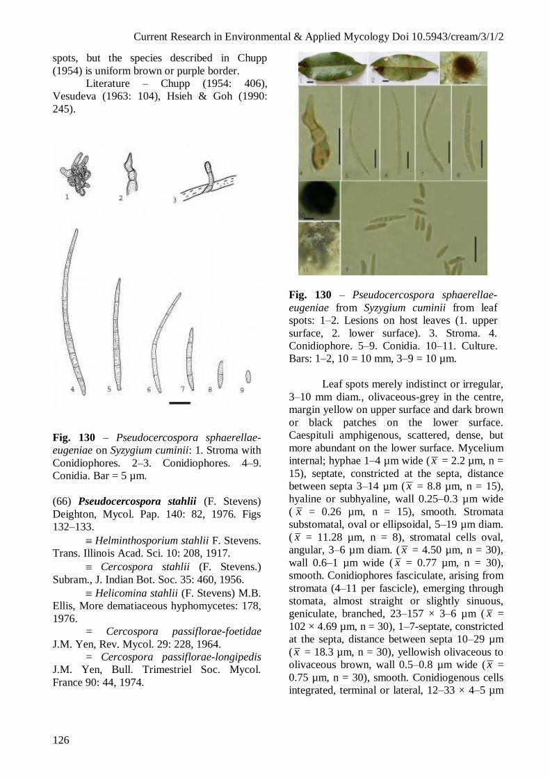
Current Research in Environmental & Applied Mycology Doi 10.5943/cream/3/1/2
126
spots, but the species described in Chupp
(1954) is uniform brown or purple border.
Literature – Chupp (1954: 406),
Vesudeva (1963: 104), Hsieh & Goh (1990:
245).
Fig. 130 – Pseudocercospora sphaerellae-
eugeniae on Syzygium cuminii: 1. Stroma with
Conidiophores. 2–3. Conidiophores. 4–9.
Conidia. Bar = 5 µm.
(66) Pseudocercospora stahlii (F. Stevens)
Deighton, Mycol. Pap. 140: 82, 1976. Figs
132–133.
Helminthosporium stahlii F. Stevens.
Trans. Illinois Acad. Sci. 10: 208, 1917.
Cercospora stahlii (F. Stevens.)
Subram., J. Indian Bot. Soc. 35: 460, 1956.
Helicomina stahlii (F. Stevens) M.B.
Ellis, More dematiaceous hyphomycetes: 178,
1976.
= Cercospora passiflorae-foetidae
J.M. Yen, Rev. Mycol. 29: 228, 1964.
= Cercospora passiflorae-longipedis
J.M. Yen, Bull. Trimestriel Soc. Mycol.
France 90: 44, 1974.
Fig. 130 – Pseudocercospora sphaerellae-
eugeniae from Syzygium cuminii from leaf
spots: 1–2. Lesions on host leaves (1. upper
surface, 2. lower surface). 3. Stroma. 4.
Conidiophore. 5–9. Conidia. 10–11. Culture.
Bars: 1–2, 10 = 10 mm, 3–9 = 10 µm.
Leaf spots merely indistinct or irregular,
3–10 mm diam., olivaceous-grey in the centre,
margin yellow on upper surface and dark brown
or black patches on the lower surface.
Caespituli amphigenous, scattered, dense, but
more abundant on the lower surface. Mycelium
internal; hyphae 1–4 µm wide ( x = 2.2 µm, n =
15), septate, constricted at the septa, distance
between septa 3–14 µm (x = 8.8 µm, n = 15),
hyaline or subhyaline, wall 0.25–0.3 µm wide
( x = 0.26 µm, n = 15), smooth. Stromata
substomatal, oval or ellipsoidal, 5–19 µm diam.
( x = 11.28 µm, n = 8), stromatal cells oval,
angular, 3–6 µm diam. ( x = 4.50 µm, n = 30),
wall 0.6–1 µm wide ( x = 0.77 µm, n = 30),
smooth. Conidiophores fasciculate, arising from
stromata (4–11 per fascicle), emerging through
stomata, almost straight or slightly sinuous,
geniculate, branched, 23–157 × 3–6 µm ( x =
102 × 4.69 µm, n = 30), 1–7-septate, constricted
at the septa, distance between septa 10–29 µm
(x = 18.3 µm, n = 30), yellowish olivaceous to
olivaceous brown, wall 0.5–0.8 µm wide (x =
0.75 µm, n = 30), smooth. Conidiogenous cells
integrated, terminal or lateral, 12–33 × 4–5 µm

Current Research in Environmental & Applied Mycology Doi 10.5943/cream/3/1/2
127
( x = 22.3 × 4.43 µm, n = 30), brownish;
conidiogenous loci small, short tapered towards
an apical conidiogenous locus, which is later
displaced to remain visible as a short lateral
peg, 1–1.5 µm wide. Conidia solitary, mostly
clavate or cylindrical, 14–46 × 3–7 µm ( x = 28
× 5 µm, n = 30), 1–5-septate, sometimes
slightly constricted at the septa, concolorous
with the conidiophores, wall 0.5–0.8 µm wide
( x = 0.73 µm, n = 30), smooth, apex broadly
rounded, based apiculate with a truncate hilum.
Colonies on PDA after 3 weeks at 25 °C
black grey, reaching 4 mm diam., hyphae 2–7
µm wide, septate, constricted at the septa,
distances between septa 5–29 µm, occasionally
with a nodulose swellings, up to 7 µm wide,
brown, wall smooth, short tapered towards a
truncate apex which is later displaced to remain
visible as a short lateral peg, 1–1.5 µm wide.
Conidia solitary, 15–32 × 4–6 µm, brownish,
wall smooth.
Hosts – Passiflora foetida L., P.
quadrangularis L. (Passifloraceae).
Distribution – Africa: Gabon, Ivory
Coast; Asia: Brunei, India, Laos, Malaysia,
Myanmar, Sabah, Singapore, Taiwan, Thailand;
North America and West Indies: Puerto Rico,
Trinidad and Tobago; Oceania: American
Samoa, Australia, Fiji, Micronesia, Palau,
Papua New Guinea, Samoa, Solomon Islands,
Tonga, Vanuatu.
Material examined – Vientiane Capital,
Xaythany District, Nakhae Village, on leaves of
Passiflora foetida, 23 April 2006, P.
Phengsintham (P20).
Notes – There is no difference in the
size of conidia formed in vivo or in vitro (Crous
& Braun 2003).
Literature – Deighton (1976: 82), Ellis
(1976: 178), Hsieh & Goh (1990: 260), Crous
& Braun (2003: 386).
(67) Pseudocercospora stizolobii (Syd. & P.
Syd.) Deighton, Mycol. Pap. 140: 153, 1976.
Figs 134–135.
Cercospora stizolobii Syd. & P. Syd.,
Ann. Mycol. 11: 270, 1913.
= Cercospora lussoniensis Sacc., Ann.
Mycol. 12: 314, 1914.
= Cercospora mucunae-capitatae
Sawada, Rep. Gov. Agric. Res. Inst. Taiwan 85:
116, 1943 (nom.inval.).
Fig. 132 – Pseudocercospora stahlii on
Passiflora foetida: 1. Conidiophore. 2. Stroma
with attached conidiophores. 5. Apex of
conidiophore. 4–8. Conidia. Bar = 10 µm.
Fig. 133 – Pseudocercospora stahlii on
Passiflora foetida: 1–2 Lesions on host leaves
(1. upper surface and 2. lower surface). 3.
Conidiophores. 4. Stroma with attached
conidiophores. 5. Apex of conidiophores. 6–
8. Conidiophores. 9–14. Conidia. 15. Culture.
Bars 1–2, 15 = 10 mm, 3–14 = 10 µm.

Current Research in Environmental & Applied Mycology Doi 10.5943/cream/3/1/2
128
Leaf spots indistinct or distinct,
orbicular or vein-limited and angular to
irregular, 1–6 mm diam., at first yellowish or
pale brown, later becoming dark brown or
reddish, and sometimes with grey center with a
darker margin; Caespituli amphigenous;
stromata small, dark brown. Mycelium internal,
inconspicuous. Stromata oval to ellipsoidal, 20–
50 µm diam. ( x = 30 µm, n = 5), brown to dark
brown, stromatal cells oval, ellipsoidal and
angular, 5–7 µm wide ( x = 5.8 µm, n = 30),
dark brown, wall 0.5–0.8 µm wide ( x = 0.58
µm, n = 30), smooth. Conidiophores
fasciculate, arising from stromata (2–11 per
fascicle), slightly geniculate, unbranched, 50–
118 × 3–4 µm ( x = 85.5 × 3.9 µm, n = 30), 3–
7-septate, constricted at the septa, distance
between septa 6–35 µm long ( x = 16 µm, n =
30), uniformly pale to medium brown, much
paler and more narrow toward the tip, wall 0.5–
1 µm ( x = 0.61 µm, n = 30), smooth.
Conidiogenous cells terminal, 14–18 × 3–4 µm
( x = 15.5 × 3.67 µm, n = 7), apex obtuse;
conidiogenous loci inconspicuous, unthickened,
not darkened. Conidia solitary, cylindrical-
obclavate, straight to slightly curved, 17–64 ×
4–5 µm ( x = 46.5 × 4.6 µm, n = 30), 1–7-
septate, pale olivaceous-brown, wall 0.3–0.5
µm wide ( x = 0.48 µm, n = 30), smooth; apex
subacute; base obconically truncate, hila 1–3
µm wide ( x = 2.08 µm, n = 30).
Hosts – Mucuna aterrima (Piper &
Tracy) Holland, M. capitata Wight & Arn., M.
cochinchinensis (Lour.) A. Chev., M.
deeringiana (Bort) Hanelt, M. ferruginea
Matsum., M. nivea (Roxb.) DC. ex Wight &
Arn., M. pruriens (L.) DC., M. prurita Wight,
M. urens (L.) Medik., Mucuna sp., Stizolobium
sp. (Fabaceae).
Distribution – Africa: Gabon, Ghana,
Malawi, Nigeria, Sierra Leone, South Africa,
Togo, Zambia, Zimbabwe; Asia: Cambodia,
China, Hong Kong, India, Japan, Java Laos,
Nepal, Philippines, Taiwan, Thailand; North
America and West Indies: Barbados, Cuba,
Guatemala, Haiti, Jamaica, Panama, Puerto
Rico, Saint Vincent and the Grenadines,
Trinidad and Tobago, USA (FL, MS, NC),
Virgin Islands; Oceania: Australia, Fiji, Papua
New Guinea; South America: Brazil,
Columbia, Guyana, Venezuela.
Material examined – Luang Namtha
Province, Luang Namtha District, Chaleunsouk
Village, fallow forest, on leaves of Mucuna
pruriens, 19 February 2010, P. Phengsintham
(P565).
Notes – The collection from Laos agrees
well with Pseudocercospora stizolobii
described by Chupp (1954).
Literature – Saccado (1931: 882),
Chupp (1954: 298), Vasudeva (1963: 198),
Deighton (1976: 153), Ellis (1976: 270), Hsieh
& Goh (1990: 203), Hsieh & Goh (1990: 203),
Guo & Hsieh (1995: 182), Crous & Braun
(2003: 389).
Fig. 134 – Pseudocercospora stizolobii on
Mucuna pruriens: 1. Stroma with attached
conidiophores. 2–3. Conidiophores. 4–9.
Conidia. Bar = 10 µm.
(68) Pseudocercospora tabernaemontanae
(Syd. & P. Syd.) Deighton, Mycol. Pap. 140:
154, 1976. Figs 136–137.
Cercospora tabernaemontanae Syd. &
P. Syd., Philipp. J. Sci. (Bot.) 8: 507, 1913.
= Cercospora tabernaemontanae
Thirum. & Govindu, Sydowia 7: 45, 1953.
= Cercospora ervataniae J.M. Yen &
Lim, Bull. Trimestestriel Soc. Mycol. France
86: 745, 1971.
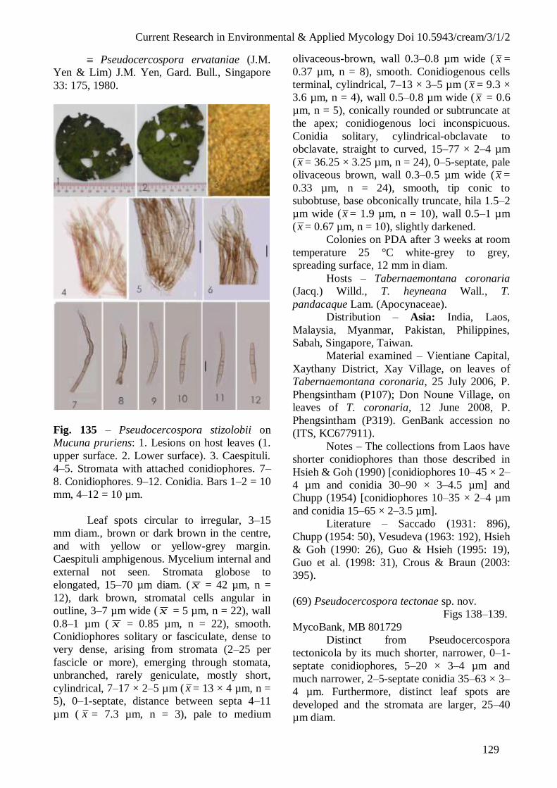
Current Research in Environmental & Applied Mycology Doi 10.5943/cream/3/1/2
129
Pseudocercospora ervataniae (J.M.
Yen & Lim) J.M. Yen, Gard. Bull., Singapore
33: 175, 1980.
Fig. 135 – Pseudocercospora stizolobii on
Mucuna pruriens: 1. Lesions on host leaves (1.
upper surface. 2. Lower surface). 3. Caespituli.
4–5. Stromata with attached conidiophores. 7–
8. Conidiophores. 9–12. Conidia. Bars 1–2 = 10
mm, 4–12 = 10 µm.
Leaf spots circular to irregular, 3–15
mm diam., brown or dark brown in the centre,
and with yellow or yellow-grey margin.
Caespituli amphigenous. Mycelium internal and
external not seen. Stromata globose to
elongated, 15–70 µm diam. ( x = 42 µm, n =
12), dark brown, stromatal cells angular in
outline, 3–7 µm wide ( x = 5 µm, n = 22), wall
0.8–1 µm ( x = 0.85 µm, n = 22), smooth.
Conidiophores solitary or fasciculate, dense to
very dense, arising from stromata (2–25 per
fascicle or more), emerging through stomata,
unbranched, rarely geniculate, mostly short,
cylindrical, 7–17 × 2–5 µm ( x = 13 × 4 µm, n =
5), 0–1-septate, distance between septa 4–11
µm ( x = 7.3 µm, n = 3), pale to medium
olivaceous-brown, wall 0.3–0.8 µm wide ( x =
0.37 µm, n = 8), smooth. Conidiogenous cells
terminal, cylindrical, 7–13 × 3–5 µm (x = 9.3 ×
3.6 µm, n = 4), wall 0.5–0.8 µm wide ( x = 0.6
µm, n = 5), conically rounded or subtruncate at
the apex; conidiogenous loci inconspicuous.
Conidia solitary, cylindrical-obclavate to
obclavate, straight to curved, 15–77 × 2–4 µm
(x = 36.25 × 3.25 µm, n = 24), 0–5-septate, pale
olivaceous brown, wall 0.3–0.5 µm wide ( x =
0.33 µm, n = 24), smooth, tip conic to
subobtuse, base obconically truncate, hila 1.5–2
µm wide ( x = 1.9 µm, n = 10), wall 0.5–1 µm
(x = 0.67 µm, n = 10), slightly darkened.
Colonies on PDA after 3 weeks at room
temperature 25 °C white-grey to grey,
spreading surface, 12 mm in diam.
Hosts – Tabernaemontana coronaria
(Jacq.) Willd., T. heyneana Wall., T.
pandacaque Lam. (Apocynaceae).
Distribution – Asia: India, Laos,
Malaysia, Myanmar, Pakistan, Philippines,
Sabah, Singapore, Taiwan.
Material examined – Vientiane Capital,
Xaythany District, Xay Village, on leaves of
Tabernaemontana coronaria, 25 July 2006, P.
Phengsintham (P107); Don Noune Village, on
leaves of T. coronaria, 12 June 2008, P.
Phengsintham (P319). GenBank accession no
(ITS, KC677911).
Notes – The collections from Laos have
shorter conidiophores than those described in
Hsieh & Goh (1990) [conidiophores 10–45 × 2–
4 µm and conidia 30–90 × 3–4.5 µm] and
Chupp (1954) [conidiophores 10–35 × 2–4 µm
and conidia 15–65 × 2–3.5 µm].
Literature – Saccado (1931: 896),
Chupp (1954: 50), Vesudeva (1963: 192), Hsieh
& Goh (1990: 26), Guo & Hsieh (1995: 19),
Guo et al. (1998: 31), Crous & Braun (2003:
395).
(69) Pseudocercospora tectonae sp. nov.
Figs 138–139.
MycoBank, MB 801729
Distinct from Pseudocercospora
tectonicola by its much shorter, narrower, 0–1-
septate conidiophores, 5–20 × 3–4 µm and
much narrower, 2–5-septate conidia 35–63 × 3–
4 µm. Furthermore, distinct leaf spots are
developed and the stromata are larger, 25–40
µm diam.

Current Research in Environmental & Applied Mycology Doi 10.5943/cream/3/1/2
130
Fig. 136 – Pseudocercospora tabernaemontanae
on Tabernaemontana coronaria: 1. Stroma with
Conidiophores. 2. Stromatal cells with
Conidiophore. 3. Conidiophore. 4–6. Conidia.
Bars = 5 µm.
Leaf spots subcircular to irregular, 1–25
mm diam. ( x = 11.3 µm, n = 6), at first yellowish,
later becoming brown to dark brown, dark brown
and yellowish at the margin. Caespituli
hypophyllous, inconspicuous. Mycelium internal,
inconspicuous. Stromata well-developed, oval to
ellipsoidal, 25–40 µm diam. ( x = 31.7 µm, n = 4),
brown to dark brown, stromatal cells oval,
ellipsoidal and angular, 3–5 µm wide ( x = 4 µm,
n = 9), dark brown, wall 0.5–0.8 µm wide ( x =
0.6 µm, n = 9), smooth. Conidiophores fasciculate,
arising from stromata (3–7 per fascicle), emerging
through stomata, unbranched, 5–20 × 3–4 µm ( x
= 11.9 × 3.56 µm, n = 9), 0–1-septate, constricted
at the septa, distance between septa 5–11 µm long
( x = 8.23 µm, n = 13), uniformly pale to medium
brown, much paler and more narrow toward the
tip, wall 0.3–0.5 µm ( x = 0.43 µm, n = 13),
smooth. Conidiogenous cells terminal, 5–10 ×
2.5–4 µm ( x = 7.89 × 3.28 µm, n = 9), apex
obtuse, conidiogenous loci inconspicuous,
unthickened, not darkened.
Conidia solitary, obclavate, obclavate-cylindrical,
straight to slightly curved, 35–63 × 3–4 µm (x =
51.75 × 3.25 µm, n = 4), 2–5-septate, pale
olivaceous-brown, wall 0.3–0.5 µm wide ( x = 0.4
µm, n = 4), smooth, tip rounded, base obconically
truncate, hila 1.5–2 µm wide (x = 1.8 µm, n = 4).
Fig. 137 – Pseudocercospora tabernaemontanae
on Tabernaemontana coronaria from leaf spots:
1–2. Lesions on host leaves (1. upper surface, 2.
lower surface). 3. Stroma with attached
Conidiophores. 4. Stromatal cells with attached
conidiophore. 5–10. Conidia. Bars 1–2 = 10 mm,
3–10 = 10 µm.
Hosts – Tectona grandis L. f.
(Verbenaceae).
Distribution – Asia: Laos.
Material examined – Vientiane Capital,
Xaythany District, Nong Viengkham Village,
on leaves of Tectona grandis, 10 August 2006,
P. Phengsintham (P138, MFLU12-2204,
Holotype).
Notes – Pseudocercospora tectonicola
J.M. Yen, A.K. Kar & B.K. Das (Yen et al.
1982), the only other Pseudocercospora species
on Tectona, is distinct from P. tectonae and has
smaller stromata (lacking or up to 20 µm
diam.), very long and broad conidiophores 36–
120 × 5.5–8 µm, much broader conidia, 30–100
× 6.5–8 µm, with up to 9 septa. In addition, leaf
spots are lacking in P. tectonicola.

Current Research in Environmental & Applied Mycology Doi 10.5943/cream/3/1/2
131
Fig. 138 – Pseudocercospora tectonae on
Tectona grandis: 1. Stroma with attached
conidiophores. 2–5. Conidia. Bars = 10 µm.
Fig. 139 – Pseudocercospora tectonae on
Tectona grandis from leaf spots: 1–2. Lesions
on host leaves (1. upper surface and 2. lower
surface). 3–4. Stromata with attached
conidiophores. 5–10. Conidia. Bars 1–2 = 10
mm, 3–10 = 10 µm.
(70) Pseudocercospora tetramelis A.N. Shukla
& Sarmah, Curr. Sci. 53(4): 204, 1984. Figs
140–141.
Leaf spots circular to irregular, 1–5 mm
diam., brown to dark brown in the centre, with a
brown to brown-greenish margin. Caespituli
hypophyllous, scattered. Mycelium internal;
hyphae branched, 2–4 µm wide ( x = 2.85 µm,
n = 28), septate, constricted at the septa,
distance between septa 2–17 µm ( x = 10.21
µm, n = 28), brown to subhyaline, wall smooth.
Stromata more developed, sub-stomatal,
subglobular, 10–45 µm diam. ( x = 26.9 µm, n
= 10), brown to dark brown, stromatal cells
oval, ellipsoidal to angular in outline, 3–10 µm
wide ( x = 6.3 µm, n = 30), dark brown, wall
0.5–1 µm wide ( x = 0.7 µm, n = 30), smooth.
Conidiophores solitary or fasciculate, arising
from stromata (2–14 per fascicle), erect, straight
or curved, unbranched, 75–202 × 4–5 µm (x =
121 × 4.73 µm, n = 11), 3–8-septate, distance
between septa 10–32 µm ( x = 21.2 µm, n =
30), pale to moderately olivaceous-brown, wall
0.5–0.8 µm wide ( x = 0.58 µm, n = 30),
smooth. Conidiogenous cells integrated,
subtruncate, cicatrized, 12–30 × 3–5 µm (x =
19.3 × 4.36 µm, n = 11), pale olivaceous or
brown; conidiogenous loci inconspicuous, 1–3
µm wide ( x = 1.7 µm, n = 5), wall 0.5–0.8 µm
wide ( x = 0.56 µm, n = 5), smooth,
unthickened and not darkened. Conidia solitary,
obclavate, straight to moderately curved, 46–87
× 6–8 µm ( x = 60.7 × 6.9 µm, n = 10), 3–8-
septate, slightly constricted at the septa, pale
olivaceous, wall 0.3–0.5 µm wide ( x = 0.48
µm, n = 10), smooth, bluntly rounded at the
apex, with subtruncate base, basal hila 1.5–2
µm wide ( x = 1.8 µm, n = 8), wall 0.3–0.5 µm
(x = 0.47 µm, n = 8) thick.
Colonies on PDA after 3 weeks at 25°C
grey, 1–2 mm diam., surface ridged and
smooth, mycelium dark brown, hyphae 3–8 µm
wide ( x = 4.6 µm, n = 30), septate, constricted
at the septa, distance between septa 7–30 µm
long (x = 18 µm, n = 30), hyaline, wall 0.3–1
µm ( x = 0.52 µm, n = 30) thick, smooth.
Conidiophores not formed in culture. Conidia
solitary, arising from apices of mycelium,
obclavate, straight to moderately curved, 20–62
× 5–9 µm ( x = 36.9 × 6.8 µm, n = 30), 1–4-
septate, slightly constricted at the septa, pale
olivaceous, wall 0.3–0.5 µm wide ( x = 0.46

Current Research in Environmental & Applied Mycology Doi 10.5943/cream/3/1/2
132
µm, n = 30), smooth, bluntly rounded at the
apex, with subtruncate base, basal hila 1.5–3
µm wide ( x = 2.05 µm, n = 30), wall 0.3–0.5
µm ( x = 0.48 µm, n = 30) thick.
Host – Tetrameles nudiflora R. Br. &
Benn. (Datiscaceae).
Distribution – Asia: India, Laos.
Material examined – Vientiane
Province, Home District, Pha En Village, mixed
deciduous forest, on leaves of Tetrameles
nudiflora, 18 November 2009, P. Phengsintham
(P462). GenBank accession no (ITS,
KC677938).
Notes – This is true intermediate
between Passalora and Pseudocercospora.
Such intermediate taxa are common in species
with synnematous conidiomata. The
conidiogenous cells are barely geniculate, rather
subcylindrical. Phaeoisariopsis griseola, the
type species of Phaeoisariopsis, has similar
conidiogenous cells. This species is now placed
in Pseudocercospora (confirmed by molecular
studies). Pseudocercospora tetramelis has a
similar range in conidiogenous loci from being
unthickened, not darkened to slightly darkened-
refractive or only the ultimate rim somewhat
darkened. The hila of the conidia are all
unthickened and not darkened. Hence it is
preferable to assign this species to
Pseudocercospora (as in the case of
Phaeoisariopsis griseola).
Literature – Shukla & Sarmah (1984:
204), Crous & Braun (2003: 27).
(71) Pseudocercospora tiliacorae (A.K. Kar &
M. Mandal) Deighton, Trans. Brit. Mycol. Soc.
88: 388, 1987. Figs 142–143.
Cercospora tiliacorae A.K. Kar & M.
Mandal, Trans. Brit. Mycol. Soc. 53: 353, 1969.
Leaf spots subcircular, 2–6 mm diam.,
with pale grey centre, and dark grey to dark
brown margin. Caespituli amphigenous,
inconspicuous. Mycelium internal; hyphae
branched, 1–7 µm wide ( x = 3.37 µm, n = 30),
septate, constricted at the septa, distance
between septa 5–20 µm ( x = 12.13 µm, n =
30), brownish, subhyaline, wall 0.25–0.9 µm
wide ( x = 0.50 µm, n = 30), smooth, forming
plate-like plectenchymatous stromatic hyphal
aggregations. Stromata globular, subglobose to
ellipsoidal, subglobose, 28–30 µm diam, brown
Fig. 140 – Pseudocercospora tetramelis on
Tetrameles nudiflora: 1. Stroma with attached
conidiophores. 2. Stroma. 3. Apices of
Conidiophores. 4–8. Conidia. Bars = 10 µm.
Fig. 141 – Pseudocercospora tetramelis on
Tetrameles nudiflora from leaf spots: 1–2.
Lesions on host leaves (1. upper surface, 2.
lower surface). 3. Internal mycelium. 4.
Stroma with attached conidiophores. 5.
Stroma. 6. Apices of conidiophores. 7.
Conidiophore with attached conidium. 8–10.
Conidia. Bars 1–2 = 10 mm, 3–10 = 10 µm.

Current Research in Environmental & Applied Mycology Doi 10.5943/cream/3/1/2
133
to dark brown, stromatal cells oval, ellipsoidal
to angular in outline, 3–5 µm wide ( x = 4.67
µm, n = 11), brownish or dark brown, wall 0.8–
1 µm wide ( x = 0.82 µm, n = 11), smooth.
Conidiophores fasciculate, arising from
stromata (up to 50 per fascicle), slightly
geniculate, unbranched, 9–20 × 2–3 µm ( x =
13.7 × 2.5 µm, n = 6), septate indistinctly, 0–1-
septate, constricted at the septa, distance
between septa 5–15 µm, uniformly pale to
medium olivaceous brown, uniform in colour,
wall 0.3–0.5 µm wide ( x = 0.43 µm, n = 6),
smooth. Conidiogenous cells terminal, 15 × 2
µm, apex subobtuse to obtuse; conidiogenous
loci inconspicuous, unthickened, not darkened.
Conidia solitary, cylindrical-obclavate,
obclavate, cylindrical, straight to slightly
curved, 15–79 × 2–3 µm ( x = 35.9 × 2.3 µm, n
= 30), 1–5-septate, pale olivaceous-brown, wall
0.3–0.5 µm wide ( x = 0.45 µm, n = 30),
smooth, tip subobtuse or obtuse, base
subobconically truncate, hilum1.5 µm wide,
wall of the hila 0.5 µm wide.
Colonies on PDA after 3 weeks at 25°C
grey, reaching 9–15 mm diam., hyphae 1–7 µm
wide ( x = 3.37 µm, n = 30), septate, constricted
at the septa, distance between septa 5–20 µm
( x = 12.13 µm, n = 30), branched, breaking
easily into parts, brownish or subhyaline, wall
0.25–0.9 µm wide ( x = 0.50 µm, n = 30),
smooth. Conidia obclavate, 12 × 3 µm,
subhyaline, wall 0.3 µm wide, smooth.
Hosts – Tiliacora acuminata Miers, T.
triandra Diels (Menispermaceae).
Distribution – Asia: India, Laos.
Material examined – Vientiane Capital,
Xaythany District, Nong Viengkham Village,
on leaves of Tiliacora triandra, 22 April 2006,
P. Phengsintham (P16).
Notes – The collection from Laos differs
from Indian type material in having shorter
conidiophores and in being indistinctly, 0–1-
septate (conidiophores in the Indian collection
are longer and distinctly 0–8-septate).
Literature – Chupp (1954: 297), Kar &
Madal (1969: 253), Crous & Braun (2003: 404).
(72) Pseudocercospora trichophila (F. Stevens)
Deighton var. punctata U. Braun & Urtiaga,
Mycosphere 3(3): 322, 2012. Figs 144, 145.
Fig. 142 – Pseudocercospora tiliacorae on
Tiliacora triandra: 1–4. Conidiophores. 5–9.
Conidia. Bar = 10 µm.
Fig. 143 – Pseudocercospora tiliacorae on
Tiliacora triandra: 1–2 Lesions on host leaves
(1. upper surface, 2. lower surface). 3. Stroma
with attached conidiophores. 4–9. Conidia. Bars
1–2 = 10 mm, 3–9 = 10 µm.

Current Research in Environmental & Applied Mycology Doi 10.5943/cream/3/1/2
134
Leaf spots suborbicular or angular, 1–13
mm diam., pale brown or grey to brown in the
centre, and with yellowish margin. Caespituli
amphigenous, but chiefly hypophyllous.
Mycelium external; hyphae branched, 2–4 µm
wide ( x = 2.25 µm, n = 30), septate, constricted
at the septa, distance between septa 4–31 µm
( x = 17.2 µm, n = 30), brownish, subhyaline,
wall 0.3–0.8 µm wide ( x = 0.4 µm, n = 17),
smooth. Stromata developed, oval to ellipsoidal,
9–40 µm diam., stromatal cells oval, ellipsoidal
to angular in outline, 2–4 µm wide, dark brown,
wall 0.6 µm wide, smooth. Conidiophores
fasciculate, arising from stromata (6–7 per
fascicle), and secondary conidiophores borne on
terminally and laterally on the external mycelial
hyphae, very variable in length in one on the
same hyphae, erect, straight or curved,
branched, obtuse, 6–50 × 3–4 µm ( x = 19.11 ×
3.46 µm, n = 30), 0–3-septate, distance between
septa 8–21 µm ( x = 11.17 µm, n = 6), pale to
medium brown; wall 0.50 µm wide, smooth.
Conidiogenous cells integrated, 6–9 × 3–4 µm,
pale olivaceous or brown, apex obtuse;
conidiogenous loci inconspicuous. Conidia
solitary, subcylindrical, slightly obclavate-
cylindrical, or sometimes slightly clavate-
cylindrical, substraight or slightly to strongly
curved, 30–60 × 3–5 µm (x = 42.45 × 3.46 µm,
n = 30), 1–6-septate, slightly note constricted at
the septa, pale olivaceous, wall 0.5–0.8 µm
wide ( x = 0.67 µm, n = 30), smooth, broadly
rounded at the apex, the basal cell rounded at
the base or more abruptly tapering towards the
base, hilum 1–2 µm wide, wall 0.6–1 µm wide,
unthickened, not darkened.
Colonies on PDA after 3 weeks at 25°C
black-grey mycelium, reaching 2–3 mm diam.,
hyphae 1–7 µm wide ( x = 3.37 µm, n = 30),
septate, constricted at the septa, distance
between septa 4–21 µm ( x = 12.33 µm, n =
30), brownish or subhyaline, wall 0.25–0.9 µm
( x = 0.43 µm, n = 30), smooth. Conidiogenous
cells terminal, consists of swollen cells 5–6 µm
wide; conidiogenous loci inconspicuous.
Conidia solitary, subcylindrical, slightly
obclavate-cylindrical, or sometimes slightly
clavate-cylindrical, substraight or slightly or
strongly curved, 11–84(–94) × 3–7 µm ( x =
58.9 × 4.23 µm, n = 30), 1–6-septate, slightly or
occasionally distinctly constricted at the septa,
pale olivaceous, wall approximately 0.85 µm
wide, smooth, broadly rounded at the apex, the
basal cell rounded toward the base or more
abruptly tapering towards the base.
Hosts – Solanum aculeatissimum Acq.,
S. biflorum Lour., S. erianthum D. Don, S. ferox
L., S. hirtum Vahl, S. jamaicensis (L.) Vahl., S.
melongena L., S. nigrum L., S. torvum Sw., S.
umbellatum Mill., S. undatum Poir and S.
verbascifolium L. (Solanaceae).
Distribution – Asia: Brunei, China,
India, Laos, Malaysia, Sabah, Taiwan; North
America and West Indies: Costa Rica, Cuba,
Dominican Republic, Jamaica, Panama, Puerto
Rico, Trinidad and Tobago, USA (FL), Virgin
Islands; Oceania: Papua New Guinea, Solomon
Islands; South America: Brazil, Columbia,
Guyana, Venezuela.
Material examined – Vientiane Capital,
Xaythany District, Xay Village, on leaves of
Solanum undatum, 15 April 2006, P.
Phengsintham (P07).
Notes – Deighton (1976) did not
describe any stromata, but in the collection of
Pseudocercospora trichophila from Laos small
stromatic hyphal aggregations are developed.
Such deviating collections with stromata have
recently been described as P. trichophila var.
punctata (Braun & Urtiaga 2012).
Literature – Braun & Urtiaga (2012:
301–329).
(73) Pseudocercospora wendlandiphila sp.
nov. Figs 146–147.
MycoBank, MB 801730
Superficially similar to Cercospora
wendlandiae, but distinct by its pluriseptate
conidiophores and longer, narrower and
pigmented conidia.
Leaf spots subcircular to irregular, 1–15
mm diam., at first reddish, later becoming
brown, brown to dark brown at the margin.
Caespituli hypophyllous, conspicuous.
Mycelium internal; hyphae branched, 2–3 µm
wide ( x = 2.5 µm, n = 7), septate, constricted at
the septa, distance between septa 5–10 µm (x =
7 µm, n = 7), brownish, subhyaline, wall 0.3–
0.5 µm wide ( x = 0.45 µm, n = 7), smooth,
forming plate-like plectenchymatous stromatic
hyphal aggregations. Stromata oval to
ellipsoidal, 15–40 µm diam. ( x = 29.5 µm, n =
11), brown to dark brown, stromatal cells oval,

Current Research in Environmental & Applied Mycology Doi 10.5943/cream/3/1/2
135
Fig. 144 – Pseudocercospora trichophila var.
punctata on Solanum undatum: 1. Stroma with
attached conidiophores. 2 Stroma with attached
conidiophores and external hyphae. 3–4.
External hyphae. 5–10. Conidia. Bars = 10 µm.
Fig. 145 – Pseudocercospora trichophila var.
punctata on Solanum undatum from leaf spots:
1–2. Lesions on host leaves (1. upper surface, 2.
lower surface). 3. Stroma with attached
conidiophores. 4–5. External hyphae. 6–8.
Conidia. 9. Culture. Bars 1–2, 9 = 10 mm, 4–8
= 10 µm.
ellipsoidal and angular, 5–10 µm wide (x = 6.9
µm, n = 9), dark brown, wall 0.5–0.8 µm wide
(x = 0.73 µm, n = 9), smooth. Conidiophores
fasciculate, arising from stromata (5–32 per
fascicle), geniculate, unbranched, 25–65 × 4–5
µm ( x = 47.6 × 4.64 µm, n = 11), 1–5-septate,
slightly constricted at the septa, distance
between septa 5–28 µm long (x = 12.5 µm, n =
30), uniformly pale to medium brown, much
paler and more narrow toward the tip, wall 0.5–
0.8 µm ( x = 0.56 µm, n = 30), smooth.
Conidiogenous cells terminal, 8–17 × 3–5 µm
( x = 12.2 × 3.89 µm, n = 9), obtuse,
conidiogenous loci inconspicuous, unthickened,
not darkened. Conidia solitary, obclavate,
straight to slightly curved, 23–72 × 2–5 µm (x
= 49.3 × 3.0 µm, n = 11), 3–5-septate, pale
olivaceous-brown, wall 0.3–0.5 µm wide (x =
0.46 µm, n = 11), smooth, tip subacute, base
truncate, hila 1.5–3 µm wide (x = 2.38 µm, n =
11).
Hosts – Wendlandia thorelii Pit.
(Rubiaceae).
Distribution – Asia: Laos.
Material examined – Xiangkhouang
Province, Paek District, Phonsavan Village,
coniferous forest, on leaves of Wendlandia
thorelii, 3 January 2010, P. Phengsintham
(P512, MFLU 12-2205, Holotype).
Notes – Pseudocercospora
wendlandiphila is morphologically comparable
with Cercospora wendlandiae (Vasudeva
1963). The generic affinity of this species is
still unclear (Crous & Braun 2003), but due to
sparsely septate conidiophores and subhyaline,
shorter and wider conidia, 28–46 × 4.5–6 µm
(Vasudeva 1963) the latter species is in any
case not conspecific. The Indian Zasmidium
wendlandiicola (U. Braun & Crous) Kamal &
U. Braun ( Stenella wendlandiicola U. Braun
& Crous, Cercospora wendlandiae M.
Mandal, nom. illeg., non C. wendlandiae T.S.
Ramakr. & Sundaram) is a quite distinct
cercosporoid fungus with verruculose
superficial hyphae, solitary conidiophores
(fascicles lacking), thickened and darkened
conidiogenous loci and very long pluriseptate
conidia formed singly (Mandal 1978).
Zasmidium rubiacearum (S. Chaudhary, N.
Sharma & Kamal) Kamal (Chaudhary et al.
2002, Kamal 2010) on Wendlandia tinctoria in
India resembles Z. tinctoriicola, but the conidia

Current Research in Environmental & Applied Mycology Doi 10.5943/cream/3/1/2
136
Fig. 146 – Pseudocercospora wendlandiphila
on Wendlandia thorelii: 1. Stroma with
attached conidiophores. 2–5. Conidia. Bar =
10 µm.
Fig. 147 – Pseudocercospora wendlandiphila
on Wendlandia thorelii from leaf spots: 1–2.
Lesions on host leaves (1. upper surface. 2.
lower surface). 3. Caespituli. 4–5. Stromata
with attached conidiophores. 6–10. Conidia.
Bars 1–2 = 10 mm, 4–10 = 10 µm.
are formed in simple or branched chains.
Literature – Vasudeva (1963),
Mandal (1978), Chaudhary et al. (2002),
Crous & Braun (2003), Kamal (2010).
(74) Pseudocercospora wrightiae (Thirum. &
Chupp) Deighton, Mycol. Pap. 140: 156, 1976.
Figs 148–149.
Cercospora wrightiae Thirum. &
Chupp, Mycologia 40: 362, 1948.
Leaf spots circular, 2–10 mm in diam.,
with brown to dark brown and yellowish
margin. Caespituli amphigenous, but chiefly
hypophyllous. Mycelium internal; hyphae
branched, 2–4 µm wide ( x = 3.5 µm, n = 6),
septate, constricted at the septa, distance
between septa 5–17 µm (x = 9.13 µm, n = 8),
subhyaline or hyaline, wall 0.3–0.5 µm wide
(x = 0.38 µm, n = 8), smooth. Stromata well-
developed, substomatal, subglobular, 20–42 µm
wide, brown to dark brown, stromatal cells
oval, ellipsoidal to angular in outline, 4–8 µm
wide (x = 6.59 µm, n = 17), dark brown, wall
0.8–1 µm wide ( x = 0.87 µm, n = 17), smooth.
Conidiophores fasciculate, arising from
stromata (6-9 per fascicle), erect, straight or
curved, unbranched, (10–)20–30 × 3–5 µm (x =
24.8 × 4.08 µm, n = 12), 0–1-septate, distance
between septa 2–22 µm (x = 11.6 µm, n = 8),
pale to moderately olivaceous-brown, paler and
narrower towards the apex, wall 0.5–0.8 µm
wide ( x = 0.55 µm, n = 12), smooth.
Conidiogenous cells integrated, 13–26 × 3–5
µm, pale olivaceous-brown; conidiogenous loci
inconspicuous. Conidia solitary, obclavate,
straight to moderately curved, (28–)37–107 ×
(3–)4–6 µm (x = 64.5 × 4.57 µm, n = 30), 1–7-
septate, slightly constricted at the septa, pale
olivaceous, wall 0.5–0.8 µm wide (x = 0.60 µm,
n = 30), smooth, obtuse at the apex, with long
obconically truncate base.
Colonies on PDA after 3 weeks at 25°C
grey, spreading surface ridged and smooth, 6–
10 mm diam, hyphae 1–6 µm wide (x = 3 µm, n
= 30), septate, constricted at the septa, distance
between septa 5–20 µm (x = 15.67 µm, n = 30),
brown, wall 0.25–0.5 µm wide ( x = 0.41 µm, n
= 30), smooth. Conidia not formed in culture.
Hosts – Plumeria obovata Müll. Arg.,
Wrightia pubescens R. Br., W. tintoria R. Br.
(Apocynaceae).

Current Research in Environmental & Applied Mycology Doi 10.5943/cream/3/1/2
137
Distribution – Asia: China, India, Laos;
South America: Brazil.
Material examined – Vientiane Capital,
Xaythany District, Xay Village, on leaves of
Wrightia pubescens, 11 May 2006, P.
Phengsintham (P40), ibid., 12 August 2007, P.
Phengsintham (P300).
Notes – The collections from Laos are
similar to the original description of this
species, based on materials from India, but
there are slight differences in size of the
conidiophores and conidia. The collection from
India has conidiophores densely fasciculate,
15–45 × 3–6.5 µm, subhyaline to pale brown
and conidia obclavate, straight to moderately
curved, 25–105 × 4–6.5 µm, medium
olivaceous.
Literature – Chupp (1954: 51), Guo &
Hsieh (1995: 19), Crous & Braun (2003: 431).
Fig. 148 – Pseudocercospora wrightiae on
Wrightia pubescens: 1. Stroma with attached
conidiophore. 2–4. Conidiophores. 5–9.
Conidia. Bar = 10 µm.
(75) Zasmidium aporosae Phengsintham, K.D.
Hyde & U. Braun, Cryptog. Mycol. 30(2): 246,
2009. Figs 150–151.
Leaf spots variable, typically deep
brown to black, more or less irregularly
orbicular, 1–15 mm in diam. Caespituli
amphigenous, but chiefly hypophyllous.
Fig. 149 – Pseudocercospora wrightiae on
Wrightia pubescens from leaf spots: 1–2.
Lesions on host leaves (1. upper surface, 2.
lower surface). 3. Caespituli. 4–5. Stromata
with attached conidiophores. 6–9. Conidia. 10.
Culture. Bars 1–2, 10 = 10 mm, 4–9 = 10 µm.
Mycelium internal and external, internal hypha
inconspicuous; external hyphae often
constricted at the septa, pale olivaceous-brown,
almost smooth to verruculose, 1–7 µm wide (x
= 2.57 µm, n = 30), distance between septa 5–
29 µm ( x = 11.67 µm, n = 30), thick-walled
0.3–1 µm ( x = 0.61 µm, n = 30). Stromata
absent. Conidiophores borne on external
mycelial hyphae, unbranched, septate, mid pale
golden brown, smooth, 2–5-septate, thin-
walled, 0.5–1 µm ( x = 0.80 µm, n = 30), (6–
)29–76(–83) × 3–4 µm ( x = 43.1 × 3.2 µm, n =
30); conidiogenous cells intergrated, terminal or
rarely intercalary, 7–15 × 1–3 µm (x = 10.3 ×
2.8 µm, n = 30), conidiogenous loci forming
minute, dark or refractive scars on lateral and
terminal denticles, 1–2 µm wide (x = 1.72 µm,
n = 30), planate, giving rise to branched
conidial chains, occasionally terminally
swollen. Conidia solitary or catenate, pale
olivaceous, small conidia ellipsoid-ovoid to
subcylindrical, but most conidia longer and
slightly obclavate to obclavate-subcylindrical,
straight or slightly curved or sinuous, smooth or

Current Research in Environmental & Applied Mycology Doi 10.5943/cream/3/1/2
138
finely verruculose, thin-walled, rounded or
subtruncate at the ends with thickened, planate
hila, short obconically truncate at the base,
variable in length and shape, occasionally with
lateral branchlets (germ tubes), 0–3-septate, (5–
)6–38(–39) × 2–3 µm ( x = 17.47 × 2.3 µm, n =
30).
Colonies on PDA after 3 weeks at 250C
with spreading mycelium, surface ridged, black
and wavy in the centre and grey margin,
reaching 10–27 mm diam.; hyphae often
constricted at the septa, distance between septa
4–20 µm ( x = 10.97 µm, n = 30), thin-walled
0.5–1 µm ( x = 0.68 µm, n = 30), hyaline,
smooth or verruculose, forming lateral and
terminal minute, dark or refractive denticle-like
scars, 1–2 μm diam., giving rise to branched
conidial chains, width of mycelial hyphae
gradually decreasing from primary to secondary
and any later colonies. Conidia solitary or
catenate, greenish, verruculose, more variable
in length and shape than those from leaves, 17–
66 × 3–4 µm ( x = 41.94 × 3.3 µm, n = 18).
Hosts: Aporosa villosa (Lindl.) H. Baill.
(Euphorbiaceae).
Distribution – Asia: Laos.
Material examined – Vientiane Capital,
Xaythany District, Nonh Saengchanh Village,
Dry dipterocarp forest, on leaves of Aporosa
villosa, 19 April 2006, P. Phengsintham (P8,
MFLU12-2206, holotype); ibid., 22 January
2007, P. Phengsintham (P201); ibid., 25 May
2007, P. Phengsintham (P171); ibid.,
Bolikhamxay Province, 5 May 2007, P.
Phengsintham (P274). GenBank accession no
(ITS, KC677912).
Notes – The young conidia of Z.
aporosae can be minutely verruculose, more
evident than in adult conidia. Several species of
the genus Zasmidium are known from hosts
belonging to the Euphorbiaceae, but all of them
are distinct from Z. aporosae. Zasmidium
bischofiae-javanicae (R.K. Chauhary, Tripathi,
P.N. Singh & S. Chaudhary) Kamal (Chaudhary
et al. 2001, Kamal 2010), described form India
on Bischofia javanica, differs in having usually
solitary conidia, up to 5 µm wide, with 2–6
septa and a surface with loosely scattered
coarse warts. S. brideliicola (K. Srivast., A.K.
Srivast. & Kamal) Kamal on Bridelia stipularis
in India (Srivastava et al. 1994, Kamal 2010)
has longer conidiophores, up to 310 × 5–7 µm,
and broader conidia 4–7 µm, formed singly.
Zasmidium manihotis (U. Braun & F.O. Freire)
U. Braun, known on Manihot sp. in Brazil, is
characterized by having solitary as well as
fasciculate conidiophores and longer conidia,
25–160 × 3–4 µm, 2–16-septate, usually formed
singly. Z. gorakhpurensis (Kamal & P. Kumar)
Kamal (de Hoog et al. 1983, Kamal 2010) on
Glochidion multiloculare in India is
characterized by having colourless hyphae and
smooth, very small, 1–3-septate conidia.
Literature – Crous & Braun (2003: key).
Fig. 150 – Zasmidium aporosae on Aporosa
villosa: 1–3. External hyphae with attached
conidiophores. 4–11. Conidia. Bar = 10 µm.
(76) Zasmidium dalbergiae sp. nov.
Figs 152–153.
Mycobank, MB 801731
Morphologically comparable with
Zasmidium pterocarpi, but hyphae wider, 3–4
µm, conidiophores much longer and wider, 45–
290 × 3–4 µm, and conidia longer, wider and
pale olivaceous. Differs from all other species of
Zasmidium on hosts of the Fabaceae s. str.
(Faboideae) in having very narrow conidia, 48–
105 × 2–3 µm.
Leaf spots variable, more or less
irregularly orbicular, up to 10 mm diam.,
typically deep brown. Caespituli amphigenous,
inconspicuous. Mycelium external; hyphae
branched, 3–4 µm wide (x =3.2 μm n = 11), sep-

Current Research in Environmental & Applied Mycology Doi 10.5943/cream/3/1/2
139
Fig. 151 – Zasmidium aporosae on Aporosa
villosa from leaf spots/Lesions: 1–2. Lesions on
host leaves (1. upper surface, 2. lower surface).
3. Caespituli. 4–5. Conidiophores. 6–7. External
hyphae. 8–10. Conidia. 11. Culture. Bars 1–2,
11 = 10 mm, 4–10 = 10 µm.
tate, constricted at the septa, distance between
septa 15–35 µm ( x = 25.4 μm, n = 11), pale
olivaceous-brown, thin-walled 0.3–0.5 µm wide
( x = 3.46 μm, n = 11), verruculose. Stromata
lacking. Conidiophores borne on external
mycelial hyphae, unbranched, cylindrical, 45–
290 × 3–4 µm ( x = 148 × 3.29 µm, n = 7), 3–
11-septate, distance between septa 8–38 µm ( x
= 22.9 µm, n = 30), mid pale golden brown,
wall 0.5–0.8 µm( x = 0.54 µm, n = 30), smooth.
Conidiogenous cells integrated, terminal or
intercalary, 9–27 × 2–4 µm ( x = 20.6 × 2.8 µm,
n = 11), cylindrical with swollen and curved at
the apex; conidiogenous loci forming minute,
dark or refractive scars on lateral and terminal
denticles, 1 × 1.5 µm diam. ( x = 1.16 µm, n =
11), giving rise to branched conidial chains,
wall 0.3–0.5 µm wide ( x = 0.43 µm, n = 11),
thickened, darkened. Conidia solitary or
catenate, sometimes ellipsoidal-ovoid or
subcylindrical, but mostly slightly obclavate,
straight or slightly curved or sinuous, 48–105 ×
2–3 µm ( x = 80.4× 2.6 µm, n = 13), 1–3-
septate, pale olivaceous, wall 0.3–0.5 µm wide
( x = 0.35 µm, n = 13), smooth or finely
verruculose; apex rounded or subtruncate at the
ends with thickened hila, 1–1.5 µm wide ( x =
1.25 µm, n = 5), wall 0.3–0.5 µm wide (x = 0.4
µm, n = 5), thickened and darkened, base
shortly tapered at the base to the hilum, 1–1.5
µm wide ( x = 1.25 µm, n = 6), wall 0.3–0.5 µm
wide ( x = 0.4 µm, n = 6), thickened and
darkened.
Hosts – Dalbergia cultrata Graham
(Fabaceae).
Distribution – Asia: Laos.
Material examined – Vientiane Capital,
Xaythany District, Dongmakkai Village, mixed
deciduous forest, on leaves of Dalbergia
cultrata, 2 April 2010, P. Phengsintham (P550,
MFLU12-2207, holotype). GenBank accession
no (ITS, KC677913).
Notes – This is the first species of
Zasmidium on Dalbergia. Zasmidium
pterocarpi (Kranz) U. Braun comb. nov. (Bas.:
Stenella pterocarpi Kranz, Sydowia 20(1/6):
214, “1966” 1968; MycoBank, MB 339665;
holotype: on Pterocarpus santalinoides DC.,
Guinea, Kinida, 7 Dec. 1963, J. Kranz, IMI
105007) is a comparable species with narrow
conidia, but differs from Z. dalbergiae in
having very narrow hyphae, 1–2(–3) µm wide,
much shorter and narrower conidiophores, 5–20
× 1.5–3 µm, and shorter, narrower, hyaline
conidia, 8–60 × 1.5–2 µm (Braun 2001).
Several additional species of Zasmidium have
been described from other hosts belonging to
the Fabaceae, but all of them [Z. browneicola
(R.K. Chaudhary, Tripathi, P.N. Singh &
Kamal) Kamal, Z. buteae (S. Misra, N. Srivast.
& A.K. Srivast.) Kamal, Z. canavaliae
(Deighton) Kamal, Z. pterocarpigena (U. Braun
& Hosag.) U. Braun, Z. crotalariicola (R.
Chaudhary, C. Gupta & Kamal) Kamal, Z.
fabacearum (K. Srivast., A.K. Srivast. &
Kamal) Kamal, Z. millettiae (R.K. Chaudhary,
Tripathi, P.N. Singh & Kamal) Kamal, Z.
tephrosiae (G.F. Atk.) Kamal & U. Braun] have
wider conidia, up to 6 µm, and differ in various
additional characters, e.g. length and septation
of conidiophores and conidia (Chupp 1954,
Chaudhary et al. 2001, Braun et al. 2003,
Kamal 2010).

Current Research in Environmental & Applied Mycology Doi 10.5943/cream/3/1/2
140
Literature – Chupp(1954), Chaudhary et
al. (2001), Braun et al. (2001, 2003), Kamal
(2010).
Fig. 152 – Zasmidium dalbergiae on
Dalbergia cultrata: 1–2. External mycelium
with attached conidiophores. 2–5. Conidia.
Bar = 10 µm.
Fig. 153 – Zasmidium dalbergiae on Dalbergia
cultrata from leaf spot: 1–2. Lesions on host
leaves (1. upper surface and 2. lower surface).
3. External mycelium with attached with
conidiophore. 4. Apex of conidiophores. 5.
Conidium. Bar = 10 µm.
(77) Zasmidium jasminicola Phengsintham,
K.D. Hyde & U. Braun, Cryptog. Mycol. 30(2):
249, 2009. Figs 174–175.
Leaf spots variable, typically deep
brown, more or less irregularly orbicular, up to
10 mm. diam. Caespituli amphigenous.
Mycelium: internal hyphae not observed;
external hyphae verruculose, often constricted
at the septa, pale olivaceous-brown, 1–2 μm
wide, distance between septa 7–15 µm, thin-
walled 0.5–0.8 µm. Stromata well-developed,
brown, 6–40 μm diam. Conidiophores borne on
external mycelial hyphae, unbranched, septate,
mid pale golden brown, smooth, thinwalled
0.5–0.8 µm, up to 3-septate, 10–46 × 2–4 µm
(x = 21.54 × 2.31 µm, n = 13), conidiogenous
cells intergrated, terminal or rarely intercalary,
7–15 × 2–3 µm, conidiogenous loci forming
minute, dark or refractive scars on lateral and
terminal denticles, 2–3 µm diam., planate,
giving rise to branched conidial chains,
occasionally terminally swollen. Conidia
solitary or catenate, pale olivaceous, ellipsoid-
ovoid or subcylindrical but mostly slightly
obclavate, straight or slightly curved or sinuous,
smooth or finely verruculose, thin-walled,
rounded or subtruncate at the ends, with
thickened hila, 1–2 μm wide, planate, short
obconically truncate at the base, about 0.5–1.5
μm wide, variable in length and shape, some
conidia occasionally with a lateral branchlet
(germ tube), 0–6-septate, (4–)5–42(–47) × (2–
)3–5(–6) µm (x = 13.98 × 2.09 µm, n = 30).
Colonies on PDA after 3 weeks at 25 0C
with spreading mycelium, surface ridged, black
and wavy in the centre and grey margin,
reaching 10–27 mm diam. Hyphae often
constricted at the septa, distances between septa
(2–)6–27 µm ( x = 14.93 µm, n = 30), thin-
walled, approximately 0.5 μm, hyaline, smooth
or verruculose, forming minute, dark or
refractive scars on lateral and terminal
denticles, 1.5–2 µm diam., giving rise to
branched conidial chains, width of mycelial
hyphae gradually decreasing from primary to
secondary and any later colonies, 1–5 µm wide
( x = 2.8 µm, n = 30). Conidia solitary or
catenate, greenish, verruculose, more variable
in length and shape than those from leaves, 5–
29 × 1.5–2.5 µm (x = 10.5 × 2.08 µm, n = 30).
Hosts – Jasminum undulatum Ker-
Gawl. (Oleaceae).

Current Research in Environmental & Applied Mycology Doi 10.5943/cream/3/1/2
141
Distribution – Asia: Laos.
Material examined – Vientiane Capital,
Xaythany District, Xay Village, fallow forest,
on leaves of Jasminum undulatum, 19 April
2006 (P10, MFLU12-2208, holotype).
Notes – Zasmidium pseudoramularia
(U. Braun) U. Braun (≡ Stenella
pseudoramularia U. Braun, the second species
of Zasmidium on a host belonging to the
Oleaceae, described from Indonesia on
Nyctanthes arbor-tristis (Braun 2001), is
distinguished from Z. jasminicola by forming
solitary or loosely aggregated (subfasciculate)
conidiophores arising from immersed hyphae
and much narrower (6–35 × 1–3 µm in vivo),
0–1(–2)-septate conidia The young conidia of
Z. jasminicola can be minutely verruculose,
more evident than in adult conidia.
Fig. 174 – Zasmidium jasminicola on Jasminum
undulatum: 1–4. Conidiophores. 5–11. Conidia.
Bars = 10 µm.
Fig. 155 – Zasmidium jasminicola on Jasminum
undulatum from leaf spot: 1–2. Lesions on host
leaves (1. upper surface, 2. lower surface). 3–4.
Stromata. 5–8. Conidiophores. 9–14. Conidia.
15. Culture. Bars: 1–2, 15 = 10 mm, 3–14 = 10
µm.
(78) Zasmidium meynae-laxiflorae (K.
Srivast., A.K. Srivast. & Kamal) Phengsintham,
K.D. Hyde & U. Braun, Cryptog. Mycol. 30(2):
255, 2009. Figs 156–157.
Stenella meynae-laxiflorae K. Srivast.,
A.K. Srivast. & Kamal, in Srivastava et al.,
Mycol. Res. 99: 235, 1995.
Leaf spots circular to irregularly,
angular, 1–12 mm in diam., brown to dark
brown in the centre and with yellowish margin.
Caespituli amphigenous, small, scattered,
brown. Mycelium internal and external: internal
hyphae inconspicuous; external hyphae
branched, 2–3 µm wide (x = 2.5 µm, n = 12),
septate, constricted at the septa, distance
between septa 5–18 μm ( x = 11.92 µm, n =
12), pale olivaceous-brown, wall 0.3–0.8 µm
wide ( x = 0.52 µm, n = 12), verruculose.
Stromata well-developed, subglobose, 10–41
µm in diam., brown; stromatal cells oval,
ellipsoidal to angular in outline, 3–9 μm wide

Current Research in Environmental & Applied Mycology Doi 10.5943/cream/3/1/2
142
( x = 5.67 µm, n = 30), brown to dark brown,
wall 0.5–1 µm wide ( x = 0.74 µm, n = 30),
smooth. Conidiophores fasciculate, arising from
stromata (9–20 per fascicle) and solitary, borne
on external mycelial hyphae, unbranched,
cylindrical, (14–)15–93(–98) × 3–4 µm ( x =
51.8 × 3.37 µm, n = 30), 0–7-septate, distance
between septa 4–23 µm ( x = 11 µm, n = 30),
brown to dark brown, wall 0.5–1 µm ( x = 0.63
µm, n = 30), smooth, 0–2-geniculate;
conidiogenous cells polyblastic, integrated,
terminal or intercalary, 9–23 × 3–4 μm,
cylindrical, pale at the apex; conidiogenous loci
forming minute, dark or refractive scars on
lateral and terminal denticle-like protuberances
giving rise to branched conidial chains, 1–1.5
µm wide ( x = 1.68 µm, n = 13), wall 0.5–1 µm
wide ( x = 0.81 µm, n = 13), thickened,
darkened, with a minute central pore. Conidia
solitary or catenate, sometimes subcylindrical
but mostly slightly obclavate, straight or
slightly curved or sinuous, (4–)16–87 × 2–4 µm
( x = 30.57 × 3.4 µm, n = 14), 0–6-septate, pale
olivaceous, wall 0.25–0.5 µm wide ( x = 0.3
µm, n = 14), smooth or finely verruculose, apex
rounded or subtruncate with a thickened hilum,
base short obconically truncate, with a basal
hilum 1–1.5 µm wide ( x = 1.32 µm, n = 9),
wall thickened 0.5–0.8 μm wide (x = 0.54 µm,
n = 9).
Colonies on PDA after 3 weeks at 250C
spreading surface ridged, black and brown in
the centre, grey margin, reaching 25 mm in
diam. 1–8 µm wide ( x = 3.38 µm, n = 30),
septate, constricted at the septa, distance
between septa 4–20 µm ( x = 14.2 µm, n = 30),
brownish to subhyaline, wall 0.25–0.80 µm ( x
= 0.45 µm, n = 30), smooth or verruculose.
Conidia not formed in the culture.
Hosts – Meyna laxiflora Robyns, M.
pubescens (Kurz) Robyns (Rubiaceae).
Distribution – Asia: India, Laos.
Materials examined – Vientiane Capital,
Xaythany District, Houay Den Meuang and
Dong Mak Khai villages, dry dipterocarp forest,
on leaves of Meyna pubescens, 26 April 2006,
P. Phengsintham (P29, MFLU12-2210,
holotype); ibid., 10 March 2007, P.
Phengsintham (P295).
Notes – The collections from Laos are
very similar to the type collection from India
which is, according to the original description,
characterized as follows: conidiophores
superficial, 16–52 × 3–5.5 µm; conidia
cylindrical, obclavate, 12–92 × 2.5–4 µm,
olivaceous-brown, verruculose. Meyna
pubescens is a new host species for this fungus.
The relation of Zasmidium meynae-laxiflorae
and Z. vangueriae (Thirum. & Mishra) Kamal
(Kamal 2010), both species described from
India on Meyna laxiflora (= Vangueria
spinosa), is not clear. The two taxa are similar,
but the latter species was described with much
longer and somewhat wider conidia, 30–258 ×
3–4.5 µm, formed singly, sometimes with
lateral branchlets.
Fig. 156 – Zasmidium meynae-laxiflorae on
Meyna pubescens: 1. Stroma with attached
conidiophore. 2–3. External mycelia with
attached conidiophores. 4–5. Conidiophores. 6.
External mycelium with attached young
conidiophore. 7–11. Conidia. Bar = 10 µm.
(79) Zasmidium micromeli Phengs., K.D. Hyde
& U. Braun, Cryptog. Mycol. 31: 319, 2010.
Figs 158–159.
Leaf spots variable, more or less
irregularly orbicular, 2–30 mm diam., typically
brown. Caespituli hypophyllous, conspicuous.
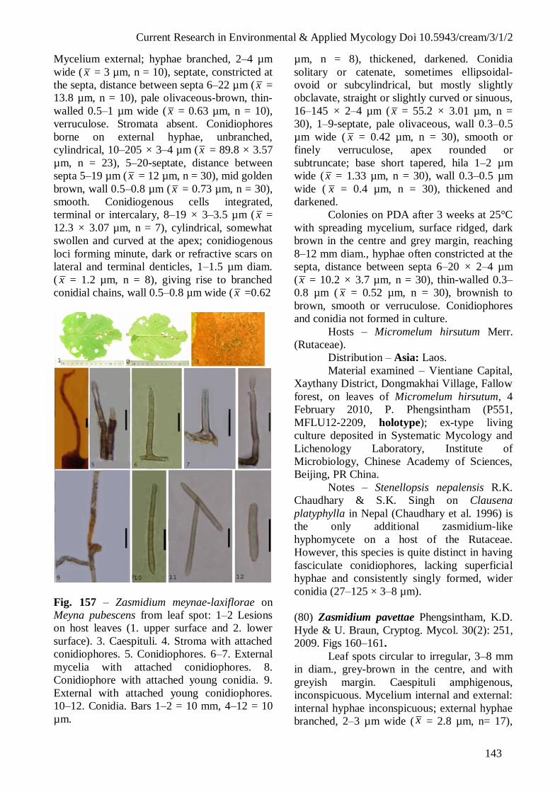
Current Research in Environmental & Applied Mycology Doi 10.5943/cream/3/1/2
143
Mycelium external; hyphae branched, 2–4 µm
wide ( x = 3 µm, n = 10), septate, constricted at
the septa, distance between septa 6–22 µm ( x =
13.8 µm, n = 10), pale olivaceous-brown, thin-
walled 0.5–1 µm wide ( x = 0.63 µm, n = 10),
verruculose. Stromata absent. Conidiophores
borne on external hyphae, unbranched,
cylindrical, 10–205 × 3–4 µm (x = 89.8 × 3.57
µm, n = 23), 5–20-septate, distance between
septa 5–19 µm ( x = 12 µm, n = 30), mid golden
brown, wall 0.5–0.8 µm ( x = 0.73 µm, n = 30),
smooth. Conidiogenous cells integrated,
terminal or intercalary, 8–19 × 3–3.5 µm ( x =
12.3 × 3.07 µm, n = 7), cylindrical, somewhat
swollen and curved at the apex; conidiogenous
loci forming minute, dark or refractive scars on
lateral and terminal denticles, 1–1.5 µm diam.
( x = 1.2 µm, n = 8), giving rise to branched
conidial chains, wall 0.5–0.8 µm wide ( x =0.62
Fig. 157 – Zasmidium meynae-laxiflorae on
Meyna pubescens from leaf spot: 1–2 Lesions
on host leaves (1. upper surface and 2. lower
surface). 3. Caespituli. 4. Stroma with attached
conidiophores. 5. Conidiophores. 6–7. External
mycelia with attached conidiophores. 8.
Conidiophore with attached young conidia. 9.
External with attached young conidiophores.
10–12. Conidia. Bars 1–2 = 10 mm, 4–12 = 10
µm.
µm, n = 8), thickened, darkened. Conidia
solitary or catenate, sometimes ellipsoidal-
ovoid or subcylindrical, but mostly slightly
obclavate, straight or slightly curved or sinuous,
16–145 × 2–4 µm ( x = 55.2 × 3.01 µm, n =
30), 1–9-septate, pale olivaceous, wall 0.3–0.5
µm wide ( x = 0.42 µm, n = 30), smooth or
finely verruculose, apex rounded or
subtruncate; base short tapered, hila 1–2 µm
wide (x = 1.33 µm, n = 30), wall 0.3–0.5 µm
wide ( x = 0.4 µm, n = 30), thickened and
darkened.
Colonies on PDA after 3 weeks at 25°C
with spreading mycelium, surface ridged, dark
brown in the centre and grey margin, reaching
8–12 mm diam., hyphae often constricted at the
septa, distance between septa 6–20 × 2–4 µm
(x = 10.2 × 3.7 µm, n = 30), thin-walled 0.3–
0.8 µm ( x = 0.52 µm, n = 30), brownish to
brown, smooth or verruculose. Conidiophores
and conidia not formed in culture.
Hosts – Micromelum hirsutum Merr.
(Rutaceae).
Distribution – Asia: Laos.
Material examined – Vientiane Capital,
Xaythany District, Dongmakhai Village, Fallow
forest, on leaves of Micromelum hirsutum, 4
February 2010, P. Phengsintham (P551,
MFLU12-2209, holotype); ex-type living
culture deposited in Systematic Mycology and
Lichenology Laboratory, Institute of
Microbiology, Chinese Academy of Sciences,
Beijing, PR China.
Notes – Stenellopsis nepalensis R.K.
Chaudhary & S.K. Singh on Clausena
platyphylla in Nepal (Chaudhary et al. 1996) is
the only additional zasmidium-like
hyphomycete on a host of the Rutaceae.
However, this species is quite distinct in having
fasciculate conidiophores, lacking superficial
hyphae and consistently singly formed, wider
conidia (27–125 × 3–8 µm).
(80) Zasmidium pavettae Phengsintham, K.D.
Hyde & U. Braun, Cryptog. Mycol. 30(2): 251,
2009. Figs 160–161.
Leaf spots circular to irregular, 3–8 mm
in diam., grey-brown in the centre, and with
greyish margin. Caespituli amphigenous,
inconspicuous. Mycelium internal and external:
internal hyphae inconspicuous; external hyphae
branched, 2–3 µm wide ( x = 2.8 µm, n= 17),
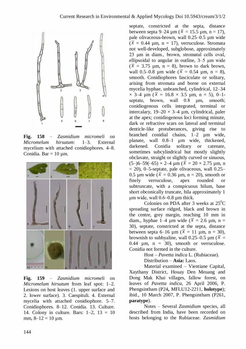
Current Research in Environmental & Applied Mycology Doi 10.5943/cream/3/1/2
144
Fig. 158 – Zasmidium micromeli on
Micromelum hirsutum: 1–3. External
mycelium with attached conidiophores. 4–8.
Conidia. Bar = 10 µm.
Fig. 159 – Zasmidium micromeli on
Micromelum hirsutum from leaf spot: 1–2.
Lesions on host leaves (1. upper surface and
2. lower surface). 3. Caespituli. 4. External
mycelia with attached conidiophore. 5–7.
Conidiophores. 8–12. Conidia. 13. Culture.
14. Colony in culture. Bars: 1–2, 13 = 10
mm, 8–12 = 10 µm.
septate, constricted at the septa, distance
between septa 9–24 µm ( x = 15.5 µm, n = 17),
pale olivaceous-brown, wall 0.25–0.5 µm wide
(x = 0.44 µm, n = 17), verruculose. Stromata
not well-developed, subglobose, approximately
23 μm in diam., brown, stromatal cells oval,
ellipsoidal to angular in outline, 3–5 μm wide
( x = 3.75 µm, n = 8), brown to dark brown,
wall 0.5–0.8 µm wide ( x = 0.54 µm, n = 8),
smooth. Conidiophores fasciculate or solitary,
arising from stromata and borne on external
mycelia hyphae, unbranched, cylindrical, 12–34
× 3–4 µm ( x = 16.8 × 3.5 µm, n = 5), 0–1-
septate, brown, wall 0.8 μm, smooth;
conidiogenous cells integrated, terminal or
intercalary, 19–20 × 3–4 µm, cylindrical, paler
at the apex; conidiogenous loci forming minute,
dark or refractive scars on lateral and terminal
denticle-like protuberances, giving rise to
branched conidial chains, 1–2 µm wide,
planate, wall 0.8–1 µm wide, thickened,
darkened. Conidia solitary or catenate,
sometimes subcylindrical but mostly slightly
obclavate, straight or slightly curved or sinuous,
(5–)6–59(–65) × 2–4 µm (x = 20 × 2.75 µm, n
= 20), 0–5-septate, pale olivaceous, wall 0.25–
0.5 µm wide ( x = 0.36 µm, n = 20), smooth or
finely verruculose, apex rounded or
subtruncate, with a conspicuous hilum, base
short obconically truncate, hila approximately 1
μm wide, wall 0.6–0.8 µm thick.
Colonies on PDA after 3 weeks at 250C
spreading surface ridged, black and brown in
the centre, grey margin, reaching 10 mm in
diam., hyphae 1–4 µm wide (x = 2.6 µm, n =
30), septate, constricted at the septa, distance
between septa 6–16 µm (x = 11 µm, n = 30),
brownish to subhyaline, wall 0.25–0.5 µm ( x =
0.44 µm, n = 30), smooth or verruculose.
Conidia not formed in the culture.
Host – Pavetta indica L. (Rubiaceae).
Distribution – Asia: Laos.
Material examined – Vientiane Capital,
Xaythany District, Houay Den Meuang and
Dong Mak Khai villages, fallow forest, on
leaves of Pavetta indica, 26 April 2006, P.
Phengsintham (P24, MFLU12-2211, holotype);
ibid., 10 March 2007, P. Phengsintham (P261,
paratype).
Notes – Several Zasmidium species, all
described from India, have been recorded on
hosts belonging to the Rubiaceae. Zasmidium

Current Research in Environmental & Applied Mycology Doi 10.5943/cream/3/1/2
145
canthii (J.M. Yen, A.K. Kar & B.K. Das)
Kamal (Yen et al. 1982a, Kamal 2010),
described from West Bengal on Canthium
dedymum, is morphologically close to the
fungus on Pavetta indica, but this species is
distinguished by lacking stromata, consistently
solitary conidiophores and somewhat longer,
wider conidia, 18–112 × 3–4.5 µm. Z. meynae-
laxiflorae (K. Srivast., A.K. Srivast. & Kamal)
Phengsintham, K.D. Hyde & U. Braun
(Srivastava et al. 1995, Phengsintham et al.
2009) is another similar species, but it is
distinct by its much longer conidiophores, up to
about 90 μm in length, often formed in well-
developed fascicles of 9–20 stalks. Most of the
other Stenella species on Rubiaceae are
characterized by having much longer
pluriseptate conidia, viz., Z. coffeae (J.M. Yen,
A.K. Kar & B.K. Das) Kamal (conidia 33–200
× 3–5(–6) µm, see Yen et al. 1982a), Stenella
hyptiantherae S.K. Singh, Arch. Singh &
Kamal (conidia 18–177 × 3–5 µm, see Singh et
al. 1997), Z. plectroniae (Ponnappa) Kamal
(conidia 40–210 × 2–3.5 µm, see Ellis 1976), Z.
vangueriae (Thirum. & Mishra) Kamal (conidia
30–258 × 3–4.5 µm, see Thirumalachar &
Mishra 1963), and Z. xeromphigena (J.M. Yen,
A.K. Kar & B.K. Das) Kamal (conidia 20–156
× 3–4 µm, see Yen et al. 1982b). Zasmidium
naucleae (A.K. Das) Kamal (Das 1990, Kamal
2010) differs in having larger, well-developed
stromata with large fascicles composed of 7–55
conidiophores, and much longer, pluriseptate
conidiophores, 33–115 × 3–5 µm.
Literature – Yen et al. (1982: 35–57),
Srivastava et al. (1995: 233–236),
Thirumalachar & Mishra (1963: 29–83).
(81) Zasmidium sp. Figs 162–163.
Leaf spots variable, more or less
irregularly orbicular, 2–4 mm diam., grey-
brown in the center, dark brown at the margin.
Caespituli hypophyllous, conspicuous.
Mycelium external; hyphae branched, 4–6 µm
wide ( x = 5 µm, n = 7), septate, constricted at
the septa, distance between septa 15–36 µm ( x
= 23.5 µm, n = 7), pale olivaceous-brown, thin-
walled 0.5–1 µm wide ( x = 0.63 µm, n = 10),
verruculose. Stromata absent. Conidiophores
borne on external hyphae, unbranched,
cylindrical, 18–96 × 3–4 µm ( x = 56.58 × 3.64
µm, n = 13), 1–5-septate, distance between sep-
Fig. 160 – Zasmidium pavettae on Pavetta
indica: 1–3. Conidiophores. 4. External
mycelium with attached conidiophores. 6–9.
Conidia. Bar = 10 µm.
Fig. 161 – Zasmidium pavettae on Pavetta
indica from leaf spot: 1–2 Lesions on host
leaves (1. upper surface and 2. lower surface). 3.
Stroma. 4–5. Conidiophores. 6. External
mycelium with attached young conidiophores.
7–10. Conidia. 11. Culture. Bars 1–2, 11 = 10
mm, 3–10 = 10 µm.
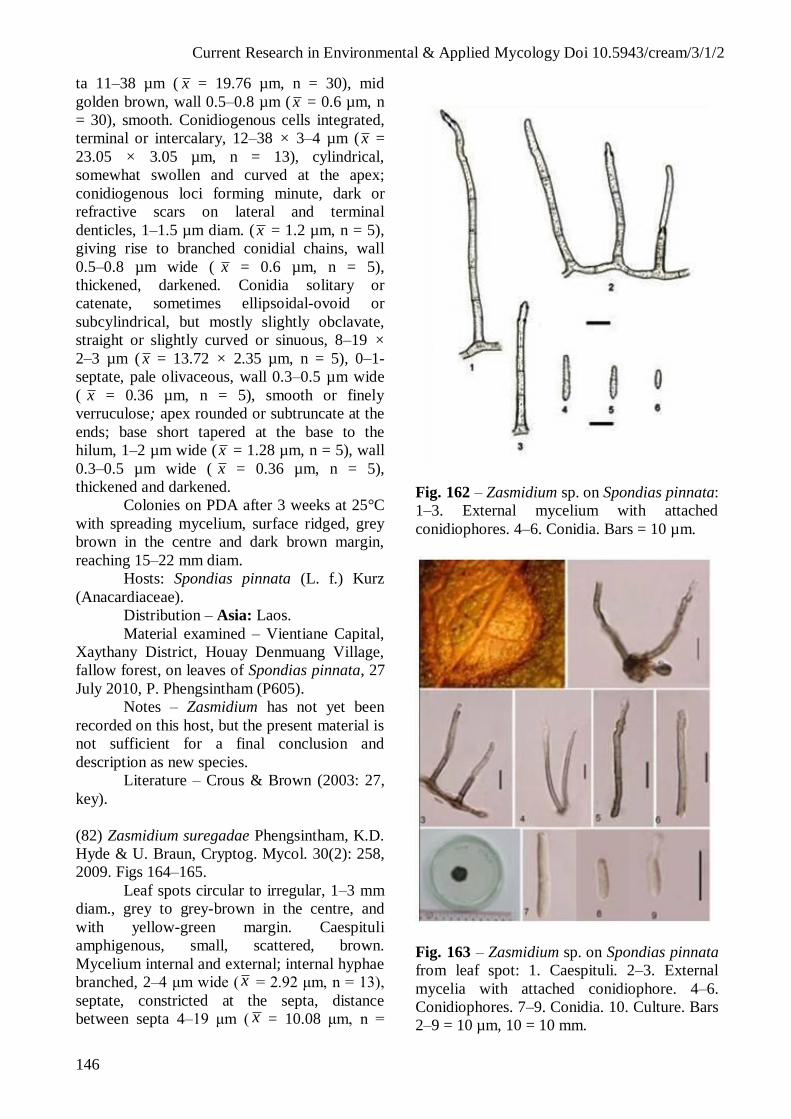
Current Research in Environmental & Applied Mycology Doi 10.5943/cream/3/1/2
146
ta 11–38 µm ( x = 19.76 µm, n = 30), mid
golden brown, wall 0.5–0.8 µm ( x = 0.6 µm, n
= 30), smooth. Conidiogenous cells integrated,
terminal or intercalary, 12–38 × 3–4 µm ( x =
23.05 × 3.05 µm, n = 13), cylindrical,
somewhat swollen and curved at the apex;
conidiogenous loci forming minute, dark or
refractive scars on lateral and terminal
denticles, 1–1.5 µm diam. ( x = 1.2 µm, n = 5),
giving rise to branched conidial chains, wall
0.5–0.8 µm wide ( x = 0.6 µm, n = 5),
thickened, darkened. Conidia solitary or
catenate, sometimes ellipsoidal-ovoid or
subcylindrical, but mostly slightly obclavate,
straight or slightly curved or sinuous, 8–19 ×
2–3 µm ( x = 13.72 × 2.35 µm, n = 5), 0–1-
septate, pale olivaceous, wall 0.3–0.5 µm wide
( x = 0.36 µm, n = 5), smooth or finely
verruculose; apex rounded or subtruncate at the
ends; base short tapered at the base to the
hilum, 1–2 µm wide (x = 1.28 µm, n = 5), wall
0.3–0.5 µm wide ( x = 0.36 µm, n = 5),
thickened and darkened.
Colonies on PDA after 3 weeks at 25°C
with spreading mycelium, surface ridged, grey
brown in the centre and dark brown margin,
reaching 15–22 mm diam.
Hosts: Spondias pinnata (L. f.) Kurz
(Anacardiaceae).
Distribution – Asia: Laos.
Material examined – Vientiane Capital,
Xaythany District, Houay Denmuang Village,
fallow forest, on leaves of Spondias pinnata, 27
July 2010, P. Phengsintham (P605).
Notes – Zasmidium has not yet been
recorded on this host, but the present material is
not sufficient for a final conclusion and
description as new species.
Literature – Crous & Brown (2003: 27,
key).
(82) Zasmidium suregadae Phengsintham, K.D.
Hyde & U. Braun, Cryptog. Mycol. 30(2): 258,
2009. Figs 164–165.
Leaf spots circular to irregular, 1–3 mm
diam., grey to grey-brown in the centre, and
with yellow-green margin. Caespituli
amphigenous, small, scattered, brown.
Mycelium internal and external; internal hyphae
branched, 2–4 μm wide (x = 2.92 μm, n = 13),
septate, constricted at the septa, distance
between septa 4–19 μm ( x = 10.08 μm, n =
Fig. 162 – Zasmidium sp. on Spondias pinnata:
1–3. External mycelium with attached
conidiophores. 4–6. Conidia. Bars = 10 µm.
Fig. 163 – Zasmidium sp. on Spondias pinnata
from leaf spot: 1. Caespituli. 2–3. External
mycelia with attached conidiophore. 4–6.
Conidiophores. 7–9. Conidia. 10. Culture. Bars
2–9 = 10 µm, 10 = 10 mm.

Current Research in Environmental & Applied Mycology Doi 10.5943/cream/3/1/2
147
13), pale olivaceousbrown, wall 0.3–0.5 μm
wide ( x = 0.39 μm, n = 13), smooth; external
hyphae superficial, branched, 1–4 μm wide ( x
= 2.67 μm, n = 30), septate, constricted at the
septa, distance between septa 6–22 μm ( x =
11.78 μm, n = 30), pale olivaceous-brown, wall
0.3–0.5 μm wide ( x = 0.44 μm, n = 30), almost
smooth to verruculose. Stromata well-
developed, subglobose, 35–70 μm diam.,
brown, stroma cells oval, ellipsoidal to angular
in outline, 3–10 μm wide ( x = 6.37 μm, n =
30), brown to dark brown, wall 0.5–1 μm wide
( x = 0.74 μm, n = 30), smooth. Conidiophores
fasciculate, arising from stromata (9–34 per
fascicle) and solitary, borne on external
mycelial hyphae, unbranched, cylindrical, (34–
)40–86(–110) × 3–4 μm (x = 65.8 × 3.3 μm, n
= 30), 2–9-septate, distance between septa 7–20
μm ( x = 13.3 μm, n = 30), brown to dark
brown, wall 0.5–0.8 μm (x = 0.58 μm, n = 30),
smooth, 0–2 times geniculate; conidiogenous
cells polyblastic, integrated, terminal or
intercalary, 8–20 × 2–3 μm ( x = 15.8 × 3.14
μm, n = 30), cylindrical, pale at the apex;
conidiogenous loci small, conspicuous, sub-
planate to planate, 1–1.5 μm wide ( x = 1.15
μm, n = 10), wall 0.5–1 μm wide ( x = 0.76 μm,
n = 10), thickened, darkened. Conidia solitary
or catenate, sometimes subcylindrical, but
mostly slightly obclavate, occasionally with
lateral branches, straight or slightly curved to
sinuous, (16–)17–128(–153) × 2–4 μm ( x =
74.13 × 2.8 μm, n = 30), 1–10-septate, pale
olivaceous, wall 0.3–0.5 μm wide ( x = 0.36
μm, n = 30), smooth or finely verruculose, apex
rounded or subtruncate, at the end of some
conidia with a thickened hilum, base truncate,
hila slightly thickened and darkened, 1–1.5 μm
wide ( x = 1.04 μm, n = 13), wall 0.5–0.8 μm
wide ( x = 0.52 μm, n = 13).
Colonies on PDA after 3 weeks at 25oC
spreading surface ridged, grey-brown in the
centre, margin greenish black, reaching 11 mm
diam., hyphae 1–4 μm wide ( x = 2.8 μm, n =
30), septate, constricted at the septa, distance
between septa 6–18 μm (x = 11.9 μm, n = 30),
greenish to brownish, wall approximately 0.25–
0.5 μm ( x = 0.34 μm, n = 30), smooth or
verruculose. Conidia not formed in the culture.
Hosts – Suregada multiflora (Juss.) H.
Baill. (Euphorbiaceae).
Distribution – Asia: Laos.
Material examined – Vientiane Capital,
Xaythany District, Houay Den Meuang Village,
on leaves of Suregada multiflora, 8 May 2006,
P. Phengsintham (P36, MFLU12-2212,
holotype). GenBank accession no (ITS,
KC677914; LSU, KC677939).
Notes – Five Zasmidium spp. have been
recorded on other hosts of the family
Euphorbiaceae, but there is no record from
Suregada spp. Zasmidium bischofiae-javanicae
(R.K. Chaudhary, M.S. Tripathi, P.N. Singh &
S. Chaudhary) Kamal (Chaudhary et al. 2001),
Z. brideliicola (K. Srivast., A.K. Srivast. &
Kamal) Kamal (Srivastava et al. 1994), Z.
gorakhpurensis (Kamal & P. Kumar) Kamal
(Kamal & Kumar 1980, de Hoog et al. 1983) as
well as Z. aporosae, described above, are
distinct from Z. suregadae by having
conidiophores that are consistently formed
singly, i.e. stromata and fasciculate
conidiophores are lacking. Zasmidium
manihotis (≡ Stenella manihotis, Braun &
Freire 2004), described from Brazil on Manihot
sp., is a similar species with solitary as well as
fasciculate conidiophores, but the stromata are
much smaller, 10–25 μm diam., or even
lacking, the conidiophores are formed in small
fascicles, they are wider, 3–7 μm, and the
conidia are also wider, 3–6 μm. Stenella
ateramnae R.F. Castaneda & B. Kendr.
(Castaneda & Kendrick 1991), described from
Cuba on Ateramnus lucidus, is close to the new
species by having large stromata, 50–80 × 25–
45 μm, with numerous conidiophores in
fascicles, but the brown conidia are much
longer, up to 280 μm, with up to 20 septa.
Furthermore, the latter species has been
described from fallen leaves (lesions lacking)
with usually internal mycelium (verruculose
external hyphae not described).
Literature – Kamal & Kumar (1980:
265–269), Hoog et al. (1983: 485–490),
Castañeda & Kendrick (1991: 1–132),
Srivastava et al. (1994: 515–520), Chaudhary et
al. (2001: 226–232), Crous & Braun (2003:
Key).
1.3.3.2 Morphologically similar cercosporoid
fungi
(83) Cladosporium colocasiae Sawada, Trans.
Nat. Hist. Soc. Taiwan 25: 125, 1916. Figs
166–167.
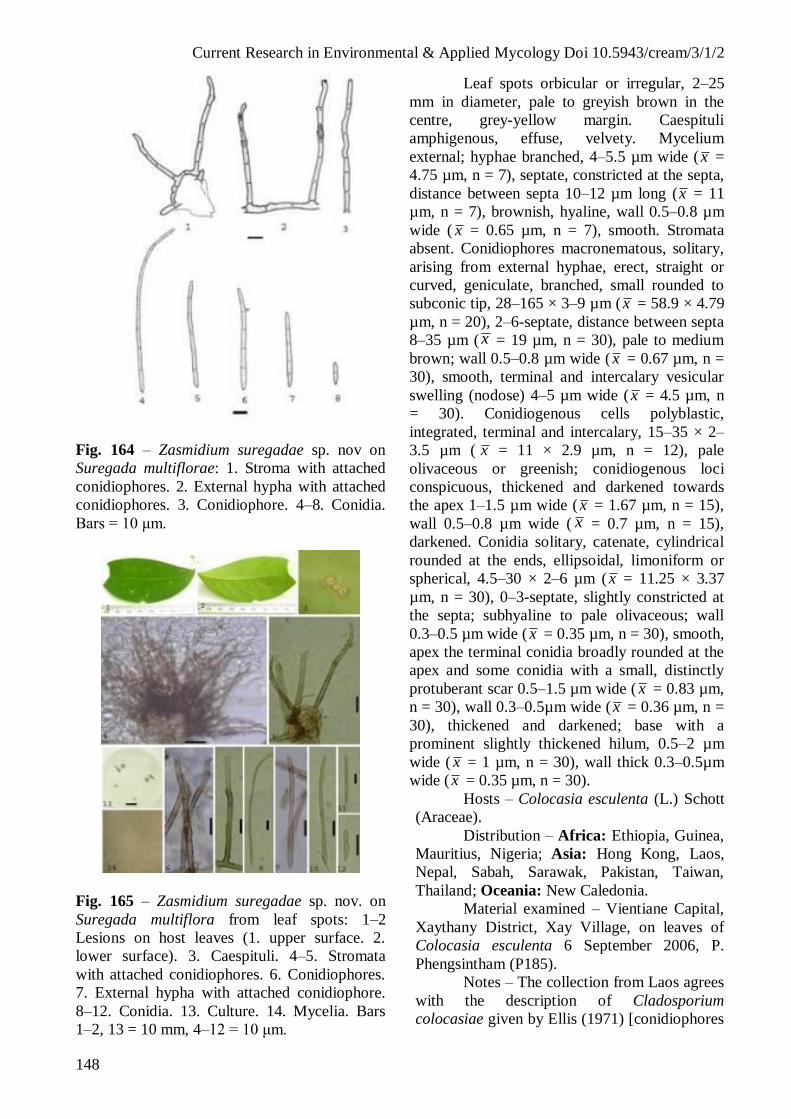
Current Research in Environmental & Applied Mycology Doi 10.5943/cream/3/1/2
148
Fig. 164 – Zasmidium suregadae sp. nov on
Suregada multiflorae: 1. Stroma with attached
conidiophores. 2. External hypha with attached
conidiophores. 3. Conidiophore. 4–8. Conidia.
Bars = 10 μm.
Fig. 165 – Zasmidium suregadae sp. nov. on
Suregada multiflora from leaf spots: 1–2
Lesions on host leaves (1. upper surface. 2.
lower surface). 3. Caespituli. 4–5. Stromata
with attached conidiophores. 6. Conidiophores.
7. External hypha with attached conidiophore.
8–12. Conidia. 13. Culture. 14. Mycelia. Bars
1–2, 13 = 10 mm, 4–12 = 10 μm.
Leaf spots orbicular or irregular, 2–25
mm in diameter, pale to greyish brown in the
centre, grey-yellow margin. Caespituli
amphigenous, effuse, velvety. Mycelium
external; hyphae branched, 4–5.5 µm wide (x =
4.75 µm, n = 7), septate, constricted at the septa,
distance between septa 10–12 µm long (x = 11
µm, n = 7), brownish, hyaline, wall 0.5–0.8 µm
wide ( x = 0.65 µm, n = 7), smooth. Stromata
absent. Conidiophores macronematous, solitary,
arising from external hyphae, erect, straight or
curved, geniculate, branched, small rounded to
subconic tip, 28–165 × 3–9 µm (x = 58.9 × 4.79
µm, n = 20), 2–6-septate, distance between septa
8–35 µm ( x = 19 µm, n = 30), pale to medium
brown; wall 0.5–0.8 µm wide (x = 0.67 µm, n =
30), smooth, terminal and intercalary vesicular
swelling (nodose) 4–5 µm wide (x = 4.5 µm, n
= 30). Conidiogenous cells polyblastic,
integrated, terminal and intercalary, 15–35 × 2–
3.5 µm ( x = 11 × 2.9 µm, n = 12), pale
olivaceous or greenish; conidiogenous loci
conspicuous, thickened and darkened towards
the apex 1–1.5 µm wide (x = 1.67 µm, n = 15),
wall 0.5–0.8 µm wide ( x = 0.7 µm, n = 15),
darkened. Conidia solitary, catenate, cylindrical
rounded at the ends, ellipsoidal, limoniform or
spherical, 4.5–30 × 2–6 µm (x = 11.25 × 3.37
µm, n = 30), 0–3-septate, slightly constricted at
the septa; subhyaline to pale olivaceous; wall
0.3–0.5 µm wide ( x = 0.35 µm, n = 30), smooth,
apex the terminal conidia broadly rounded at the
apex and some conidia with a small, distinctly
protuberant scar 0.5–1.5 µm wide ( x = 0.83 µm,
n = 30), wall 0.3–0.5µm wide (x = 0.36 µm, n =
30), thickened and darkened; base with a
prominent slightly thickened hilum, 0.5–2 µm
wide ( x = 1 µm, n = 30), wall thick 0.3–0.5µm
wide (x = 0.35 µm, n = 30).
Hosts – Colocasia esculenta (L.) Schott
(Araceae).
Distribution – Africa: Ethiopia, Guinea,
Mauritius, Nigeria; Asia: Hong Kong, Laos,
Nepal, Sabah, Sarawak, Pakistan, Taiwan,
Thailand; Oceania: New Caledonia.
Material examined – Vientiane Capital,
Xaythany District, Xay Village, on leaves of
Colocasia esculenta 6 September 2006, P.
Phengsintham (P185).
Notes – The collection from Laos agrees
with the description of Cladosporium
colocasiae given by Ellis (1971) [conidiophores

Current Research in Environmental & Applied Mycology Doi 10.5943/cream/3/1/2
149
up to 180 µm long and 4–6 µm and conidia size
12–32 × 6–9 µm, 1–3-septate], but the conidia
from Laos vary from 0–3-septate.
Literature – Ellis (1971: 312).
Fig. 166 – Cladosporium colocasiae on
Colocasia esculenta from leaf spots: 1–4.
Conidiophores. 5–9. Conidia. Bars = 10 µm.
(84) Periconiella lygodii Arch. Singh, Bhalla &
S.K. Singh ex U. Braun, Feddes Repert. 115:
53, 2004. Figs 168–169.
Leaf spots black, striiform, restricted by
lateral leaf veins and therefore appearing
oblique, 1–5×1 mm, becoming confluent and
eventually causing almost complete blackening
of the leaflet. Caespituli amphigenous,
conspicuous. Mycelium intercellar,
inconspicuous. Stromata absent or small,
intraepidermal, up to 15 µm diam, consisting of
swollen, brown hyphal cells. Conidiophores
single, arising through stomata, medium brown,
smooth, 60–175 × 7–8 μm (x = 118 × 7.5 µm,
n = 5), 5–11-septate, distance between septa 7–
40 µm ( x = 18.8 µm, n = 17), pale to medium
brown, wall 0.5–0.8 µm wide ( x = 0.66 µm, n
= 17), smooth. Conidiogenous cells terminal,
17–22 × 5–6 μm (x = 20.5 × 4.5 µm, n = 7);
conidiogenous loci usually restricted to the apex
of the cell, slightly raised, slightly thickened
and rather refractive than darkened,
Fig. 167 – Cladosporium colocasiae on
Colocasia esculenta from leaf spots: 1. Lesions
on host leaves (upper surface). 2–4.
Conidiophores. 9–10. Conidia. Bars 1 = 10 mm,
2–10 = 10 µm.
1.5 μm diam. Conidia solitary, obclavate,
tapering towards the apex, straight or slightly
curved, pale brown, smooth, 17–58 × 4–5
μm( x = 32.8 × 4.5 µm, n = 12), 0–7-septate,
wall 0.3–0.5 µm wide ( x = 0.47 µm, n = 12),
smooth, basal hilum unthickened to thickened
and not darkened to darkened, 1.5 μm wide.
Hosts – Lygodium flexuosum (L.) Sw.,
L. japonicum (Thunb.) Sw. (Schizaeaceae).
Distribution – Asia: India, Laos,
Taiwan.
Material examined – Khammoune
Province, Nakai District, Nahao Village, fallow
forest, on leaves of Lygodium flexuosum, 3 June
2010, P. Phengsintham (P579).
Notes – The collection from Laos agrees
with the description of Periconiela lygodii
published by Singh et al. (1998) [conidiophores,
90–345 × 9–12 μm, with 3–6 septa, septa 8–75
μm apart; conidia solitary, obclavate, tapering
towards the apex, straight or slightly curved,
pale brown, smooth, 3–9-septate, 27–5 × 5–7
μm].

Current Research in Environmental & Applied Mycology Doi 10.5943/cream/3/1/2
150
Literature – Braun (2004), Singh et al.
(1998).
Fig. 168 – Periconiela lygodii on Lygodium
flexuosum from leaf spots: 1. Stromata with
attached conidiophores. 2. Base
conidiophores. 3. Apex of conidiophores. 4–
8. Conidia. Bar = 10 µm.
(85) Pseudocercosporella bakeri (Syd. & P.
Syd.) Deighton, Mycol. Pap. 133: 41, 1973.
Figs 170–171.
≡ Cylindrosporium bakeri Syd. & P.
Syd., Ann. Mycol. 14: 372, 1916.
= Ramularia ipomoeae F. Stevens, Bern.
Bishop Mus. Bull. 19: 150, 1925.
= Cercosporella ipomoeae Sawada,
Rep. Govt. Res. Inst. Dept. Agric. Formosa 86:
161, 1943.
= Cercosporella ipomoeicola Sawada,
Spec. Publ. Coll. Agric. Natn. Taiwan Univ. 8:
192, 1959.
= Pseudocercosporella ipomoeae
Deighton, Mycol. Pap. 133: 39, 1973.
Leaf spots subcircular, angular to
irregular, 2–20 mm diam, pale greenish,
becoming pale brown, finally greyish white;
margin indefinite or with a narrow brown
border. Caespituli amphigenous, punctiform to
effuse, whitish.
Fig. 169 – Periconiela lygodii on Lygodium
flexuosum from leaf spots: 1. Lesions on host
leaf. 2. Conidiophores. 3. Base of conidia. 4.
Apices of conidiophores. 5–9. Conidia. Bars 3–
9 = 10 µm.
Mycelium internal, consisting of hyaline,
septate, sparingly branched, 1.5–3.5 µm wide
hyphae; Stromata absent or small,
intraepidermal, 10–40 µm diam, consisting of
swollen, hyaline hyphal cells. Conidiophores
usually aggregated, occasionally subfasciculate,
up to about 20, arising from inner hyphae or
hyphal aggregations, erumpent through the
cuticle, short, erect, subcylindrical, conical,
straight, curved to geniculate-sinuous, (2.5–
)20–40 × (2–)2.5–4(–6) µm, 0–2-septate.
Conidiogenous cells terminal, integrated, (2.5–
)10–20 × 3.5–4 µm; conidial scars usually more
or less truncate, 1.5–3 µm diam, unthickened,
not darkened. Conidia solitary, subcylindrical,
somewhat acicular to slightly obclavate, (35–
)40–65 × (2.5–)3.5–4 µm, 1–3-septate in vivo,
not constricted, hyaline, smooth, apex obtuse,
base truncate or slightly obconically truncate,
unthickened (adapted from Braun 1995). In
vitro on OA, conidia (30–)46–60(–75) × (3.5–
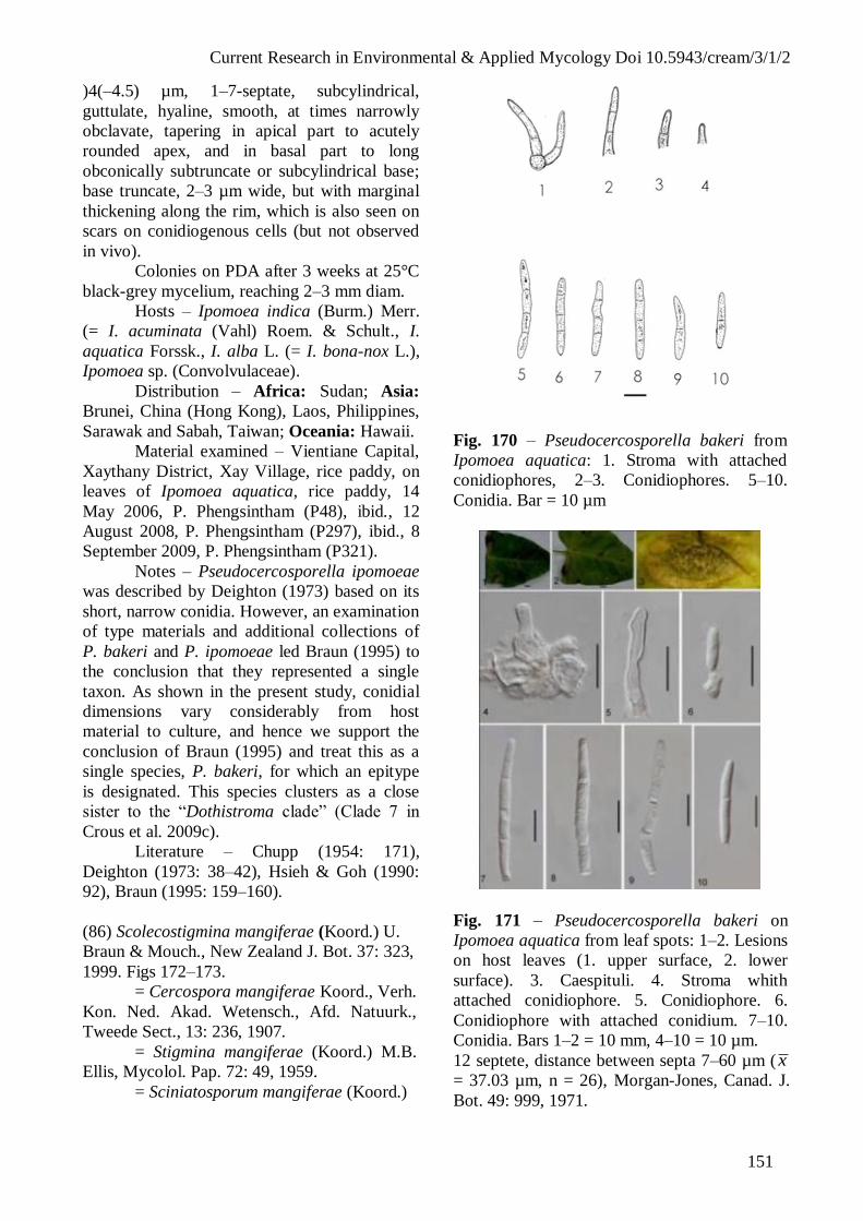
Current Research in Environmental & Applied Mycology Doi 10.5943/cream/3/1/2
151
)4(–4.5) µm, 1–7-septate, subcylindrical,
guttulate, hyaline, smooth, at times narrowly
obclavate, tapering in apical part to acutely
rounded apex, and in basal part to long
obconically subtruncate or subcylindrical base;
base truncate, 2–3 µm wide, but with marginal
thickening along the rim, which is also seen on
scars on conidiogenous cells (but not observed
in vivo).
Colonies on PDA after 3 weeks at 25°C
black-grey mycelium, reaching 2–3 mm diam.
Hosts – Ipomoea indica (Burm.) Merr.
(= I. acuminata (Vahl) Roem. & Schult., I.
aquatica Forssk., I. alba L. (= I. bona-nox L.),
Ipomoea sp. (Convolvulaceae).
Distribution – Africa: Sudan; Asia:
Brunei, China (Hong Kong), Laos, Philippines,
Sarawak and Sabah, Taiwan; Oceania: Hawaii.
Material examined – Vientiane Capital,
Xaythany District, Xay Village, rice paddy, on
leaves of Ipomoea aquatica, rice paddy, 14
May 2006, P. Phengsintham (P48), ibid., 12
August 2008, P. Phengsintham (P297), ibid., 8
September 2009, P. Phengsintham (P321).
Notes – Pseudocercosporella ipomoeae
was described by Deighton (1973) based on its
short, narrow conidia. However, an examination
of type materials and additional collections of
P. bakeri and P. ipomoeae led Braun (1995) to
the conclusion that they represented a single
taxon. As shown in the present study, conidial
dimensions vary considerably from host
material to culture, and hence we support the
conclusion of Braun (1995) and treat this as a
single species, P. bakeri, for which an epitype
is designated. This species clusters as a close
sister to the “Dothistroma clade” (Clade 7 in
Crous et al. 2009c).
Literature – Chupp (1954: 171),
Deighton (1973: 38–42), Hsieh & Goh (1990:
92), Braun (1995: 159–160).
(86) Scolecostigmina mangiferae (Koord.) U.
Braun & Mouch., New Zealand J. Bot. 37: 323,
1999. Figs 172–173.
= Cercospora mangiferae Koord., Verh.
Kon. Ned. Akad. Wetensch., Afd. Natuurk.,
Tweede Sect., 13: 236, 1907.
= Stigmina mangiferae (Koord.) M.B.
Ellis, Mycolol. Pap. 72: 49, 1959.
= Sciniatosporum mangiferae (Koord.)
Fig. 170 – Pseudocercosporella bakeri from
Ipomoea aquatica: 1. Stroma with attached
conidiophores, 2–3. Conidiophores. 5–10.
Conidia. Bar = 10 µm
Fig. 171 – Pseudocercosporella bakeri on
Ipomoea aquatica from leaf spots: 1–2. Lesions
on host leaves (1. upper surface, 2. lower
surface). 3. Caespituli. 4. Stroma whith
attached conidiophore. 5. Conidiophore. 6.
Conidiophore with attached conidium. 7–10.
Conidia. Bars 1–2 = 10 mm, 4–10 = 10 µm.
12 septete, distance between septa 7–60 µm (x
= 37.03 µm, n = 26), Morgan-Jones, Canad. J.
Bot. 49: 999, 1971.

Current Research in Environmental & Applied Mycology Doi 10.5943/cream/3/1/2
152
Leaf spots round to angular black spots,
1–13 mm diam. ( x = 4 mm, n = 13), pale grey or
black in the centre, and with yellowish margin.
Caespituli hypophyllous, minute, pucktiform,
dark olivaceous-brown. Mycelium internal,
inconspicuous. Stromata oval or ellipsoidal, 13–
25 µm diam. (x = 18.44 µm, n = 9), dark brown,
stromatal cells angular in outline, 3–7 µm wide
( x = 3.84 µm, n = 20), wall 0.8–1 µm (x = 0.84
µm, n = 20), smooth. Conidiophores fasciculate,
arising from stomata (1–4 per fascicle),
unbranched, 0–1- geniculate, lageniform,
straight or curved, 8–24 × 3–5 µm ( x = 12 ×
4.11 µm, n = 9), with up to 3 annellations, 0–2-
septate, distance between septa 8–9 µm ( x = 8.5
µm, n = 5), olivaceous-brown, paler and narrow
at the apex, wall 0.5–1 µm wide ( x = 0.9 µm, n
= 9), smooth. Conidiogenous cells terminal,
lageniform, 9 × 5 µm; conidiogenous loci small,
at the apex, conspicuous, ovoid to oval, up to 2
µm, wall 0.8 µm thickened. Conidia solitary,
cylindrical to obclavate, straight to curved, 21–
77 × 3–5 µm ( x = 50.53 × 3.84 µm, n = 13), 2–
10-septate, reddish brown, pale near the apex,
wall 0.3–0.8 µm wide ( x = 0.53 µm, n = 13),
smooth, tip subobtuse, base truncate, hila 2–3
µm wide ( x = 2.85 µm, n = 9), wall 0.5–1 µm
( x = 0.8 µm, n = 9), thickened and darkened.
Hosts – Mangifera foetida Lour., M.
indica L. (Anacardiaceae).
Distribution – Africa: Angola, Congo,
Gabon, Ghana, Malawi, Mozambique, Nigeria,
São Tomé and Principe, Sierra Leone, Somalia,
Sudan, Tanzania, Uganda; Asia: Brunei, China,
Indonesia, Japan, Java, Laos, Malaysia, Nepal,
Philippines, Sri Lanka, Taiwan, Thailand;
North America and West Indies: Cuba,
Dominican Republic, Haiti, Honduras, Jamaica,
Mexico, Panama, Puerto Rico, Trinidan and
Tobago, Virgin Islands; Australia; Oceania:
Cook Islands, Fiji, New Caledonia, Papua New
Guinea, Samoa, Solomon Islands, Vanuatu;
South America: Colombia, Suriname,
Venezuela.
Material examined – Vientiane Capital,
Xaythany District, Dong Makkhai Village,
Garden, on leaves of Mangifera indica, 18
December 2008, P. Phengsintham (P388).
Notes – The collection from Laos has
conidiophores and conidia of a similar size to
those described by Ellis (1971) and Hsieh &
Goh (1990), but the conidiophores have only
three annellations.
Literature – Saccardo (1910: 253), Ellis
(1971: 146), Hsieh & Goh (1990: 21), Braun et
al. (1999: 323), Crous & Braun (2003: 265).
Fig. 172 – Scolecostigmina mangiferae from
leaf spots of Mangifera indica: 1. Stroma with
Conidiophore. 2–3. Conidiophores. 4–8.
Conidia. Bars = 10 µm.
(87) Spiropes clavatus (Ellis & Martin) M.B.
Ellis. Dematiaceous hyphomycetes: 256,
1971. Figs 174–175.
Leaf spots circular to irregular, 2–15 mm
diam., black in the centre, and with dark
brown or black margin. Caespituli effuse,
epiphyllous, scattered, dark blackish brown,
hairy. Mycelium external, hyperparasitic on
Meliora; hyphae branched, 3–4 µm wide (x =
3.68 µm, n = 16), septate, constricted at the
septa, distance between septa 5–25 µm (x =
11.68 µm, n = 16), brownish, wall 0.5–1 µm
wide ( x = 0.62 µm, n = 16). Stromata absent.
Conidiophores arising from secondary
mycelium, threads very tightly packed
together to form erect, dark blackish brown to
black synnemata, 410–609 × 18–35 µm (x =
536.53 × 29.28 µm, n = 5), spraying out at the
apex and base, individually brown to dark
brown, smooth, cylindrical, not branched,
straight to curved, 410–609 × 3–6 µm (x =
525.43 × 4.2 µm, n = 19), 8–12-septate,
distance between septa 7–60 µm ( x = 37.03
µm, n = 26), medium brown, paler at the apex,

Current Research in Environmental & Applied Mycology Doi 10.5943/cream/3/1/2
153
wall 0.5–1 µm wide ( x = 0.71 µm, n = 26),
smooth.
Fig. 173 – Scolecostigmina mangiferae from
leaf spots of Mangifera indica: 1–2. Lesions
on host leaves (1. upper surface, 2. lower
surface). 3–4. Stroma with attached
conidiophores. 5. Conidiophores. 6–12.
Conidia (7 and10. Apex of conidia). Bars 1–2
= 10 mm, 3–12 = 10 µm.
Conidiogenous cells integrated, terminal,
cylindrical, 25–60 × 4–7 µm ( x = 45.5 × 5.57
µm, n = 14), pale brown; conidiogenous loci
conspicuous, scattered, subcircular, 0.5–2 µm
wide ( x = 1.45 µm, n = 20), dark brown, wall
0.5–1.5 µm thick ( x = 1.02 µm, n = 20).
Conidia straight or slightly flexuous, obclavate
or clavate, rostrate, dark brown in the broadest
part, paler towards the ends, 26–55 × 8–11 µm
( x = 36.43 × 9.63 µm, n = 30), 1–3(–4)-septate,
subhyaline to olivaceous brown or dark brown,
smooth, or verruculose, tip rounded, 2–6 µm
wide ( x = 2.68 µm, n = 25), wall of apex 0.8–1
µm ( x = 0.88 µm, n = 25) thick., base truncate
3–4 µm wide ( x = 3.43 µm, n = 30), wall of the
hila 0.8–1 µm ( x = 0.94 µm, n = 30) thick.
Hosts – Mangifera indica L.
(Anacardiaceae).
Distribution – Africa: Ghana, Malawi,
Sierra Leone, Uganda; Asia: Laos, Malaysia;
North America and West Indies: Puerto Rica;
South America: Argentina, Brazil.
Material examined – Vientiane Capital,
Xaythani District, Phonsaath Village, on leaves
of Mangifera indica, 19 December 2008, P.
Phengsintham (P390).
Notes – This species is newly recorded
in Laos, and it is morphological very similar to
Spiropes clavatus, but the size of conidia of the
collection from Laos is bigger and longer than
described in Ellis (1971) [18–33 × 5–7 µm].
Literature – Ellis (1971: 256).
Fig. 174 – Spiropes clavatus on Mangifera
indica from leaf spots. 1. Conidiophores, 2.
Apical cells of conidiophores with attached
young conidia; 3–4. Apical cells of
Conidiophores, 5–19. Conidia. Bars = 10 µm.
Additonal cercosporoid fungi
(88) Cercospora canescens Ellis & G. Martin,
Amer. Naturalist 16: 1003, 1882.
Material examined – Vientiane Capital,
Xaythany District, Xay Village, on leaves of
Lablab purpureus subsp. bengalensis (Jacq.)
Verdc. (Fabaceae), 12 September 2006, P.
Phengsintham (P172).
(89) Cercospora citrulina Cooke, Grevillea: 12:
31. 1883
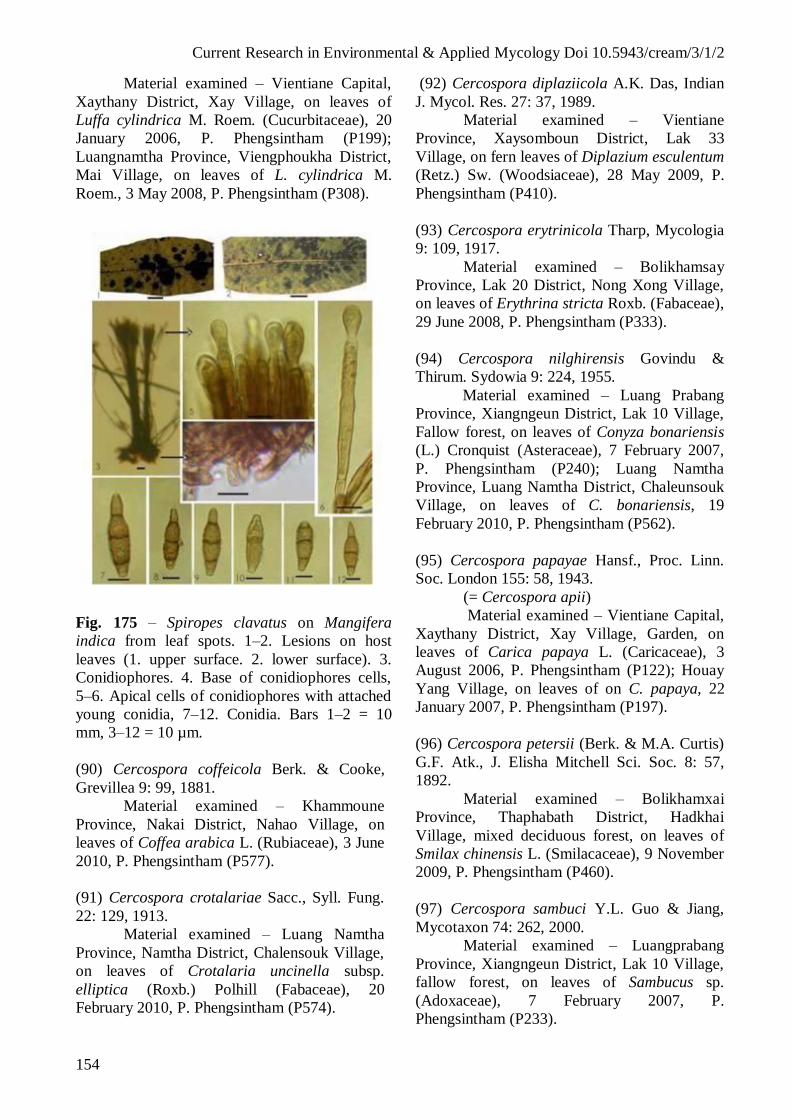
Current Research in Environmental & Applied Mycology Doi 10.5943/cream/3/1/2
154
Material examined – Vientiane Capital,
Xaythany District, Xay Village, on leaves of
Luffa cylindrica M. Roem. (Cucurbitaceae), 20
January 2006, P. Phengsintham (P199);
Luangnamtha Province, Viengphoukha District,
Mai Village, on leaves of L. cylindrica M.
Roem., 3 May 2008, P. Phengsintham (P308).
Fig. 175 – Spiropes clavatus on Mangifera
indica from leaf spots. 1–2. Lesions on host
leaves (1. upper surface. 2. lower surface). 3.
Conidiophores. 4. Base of conidiophores cells,
5–6. Apical cells of conidiophores with attached
young conidia, 7–12. Conidia. Bars 1–2 = 10
mm, 3–12 = 10 µm.
(90) Cercospora coffeicola Berk. & Cooke,
Grevillea 9: 99, 1881.
Material examined – Khammoune
Province, Nakai District, Nahao Village, on
leaves of Coffea arabica L. (Rubiaceae), 3 June
2010, P. Phengsintham (P577).
(91) Cercospora crotalariae Sacc., Syll. Fung.
22: 129, 1913.
Material examined – Luang Namtha
Province, Namtha District, Chalensouk Village,
on leaves of Crotalaria uncinella subsp.
elliptica (Roxb.) Polhill (Fabaceae), 20
February 2010, P. Phengsintham (P574).
(92) Cercospora diplaziicola A.K. Das, Indian
J. Mycol. Res. 27: 37, 1989.
Material examined – Vientiane
Province, Xaysomboun District, Lak 33
Village, on fern leaves of Diplazium esculentum
(Retz.) Sw. (Woodsiaceae), 28 May 2009, P.
Phengsintham (P410).
(93) Cercospora erytrinicola Tharp, Mycologia
9: 109, 1917.
Material examined – Bolikhamsay
Province, Lak 20 District, Nong Xong Village,
on leaves of Erythrina stricta Roxb. (Fabaceae),
29 June 2008, P. Phengsintham (P333).
(94) Cercospora nilghirensis Govindu &
Thirum. Sydowia 9: 224, 1955.
Material examined – Luang Prabang
Province, Xiangngeun District, Lak 10 Village,
Fallow forest, on leaves of Conyza bonariensis
(L.) Cronquist (Asteraceae), 7 February 2007,
P. Phengsintham (P240); Luang Namtha
Province, Luang Namtha District, Chaleunsouk
Village, on leaves of C. bonariensis, 19
February 2010, P. Phengsintham (P562).
(95) Cercospora papayae Hansf., Proc. Linn.
Soc. London 155: 58, 1943.
(= Cercospora apii)
Material examined – Vientiane Capital,
Xaythany District, Xay Village, Garden, on
leaves of Carica papaya L. (Caricaceae), 3
August 2006, P. Phengsintham (P122); Houay
Yang Village, on leaves of on C. papaya, 22
January 2007, P. Phengsintham (P197).
(96) Cercospora petersii (Berk. & M.A. Curtis)
G.F. Atk., J. Elisha Mitchell Sci. Soc. 8: 57,
1892.
Material examined – Bolikhamxai
Province, Thaphabath District, Hadkhai
Village, mixed deciduous forest, on leaves of
Smilax chinensis L. (Smilacaceae), 9 November
2009, P. Phengsintham (P460).
(97) Cercospora sambuci Y.L. Guo & Jiang,
Mycotaxon 74: 262, 2000.
Material examined – Luangprabang
Province, Xiangngeun District, Lak 10 Village,
fallow forest, on leaves of Sambucus sp.
(Adoxaceae), 7 February 2007, P.
Phengsintham (P233).

Current Research in Environmental & Applied Mycology Doi 10.5943/cream/3/1/2
155
(98) Cercospora scrophulariae (Moesz)
Chupp, A monograph of the fungus genus
Cercospora: 525, 1954.
Material examined – Luangnamtha
Province, Namtha District, Chaleunsouk
Village, fallow forest, on leaves of
Scrophularia sp. (Scrophulariaceae), 20
February 2010, P. Phengsintham (P570).
(99) Cercospora sonchi Chupp., A Monograph
of the fungus genus Cercospora: 154, 1954.
Material examined – Phongsaly
Province, Phongsaly District, on leaves of
Taraxacum officinale Wigg. (Asteraceae), 23
June 2010, P. Phengsintham (P591).
(100) Cercospora tridacis-procumbentis
Govindu & Thirum., Sydowia 7: 49, 1953.
(= Cercospora apii)
Material examined – Vientiane Capital,
Xaythany District, Xay Village, fallow forest,
on leaves of Tridax procumbens L.
(Asteraceae), 9 June 2007, P. Phengsintham
(P282); Xaythany District, Xay Village, on
leaves of T. procubens, 18 November 2011, P.
Phengsintham (P649).
(101) Pseudocercospora centromaticola (J.M.
Yen & Lim) J.M. Yen, in Yen & Lim, Gard.
Bull. Singapore 33: 171, 1980.
Material examined – Vientiane Capital,
Xaythany District, Xay Village, fallow forest,
on leaves of Centrosema pubescens Benth.
(Fabaceae), 11 May 2006, P. Phengsintham
(P44).
(102) Pseudocercospora cycleae (Chidd.)
Deighton, Mycol. Pap. 140: 143, 1976.
Material examined – Sayabouli
Province, Paklay District, Sisa-art Somphou
Village, fallow forest, on leaves of Cyclea
peltata Hook. f. & Thomson (Menispermaceae),
19 August 2006, P. Phengsintham (P90).
(103) Pseudocercospora ecdysantherae (J.M.
Yen) J.M. Yen, Bull. Trimestriel Soc. Mycol.
France 97: 94, 1987.
Material examined – Vientiane Capital,
Xaythany District, Nongviengkham Village, on
leaves of Ecdysanthera rosea Hook. & Arn.
(Apocynaceae), 9 August 2006, P.
Phengsintham (P133).
(104) Pseudocercospora giranensis Sawada ex
Goh & W.H. Hsieh, Trans. Mycol. Soc. Republ.
China 2: 92, 1987.
Material examined – Vientiane
Province, Toulakhom District,
Phoukhaukhouay Protected Area, on leaves of
Glochidion eriocarpum Champ. ex Benth.
(Euphorbiaceae), 16 October 2006, P.
Phengsintham (P181); Xiangkhouang Province,
Paek District, Phonsavan Village, on leaves of
G. eriocarpum, 1 March 2010, P. Phengsintham
(P509).
(105) Pseudocercospora ixorae (Solheim)
Deighton, Mycol. Pap. 140: 145, 1976.
Material examined – Vientiane Capital,
Xaysetha District, Patouxay Garden, on leaves
of Ixora stricta Roxb. (Rubiaceae), 15 May
2006, P. Phengsintham (P50).
(106) Pseudocercospora malloticola Goh &
W.H. Hsieh., Cercospora and similar fungi
from Taiwan: 124, 1990.
Material examined – Savannakhet
Province, Vilaboury District, Naloumai Village,
on leaves of Mallotus thorelii Gagnep.
(Euphorbiaceae), 23 June 2010, P.
Phengsintham (P588).
(107) Pseudocercospora namae (Dearn. &
House) U. Braun & Crous. CBS Biodiversity
Series 1: 288, 2003.
Material examined – Vientiane Capital,
Xaythany district, Xay Village, rice paddy, on
leaves of Hydrolea zeylanica (L.) Vahl
(Hydroleaceae), 6 September 2006, P.
Phengsintham (P159); Xaythany District, Xay
Village, rice paddy, on leaves of Hydrolea
zeylanica, 20 November 2009, P. Phengsintham
(P466).
(108) Pseudocercospora olacicola (Muthappa)
Kamal, M.K. Khan & R.K. Verma, Mycol. Res.
94: 241, 1990.
Material examined – Vientiane Capital,
Xaythany District, Dongmakkhai Village,
dipterocarp forest, on leaves of Olax scandens
Roxb. (Olacaceae), 22 January 2007, P.
Phengsintham (192).
(109) Pseudocercospora puderi Deighton,
Mycol. Pap. 140: 90, 1976.
Current Research in Environmental & Applied Mycology Doi 10.5943/cream/3/1/2
156
Material examined – Vientiane Capital,
Xaythany District, Xay Village, on leaves of
Rosa chinensis Jacq. (Rosaceae), 9 Septemebr
2006, P. Phengsintham (P164).
(110) Pseudocercospora punicae (Henn.)
Deighton, Mycol. Pap. 140: 151, 1976.
Material examined – Xiangkhouang
Province, Phonsavan Village, on leaves of
Punica granatum L. (Lythraceae), 26 April
2011, P. Phengsintham (P623).
(111) Pseudocercospora sarcocephali (Vienn.-
Bourg.) Deighton, Mycol. Pap. 140: 152, 1976.
Figs 187-188
Material examined – Bolikhamsay
Province, Lak 20 District, Nongxong Village,
on leaves of Sarcocephalus cordatus (Roxb.)
Miq. (Rubiaceae), 10 August 2008, P.
Phengsintham (P358); Luangnamtha Province,
Luangnamtha District, Chaleunsouk Village, on
leaves of S. cordatus, 19 February 2010, P.
Phengsintham (P557).
(112) Pseudocercospora scopariicola (J.M.
Yen) Deighton, Mycol. Pap. 140: 152, 1976
Material examined – Khammoune
Province, Nakai District, Nahao Village, on
leaves of Scoparia dulcis L. (Plantagiaceae), 20
July 2011, P. Phengsintham (P644).
(113) Pseudocercospora tremicola (J.M. Yen)
Deighton [as 'trematicola'], Mycol. Pap. 140:
154, 1976.
Material examined – Luangnamtha
Province, Luangnamtha District, Chaleunsouk
Village, on leaves of Trema orientalis (L.)
Blume (Cannabaceae), 19 February 2010, P.
Phengsintham (P563).
Acknowledgements
The authors thank the Mushroom
Research Foundation (MRF) for financial
support. Special thanks also go to the MRF
organizers and members of K.D. Hyde’s
laboratory, Mae Fah Luang University (MFU)
for their assistance.
References
Bagyanarayana G, Braun U, Jagadeeswar P.
1995 – Notes on Indian Cercosporeae
and allied genera (IV). Cryptogamic
Botany 5, 363–366.
Braun U. (1989) – Cercospora-like fungi on
Cassia. International Journal of
Mycology and Lichenology 4(1–2),
191–204.
Braun U. 1995 – A monograph of
Cercosporella, Ramularia and allied
genera (phytopathogenic hyphomy-
cetes). Vol. 1. IHW-Verlag, Eching.
Braun U. 2000 – Miscellaneous notes on some
micromycetes. Schlechtendalia 5, 31–
56.
Braun U. 2003 – Miscellaneous notes on some
cercosporoid hyphomycetes. Bibliotheca
Lichenologica 86, 79– 98.
Braun U. 2004 – Periconiella species occurring
on ferns. Feddes Repert 115, 50–55.
Braun U, Castañeda RF. 1991 – Cercospora
and allied genera of Cuba (II).
Cryptogamic Botany 2, 289–297.
Braun U, Mel’nik V . 1997 – Cercosporoid
fungi from Russia and adjacent
countries. Trudy Botanicheskogo Insti-
tuta Imeni V.L. Komarova, Rossijskaya
Akademiya Nauk St. Petersburg 20, 1–
130.
Braun U, Sivapalan A. 1999 – Cercosporoid
hyphomycetes from Brunei. Fungal
Diversity 3, 1–27.
Braun U. Urtiaga R. 2012 – New species and
new records of cercosporoid
hyphomycetes from Cuba and
Venezuela (Part 1). Mycosphere 3(3),
301–329.
Braun U, Crous PW, Kamal 2006 – New
species of Pseudocercospora,
Pseudocercosporella, Ramularia and
Stenella (cercosporoid hyphomycetes).
Mycological Progress 2, 197–208.
Casta eda RF, Braun U. 1989 – Cercospora
and allied Genera of Cuba (I).
Cryptogamic Botany 1, 42–55.
Castañeda RF, Kendrick B. 1991 – Ninety-nine
conidial fungi from Cuba and three from
Canada Journal University of Waterloo,
Biology Series 35, 1–132.
Chaudhary RK, Tripathi MS, Singh PN,
Chaudhary S. 2001 – Novel taxa of
Stenella from forest flora of North-
Eastern Uttar Pradesh. Indian

Current Research in Environmental & Applied Mycology Doi 10.5943/cream/3/1/2
157
Phytopathology 54, 226–232.
Chaudhary S, Sharma N, Kamal 2002 – Three
new species of Stenella. Indian
Phytopathology 55, 57–60.
Choi YW, Hyde KD, Ho WH. 1999 – Single
spore isolation of fungi. Fungal
Diversity 3, 29–38.
Chupp C. 1954 – A Monongraph of the Fungus
Genus Cercospora. Ithaca, New York.
Published by the author.
Crous PW, Braun U. 2003 – Mycosphaerella
and its anamorphs: Names published in
Cercospora and Passalora. CBS
Biodiversity Series 1. CBS Utrecht, The
Netherlands.
Deighton FC. 1967 – Studies on Cercospora
and allied genera. II. Passalora,
Cercosporidium, and some species of
Fusicladium on Euphorbia. Mycological
Papers 112, 1–80.
Deighton FC. 1971 – Studies on Cercospora
and allied genera. III. Centrospora.
Mycological Papers 124, 1–13.
Deighton FC. 1973 – Studies on Cercospora
and allied genera. IV. Cercosporella
Sacc., Pseudocercosporella gen. nov.
and Pseudocercosporidium gen. nov.
Mycological Papers 133:1–62.
Deighton FC. 1974 – Studies on Cercospora
and allied genera. V. Mycovellosiella
Rangel and a new species of
Ramulariopsis. Mycological Papers137,
1–75.
Deighton FC. 1976 – Studies on Cercospora
and allied genera. VI. Pseudocercospora
Speg., Pantospora Cif., and
Cercoseptoria Petr. Mycological Papers
140, 1–168.
Deighton FC. 1979 – Studies on Cercospora
and allied genera. VII. New species and
redispositions. Mycological Papers 144,
1–56.
Deighton FC. 1987 – New species of
Pseudocercospora and Mycovellosiella,
and new combination into Pseudocer-
cospora and Phaeoramularia.
Transactions of the British Mycological
Society 88, 365–391.
Ellis MB. 1971 – Dematiaceous hyphomycetes.
Commonwealth Mycological Institute
Kew, Surrey, England.
Ellis MB. 1976 – More dematiaceous
hyphomycetes. Commonwealth Myco-
logical Institute Kew, Surrey, England.
Frank J, Crous PW, Groenewald JZ, Oertel B,
Hyde KD, Phengsintham P, Schroers
HJ. 2010 – Microcyclospora and
Microcyclosporella: novel genera
accommodating epiphytic fungi causing
sooty blotch on apple. Persoonia 24, 93–
105
Groenewald JZ, Nakashima C, Nishikawa J,
Shin H-, Park J-H, Jama AN,
Groenewald M, Braun U, Crous PW.
2012 – Species concepts in Cercospora:
spotting the weeds among the roses.
Studies in Mycology 75, 115–170.
Guo YL, Hsieh WH. 1995 – The genus
Pseudocercospora in China.
Mycosystema Monographicum Series 2,
1–388.
Hoog GS de, Oorschot CAN van, Hijwegen T.
1983 – Taxonomy of the Dactylaria
complex. II. Dissoconium. gen. nov. and
Cordana Preuss. Proceedings of the
Koninklijke Nederlandse Akademie van
Wetenschappen Ser. C, Biological and
Medical Sciences 86(2), 197–206.
Index of Fungi 4. 1981, 603.
Kamal 2010 – Cercosporoid Fungi of India.
Sishen Singh Mahendra Pal Singh,
Dehra Dun.
Kar AK, Madal M. 1969 – New Cercospora
spp. from West Begal. Transactions of
the British Mycological Society 53, 337-
360.
Katsuki S. 1965 – Cercosporoid of Japan.
Transacions of the Mycological Society
of Japan. Extra Issue No 1, 1–100.
Khan MK, Kamal, Morgan-Jones G. 1995
Notes on hyphomycetes. LXIV New
species of Mycovellosiella, Phaeoisa-
riopsis, Sirosporium and Stenella from
India. Mycotaxon 54, 27–36.
Laos 2011 – Laos Country Report.
http://www.tititudorancea.com/z/laorepo
rt.htm.
MAF, STEA. 2003 – Lao PDR Biodiversity
Country report. Vientiane, Lao PDR.
Mandal M. 1978 – New Cercospora spp. from
West Bengal. Indian Journal of
Mycological Research 16(2), 371–376.

Current Research in Environmental & Applied Mycology Doi 10.5943/cream/3/1/2
158
Meeboon J. 2009 – Diversity and phylogeny of
true cercosporoid fungi from northern
Thailand. Ph.D. Thesis, Chiang Mai
University, Thailand (unpublished).
Meeboon J, Hidayat I, To-anun C. 2007 –
Annotated list of cercosporoid fungi in
northern Thailand. Journal of
Agricultural Technology 3, 51–63.
Mehrotra MD, Verma RK. 1991 – Some new
hyphomycetes associated with leaf spots
of trees in India. Mycological Research
95, 1163–1168.
Nakashima C, Motohashi K, Meeboon J, To-
anun C. 2007 – Studies on Cercospora
and allied genera in northern Thailand.
Fungal Diversity 26, 257–270.
Phengsintham P, Hyde KD. 2003a – Check list
of Lao fungi. Building Capacity in
Biodiversity Information Sharing 2003.
Ksukuba Japan, 184–190.
Phengsintham P, Hyde KD. 2003b – Fungi of
Laos I: Ascomycetes from Palms.
Building Capacity in Biodiversity
Information Sharing 2003. Ksukuba
Japan, 174–183.
Phengsintham P, Chukeatirote E, Hyde KD,
Braun U. 2012 – Mycology in Laos.
NAMC–Newsletter of the Asian
Mycological Committee.
Saccardo PA. 1886 – Sylloge Fungorum
omnium hucusque cognitorum, Vol. 4.
Padova.
Saccardo PA.1902 – Sylloge Fungorum
omnium hucusque cognitorum, Vol. 16.
Padova.
Saccardo PA. 1910 – Sylloge Fungorum
omnium hucusque cognitorum, Vol. 19.
Padova.
Saccardo PA. 1931 – Sylloge Fungorum
omnium hucusque cognitorum, Vol. 25.
Avellino
Saccardo PA. 1972 – Sylloge Fungorum
omnium hucusque cognitorum, Vol. 26
Trotter A (ed.), published by Cash K.),
Johnson Reprint Corporation, New York,
London.
Shin HD, Kim JD. 2001 – Cercospora and
allied genera from Korea. Plant
Pathogens from Korea 7, 1–302.
Shukla AN, Sarman PC. 1984 – A new species
of Pseudocercospora on Bhelu
(Tetrameles nudiflora R.Br.). Current
Science 53(4), 204.
Singh A, Bhalla K, Singh SK. 1998 – Three
phytoparasitic species of Periconiella
from Indian sub-continent. Indian
Phytopathology 51:30–35.
Srivastava K, Srivastava AK, Kamal, Rai AN.
1995 – Additions to Stenella from India.
Mycological Research 99, 233–236.
Thirumalachar MJ, Govindu HC. 1953 – Notes
on some Indian Cercosporae-V.
Sydowia 8, 342–348.
To-anun C, Hidayat I, Meeboon J. 2011 –
Genus Cercospora in Thailand:
taxonomy and phylogeny (with a
dichotomous key to species). Plant
Pathology and Quarantine 1(1), 11–87.
Vasudeva RS. 1963 – India Cercosporae.
Indian Council of Agricultural
Research. New Delhi.
Vidal J. 1959 – Noms vernacularis de Plantes
en usage au Laos. Ecole Francaise d’
Extreme-Orient. Paris.
Yen JM. 1979 – Étude sur les champignions
parasites du sud–est asiatique. 33. Les
Cercospora de Formose. V. Pseudo-
cercospora. Bulletin Trimestriel de la
Société Mycologique de France 94,
385–389.
Yen JM, Lim G. 1980 – Cercospora and allied
genera of Singapore and the Malay
Peninsula. Garden Bulletin Singapore 3,
175–176.
Yen JM, Kar AK, Das BK. 1982 – Studies on
hyphomycetes from West Bengal, India,
II. Cercospora and allied genera of
West Bengal, 2. Mycotaxon 16, 58–79.