monet's painting under the microscope - opus at … · key words: claude monet, optical...
TRANSCRIPT
Monet’s Painting under the Microscope
Paula Dredge,1 Richard Wuhrer,2* and Matthew R. Phillips2
1Art Gallery of New South Wales, Art Gallery Road, The Domain, Sydney, NSW, 2000, Australia2University of Technology, Sydney, Microstructural Analysis Unit, P.O. Box 123, Broadway, NSW, 2007, Australia
Abstract: An oil painting by Claude Monet, Port-Goulphar, Belle-Ile 1887 ~collection of the Art Gallery of New
South Wales!, was examined to determine both the identity of the pigments used by the artist in this painting
and his technique of mixing colors and laying paint on the canvas. The extremely complex construction of the
painting was revealed by optical microscopy, scanning electron microscopy ~SEM!, energy dispersive X-ray
analysis ~EDS!, and X-ray mapping ~XRM! analysis of cross sections of paint flakes excised from damaged
regions of Port-Goulphar, Belle-Ile. Nine different pigments were found on the painting. Many of the identified
colors were modern pigments that became available only late in the 19th century as a result of scientific
advances in pigment chemistry. Although similar colors were available in a natural mineral form, they lacked
the vivid color of their manufactured counterparts. The use of these new synthetic metallic oxide colors by
Monet accounts for the brilliance of his paintings. In addition, a separation between successive paint layers was
observed in some areas of paint chip cross sections, indicating that oil-based paint was applied to paint that had
dried, and consequently, Port-Goulphar, Belle-Ile was painted over a long period of time. This observation is
contrary to the general perception of Monet’s technique of painting freely and quickly.
Key words: Claude Monet, optical microscopy, scanning electron microscopy, energy dispersive spectroscopy,
X-ray mapping
INTRODUCTION
The Art Gallery of New South Wales is conducting a com-
parative study of the painting techniques of Claude Monet
~1840–1926!, John Russell ~1858–1930!, and Henri Matisse
~1869–1954!. This work has focused on two artistic encoun-
ters between these three painters; the first in 1886, when
John Russell met Claude Monet while they were both paint-
ing at Belle-Ile, an island in Brittany off the northwest coast
of France, and the second in 1896–1897, when the young
Matisse visited Belle-Ile and was befriended by John Russell,
who moved permanently to the island in 1887. The aim of
our on-going study is to determine if these two encounters
between the painters influenced the painting style and tech-
nique of each painter.
The Art Gallery of New South Wales has a number of
works by John Russell in its collection which have been
previously studied, as well as a painting by Claude Monet,
Port-Goulphar, Belle-Ile ~Fig. 1!. It is this painting by Monet
which is the subject of this collaborative research project by
the authors.
Energy dispersive X-ray spectrometry ~EDS! is an estab-
lished technique for the analysis of the chemical composi-
tion of paint layers in a scanning electron microscope
~SEM!. X-ray mapping ~XRM! is the collection of character-Received September 27, 2002; accepted April 16, 2002.*Corresponding author. E-mail: [email protected]
Microsc. Microanal. 9, 139–143, 2003DOI: 10.1017/S1431927603030198 MicroscopyAND
Microanalysis© MICROSCOPY SOCIETY OF AMERICA 2003

Figure 1
Figure 2
Figure 3
Figure 4
Figure 5
Figure 6
Figure 1. Port-Goulphar, Belle-Ile painted by Claude Monet in 1887 from the collection of the Art Gallery of New South
Wales.
Figure 2. A magnified section of the lower right-hand side of the painting in Figure 1 showing the complexity of color
used by Monet in this painting.
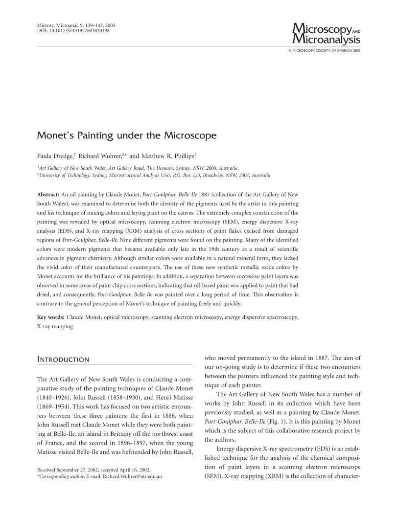
Figure 3. Optical micrograph with visible incident illumination of the cross section of a paint chip excised from the
rock in the lower right-hand corner of the painting in Figure 1. Width of field is 360 mm.
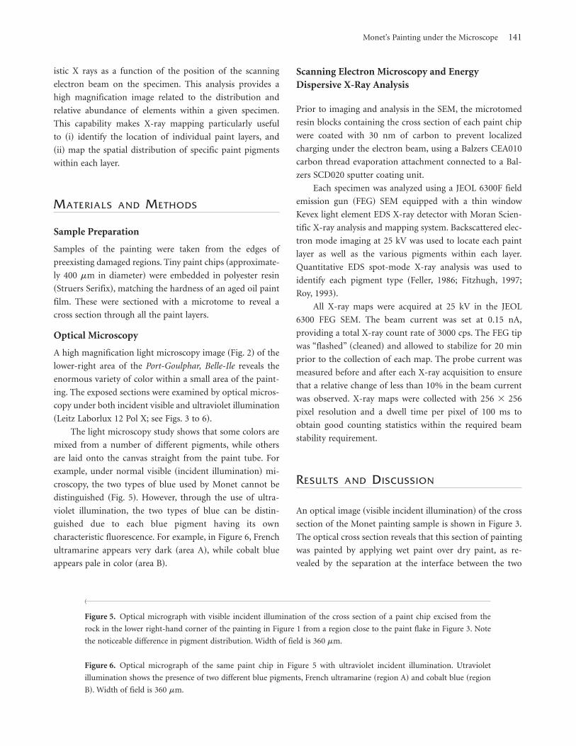
Figure 4. Optical micrograph of the same paint chip in Figure 3 with ultraviolet incident illumination. Width of field is
360 mm.
140 Paula Dredge et al.
istic X rays as a function of the position of the scanning
electron beam on the specimen. This analysis provides a
high magnification image related to the distribution and
relative abundance of elements within a given specimen.
This capability makes X-ray mapping particularly useful
to ~i! identify the location of individual paint layers, and
~ii! map the spatial distribution of specific paint pigments
within each layer.
MATERIALS AND METHODS
Sample Preparation
Samples of the painting were taken from the edges of
preexisting damaged regions. Tiny paint chips ~approximate-
ly 400 mm in diameter! were embedded in polyester resin
~Struers Serifix!, matching the hardness of an aged oil paint
film. These were sectioned with a microtome to reveal a
cross section through all the paint layers.
Optical Microscopy
A high magnification light microscopy image ~Fig. 2! of the
lower-right area of the Port-Goulphar, Belle-Ile reveals the
enormous variety of color within a small area of the paint-
ing. The exposed sections were examined by optical micros-
copy under both incident visible and ultraviolet illumination
~Leitz Laborlux 12 Pol X; see Figs. 3 to 6!.
The light microscopy study shows that some colors are
mixed from a number of different pigments, while others
are laid onto the canvas straight from the paint tube. For
example, under normal visible ~incident illumination! mi-
croscopy, the two types of blue used by Monet cannot be
distinguished ~Fig. 5!. However, through the use of ultra-
violet illumination, the two types of blue can be distin-
guished due to each blue pigment having its own
characteristic fluorescence. For example, in Figure 6, French
ultramarine appears very dark ~area A!, while cobalt blue
appears pale in color ~area B!.
Scanning Electron Microscopy and EnergyDispersive X-Ray Analysis
Prior to imaging and analysis in the SEM, the microtomed
resin blocks containing the cross section of each paint chip
were coated with 30 nm of carbon to prevent localized
charging under the electron beam, using a Balzers CEA010
carbon thread evaporation attachment connected to a Bal-
zers SCD020 sputter coating unit.
Each specimen was analyzed using a JEOL 6300F field
emission gun ~FEG! SEM equipped with a thin window
Kevex light element EDS X-ray detector with Moran Scien-
tific X-ray analysis and mapping system. Backscattered elec-
tron mode imaging at 25 kV was used to locate each paint
layer as well as the various pigments within each layer.
Quantitative EDS spot-mode X-ray analysis was used to
identify each pigment type ~Feller, 1986; Fitzhugh, 1997;
Roy, 1993!.
All X-ray maps were acquired at 25 kV in the JEOL
6300 FEG SEM. The beam current was set at 0.15 nA,
providing a total X-ray count rate of 3000 cps. The FEG tip
was “flashed” ~cleaned! and allowed to stabilize for 20 min
prior to the collection of each map. The probe current was
measured before and after each X-ray acquisition to ensure
that a relative change of less than 10% in the beam current
was observed. X-ray maps were collected with 256 3 256
pixel resolution and a dwell time per pixel of 100 ms to
obtain good counting statistics within the required beam
stability requirement.
RESULTS AND DISCUSSION
An optical image ~visible incident illumination! of the cross
section of the Monet painting sample is shown in Figure 3.
The optical cross section reveals that this section of painting
was painted by applying wet paint over dry paint, as re-
vealed by the separation at the interface between the two
^
Figure 5. Optical micrograph with visible incident illumination of the cross section of a paint chip excised from the
rock in the lower right-hand corner of the painting in Figure 1 from a region close to the paint flake in Figure 3. Note
the noticeable difference in pigment distribution. Width of field is 360 mm.
Figure 6. Optical micrograph of the same paint chip in Figure 5 with ultraviolet incident illumination. Utraviolet
illumination shows the presence of two different blue pigments, French ultramarine ~region A! and cobalt blue ~region
B!. Width of field is 360 mm.
Monet’s Painting under the Microscope 141

layers. This observation indicates that Monet worked on
this painting over a long period of time, as oil paints take a
long time to dry, and this is contrary to the generally held
belief that Monet painted quickly and freely. However, the
optical images ~Figs. 3, 4, 5, and 6! also show regions with
no separation between the paint layers, indicating that these
areas were painted wet paint over wet paint, while mixing
the paints directly on the canvas.
A SEM backscattered electron image ~Fig. 7! of a paint
cross section shows the abundance of pigments used within
a small section of painting. A high magnification backscat-
tered electron image of a yellow region of the painting re-
veals that the areas contain rod-like pigments ~Fig. 8!, which
contain lead, chromium, and oxygen. This pigment is known
as chrome yellow, a lead chromate ~PbCrO4 and PbCrO4{
PbSO4!. Interestingly, the chrome yellow pigment was re-
placed by cadmium yellow late in the 19th century. However,
Port-Goulphar, Belle-Ile seems to have a mixture ~blended!
of the two types of yellow pigment ~chrome and cadmium!.
Background corrected X-ray maps for aluminium, sili-
con, calcium, chromium, cobalt, copper, cadmium, mer-
cury, and lead from a representative paint chip are shown in
Figure 9. These maps, as well as extensive spot mode EDS
analysis, were used to identity the following pigments: lead
white @basic lead carbonate 2PbCO3{Pb~OH!2# , vermilion
@mercuric sulphide HgS# , cadmium yellow @cadmium sul-
fide CdS# , chrome yellow @lead chromate PbCrO4 and
PbCrO4{PbSO4# , viridian @hydrated chromium oxide
Cr2O3{2H2O or Cr2O~OH!4# , emerald green @copper aceto-
arsenite 3Cu~AsO2!2{Cu~CH3COO!2# , cobalt blue @cobalt
aluminate CoO{Al2O3# , and French ultramarine @approxi-
mately Na6–10Al6Si6O24S2–4# . The complex distribution of
these pigments within a paint chip cross section from the
painting is shown in Figure 10.
Two organic lake colors ~a dye precipitated onto an
inert base! were also found in Port-Goulphar, Belle-Ile; a red
lake ~presumably madder! on an aluminium hydroxide base,
and a yellow lake ~unidentified! on a base of natural chalk
sphericals.
This present work shows that Monet worked on Port-
Goulphar, Belle-Ile with a limited number of pigments; two
reds, three yellows ~the yellow lake may be an impurity
added by the color merchant to enhance the color of the
cadmium yellow!, two blues, two greens, and white. These
pigments were premixed in an impressive number of com-
binations to give enormous variation in color on the paint-
ing. Some single brush strokes, such as those in the dark
rocks, contained all 10 pigments mixed together. Subtle
variations in color were achieved by slight alterations in the
proportions of each pigment in the paint mix.
CONCLUSIONS
The combination of these microscopy methods used in this
study enabled ~a! the identification of each of the pigment
types used in the painting, and ~b! an insight into the paint
application techniques used by Monet, in particular whether
the paint was premixed or applied pure from the tube and
mixed wet-in-wet on the canvas. The complex construction
of the paint layers studied in this work confirmed that
Figure 7. A backscattered electron image of the paint chip cross
section excised from the lower right-hand corner of the painting
showing the abundance of pigments used by Monet in this work.
Width of field is 340 mm.
Figure 8. Backscattered electron image of a yellow section of the
painting containing a rodlike pigment known as chrome yellow, a
lead chromate ~PbCrO4 and PbCrO4{PbSO4!. Width of field 23 mm.
142 Paula Dredge et al.

Monet possessed a mastery of color through a deep under-
standing of the pigments available at that time. In addition,
contrary to popular belief, certain sections of Port-Goulphar,
Belle-Ile were painted over intervals long enough for the
previously painted area to dry.
ACKNOWLEDGMENTS
The authors gratefully acknowledge the Art Gallery of New
South Wales and the University of Technology, Sydney for
their support.
REFERENCES
Feller, R.L. (Ed.). ~1986!. Artists’ Pigments. A Handbook of Their
History and Characteristics, vol 1. Washington, D.C.: National
Gallery of Art.
Fitzhugh E.W. (Ed.). ~1997!. Artists’ Pigments. A Handbook of
Their History and Characteristics, vol 3. Washington, D.C.:
National Gallery of Art.
Roy, A. (Ed.) ~1993!. Artists’ Pigments. A Handbook of Their
History and Characteristics, vol 2. Washington, D.C.: National
Gallery of Art.
Figure 9. X-ray maps ~XRM! for aluminum, silicon, calcium, chromium, cobalt, copper, cadmium, mercury, and lead
from a representative paint chip cross section. Maps were collected at an accelerating voltage of 25 kV, beam current of
0.15 nA, 256 3 256 pixels and a dwell time per pixel of 100 ms. Width of field, 110 mm.
Figure 10. Optical micrograph image showing the complex distribution of pigments identified using EDS spot mode
analysis. Width of field 50 mm.
Monet’s Painting under the Microscope 143

Thomson lSI - Journal Search Results
THOMSON+:
http://www.isinet.comlcgi-bin/jrnIst/jlresults.cgi?PC=MASTER&I ...
11i'Wfij'i: _tH·be_Mlde'_iShiilW'&,
SEARCH RESULTS
1 of 1
lSI Master Journal ListSEARCH RESULTS
Search Terms: 1431-9276Total [ournals founej: 1
The fcHovdng titJe(s) marcneo your request:
Jcumats ~-1 (of 1) I-t; , I fORMAT fOR PRHH
~....HCROSCOPY AND ~.MCROANALYSIS
8imc'n!t1IyISSN: 1431·9275CA~...1BRIDGEUNIV PRESS, 40 WEST 20TH ST. NEW '{ORK. USA. NY. , 0011·4211
Journals: ~-1 (of 1) I FORMAT FOR PlUtH
Horne i Sfte Mac I About Us I Press Room Careers Contact Us i Thomson cornSliWOf1 ; Joumal Lists IS; Links I !SI Essays I HGt Research
3110312004 2:53 PM