mon 12-05-2005 os lecture 6 - valve disease regurgitant - dr
TRANSCRIPT
Valvular Regurgitation Sheldon Litwin, M.D.
• Normal heart valves have minimal leakage (back flow, insufficiency, regurgitation) when they close
• Significant valvular regurgitation causes the heart to do excess work (like walking up a sandy hill)
• Over time, significant regurgitation usually leads to cardiac enlargement and contractile dysfunction

Water flows down hill!

Clinical goals in Valvular Disease
Make the diagnosis Slow the disease progression Prevent complications Intervene at just the right time Not too soon, not too late
Key Concept in Valve Regurgitation
• Timing of intervention is tricky!!• Chamber enlargement and contractile
dysfunction develop very gradually• Don’t subject your patient to risks
before it it necessary• Fix the problem before damage
becomes irreversible (may need to intervene before symptoms developbefore symptoms develop)

Mitral Valve Disease
• Annulus• Leaflets• Chordae• Papillary muscles• LV
LA
Acute MR: Etiology
• Ischemia/MI– Posterolateral hypokinesis– Papillary muscle rupture
• Ruptured chordae• Endocarditis• Systolic Anterior Motion (SAM) of
mitral leaflet(s)

Ischemia or MI
QuickTime™ and aVideo decompressorare needed to see this picture. QuickTime™ and aVideo decompressorare needed to see this picture.

Ruptured Papillary Muscle
LA
LV
MV
Always causes acute, severe MR - a surgical emergency.
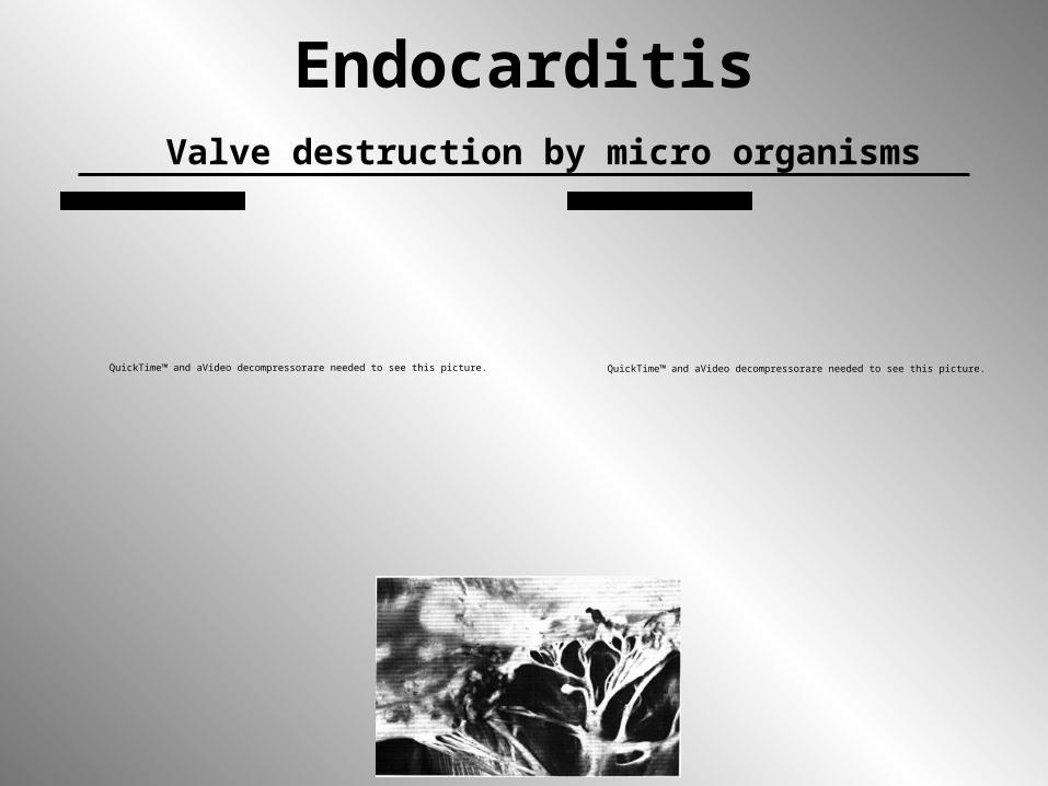
Endocarditis Valve destruction by micro organisms
QuickTime™ and aVideo decompressorare needed to see this picture. QuickTime™ and aVideo decompressorare needed to see this picture.

Acute MR: Pathophysiology
• Flow from LV (high pressurehigh pressure) to LA (low pressurelow pressure) LA pressure PCWP pulmonary edema
• Low compliance atria transmits LV pressure more directly to pulmonary capillaries
“forward” stroke volume (low cardiac output)
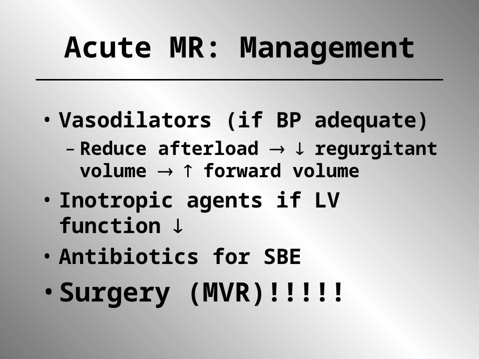
Acute MR: Management
• Vasodilators (if BP adequate)– Reduce afterload regurgitant volume
forward volume• Inotropic agents if LV function • Antibiotics for SBE• Surgery (MVR)!!!!!
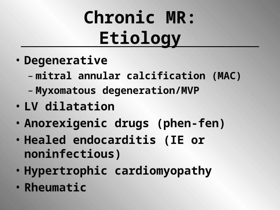
Chronic MR:Etiology
• Degenerative – mitral annular calcification (MAC)– Myxomatous degeneration/MVP
• LV dilatation• Anorexigenic drugs (phen-fen)• Healed endocarditis (IE or noninfectious)• Hypertrophic cardiomyopathy• Rheumatic
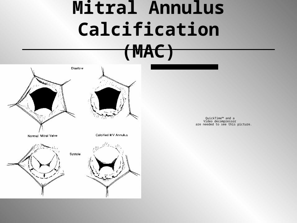
Mitral Annulus Calcification(MAC)
QuickTime™ and aVideo decompressor
are needed to see this picture.

Mitral Valve Prolapse
• “Syndrome” vs. disease– overdiagnosis
• Myxomatous degeneration of leaflets & chordae (increased MMP activity)
• SBE prophylaxis if significant MR present (> mild)
• Early valve repair for severe MR
QuickTime™ and aVideo decompressorare needed to see this picture.
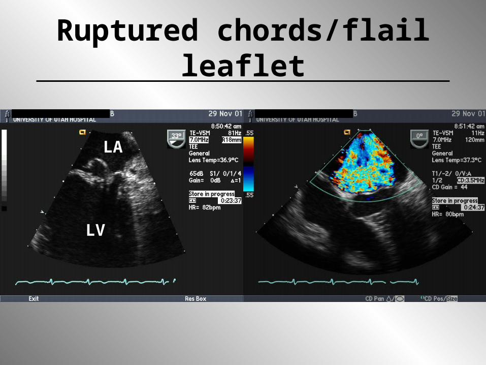
Ruptured chords/flail leaflet
LV
LA

Nonvalvular causes of MR
• May occur without structural abnormalities o f valve
• Usually LV dilatation and remodeling with increased sphericity
• Stretch of annulus and lateral displacement of papillary muscles may cause malcoaptation of the leaflets
LV dilatationLateral displacement Pap’s
QuickTime™ and aVideo decompressor
are needed to see this picture.
Quantification of MR:size of jet on echo
Mild-Moderate Mod-Severe
QuickTime™ and aVideo decompressor
are needed to see this picture.
QuickTime™ and aAnimation decompressor
are needed to see this picture.

Quantitation of MR: Regurgitant Volume
• < 20 ml = trace• 20-30 ml = mild• 30-50 ml = mod• > 50 ml = severe
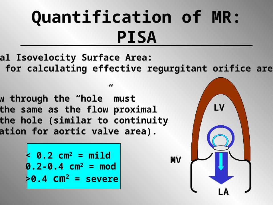
Quantification of MR: PISAProximal Isovelocity Surface Area:Method for calculating effective regurgitant orifice area (ERO)
Flow through the “hole” mustbe the same as the flow proximalto the hole (similar to continuityequation for aortic valve area).
< 0.2 cm2 = mild0.2-0.4 cm2 = mod>0.4 cm2 = severe
LV
LA
MV

QuickTime™ and aVideo decompressor
are needed to see this picture.
TEE: Severe MR PISA
The bigger the PISA, the bigger the hole
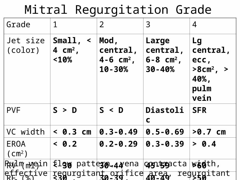
Mitral Regurgitation GradeGrade 1 2 3 4
Jet size (color)
Small, < 4 cm2, <10%
Mod, central, 4-6 cm2, 10-30%
Large central, 6-8 cm2, 30-40%
Lg central, ecc, >8cm2, > 40%, pulm vein
PVF S > D S < D Diastolic SFRVC width < 0.3 cm 0.3-0.49 0.5-0.69 >0.7 cmEROA (cm2)
< 0.2 0.2-0.29 0.3-0.39 > 0.4
RV (ml) < 30 30-44 45-59 >60RF (%) <30 30-39 40-49 >50
Pulm vein flow pattern, vena contracta width, effective regurgitant orifice area, regurgitant volume, regurgitant fraction
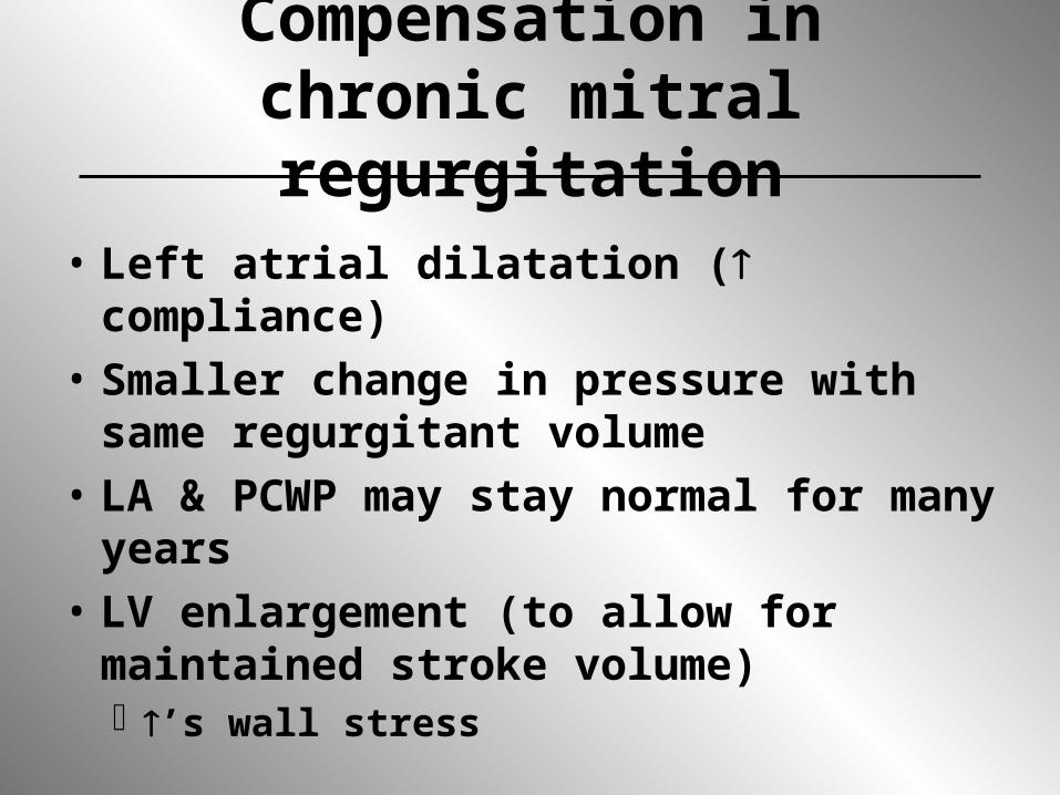
Compensation in chronic mitral regurgitation
• Left atrial dilatation ( compliance)• Smaller change in pressure with same
regurgitant volume• LA & PCWP may stay normal for many
years• LV enlargement (to allow for maintained
stroke volume) ’s wall stress

Afterload in MR• LV afterload (resistance to LV ejection) is
reduced because the LA is a low pressure alternate pathway for ejection
• LV chamber function (EF) should theoretically be greater than normal if myocardial contractility is preserved
• Once EF is below normal, significant LV dysfunction exists and it is likely to get worse once the mitral valve is replaced

Complications of chronic MR
• Atrial fibrillation• LV dilatation and systolic
dysfunction• Passive pulmonary hypertension
with RV dysfunction
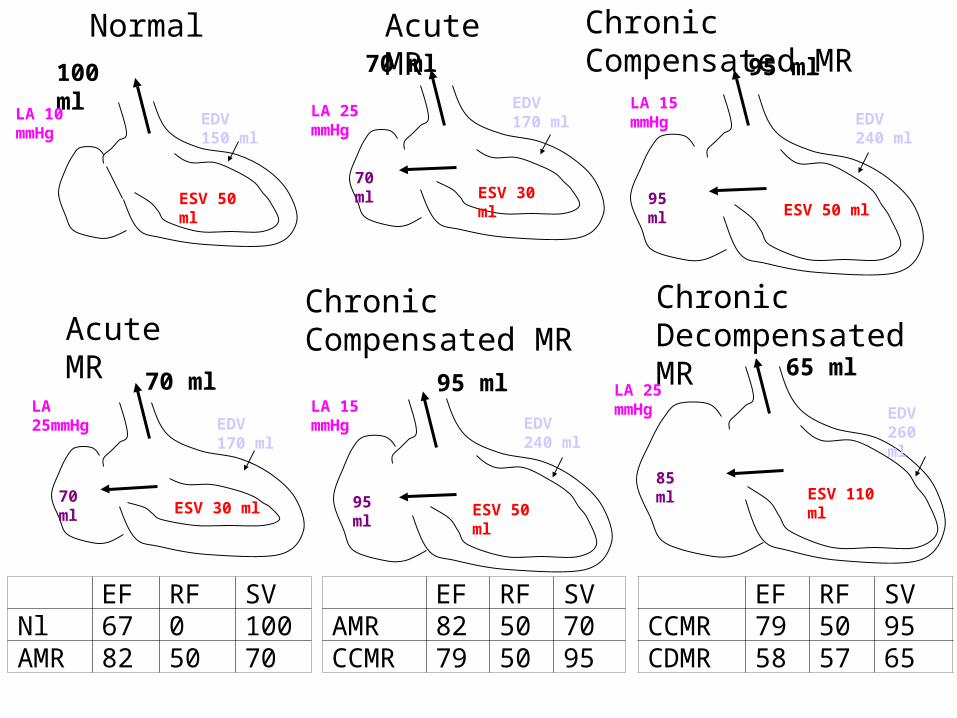
Normal Acute MR Chronic Compensated MR
Acute MRChronic Compensated MR
Chronic Decompensated MR
100 ml
70 ml 95 ml65 ml
70 ml 95 ml
EF RF SVNl 67 0 100AMR 82 50 70
EF RF SVAMR 82 50 70CCMR 79 50 95
EF RF SVCCMR 79 50 95CDMR 58 57 65
LA 10 mmHg
EDV 150 ml
LA 25 mmHgLA 15
mmHgLA 25mmHg
LA 15 mmHg
LA 25 mmHg
ESV 50 ml
EDV 260 ml
ESV 110 ml
EDV 240 ml
ESV 50 ml
EDV 170 ml
ESV 30 ml
EDV 170 ml
ESV 30 ml
EDV 240 ml
ESV 50 ml
70 ml95 ml
70 ml 95 ml85 ml

Medical management of MR
• Afterload reduction, Rx of arterial HTN
• Rx heart failure (if not surgical candidate)
• SBE prophylaxis• Rheumatic fever prophylaxis• Rx of atrial fibrillation• Rx of ischemia
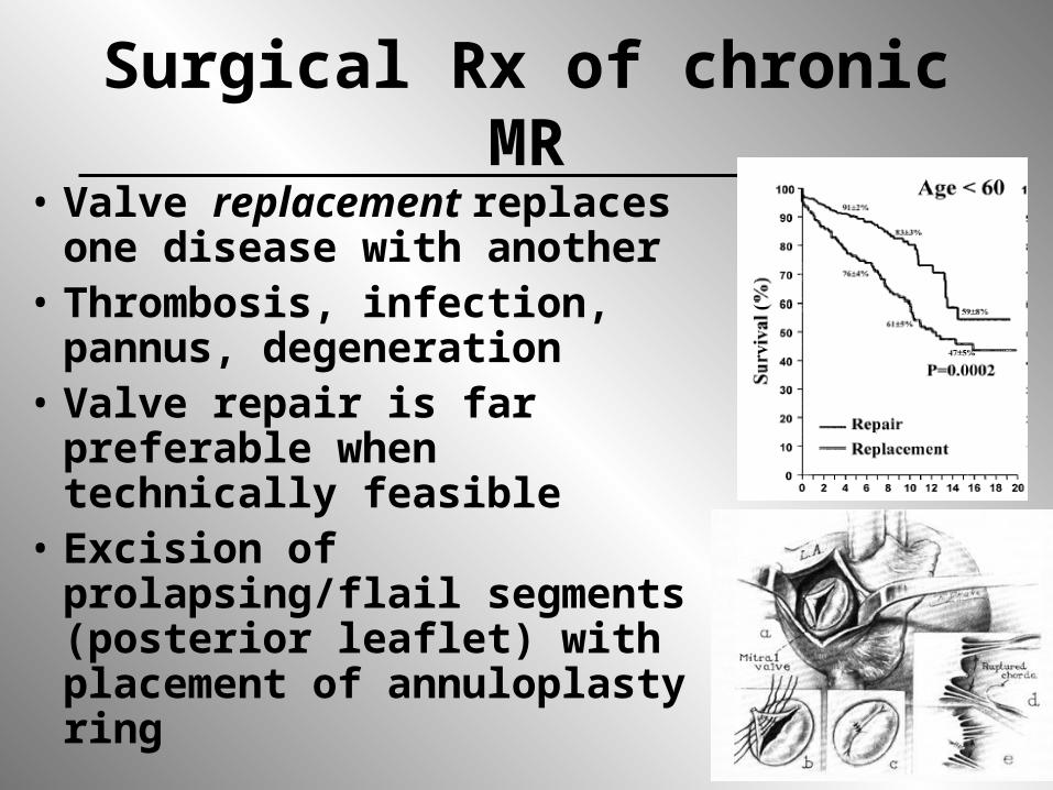
Surgical Rx of chronic MR• Valve replacement replaces
one disease with another• Thrombosis, infection,
pannus, degeneration• Valve repair is far preferable
when technically feasible• Excision of prolapsing/flail
segments (posterior leaflet) with placement of annuloplasty ring

Timing of surgery for MR• Old approach was to wait for symptoms,
LV enlargement or systolic dysfunction (typically serial echoes were performed)
• Problem: wait too long?• With low morbidity/mortality of repair
and low need for reoperation, trend is to recommend early repair for severe MR, even in asymptomatic patients with normal LV function
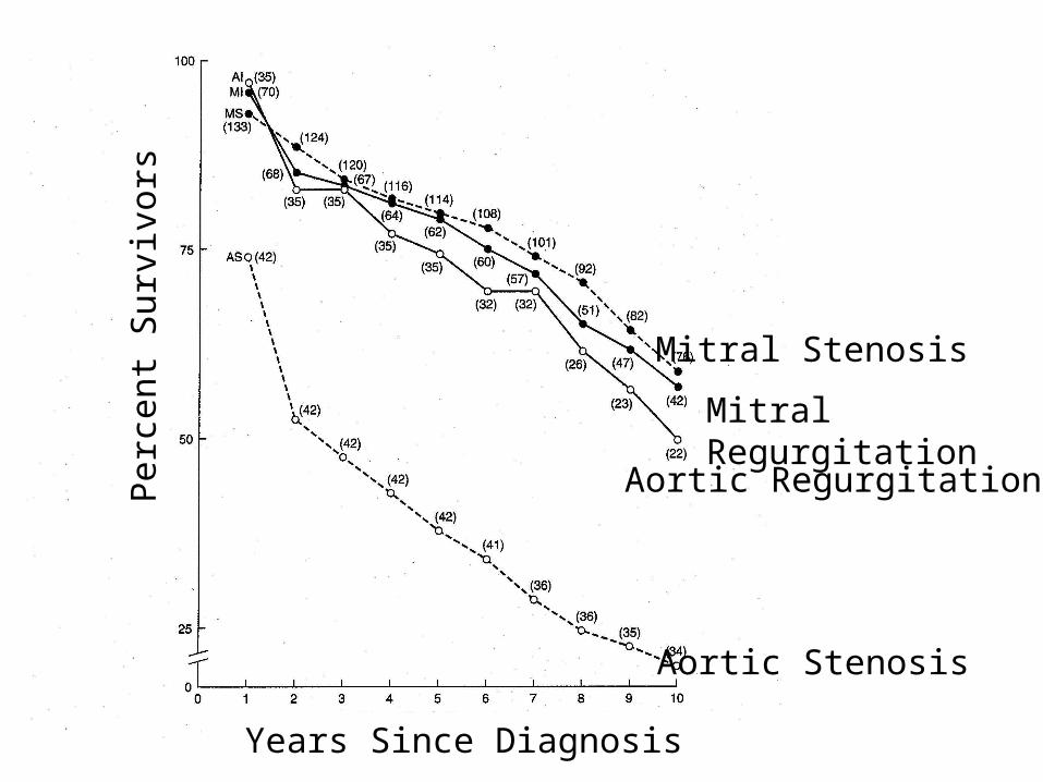
Perc
ent S
urvi
vors
Years Since Diagnosis
Aortic Stenosis
Aortic Regurgitation
Mitral Regurgitation
Mitral Stenosis

Indications for surgery in chronic, nonischemic MR
• Class I– Acute symptomatic MR in which repair is
likely– NYHA Class II-IV symptoms with normal LV
function (EF > 60%) and LVESD < 45 mm– Symptomatic or asymptomatic with mild LV
dysfunction (EF 50-60%) and/or LVESD 50-55 mm
– Symptomatic or asymptomatic with moderate LV dysfunction (EF 30-50%) and/or LVESD 50-55 mm
Prosthetic Mitral ValvesMechanical: bileaflet tilting disc
QuickTime™ and aVideo decompressor
are needed to see this picture.
Tricuspid valve regurgitation: etiology
• Pulmonary hypertension• RV enlargement (infarct, dysplasia)• Primary tricuspid disease
– Infectious endocarditis– trauma– Carcinoid – Pacing wires/catheters– Ebstein's anomaly– Iatrogenic/bioptome
QuickTime™ and aVideo decompressor
are needed to see this picture.QuickTime™ and a
Video decompressorare needed to see this picture.
Tricuspid Endocarditis
Aortic Insuffiency (regurgitation)
• Valve normally open during systole, closed during diastole
• AI occurs during diastole as aortic pressure is higher than LV pressure during this part of the cardiac cycle
QuickTime™ and aMicrosoft Video 1 decompressorare needed to see this picture.

Etiology of Aortic Valve Regurgitation
• Annulus• Leaflets• Root
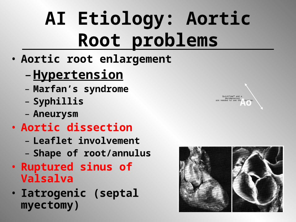
AI Etiology: Aortic Root problems
• Aortic root enlargement– Hypertension– Marfan’s syndrome– Syphillis– Aneurysm
• Aortic dissection– Leaflet involvement– Shape of root/annulus
• Ruptured sinus of Valsalva• Iatrogenic (septal
myectomy)
QuickTime™ and a decompressor
are needed to see this picture.Ao

AI Etiology: leaflet problems
• Calcification/fibrosis– Degenerative– Rheumatic
• Bicuspid• Endocarditis
– Infectious– Noninfectious
• Diet drugs
QuickTime™ and aMicrosoft Video 1 decompressorare needed to see this picture.

Aortic Valve Endocarditis
QuickTime™ and aMicrosoft Video 1 decompressorare needed to see this picture.
QuickTime™ and aMicrosoft Video 1 decompressorare needed to see this picture.

Aortic Dissection
LV
QuickTime™ and aH.263 decompressor
are needed to see this picture.QuickTime™ and a
H.263 decompressorare needed to see this picture.

Quantification of AI:size of color jet on echo
Mild-Moderate Mod-Severe
QuickTime™ and aAnimation decompressor
are needed to see this picture.QuickTime™ and a
Animation decompressorare needed to see this picture.
Quantification is important because we usually only operate on patients with severe AI and it is necessary to track progression of disease
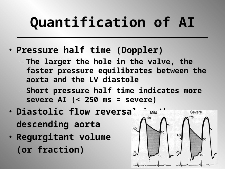
Quantification of AI
• Pressure half time (Doppler)– The larger the hole in the valve, the faster pressure
equilibrates between the aorta and the LV diastole– Short pressure half time indicates more severe AI
(< 250 ms = severe)• Diastolic flow reversal in the
descending aorta• Regurgitant volume
(or fraction)
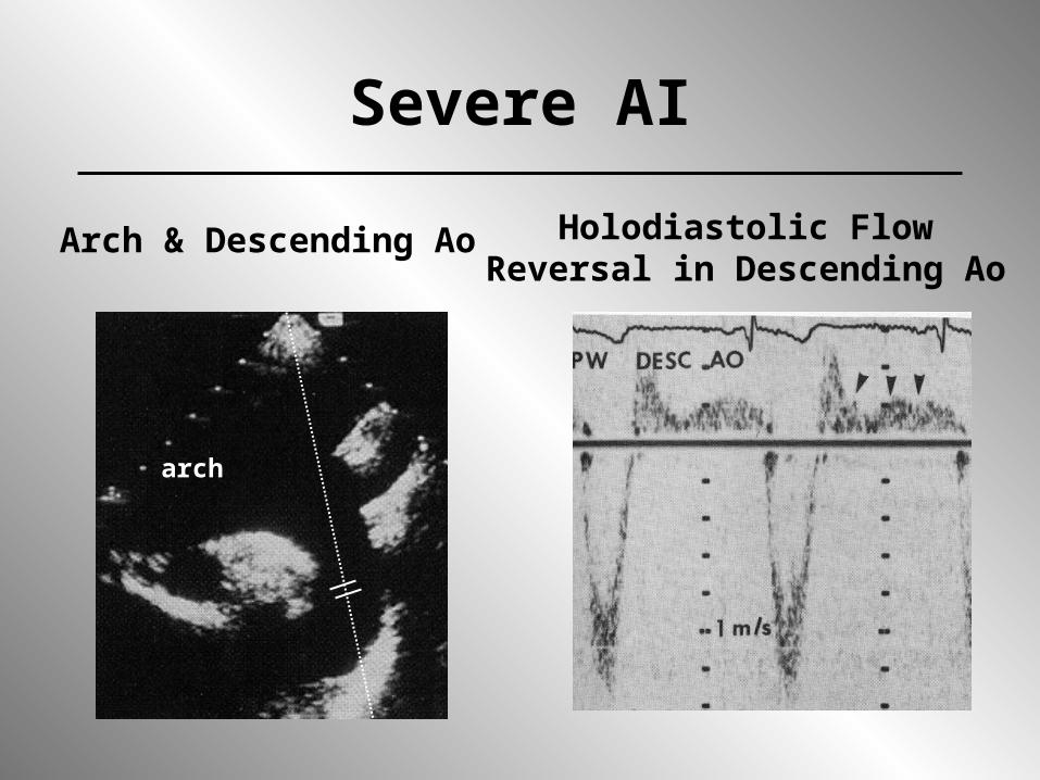
Severe AI
Arch & Descending Ao Holodiastolic FlowReversal in Descending Ao
arch
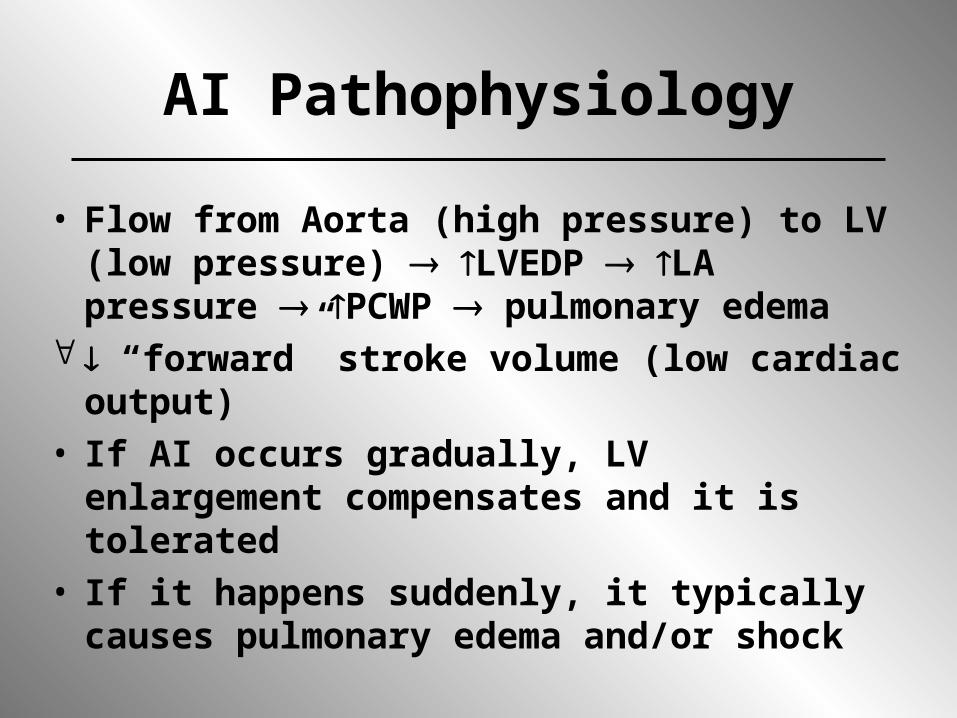
AI Pathophysiology
• Flow from Aorta (high pressure) to LV (low pressure) LVEDP LA pressure PCWP pulmonary edema
“forward” stroke volume (low cardiac output)
• If AI occurs gradually, LV enlargement compensates and it is tolerated
• If it happens suddenly, it typically causes pulmonary edema and/or shock
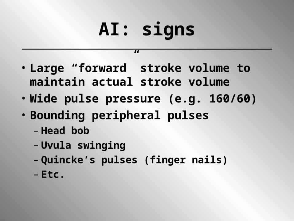
AI: signs
• Large “forward” stroke volume to maintain actual stroke volume
• Wide pulse pressure (e.g. 160/60)• Bounding peripheral pulses
– Head bob– Uvula swinging– Quincke’s pulses (finger nails) – Etc.

Acute AI: Management
• Vasodilators (if BP adequate)– Reduce afterload regurgitant volume
forward volume• Inotropic agents if LV function • Antibiotics for SBE• Avoid intra-aortic balloon pump
(makes AI worse)• Surgery (AVR)!!!!!

Chronic aortic insufficiency
• LV enlargement to maintain forward stroke volume (’s wall stress)
• LA & PCWP may stay normal for years• LV chamber function (EF) should be
normal or greater than normal if myocardial contractility is preserved
• Once EF is below normal, significant LV dysfunction exists and the outcome following valve replacement is worse
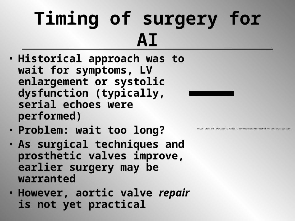
Timing of surgery for AI• Historical approach was to wait
for symptoms, LV enlargement or systolic dysfunction (typically, serial echoes were performed)
• Problem: wait too long?• As surgical techniques and
prosthetic valves improve, earlier surgery may be warranted
• However, aortic valve repair is not yet practical
QuickTime™ and aMicrosoft Video 1 decompressorare needed to see this picture.

Surgical Rx of chronic AI• Low EF increases mortality
• Don’t wait too long

Medical management of AI
• Afterload reduction (nifedipine, ACE inhibitors) - recent study suggests not helpful
• Control arterial HTN• Treat heart failure (if not surgical
candidate)• SBE prophylaxis• Rheumatic fever prophylaxis• All palliative
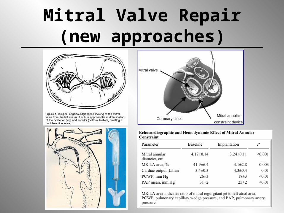
Mitral Valve Repair(new approaches)

What is the best parameter to describe MR severity?
• Quantify regurgitant volume, regurgitant fraction, and/or regurgitant orifice area
– Echo– MRI
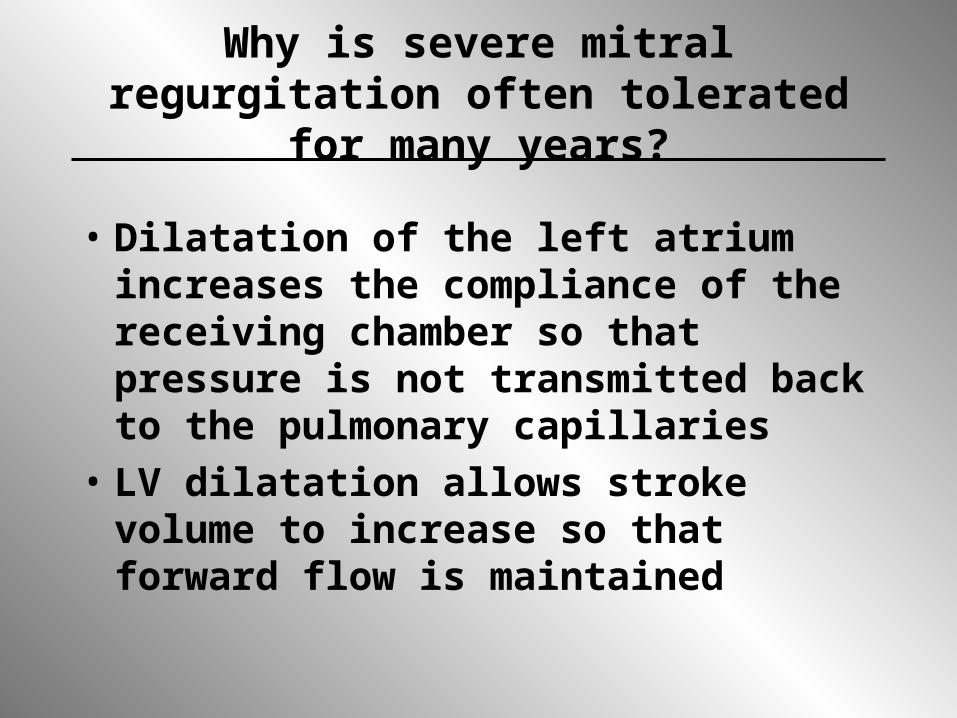
Why is severe mitral regurgitation often tolerated for many years?
• Dilatation of the left atrium increases the compliance of the receiving chamber so that pressure is not transmitted back to the pulmonary capillaries
• LV dilatation allows stroke volume to increase so that forward flow is maintained
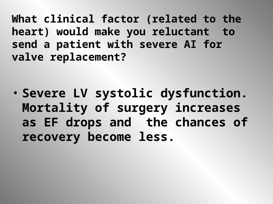
What clinical factor (related to the heart) would make you reluctant to send a patient with severe AI for valve replacement?
• Severe LV systolic dysfunction. Mortality of surgery increases as EF drops and the chances of recovery become less.
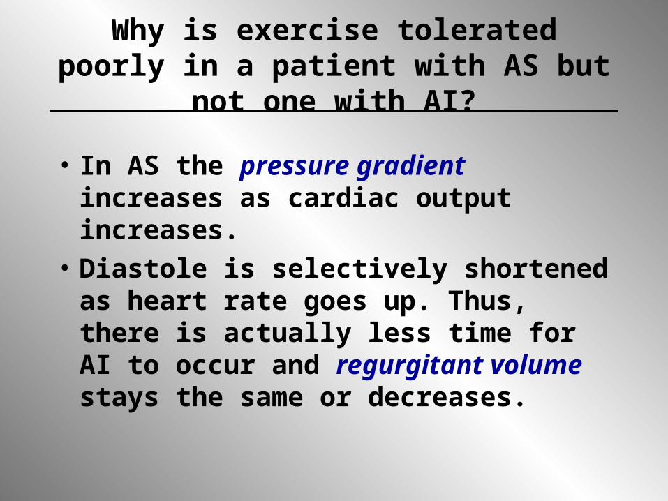
Why is exercise tolerated poorly in a patient with AS but not one with AI?
• In AS the pressure gradient increases as cardiac output increases.
• Diastole is selectively shortened as heart rate goes up. Thus, there is actually less time for AI to occur and regurgitant volume stays the same or decreases.
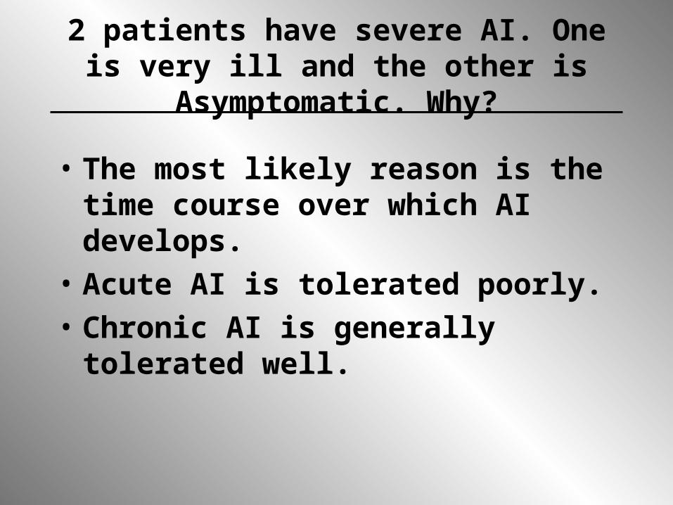
2 patients have severe AI. One is very ill and the other is Asymptomatic.
Why?
• The most likely reason is the time course over which AI develops.
• Acute AI is tolerated poorly.• Chronic AI is generally tolerated well.

Indications for surgery in chronic nonischemic MR
• Class IIa– Asymptomatic patients with preserved LV function
and atrial fibrillation– Asymptomatic patients with preserved LV function
and pulmonary hypertension (PASP > 50 mmHg at rest or > 60 mmHg with exercise)
– Asymptomatic with LV EF 50-60% andLVESD < 45 mm, or EV > 60% and LVESD 45-55 mm
– Patients with severe LV dysfunction (EF < 30% and/or LVESD > 55 mmHg) in whom chordal preservation is highly likely
– Asymptomatic with chronic MR with preserved LV function in whom valve repair is highly likely
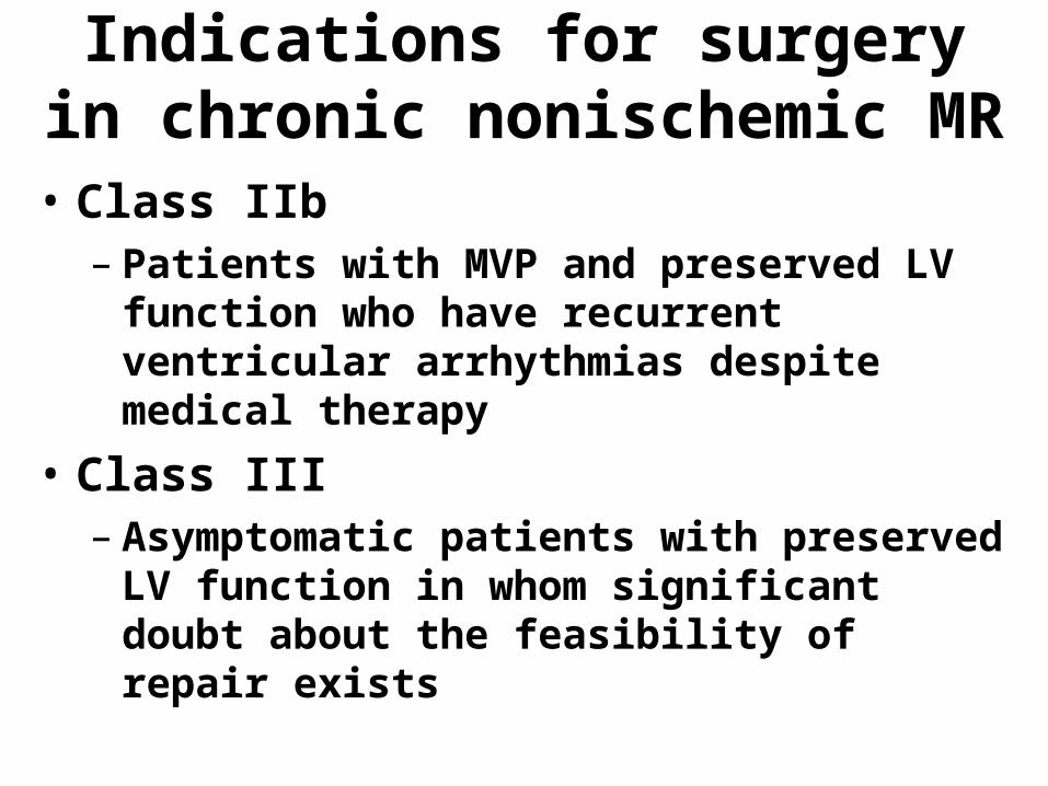
Indications for surgery in chronic nonischemic MR
• Class IIb– Patients with MVP and preserved LV function
who have recurrent ventricular arrhythmias despite medical therapy
• Class III– Asymptomatic patients with preserved LV
function in whom significant doubt about the feasibility of repair exists