molecular regulation of embryo development in norway spruce
TRANSCRIPT
Molecular Regulation of Embryo Development in Norway Spruce
Polar Auxin Transport and Transcription Factors
Emma Larsson Faculty of Natural Resources and Agricultural Sciences
Department of Plant Biology and Forest Genetics Uppsala
Doctoral Thesis Swedish University of Agricultural Sciences
Uppsala 2011
Acta Universitatis agriculturae Sueciae
2011:68
ISSN 1652-6880 ISBN 978-91-576-7612-2 © 2011 Emma Larsson, Uppsala Print: SLU Service/Repro, Uppsala 2011
Cover: Auxin response in the beginning of late embryogeny in Norway spruce
Molecular Regulation of Embryo Development in Norway Spruce. Polar Auxin Transport and Transcription Factors
Abstract Early events in embryo development are critical for the plant body formation. During this phase the apical-basal axis, the radial symmetry, and the primary meristems are specified. The shoot apical meristem (SAM) and the root apical meristem (RAM) will subsequently give rise to all above-ground and below-ground tissues. Despite the environmental and economical importance of conifers, the regulation of their embryonic development is still relatively unknown. We are using somatic embryos of Norway spruce as a model system for studying embryology in conifers.
In this thesis, I show that polar auxin transport (PAT) is essential for proper patterning of both apical and basal parts of conifer embryos. Blocked PAT caused increased auxin levels, increased differentiation of early somatic embryos, a skewed balance between the embryonal mass and the suspensor cells in late embryos, and an abnormal morphology with irregular RAM, fused or aborted cotyledons and absence of a functional SAM in mature embryos.
Two NAC gene family members (PaNAC01 and PaNAC02), closely related to the Arabidopsis CUP-SHAPED COTYLEDON (CUC) genes, were characterized. PaNAC01 harbors previously characterized functional motifs, and could complement the cuc1cuc2 double mutant. The temporal expression of PaNAC01 was dependent on PAT, and coincided with the formation of separated cotyledons and a functional SAM. Furthermore, the expression profiles of four KNOXI genes (HBK1 to 4) showed that also the expression of HBK2 and HBK4 depend on PAT, and indicated their role in embryo differentiation and SAM formation, while HBK1 and HBK3 seemingly have a more general role during embryo development.
In addition, a global gene expression analysis revealed important processes during early somatic embryogenesis in Norway spruce.
Taken together the results show that central parts of the regulatory network for embryo development are still conserved between angiosperms and gymnosperms, despite their separation 300 million years ago.
Keywords: Auxin, cotyledons, CUC, conifers, KNOX, NAC, Norway spruce, polar auxin transport (PAT), shoot apical meristem (SAM), somatic embryogenesis.
Author’s address: Emma Larsson, Swedish University of Agricultural Sciences, Uppsala BioCenter, Department of Plant Biology and Forest Genetics, P.O. Box 7080, 750 07 Uppsala, Sweden E-mail: [email protected]

Contents
List of Publications 7
1 Introduction 9 1.1 Pattern formation during embryo development in seed plants 10
1.1.1 Angiosperms (Arabidopsis) 11 1.1.2 Gymnosperms (conifers) 13 1.1.3 Somatic embryogenesis in Norway spruce 14
1.2 Molecular regulation of embryo development 15 1.2.1 Auxin 16 1.2.2 Transcription factors 20 1.2.3 Gymnosperms 23
2 Aims of the study 25
3 Results and Discussion 27 3.1 Polar auxin transport is important for normal embryo development in
Norway spruce (I, II, III and appendix) 27 3.1.1 Blocked PAT results in increased auxin content (I) 27 3.1.2 Blocked PAT results in decreased PCD (I) 28 3.1.3 Increased auxin response in the suspensors of NPA-treated
embryos (appendix) 28 3.1.4 Blocked PAT causes fused cotyledons, aborted SAM and an
irregular RAM (I, II, III) 32 3.2 NAC-regulation of embryo development in conifers (II and unpublished
results) 33 3.2.1 NAC-genes in conifers 33 3.2.2 CUC-orthologs in Norway spruce 34 3.2.3 The expression of PaNAC01 is dependent on PAT 35 3.2.4 PaNAC01 can stimulate cotyledon separation in the cuc1cuc2
mutant background 37 3.2.5 The expression of PaNAC02 decreases as embryos develop 37
3.3 KNOX-diversification during embryo development in conifers (III) 38 3.3.1 Differential expression of HBK2 and HBK4 during embryo
development is dependent on the formation of a functional
SAM 38 3.3.2 HBK overexpression in Arabidopsis induces morphologies
characteristic of ectopic KNOXI expression 40

3.4 Identification of putative processes associated with pattern formation
during the earliest stages of somatic embryogenesis (IV) 40 3.4.1 Changes in global gene expression during differentiation and
development of early somatic embryos 41 3.4.2 A model for putative processes regulating early embryo
differentiation and development 43
4 Conclusions 45
5 Future perspectives 47
References 49
Acknowledgements 61
7
List of Publications
This thesis is based on the work contained in the following papers, referred to by Roman numerals in the text:
I Larsson, E., Sitbon, F., Ljung, K., von Arnold, S. (2008). Inhibited polar auxin transport results in aberrant embryo development in Norway spruce. New Phytologist 177, 356-366.
II Larsson, E., Sundström, JF., Sitbon, F., von Arnold, S. Characterisation of two CUP-SHAPED COTYLEDON orthologues (PaNAC01 and PaNAC02) in Picea abies (Norway spruce). Manuscript
III Larsson, E., Sitbon, F., von Arnold, S. Expression of Knotted-1 like genes during the establishment of the shoot apical meristem in Picea abies (Norway spruce). Manuscript
IV Vestman, D., Larsson, E., Uddenberg, D., Cairney, J., Clapham, D., Sundberg, E., von Arnold, S. (2011). Important processes during differentiation and early development of somatic embryos of Norway spruce as revealed by changes in global gene expression. Tree Genetics and Genomes 7, 347–362.
Appendix
Larsson, E., Sitbon, F., von Arnold, S. (2008). Polar auxin transport controls suspensor fate. Plant Signaling and Behavior 3:7, 469-470.
Papers I, IV and appendix are reproduced with the permission of the publishers.

8
The contribution of Emma Larsson to the papers included in this thesis was as follows:
I Planned the work and accomplished the experimental work, except for the auxin analyses. Summarized the results and wrote the manuscript.
II Planned the work and accomplished the experimental work. Participated in the phylogenetic analyses. Summarized the results and wrote the manuscript.
III Planned the work and accomplished the experimental work. Summarized the results and wrote the manuscript.
IV Participated in planning the experiments. Sampled and prepared the material for microarray analysis. Took part in analyzing the results and in writing the manuscript.
Appendix
Planned the work and accomplished the experimental work. Summarized the results and wrote the manuscript.

9
1 Introduction
Wood is one of the economically most important raw materials in the world. It is used for paper and pulp, lumber, energy, and textiles. From an environmental point of view forest resources are crucial; growing forests bind carbon dioxide, and wood- and paper-based products continue to store carbon dioxide throughout their whole life time. Furthermore, there is an urgent need to find alternatives to the ever diminishing fossil fuels, and forest by-products are regarded as important renewable and environmentally friendly feedstock for bio-ethanol production (Ohlrogge et al., 2009).
In Sweden, more than 55 % of the land area is covered by forests, of which 42 % consists of spruce and 39 % consists of pine (Swedish Statistical Yearbook of Forestry, 2010). Although Sweden holds just below 1 % of the world’s commercial forest area, the country provides about 10 % of the world’s sawn timber, paper and pulp (Swedish Forest Industries Federation, 2010). In 1994 the Swedish government decided that high forest production and forest sustainability with conserved biological diversity are equally important goals in the Swedish forestry. Although conifers are generally regarded as undomesticated trees, improvement through crossing, selection and testing has had a significant input on the productivity and quality. In order to make use of the genetic gain obtained in breeding programs, selected trees must be mass-propagated. Traditionally this has been achieved by establishing seed orchards. However, vegetative propagation is an attractive alternative since it saves both time and increases genetic gain more effectively. Forest trees can be propagated vegetatively through grafting, rooting of cuttings, layering or through micro-propagation such as organogenesis or somatic embryogenesis.
The development of somatic embryos will be described more thoroughly later. Briefly, the method is based on embryogenic cultures, which are established from somatic cells, and from which somatic embryos differentiate in response to addition and withdrawal of different plant growth regulators (PGRs). Today it is only possible to initiate embryogenic cell lines from
10
zygotic conifer embryos. However, work is in progress to overcome the difficulties with using more mature starting material such as seedlings or buds (Uddenberg et al., 2011). One major advantage of somatic embryogenesis is that the embryogenic cell lines can be cryo-preserved in liquid nitrogen. After initiation of an embryogenic cell line, one part is cryo-preserved and the rest is propagated. Subsequently, somatic embryo plants are regenerated and tested in field trials. This makes it possible at any time to thaw cultures originating from genotypes that possess valuable traits based on the results from field trials. A large number of somatic embryo plants can then be regenerated from the thawed cultures, and these plants can be used in different reforestation programs.
Although the somatic embryo protocol today can be used for several conifer species, there are still many genotypes that are difficult to propagate using this method. A deeper understanding of the genetic regulation may help indicating what goes wrong in the development of somatic embryos in recalcitrant genotypes and species. This can provide clues on how to improve the culture conditions in order to propagate economically important conifers via somatic embryos.
This thesis describes molecular regulation of somatic embryogenesis in Norway spruce (Picea abies), with specific emphasis on the interplay between polar auxin transport (PAT) and transcription factors (TFs) for the establishment of the shoot apical meristem (SAM) and formation of separated cotyledons. The developmental pathway has been dissected to pinpoint specific developmental stages. Furthermore, a protocol has been established to induce the formation of fused cotyledons and aborted SAM, which makes it possible to study genes that are of importance for SAM formation and cotyledon separation.
1.1 Pattern formation during embryo development in seed plants
Unlike animals, plants continue to grow and develop new organs throughout their life time. This is a prerequisite for their survival, since plants are sessile and have to cope with changes in their surroundings. The high degree of plasticity and adaptability that plants possess largely depends on their capacity to perceive environmental cues and orchestrate the developmental programs that best suit the conditions at their habitat.
The primary shoot and root apical meristems (SAM and RAM) are small niches of cells harboring the pluripotent stem cells that give rise to all above-ground and below-ground organs, respectively, during the development of a
11
plant. The SAM and the RAM are established at opposite ends along the apical-basal axis early in embryogenesis. Failure to correctly form these two meristems usually leads to abortion or to progressive accumulation of morphogenic errors throughout the life time of the plant.
In addition to the apical-basal polarization, the radial pattern of concentric tissue layers, perpendicular to the apical-basal axis, is specified during embryogenesis. Although embryo development differs greatly between different plant species, this basic body plan is similar (De Smet et al., 2010; Lau et al., 2010).
Current knowledge about the genetic regulation of embryonic pattern formation in plants is to a large extent derived from studies on embryo-defective mutants of the angiosperm Arabidopsis thaliana. Knowledge from this species is often regarded as a paradigm for all plant species despite the vast morphological differences within the plant kingdom. Angiosperms and gymnosperms separated approximately 300 million years ago (Smith 2010), and several important embryonal patterning processes differ between the two groups of seed plants. From an evolutionary point of view, it is therefore of interest to study embryo development in conifers.
1.1.1 Angiosperms (Arabidopsis)
Early embryogenesis in Arabidopsis proceeds through highly regular cell divisions and gene expression patterns that cannot be directly translated to other plant species (De Smet et al., 2010). However, since there is so much more knowledge available about Arabidopsis embryogenesis (Mansfield and Briarty, 1991 and reviewed by Jenik et al., 2007; Capron et al., 2009; De Smet et al., 2010 and Lau et al., 2010) compared to conifer embryogenesis, it is valuable to use that knowledge as a foundation when studying similarities and differences between distantly related species.
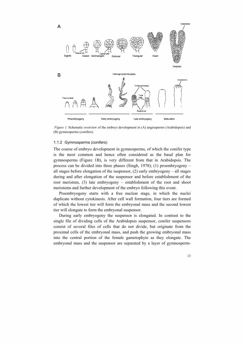
A schematic picture of the different developmental stages during Arabidopsis embryo development is shown in Figure 1A. The embryo pattern formation starts with an asymmetric division of the zygote, which gives rise to a small apical cell and a larger, elongated basal cell. Already after the first asymmetric cell division specific transcription programs are initiated resulting in different fates of the two progenitor cells. While the basal cell and its descendants divide only transversely to form one long file of cells called the suspensor, the apical cell undergoes a series of transversal and longitudinal divisions in order to develop the embryo proper.
At the octant stage the embryo proper consists of eight cells divided between the upper and the lower tier. The upper tier will subsequently give rise
12
to much of the apical part of the seedling, including the SAM and the cotyledons, while the lower tier will form much of the root. The uppermost suspensor cell will at later stages become incorporated into the embryo as the hypophysis, the founder of parts of the future root meristem.
During the transition from the octant stage to the dermatogen stage, the cells of the embryo proper divide tangentially and give rise to an inner cell layer, and an outer cell layer designated the protoderm, which subsequently will become the epidermis.
The next round of cell divisions (longitudinal among the inner cells and anticlinal among the protodermal cells) gives rise to a 32 cell globular embryo. From this stage on, the cell divisions are less coordinated. However, the different tissues are becoming evident so that by the mid-globular stage all main tissues, such as epidermis, ground tissue and provascular tissue as well as the primary shoot and root meristems, have been specified or predestined.
The hypophysis divides asymmetrically, giving rise to a smaller lens-shaped cell, which is the precursor of the quiescent centre, and a larger basal cell, which subsequently will give rise to the lower tier of stem cells in the root meristem.
The cells of the embryo proper continue to divide so that the embryo gradually assumes a triangular and then a heart shape as the cotyledons grow out on opposite sides of the incipient SAM. Further refinement of the embryonic pattern occurs during the subsequent developmental stages, the torpedo and bent cotyledon stages.
13
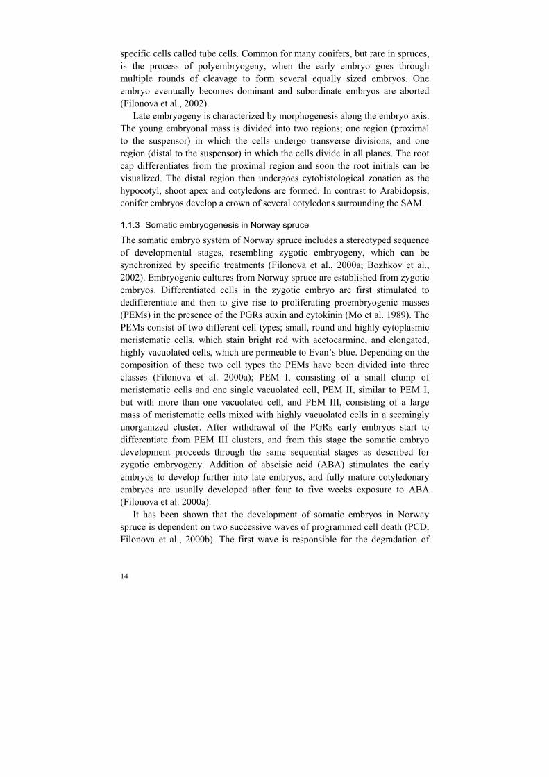
Figure 1. Schematic overview of the embryo development in (A) angiosperms (Arabidopsis) and (B) gymnosperms (conifers).
1.1.2 Gymnosperms (conifers)
The course of embryo development in gymnosperms, of which the conifer type is the most common and hence often considered as the basal plan for gymnosperms (Figure 1B), is very different from that in Arabidopsis. The process can be divided into three phases (Singh, 1978); (1) proembryogeny – all stages before elongation of the suspensor, (2) early embryogeny – all stages during and after elongation of the suspensor and before establishment of the root meristem, (3) late embryogeny – establishment of the root and shoot meristems and further development of the embryo following this event.
Proembryogeny starts with a free nuclear stage, in which the nuclei duplicate without cytokinesis. After cell wall formation, four tiers are formed of which the lowest tier will form the embryonal mass and the second lowest tier will elongate to form the embryonal suspensor.
During early embryogeny the suspensor is elongated. In contrast to the single file of dividing cells of the Arabidopsis suspensor, conifer suspensors consist of several files of cells that do not divide, but originate from the proximal cells of the embryonal mass, and push the growing embryonal mass into the central portion of the female gametophyte as they elongate. The embryonal mass and the suspensor are separated by a layer of gymnosperm-
14
specific cells called tube cells. Common for many conifers, but rare in spruces, is the process of polyembryogeny, when the early embryo goes through multiple rounds of cleavage to form several equally sized embryos. One embryo eventually becomes dominant and subordinate embryos are aborted (Filonova et al., 2002).
Late embryogeny is characterized by morphogenesis along the embryo axis. The young embryonal mass is divided into two regions; one region (proximal to the suspensor) in which the cells undergo transverse divisions, and one region (distal to the suspensor) in which the cells divide in all planes. The root cap differentiates from the proximal region and soon the root initials can be visualized. The distal region then undergoes cytohistological zonation as the hypocotyl, shoot apex and cotyledons are formed. In contrast to Arabidopsis, conifer embryos develop a crown of several cotyledons surrounding the SAM.
1.1.3 Somatic embryogenesis in Norway spruce
The somatic embryo system of Norway spruce includes a stereotyped sequence of developmental stages, resembling zygotic embryogeny, which can be synchronized by specific treatments (Filonova et al., 2000a; Bozhkov et al., 2002). Embryogenic cultures from Norway spruce are established from zygotic embryos. Differentiated cells in the zygotic embryo are first stimulated to dedifferentiate and then to give rise to proliferating proembryogenic masses (PEMs) in the presence of the PGRs auxin and cytokinin (Mo et al. 1989). The PEMs consist of two different cell types; small, round and highly cytoplasmic meristematic cells, which stain bright red with acetocarmine, and elongated, highly vacuolated cells, which are permeable to Evan’s blue. Depending on the composition of these two cell types the PEMs have been divided into three classes (Filonova et al. 2000a); PEM I, consisting of a small clump of meristematic cells and one single vacuolated cell, PEM II, similar to PEM I, but with more than one vacuolated cell, and PEM III, consisting of a large mass of meristematic cells mixed with highly vacuolated cells in a seemingly unorganized cluster. After withdrawal of the PGRs early embryos start to differentiate from PEM III clusters, and from this stage the somatic embryo development proceeds through the same sequential stages as described for zygotic embryogeny. Addition of abscisic acid (ABA) stimulates the early embryos to develop further into late embryos, and fully mature cotyledonary embryos are usually developed after four to five weeks exposure to ABA (Filonova et al. 2000a).
It has been shown that the development of somatic embryos in Norway spruce is dependent on two successive waves of programmed cell death (PCD, Filonova et al., 2000b). The first wave is responsible for the degradation of
15
PEM cells as early embryos differentiate in response to withdrawal of PGRs, and the second wave eliminates the terminally differentiated suspensor cells during early embryogeny. The number of cells undergoing PCD in embryogenic cultures of Norway spruce has successfully been studied by analyzing the amount of DNA fragmentation (laddering), by using terminal deoxynucleotidyl transferase (TdT)-mediated dUTP nick end labelling (TUNEL), which is an in situ method to label fragmented DNA, and by ultrastructure analysis of cell dismantling using transmission electron microscopy (Filonova et al., 2000b).
The process of somatic embryogenesis has proven to be a valuable tool for studying embryo development in conifers. First, it is because the later stages of somatic embryogenesis are identical to the equivalent stages of zygotic embryogenesis. Second, zygotic embryogenesis occurs under multiple layers of tissues and is therefore hard to study, while somatic embryos are devoid of such obstructing tissues. Third, somatic embryogenesis can be synchronized so that many embryos at the same developmental stage can be obtained at a specific time point, which is practically impossible with zygotic embryos of Norway spruce. Somatic embryos that have been propagated from the same culture are genetically identical, which simplifies molecular studies. Furthermore, there are no embryo-specific mutants available in conifers, but the possibility to genetically transform somatic embryos makes it possible to study gene function also in conifer embryos.
We are using the somatic embryo system in Norway spruce as a model for elucidating the regulation of embryo development in conifers (von Arnold et al., 2002).
1.2 Molecular regulation of embryo development
The molecular and cellular mechanisms that coordinate the precise developmental patterning during early embryogenesis are still rather poorly understood. However, emerging knowledge, mainly from studies of Arabidopsis embryogenesis, is drawing a picture of an intricate network of signaling molecules, TFs and PGRs that together orchestrate specific developmental programs in both temporally and spatially restricted locations (De Smet et al., 2010). In this thesis I focus on the importance of polar transport of the PGR auxin, and on the roles played by members of two large families of TFs. Of course, many other players are essential for the pattern formation during embryogenesis, but those will not be discussed in detail here.

16
1.2.1 Auxin
The small molecule indole-3-acetic acid (IAA) is one of the most important compounds for the plasticity of a living plant. Already Charles Darwin and his son Francis suggested the occurrence of a compound that in response to light travelled from the coleoptile tip of the grass Phalaris canariensis to a more basal position, where it stimulated growth (Darwin and Darwin, 1880). The compound was later named auxin, derived from the Greek word ‘auxein’ for ‘to grow’, and is today used as the generic name for a group of important endogenous and synthetic molecules with similar molecular properties (reviewed by Teale et al., 2006). In plants, the most important auxin seems to be IAA, although other substances with auxin activity have been identified. It has been debated if auxin should be considered as a hormone or a morphogen, since it shows properties of both. It is synthesized at one location and then transported to another where it is perceived by a receptor and thereby induces its response, just like a hormone. On the other hand, auxin regulates several aspects of plant development by forming concentration gradients and thereby altering cell fate decisions in a dose-dependent manner, just like a morphogen (Bhalerao and Bennett, 2003). Thus, here I use the term PGR to denote auxin and similar substances. Certainly, there are other PGRs which greatly influence the growth and morphology of a plant, and also ratios between different PGRs influence the fate of a cell. However, since the projects within this thesis have mainly dealt with the effects of auxin, I focus on describing this particular PGR.
Considerable efforts have been made to understand how such a small molecule can affect so many different developmental processes in so many different locations during so many different developmental stages of the plant life cycle. Three aspects of auxin are considered to contribute to its role during plant development: (1) biosynthesis, to create local auxin sources and maxima, (2) transport, to generate gradients or local auxin maxima, and (3) perception, to induce a response, which eventually will affect development. However, the amount of active auxin available to exert a response in a specific tissue is also dependent on conjugation, methylation, degradation and conversion to other auxins (Chandler, 2009).
Biosynthesis
Comprehensive knowledge about auxin biosynthesis is still missing, one reason probably being the lack of auxin-deficient mutants. However, four routes originating from the amino acid tryptophan (trp), and one trp-independent route have been postulated for the biosynthesis of IAA, although their complete pathways and relationships remain elusive (reviewed by Zhao,
17
2010). One of the suggested pathways involves a family of flavin monooxygenases called the YUCCAs (YUC). These enzymes have been suggested to have the capacity to catalyze the conversion of tryptamine into N-hydroxyl tryptamine, which subsequently can proceed to IAA via a few intermediates (Zhao et al., 2001). Another pathway is called the Indole-3-pyruvate (IPA) pathway and suggests IPA as an intermediate in the IAA biosynthesis. It involves the TRYPTOPHAN AMINOTRANSFERASE of ARABIDOPSIS (TAA1) enzyme, which catalyzes the production of IPA from tryptophan (Stepanova et al., 2008).
Both YUC and TAA genes are expressed in the apical parts of globular embryos. In more mature embryos, the expression of YUC becomes restricted to the SAM and the tips of the cotyledons (Cheng et al., 2007), while TAA1 expression remains in a protoderm stripe around the shoot apex, but can also be found in the developing root meristem (Stepanova et al., 2008). The quadruple mutant yuc1 yuc4 yuc10 yuc11 fails to develop hypocotyl and a root (Cheng et al., 2007), as does a large fraction of triple mutants of taa1 and its close homologs tryptophan aminotransferase related 1 and 2 (tar1 tar2; Stepanova et al., 2008).
Another pathway that influences the active pool of IAA is the indole-3-acetaldoxime (IAOx) and glucosinolate pathway (Zhao et al., 2010). It was defined by the auxin overproducing mutants superroot 1 (sur1; Boerjan et al., 1995) and superroot2 (sur2; Delarue et al., 1998). In this pathway, trp is converted to IAOx, which in turn can be converted to IAA or to indolic glucosinolates. This last conversion is catalyzed by the SUR enzymes, which thus control the available amount of IAOx for biosynthesis of IAA (Mikkelsen et al., 2004).
Transport
As mentioned above, auxin was first discovered as a compound transported in an apical-basal direction in response to environmental factors, and the classical view of organ differentiation is that new organs initiate at local auxin maxima created by PAT (Benková et al., 2003). Much effort has been put into understanding the different mechanisms regulating auxin transport. This has therefore been reviewed many times, most recently by Petrášek and Friml (2009) and Grunewald and Friml (2010).
The bulk flow of auxin occurs in mature phloem away from auxin sources such as young leaves and flowers. However, it has been shown that the generation of local auxin maxima and gradients, which influence different developmental processes, is dependent on a slower and highly regulated cell-

18
to-cell transport. This intercellular transport is mediated by different types of auxin influx and efflux carriers.
As a weak organic acid, auxin is protonated in the cell wall and in the extracellular space, where the pH is about 5.5. Thus, it can penetrate the hydrophobic plasma membrane through diffusion, or via the AUXIN RESISTANT 1/LIKE AUX1 (AUX1/LAX) family of influx carriers (Bennet et al., 1996; Swarup et al., 2008). However, inside the cell, where the pH is about 7, auxin becomes dissociated and therefore trapped within the cell. In this way auxin can be transported directionally through a family of PIN-FORMED (PIN) efflux carriers that localize polarly at the plasma membrane of a cell (Petrášek et al., 2006). The PIN proteins are continuously recycled between the plasma membrane and endosomes, thus enabling a flexibility of the transport system to change direction at the cellular level (Geldner et al., 2003). Apical targeting of the PIN proteins is dependent on phosphorylation by the serine/threonine protein kinase PINOID (PID; Friml et al., 2004). This subcellular apical targeting is antagonized by dephosphorylation mediated by the PROTEIN PHOSPHATASE 2A (PP2A), which thus leads to basal targeting (Michniewicz et al., 2007).
Another type of auxin efflux carriers are homologs of the mammalian ATP-BINDING CASSETTE SUBFAMILY B (ABCB)-transporters of the MULTIDRUG RESISTANCE/PHOSPHOGLYCOPROTEIN (ABCB/MDR/PGP) protein family (Noh et al., 2001; Verrier et al., 2008). These proteins are localized more symmetrically in the plasma membrane than the PIN proteins, and are therefore believed to regulate the amount of auxin available in the cell for PIN-mediated directional transport (Mravec et al., 2008).
The phytotropin 1-N-naphtylphthalamic acid (NPA) is a well-established polar auxin efflux inhibitor that has been widely used to study auxin transport-dependent plant development (Petrášek and Friml, 2009). It has been shown to reversibly inhibit auxin efflux when applied to plant material (Lembi et al. 1971), and wild-type Arabidopsis plants as well as embryos of Indian mustard (Brassica juncea) phenocopy Atpin1 mutants when treated with NPA (Okada et al., 1991; Liu et al., 1993; Hadfi et al., 1998). In transgenic Arabidopsis plants carrying functional PIN1:GFP constructs, PIN1:GFP relocation was prevented by NPA (Benková et al., 2003), and PIN-mediated transport in tobacco (Nicotiana tabacum) BY-2 cells was completely inhibited by NPA (Petrášek et al., 2006). NPA has also been used to study the role of auxin transport in compression wood formation in Scots pine (Pinus sylvestris; Sundberg et al., 1994). Interestingly, some of the ABCB proteins show binding affinity to NPA (Murphy et al., 2000; Noh et al., 2001), and NPA regulates the

19
activity of both ABCB1 and ABCB19 (Murphy et al., 2000; Noh et al., 2001; Geisler et al, 2005; Rojas-Pierce et al., 2007; Bailly et al., 2008).
PIN-mediated PAT has been shown to be of major importance for early embryo patterning in Arabidopsis (Friml et al., 2003, Figure 2). Already after the first cell division, auxin is transported from the larger basal cell to the smaller apical cell through the activity of PIN7, which is apically localized in the basal cell. This transport is maintained until the globular stage, and aided by the suspensor-specific localization of ABCB19 (Mravec et al., 2008). During these first stages PIN1 probably mediates a uniform auxin distribution within the apical cell lineage. At the globular stage there is a polar switch of PIN1 and PIN7 localisation so that the PAT is reversed towards the suspensor. This reversion creates an auxin response maximum in the uppermost suspensor cell, which has been correlated with its specification as the hypophysis. In accordance, auxin biosynthesis and response mutants show severe defects in embryonic root formation (Hardke and Berleth, 1998; Hamann et al., 2002; Dharmasiri et al., 2005; Cheng et al., 2007; Stepanova et al., 2008). During the development of the heart stage embryo, new auxin response maxima are formed at the tip of the cotyledon primordia. These maxima are correlated with the apical localization of PIN1 in the protoderm (Benková et al., 2003), but are probably also dependent on ABCB1 and ABCB19 since pin1 abcb1 abcb19 triple mutants are compromised in establishing auxin response maxima and form fused cotyledons with a much higher frequency than pin1 or abcb1 abcb19 mutants do (Mravec et al., 2008).
Figure 2. Schematic depiction of the auxin distribution and the localization of auxin transporters during early Arabidopsis embryogenesis. Reproduced with permission from the Company of Biologists: Petrášek and Friml (2009), Development 136, 2681.

20
Perception
Auxin is perceived by a small group of F-box protein receptors known as TRANSPORT INHIBITOR RESPONSE1 (TIR1) and AUXIN SIGNALING F-BOX PROTEIN1 (AFB1), AFB2, AFB3, AFB4 and AFB5 (Chapman and Estelle, 2009). TIR1 interacts with AUXIN/INDOLE-3-ACETIC ACID (AUX/IAA) proteins, targeting them for degradation by the 26S proteasome. Auxin binds to a pocket between TIR1 and the AUX/IAA protein, thereby acting as a “molecular glue” stabilizing the interaction of the two proteins (Tan et al., 2007). The two proteins then interact with several other proteins to form the SCFTIR1 complex, which mediates ubiquitylation and subsequent degradation of the AUX/IAA protein. If not degraded, the AUX/IAA proteins block auxin responses by forming dimers with the DNA-binding auxin response factors (ARFs), thus preventing them from regulating their target genes (reviewed by Chapman and Estelle, 2009). The ARFs bind to auxin-responsive elements (AuxRE) in the promoters of auxin-induced genes belonging to families of SMALL AUXIN-UP RNAs (SAUR), AUX/IAA and GRETCHEN HAGEN3 (GH3) (reviewed by Hagen and Guilfoyle, 2002). Several auxin-regulated promoters have been fused to reporter genes and used to monitor auxin responses in vivo. In addition, studies of auxin-regulated promoters revealed the six bp sequence TGTCTC as the AuxRE. This led to the construction of several synthetic auxin-responsive promoters, such as DR5, that is now widely used to visualize auxin response and/or levels (reviewed by Chapman and Estelle, 2009). However, it has to be kept in mind that these kinds of reporters merely show auxin response and not actual auxin accumulation. In some tissues the levels of auxin might be too low to elicit a response, and in some tissues the auxin level might be high, but the response low. Furthermore, auxin response reporters might become activated by other PGRs, as has been shown for brassinolide and DR5 (Nakamura et al., 2003).
1.2.2 Transcription factors
TFs constitute master regulators of dynamic transcriptional networks that have fundamental roles in almost all biological processes. Certain TFs are key developmental regulators, as their binding to cis-acting promoter elements activate or repress the expression of target genes to promote differentiation of an organism. Genes regulating important developmental processes are often conserved, even between distantly related organisms. However, it has been suggested that morphological evolution is not a result of sequence differences in the genes per se, but may be driven by cis-regulatory evolution (Carrol, 2008). This type of evolution involves mutations in regulatory elements (cis-elements) in promoters of genes encoding regulatory proteins, such as TFs or
21
signaling proteins. The cis-element mutations may alter the expression of regulatory genes, thereby causing a cascade of differential expression of down-stream target genes leading to morphological differences between species. Therefore, it is interesting to compare the expression patterns of different TFs in distantly related plant species.
Homeobox genes
Almost two decades ago, it was shown that the establishment of the embryonal SAM in Arabidopsis was dependent on the homeodomain-containing TF SHOOT MERISTEMLESS (STM; Barton and Poethig, 1993; Long et al., 1996). The discovery drew a lot of attention since the characterization of homeobox genes in animals a few years earlier had revolutionized the understanding of the molecular regulation of animal development and evolution (reviewed by Hay and Tsiantis, 2010). STM belongs to the KNOTTED-like homeobox (KNOX) family of TFs (Long et al., 1996). These constitute a small sub-family of the larger TALE (three amino acid loop extension) family of homeobox genes, and are characterised by several conserved regions including a long homeobox domain involved in DNA binding, a KNOX domain, a GSE Box and an ELK domain (Bürglin, 1997). Based on sequence similarity, intron positions and expression patterns, the KNOX family has been divided into two classes, class I and class II (Kerstetter et al., 1994). The proteins in class I have proven to be essential for maintaining the pluripotent stem-cell pool in the SAM of land plants (Hay and Tsiantis, 2010 with refs.), while the functions of class II genes are more elusive. There are four KNOXI genes in Arabidopsis, STM, KNOTTED-like from Arabidopsis thaliana 1/BREVIPEDICELLUS (KNAT1/BP), KNAT2 and KNAT6.
STM is the best characterized gene in the KNOX family. Since the other KNOX genes act redundantly with STM to maintain SAM characteristics, stimulate organ separation, induce leaf serration and establish carpel identity, it has been difficult to assign distinct functions to each gene. STM is first expressed in a single cell in the centre of the apical part of the globular stage embryo, before the SAM is established (Long et al., 1996). At later stages, STM expression is confined to the incipient SAM between the emerging cotyledons. In adult plants STM expression can be detected in vegetative, axillary, inflorescence and floral meristems. Strong stm-1 homozygous mutants do not form an embryonal SAM, nor do they regenerate flowering shoots in tissue culture (Barton and Poethig, 1993). In accordance, plants constitutively overexpressing STM develop ectopic meristems on their adaxial leaf surfaces (Brand et al., 2002). Taken together, the results suggest that STM provides the critical function for the formation of both vegetative and reproductive
22
meristematic tissues, and that its expression is required to repress differentiation and to maintain the pluripotency of the meristematic cells.
KNAT1/BP is expressed in the shoot apex at the seedling stage (Dockx et al., 1995). When ectopically expressed, the gene can induce ectopic meristems (Chuck et al., 1996) and rescue the stm mutant phenotype (Scofield et al., 2008). However, there is no disruption of the SAM in bp mutants (Venglat et al., 2002).
KNAT6 acts redundantly with STM to maintain meristem identity and organ separation (Belles-Boix et al., 2006). Its encoding gene is, just like STM, expressed in the embryonal SAM, but first when the bilateral symmetry has been established.
Mutants of knat2 have no obvious phenotype, probably because of its redundancy with KNAT6 (Byrne et al., 2002). However, ectopically expressed KNAT2 induces a conversion of ovules to carpels, suggesting a function related to carpel development (Pautot et al., 2001), which later has been shown also for STM (Scofield et al., 2007).
NAC-genes
The NAC-gene family is one of the largest plant-specific families of TFs (Riechmann et al., 2000). Members of this family possess a conserved NAC-domain (from petunia NAM and Arabidopsis ATAF1, ATAF2, and CUC2; Aida et al., 1997) in their N-terminal region, which has been implicated in DNA-binding, nuclear localization and homo- or heterodimerization (Xie et al., 2000; Duval et al., 2002; Ernst et al., 2004), and has for some proteins been shown to confer functional specificity (Taoka et al., 2004). In contrast, the C-terminal domains of NAC-family members are highly diverse and have been shown to confer transactivation capacity (Taoka et al., 2004). The functions and expression patterns of reported NAC genes are highly diverse, and include SAM formation and organ separation, hormone signalling, cell division, growth of floral organs, lateral root formation, stress response, senescence and wood formation (reviewed by Olsen et al., 2005 and Shen et al., 2009).
The NAC-genes in various species have been divided into sub-groups based on phylogenetic analysis (Ooka et al., 2003, Fang et al., 2008, Shen et al., 2009, Jensen et al., 2010). This has revealed that NAC-genes that cluster together tend to share conserved motifs in the C-terminal encoding regions, are co-expressed, and exert similar functions (Shen et al., 2009, Jensen et al., 2010).
Some of the first NAC genes to be characterized were identified in Arabidopsis mutants forming cup-shaped cotyledons and lacking a SAM, and these were therefore designated CUP-SHAPED COTYLEDON 1 (CUC1) and
23
CUC2 (Aida et al., 1997). Today three CUC genes have been characterized in Arabidopsis (Aida et al., 1997; Takada et al., 2001; Vroemen et al., 2003). These are expressed in the boundaries in-between shoot organs and between shoot organs and meristems during embryonic, vegetative and reproductive development, where they presumably prevent proliferation and thereby organ outgrowth (Aida and Tasaka, 2006 with refs.). The restriction of CUC1 and CUC2 expression is regulated by auxin response and/or transport (Aida et al., 2002; Furutani et al., 2004). The mRNA levels of CUC1 and CUC2 are in addition regulated by the microRNA miR164 (Laufs et al., 2004; Mallory et al., 2004), which appears to control the balance between separation and fusion in several different plant tissues (Nikovics et al., 2006; Sieber et al., 2007; Larue et al., 2009).
A model has been suggested for how organ primordia and boundaries are established in inflorescence meristems (Heisler et al., 2005). In this model, auxin starts to accumulate at a specific position, which leads to primordium specification. As soon as the primordium has been initiated, auxin is translocated from that region, leading to an accumulation at another position. The expression of CUC2 is up-regulated after the translocation of auxin in the primordial boundaries, which thus appears depleted from auxin. Whether auxin regulates CUC expression similarly during embryo development as during flower development is not known.
1.2.3 Gymnosperms
If the molecular regulation of pattern formation in angiosperms is still rather poorly understood, the knowledge about how pattern formation is regulated in gymnosperms is practically non-existent. One of the reasons for this is that gymnosperms, and specifically conifers, have several disadvantages as experimental organisms; they are big in size, have long generation times, and have large genomes (about 200 to 400 times larger than that of Arabidopsis) and no conifer genome has been fully sequenced at this point. Furthermore, many of the standard protocols for molecular analysis are not yet well established for conifers. However, as mentioned earlier, the somatic embryo system in Norway spruce has successively been developed to such an extent that it has become a valuable tool for studying embryology in conifers. Up to now, however, knowledge about regulatory mechanisms for pattern formation in conifers is mainly based on similarities with corresponding events in Arabidopsis.
WUSCHEL RELATED HOMEOBOX (WOX) genes encode TFs of major importance for the earliest embryo patterning events in Arabidopsis (Haecker et al., 2004; Wu et al., 2007; Breuninger et al., 2008). In accordance, WOX2

24
and WOX8/9 orthologous genes have been shown to be expressed during early embryogenesis in Norway spruce (Palovaara et al., 2010).
An aquaglyceroporin-encoding gene, PtNIP1:1, is specifically expressed in the suspensor and tube cells during early embryogeny in Loblolly pine (Pinus taeda), indicating that suspensor elongation and/or solute transport might be important during early somatic embryo development (Ciavatta et al., 2001, Ciavatta et al., 2002). The suspensor-specific expression of a metacaspase (mcII-Pa) is essential for PCD-related suspensor differentiation (Suarez et al., 2004), and silencing of mcII-Pa or in vivo inhibition of the VEIDase proteolytic activity blocks the transition from PEM proliferation to embryo differentiation (Bozhkov et al., 2004; Suarez et al., 2004).
The differentiation of a protoderm occurs early during embryo development in Arabidopsis, and is accompanied by the establishment of complementary expression domains of previously co-expressed genes (De Smet et al., 2010). For example, expression of ARABIDOPSIS THALIANA MERISTEM LAYER 1 (ATML1) and PROTODERMAL FACTOR 2 (PDF2) become restricted to the protodermal layer of cells, while the expression of the ARGONAUTE (AGO) family member ZWILLE (ZLL), suggested to be important for the maintenance of an undifferentiated stem cell pool (Moussian et al., 1998), becomes restricted to the inner cells. In accordance, differentiation of an outer cell layer in the embryonal mass of Norway spruce embryos is suggested to be regulated by Picea abies Homeobox1 (PaHB1), whose encoded protein is highly similar to that of ATML1 (Ingouff et al., 2001). Furthermore, a member of the AGO family from white spruce (Picea glauca), PgAGO, is required for proper shoot and root meristem differentiation during embryo development, suggesting a similar function as for ZLL (Tahir et al. 2006).
As in angiosperms, a proper function of the outer cell layer in Norway spruce requires a specific expression pattern of a lipid transfer protein encoding gene (Sabala et al., 2000), and during late embryogeny PaHB2 participates in the maintenance of the radial pattern by specifying the identity of the cortical cell layers (Ingouff et al., 2003). In addition, the expression pattern of Picea abies viviparous 1 (Pavp1) during maturation of Norway spruce embryos is similar to that of angiosperm VIVIPAROUS1/ABSCISIC ACID-INSENSITIVE 3 (VP1/ABI3) homologues during somatic and zygotic embryogenesis (Footitt et al., 2003).
25
2 Aims of the study
Considering the economical and environmental importance of forest trees, the objective of this study has been to increase our knowledge about molecular mechanisms that regulate embryo development in conifers. My main focus has been to study regulation of the shoot apical meristem differentiation and the formation of separated cotyledons. Specific aims were to investigate: the importance of polar auxin transport during embryo development. the importance of NAC and KNOX transcription factor family members
during embryo development. changes in global gene expression during differentiation and development
of early embryos.

26
27
3 Results and Discussion
3.1 Polar auxin transport is important for normal embryo development in Norway spruce (I, II, III and appendix)
Already from the very first cell division in Arabidopsis embryos, auxin is transported towards the apical cell (Friml et al., 2003). The auxin gradient is maintained through effective PAT despite fluctuations in auxin biosynthesis or conjugation (Weijers et al., 2005). It should be kept in mind though, that an active pool of auxin has to be available for the transport to occur.
The role of auxin and PAT during somatic embryo development in Norway spruce was analysed by treating embryogenic cultures and developing somatic embryos with the well-characterized PAT inhibitor NPA.
3.1.1 Blocked PAT results in increased auxin content (I)
Norway spruce somatic embryos differentiate from PEM III in response to withdrawal of auxin and cytokinin. Addition of NPA to culture medium lacking these two PGRs resulted in an increased number of early embryos differentiating from the PEMs. IAA quantification using gas chromatography-mass spectrometry revealed that the endogenous free IAA level in NPA-treated cultures increased almost two-fold compared to control cultures. Presumably, this reflects decreased efflux, but increased synthesis or decreased degradation via a feed-back system is also possible.
We suggest that early somatic embryos differentiate at appropriate auxin levels generated by active PAT. The increased endogenous auxin level in NPA-treated cultures could be sufficient for an increased differentiation of early embryos. However, it is tempting to speculate that in the absence of PAT, auxin accumulates randomly in different cells, leading to increased differentiation. This would implicate that PAT to some extent counteracts random embryo differentiation. Alternatively, auxin biosynthesis might in the
28
absence of PAT become more important for the creation of auxin peaks, which might be more frequent in NPA-treated cultures than those created through active PAT in control cultures.
3.1.2 Blocked PAT results in decreased PCD (I)
Early somatic embryos of Norway spruce are polar structures consisting of three major cell types: the meristematic cells of the embryonal mass, the embryonal tube cells and the terminally differentiated suspensor cells. The suspensor cells are degraded by PCD, which is induced by the withdrawal of PGRs (Filonova et al., 2000b; Suarez et al., 2004; Bozhkov et al., 2005). Dysregulation of PCD leads to embryonic aberrations (Smertenko et al., 2003).
At the beginning of late embryogeny, NPA-treated embryos often carried supernumerary suspensor cells, giving the embryos a more cone-shaped morphology compared to the cylinder-shaped control embryos. Another typical feature of NPA-treated embryos was meristematic cells in the suspensor, which in extreme cases resulted in ball-shaped embryos without any suspensor cells at all. The number of cells undergoing PCD in control and NPA-treated early embryos was analyzed using a TUNEL-assay. The number of TUNEL-positive nuclei was significantly decreased in developing embryos treated with NPA. This indicates that the suspensor cells of the NPA-treated embryos underwent PCD to a lesser extent, presumably because of the increased auxin content in the suspensor (see below). The decreased PCD resulted in a skewed balance between embryonal mass and suspensor cells, and as a consequence, NPA-treated embryos developed abnormally.
3.1.3 Increased auxin response in the suspensors of NPA-treated embryos (appendix)
Auxin response has been visualized using auxin responsive promoters fused to reporter genes. The pea (Pisum sativum) gene PsIAA4/5 has been characterized as an early auxin responsive gene, harbouring several AuxREs in its promoter region (Ballas et al., 1993; Oeller et al., 1993). Tobacco plants, carrying a -318 bp deletion of the PsIAA4/5 promoter, containing the auxin responsive domains A and B, fused to the β-glucuronidase (GUS) reporter gene, expressed GUS in rapidly elongating hypocotyls (Wong et al., 1996, personal observations). Furthermore, strong induction by auxin was observed in elongating zones of both roots and hypocotyls in transgenic pIAA4/5-GUS Arabidopsis plants (Oono et al., 1998).
In order to monitor auxin responses during somatic embryogenesis, a pIAA4/5-GUS construct was introduced into an embryogenic cell line of
29
Norway spruce. At early stages of somatic embryo development pIAA4/5-GUS activity was detected in PEMs, tube cells and suspensor cells, but not in the embryonal mass. The pIAA4/5-GUS construct can thus be considered as a biosensor of auxin activity in most cells during spruce embryo development, but not in the embryonal mass. When early embryos of Norway spruce were treated with NPA, the pIAA4/5-GUS activity increased in tube cells and suspensor cells, well in line with the two-fold increment of free IAA levels (I).
Similarly, also the GH3 promoter from soybean (Glycine max) fused to the GUS reporter gene has been used in various plant species to visualize auxin levels and/or response (Hagen et al., 1991; Larkin et al., 1996; Li et al., 1999; Bierfreund et al., 2003; Teichmann et al., 2008). To gain a better understanding of the auxin response in the embryonal mass, this construct was introduced into embryogenic cell lines of Norway spruce.
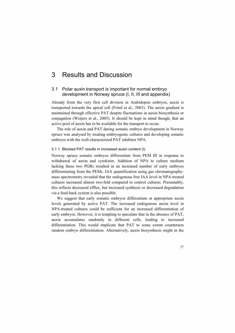
One week after subculture, PGRs start to become depleted from the proliferation medium and early embryos start to differentiate. At this stage, small patches of stronger GUS activity was surrounded by cells with faint GUS staining (Figure 3A), in accordance with the hypothesis that early embryos start to differentiate from cells with appropriate auxin contents (see above).
During early embryogeny and in the beginning of late embryogeny an auxin response maximum was generated in the basal part of the embryonal mass and in the differentiating tube cells (Figure 3B,C). We assume that the basal part of the embryonal mass harbours stem cells that provide both the developing embryonal mass and the differentiating suspensor with new cells (i.e. meristematic cells and tube cells). Thus, the GUS activity in this region indicates that auxin might play a role in specifying the embryonal stem cells in conifers.
In late embryos that had developed a protoderm, but not yet established a SAM, GUS staining was restricted to the basal cells of the embryonal mass (Figure 3D), which presumably contain the stem cells, but also will form the root cap of the maturing embryo. Early embryos of Norway spruce that were treated with NPA showed no GUS activity in the embryonal mass, but increased GUS staining in the tube cells and in the suspensor cells (Figure 3E), suggesting that polar transport of auxin from the suspensor to the embryonal mass is blocked.
To exclude the possibility that the specific GUS staining was due to the inability of GH3 to be expressed in the apical part of early Norway spruce embryos, the embryos were treated with 2.4-D for 15 h, which induced GUS activity in all cells of the embryonal mass (Figure 3F).
30
Figure 3. Auxin response as visualized by the expression of GH3-GUS in early somatic embryos of Norway spruce. (A) Proliferating proembryogenic masses, note the patches of increased GUS activity. (B) Early differentiating embryo. (C) Beginning of late embryogeny, note the gradient of GUS activity from the suspensor cells to the basal part of the embryonal mass. (D) Late embryo, note strong GUS activity in the basal part of the embryonal mass. (E) NPA-treated early embryo, note the lack of GUS activity in the meristematic cells. (F) Embryo in the beginning of late embryogeny treated with 2.4-D over night before stained for GUS activity, note the strong GUS activity in the whole embryo.
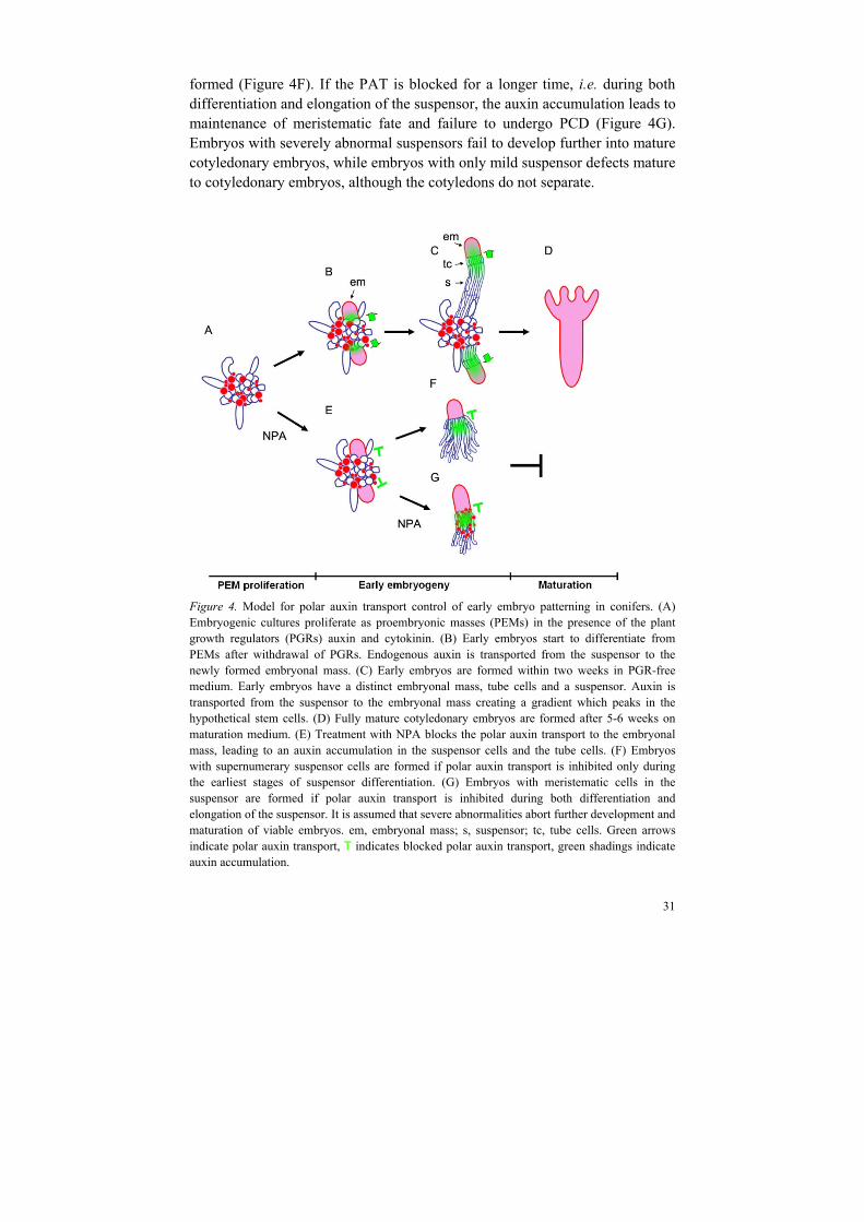
Taken together, the effects of blocked PAT on embryo differentiation and early development enables a model for auxin regulation of early somatic embryo development in Norway spruce (Figure 4, modified from appendix). The results indicate that IAA under normal conditions is transported from the suspensor cells to the cells in the embryonal mass, creating a gradient with maximum, or appropriate, concentration in the stem cells (Figure 4B,C). In accordance, auxin gradients with maxima in meristematic cells have been shown to organize cell specification in both the vascular cambium (Uggla et al., 1996) and in primary root apex (Sabatini et al., 1999). Furthermore, auxin gradients seem to specify cell identity in female gametophytes (Pagnussat et al., 2009). NPA-treatment blocks the polar transport of IAA, which results in an accumulation of IAA and thus increased auxin response in the suspensor cells and in the tube cells (Figure 4F,G). Blocked PAT during early differentiation of the suspensor stimulates abnormal cell divisions of the tube cells, and perhaps also of the most recently developed suspensor cells. Consequently, embryos with supernumerary tube and suspensor cells are
31
formed (Figure 4F). If the PAT is blocked for a longer time, i.e. during both differentiation and elongation of the suspensor, the auxin accumulation leads to maintenance of meristematic fate and failure to undergo PCD (Figure 4G). Embryos with severely abnormal suspensors fail to develop further into mature cotyledonary embryos, while embryos with only mild suspensor defects mature to cotyledonary embryos, although the cotyledons do not separate.
Figure 4. Model for polar auxin transport control of early embryo patterning in conifers. (A) Embryogenic cultures proliferate as proembryonic masses (PEMs) in the presence of the plant growth regulators (PGRs) auxin and cytokinin. (B) Early embryos start to differentiate from PEMs after withdrawal of PGRs. Endogenous auxin is transported from the suspensor to the newly formed embryonal mass. (C) Early embryos are formed within two weeks in PGR-free medium. Early embryos have a distinct embryonal mass, tube cells and a suspensor. Auxin is transported from the suspensor to the embryonal mass creating a gradient which peaks in the hypothetical stem cells. (D) Fully mature cotyledonary embryos are formed after 5-6 weeks on maturation medium. (E) Treatment with NPA blocks the polar auxin transport to the embryonal mass, leading to an auxin accumulation in the suspensor cells and the tube cells. (F) Embryos with supernumerary suspensor cells are formed if polar auxin transport is inhibited only during the earliest stages of suspensor differentiation. (G) Embryos with meristematic cells in the suspensor are formed if polar auxin transport is inhibited during both differentiation and elongation of the suspensor. It is assumed that severe abnormalities abort further development and maturation of viable embryos. em, embryonal mass; s, suspensor; tc, tube cells. Green arrows indicate polar auxin transport, T indicates blocked polar auxin transport, green shadings indicate auxin accumulation.

32
3.1.4 Blocked PAT causes fused cotyledons, aborted SAM and an irregular RAM (I, II, III)
NPA-treated embryos that escaped severe abnormalities during early embryogeny developed significantly slower than control embryos. Beside this effect, there were no other apparent differences between NPA-treated and control maturing embryos until the cotyledons were clearly visible. After a few weeks on maturation medium, control embryos started to form cotyledons as small protuberances in a circle around the incipient SAM. In contrast, in NPA-treated embryos, the cotyledons could not be distinguished from each other, and developed as a thick ring with a cavity in the centre. The ring continued to grow, but thinned out so that the apical part of the mature embryo looked like a hollow cylinder. Sectioning and histological analysis of such embryos revealed that no organised SAM could be visualized, and the RAM was highly irregular. Embryos with fused cotyledons and unorganized RAM formed neither a shoot nor a root when placed on germination medium.
Fused or aborted cotyledons are characteristic for angiosperm embryos with impaired auxin transport or response (Liu et al., 1993; Hadfi et al., 1998; Aida et al., 2002; Mravec et al., 2008). In dicotyledonous embryos it has been postulated that auxin accumulates at incipient cotyledon primordia, thereby stimulating the transition from radial to bilateral symmetry, and cotyledon outgrowth (Benkovà et al., 2003). In conifers, PAT is not involved in establishing bilateral symmetry. As the cotyledons in conifers often occur in odd numbers (personal observation), the cotyledonary crown cannot even be regarded as having several planes of symmetry. However, the transition from late embryogeny to maturation can still be considered as a symmetry-breaking event, as the embryo goes from a radial symmetry to an asymmetric organisation. Furthermore, PAT might still be important for depleting cotyledon boundaries from auxin and for directing the cotyledon outgrowth.
In Arabidopsis the establishment of the root meristem is dependent on an accumulation of auxin response in the hypophysis, and several auxin response and biosynthesis mutants show pronounced defects in embryonic root formation (see 1.2.1).
Based on the fused cotyledons and the irregular cell divisions in the area of the RAM in NPA-treated embryos, we suggest that an apical-basal transport of auxin is important for pattern formation also in conifers. It is tempting to speculate that the NPA-treatment of Norway spruce somatic embryos blocks the transport of auxin towards the cotyledon primordia, as well as the apical-basal transport, so that auxin accumulates all over the apical part of the embryo instead of in the emerging cotyledon tips and in the incipient root meristem. In accordance, Arabidopsis PID overexpressors fail to specify the hypophysis and
33
develop malformed or fused cotyledons, as a consequence of an auxin accumulation in the apical part of the embryo, due to a basal-to-apical shift in PIN polarity (Friml et al., 2004).
Since all maturing embryos treated with NPA formed fused cotyledons without an apparent SAM, NPA-treatment can in future experiments be used as a valuable tool for studies of SAM formation in conifer embryos.
3.2 NAC-regulation of embryo development in conifers (II and unpublished results)
The SAM is established during embryo development in conifers concomitantly with the formation of a surrounding cotyledonary crown. Cotyledons are highly diverse in size, form and morphology across the plant kingdom. However, the genetic regulation of cotyledon differentiation is elusive and has been studied primarily in Arabidopsis (reviewed by Chandler, 2008). Hardly anything is known about the regulation of SAM and cotyledon formation in conifers, which morphologically differs considerably from the dicotyledonous Arabidopsis, and also varies between different individuals of the same species.
In Arabidopsis, the formation of the cotyledon boundaries and the establishment of the embryonal SAM are dependent on the redundant function of the three CUC genes (Aida et al., 1997; Takada et al., 2001; Vroemen et al., 2003). Hence, it was of interest to analyze the role of CUC orthologous genes during embryo development also in Norway spruce.
3.2.1 NAC-genes in conifers
The CUC genes belong to the large plant-specific NAC gene family of TFs. Despite the diversity of NAC genes and their importance for various developmental processes in seed plants (Olsen et al., 2005; Shen et al., 2009), no reports about the roles of NAC genes in conifers have been found. By performing BLAST searches on publicly available EST sequences from white spruce (Picea glauca) in GenBank, using various NAC-domains from Arabidopsis as the query, we identified 16 different DNA sequences with full-length NAC domains, designated PgNAC01-16. Based on the assumption that NAC-encoding genes that are phylogenetically closely related also exert similar functions (Shen et al., 2009; Jensen et al., 2010), it was of interest to deploy the spruce sequences into different clades within the NAC gene family. The phylogenetic analysis tools PAUP* and MrBayes were used to generate consensus trees of the evolutionary relationship between NAC genes from Arabidopsis, spruce, Medicago truncatula and moss (Physcomitrella patens).

34
Two spruce sequences, PgNAC14 and PgNAC15, clustered with the VASCULAR-RELATED NAC-DOMAIN (VND) and NAC SECONDARY WALL THICKENING PROMOTING FACTOR (NST) genes, which are regarded as master regulators for wood formation (reviewed by Zhang et al., 2011), suggesting that these sequences are interesting candidates for studying wood formation in economically important conifers. PgNAC07 clustered together with drought-inducible ATAF1 (Lu et al., 2007), which could also be of interest for future studies. Two sequences, PgNAC01 and PgNAC02, clustered with the CUC genes, and were used as templates to amplify CUC orthologs in Norway spruce
3.2.2 CUC-orthologs in Norway spruce
Two full-length cDNAs, denoted PaNAC01 and PaNAC02, were cloned from Norway spruce. Although it was clear that both genes belong to the CUC-clade, the sequences did not contain enough information to resolve the exact position of individual genes within this clade. However, motif analysis showed that PaNAC01, but not PaNAC02 harbored all previously characterized motifs that have been shown to be of functional importance for CUC1 and CUC2 (Figure 5), including the microRNA, miR164, recognition site. In Arabidopsis, CUC1 and CUC2 are post-transcriptionally down-regulated by miR164-cleavage (Laufs et al., 2004; Mallory et al., 2004), and a microRNA screen in Norway spruce has identified a miR164 transcript (U. Lagercrantz, personal communication). Based on sequence similarity it seems likely that PaNAC01 can be recognized by this microRNA, although it remains to be verified experimentally. Interestingly, the miR164 recognition site could not be found in any of the NAC genes that are present in moss or spike moss. Large-scale sequencing of small RNAs did not identify miR164 in these species (Axtell et al., 2007), which suggests that the miR164-mediated post-transcriptional regulation of the CUC genes was established after the origin of seed plants, but before the separation between angiosperms and gymnosperms.
Using genome walking, a 2560 bp sequence upstream of PaNAC01 and a 1247 bp sequence upstream of PaNAC02 were isolated. The sequences were analyzed together with 3000 bp upstream sequences of Arabidopsis NAC genes in order to find motifs, conserved between the NAC genes in angiosperms and gymnosperms. Interestingly, a 21 bp long element located 300 – 1000 bp up-stream of the translation start site was found to be highly conserved between PaNAC01, PaNAC02 and the three CUC genes in Arabidopsis (Figure. 5). The element has not been characterized previously and could not be found in any other NAC gene in Arabidopsis. It is likely that TFs restricting the expression

35
of the CUC genes bind to this element, since an element deletion in the Arabidopsis CUC2 promoter resulted in a broadened expression domain in seedlings.
Figure 5. Schematic comparison of the two CUC-like sequences (PaNAC01 and PaNAC02) that were cloned from Norway spruce, and CUC1, CUC2 and CUC3 from Arabidopsis. All sequences harbor the highly conserved DNA-binding NAC-domain in their N-terminal region, and all genes have the 21 bp conserved promoter element, specific for the CUC genes. In addition PaNAC01 contain all previously characterized motifs (L, V and W), including the miR164 recognition site, that have been shown to have functional importance for CUC1 and CUC2.
3.2.3 The expression of PaNAC01 is dependent on PAT
The temporal expression pattern of PaNAC01 was analysed using quantitative RT-PCR in eight consecutive stages of somatic embryos that had been treated or not with NPA. In control embryos, the relative expression of PaNAC01 increased as early embryos started to differentiate, and remained high until the SAM was clearly visible and the cotyledons had grown out. In NPA-treated embryos that formed fused cotyledons and lacked a distinct SAM, the up-regulation of PaNAC01 was blocked compared to controls, and the relative expression of PaNAC01 was significantly lower throughout all developmental stages until maturation. In Arabidopsis, the spatial expression pattern of both
36
CUC1 and CUC2 is affected in mutants with impaired auxin transport and/or response (Aida et al., 2002; Furutani et al., 2004), but the expression levels of the CUC genes have not been reported. However, it has been shown that the expression of NAC1 initially increases in response to auxin treatment, indicating that NAC1 may be an early auxin-responsive gene (Xie et al., 2000).
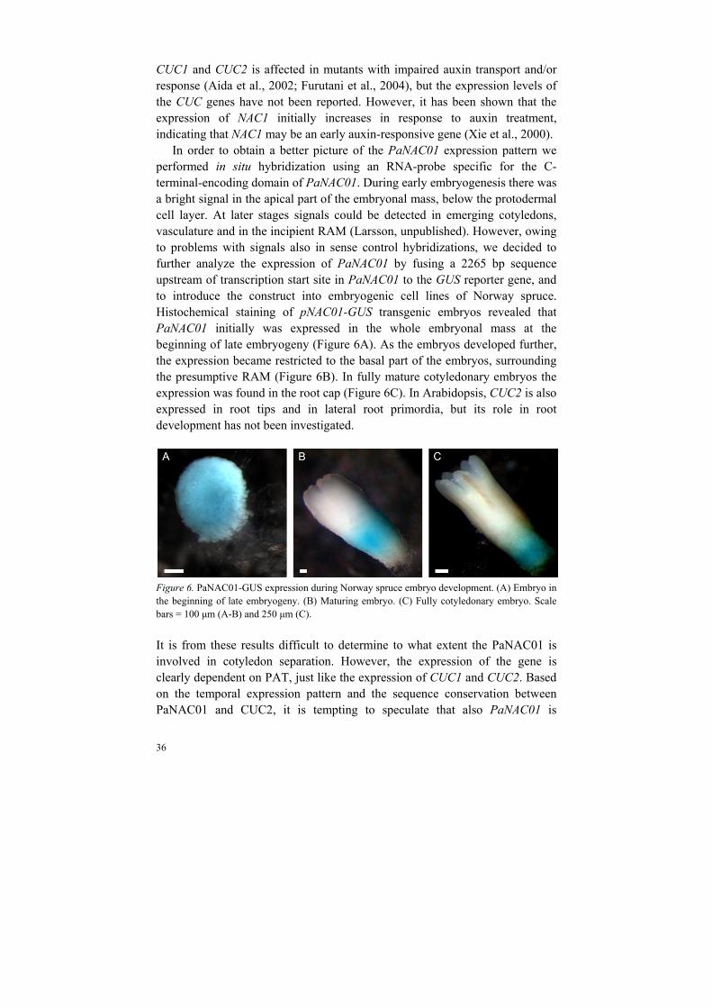
In order to obtain a better picture of the PaNAC01 expression pattern we performed in situ hybridization using an RNA-probe specific for the C-terminal-encoding domain of PaNAC01. During early embryogenesis there was a bright signal in the apical part of the embryonal mass, below the protodermal cell layer. At later stages signals could be detected in emerging cotyledons, vasculature and in the incipient RAM (Larsson, unpublished). However, owing to problems with signals also in sense control hybridizations, we decided to further analyze the expression of PaNAC01 by fusing a 2265 bp sequence upstream of transcription start site in PaNAC01 to the GUS reporter gene, and to introduce the construct into embryogenic cell lines of Norway spruce. Histochemical staining of pNAC01-GUS transgenic embryos revealed that PaNAC01 initially was expressed in the whole embryonal mass at the beginning of late embryogeny (Figure 6A). As the embryos developed further, the expression became restricted to the basal part of the embryos, surrounding the presumptive RAM (Figure 6B). In fully mature cotyledonary embryos the expression was found in the root cap (Figure 6C). In Arabidopsis, CUC2 is also expressed in root tips and in lateral root primordia, but its role in root development has not been investigated.
Figure 6. PaNAC01-GUS expression during Norway spruce embryo development. (A) Embryo in the beginning of late embryogeny. (B) Maturing embryo. (C) Fully cotyledonary embryo. Scale bars = 100 μm (A-B) and 250 μm (C).
It is from these results difficult to determine to what extent the PaNAC01 is involved in cotyledon separation. However, the expression of the gene is clearly dependent on PAT, just like the expression of CUC1 and CUC2. Based on the temporal expression pattern and the sequence conservation between PaNAC01 and CUC2, it is tempting to speculate that also PaNAC01 is

37
expressed in the intercotyledonary regions, but that the expression is too weak to be visualized by GUS staining.
3.2.4 PaNAC01 can stimulate cotyledon separation in the cuc1cuc2 mutant background
The coding sequence of PaNAC01 was expressed from the Arabidopsis CUC2 promoter, and transformed into cuc1/cuc1 cuc2/+ double mutants. In the progeny of untransformed cuc1/cuc1 cuc2/+ plants about 25 % of the seedlings had a cup-shaped phenotype, while the progeny of cuc1/cuc1 cuc2/+ plants transformed with the pAtCUC2::PaNAC01 construct did not show this phenotype, indicating a complementation of the mutation by the construct. Three independently rescued lines were taken to the T2 generation. As determined by PCR analysis, all T2 plants were homozygous for the cuc2 mutation. However, up to 90 % had a wild-type appearance with two separated cotyledons bilaterally positioned on opposite sides of the SAM. The rest of the seedlings showed a variable degree of cotyledon fusion, but not as complete as in the cup-shaped phenotype. These results show that the PaNAC01 protein can function in a similar pathway as CUC1 and CUC2, and that this pathway is evolutionarily conserved between angiosperms and gymnosperms.
To explore the function of PaNAC01 during embryo development, the coding sequence was expressed from the constitutive CaMV 35S promoter in embryogenic cell lines of Norway spruce. In Arabidopsis, seedlings overexpressing CUC1 develop lobed cotyledons with ectopic shoots formed on the adaxial surface (Takada et al., 2001). Somatic embryos overexpressing PaNAC01 grew and developed much faster than control embryos (Larsson, unpublished), but there were no other distinct morphological differences between transgenes and controls.
3.2.5 The expression of PaNAC02 decreases as embryos develop
The relative expression of PaNAC02 was high in PEMs and early differentiating embryos, but decreased as the embryos developed and matured. There was no significant difference between the PaNAC02 expression in NPA-treated embryos compared to controls. Analysis of a pPaNAC02::GUS fusion (Larsson, unpublished) revealed that PaNAC02 was first expressed in the whole embryonal mass, except for the outer cell layer in early differentiating embryos. In the beginning of late embryogeny, PaNAC02 expression could be seen in the upper half of the embryonal mass, but as the embryo developed further and the SAM and cotyledons were formed, the expression of PaNAC02 became restricted to a few cells in the epidermis around the RAM until it finally disappeared. It is based on these results difficult to suggest a function of
38
PaNAC02. However, its differential expression during embryo development makes it interesting to study the role of the gene further.
Taken together, the results suggest that PaNAC01 plays an important role during embryo development in Norway spruce. PaNAC01, but not PaNAC02, harbors motifs that have been shown to be of functional importance for CUC1 and CUC2. The ability of PaNAC01 to functionally substitute for CUC2 in Arabidopsis cuc1cuc2 double mutants, its response to altered PAT, and its strong expression during SAM differentiation and cotyledon formation, together indicate that PaNAC01 function in regulating cotyledon boundaries and SAM formation analogous to the CUC genes in Arabidopsis. The function of PaNAC02 is more difficult to interpret. However, there are several CUC-like genes in Arabidopsis with unknown functions that could be analogous to PaNAC02. Alternatively, PaNAC02 might exert an ancient CUC-function or a conifer-specific function. Interestingly, both PaNAC01 and PaNAC02 harbor the promoter element that could be distinguished only in the promoters of the CUC genes in Arabidopsis. This at least suggest of a similar regulation of the five genes. Clearly, the regulation and function of PaNAC genes merits further investigations.
3.3 KNOX-diversification during embryo development in conifers (III)
Four KNOXI genes, HBK1, HBK2, HBK3 and PaKN4 (here denoted HBK4), have been identified in Norway spruce (Sundås-Larsson et al., 1998; Hjortswang et al., 2002; Gulliet-Claude et al., 2004). In order to distinguish the function of these four genes, their expression pattern was analyzed during embryo development in Norway spruce, as well as in embryos that lack a functional SAM. In addition, the effects of constitutive expression in Arabidopsis were studied.
3.3.1 Differential expression of HBK2 and HBK4 during embryo development is dependent on the formation of a functional SAM
In Arabidopsis, the KNOXI gene STM is essential for the establishment of the embryonal SAM, and the expression of STM is regulated by CUC and PAT (Aida et al., 1999; Furutani et al., 2004). As the regulatory connection between a CUC-ortholog and PAT seems to be conserved between Arabidopsis embryos and Norway spruce embryos, it is not unlikely that also a third partner in the regulatory network for SAM establishment, a KNOXI gene, could have a conserved function.

39
To determine the temporal expression, and PAT-responsiveness, of the four KNOXI genes during somatic embryogenesis in Norway spruce, the relative expression of each gene was analyzed at eight consecutive stages during development of somatic embryos, as well as in embryos that had been treated with NPA. This showed that HBK1 and HBK3 were up-regulated in the beginning of late embryogeny. The level of HBK1 expression decreased at the end of maturation, while the expression of HBK3 remained high. The temporal expression of the genes was not affected in embryos that had been treated with NPA. Since NPA-treated embryos lack a visible SAM, the results indicate that HBK1 and HBK3 have a more general role during embryo development than to specify the SAM.
The relative expression of HBK2 decreased dramatically as early embryos began to differentiate. However, as the cotyledon primordia started to protrude around the incipient SAM, the expression increased. Coinciding with the time of SAM establishment, the expression of HBK2 was almost 40 times higher than during early embryogeny. In NPA-treated embryos, the increased expression of HBK2 was delayed compared to controls, and it was not until the embryos were almost fully mature that the relative expression of HBK2 became comparable in NPA-treated and control embryos. The importance of narrow temporal windows for KNOXI expression has been shown (Shani et al., 2009), making it tempting to speculate that HBK2 has to be up-regulated at a specific developmental stage for correct establishment of the embryonal SAM. The relative expression of HBK4 was low during early embryo differentiation, but increased as the cotyledons began to differentiate around the developing SAM. The expression then remained at a steady level throughout maturation. In NPA-treated embryos, the relative expression of HBK4 was delayed similarly to that of HBK2, suggesting that the expression timing of both these genes is important for the correct establishment of the embryonal SAM.
It has been shown that HBK1 and HBK3 are expressed in embryogenic cell lines irrespective of a blocked embryo development or not. In contrast, HBK2 is expressed only in lines that develop fully mature cotyledonary embryos (Hjortswang et al., 2002). In order to test the regulation of HBK4 in this aspect, the temporal expression of each gene was analyzed by RT-PCR in one cell line blocked at the proliferation stage, and in one cell line that develops fully mature cotyledonary embryos. The results were consistent with those from Hjortswang et al (2002), and it was evident that HBK4, just like HBK2, is expressed only in cell lines capable to develop fully mature cotyledonary embryos. This supports the hypothesis that HBK2 and HBK4 are important for normal somatic embryo development in Norway spruce.
40
3.3.2 HBK overexpression in Arabidopsis induces morphologies characteristic of ectopic KNOXI expression
In order to further test the different functions of the four HBK genes, they were fused to the 35S-promoter and introduced into Arabidopsis. Typically, KNOXI overexpression in Arabidopsis results in serrated and lobed leaves, and ectopic meristems are occasionally formed on the leaves (Hay and Tsiantis, 2010). Similarly, overexpression of all four HBK genes resulted in serrated leaves. However, the degree of serration was stronger in HBK1 and HBK3 overexpressors than it was in transformants overexpressing HBK2 and HBK4. Furthermore, plants expressing HBK1 and HBK3 developed abnormal flowers, reminiscent of those produced by plants overexpressing BP, KNAT2 and STM (Scofield et al., 2008). In contrast, the HBK2 and HBK4 overexpressors developed flowers that were similar to wild-type Arabidopsis flowers, although the most severe lines showed an extremely delayed abscission of the outer floral organs. This is a characteristic that has not been described previously for KNOXI overexpressors, suggesting that HBK2 and HBK4 can function in different pathways than previously characterized KNOXI proteins.
None of the HBK overexpressors induced ectopic meristems on the leaves.
Taken together our results show that HBK1 and HBK3 are regulated similarly during somatic embryo development in Norway spruce, and indicate that they have a general function during this process. The role of HBK2 and HBK4 is more elusive, but it seems likely that they have important and specific functions during somatic embryo differentiation and SAM establishment in Norway spruce. Expression of both genes marks the potential to fully complete maturation, and is affected by blocked PAT. Based on the overexpressor studies in Arabidopsis it seems likely that the function of KNOXI proteins to a large extent is conserved between angiosperms and gymnosperms, but that each HBK protein, and specifically HBK2 and HBK4, also have unique and conifer-specific functions. Therefore, it would be of great interest to study the function of these proteins during somatic embryo development in Norway spruce more thoroughly.
3.4 Identification of putative processes associated with pattern formation during the earliest stages of somatic embryogenesis (IV)
The protocol for somatic embryogenesis in Norway spruce used in this thesis is well-synchronized and produces high numbers of fully mature cotyledonary embryos with high germination capacity. However, not all genotypes of

41
Norway spruce, or other economically important conifers, share this ability. To identify molecular processes important for the regulation of early embryo differentiation and development in the somatic embryogenesis system, a global gene expression study was performed. This might be helpful for identifying aberrant expression patterns in recalcitrant genotypes of Norway spruce and other conifers.
3.4.1 Changes in global gene expression during differentiation and development of early somatic embryos
The expression profile of 12,536 cDNA clones was followed during the earliest stages of somatic embryo development in Norway spruce, using a 17 k loblolly pine cDNA microarray. In total, 720 unique transcripts were differentially expressed. Of these, 152 clones expressed during the transition from proliferation to early embryo differentiation, and 383 clones expressed in the beginning of late embryo development, could be assigned to putative homologous genes in Arabidopsis. These genes were used to set up a model for the molecular regulation of the early somatic embryo development in conifers.
Nurse cells
It has been shown that several genes expressed in the female gametophyte affect embryo development, without being expressed in the embryo itself. These genes have collectively been called MATERNAL EFFECT EMBRYO ARREST (MEE) genes (Pagnussat et al., 2005). The phenomenon has been shown also in somatic embryos of Norway spruce, in which ‘nurse cells’, positioned close to the embryo but not in the embryo itself, express chitinase-encoding genes that affect the early embryo differentiation (Wiweger et al., 2003). The microarray analysis further highlighted the importance of nurse cells during the differentiation of early embryos, as putative homologs to ARABIDOPSIS THALIANA HOMEOBOX PROTEIN 22(ATHB22)/ MEE 68, as well as MEE49 and MEE66, were differentially expressed at this stage. Furthermore, a putative homolog to NAC-REGULATED SEED MORPHOLOGY2 (NARS2) was up-regulated during early embryo differentiation. In Arabidopsis NARS1 and NARS2 are expressed in the integuments of the seed and are redundantly required for proper embryo development (Kunieda et al., 2008).
PCD
The microarray analysis also verified the importance of suspensor differentiation and PCD, since the suspensor-specific aqua-glycoprotein encoding gene TRANSMEMBRANE PROTEIN C (TMP-C, Ciavatta et al.,
42
2002), the F-actin capping protein encoding gene CATHEPSIN B-LIKE CYSTEINE PROTEASE (McLellan et al., 2009) and METACASPASE 9 (MC9, Tsiantsiani et al., 2011) were up-regulated already 24 h after withdrawal of PGRs.
Auxin
The down-regulation of a suggested homolog to SUR1, and the concomitant up-regulation of a putative SAUR, suggest that auxin biosynthesis increases during early embryogeny. Furthermore, clones homologous to genes encoding the auxin-induced protein IAA11 and the auxin receptor TIR1 were up-regulated during the transition from differentiation to development together with a putative homolog of MYB77, a transcription factor involved in auxin-response (Shin et al., 2007). This indicates that the auxin-response machinery is up-regulated in the beginning of late embryo development.
Cell-identity genes
Early embryogeny in conifers can be compared to the globular stage in Arabidopsis, and already at this stage genes involved in tissue specification were differentially expressed. For example, the protoderm specification gene PaHB1 (Ingoff et al., 2001) was up-regulated, as well as a putative homolog of the floral organ identity gene LEUNIG (LUG, Navarro et al., 2004). In Arabidopsis double mutants of lug and its close homolog leunig-homolog (luh) are embryo lethal (Sitarman et al., 2008), suggesting that the two genes have a function also in pattern formation. Furthermore a clone homologous to the ROXY1 gene, which in Arabidopsis encodes a glutaredoxine important for petal induction (Xing et al., 2005) was down-regulated during the transition from differentiation to embryo development. Nothing is known about domain specification in conifer embryos. However, it appears that this occurs already during early embryogeny, and that genes involved in homeotic floral functions may function in the specification of cell identity in the embryonal mass.
Gibberellin (GA)
During the transition from early to late embryogeny, there was a general increase in the differential expression of clones putatively involved in GA-signaling. GA content seemed to increase already during early embryo differentiation, since a putative GA inactivation gene, GIBBERELLIN 2-OXIDASE 2 (GA2OX2, Rieu et al., 2008) was down-regulated. Concomitantly, a homolog of a negative regulator of GA response, RGA-LIKE1 (RGL1, Busov et al., 2006) was up-regulated, indicative of reduced GA-response. In the beginning of late embryo development another GA2-oxidase (GA2OX6) was
43
down-regulated and the RGL1 homolog was continuously up-regulated. In addition, a clone homologous to another GA signaling repressor, SPINDLY (SPY), was up-regulated, and a GA-regulated gene was down-regulated. Together these results indicate that the content of active GAs increase during embryo development, but that the GA-response is reduced. In Arabidopsis reduced activity of the GA pathway promotes meristematic activity in the SAM (Hay et al., 2002), suggesting that the SAM anlagen could be initiated already in the beginning of late embryogeny in conifers.
Phase change
In Arabidopsis the LEAFY COTYLEDON genes LEC1, LEC2 and FUSCA3 (FUS3) are considered to act as master regulators of embryogenesis. Ectopic expression of all three LEC genes causes cells in vegetative tissues to adopt characteristics of maturation phase embryos, and their expression must be repressed post-embryonically to allow vegetative development (reviewed by Braybrook and Harada, 2008). Together with ABSISIC ACID INSENSITIVE3 (ABI3), the LEC genes promote embryo maturation (To et al., 2006). In our microarray analysis, a putative homolog of LEC1 and its close relative LEC1-like (L1L) was found to be down-regulated during late embryo development. Concomitantly, the ABI3 homolog PaVP1 (Footitt et al., 2003) was up-regulated. These results, together with recently published data (Uddenberg et al., 2011), suggest that the conifer homologs of LEC1 and ABI3 play central roles, already in the beginning of late embryogeny, in preparing the embryo for the phase change from embryonic to vegetative development.
3.4.2 A model for putative processes regulating early embryo differentiation and development
Based on the results from the microarray analysis a model was set up for processes putatively regulating the differentiation of early somatic embryos and the development of late somatic embryos in conifers (Figure 7). The importance of some of these processes, such as PCD and PAT, have been verified through molecular and biochemical analyses (Filonova et al., 2000b; Suarez et al., 2004; Bozhkov et al., 2005; Larsson et al., 2008). Additional studies are now needed to confirm the relevance of other processes indicated by the microarray analysis.
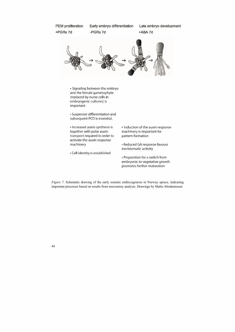
44
Figure 7. Schematic drawing of the early somatic embryogenesis in Norway spruce, indicating important processes based on results from microarray analysis. Drawings by Malin Abrahamsson

45
4 Conclusions
Polar auxin transport (PAT) is essential for the correct patterning of both apical and basal parts of conifer embryos throughout the whole developmental process. The results indicate that the role of PAT during embryo development is conserved between angiosperms and gymnosperms.
CUC-like genes with distinct signature motifs existed before the separation of angiosperms and gymnosperms. Several lines of evidence suggest a conserved function between PaNAC01 and CUC1/CUC2 in regulating cotyledonary boundaries and SAM formation.
KNOXI genes have diversified after the split between gymnosperms and angiosperms. The expression of HBK2 and HBK4 is dependent on PAT, and both genes seem to be important for embryo differentiation and SAM formation.
Processes that based on microarray analysis are important for early somatic embryo development are: signaling between nurse cells and differentiating embryos; PCD and suspensor differentiation; auxin synthesis, transport and response; GA response; establishment of cell identity, and preparation for a switch from embryonic to vegetative growth.

46
47
5 Future perspectives
The molecular regulation of embryo development and pattern formation in conifers is still poorly understood. However, the results presented in this thesis can serve as a frame-work for future studies. Techniques for localizing expression patterns, and transformation need to be standardized and possible to perform in high through-put in order to reveal the true function of each gene. The possibility to down-regulate expression of redundant genes using artificial microRNAs is currently investigated.
Clearly, auxin is of great importance for both somatic and zygotic embryo development in Norway spruce. It would be very interesting to elucidate auxin biosynthesis pathways, transport routes and perception mechanisms in conifers, since this might reveal how and why pattern formation differs between distantly related plant species. Work is in progress to obtain a higher resolution of the auxin response during embryo development. However, methods to visualize true auxin gradients in any plant species are still missing.
As long as we refer our results to knowledge about Arabidopsis, it will be difficult to find regulatory pathways specific to conifers. Swedish researchers have undertaken the massive task to sequence the whole genome of Norway spruce. Hopefully, this will aid to identify and shed some more light on genes that are important for conifer development. With the genome sequence available it will be easier to make intelligent guesses about how to set up transgenic analyses, and promoter studies will be simplified. In addition it will be much easier to look at natural variation within the species. Our microarray analysis revealed differential expression of many putative genes with unknown function, and from an evolutionary point of view it would be of great interest to characterize the functions of these genes. Future studies of global gene expression patterns will most likely involve RNA sequencing in specific tissues and at specific stages, which will increase the resolution compared to microarray studies.

48

49
References
Aida, M., Ishida, T., Fukaki, H., Fujisawa, H., Tasaka, M. (1997). Genes involved in organ
separation in Arabidopsis: an analysis of the cup-shaped cotyledon mutant. The Plant Cell
9(6), 841-857.
Aida, M., Ishida, T., Tasaka, M. (1999). Shoot apical meristem and cotyledon formation during
Arabidopsis embryogenesis: interaction among the CUP-SHAPED COTYLEDON and
SHOOT MERISTEMLESS genes. Development 126, 1563-1570.
Aida, M., Vernoux, T., Furutani, M., Traas, J., Tasaka, M. (2002). Roles of PIN-FORMED1 and
MONOPTEROS in pattern formation of the apical region of the Arabidopsis embryo.
Development 129, 3965-3974.
Aida, M., Tasaka, M. (2006). Morphogenesis and patterning at the organ boundaries in the higher
plant shoot apex. Plant Molecular Biology 60, 915-928.
Axtell, MJ., Snyder, JA., Bartel, DP. (2007). Common Functions for Diverse Small RNAs of
Land Plants. The Plant Cell 19, 1750–1769.
Bailly, A., Sovero, V., Vincenzetti, V., Santelia, D., Bartnik, D., Koenig, BW., Mancuso, S.,
Martinoia, E., Geisler, M. (2008). Modulation of P-glycoproteins by auxin transport inhibitors
is mediated by interaction with immunophilins. The Journal of Biological Chemistry 283,
21817 -21826.
Ballas, N., Wong, L-M., Theologi,s A. (1993). Identification of the auxin-responsive element,
AuxRE, in the primary indoleacetic acid-inducible gene, PS-IAA4/5, of pea (Pisum sativum).
Journal of Molecular Biology 233, 580-596.
Barton, MK., Poethig, RS. (1993). Formation of the shoot apical meristem in Arabidopsis
thaliana: an analysis of development in the wild type and in the shoot meristemless mutant.
Development 119, 823-831.
Belles-Boix, E., Hamant, O., Witiak, SM., Morin, H., Traas, J., Pautot, V. (2006). KNAT6: An
Arabidopsis homeobox gene involved in meristem activity and organ separation. The Plant
Cell 18:1900-1907.
Benková, E., Michniewicz, M., Sauer, M., Teichmann, T., Seifertová, D., Jürgens, G., Friml, J.
(2003). Local, efflux-dependent auxin gradients as a common module for plant organ
formation. Cell 115, 591-602.
50
Bennett, MJ., Marchant, A., Green, HG., May, ST., Ward, SP., Millner, PA., Walker, AR.,
Schulz, B. and Feldmann, KA. (1996). Arabidopsis AUX1 gene: a permease-like regulator of
root gravitropism. Science 273, 948 -950.
Bhalerao, RP., Bennet, MJ. (2003). The case of morphogens in plants. Nature Cell Biology 5,
939-943.
Bierfreund, NM., Reski, R., Decker, EL. (2003). Use of an inducible reporter gene system for the
analysis of auxin distribution in the moss Physcomitrella patens. Plant Cell Reports 21, 1143-
1152.
Boerjan,W., Cervera, MT., Delarue, M., Beeckman, T., Dewitte, W., Bellini, C., Caboche, M.,
Van Onckelen, H., Van Montagu, M., lnzé, D. (1995). superroot, a recessive mutation in
Arabidopsis, confers auxin overproduction. The Plant Cell 7, 1405–1419.
Bozhkov, PV., Filonova, LH., von Arnold, S. (2002). A key developmental switch during Norway
spruce somatic embryogenesis is induced by withdrawal of growth regulators and is
associated with cell death and extracellular acidification. Biotechnology and Bioengineering
77, 658-667.
Bozhkov, PV., Filonova, LH., Suarez, MF., Helmersson, A., Smertenko, AP., Zhivotovsky, B.,
von Arnold, S. (2004). VEIDase is a principal caspase-like activity involved in plant
programmed cell death and essential for embryonic pattern formation. Cell Death and
Differentiation 11, 175-182
Bozhkov, PV., Filonova, LH., Suarez, MF. (2005). Programmed cell death in plant
embryogenesis. Current Topics in Developmental Biology 67, 135-179.
Brand, U., Grünewald, M., Hobe, M., Simon, R. (2002). Regulation of CLV3 expression by two
homeobox genes in Arabidopsis. Plant Physiology 129, 565-575.
Braybrook, SA., Harada, JJ. (2008). LECs go crazy in embryo development. Trends in Plant
Sciences 13, 624–630.
Breuninger, H., Rikirsch, E., Hermann, M., Ueda, M., and Laux, T. (2008). Differential
expression of WOX genes mediates apical-basal axis formation in the Arabidopsis embryo.
Developmental Cell 14, 867–876.
Bürglin, TR. (1997). Analysis of TALE superclass homeobox genes (MEIS, PBC, KNOX,
Iroquois, TGIF) reveals a novel domain conserved between plants and animals. Nucleic Acids
Research 25, 4173-4180.
Busov, V., Meilan, R., Pearce, DW., Rood, SB., Ma, C., Tschaplinski, TJ., Strauss, SH. (2006).
Transgenic modification of gai or rgl1 causes dwarfing and alters gibberellins, root growth,
and metabolite profiles in Populus. Planta 224, 288-299.
Byrne, ME., Simorowski, J., Martienssen, RA. (2002). ASYMMETRIC LEAVES1 reveals knox
gene redundancy in Arabidopsis. Development 129, 1957-1965.
Capron, A., Chatfield, S., Provart, N., Berleth, T. (2009). Embryogenesis: Pattern formation from
a single cell. The Arabidopsis Book. Rockville, MD: American Society of Plant Biologists.
doi: 10.1199/tab.0126, http://www.aspb.org/publications/arabidopsis/
Carroll, SB. (2008). Evo-devo and an expanding evolutionary synthesis: a genetic theory of
morphological evolution. Cell 134, 25-36.
Chandler, J. (2008). Cotyledon organogenesis. Journal of Experimental Botany 59, 2917-2931.

51
Chandler, JW. (2009). Local auxin production: a small contribution to a big field. BioEssays 31,
60-70.
Chapman, EJ., Estelle, M. (2009). Mechanisms of auxin-regulated gene expression in plants.
Annual Review of Genetics 43, 265-285.
Cheng, Y., Dai, X., Zhao, Y. (2007). Auxin synthesized by the YUCCA flavin monooxygenases
is essential for embryogenesis and leaf formation in Arabidopsis. The Plant Cell 19, 2430-
2439.
Chuck, G., Lincoln, C., Hake, S. (1996). KNAT1 induces lobed leaves with ectopic meristems
when overexpressed in Arabidopsis. The Plant Cell 8, 1277-1289.
Ciavatta, VT., Morillon, R., Pullman, GS., Chrispeels, MJ., Cairney, J. (2001). An
aquaglyceroporin is abundantly expressed early in the development of the suspensor and the
embryo proper of Loblolly pine. Plant Physiology 127, 1556–1567.
Ciavatta, VT., Egertsdotter, U., Clapham, D., von Arnold, S., Cairney, J. (2002) A promoter from
the loblolly pine PtNIP1;1 gene directs expression in an early-embryogenesis and suspensor-
specific fashion. Planta 215, 694-698.
Dharmasiri, N., Dharmasiri, S., Weijers, D., Lechner, E., Yamada, M., Hobbie, L., Ehrismann,
JS., Jürgens, G., Estelle, M. (2005). Plant development is regulated by a family of auxin
receptor F box proteins. Developmental Cell 9, 109-119.
Darwin, C., Darwin, F. (1880). The power of movement in plants. Murray, J. (ed). London, UK.
Delarue, M., Prinsen, E., Onckelen, HV., Caboche, M., Bellini, C. (1998). sur2 mutations of
Arabidopsis thaliana define a new locus involved in the control of auxin homeostasis. The
Plant Journal 14, 603–11
De Smet, I., Lau, S., Mayer, U., Jürgens, G. (2010). Embryogenesis – The humble beginnings of
plant life. The Plant Journal 61, 959-970.
Dockx, J., Quaedvlieg, N., Keultjes, G., Kock, P., Weisbeek, P., Smeekens, S. (1995). The
homeobox gene ATK1 of Arabidopsis thaliana is expressed in the shoot apex of the seedling
and in flowers and inflorescence stems of mature plants. Plant Molecular Biology 28, 723-
737.
Duval, M., Hsieh, T-F., Kim, SY., Thomas, TL. (2002). Molecular characterization of AtNAM: a
member of the Arabidopsis NAC domain superfamily. Plant Molecular Biology 50, 237–248.
Ernst, HA., Olsen, AN., Karen Skriver, K., Larsen, S., Leggio, LL. (2004). Structure of the
conserved domain of ANAC, a member of the NAC family of transcription factors. EMBO
reports 5, 297–303.
Fang, Y., You, J., Xie, K., Xie, W., Xiong, L. (2008). Systematic sequence analysis and
identification of tissue-specific or stress-responsive genes of NAC transcription factor family
in rice. Molecular Genetics and Genomics 280, 547-563.
Filonova, LH., Bozhkov, PV., von Arnold, S. (2000a). Developmental pathway of somatic
embryogenesis in Picea abies as revealed by time-lapse tracking. Journal of Experimental
Botany 51, 249-264.
Filonova, LH., Bozhkov, PV., Brukhin, VB., Daniel, G., Zhivotovsky, B., von Arnold, S.
(2000b). Two waves of programmed cell death occur during formation and development of
somatic embryos in the gymnosperm, Norway spruce. Journal of Cell science 113, 4399-
4411.
52
Filonova LH., von Arnold S., Daniel G., Bozhkov, PV. (2002). Programmed cell death eliminates
all but one embryo in a polyembryonic plant seed. Cell Death and Differentiation 9, 1057-
1062.
Footitt, S., Ingouff, M., Clapham, D., von Arnold, S. (2003). Expression of the viviparous 1
(Pavp1) and p34cdc2 protein kinase (cdc2Pa) genes during somatic embryogenesis in Norway
spruce (Picea abies [L.] Karst). Journal of Experimental Botany 54, 1711-1709.
Friml, J., Vieten, A., Sauer, M., Weijers, D., Schwartz, H., Hamann, T., Offringa, R., Jürgens, G.
(2003). Efflux-dependent auxin gradients establish the apical-basal axis of Arabidopsis.
Nature 426, 147-153.
Friml, J., Yang, X., Michniewicz, M., Weijers, D., Quint, A., Tietz, O., Benjamins, R.,
Ouwerkerk, PBF., Ljung, K., Sandberg, G., Hooykaas PJJ., Palme, K., Offringa, R. (2004). A
PINOID-dependent binary switch in apical-basal PIN polar targeting directs auxin efflux.
Science 306, 862 -865.
Furutani, M., Vernoux, T., Traas, J., Kato, T., Tasaka, M., Aida, M. (2004). PIN-FORMED1 and
PINOID regulate boundary formation and cotyledon development in Arabidopsis
embryogenesis. Development 131, 5021-5030.
Geisler, M., Blakeslee, JJ., Bouchard, R., Lee, OR., Vincenzetti, V., Bandyopadhyay, A.,
Titapiwatanakun, B., Peer, WA., Bailly, A., Richards, EL., Ejendal, KF., Smith, AP., Baroux,
C., Grossniklaus, U., Müller, A., Hrycyna, CA., Dudler, R., Murphy, AS., Martinoia, E.
(2005). Cellular efflux of auxin catalyzed by the Arabidopsis MDR/PGP transporter AtPGP1.
The Plant Journal 44, 179 -194.
Geldner, N., Anders, N., Wolters, H., Keicher, J., Kornberger, W., Muller, P., Delbarre, A., Ueda,
T., Nakano, A., Jürgens, G. (2003). The Arabidopsis GNOM ARF-GEF mediates endosomal
recycling, auxin transport, and auxin-dependent plant growth. Cell 112, 219–23.
Grunewald, W., Friml, J. (2010). The march of the PINs: developmental plasticity by dynamic
polar targeting in plant cells. The EMBO Journal 29, 2700-2714.
Guillet-Claude, C., Isabel, N., Pelgas, B., Bousquet, J. (2004). The evolutionary implications of
knox-I gene duplication in conifers:correlated evidence from phylogeny, gene mapping, and
analysis of functional divergence. Molecular Biology and Evolution 21, 2232-2245.
Hadfi, K., Speth, V., Neuhaus, G. (1998). Auxin-induced developmental patterns in Brassica
juncea embryos. Development 125, 879-887.
Haecker, A., Gross-Hardt, R., Geiges, B., Sarkar, A., Breuninger, H., Herrmann, M., and Laux, T.
(2004). Expression dynamics of WOX genes mark cell fate decisions during early embryonic
patterning in Arabidopsis thaliana. Development 131, 657–668.
Hagen, G., Martin, G., Li, Y., Guilfoyle, TJ. (1991). Auxin-induced expression of GH3 promoter
in transgenic tobacco plants. Plant Molecular Biology 17, 567-579.
Hagen, G., Guilfoyle, T. (2002). Auxin-resonsive gene expression: genes, promoters and
regulatory factors. Plant Molecular Biology 49, 373-385.
Hamann, T., Benková, E., Bäurle, I., Kientz, M., Jürgens, G. (2002). The Arabidopsis
BODENLOS gene encodes an auxin response protein inhibiting MONOPTEROS-mediated
embryo patterning. Genes and Development 16, 1610 -1615.
53
Hardtke, CS., Berleth, T. (1998). The Arabidopsis gene MONOPTEROS encodes a transcription
factor mediating embryo axis formation and vascular development. The EMBO Journal 17,
1405 -1411.
Hay, A., Kaur, H., Phillips, A., Hedden, P., Hake, S., Tsiantis, M. (2002). The gibberellin
pathway mediates KNOTTED1-type homeobox function in plants with different body plans.
Current Biology 12, 1557-1565.
Hay, A., Tsiantis, M. (2010). KNOX genes: versatile regulators of plant development and
diversity. Development 137, 3153-3165.
Heisler, MG., Ohno, C., Das, P., Sieber, P., Reddy, GV., Long, JA., Meyerowitz, EM. (2005).
Patterns of auxin transport and gene expression during primordium development revealed by
live imaging of the Arabidopsis inflorescence meristem. Current Biology 15, 1899-1911.
Hjortswang, HI., Sundås-Larsson, A., Bharathan, G., Bozhkov, PV., von Arnold, S., Vahala, T.
(2002). KNOTTED1-like homeobox genes of a gymnosperm, Norway spruce, expressed
during somatic embryogenesis. Plant Physiology and Biochemistry 40, 837-843.
Ingouff, M., Farbos, I., Lagercrantz, U., von Arnold, S. (2001). PaHB1 is an evolutionary
conserved HD-GL2 homeobox gene expressed in the protoderm during Norway spruce
embryo development. Genesis 30, 220-230.
Ingouff, M., Farbos, I., Wiweger, M., von Arnold, S. (2003). The molecular characterization of
PaHB2, a homeobox gene of the HD-GL2 family expressed during embryo development in
Norway spruce. Journal of Experimental Botany 54, 1343-1350.
Jenik, PD., Gillmor, CS., Lukowitz, W. (2007). Embryonic patterning in Arabidopsis thaliana.
Annual Review of Cell and Developmental Biology 23, 207-236.
Jensen, MK., Kjaersgaard, T., Nielsen, MM., Galberg, P., Petersen, K., O'Shea, C., Skriver, K.
(2010). The Arabidopsis thaliana NAC transcription factor family: structure-function
relationships and determinants of ANAC019 stress signalling. Biochemical Journal 426, 183-
196.
Kerstetter, R., Vollbrecht, E., Lowe, B., Veit, B., Yamaguchi, J., Hake, S. (1994). Sequence
analysis and expression patterns divide the maize knotted-1-like homeobox genes into two
classes. The Plant Cell 6, 1877-1887.
Kunieda, T., Mitsuda, N., Ohme-Takagi, M., Takeda, S., Aida, M., Tasaka, M., Kondo, M.,
Nishimura, M., Hara-Nishimura, I. (2008). NAC family proteins NARS1/NAC2 and
NARS2/NAM in the outer integument regulate embryogenesis in Arabidopsis. The Plant Cell
20:2631–2642.
Larkin, PJ., Gibson, JM., Mathesius, U., Weinman, JJ., Gartner, E., Hall, E., Tanner, GJ., Rolfe,
BG. Djordjevic, MA. (1996). Transgenic white clover. Studies with the auxin-responsive
promoter, GH3, in root gravitropism and lateral root development. Transgenic Research 5,
325-335.
Larue, CT., Wen, J., Walker, JC. (2009). Genetic interactions between the miRNA164-CUC2
regulatory module and BREVIPEDICELLUS in Arabidopsis developmental patterning. Plant
Signalling and Behavior 4, 666-668.
Lau, S., Ehrismann, JS., Schlereth, A., Takada, S., Mayer, U., Jürgens, G. (2010). Cell-cell
communication in Arabidopsis early embryogenesis. European Journal of Cell Biology 89,
225-230.
54
Laufs, P., Peaucelle, A., Morin, H., Traas, J. (2004). MicroRNA regulation of the CUC genes is
required for boundary size control in Arabidopsis meristems. Development 131, 4311-4322.
Lembi, CA., Morré, DJ. (1971). N-1-Napthylphthalamic-acid-binding activity of a plasma
membrane-rich fraction from maize coleoptiles. Planta 99, 37-45.
Li, Y., Wu, YH., Hagen, G., Guilfoyle, T. (1999). Expression of the eauxin-inducible GH3
promoter/GUS fusion gene as a useful marker for auxin physionolgy. Plant Cell Physiology
40, 675-682.
Liu, C-M., Xu, Z-H., Chua, N-H. (1993). Auxin polar transport is essential for the establishment
of bilateral symmetry during early plant embryogenesis. The Plant Cell 5, 621-630.
Long, JA., Maon, EI., Medford, JI., Barton, MK. (1996). A member of the KNOTTED class of
homeodomain proteins encoded by the STM gene of Arabidopsis. Nature 379, 66-69.
Lu, P-L., Chen, N-Z., An, R., Su, Z., Qi, B-S., Ren, F., Chen, J., Wang, W-C. (2007). A novel
drought-inducible gene, ATAF1, encodes a NAC family protein that negatively regulates the
expression of stress-responsive genes in Arabidopsis. Plant Molecular Biology 63, 289-305.
Mallory, AC., Dugas, DV., Bartel, DP., Bartel, B. (2004). MicroRNA regulation of NAC-domain
targets is required for proper formation and separation of adjacent embryonic, vegetative, and
floral organs. Current Biology 14(12), 1035-1046.
Mansfield, SG., Briarty, LG. (1991). Early embryogenesis in Arabidopsis thaliana. II. The
developing embryo. Canadian Journal of Botany 69, 461-476.
McLellan, H., Gilroy, EM., Yun, B-W., Birch, PRJ., Loake, GJ., (2009). Functional redundancy
in the Arabidopsis Cathepsin B gene family contributes to basal defense, the hypersensitive
response and senescence. New Phytologist 183, 408-418.
Michniewicz, M., Zago, MK., Abas, L., Weijers, D., Schweighofer, A., Meskiene, I., Heisler,
MG., Ohno, C., Zhang, J., Huang, F., Schwab, R., Weigel, D., Meyerowitz, EM., Luschnig,
C., Offringa, R., Friml, J. (2007). Antagonistic regulation of PIN phosphorylation by PP2A
and PINOID directs auxin flux. Cell 130, 1044 -1056.
Mikkelsen, MD., Naur, P., Halkier, BA., (2004). Arabidopsis mutants in the C-S lyase of
glucosinolate biosynthesis establish a critical role for indole-3-acetaldoxime in auxin
homeostasis. The Plant Journal 37, 770-777.
Mo, LH., Von, Arnold S., Lagercrantz, U. (1989). Morphogenetic and genetic stability in
longterm embryogenic cultures and somatic embryos of Norway spruce (Picea abies (L)
Karst). Plant Cell Reports 8, 375-378.
Moussian, B., Heiko Schoof, H., Haecker, A., Jürgens, G., Laux, T. (1998). Role of ZWILLE gene
in the regulation of central shoot meristem cell fate during Arabidopsis embryogenesis. The
EMBO Journal 17, 1799–1809.
Mravec, J., Kubes, M., Bielach, A., Gaykova, V., Petrásek, J., Skupa, P., Chand, S., Benková, E.,
Zazímalová, E. & Friml, J. (2008). Interaction of PIN and PGP transport mechanisms in auxin
distribution-dependent development. Development 135, 3345-3354.
Murphy, A., Peer, WA., Taiz, L. (2000). Regulation of auxin transport by aminopeptidases and
endogenous flavonoids. Planta 211, 315 -324.
Nakamura, A., Higuchi, K., Goda, H., Fujiwara MT., Sawa, S., Koshib, T., Shimada, Y., Yoshida,
S. (2003). Brassinolide induces IAA5, IAA19, and DR5, a synthetic auxin response element in
55
Arabidopsis, implying a cross talk point of brassinosteroid and auxin signaling. Plant
Physiology 133, 1843–1853.
Navarro, C., Efremova, N., Golz, JF., Rubiera, R., Kuckenberg, M., Castillo, R., Tietz, O.,
Saedler, H., Schwarz-Sommer, Z. (2004). Molecular and genetic interactions between
STYLOSA and GRAMINIFOLIA in the control of Antirrhinum vegetative and reproductive
development. Development 131, 3649-3659.
Nikovics, K., Blein, T., Peaucelle, A., Ishida, T., Morin, H., Aida, M., Laufs, P. (2006). The
balance between the MIR164A and CUC2 genes controls leaf margin serration in Arabidopsis.
The Plant Cell 18, 2929-2945.
Noh, B., Murphy, AS., Spalding, EP. (2001). Multidrug resistance-like genes of Arabidopsis
required for auxin transport and auxin-mediated development. The Plant Cell 13, 2441 -2454.
Oeller, PW., Keller, JA., Parks, JE., Silbert, JE., Theologis, A. (1993). Structural characterization
of the early indoleacetic acis-inducible genes, PS-IAA4/5 and PS-IAA6, of Pea (Pisum sativum
L.). Journal of Molecular Biology 233, 789-798.
Ohlrogge, J., Allen, D., Berguson, B., DellaPenna, D., Shachar-Hill, Y., Stymne, S. (2009).
Driving on Biomass. Science 22, 1019-1020.
Okada, K., Ueda, J., Komaki, MK., Bell, CJ., Shimura, Y. (1991). Requirement of the auxin polar
transport system in early stages of Arabidopsis floral bud formation. The Plant Cell 3, 677-
684.
Olsen, AN., Ernst, HA., Leggio, LL., Skriver, K. (2005). Transcriptional networks in plants -
NAC transcription factors: structurally distinct, functionally diverse. Trends in Plant Science
10, 79-87.
Ooka, H., Satoh, K., Doi, K., Nagata, T., Otomo, Y., Murakami, K., Matsubara, K., Osato, N.,
Kawai, J., Carninci, P., Hayashizaki, Y., Suzuki, K., Kojima, K., Takahara, Y., Yamamoto,
K., Kikuchi, S. (2003). Comprehensive analysis of NAC family genes in Oryza sativa and
Arabidopsis thaliana. DNA Research 10, 239-247.
Oono, Y., Chen, QG., Overvoorde, PJ., Köhler, C., Theologis, A. (1998). age mutants of
Arabidopsis exhibit altered auxin-regulated gene expression. The Plant Cell 10, 1649-1662.
Pagnussat, GC., Yu, H., Ngo, QA., Rajani, S., Mayalagu, S., Johnson, CS., Capron, A., Xie, L.,
Ye, D., Sundaresan, V. (2005). Genetic and molecular identification of genes required for
female gametophyte development and function in Arabidopsis. Development 132, 603-614.
Pagnussat., GC. Alandete-Saez, M. Bowman, JL., Sundaresan V. (2009). Auxin-dependent
patterning and gamete specification in the Arabidopsis female gametophyte. Science 324,
1684-1689.
Palovaara, J., Hallberg, H., Stasolla, C., Hakman, I. (2010). Comparative expression pattern
analysis of WUSCHEL-related homeobox 2 (WOX2) and WOX8/9 in developing seeds and
somatic embryos of the gymnosperm Picea abies. New Phytologist 188, 122-135.
Pautot, V., Dockx, J., Hamant, O., Kronenberger, J., Grandjean, O., Jublot, D., Traas, J.( 2001).
KNAT2: Evidence for a link between Knotted-like genes and carpel development. The Plant
Cell 13, 1719-1734.
Petrášek, J., Mravec, J., Bouchard, R., Blakeslee, JJ., Abas, M., Seifertová, D., Wiśniewska, J.,
Tadele, Z., Kubeš, M., Čovanová, M., Dhonukshe, P., Skůpa, P., Benková, E., Perry, L.,
Křeček, P., Ran Lee, O., Fink, GR., Geisler, M., Murphy, AS., Luschnig, C., Zažimalová, E.,
56
Friml, J. (2006). PIN protein perform a rate-limiting function in cellular auxin efflux. Science
312, 914-918.
Petrášek, J., Friml, J. (2009). Auxin transport routes in plant development. Development 136,
2675-2688.
Riechmann, JL., Heard, J., Martin, G., Reuber, L., Jiang, C-Z., Keddie, J., Adam, L., Pineda, O.,
Ratcliffe, OJ., Samaha, RR., Creelman, R., Pilgrim, M., Broun, P., Zhang, JZ., Ghandehari,
D., Sherman, BK., Yu, G-L. (2000). Arabidopsis transcription factors: Genome-wide
comparative analysis among eukaryotes. Science 290, 2105-2110.
Rieu, I., Eriksson, S., Powers, SJ., Gong, F., Griffiths, J., Woolley, L., Benlloch, R., Nilsson, O.,
Thomas, SG., Hedden, P., Phillips, AL. (2008). Genetic analysis reveals that C19-GA 2-
oxidation is a major gibberellin inactivation pathway in Arabidopsis. Plant Cell 20, 2420-
2436.
Rojas-Pierce, M., Titapiwatanakun, B., Sohn, EJ., Fang, F., Larive, CK., Blakeslee, J., Cheng, Y.,
Cutler, SR., Peer, WA., Murphy, AS., Raikhel, NV. (2007). Arabidopsis P-glycoprotein19
participates in the inhibition of Gravitropism by gravacin. Chemestry and Biology 14, 1366 -
1376.
Sabala, I., Elfstrand, M., Farbos, I., Clapham, D., von Arnold, S. (2000). Tissue-specific
expression of Pa18, a putative lipid transfer protein gene, during embryo development in
Norway spruce (Picea abies). Plant Molecular Biology 42, 461-478.
Sabatini, S., Beis, D., Wolkenfelt, H., Murfett, J., Guilfoyle, T., Malamy, J., Benfey, P., Leyser,
O., Bechtold, N., Weisbeek, P., Scheres, B. (1999). An auxin-dependent distal organizer of
pattern and polarity in the Arabidopsis root. Cell 99, 463-472.
Scofield, S., Dewitte, W., Murray, JAH. (2007). The KNOX gene SHOOT MERISTEMLESS is
required for the development of reproductive meristematic tissues in Arabidopsis. The Plant
Journal 50, 767-781.
Scofield, S., Dewitte, W., Murray, JAH. (2008). A model for Arabidopsis class-1 KNOX gene
function. The Plant Signaling and Behavior 3, 257-259.
Shani, E., Burko, Y., Ben-Yaakov, L., Berger, Y., Amsellem, Z., Goldshmidt, A., Sharon, E., Ori,
N.( 2009). Stage-specific regulation of Solanum lycopersicum leaf maturation by class 1
KNOTTED1-LIKE HOMEOBOX proteins. The Plant Cell 21, 3078-3092.
Shen, H., Yin, Y., Chen, F., Xu, Y., Dixon, RA. (2009). A bioinformatic analysis of NAC genes
for plant cell wall development in relation to lignocellulosic bioenergy production. BioEnergy
Research 2, 217-232.
Shin, R., Burch, AY., Huppert, KA., Tiwari, SB., Murphy, AS., Guilfoyle, TJ., Schachtman, DP.
(2007) The Arabidopsis transcription factor MYB77 modulates auxin signal transduction. The
Plant Cell 19, 2440-53.
Sieber, P., Wellmer, F., Gheyselinck, J., Riechmann, JL., Meyerowitz, EM. (2007). Redundancy
and specialization among plant microRNAs: role of the MIR164 family in developmental
robustness. Development 134, 1051-1060.
Singh H. (1978). Embryology of gymnosperms. In: Zimmerman W, Carlquist Z, Ozenda P, Wulff
H, eds. Handbuch der pflanzeanatomie. Gebrüder Borntraeger, Berlin, Stuttgart. 187-241.

57
Sitarman, J., Bui, M., Liu, Z. (2008). LEUNIG_HOMOLOG and LEUNIG preform partially
redundant functions during Arabidopsis embryo and floral development. Plant Physiology
147, 672-681.
Smertenko, AP., Bozhkov, PV., Filonova, LH., von Arnold, S., Hussey, PJ. (2003). Re-
organisation of the cytoskeleton during developmental programmed cell death in Picea abies
embryos. The Plant Journal 33, 813-824.
Smith, SA., Beaulieu, JM., Donoghue, MJ. (2010). An uncorrelated relaxed-clock analysis
suggests an earlier origin for flowering plants. Proceedings of the National Academy of
Sciences of the United States of America. 107, 5897-5902.
Stepanova, AN., Robertson-Hoyt, J., Yun, J., Benavente, LM., Xie, DY., Dolezal, K., Schlereth,
A., Jürgens, G., Alonso, JM. (2008). TAA1-mediated auxin biosynthesis is essential for
hormone crosstalk and plant development. Cell 133, 177-191.
Suarez, MF., Filonova, LH., Smertenko, A., Savenkov, EI., Clapham, DV., von Arnold, S.,
Zhivotovsky, B., Bozhkov, PV. (2004). Metacaspase-dependent programmed cell death is
essential for plant embryogenesis. Current Biology 14, R339-R240.
Sundberg, B., Tuominen, H., Little, CHA. (1994). Effects of the Indole-3-Acetic Acid (IAA)
transport inhibitors N-1-naphthylphthalamic acid and morphactin on endogenous IAA
dynamics in relation to compression wood formation in 1 -year-old Pinus sylvestris (L.)
shoots. Plant Physiology 106, 469-476.
Sundås-Larsson, A., Svenson, M., Liao, H., Engström, P. (1998). A homeobox gene with
potential developmental control function in the meristem of the conifer Picea abies.
Proceedings of the National Academy of Sciences of the United States of America. 95, 15118-
15122
Swarup, K., Benková, E., Swarup, R., Casimiro, I., Péret, B., Yang, Y., Parry, G., Nielsen, E., De
Smet, I., Vanneste, S. Levesque, MP., Carrier, D., James, N., Calvo, V., Ljung, K., Kramer,
E., Roberts, R., Graham, N., Marillonnet, S., Patel, K., Jones, JD., Taylor, CG., Schachtman,
DP., May, S., Sandberg, G., Benfey, P., Friml, J., Kerr, I., Beeckman, T., Laplaze, L., Bennett,
MJ. (2008). The auxin influx carrier LAX3 promotes lateral root emergence. Nature Cell
Biology 10, 946 -954.
Tahir, M., Law, DA., Stasolla, C. (2006). Molecular characterization of PgAGO, a novel conifer
gene of the Argonaute family expressed in apical cells and required for somatic embryo
development in spruce. Tree Physiology 26, 1257-1270.
Takada, S., Hibara, K-I., Ishida, T., Tasaka, M. (2001). The CUC-SHAPED COTYLEDON1 gene
of Arabidopsis regulates shoot apical meristem formation. Development 128, 1127-1135.
Tan, X., Calderon-Villalobos, LIA., Sharon, M., Zheng, CX., Robinson, CV., Estelle, M., Zheng,
N. (2007). Mechanism of auxin perception by the TIR1 ubiquitin ligase. Nature 446, 640-645.
Taoka, K., Yanagimoto, Y., Daimon, Y., Hibara, K., Aida, M., Tasaka, M. (2004). The NAC
domain mediates functional specificity of CUP-SHAPED COTYLEDON proteins. The Plant
Journal 40, 462-473.
Teale, WD., Paponov, IA., Palme, K. (2006). Auxin in action: signaling, transport and the control
of plant growth and development. Nature Reviews Molecular Cell Biology 7, 847-859.
Teichmann, T., Bolu-Arianto, WH., Olbrich, A., Langenfeld-Heyser, R., Göbel, C., Grzeganek,
P., Feussner, I., Hänsch, R., Polle, A. (2008). GH3::GUS reflects cell-specific developmental
58
patterns and stress-induced changes in wood anatomy in the poplar stem. Tree Physiology 28,
1305-1315.
To, A., Valon, C., Savino, G., Guilleminot, J., Devic, M., Giraudat, J., Parcy, F. (2006). A
network of local and redundant gene regulation governs Arabidopsis seed maturation. The
Plant Cell 18, 1642-1651.
Tsiatsiani, L., Van Breusegem, F., Gallois, P., Zavialov, A., Lam, E., Bozhkov, PV. (2011).
Metacaspases. Cell death and Differentiation 18, 1279-1288.
Uddenberg, D., Valladares, S., Abrahamsson, M., Sundström, JF., Sundås-Larsson, A., von
Arnold, S. (2011). Embryogenic potential and expression of embryogenesis-related genes in
conifers are affected by treatment with a histone deacetylase inhibitor. Planta DOI10.1007/s00425‐011‐1418‐8
Uggla, C., Moritz, T., Sandberg, G., Sundberg, B. (1996). Auxin as a positional signal in pattern
formation in plants. Proceedings of the National Academy of Sciences of the United States of
America 93, 9282-9286.
Weijers, D., Sauer, M., Meurette, O., Friml, J., Ljung, K., Sandberg, G., Hooykaas, P., Offringa,
R. (2005). Maintenance of embryonic auxin distribution for apical-basal patterning by PIN-
formed-dependent auxin transport in Arabidopsis. The Plant Cell 17, 2517-2526.
Venglat, SP., Dumonceaux, T., Rozwadowski, K., Parnell, L., Babic, V., Keller, W., Martienssen,
R., Selvaraj, G., Datla, R. (2002). The homeobox gene BREVIPEDICELLUS is a key
regulator of inflorescence architecture in Arabidopsis. Proceedings of the National Academy
of Sciences of the United States of America 99, 4730-4735.
Verrier, PJ., Bird, D., Burla, B., Dassa, E., Forestier, C., Geisler, M., Klein, M., Kolukisaoglu, U.,
Lee, Y., Martinoia, E., Murphy, A., Rea, PA., Samuels, L., Schulz, B., Spalding, EJ., Yazaki,
K., Theodoulou, FL. (2008). Plant ABC proteins - a unified nomenclature and updated
inventory. Trends in Plant Sciences 13, 151 -159.
Wiweger, M., Farbos, I., Ingouff, M., Lagercrantz, U., von Arnold, S. (2003) Expression of
Chia4-Pa chitinase genes during somatic and zygotic embryo development in Norway spruce
(Picea abies): similarities and differences between gymnosperm and angiosperm class IV
chitinases. Journal of Experimental Botany 54, 2691-2699.
von Arnold, S., Sabala I., Bozhkov P., Dyachok J., Filonova LH.( 2002). Developmental
pathways of somatic embryogenesis. Plant Cell, Tissue and Organ Culture 69, 233-249.
Wong, LM., Abel, S., Shen, N., de la Foata, M., Mall, Y., Theologi,s A. (1996). Differential
activation of the primary auxin response genes, PS-IAA4/5 and PS-IAA6, during early plant
development. The Plant Journal 9, 587–599.
Vroemen, CW., Mordhorst, AP., Albrecht, C., Kwaaitaal, MA., de Vries, SC. (2003) The CUP-
SHAPED COTYLEDON3 gene is required for boundary and shoot meristem formation in
Arabidopsis. The Plant Cell 15(7), 1563-1577.
Wu, X., Chory, J., and Weigel, D. (2007). Combinations of WOX activities regulate tissue
proliferation during Arabidopsis embryonic development. Developmental Biology 309, 306–
316.
Xie, Q., Frugis, G., Colgan, D., Chua, N-H. (2000). Arabidopsis NAC1 transduces auxin signal
downstream of TIR1 to promote lateral root development. Genes and Development 14, 3024-
3036.

59
Xing, S., Rosso, MG., Zachgo, S. (2005). ROXY1, a member of the plant glutaredoxin family, is
required for petal development in Arabidopsis thaliana. Development 132, 1555-1565.
Zhang, J., Elo, A., Helariutta, Y. (2011). Arabidopsis as a model for wood formation. Current
Opinion in Biotechnology 22, 293–299.
Zhao, Y., Christensen, SK., Fankhauser, C., Cashman, JJ., Cohen, JD., Weigel, D., Chory, J.
(2001). A role for flavin monooxygenases in auxin biosynthesis. Science 291, 306-309.
Zhao, Y. (2010). Auxin biosynthesis and its role in plant development. Annual Review of Plant
Biology 61, 49-64.

60

61
Acknowledgements
Doing a PhD is like riding a rollercoaster, with high peaks and very deep valleys. I have been fortune to have supervisors, friends and family that have carried me through the deepest passages, and when I look back, it feels like I have enjoyed most of it.
First of all I want to thank my supervisors for giving me the opportunity to work with an interesting and stimulating project. Many thanks to Sara, for your never ending optimism and encouragement, for always taking time when I needed advise, and for caring about me and my family. Thanks to Folke, for technical advises in the lab and for critical reading of manuscripts and thesis.
Tanks to all past and present members of the “forest group”. Johan and Andreas, for teaching me everything when I first started at GC. David and Peter, for all your knowledge and for always taking your time to help me when I needed it. Alyona and Panos, for skillful help and advises in the lab. Kristen, Xiaoze, Marta, Silvia, Gunnar, Henrik, Harald and Tianqing for bringing fresh air to the group. Joel for interesting conversations about both scientific and not so scientific stuff, and for letting me try with the PINs. Loads of hugs and kisses to my roommates in the “smelly ally” Malin and Daniel V, and our little appendix down the hall, Daniel U for all laughter, strange conversations and for making it fun to go to work, you are crazy, I love it, and I miss you already!
All past and present colleagues at VBSG, I want to thank each and every one of you for creating such a friendly and enjoyable working environment. Lunches, coffee breaks, small chats or even just a friendly smile in the corridor have made my days, and I’m really happy that I don’t have to leave just yet.

62
Special thanks to Eva, for being an excellent prefect and a great role model, and for all encouragement during the thesis writing. To Jesper, Magnus and others for taking care of me when I first came to Uppsala. To everyone who played innebandy with me at Tuesday afternoons, and to everyone who participated in the enjoyable WESEs and wine-tastings. To “the girls” for nice dinners and chats about things that girls need to talk about. To “the boys” for fun conversations that always tended to end with a specific subject. To my friends from Pakistan for interesting and informative talks about cultural differences.
Special, special thanks to Tom, for being one of my best friends at GC, for going through these last though month’s together with me, for caring so much about me and my family, and for encouraging me to have fun. You better get a post-doc at BioCentrum so we can continue having lunch and coffee together!
Many thanks also to: Björn, for taking care of my computer. Yvonne, for imbedding my embryos
when I was pregnant. Lada, for instructions about sectioning. Jens, for help with phylogenetic analysis and for valuable scientific discussions. Niclas, for help with genome walker and difficult PCRs. Ingrid, for knowing everything about everything in the lab. Gunilla, for making great transformations. Urban, for taking care of my plants, and for wonderful wine-tastings. Mattias and Petra, for creating and maintaining an excellent research school. Sara, Folke, Eva, David, Joel, Daniel V, Daniel U and Peter for carefully reading through and commenting on this thesis, it helped me a lot.
A whole bunch of roses to the TA personnel for keeping track and order of everything at VBSG. To Lotta, Birgitta, Mona and Monica, for perfect administration. To Shahid and Lena, for keeping our office, lab and corridor neat and clean. To Randi, Pia and Laura for taking care of our dirty dishes.
I also want to thank all friends outside work. The Enwall/Gustafsson family, the Svensson/Eklund family and Tom & Frida for everything from nice dinner parties to fun afternoons in the park. Charlotte, Emma, Jane, Nathalie and Anna with families, for staying friends with me, even though I live far away and have become a science nerd.
Till sist vill jag tacka alla mina underbara familjer för värme, stöd, glädje och gemenskap. Tack till familjen Jansson i Alunda för att ni alltid är så gästvänliga och välkomnar oss och våra barn med öppna armar. Stort tack till

63
Familjerna Larsson/Peterson i Småland för att ni från första stund fått mig att känna mig som en i familjen, tack för härliga somrar vid havet, god mat och dryck och framför allt för de gånger ni släppt allt och åkt hela vägen till Uppsala för att hjälpa oss när vi haft mycket på jobbet eller varit sjuka. Tack mamma och pappa för att ni alltid trott på mig och alltid stöttat mig vad jag än tagit mig för. Det är tack vare er jag har kommit så långt som jag har gjort och hunnit uppleva så mycket i mitt liv. Tack för att ni alltid finns där och för all hjälp med barnen. Tack mormor för alla fina samtal, du är så klok. Jag vet att morfar hade velat vara här idag, men kanske är han det på något sätt. Tack till världens bästa bror, Olle och Ullis för trevliga middagar, roliga fester och för all hjälp med barnen. Det känns otroligt skönt att ha lite familj nära en. Sist men inte minst vill jag tacka dig älskade Eric för att du alltid stått vid min sida och alltid stöttat mig vare sig det har gällt att resa till andra sidan jorden eller flytta till Uppsala. Tack för att du följt med mig till labbet på nätter och helger innan vi fick barn, och för att du tagit hand om barnen när jag jobbat kvällar och helger efter det att vi fick dem. ”You’re simply the best” kommer du ihåg det? Framför allt vill jag tacka dig för att du hjälpt mig med de två viktigaste och mest lyckade experimenten under den här tiden, Alva och Tuva! jag älskar er!