molecular mechanisms underlying tolerance to acetic acid ... · natural resources and life ......
TRANSCRIPT

Molecular mechanisms underlying tolerance to acetic acid in
vaginal Candida glabrata clinical isolates: role of the CgHaa1-
dependent system
Diana Varandas da Cunha
Thesis to obtain the Master in Science Degree in
Biotechnology
Supervisor: Prof. Nuno Gonçalo Pereira Mira
Examination Committee
Chairperson: Prof. Isabel Maria de Sá Correia Leite de Almeida
Supervisor: Prof. Nuno Gonçalo Pereira Mira
Member of the committee: Prof. Mariana Contente Rangel Henriques
October 2014

ii
Acknowledgments
At first I would like to express my gratitude to my supervisor, Professor Nuno Mira, for giving
me the opportunity of becoming a part of this project. I specially would like to thank him for always
believing in me and in my work and for his positive attitude and patience even when things went wrong.
That was essential to keep me always motivated and to learn to enjoy more and more this project.
I would also like to acknowledge Professor Isabel Sá-Correia for her scientific contribution to
the development of this project which is imprinted in the experience of the group in the study of Yeasts
responses to stress imposed by weak acids. I would also like to thank Prof Isabel for the financial support
and for allowing me to join the Biological Science Research Group. My acknowledgements are also going
to Professor Hiroji Chibana (Chiba University, Japan) and Professor Christoph Schüeller (University of
Natural Resources and Life Sciences, Vienna, Austria) for kindly providing me some of the Candida
glabrata strains used in this thesis. My appreciations are also going to Professor Geraldine Butler
(College University of Dublin, Ireland) for the ongoing collaboration in the execution of microarrays
whose analysis allowed me to achieve some goals in my thesis, and to Professor Maria Manuel Lopes
(Faculdade de Farmácia da Universidade de Lisboa) for generously granting all the C. glabrata clinical
isolates studied. I would also like to thank the financial support given by project “PanCandida – Towards
the development of a pangenomic DNA chip for the early detection of invasive candidiasis”, sponsored
by Gilead Pharmaceuticals, and to project PEst-OE/EQB/LA0023/2011_research line: Systems and
Synthetic Biology, sponsored by FCT.
My great appreciations are going to my Msc colleagues who accompanied me along this
journey, in particular to Nicole Rodrigues, José Rodrigues, Sara Salazar, Pedro Pais and Catarina Prata,
for all the support and for always making me laugh when I needed. I would like to express my gratitude
especially to Ruben Bernardo who taught me how to “survive” in the laboratory and whose support was
crucial along this year. Also to Catarina Costa and everyone in the BRSG group who helped me whenever
I needed. Last but not least, I would like to thank my parents and my big sister for all the indispensable
love, dedication and care.

iii
Abstract
To successfully colonize the acidic vaginal tract Candida glabrata needs to adapt to multiple
environmental insults including the presence of acetic acid which is produced, together with other
organic acids, by the bacterial flora that co-colonizes that niche. Little is known on the genes/pathways
underlying C. glabrata ability to tolerate acetic acid at a low pH, although these represent a highly
interesting set of targets that can be used for the development of novel strategies for the treatment of
vaginal candidiasis, a highly recurrent infection. Recently, the transcription factor CgHaa1 was identified
as mediating response and tolerance of the C. glabrata laboratory strain CBS138 to acetic acid
regulating, directly or indirectly, the expression of around 30% of the acetic acid-responsive genes. The
protective effect of the CgHaa1-regulon against acetic acid was attributed to its involvement in the
activation of the plasma membrane proton pump CgPma1 and, less significantly, to its contribution for
the reduction of acetic acid internal accumulation. In this work the role played by the CgHaa1-regulon
in C. glabrata response to acetic acid was further studied being demonstrated the involvement of five
new genes (CgCMR3, CgPPZ1, ORF CAGL0E03740g, CgHRK1 and CgPEP1) in tolerance of this yeast
species to acetic acid.
Another objective of this work was to obtain mechanistic insights into the adaptive responses
of vaginal C. glabrata clinical isolates to acetic acid stress at low pH, with emphasis on the role played by
the CgHaa1-regulatory system. A phenotypic screening demonstrated that, despite some inter-strain
variability has been observed, C. glabrata isolates harvested from the vaginal tract are significantly more
tolerant to acetic acid than laboratory strains or isolates recovered from the GI tract. This increased
tolerance of vaginal C. glabrata isolates to acetic acid did not correlated with a generalized resilience to
stress indicating that specific responses seem to have been evolved by these isolates to cope with acetic
acid stress at low pH. Tolerance of more acetic-acid tolerant vaginal C. glabrata isolates was correlated
with a reduced accumulation of the acid inside these cells, partly attributed to a reduced permeability of
these cells’ envelope to the undissociated form of the acid, and to a higher activity of the proton pump
CgPma1. A distinctive phenotypic trait of C. glabrata uncovered in this work was the demonstration that
all the tested strains were able to co-consume glucose and acetic acid, with the vaginal isolates
exhibiting higher tolerance to acetic acid consuming these carbon sources at much higher rates than the
more susceptible strains.
Key words: C. glabrata; CgHaa1; acetic acid stress; response and tolerance to weak organic acids

iv
Resumo
Para colonizar com sucesso o acídico tracto vaginal (pH 4.0 ± 0.5) a Candida glabrata necessita
de se adaptar às diferentes adversidades ambientais, incluindo a presença de ácido acético que é
produzido pela flora bacteriana co-colonizadora juntamente com outros ácidos orgânicos. Pouco se
conhece sobre os genes/vias subjacentes à habilidade da C. glabrata tolerar o ácido acético a pH baixo,
embora estes representem um interessante conjunto de alvos que pode ser utilizados para desenvolver
novas estratégias de tratamento de Candidíase, uma infecção altamente recorrente. Recentemente, o
factor de transcrição CgHaa1 foi identificado por mediar a resposta e tolerância da estirpe de referência
CBS138 a ácido acético ao regular, directa ou indirectamente, a expressão de cerca de 30% dos genes
que respondem a ácido acético. O efeito protector do regulão do CgHaa1 contra o ácido acético foi-lhe
atribuído devido ao seu envolvimento na activação da bomba de protões CgPma1 da membrana
plasmática e, embora com menos significado, à sua contribuição para a redução da acumulação interna
de ácido acético. Neste trabalho, o papel desempenhado pelo regulão do CgHaa1 na resposta da C.
glabrata a ácido acético foi estudado, demonstrando-se o envolvimento de cinco novos genes (CgCMR3,
CgPPZ1, ORF CAGL0E03740g, CgHRK1 e CgPEP1) na tolerância este ácido.
Outro objectivo deste trabalho foi a compreensão de mecanismos envolvidos na resposta
adaptativa de isolados clínicos vaginais de C. glabrata a ácido acético a pH baixo, dando ênfase ao papel
desempenhado pelo sistema regulatório do CgHaa1. Um screening fenotípico demonstrou que, apesar
de existir alguma variabilidade entre estirpes, os isolados de C. glabrata recolhidos do tracto vaginal são
significativamente mais tolerantes a ácido acético do que as estirpes laboratoriais ou do que os isolados
provenientes do tracto gastrointestinal. Esta maior tolerância a ácido acético dos isolados clínicos
vaginais de C. glabrata não foi correlacionada com uma resiliência geral a stress, indicando que estes
parecem ter desenvolvido respostas específicas para lidar com a presença deste ácido a baixo pH. A
tolerância dos isolados clínicos vaginais mais tolerantes foi correlacionada com uma reduzida
acumulação interna de ácido, parcialmente atribuída a uma permeabilidade reduzida do envelope
celular destes isolados clínicos à forma não-dissociada do ácido e a uma maior actividade da bomba de
protões CgPma1. Uma característica fenotípica distinta de C. glabrata descoberta neste trabalho foi a
demonstração de que todos os isolados clínicos vaginais testados são capazes de co-consumir glucose e
ácido acético, sendo que os isolados vaginais que exibem maior tolerância a ácido acético consomem
estas fontes de carbono a taxas mais elevadas de consumo do que os isolados mais susceptíveis.
Palavras-chave: C. glabrata; CgHaa1; stress a ácido acético; resposta e tolerância a ácidos orgânicos
fracos

v
Table of Contents
Acknowledgments ........................................................................................................................ ii
Abstract ....................................................................................................................................... iii
Resumo ....................................................................................................................................... iv
Table of Contents ......................................................................................................................... v
List of Figures ............................................................................................................................. vii
List of Tables ................................................................................................................................ x
Abbreviations .............................................................................................................................. xi
1. Introduction .......................................................................................................................12
1.1. Overview ....................................................................................................................12
1.2. Adaptive response and tolerance to the toxic effects exerted by weak acids in yeast
cells: insights from the eukaryotic model Saccharomyces cerevisiae .....................................14
1.2.1. Transcriptional networks involved in the adaptive response and tolerance to
weak acids in S. cerevisiae: the Haa1-regulon ....................................................................17
1.3. Adaptive response to weak acid stress in C. glabrata ................................................17
1.4. Thesis outline .............................................................................................................19
2. Materials and Methods ......................................................................................................22
2.1. Strains and Growth Media ..........................................................................................22
2.2. Susceptibility Assays ...................................................................................................24
2.3. Growth curves in 96-well plates .................................................................................25
2.4. Real Time Reverse Transcription Polymerase Chain Reaction (qRT-PCR) ...................25
2.5. [1-14C]-Acetic acid accumulation assays .....................................................................27
2.6. Quantification of consumption of acetic acid and glucose .........................................27
2.7. In vivo estimation of C. glabrata PM-H+-ATPase (CgPMA1, ORF CAGL0A00495g)
activity 28
2.8. β-1,3-glucanase susceptibility assay ...........................................................................28
3. Results ................................................................................................................................29
The transcription factors CgMsn2 and CgMsn4 are not required for C. glabrata tolerance to
acetic acid ..............................................................................................................................29
Effect of the expression of CgHaa1-target genes in C. glabrata tolerance to acetic acid-
induced stress .........................................................................................................................30
C. glabrata clinical isolates recovered from the vaginal tract are highly tolerant to acetic
acid, compared to laboratory strains or clinical isolates recovered from other infection sites
...............................................................................................................................................32

vi
Increased tolerance to acetic acid of vaginal C. glabrata clinical isolates does not correlate
with a generalized stress resilience ........................................................................................36
Tolerance of vaginal C. glabrata clinical isolates to acetic acid correlates with tolerance to
other short-chain fatty acids and to lactic acid ......................................................................37
Effect of the expression of CgHaa1 and of CgHaa1-regulated genes in the tolerance to acetic
acid exhibited by vaginal clinical isolates ...............................................................................38
Acetic acid-tolerant vaginal C. glabrata clinical isolates accumulate less radiolabelled acetic
acid, compared to susceptible strains ....................................................................................40
The structure of cell envelop of acetic acid-tolerant vaginal C. glabrata clinical isolates seems
different from the one of tolerant isolates, based on their different resistances to lyticase
activity ....................................................................................................................................41
C. glabrata vaginal isolates and laboratory strains co-consume glucose and acetic acid, the
strains more tolerant to acetic acid exhibiting higher consumption rates ..............................43
Vaginal isolates tolerant to acetic acid exhibit higher activity of the proton pump CgPma1, in
comparison with susceptible isolates .....................................................................................45
4. Discussion ...........................................................................................................................47
5. References..........................................................................................................................53

vii
List of Figures
Figure 1 - Adaptive response in yeast cells: weak organic acids ability to cross plasma membrane,
intracellular pH recovery and reconfiguration of cellular envelop (Mira et al. 2010c) .................. 16
Figure 2 - Entry of undissociated acetic acid into yeast cells is facilitated by the aquaglyceroporin Fps1.
The undissociated acid that enters the cell dissociates due to the near-neutral pH, generating an
intracellular pool of acetate anions that activate the HOG pathway, an activity that in turn
downregulates the Fps1-mediated acid influx into the cell. (Mollapour and Piper 2007) ............. 17
Figure 3 – HOG pathways operating in S. cerevisiae and C. glabrata. Activation of the MAPKK Pbs2 can
occur through at least two distinct upstream osmosensing mechanisms: through the branch Sho1
or Sln1. The two branches end up activating the MAPKK Pbs2, which phosphorylates the MAPK
Hog1 that activates a variety of transcription factors. As indicated, MAPKKK Ssk22 does not exist
in C. glabrata, only in S. cerevisiae. The C. glabrata ATCC 2001/CBS 138, encodes a truncated and
nonfunctional Ssk2 version, which confers increased sensitivity to several weak organic acids,
including acetic acid. (Gregori et al. 2007) ................................................................................... 19
Figure 4 - Susceptibility of the single mutant ΔCgMsn2 and of the double mutant ΔCgMsn2ΔCgMsn4 to
inhibitory concentrations of acetic acid (25 and 30 mM, at pH 4.5) in comparison with the
parental strain Htu. The mutant ΔCgHaa1 and respective parental strain KCHr606 were also
compared under the same conditions. Cells used to prepare the spots were cultivated in
unsupplemented MM4 (except in the cases requiring uracil supplementation) liquid medium until
mid-exponential phase at standardized OD600nm of 0.8±0.05 and then diluted in deionized water
to an OD600nm of 0.05±0.005 (lane a). Lanes (b) and (c) are 1:5 and 1:25 dilutions of (a),
respectively. The results obtained were representative of, at least, two independent
experiments. ............................................................................................................................... 30
Figure 5 - A: Growth curves of the parental strain KCHr606 and of the deletion mutants ΔCgHAA1,
ΔCgFPS1 and ΔCgFPS2 in MM4 growth medium at pH 4.0 (left) or in acetic acid (60 mM)
supplemented MM4 growth (right).; B: Growth of the deletion mutants ΔCgHAA1, ΔCgFPS1,
ΔCgFPS2 and laboratory and parental strain KCHr606 at 40, 50 and 60 mM of acetic acid. Cells
used to prepare the spots were cultivated in unsupplemented MM4 (except in the cases
requiring uracil supplementation) liquid medium until mid-exponential phase at standardized
OD600nm of 0.8±0.05 and then diluted in deionized water to an OD600nm of 0.05±0.005 (lane a).
Lanes (b) and (c) are 1:5 and 1:25 dilutions of (a), respectively. The results obtained were
representative of, at least, two independent experiments. ......................................................... 32
Figure 6 - A: Growth curves of the parental strain KCHr606 and of the deletion mutants ΔCgSUT2,
ΔCgRSB1, ΔCgCMR3, ΔCgPPZ1 and ΔCAGL0E03740g in MM4 growth medium at pH 4.0 (left) or in
acetic acid (60 mM) supplemented MM4 growth (right); B: Growth of the deletion mutants
ΔCgSUT2, ΔCgRSB1, ΔCgCMR3, ΔCgPPZ1 and ΔCAGL0E03740g and laboratory and parental strain
KCHr606 at 40, 50, 60 and 80 mM of acetic acid. Cells used to prepare the spots were cultivated
in unsupplemented MM4 (except in the cases requiring uracil supplementation) liquid medium
until mid-exponential phase at standardized OD600nm of 0.8±0.05 and then diluted in deionized
water to an OD600nm of 0.05±0.005 (lane a). Lanes (b) and (c) are 1:5 and 1:25 dilutions of (a),
respectively. The results obtained were representative of, at least, two independent
experiments. ............................................................................................................................... 32
Figure 7 - Growth curves of the parental strain KCHr606 and of the deletion mutants ΔCgHRK1 and
ΔCgPEP1 in tetracycline supplemented MM4 growth medium at pH 4.0 (left) or in tetracycline
supplemented MM4 growth medium at pH 4.0 supplemented with 60 mM of acetic acid (right).
Cells used in this assay were grown in unsupplemented MM4 liquid medium until mid-
exponential phase at the standardized OD600nm of 0.8±0.05. The results obtained were
representative of, at least, two independent experiments. ......................................................... 33

viii
Figure 8 - Example of the screnning of clinical isolates recovered from the GU tract, in this case to
tolerance to acetic acid (80 mM) at pH 4.5. Cells used to prepare the spots were cultivated in
unsupplemented MM4 liquid medium until mid-exponential phase at standardized OD600nm of
0.8±0.05 and then diluted in deionized water to an OD600nm of 0.05±0.005 (lane a). Lanes (b) and
(c) are 1:5 and 1:25 dilutions of (a), respectively. The results obtained were representative of, at
least, two independent experiments. .......................................................................................... 33
Figure 9 – Screening of C. glabrata clinical isolates recovered from vaginal and GI tracts to acetic acid
tolerance at different pH values. Growth of vaginal clinical isolates, laboratory strain KCHr606 and
reference strain CBS138 at different concentrations of acetic acid (40, 50, 60 and 80 mM): A – at
pH 4.5; and B – at pH 6.4. Growth of GI clinical isolates, laboratory strain KCHr606 and reference
strain CBS138 at different concentrations of acetic acid (40, 50, 60 and 80 mM): C – at pH 4.5; and
D – at pH 6.4. Cells used to prepare the spots were cultivated in unsupplemented MM4 liquid
medium until mid-exponential phase at standardized OD600nm of 0.5±0.005 and then diluted in
deionized water to an OD600nm of 0.05±0.005. Cell density of each spot was measured using the
software ImageJ and for the heat-map construction the cell density values were rearrange in
colors by intensity through the programming software R. ........................................................... 35
Figure 10 - Comparison of the growth curves of laboratory strains KCHr606 and CBS138 and vaginal
clinical isolates VG281F, VG216F, VG99 and VG49F in absence (black marks) and in the presence
of 60 mM (dark grey marks), 80 mM (light grey marks) and 100 mM (light blue marks) of acetic
acid. Cells were cultivated in unsupplemented MM4 liquid medium until mid-exponential phase
at standardized OD600nm of 0.8±0.005 and then re-inoculated into a 96-well plate with MM4
supplemented with the different concentrations of acetic acid. The results obtained were
representative of, at least, two independent experiments. ......................................................... 36
Figure 11 - Variation of the lag phase in hours (left) and of the specific growth rates in hour-1 (right) of
the laboratory strains KCHr606 (-) and CBS138 (-) and vaginal clinical isolates VG281F (+), VG216F
(++), VG99 (++) and VG49F (++). Control (black bars), 60 mM acetic acid (dark grey bars); 80 mM
acetic acid (light grey bars), 100 mM acetic acid (light blue marks). (*) No growth of the strain was
observed. The results obtained were representative of, at least, two independent experiments. 37
Figure 12 - Screening of C. glabrata clinical isolates recovered from GU tract to environmental stress
tolerance at pH 4.5. Growth of vaginal clinical isolates, laboratory strain KCHr606 (-) and
reference strain CBS138 (-) at different temperatures (30ᴼC, 37ᴼC and 42ᴼC) and at different
concentrations of H2O2 (5 and 10 mM). Cells used to prepare the spots were cultivated in
unsupplemented MM4 liquid medium until mid-exponential phase at standardized OD600nm of
0.8±0.05 and then diluted in deionized water to an OD600nm of 0.05±0.005. Cell density of each
spot was measured using the software ImageJ and for the heat-map construction the cell density
values were rearrange in colors by intensity through the programming software R. .................... 38
Figure 13 - Screening of C. glabrata clinical isolates recovered from GU tract to weak acid stress at pH
4.5. Growth of vaginal clinical isolates, laboratory strain KCHr606 (-) and reference strain CBS138
(-) at different concentrations of lactic acid (30 and 50 mM), butyric acid (8 and 10 mM) and
propionic acid (15 and 17 mM). Control represent cells grown in unsupplemented MM4 at 30ᵒC.
Cells used to prepare the spots were cultivated in unsupplemented MM4 liquid medium until
mid-exponential phase at standardized OD600nm of 0.8±0.05 and then diluted in deionized water
to an OD600nm of 0.05±0.005. Cell density of each spot was measured using the software ImageJ
and for the heat-map construction the cell density values were rearrange in colors by intensity
through the programming software R.......................................................................................... 39
Figure 14 - Comparison by real time RT-PCR of the transcript levels of CgHAA1, CgTPO2/3 and CgPMA1
genes in C. glabrata laboratory strain KCHr606 (-) and in vaginal clinical isolates showing highly
(VG216F, VG99 and VG49F) and moderately (VG281F) tolerance to acetic acid under acetic acid-
induced stress. Levels of mRNA of those genes were compared in all populations in exponential
cells (dark bars) or after 1 hour of cultivation in MM4 growth media (at pH 4.0) supplemented

ix
with 60 mM of acetic acid (white bars). The values of the transcript levels were normalized using
as internal control the levels of CgACT1 mRNA and the values presented are relative to those
registered in unstressed KCHr606 cells which was considered to be equal to 1. The graphics on
right show the distribution of gene expression in the population. The results obtained were
representative of, at least, two independent biological replicates. .............................................. 40
Figure 15 – A: Time-course representation of the accumulation ratio (A) of [1-14C]-acetic acid in C.
glabrata moderately (VG281F (+)) and highly (VG216F (++), VG99 (++) and VG49F (++)) acetic acid
tolerant vaginal clinical isolates, represented by (□), in comparison with the susceptible
laboratory strain KCHr606 (-), represented by (•), during cultivation in MM4 (at pH 4.0)
supplemented with 60 mM of cold acetic acid. The accumulation values shown are means of 3 or
more independent assays. B: Accumulation levels of each GU clinical isolates. The values of
accumulation levels are relative to those registered in KCHr606 cells which were considered to be
equal to 1. The results obtained were representative of, at least, three independent experiments.
.................................................................................................................................................... 42
Figure 16 - Comparison of the susceptibilities to lyticase of C. glabrata laboratory strain KCHr606 and
moderately (VG281F) and highly (VG216F, VG99 and VG49F) acetic acid tolerant vaginal clinical
isolates’ cells. Cells were cultivated in unsupplemented MM4 liquid medium until mid-
exponential phase at standardized OD600nm of 0.8±0.005 and then re-inoculated into 0.1 mM
sodium phosphate buffer (pH 7.0) supplemented with 10 µg/ml lyticase from Arthrobacter
luteus. The growth was followed by accompanying the increase in OD600nm of the cultures. The
results obtained were representative of, at least, three independent experiments. .................... 43
Figure 17 - Time-course representation of glucose and acetic acid external concentrations during
cultivation of the laboratory strain KCHr606 (-) and of the moderately (VG281F) and highly
(VG216F, VG99 and VG49F) acetic acid tolerant vaginal clinical isolates during acetic acid-induced
stress. Cells were cultivated in MM4 growth medium (at pH 4.0) until mid-exponential phase and
then re-inoculated into MM4 growth medium either or not supplemented with 60 mM acetic
acid. Samples of culture supernatants were harvested by centrifugation and used for the
quantification of acetic acid and glucose concentrations by HPLC. The variation of glucose and
acetic acid can be compared with the growth curves of each clinical isolate in absence and
presence of acetic acid (60 mM). Legend: Growth in control conditions (); Growth in the presence
of acetic acid (60 mM) (Δ); Variation of glucose concentration (•); Variation of acetic acid
concentration (□). The results obtained were representative of two independent experiments.45
Figure 18 - External medium acidification promoted by PMA1 H+-ATPase in laboratory strain KCHr606
and in moderately (VG281F) and highly (VG216F, VG99 and VG49F) acetic acid tolerant vaginal
clinical isolates before acetic acid-induced stress. De-energized cell suspensions from mid-
exponential phase of all clinical isolates were exposed to increased concentrations of acetic acid
(0, 0.4, 0.8 and 1.2 mM) and then energized with a pulse of glucose to determine the effect of the
acid in PMA1 H+-ATPase activity. External acidification of the growth medium was taken as an in
vivo measurement of the enzyme activity. Results are means of several independent
experiments. C. glabrata laboratory strain KCHr606 (grey marks); moderately acetic acid tolerant
clinical isolate VG281F (blue marks); highly acetic acid tolerant clinical isolates VG216F (green
marks), VG99 (purple marks) and VG49F (red marks). ................................................................. 47
Figure 19 - Purposed model for the adaptive response CgHaa1-dependent to acetic acid tolerance in
vaginal C. glabrata clinical isolates............................................................................................... 53
x
List of Tables
Table 1 – Most relevant results of the microarray analysis of the transcriptome of the C.
glabrata wild-type strain KCHr606 and the mutant strain KCHr606_ΔCgHaa1. Genes
found to contribute for C. glabrata tolerance to acetic acid are highlighted in grey
(Master thesis of Ruben Bernardo, 2013). .......................................................................20
Table 2 – C. glabrata strains used in this study. 1 indicates that the gene expression is
repressed by tetracycline-controlled transcriptional repression, by adding to the
medium 20 mg/l of tetracycline; 2 indicates that the strain has an auxotrophy and
requires an uracil supplementation of the medium (0.4 g/l). ..........................................22
Table 3 – C. glabrata vaginal and GI clinical isolates used in this study .....................................23
Table 4 – Range of concentrations of weak acids and H2O2 used in the susceptibility assays in
solid media described in this work. Stock solutions were prepared in water. .................24
Table 5 – Range of concentrations of acetic acid used in the liquid susceptibility assays
described in this work ......................................................................................................25
Table 6 – Primer sequences used to perform qRT-PCR ..............................................................26
Table 7 – Blastp analysis of proteins/enzymes of the glyoxylate cycle found in C. glabrata and
their identity percentage with their orthologues in C. albicans and S. cerevisiae ............50

xi
Abbreviations
ABC ATP binding cassette
ATP Adenosine triphosphate
C. albicans Candida albicans
C. glabrata Candida glabrata
C. krusei Candida krusei
C. parapsilosis Candida parapsilosis
C. tropicalis Candida tropicalis
ESR Environmental stress response
GI tract Gastrointestinal tract
GU tract Genitourinary tract
HPLC High Performance Liquid Chromatograph
L. crispatus Lactobacillus crispatus
L. iners Lactobacillus iners
L. jensenii Lactobacillus jensenii
MAPK Mitogen-activated protein kinase
MAPKK Mitogen-activated protein kinase kinase
MAPKKK Mitogen-activated protein kinase kinase kinase
MDR Multidrug resistance
MFS Major facilitator (MF) superfamily
MM4 Minimal media 4
NCAC Non-Candida albicans Candida species
OD600nm Optical density at 600 nm
pKa Acid dissociation constant
PM-H+-ATPase Plasma Membrane Proton Pump
qRT-PCR Real Time or Quantitative Reverse Transcription Polymerase Chain Reaction
S. cerevisiae Saccharomyces cerevisiae
SCFA Short-chain fatty acids
V-H+-ATPase Vacuolar Membrane Proton Pump
YNB Yeast Nitrogen Base
YPD Yeast Peptone Dextrose

12
1. Introduction
1.1. Overview
Candida species are common commensals of the human gastrointestinal (GI) and genitourinary
(GU) tracts but under certain conditions these yeasts can trigger more serious infections that can range
from superficial infections in mucosal membranes (e.g. buccal or vaginal cavities) to disseminated
mycoses in which the yeasts cross the bloodstream and may colonize any major organ. In the last few
decades the incidence of fungal infections caused by Candida spp. has increased significantly (Lim et al.
2012), this being attributed to the massive use of antifungals drugs, to the considerable increase in the
use of immunosuppressive therapies and to the use of prophylactic broad-spectrum antimycotic therapy
(Fidel et al. 1999). Systemic infections, known as invasive candidiasis, are almost exclusively observed in
susceptible populations such as the elderly or hospitalized patients and have associated high mortality
and morbidity rates (46-75%) (Lim, Rosli et al. 2012). Mucosal candidiasis is frequent even among the
healthy population, vulvovaginal candidiasis being the more common as it is estimated that around 75%
of all women experience at least one episode during their life (Sobel et al. 1998). More than 17 different
species are known to cause candidiasis in humans, C. albicans, C. glabrata, C. tropicalis, C. parapsilosis
and C. krusei being the species exhibiting higher percentages of infection (65.0%, 11.7%, 8.0%, 5.6% and
2.5% incidence percentage between 2005 and 2007, respectively) (Pfaller et al. 2010). In the past C.
albicans was always the most prevalent species, however, a steep increase in the incidence of infections
caused by non-Candida albicans Candida species (known as NCAC or NCAS) is observed (Pfaller, Diekema
et al. 2010), in some cases surpassing the levels reported for C. albicans (for example, in women
suffering from diabetes mellitus) (Goswami et al. 2006). C. glabrata and C. parapsilosis are the NCAC
increasing more its infection levels (10.2% to 11.7% and 5.4% to 8.0%, respectively, from 1997 to 2007),
being C. glabrata the second most frequent cause of invasive and vulvovaginal candidiasis nowadays
reaching 5-24% and 7-20% of incidence, respectively, although C. albicans is still the major pathogen
responsible for 50-70% and 60-90% of the cases (Saporiti et al. 2001, Goswami, Goswami et al. 2006,
Arendrup 2010, Zhang et al. 2014). Comparing with C. albicans, C. glabrata infections are especially
difficult to treat since this pathogen is innately resistant to azole drugs (Pfaller et al. 2004) and is
showing a persistent increase in resistance to echinocandins (Pfaller et al. 2012). The rising prevalence
of infections caused by NCAC has exacerbated the need for new effective antifungal agents and one way
to develop these drugs is to understand in a more through manner the underlying mechanisms of
pathogenicity of each NCAC species (Yang 2003). The number of studies addressing NCAC’s mechanisms
of pathogenicity is much smaller than those addressing C. albicans. Nevertheless the results obtained so
far clearly show that the knowledge gathered in C. albicans cannot, in most cases, be used as a model to
understand infections caused by NCAC since significant differences had been observed in the

13
“infectivity” of these species (Brunke and Hube 2013). One example of those divergences is
demonstrated in recent studies performed in murine models in which was found that while C. albicans
follows an aggressive strategy to subvert the host response and to obtain nutrients for its survival, while
C. glabrata has apparently evolved a strategy based on stealth, evasion and persistence, without causing
severe damage (Brunke and Hube 2013).
The progress of candidiasis is naturally restrained in human infection sites by the activity of
the commensal bacterial microflora in a mechanism known as “ecological balance”, which refers to the
ability of the human normal microflora to prevent the overgrowth of pathogens. Consistently, a
reduction in the activity of the normal bacterial flora and the concomitant reduction in production of
acetic and lactic acids, occurring for example as a result of antibiotic therapy, are considered a risk
factor for the development of invasive candidiasis (Huang et al. 2011). Metagenomic studies have
demonstrated that the vaginal microbiota is mainly composed of Lactobacilli out of which L. crispatus, L.
iners and L. jensenii are those more abundant (Zhou et al. 2010, Ravel et al. 2011, Hickey et al. 2012).
The predominance of homo-fermentative lactic acid bacteria in the vaginal communities so far
described, which already includes various cohorts of women, suggests that production of organic acids,
in particular of lactic acid, is critical to control overgrowth of pathogens in this niche (Boskey et al. 2001,
Zhou, Hansmann et al. 2010, Ravel, Gajer et al. 2011). Consistently, significant amounts of lactic and
acetic acids (11.1-55.5 mM and 3.33-51.6 mM, respectively) are found present in the vaginal tract
(Owen and Katz 1999). Around 80% of the lactic acid found in the vaginal tract is estimated to come
from glycogen breakdown promoted by Lactobacilii and other bacteria that colonize that niche, the
remaining 20% being produced by the vaginal epithelium itself (Boskey, Cone et al. 2001). Acetic acid is
another carboxylic acid present in the vaginal tract particularly in conditions of dysbyosis (e.g. in
conditions of Bacterial Vaginosis in which it is observed an overgrowth of bacteria) (Chaudry et al. 2004,
O'Hanlon et al. 2011). The precise origin of acetic acid in the vaginal tract is not fully elucidated although
it is thought that it might be produced by anaerobic bacteria (Chaudry, Travers et al. 2004). Besides the
vaginal tract, acetic acid is also found in the GI tract as the result of the activity of the human gut
microbiota that ferment the non-digestive carbohydrates releasing short-chain fatty acids (SCFA).
Acetate is the dominant component of this SCFA pool, followed by butyrate and propionate acids, and
these SCFA’s distribution over the intestine varies according to pH values (Sun and O'Riordan 2013).
Interestingly, it was demonstrated that infections prompted by C. albicans in mice models of intestinal
candidiasis are severely reduced in mice producing higher amounts of acetic acid indicating that
modulation of the concentration of this organic acid might represent a response to prevent the
overgrowth of Candida spp. (Yamaguchi et al. 2005). Interestingly, the presence of acetic acid in the
environment was found to potentiate the antifungal properties of fluconazole, but only at low pH values
(Moosa et al. 2004). Although effective, the presence of acetic and lactic acids is not sufficient to
eradicate C. glabrata from the vaginal tract suggesting that this yeast is equipped with appropriate
mechanisms of defense to surpass the deleterious effects of these weak acids. In this work the

14
physiological mechanisms involved in C. glabrata response to acetic acid will be evaluated in laboratory
and in clinical vaginal C. glabrata isolates differently tolerant to this weak acid.
1.2. Adaptive response and tolerance to the toxic effects exerted
by weak acids in yeast cells: insights from the eukaryotic
model Saccharomyces cerevisiae
Much of the knowledge that has been gathered on Yeasts response to organic acids has been
obtained in the experimental model yeast Saccharomyces cerevisiae (Mira, Teixeira et al. 2010c). C.
glabrata is the Candida spp. most closely related to S. cerevisiae and the only species of the genus that
belongs to a different clade (Roetzer et al. 2011). It is thus conceivable that some of the mechanisms
involved in S. cerevisiae response and resistance to organic acids might also be active in C. glabrata. In
this section it will be discussed relevant results obtained until so far in S. cerevisiae and in the next
section it will be described what is known on this subject in C. glabrata.
The antimicrobial effect exerted by weak organic acids, including lactic and acetic acids, is
different from the inhibitory effect exerted by low pH itself. When a strong acid is used to acidify the
growth medium (low pH) the high concentration of protons (H+) in the medium may affect the cell wall
structure and alter the conformation of plasma membrane’ proteins (reviewed by Mira, Teixeira et al.
(2010c)). However, protons diffuse poorly through the plasma membrane thereby being almost
exclusively maintained in the cell exterior. Differently, the undissociated form of organic acids (RCOOH),
the more abundant form when the pH of the external environment is below the acids’ pKa, may
permeate the plasma membrane by simple diffusion. At the acidic environment of vaginal pH (pH 3.6-
4.5, depending on dominant Lactobacillus spp.) (Boskey et al. 1999) approximately 80% of acetic acid
and 42% of lactic acid (values estimated for a pH of 4.0) are expected to be found in their undissociated
form (pka acetic acid=4.76; pka lactic acid=3.86). Once in the near-neutral pH cytosol the organic acid will
dissociate leading to the consequent accumulation of protons and of the negatively charged counter-ion
(Mira, Teixeira et al. 2010c) (see Figure 1).
To offset the increased flux of protons that result from dissociation of the acid and from
membrane permeabilization, S. cerevisiae cells rely on the activity of two proton pumps, one located in
the plasma membrane, the PM-H+-ATPase protein, encoded by the PMA1 gene, and the other located in
the vacuolar membrane, the V-ATPase (see Figure 1). Pma1p excretes the exceeding protons to the cell
exterior while V-ATPases catalyzes their efflux to the lumen of the vacuole. The activity of these two
proton pumps counter-acts the dissipation of the plasma and vacuolar membrane, respectively, also
contributing for the maintenance of intracellular pH within physiological values. Both these two proton

15
pumps have been described to have a role in yeast response and resistance to acetic and lactic acids
(Kawahata et al. 2006, Mira et al. 2010b).
Besides permeating the plasma membrane by simple diffusion, the undissociated form of acetic
acid can also enter S. cerevisiae cells through the plasma membrane aquaglyceroporin Fps1 (Mollapour
and Piper 2007). To prevent this, under acetic acid stress S. cerevisiae cells trigger the activation of the
Hog1 signaling kinase which results in phosphorylation of Fps1, a signal that generates the endocytosis
and degradation of this protein (Mollapour and Piper 2007) (see Figure 2). Interestingly, this Fps1
destabilization promoted by Hog1 seems to be specific for acetic acid stress (Mollapour and Piper 2006,
Mollapour and Piper 2007).
Due to its electric charge, the resulting counter-ions (RCOO-) are not able to cross the
hydrophobic lipid plasma membrane bilayer and accumulate in the cell interior. This accumulation can
exert different deleterious effects for the cells which will depend on the molecule in question being
described the increase in turgor pressure and the disturbance of the organization and function of
cellular membranes which leads to a subsequent increased cell permeability to protons (aggravating the
reduction of internal pH) and to the dissipation of the electrochemical potential maintained across the
membrane (reviewed by Mira, Teixeira et al. (2010c)). To reduce the internal accumulation of acid
counter-ions S. cerevisiae cells rely on the activity of specific inducible transporters. Several transporters
of the Major Facilitator Superfamily (MFS) involved in multidrug resistance (MDR) have been implicated
in S. cerevisiae tolerance to acetic and lactic acids including Azr1, Aqr1, Tpo2 and Tpo3 (Tenreiro et al.
Figure 1 - Adaptive response in yeast cells: weak organic acids ability to cross plasma
membrane, intracellular pH recovery and reconfiguration of cellular envelop (Mira et al.
2010c)

16
2000, Tenreiro et al. 2002, Abbott et al. 2008). Although these MFS-MDR transporters are described as
drug efflux pumps, evidences support that these must have a natural substrate, being the drug transport
only an opportunistic event (Sá-Correia et al. 2009).
The active expulsion of weak acid anions from the cell interior would be energetically expensive
and futile if the undissociated acid could reenter the cells at a similar rate. Consequently, one of the
mechanisms proposed to reduce the diffusion rate of weak acids is the reinforcement of cell wall
structure to decrease its porosity (Simões et al. 2006, Ullah et al. 2013b) (see Figure 1). Consistently,
several genes encoding components of the cell wall (CTS1, DSE2, EGT2, SCW11 and SED1) were found to
confer resistance against lactic acid-induced stress (Kawahata, Masaki et al. 2006). From those genes,
only two (EGT2 and SCW11) were proved to have a prominent role in cells challenged by inhibitory
concentrations of acetic acid (Kawahata, Masaki et al. 2006). Other genes related to cell wall function
were also identified as determinants of resistance to acetic acid including genes involved in the
assembly and remodeling of the cell wall structure (e.g. BPH1, GAS1, CWH43) involved in the synthesis
of cell wall polysaccharides (e.g. FKS1, KRE1, CHS1) and proteins responsible for the mannosylation of
proteins to be incorporated in the mannan layer (e.g. MNN2, MNN9, KTR4, GON7) (Mira, Palma et al.
2010b).
Figure 2 - Entry of undissociated acetic acid into yeast cells is facilitated by the aquaglyceroporin Fps1.
The undissociated acid that enters the cell dissociates due to the near-neutral pH, generating an
intracellular pool of acetate anions that activate the HOG pathway, an activity that in turn
downregulates the Fps1-mediated acid influx into the cell. (Mollapour and Piper 2007)

17
1.2.1. Transcriptional networks involved in the adaptive response and
tolerance to weak acids in S. cerevisiae: the Haa1-regulon
In S. cerevisiae, the transcription factors Msn2p and Msn4p govern the expression of genes
required for environmental stress response (Gasch et al. 2000) and they also participate in the control of
transcriptional response to different weak acids, including acetic acid (Schuller et al. 2004, Mira, Palma
et al. 2010b). Consistently, the expression of MSN2 was demonstrated to increase S. cerevisiae tolerance
to acetic, propionic and benzoic acids and a very significant number of Msn2-target genes was found to
be required for maximal S. cerevisiae tolerance to acetic acid (Mira, Palma et al. 2010b, Mira, Teixeira et
al. 2010c). The exploration of transcriptomic studies performed in organic acid-stressed yeast cells led to
the identification of other relevant transcriptional regulatory systems including those controlled by the
transcription factors Rim101p, War1p and Haa1p (Schuller, Mamnun et al. 2004, Mira et al. 2009, Mira
et al. 2010a). Out of these transcription factors, Haa1 emerged as the main player in the control of yeast
genomic expression in response to acetic acid stress by regulating, directly or indirectly, the
transcription of 80% of the genes that are activated by the acid (Mira, Becker et al. 2010a, Mira et al.
2011). The effect of Haa1p in reducing the acetic acid-induced lag phase was correlated with its role in
reducing the internal accumulation of the acid by regulating the expression of the drug:efflux pumps
TPO2 and TPO3 (Fernandes et al. 2005, Mira, Becker et al. 2010a). Other Haa1 regulated genes required
for tolerance to acetic acid include SAP30, a component of a histone deacetylase complex involved in
the regulation of ESR; and HRK1, encoding a protein kinase involved in the post-translational regulation
of plasma transporters (Mira, Henriques et al. 2011). Haa1p was also found to be required for S.
cerevisiae tolerance to lactic acid, this protective effect being more pronounced at low-pH cultures (pH ~
3.0) where the acid is majority on its undissociated form (Abbott, Suir et al. 2008). Accordingly to
Sugiyama et al. (2014), when cells are expose to lactic acid stress the subcellular localization of Haa1p
changes rapidly from the cytoplasm to the nucleus, being observe an induced up-regulation of some of
the Haa1p target genes.
1.3. Adaptive response to weak acid stress in C. glabrata
Although a high resilience to stress is a very well known characteristic of C. glabrata, little is
known on the mechanisms by which this yeast species tolerates and responses to carboxylic acids. In
this section some of the more relevant results already obtained will be discussed, with emphasis on the
knowledge gathered regarding tolerance to acetic acid since until so far the responses of C. glabrata to
lactic acid had not been explored. The genome of C. glabrata encodes only one orthologue of Pma1,
named CgPma1, which differs from what is observed in S. cerevisiae which expresses Pma1, but also
Pma2, an isophorm of Pma1. The involvement of CgPma1 in C. glabrata response to organic acids has

18
also been recently described in the literature although this was anticipated from the knowledge
gathered in S. cerevisae. The results obtained suggest that under acetic acid stress CgPma1 might
become activated by the yapsin CgYps1 through proteolytic processing (Bairwa and Kaur 2011). Until so
far only the drug:H+- antiporters CgArq1 and CgTpo3 have been described to provide protection against
acetic acid in C. glabrata (Costa et al. 2013, Costa et al. 2014), as observed with its S. cerevisiae counter-
partners, remaining to be clarified if there is a role for other drug-efflux pumps in tolerance of this yeast
species to other organic acids. The Hog1-pathway was also found to be involved in mediating C. glabrata
response to organic acids (Gregori, Schuller et al. 2007, Jandric et al. 2013). Weak acids were shown to
activate this pathway however the specific physiological mechanisms underlying this mediated tolerance
are not elucidated (Gregori, Schuller et al. 2007, Jandric, Gregori et al. 2013). Interestingly, the reference
strain C. glabrata ATCC2001 was found to exhibit an increased sensitivity to acetic acid because it
encodes a truncated version of the Ssk2 kinase thus forcing the Hog1 pathway to function only through
the Sho1-branch.
C. glabrata encodes orthologues to both Msn2 and Msn4 and studies have revealed that
CgMsn2/4 responds to several environmental stress conditions such heat shock, oxidative stress and
Figure 3 – HOG pathways operating in S. cerevisiae and C. glabrata. Activation of the MAPKK Pbs2 can occur
through at least two distinct upstream osmosensing mechanisms: through the branch Sho1 or Sln1. The two
branches end up activating the MAPKK Pbs2, which phosphorylates the MAPK Hog1 that activates a variety of
transcription factors. As indicated, MAPKKK Ssk22 does not exist in C. glabrata, only in S. cerevisiae. The C.
glabrata ATCC 2001/CBS 138, encodes a truncated and nonfunctional Ssk2 version, which confers increased
sensitivity to several weak organic acids, including acetic acid. (Gregori et al. 2007)

19
ethanol stress (Roetzer et al. 2008). However, unlike what happens in S. cerevisiae (Schuller, Mamnun et
al. 2004), CgMsn2 and CgMsn4 fail to respond to weak acid stress (Roetzer, Gregori et al. 2008).
Orthologues of the S. cerevisiae transcription factors War1, Rim101 and Haa1 are also found in the
genome of C. glabrata (CAGL0H04367g, CAGL0E03762g and CAGL0L09339g, respectively), however the
role of these transcription factors in response to organic acids has only been poorly explored. Until so
far it was only demonstrated the involvement of CgWar1 in contributing for C. glabrata tolerance to
sorbic and propionic acids’ stress but does not seem to respond to acetic acid stress (Kren et al. 2003,
Mundy and Cormack 2009).
1.4. Thesis outline
The work developed in this thesis was based on a previous study performed in our laboratory in
which it was demonstrated the involvement of C. glabrata transcription factor CgHaa1 (orthologue of S.
cerevisiae transcription factor Haa1, encoded by the ORF CAGL0L09339g) in tolerance of this species to
acetic and lactic acids (Bernardo 2013a). The results obtained demonstrated that the expression of
CgHAA1 is dispensable for C. glabrata growth acidic growth medium, when a strong acid is used as the
acidulant, indicating that the CgHaa1-regulatory system is specifically required for tolerance to acetic
acid and not to low pH itself. The deletion of CgHAA1 did not increased susceptibility of C. glabrata cells
to a wide range of azole drugs nor to other environmental stresses such as exposure to inhibitory
concentrations of H2O2 or to increased temperatures (37ᵒC-40ᵒC). By exploring a transcriptomic analysis
it was possible to identify the CgHaa1-regulon active under acetic acid-induced stress (Bernardo 2013b).
Around 135 genes (30% of the total of the acetic acid-activated genes) were found to be activated by
CgHaa1 under acetic acid stress, while only 13 genes were found to be down-regulated in a CgHaa1-
dependent manner. These observations suggest that CgHaa1 functions primarily as a transcriptional
activator. The genes of the CgHaa1-regulon were found to have widespread biological functions
including control of internal pH homeostasis, stress response, transport, cell wall maintenance,
signaling, RNA synthesis and regulation of glycolysis. The more relevant identified CgHaa1-regulated
genes are listed in Table 1. Three of the CgHaa1-regulated genes were found to increase C. glabrata
susceptibility to acetic acid: CgGAD1, encoding a glutamate decarboxylase; CgTPO3, encoding a drug
efflux pump; and CgYPS1, encoding an aspartyl protease that positively regulates CgPma1 under acetic
acid stress. These results were consistent with the concept that the CgHaa1 signaling system increases
C. glabrata tolerance to acetic acid by contributing to reduce the internal accumulation of the acid
through the up-regulation of the activity of the plasma membrane proton pump CgPma1 and of CgTpo3.
Under acetic acid stress CgHaa1 was also found to increase biofilm formation, this being attributed to
the positive effect exerted by this transcription factor in the up-regulation of the adhesin-encoding
genes ALS1, 3, 5 and 6.

20
The objective of the present work was to examine the role of other genes of the CgHaa1-
regulon in mediating C. glabrata tolerance to acetic acid extending the analyses that had been
previously performed. Furthermore, it was also aimed to examine the relevance of the CgHaa1-system
in tolerance to acetic acid of vaginal clinical isolates in order to see if this signaling system could play a
role in improving adaptation of the isolates to the vaginal environment. Other mechanistic insights
behind the extreme tolerance to acetic acid of vaginal C. glabrata isolates were also studied.
Table 1 – Most relevant results of the microarray analysis of the transcriptome of the C. glabrata wild-type strain
KCHr606 and the mutant strain KCHr606_ΔCgHaa1. Genes found to contribute for C. glabrata tolerance to acetic
acid are highlighted in grey (Master thesis of Ruben Bernardo, 2013).
ORF
Function
S. cerevisiae
orthologue
CAGL0K03421g 9.0
Phosphoglucomutase; catalyzes the
conversion from glucose-1-phosphate to
glucose-6-phosphate
PGM2
CAGL0I04246g 7.1 Regulates sterol uptake under anaerobic
conditions SUT2
CAGL0E03740g 6.7 Putative protein of unknown function YHL026C
CAGL0F03707g 6.5 Implicated in activation of the plasma
membrane H+-ATPase Pma1p
HRK1
CAGL0L10142g 5.5 Suppressor of sphingoid long chain base (LCB)
sensitivity RSB1
*CAGL0I10384g 5.3 Polyamine transporter of the major facilitator
superfamily (MFS-MDR) TPO3
CAGL0A00495g 4.3 Plasma membrane H+-ATPase PMA1
CAGL0L05786g 4.3 Putative zinc finger protein CMR3
CAL0K12078g 4.2
Mediates glucose repression and negatively
regulates a variety of processes including
filamentous growth and alkaline pH response
NRG1
*CAGL0E01749g 4.0 Member of the yapsin family of proteases
involved in cell wall growth and maintenance YPS1
CAGL0A01870g 3.8 Type I transmembrane sorting receptor for
multiple vacuolar hydrolases PEP1
CAGL0H04851g 3.2 Involved in regulation of potassium transport PPZ1
CAGL0C03267g 2.9
Aquaglyceroporin: plasma membrane
channel. Involved in efflux of glycerol and
xylitol and in uptake of acetic acid
FPS1
*CAGL0H02585g 2.8 Glutamate decarboxylase: converts GAD1

21
glutamate into gamma-aminobutyric acid
(GABA)
CAGL0L10912g 2.5 Polyamine transporter of the major facilitator
superfamily (MFS-MDR) TPO4
CAGL0A04829g 2.1 Hexokinase isoenzyme 2: catalyzes
phosphorylation of glucose in cytosol HXK2

22
2. Materials and Methods
2.1. Strains and Growth Media
C. glabrata strains used during the master thesis are described in Table 2 and Table 3. Mutant
strains of the KCHr606 background were kindly provided by Professor Hiroji Chibana from Medical
Mycology Research Center, Chiba University, Japan. Mutants of the Htu background were kindly
provided by Professor Christoph Schüller from Department of Applied Genetics and Cell Biology
(DAGCB), University of Natural Resources and Life Sciences, Vienna, Austria. The clinical isolates used in
this work were recovered by Professor Maria Manuel Lopes from Faculdade de Farmácia from
Universidade de Lisboa during the course of longitudinal epidemiological surveys carried out in three
hospitals of Lisbon area. All the clinical isolates and mutant strains used were stocked and -80°C in rich
growth medium Yeast Peptone Dextrose (YPD) supplemented with 30% glycerol (v/v).
Table 2 – C. glabrata strains used in this study. 1 indicates that the gene expression is repressed by tetracycline-
controlled transcriptional repression, by adding to the medium 20 mg/l of tetracycline; 2 indicates that the strain
has an auxotrophy and requires an uracil supplementation of the medium (0.4 g/l).
Strain Genotype/Description Source
KCHr606 Laboratory strain derived from CBS
138/ATCC2001
Prof. Hiroji Chibana
(Medical Mycology Research
Center, Chiba University,
Japan)
CBS 138 or ATCC2001 Reference strain
(intestinal source)
CgΔHAA1 KCHr606_ΔCAGL0L09339g
CgΔSUT2 KCHr606_ΔCAGL0I04246g
CgΔRSB1 KCHr606_ΔCAGL0L10142g
CgΔCMR3 KCHr606_ΔCAGL0L05786g
CgΔPPZ1 KCHr606_ΔCAGL0H04851g
CgΔHRK1 KCHr606_ΔCAGL0F03707g1
CgΔCAGL0E03740g KCHr606_ΔCAGL0E03740g
CgΔPEP1 KCHr606_ΔCAGL0A01870g1
CgΔFPS1 KCHr606_ΔCAGL0C03267g
CgΔFPS2 KCHr606_ΔCAGL0E03894g
Htu Reference strain (Genotype: his3Δ trp1Δ ura3Δ) 2 Prof. Christoph Schüller
(DAGCB, University of Natural
Resources and Life Sciences,
Vienna, Austria)
CgΔMSN2 Htu_ΔCAGL0F05995g2
CgΔMSN2/4 Htu_ΔCAGL0F05995g ΔCAGL0M13189g2

23
Table 3 – C. glabrata vaginal and GI clinical isolates used in this study
GU clinical isolates GI clinical isolates (anal source) Source
VG49F FFUL24
Prof. Maria Manuel Lopes
(Faculdade de Farmácia da
Universidade de Lisboa)
VG79C FFUL75
VG95 FFUL76
VG99 FFUL92
VG102F FFUL93
VG111F FFUL97
VG124F FFUL98
VG137F FFUL246
VG216F FFUL247
VG229F FFUL267
VG241F FFUL268
VG242F FFUL281
VG262F
VG281F
VG318F
VG1681
VGP
Both the C. glabrata laboratory stains and the clinical isolates were batch-cultured at 30°C, with
orbital agitation (250 rpm), in minimal media MM4. MM4 contains, per liter, 1.70 g yeast nitrogen base
(YNB) without amino acids and NH4+ (Difco Laboratories, Detroit, Mich.), 20 g Glucose (Merck Millipore,
Darmstadt, Germany) and 2.65 g (NH4)2SO4 (Merck Millipore). When required this growth medium was
adjusted to pH 4.0 or to pH 6.4 using HCl or NaOH. The different C. glabrata strains were maintained at -
80°C in rich growth medium Yeast Peptone Dextrose (YPD) (per liter, 20 g glucose (Merck Millipore), 10 g
yeast extract (HiMedia Laboratories, Mumbai, India) and 20 g peptone (HiMedia Laboratories) and 30%
glycerol (v/v) (Merck). All media were prepared in deionized water and sterilized by autoclaving for 15
minutes at 121°C and 1 atm. Solid media were obtained by supplementing the liquid growth medium
with 20 g per liter of agar (Iberagar). If required, the adjustment of solid media pH was made after agar
supplementation to pH 4.5 or pH 6.4 and before autoclaving the media. Due to agar liquefaction at low
pH (below pH 4.5) was not possible to set the pH value to 4.0 as was done on the other experiments.

24
2.2. Susceptibility Assays
The susceptibility assays performed were based on spot assays and/or on the comparison of
the growth curve of the different strains in liquid MM4 growth medium. For the spot assays cell
suspension of the different C. glabrata strains were batch-cultured in MM4 liquid medium (adjusted at
pH 4.0 or 6.4) at 30°C with orbital agitation (250 rpm) until mid-exponential phase (OD600nm 0.8-
1.0±0.05). The cellular suspension was diluted to a standardized OD600nm of 0.05±0.005 in 1 ml of
sterilized-deionized water and subsequent dilutions (1:5 and of 1:25) were prepared. The initial cell
suspension and the dilutions prepared were applied as spots (4 μl) onto the surface of agarized MM4
plates (at pH 4.5 or pH 6.4) supplemented with different concentrations of the environmental stressors
tested which include acetic acid, propionic acid, butyric acid, lactic acid and H2O2. The range of
concentrations of the stressors is indicated in Table 4. After inoculation, the agar plates were incubated
at 30°C for 1 to 2 days depending on the severity of growth inhibition. To assess the effect of
temperature in growth of the isolates the agar plates were incubated at 30°C, 37°C or 42°C. In order to
have a quantitative analysis of the results obtained each spot density was estimated using ImageJ
software and the results obtained were compiled in a matrix that was used to build the heat-maps
shown which was prepared using the R software.
Table 4 – Range of concentrations of weak acids and H2O2 used in the susceptibility assays in solid media described in this work. Stock solutions were prepared in water.
Stressors Stock solution Concentration Stress condition range tested
Acetic Acid 3 M 25, 30, 40, 50, 60 and 80 mM
Propionic Acid 3 M 15, 17 and 20 mM
Butyric Acid 3 M 8, 10 and 12 mM
Lactic acid 3 M 30, 50 and 75 mM
Hydrogen Peroxide 1 M 5 and 10 mM
Growth curves of selected strains was performed in liquid MM4 growth media either or not
supplemented with acetic acid (at pH 4.0). Cells of the different strains were cultivated in MM4 growth
medium (at pH 4.0) until mid-exponential phase (OD600nm of the culture between 0.5 and 0.8±0.05) and
then re-inoculated at a standardized OD600nm into MM4 growth medium either or not supplemented
with acetic acid. Cells were cultivated at 30°C with orbital agitation (250 rpm) and growth was followed
by accompanying the increase in OD600nm of the cultures.
25
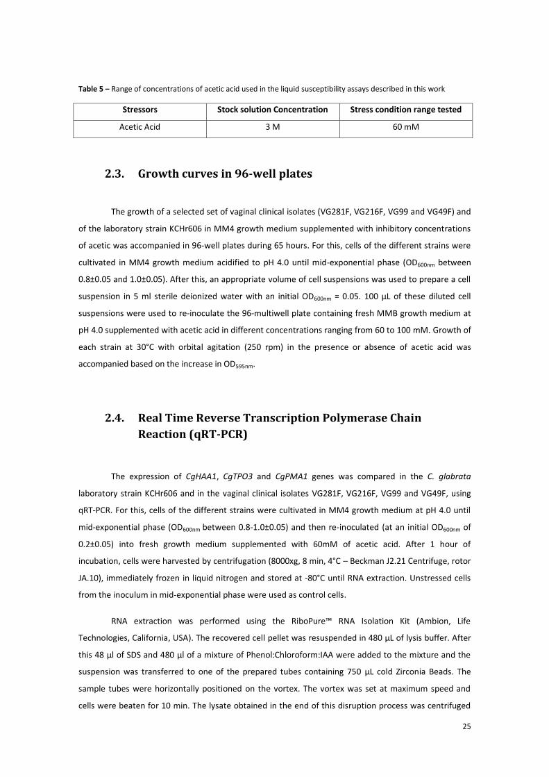
Table 5 – Range of concentrations of acetic acid used in the liquid susceptibility assays described in this work
Stressors Stock solution Concentration Stress condition range tested
Acetic Acid 3 M 60 mM
2.3. Growth curves in 96-well plates
The growth of a selected set of vaginal clinical isolates (VG281F, VG216F, VG99 and VG49F) and
of the laboratory strain KCHr606 in MM4 growth medium supplemented with inhibitory concentrations
of acetic was accompanied in 96-well plates during 65 hours. For this, cells of the different strains were
cultivated in MM4 growth medium acidified to pH 4.0 until mid-exponential phase (OD600nm between
0.8±0.05 and 1.0±0.05). After this, an appropriate volume of cell suspensions was used to prepare a cell
suspension in 5 ml sterile deionized water with an initial OD600nm = 0.05. 100 μL of these diluted cell
suspensions were used to re-inoculate the 96-multiwell plate containing fresh MMB growth medium at
pH 4.0 supplemented with acetic acid in different concentrations ranging from 60 to 100 mM. Growth of
each strain at 30°C with orbital agitation (250 rpm) in the presence or absence of acetic acid was
accompanied based on the increase in OD595nm.
2.4. Real Time Reverse Transcription Polymerase Chain
Reaction (qRT-PCR)
The expression of CgHAA1, CgTPO3 and CgPMA1 genes was compared in the C. glabrata
laboratory strain KCHr606 and in the vaginal clinical isolates VG281F, VG216F, VG99 and VG49F, using
qRT-PCR. For this, cells of the different strains were cultivated in MM4 growth medium at pH 4.0 until
mid-exponential phase (OD600nm between 0.8-1.0±0.05) and then re-inoculated (at an initial OD600nm of
0.2±0.05) into fresh growth medium supplemented with 60mM of acetic acid. After 1 hour of
incubation, cells were harvested by centrifugation (8000xg, 8 min, 4°C – Beckman J2.21 Centrifuge, rotor
JA.10), immediately frozen in liquid nitrogen and stored at -80°C until RNA extraction. Unstressed cells
from the inoculum in mid-exponential phase were used as control cells.
RNA extraction was performed using the RiboPure™ RNA Isolation Kit (Ambion, Life
Technologies, California, USA). The recovered cell pellet was resuspended in 480 μL of lysis buffer. After
this 48 μl of SDS and 480 μl of a mixture of Phenol:Chloroform:IAA were added to the mixture and the
suspension was transferred to one of the prepared tubes containing 750 μL cold Zirconia Beads. The
sample tubes were horizontally positioned on the vortex. The vortex was set at maximum speed and
cells were beaten for 10 min. The lysate obtained in the end of this disruption process was centrifuged

26
for 5 min at 16,100 x g at room temperature to separate the aqueous phase, containing the RNA, from
the organic phase. The aqueous phase was collected and added to 1.90 ml of Binding Buffer and 1.25 mL
of cold 100% Ethanol. The total volume was centrifuged through a filter cartridge and washed with 700
μL of Wash Solution 1. After new centrifugation the filter was washed two times with 500 μL of Wash
Solution 2/3 followed by an extra minute to ensure the complete removal of wash solution. Total RNA
obtained was eluted in two times 50 μL of Elution Solution, previously heated at 95°C. The isolated RNA
was treated with DNase I to remove traces of chromosomal DNA. For this, 100μl RNA sample (50μl +
50μl) was added to 8U of DNase I and 10 μl of 10X DNase I Buffer. The mixture was incubated at 37°C
during 30 minutes. After this incubation period 10 μl of DNase Inactivation Reagent were added to the
mixture, which was then vortexed and left for 5 minutes at room temperature. The purified RNA (in the
supernatant fraction) was collected into a fresh tube by centrifugation (>10000 rpm, 3 min).
Conversion of total RNA into cDNA was performed in a mixture of 10 μl using 500 g RNA, 2.2
μl MgCl2 25mM, 1.0 μl Buffer 10x, 2.0 μl dNTPs 2.5mM, 0.5 μl Random Hexamers, 0.2 μl RNAse
inhibitor, 1.85 μl ddH20 and 0.25 μl reverse transcriptase. This step was performed in a C1000 Thermal
Cycler (Bio-Rad, Hercules, USA) with the following PCR conditions: 10 min at 25°C, 30 min at 48°C, 5min
at 95°C. The cDNA obtained was diluted 1:2 using deionized water and the transcript levels of the
selected genes were compared using a set of primers that were specifically designed for this purpose
(primers sequences available in Table 6). The expression of a constitutive gene, CgACT1, was used as an
internal control. For the quantification of gene transcript levels 2.5 μl of the total cDNA produced from
each sample was used. 12.5 μl MasterMix (SYBR® Select Master Mix, Applied Biosystems), 2.5 μl Primer
Forward (4pmol/ml), 2.5 μl Primer Reverse (4pmol/ml) and 5 μl deionized water were added to the
cDNA and this mixture was subjected to a 40 cycles of PCR amplification using the following setup: 2 min
at 50°C, 10 min at 95°C, 15 seconds at 95°C, 1 min at 60°C. The primers used for the amplification of the
genes tested were designed using Primer Express® Software (Applied Biosystems).
Table 6 – Primer sequences used to perform qRT-PCR
Primer Identification Primer Sequence
CgACT1 primer forward 5’-AGA GCC GTC TTC CCT TCC AT-3’
CgACT1 primer reverse 5’-TTG ACC CAT ACC GAC CAT GA-3’
CgHAA1 primer forward 5’-GCC GGA CAT AAA CGG AAT AGG-3’
CgHAA1 primer reverse 5’-AGG CCA GTC TTG AGC TGT TAA TG-3’
CgTPO2/3 primer forward 5’-GCC GAT ATG TTC CCA AGT GAA-3’
CgTPO2/3 primer reverse 5’-TGG AGC GAA AGC GAA GAA AG -3’
CgPMA1 primer forward 5’-CAC CTC AGG ACG TCT ACG AAG A -3’
CgPMA1 primer reverse 5’-TCG ATC AAG GCG TCG ATG T -3’

27
2.5. [1-14C]-Acetic acid accumulation assays
The accumulation ratio of [1-14C]-acetic acid (defined as the ratio between the intracellular and
extracellular concentrations of radiolabelled acetic acid) was compared in the laboratory strain KCHr606
and in the clinical isolates VG281F, VG216F, VG99 and VG49F. Cells of the different strains were
cultivated in MM4 growth medium (at pH 4.0) at 30°C with orbital agitation (250 rpm) until mid-
exponential phase (OD600nm = 0.8 ± 0.05), harvested by filtration, washed one time with fresh medium
and finally resuspended in 5 ml of this same medium to obtain a dense cell suspension (OD600nm = 0.7±
0.05). The cell suspensions were incubated for 5 minutes at 30°C in a water bath with orbital agitation
(150 rev/min) to allow cells to recover from the harvesting process. After this time, 21.12 μM of labeled
[1-14C]-acetic acid were added to the cell suspension as well as 60 mM of cold acetic acid. Culture
samples were taken after 1, 5, 10, 15, 20, 25 and 30 minutes of incubation in the presence of the labeled
[1-14C]-acetic acid. In order to measure extracellular [1-14C]-acetic acid, a 100 μl culture sample was
collected and the supernatant was recovered by centrifugation in a tabletop centrifuge (12000 rpm, 1
minute). For quantification of intracellular [1-14C]-acetic acid, a 200 μl culture sample was filtered
through pre-wetted glass microfiber filters (Whatman GF/C) and washed with cold water. Both
supernatant and filter were added to 7 ml of scintillation liquid (Beckman) and their radioactivity was
measured in a Beckman LS 5000TD scintillation counter. Internal volume of C. glabrata was considered
equal to 2.5 μl (Rosa and Sa-Correia 1996).
.
2.6. Quantification of consumption of acetic acid and glucose
The consumption of acetic acid and glucose during growth was compared in the laboratory
strain KCHr606 and in the vaginal isolates VG216F, VG99, VG49F, VG281F. For this, strains were
cultivated in liquid MM4 growth media either or not supplemented with 60 mM acetic acid (at pH 4.0).
Cells were cultivated at 30°C with orbital agitation (250 rpm) and growth was followed by accompanying
the increase in OD600nm of the cultures. Samples of culture supernatants were harvested by
centrifugation and used for the quantification of acetic acid and glucose concentrations by HPLC.
Cultures supernatants were analyzed on an Aminex HPX-87H column, eluted at room temperature with
0.005 M H2SO4 at a flow-rate of 0.6 ml/min during 30 minutes, using a refractive-index (RI) (for glucose
quantification) and a UV (for acetic acid quantification) detector. Under the conditions used glucose and
acetic acid had retention times of 9.2 and 14.3 minutes, respectively. Reproducibility and linearity of the
method were tested and concentrations were estimated based on appropriate calibration curves.

28
2.7. In vivo estimation of C. glabrata PM-H+-ATPase (CgPMA1,
ORF CAGL0A00495g) activity
The in vivo activity of C. glabrata PM-H+-ATPase was estimated based on the ability of cells to
acidify the extracellular medium, an experimental methodology that was largely used to assess activity
of PM-H+-ATPase in S. cerevisiae (Carmelo et al. 1996, Mira, Becker et al. 2010a) and in C. glabrata
(Bairwa and Kaur 2011). The proton pumping activity was compared in the laboratory strain C. glabrata
KCHr606 and in the vaginal clinical isolates VG216F, VG99 and VG49F and VG281F. For this cells of the
different strains were cultivated until mid-exponential phase in MM4 growth medium (at pH 4.0),
harvested by filtration, washed twice with distilled water and incubated at 30°C in sorbitol solution
(20g/L, pH4) for 30 minutes to deactivate the plasma membrane PM-H+-ATPase. After this time, cells
were filtrated, washed with water (at pH 4) to remove any sorbitol residue and finally resuspended in
distilled water (at pH 4) to obtain a dense cell suspension (OD600nm ~ 20.0 ± 2.0). Each assessment of C.
glabrata PM-H+-ATPase activity was conducted in a water-jacketed cell (capacity 5 ml), at 30°C, with
agitation, by adding 3.0 ml of water (at pH 4.0), 1 ml of cell suspension and, when required, 0.4, 0.8 or
1.2 mM acetic acid. The stock solution of acetic acid used (3M) was adjusted to pH 4.0 using HCl as the
acidulant. After mixing, pH of the suspension was rapidly adjusted to 4.0±0.1 using HCl or NaOH.
Activation of C. glabrata PM-H+-ATPase in each assay was initiated by the addition of 1 ml of glucose
100g/L (at pH 4) to the mixture (yielding a final glucose concentration in the reaction mixture of 2%). pH
of the mixture was measured every 10 seconds by potentiometry using a pH microelectrode
(6.0202.100, Metrohm) attached to a Metrohm 691 pH meter.
2.8. β-1,3-glucanase susceptibility assay
Susceptibility of the laboratory strain KCHr606 and of the vaginal clinical isolates VG216F, VG99,
VG49F and VG281F to lyticase (β-1,3-glucanase, Sigma) was performed according to protocols found to
be successful in S. cerevisiae (Simões et al. 2003). Cells from all strains were grown in MM4 minimal
medium (at pH 4.0), harvested at the exponential growth phase, until a culture OD600nm of 0.8 ± 0.1 was
reached. The harvested cells were washed with distilled water and ressuspended in 0.1 mM sodium
phosphate buffer (pH 7.0). After the addition of 10µg/ml lyticase from Arthrobacter luteus (Sigma), cell
lysis was monitored by measuring the percent decrease of the initial OD600nm of the cell suspensions
every 20 minutes for a total period of 3 hours.

29
Figure 4 - Susceptibility of the single mutant ΔCgMsn2 and of the double mutant ΔCgMsn2ΔCgMsn4 to inhibitory
concentrations of acetic acid (25 and 30 mM, at pH 4.5) in comparison with the parental strain Htu. The mutant
ΔCgHaa1 and respective parental strain KCHr606 were also compared under the same conditions. Cells used to
prepare the spots were cultivated in unsupplemented MM4 (except in the cases requiring uracil supplementation)
liquid medium until mid-exponential phase at standardized OD600nm of 0.8±0.05 and then diluted in deionized water
to an OD600nm of 0.05±0.005 (lane a). Lanes (b) and (c) are 1:5 and 1:25 dilutions of (a), respectively. The results
obtained were representative of, at least, two independent experiments.
3. Results
The transcription factors CgMsn2 and CgMsn4 are not required for C. glabrata
tolerance to acetic acid
CgMSN2 and CgMSN4 have been described as not being involved in mediating C. glabrata
response to propionic and sorbic acids (Roetzer, Gregori et al. 2008) however their role against acetic
acid had not been previously examined. Given this, it was compared the susceptibility of the single
mutant ΔCgMsn2 and of the double mutant ΔCgMsn2ΔCgMsn4 to inhibitory concentrations of acetic
acid (at pH 4.5) with the susceptibility of the parental strain Htu and with the susceptibility of a deletion
mutant devoid of the CgHAA1 gene. The results obtained showed no significant differences between
growth of the parental strain Htu and of the two tested deletion mutants when cultivated in solid MM4
growth medium (at pH 4.5) supplemented with 25 and 30 mM acetic acid (see Figure 4). Although
growth of the double deletion mutant in the presence of acetic acid seems to be slightly below the one
registered for the parental strain, this difference is also observed in control conditions probably
reflecting a reduced fitness of this strain. As demonstrated before, the mutant devoid of CgHaa1
expression is highly susceptible to the tested concentrations of acetic acid. The absence of a significant
protective effect exerted by CgMsn2 and CgMsn4 against acetic acid was also confirmed in liquid growth
medium (results not shown). Altogether these results suggest that CgHaa1 is a major player in mediating
C. glabrata tolerance and response to acetic acid stress.

30
Effect of the expression of CgHaa1-target genes in C. glabrata tolerance to
acetic acid-induced stress
In this work it was examined the protective effect against acetic acid exerted by a set of genes
regulated by CgHaa1 which had not previously examined by Bernardo (2013b). In specific it was
assessed the involvement in tolerance to acetic acid of CgFps1 and CgFps2, two orthologues of the S.
cerevisiae Fps1 aquoglyceroporin; of CgHrk1, implicated in activation of the plasma membrane H+-
ATPase Pma1p; of CgSut2, involved in the regulation of sterol uptake under anaerobic conditions; of
CgRsb1, a suppressor of sphingoid long chain base sensitivity; of CgCmr3, a putative zinc finger protein
with unknown function; of CgPpz1, involved in regulation of potassium transport; of CgPep1, a type I
transmembrane sorting receptor for multiple vacuolar hydrolases; and of ORF CgCAGL0E03740g,
encoding putative protein of unknown function. The selection of these genes was based on their
significant up-regulation by CgHaa1 under acetic acid stress (see Table 1) and/or on previous indicatives
obtained in S. cerevisiae suggesting their involvement in tolerance to this weak acid. Out of the genes
tested only CgCMR3, CgPPZ1, ORF CAGL0E03740g, CgHRK1 and CgPEP1 were found to increase C.
glabrata tolerance to acetic acid, although this protective effect was below the one registered for
CgHAA1 (see Figure 5 and Figure 6). The protective effect exerted by CgCMR3, CgPPZ1, ORF
CAGL0E03740g was also confirmed in liquid growth medium. Interestingly, the individual elimination of
the two copies of the CgFPS1 and CgFPS2 genes had no significant effect in tolerance of C. glabrata to
acetic acid (see Figure 5). This result suggests that these Fps aquaporins might not be involved in
mediating the entry of undissociated acetic acid into the C. glabrata cytosol, different from the function
described for its S. cerevisiae counter-partner (Mollapour and Piper 2007).
.

31
Figure 6 - A: Growth curves of the parental strain KCHr606 and of the deletion mutants ΔCgSUT2, ΔCgRSB1,
ΔCgCMR3, ΔCgPPZ1 and ΔCAGL0E03740g in MM4 growth medium at pH 4.0 (left) or in acetic acid (60 mM)
supplemented MM4 growth (right); B: Growth of the deletion mutants ΔCgSUT2, ΔCgRSB1, ΔCgCMR3, ΔCgPPZ1 and
ΔCAGL0E03740g and laboratory and parental strain KCHr606 at 40, 50, 60 and 80 mM of acetic acid. Cells used to
prepare the spots were cultivated in unsupplemented MM4 (except in the cases requiring uracil supplementation)
liquid medium until mid-exponential phase at standardized OD600nm of 0.8±0.05 and then diluted in deionized water
to an OD600nm of 0.05±0.005 (lane a). Lanes (b) and (c) are 1:5 and 1:25 dilutions of (a), respectively. The results
obtained were representative of, at least, two independent experiments.
Figure 5 - A: Growth curves of the parental strain KCHr606 and of the deletion mutants ΔCgHAA1, ΔCgFPS1 and
ΔCgFPS2 in MM4 growth medium at pH 4.0 (left) or in acetic acid (60 mM) supplemented MM4 growth (right).; B:
Growth of the deletion mutants ΔCgHAA1, ΔCgFPS1, ΔCgFPS2 and laboratory and parental strain KCHr606 at 40,
50 and 60 mM of acetic acid. Cells used to prepare the spots were cultivated in unsupplemented MM4 (except in
the cases requiring uracil supplementation) liquid medium until mid-exponential phase at standardized OD600nm
of 0.8±0.05 and then diluted in deionized water to an OD600nm of 0.05±0.005 (lane a). Lanes (b) and (c) are 1:5 and
1:25 dilutions of (a), respectively. The results obtained were representative of, at least, two independent
experiments.

32
Elimination of CgHRK1 and of CgPEP1 from C. glabrata genome was not successful indicating
that these genes might be essential for growth of these yeast species or that they are somehow involved
in homologous recombination which was the method used to promote gene elimination. In order to
reduce the expression of these two genes their natural promoter was replaced by a Tet-promoter which
is repressible upon supplementation of the growth medium with 20 mg/l of tetracycline (see Figure 7).
In the absence of acetic acid the reduced expression of the CgHRK1 gene slightly reduced the growth
rate while no significant effect was registered upon silencing of the CgPEP1 gene. The reduced
expression of CgHRK1 or CgPEP1 significantly increased C. glabrata susceptibility to acetic acid
suggesting that these genes are required for maximal tolerance to this weak acid.
C. glabrata clinical isolates recovered from the vaginal tract are highly
tolerant to acetic acid, compared to laboratory strains or clinical isolates
recovered from other infection sites
As said in the Introduction section acetic acid is found both in the GI and in the GU tracts. The
pH of these two sites can be below the pKa of acetic acid (pKa of 4.76), the vaginal pH being estimated
to vary from 3.6-4.5, in healthy women (Boskey, Telsch et al. 1999) and the intestinal pH ranging from
5.7 in the caecum and 7.4 in the ileum (Fallingborg 1999). For this reason tolerance to acetic acid of a
cohort of C. glabrata’s clinical isolates collected from the GU and GI tracts was assessed at two different
pHs: pH 4.5, to mimic the acidic environment of the vaginal tract and pH 6.4, to characterize the near-
neutral environment of the GI tract. Tolerance of the isolates to acetic acid was assessed using spot
Figure 7 - Growth curves of the parental strain KCHr606 and of the deletion mutants ΔCgHRK1 and ΔCgPEP1 in
tetracycline supplemented MM4 growth medium at pH 4.0 (left) or in tetracycline supplemented MM4 growth
medium at pH 4.0 supplemented with 60 mM of acetic acid (right). Cells used in this assay were grown in
unsupplemented MM4 liquid medium until mid-exponential phase at the standardized OD600nm of 0.8±0.05. The
results obtained were representative of, at least, two independent experiments.

33
assays in solid MM4 growth medium (adjusted at the indicated pH) as described in materials and
methods section. In Figure 8 it is shown one example of the results obtained (results corresponding to
growth of vaginal clinical isolates in solid MM4 supplemented with 80 mM of acetic acid, at pH 4.5).
Figure 8 - Example of the screnning of clinical isolates recovered from the GU tract, in this case to tolerance to
acetic acid (80 mM) at pH 4.5. Cells used to prepare the spots were cultivated in unsupplemented MM4 liquid
medium until mid-exponential phase at standardized OD600nm of 0.8±0.05 and then diluted in deionized water to an
OD600nm of 0.05±0.005 (lane a). Lanes (b) and (c) are 1:5 and 1:25 dilutions of (a), respectively. The results obtained
were representative of, at least, two independent experiments.
The cell density in each of the spots was quantified and the overall data was gathered in a heat-
map that is represented in Figure 9. The results obtained show that despite the inter-strain variability
observed, the vaginal isolates are significantly more tolerant to acetic acid than the isolates recovered
from the GI tract or the laboratory strains KCHr606 or CBS 138 which also have an intestinal origin (see
Figure 9). This observation probably reflects the development of adaptive responses by vaginal isolates
to survive in the acidic vaginal tract where most acetic acid will be present in the undissociated form. As
expected, the deleterious effect of acetic acid is much less pronounced at pH 6.4 than at pH 4.5, this
being visible for both sets of isolates (see Figure 9). In the absence of acetic acid it was visible some
differences in growth fitness of the different isolates but isolates more tolerant to acetic acid were not
those exhibiting higher fitness in control conditions. This is for example the case of isolates VG216F and
VG318F at pH 4.5 and VG242F and FFUL98 at pH 6.4. Interestingly the reference strain CBS138,
recovered from the GI tract, is less susceptible to acetic acid-induced stress than the remaining group of
GI-derived clinical isolates at pH 6.4.

34
pH 4.5 pH 6.4
Figure 9 – Screening of C. glabrata clinical isolates recovered from vaginal and GI tracts to acetic acid tolerance at different pH values. Growth of vaginal clinical isolates, laboratory strain
KCHr606 and reference strain CBS138 at different concentrations of acetic acid (40, 50, 60 and 80 mM): A – at pH 4.5; and B – at pH 6.4. Growth of GI clinical isolates, laboratory strain
KCHr606 and reference strain CBS138 at different concentrations of acetic acid (40, 50, 60 and 80 mM): C – at pH 4.5; and D – at pH 6.4. Cells used to prepare the spots were cultivated in
unsupplemented MM4 liquid medium until mid-exponential phase at standardized OD600nm of 0.5±0.005 and then diluted in deionized water to an OD600nm of 0.05±0.005. Cell density of
each spot was measured using the software ImageJ and for the heat-map construction the cell density values were rearrange in colors by intensity through the programming software R.

35
To obtain more quantitative information regarding the effect of acetic acid in growth kinetic
parameters of the vaginal isolates the growth curves of the different isolates VG281F (+), VG216F (++),
VG99 (++) and VG49F (++) and of the laboratory strains KCHr606 (-) and CBS138 (-) was compared in
liquid growth medium. According to Figure 10, it is clear the high acetic acid tolerance of the vaginal
isolates VG216F, VG99 and VG49F, which exhibit short lag phases mainly when cultivated in the
presence of 60 mM of acetic acid (at pH 4). Clinical isolate VG49F (++) seems to be particularly tolerant
showing the lower lag phase at 80 mM of acetic acid (at pH4) also exhibiting the highest growth rate
among the cohort of isolates tested. Altogether, this analysis allows the confirmation of the results
obtained in the heat-map (see Figure 10), being possible to identify a cohort of clinical isolates exhibiting
a moderate (VG281F) and high (VG216F, VG99 and VG49F) tolerance to acetic acid in comparison with
the susceptible strains KCHr606 and CBS138. It is also possible to observe that the highest degree of
differences between the susceptible laboratory strains and the tolerant vaginal isolates is observed at
the level of the lag phase since the growth rates of the different isolates when growing in the presence
of acetic acid is not significantly different. In order to facilitate the identification of these different
clinical isolates accordingly to their acetic acid tolerance, they will be further designated as moderately
(+) or highly tolerant (++), always in comparison with the susceptible (-) strain KCHr606.
Figure 10 - Comparison of the growth curves of laboratory strains KCHr606 and CBS138 and vaginal clinical isolates
VG281F, VG216F, VG99 and VG49F in absence (black marks) and in the presence of 60 mM (dark grey marks), 80
mM (light grey marks) and 100 mM (light blue marks) of acetic acid. Cells were cultivated in unsupplemented MM4
liquid medium until mid-exponential phase at standardized OD600nm of 0.8±0.005 and then re-inoculated into a 96-
well plate with MM4 supplemented with the different concentrations of acetic acid. The results obtained were
representative of, at least, two independent experiments.

36
Increased tolerance to acetic acid of vaginal C. glabrata clinical isolates does
not correlate with a generalized stress resilience
In the vaginal tract C. glabrata cells have to cope with other environmental challenges apart
from the presence of acetic acid. Given this it was examined whether higher tolerance of vaginal isolates
to acetic acid correlates with a generalized resistance to stress. Two environmental stresses not related
to weak acid-induced stress that may challenge C. glabrata in the vaginal tract were examined: exposure
to H2O2 and heat stress. Overall the clinical isolates showed different levels of tolerance against the
different stressors, being that behavior specific for each clinical isolate (see Figure 12). Nevertheless, the
clinical isolates previously identified as being highly tolerant to acetic acid stress (e.g. VG216F, VG262F,
VG318F, VG49F – see Figure 9) were not those found to be tolerant isolates to the other stresses (see
Figure 12). For example the isolate exhibiting the highest tolerance against H2O2-stress was VG281F
which was only moderately susceptible to acetic acid (see Figure 9). This observation indicates that the
increased tolerance to acetic acid of vaginal clinical isolate does not seem to result from an increased
activity of a general stress-response mechanism but it rather seems to result from the development of
specific adaptive responses.
Figure 11 - Variation of the lag phase in hours (left) and of the specific growth rates in hour-1
(right) of the
laboratory strains KCHr606 (-) and CBS138 (-) and vaginal clinical isolates VG281F (+), VG216F (++), VG99 (++)
and VG49F (++). Control (black bars), 60 mM acetic acid (dark grey bars); 80 mM acetic acid (light grey bars),
100 mM acetic acid (light blue marks). (*) No growth of the strain was observed. The results obtained were
representative of, at least, two independent experiments.

37
Tolerance of vaginal C. glabrata clinical isolates to acetic acid correlates with
tolerance to other short-chain fatty acids and to lactic acid
To see if tolerance to acetic acid of vaginal clinical isolates was also observed in response to
other organic acids the cells were subjected to butyric (pKa=4.82) and propionic acids (pka=4.88) which,
like acetic acid, are also short-chain fatty acids (see Figure 13). The assays were performed in solid MM4
growth media at pH 4.5 which mimics the acidic vaginal pH. In this assay we have tried to use toxic
concentrations of the different weak acids (that is, concentrations that induced similar lag phases in the
laboratory strain KCHr606). The results obtained show that vaginal isolates are also tolerant to propionic
and butyric acids thus indicating that the cells may have triggered adaptive responses that turn them
more tolerant to short-chain fatty acids in general. It is interesting to observe that isolates found to be
more tolerant to acetic acid (for example VG79C or VG262) were not necessarily those exhibiting higher
fitness when cultivated in the presence of propionic and butyric acids. Since lactic acid is another
carboxylic acid that is found in the vaginal tract tolerance of the vaginal isolates to this weak acid was
also compared. The pKa of lactic acid (pka=3.86) is much lower than the one of the other SCFA tested and
therefore at pH 4.5 this acid exerts a much lower inhibitory effect against all the vaginal isolates.
Interestingly, isolates VG79C and VG95 which were only moderately tolerant to acetic acid exhibited
higher tolerance to lactic acid (Fig.9).
Figure 12 - Screening of C. glabrata clinical isolates recovered from GU tract to environmental stress tolerance
at pH 4.5. Growth of vaginal clinical isolates, laboratory strain KCHr606 (-) and reference strain CBS138 (-) at
different temperatures (30ᴼC, 37ᴼC and 42ᴼC) and at different concentrations of H2O2 (5 and 10 mM). Cells
used to prepare the spots were cultivated in unsupplemented MM4 liquid medium until mid-exponential phase
at standardized OD600nm of 0.8±0.05 and then diluted in deionized water to an OD600nm of 0.05±0.005. Cell
density of each spot was measured using the software ImageJ and for the heat-map construction the cell
density values were rearrange in colors by intensity through the programming software R.

38
Effect of the expression of CgHaa1 and of CgHaa1-regulated genes in the
tolerance to acetic acid exhibited by vaginal clinical isolates
Considering the previously demonstrated role of the CgHaa1-regulon in mediating response
and tolerance of C. glabrata laboratory strains to acetic acid it was examined if the higher tolerance to
acetic acid of the vaginal clinical isolates VG216F (++), VG99 (++) and VG49F (++) could be attributed to a
higher expression/activity of CgHaa1. For this the expression of the CgHAA1 and of the CgHaa1-
regulated genes CgTPO3 and CgPMA1 was compared in the different isolates in the presence or absence
of acetic acid (see Figure 14). The selection of these genes among those found to be regulated by
CgHaa1 is based on their demonstrated involvement in C. glabrata tolerance to acetic acid (Bernardo
2013b). The laboratory strain KCHr606 (-) was used as standard susceptible strain and VG281F (+) was
used as an example of an isolate with an intermediate tolerance.
Figure 13 - Screening of C. glabrata clinical isolates recovered from GU tract to weak acid stress at pH
4.5. Growth of vaginal clinical isolates, laboratory strain KCHr606 (-) and reference strain CBS138 (-)
at different concentrations of lactic acid (30 and 50 mM), butyric acid (8 and 10 mM) and propionic
acid (15 and 17 mM). Control represent cells grown in unsupplemented MM4 at 30ᵒC. Cells used to
prepare the spots were cultivated in unsupplemented MM4 liquid medium until mid-exponential
phase at standardized OD600nm of 0.8±0.05 and then diluted in deionized water to an OD600nm of
0.05±0.005. Cell density of each spot was measured using the software ImageJ and for the heat-map
construction the cell density values were rearrange in colors by intensity through the programming
software R.

39
Figure 14 - Comparison by real time RT-PCR of the transcript levels of CgHAA1, CgTPO3 and CgPMA1 genes in C.
glabrata laboratory strain KCHr606 (-) and in vaginal clinical isolates showing highly (VG216F, VG99 and VG49F)
and moderately (VG281F) tolerance to acetic acid under acetic acid-induced stress. Levels of mRNA of those
genes were compared in all populations in exponential cells (dark bars) or after 1 hour of cultivation in MM4
growth media (at pH 4.0) supplemented with 60 mM of acetic acid (white bars). The values of the transcript
levels were normalized using as internal control the levels of CgACT1 mRNA and the values presented are
relative to those registered in unstressed KCHr606 cells which was considered to be equal to 1. The graphics on
right show the distribution of gene expression in the population. The results obtained were representative of, at
least, two independent biological replicates.
In the absence of acetic acid the basal expression of CgHAA1 gene is similar in all the tested
isolates with exception of the clinical isolate VG281F (+) which exhibited almost three times more mRNA
than the laboratory strain KCHr606 (see Figure 14). Also, no significant differences were observed in the
expression of the CgTPO3 or of CgPMA1 genes (Fig.14). These results indicate that the higher tolerance
to acetic acid of vaginal isolates VG49F, VG99 and VG216F cannot be explained by a higher basal level of
expression of CgHAA1, CgPMA1 and CgTPO3 which would turn these cells a priori more prone to
tolerate acetic acid. After one hour of exposure to acetic acid a strong up-regulation of CgHAA1 gene
was registered in the more tolerant isolates reaching mRNA levels that are about 4-fold higher than

40
those registered for the more susceptible strain KCHr606. The moderately tolerant isolate VG281F also
exhibited slightly higher values of CgHAA1 mRNA (around 2.5-fold). Surprisingly, the increase in CgHAA1
mRNA did not led to an increase in CgTPO3 and CgPMA1 mRNAs indicate that in the tested vaginal the
effect of CgHAA1 over CgTPO3 and CgPMA1 transcription is overcome by a repressor or that in these
isolates the regulatory connection between CgHaa1 and these two target genes is lost. On the overall
the results obtained indicate that only the expression of CgHAA1 correlates with increased tolerance to
acetic acid.
Acetic acid-tolerant vaginal C. glabrata clinical isolates accumulate less
radiolabelled acetic acid, compared to susceptible strains
The reduction of the internal accumulation of acetic acid is one of the mechanisms contributing
for increased tolerance to this weak acid in yeasts (Tenreiro, Rosa et al. 2000, Tenreiro, Nunes et al.
2002, Fernandes, Mira et al. 2005). Given this the levels of accumulation of acetic acid in the vaginal
isolates VG281F (+), VG216F (++), VG99 (++) and VG49F (++) and in the susceptible strain KCHr606 strain
were compared. The assay was performed using unstressed cells of the different strains that were
suddenly exposed to a cold concentration of acetic acid (60 mM) together with 21.12 μM radiolabelled
acetic acid. The accumulation of the radiolabelled acid (taken as the ratio between the extracellular and
intracellular concentrations) was followed during the first 30 minutes of incubation in the presence of
the acid (see Figure 15). The results clearly show a reduced accumulation of radiolabelled acetic acid
inside the vaginal isolates this being more prominent for isolate VG216F and VG99 (Fig.15). In S.
cerevisiae it was demonstrated that Haa1p, and its target regulated gene Tpo3p, contribute to reduce
the internal accumulation of radiolabelled acetic acid when cells are cultivated in the presence of
inhibitory concentration of cold acetic acid (Fernandes, Mira et al. 2005). The results obtained by
Bernardo (2013b) showed that CgTpo3 is also required to reduce the internal accumulation of acetic
acid in C. glabrata under acetic acid stress, although in this case the effect exerted by CgHaa1 was
marginal. No correlation was registered between the expression of CgTpo3 and the levels of acetic acid
accumulated inside the different clinical isolates indicating that the reason why these isolates
accumulate less acid is independent of the expression of this drug efflux pump (see Figure 14 and Figure
15).

41
The structure of cell envelop of acetic acid-tolerant vaginal C. glabrata clinical
isolates seems different from the one of tolerant isolates, based on their
different resistances to lyticase activity
The fact that after only 5 minutes of incubation the VG281F (+), VG216F (++) and VG99 (++)
clinical isolates already accumulate a lower concentration of radiolabelled acetic acid (see Figure 15)
Figure 15 – A: Time-course representation of the accumulation ratio (A) of [1-14
C]-acetic acid in C. glabrata moderately
(VG281F (+)) and highly (VG216F (++), VG99 (++) and VG49F (++)) acetic acid tolerant vaginal clinical isolates,
represented by (□), in comparison with the susceptible laboratory strain KCHr606 (-), represented by (•), during
cultivation in MM4 (at pH 4.0) supplemented with 60 mM of cold acetic acid. The accumulation values shown are
means of 3 or more independent assays. B: Accumulation levels of each GU clinical isolates and respective acetic acid-
induced expression of CgTPO3. The values of accumulation levels are relative to those registered in KCHr606 cells
which were considered to be equal to 1. The results obtained were representative of, at least, three independent
experiments.

42
Figure 16 - Comparison of the susceptibilities to lyticase of C. glabrata laboratory strain KCHr606 and
moderately (VG281F) and highly (VG216F, VG99 and VG49F) acetic acid tolerant vaginal clinical isolates’ cells.
Cells were cultivated in unsupplemented MM4 liquid medium until mid-exponential phase at standardized
OD600nm of 0.8±0.005 and then re-inoculated into 0.1 mM sodium phosphate buffer (pH 7.0) supplemented
with 10 µg/ml lyticase from Arthrobacter luteus. The growth was followed by accompanying the increase in
OD600nm of the cultures. The results obtained were representative of, at least, three independent experiments.
suggests that these isolates might have intrinsic characteristics that turn them less permeable to the
undissociated form of the acid. Modifications of the cell envelope occurring both at the level of plasma
membrane and cell wall composition have been described to influence the diffusion rate of
undissociated organic acids in S. cerevisiae thereby modulating tolerance (Simões, Mira et al. 2006, Ullah
et al. 2013a). Therefore the structure of the cell wall of the different vaginal isolates and of the
laboratory strain KCHr606 was compared by comparing the susceptibility of these cells to lyticase, a β-
1,3-glucanase. Susceptibility to lyticase has been used to get a rough comparison of the cell wall
structure in yeasts (e.g. Simões, Teixeira et al. 2003). The highly acetic acid tolerant vaginal clinical
isolates (VG99, VG49F and VG216F) show an increased resistance to lyticase activity since the after 4
hours of exposure to the acid the OD600nm of the culture was only reduced by 5% (see Figure 16). In
contrast the susceptible strain KCHr606 and the clinical isolate VG281F exhibit higher susceptibility to
lyticase activity showing a reduction in the OD600nm of the culture of around 20% (see Figure 16). The
different susceptibility of these isolates to lyticase suggest that the composition of their cell walls is
considerably different, at least to what concerns to the -1,3-glucan contents, which might underlie
their different permeability to acetic acid.

43
C. glabrata vaginal isolates and laboratory strains co-consume glucose and
acetic acid, the strains more tolerant to acetic acid exhibiting higher
consumption rates
In the absence of glucose C. glabrata cells are capable of assimilate acetate, pyruvate and,
preferentially, lactate (Ueno et al. 2011). This ability to assimilate other carbon sources may represent
an advantage during host infection due to the limited glucose availability in the infection sites. Tolerance
to acetic acid in yeast species of the Zygosaccharomyces bailii genus had been demonstrated to rely on
their ability to co-consume acetic acid in the presence of glucose (Guerreiro et al. 2012, Rodrigues et al.
2012). The repressive effect exerted by glucose on C. glabrata ability to metabolize other carbon
sources had not been thoroughly studied although previous results sustained the hypothesis that this
yeast could also mechanisms of glucose repression similar to those of S. cerevisiae (Petter and Kwon-
Chung 1996). In this context it was examined whether the vaginal clinical isolates and the laboratory
strain KCHr606 strains could consume acetic acid in the presence of glucose when cultivated in MM4
(which has 2% glucose) at pH 4.0. The levels of glucose and acetic acid present in the supernatant along
the growth curve were quantified by HPLC (see Figure 17). In the absence of acetic acid, glucose was
rapidly consumed by all the clinical isolates, being the consumption rate of the laboratory strain
KCHr606 slightly lower than the one exhibited by the vaginal isolates. It was also possible to observe
that all clinical isolates and the laboratory strain KCHr606 produced slight amounts of acetic acid,
presumably as a side-product of glucose fermentation to ethanol, also detected during HPLC
quantification (results not shown). Inoculation in the acetic acid-supplemented growth medium led to a
severe reduction in glucose consumption of the susceptible strains KCHr606 (-) and VG281F (+), while in
the more tolerant isolates VG99 (++) and VG49F (++) no significant effect was detected. In fact, a closer
inspection to the consumption graphs shows clearly that increased tolerance to acetic acid correlates
with a increased capacity to consume glucose (Figure 17). Notably, it is visible in all tested isolates the
co-consumption of glucose and acetic acid being evident that the more tolerant isolates start to
consume the acid as soon as they are inoculated in the growth medium (Figure 17). Despite this, the
results obtained indicate that the consumption of the acid does not seem to serve itself as a detoxifying
mechanism since for example strain KCHr606 starts to actively consume acetic acid after approximately
20 hours, a period at which the culture is already exponentially growing in the presence of the acid. This
is also observed for the intermediate tolerant isolate VG216F which starts consumption of acetic acid
much later than it begins to grow in the presence of the acid.

44
Figure 17 - Time-course representation of glucose and acetic acid external concentrations during cultivation of the laboratory strain KCHr606 (-) and of the moderately (VG281F) and highly (VG216F, VG99 and
VG49F) acetic acid tolerant vaginal clinical isolates during acetic acid-induced stress. Cells were cultivated in MM4 growth medium (at pH 4.0) until mid-exponential phase and then re-inoculated into MM4
growth medium either or not supplemented with 60 mM acetic acid. Samples of culture supernatants were harvested by centrifugation and used for the quantification of acetic acid and glucose concentrations
by HPLC. The variation of glucose and acetic acid can be compared with the growth curves of each clinical isolate in absence and presence of acetic acid (60 mM). Legend: Growth in control conditions ( );
Growth in the presence of acetic acid (60 mM) (Δ); Variation of glucose concentration (•); Variation of acetic acid concentration (□). The results obtained were representative of two independent experiments.

45
Vaginal isolates tolerant to acetic acid exhibit higher activity of the proton
pump CgPma1, in comparison with susceptible isolates
Considering the essential role of the PM-H+-ATPase in control of internal pH in this work it was
compared the activity of this protein in the susceptible laboratory strain KCHr606 and in the acetic acid
tolerant’ vaginal isolates (VG281, VG216F, VG99 and VG49F). The in vivo activity of CgPma1 was
estimated based on the ability of cells to acidify the extracellular medium in the presence of glucose, an
experimental methodology that was previously used with success in S. cerevisiae (Mira, Becker et al.
2010a) and in C. glabrata (Bairwa and Kaur 2011, Bernardo 2013b). The addition of glucose to the cell
suspensions of all the tested strains lead to an immediate activation of CgPma1 reflected in the
acidification of the medium, although it is clear that the vaginal isolates exhibited higher rates of
acidification, being the moderately acetic acid tolerant clinical isolate VG281F (+) and highly tolerant
VG49F (++) those exhibiting the higher acidification rates (see Figure 18- Control). The expression levels
of CgPMA1 gene in these isolates in the absence of acetic acid were identical (see Figure 14) indicating
that the differences found in the rates of extracellular acidification probably reflect a different activity of
the proton pump in the different isolates. Supplementation of cell suspensions with increasing
concentrations of acetic acid (0.4, 0.8 and 1.2 mM) led to the inhibition of the activity of the C. glabrata
proton pump, consistent with the permeabilization of the plasma membrane induced by the acid,
causing the dissipation of the plasma membrane potential required for maximal PM-H+-ATPase activity
(see Figure 18). At all concentrations tested the susceptible strain KCHr606 (-) was not able to acidify the
medium efficiently as clinical isolates whereas clinical isolate VG216F (++) proved to be the most
effective (particularly at 0.8 and 1.2 mM).

46
Figure 18 - External medium acidification promoted by CgPma1 H+-ATPase in laboratory strain KCHr606 and in moderately
(VG281F) and highly (VG216F, VG99 and VG49F) acetic acid tolerant vaginal clinical isolates before acetic acid-induced
stress. De-energized cell suspensions from mid-exponential phase of all clinical isolates were exposed to increased
concentrations of acetic acid (0, 0.4, 0.8 and 1.2 mM) and then energized with a pulse of glucose to determine the effect of
the acid in CgPma1 H+-ATPase activity. External acidification of the growth medium was taken as an in vivo measurement of
the enzyme activity. Results are means of several independent experiments. C. glabrata laboratory strain KCHr606 (grey
marks); moderately acetic acid tolerant clinical isolate VG281F (blue marks); highly acetic acid tolerant clinical isolates
VG216F (green marks), VG99 (purple marks) and VG49F (red marks).

47
4. Discussion
Previous work developed in our laboratory has demonstrated the involvement of the
transcription factor CgHaa1 (orthologue of S. cerevisiae transcription factor Haa1, encoded by the ORF
CAGL0L09339g) in conferring tolerance to acetic and lactic acids in C. glabrata (Bernardo 2013a). A
transcriptomic analysis revealed that a high number of CgHaa1-regulated genes is involved in stress
response, transport, cell wall maintenance, signaling, RNA synthesis, regulation of glycolysis and internal
pH homeostasis (Bernardo 2013b), being some of these features explored in this work. In S. cerevisae
response to acetic acid stress is controlled by the transcription factors Msn2/Msn4, Rim101 and Haa1. In
C. glabrata CgHaa1 emerges has the only transcription factor associated to the adaptive response
against acetic acid-induced stress, since CgWar1 was confirmed only to confer tolerance to sorbic and
propionic acids (Kren, Mamnun et al. 2003, Mundy and Cormack 2009) and our results show that
CgMsn2, CgMsn4 and CgRim101 are dispensable for tolerance to acetic acid (Figure 4 and results not
shown). This result is a very good example to show how stress responsive pathways in S. cerevisiae and
C. glabrata are orchestrated in different manners thus being crucial to study each species individually.
A screening of a new set of C. glabrata mutants allowed the identification of five new genes of
the CgHaa1 regulon that emerged as novel determinants of tolerance to acetic acid this being CgCMR3,
CgPPZ1, CgHRK1, CgPEP1 and the ORF CAGL0E03740 (see Figure 5 and Figure 6). CgCMR3 and ORF
CAGL0E03740g encode proteins of uncharacterized function and therefore it is not possible to evaluate
what could be their role in mediating C. glabrata response to acetic acid stress. Interestingly, CgCMR3
encodes a putative zinc finger protein indicating that this might be a novel player in the regulatory
network that governs C. glabrata response to acetic acid stress. CgPEP1 gene is an orthologue of PEP1
and CaPEP1 which encodes a type I transmembrane sorting receptor for multiple vacuolar hydrolases.
No role associated with acetic acid or other weak acid stresses has been designed for this gene in both S.
cerevisiae and C. albicans thereby not being easy to anticipate what could be the role played by this
gene in C. glabrata tolerance to acetic acid. Regarding CgPPZ1, the PPZ phosphatases had been
described to play similar but not identical roles in S. cerevisiae and in C. albicans. Both PPZ1 and CaPPZ1
are described to play a role in regulating sodium (Na+) and potassium (K+) transport thereby affecting
osmotic stability, intracellular pH, cell wall integrity and regulation of the membrane potential (Yenush
et al. 2002, Adam et al. 2012). CaPPZ1 contributes additionally to the regulation of hyphal formation
(Adam, Erdei et al. 2012). Ppz1 has been found to function as a repressor of the activity of the high-
affinity potassium import system mediated by the Trk system, playing no direct role in the activity of the
PM-H+-ATPase Pma1 (Yenush, Mulet et al. 2002). In strains lacking PPZ1 and PPZ2 it is observe an
increase in intracellular pH due to the enhanced uptake of K+ that leads to a higher efflux of H+ in order
to maintain the electrical balance (Yenush, Mulet et al. 2002). The alkalinization of cytosol ultimately
leads to a decrease in Pma1 activity. Having this in mind, the up-regulation of CgPPZ1 by CgHaa1 under

48
acetic acid stress may be determinant to maximize the activity of CgPma1 thereby counteracting the
dissipation of the electrochemical potential and the intracellular acidification caused by acetic acid
stress. Another gene uncovered in this work as contributing for maximal C. glabrata tolerance to acetic
acid is CgHRK1. This gene is an orthologue of S. cerevisiae’ HRK1, which encodes a protein kinase
involved in the activation of the plasma membrane H+-ATPase Pma1 in response to glucose metabolism
(Goossens et al. 2000) but that has a negligible role in the regulation of this proton pump in acetic acid
stressed yeast cells (Mira, Becker et al. 2010a). It remains to be evaluated whether CgHrk1 plays a role in
the control of CgPma1 activity under stress imposed by acetic acid. As described in Introduction section,
activation of Pma1 is important to offset the increased flux of protons during acetic acid stress. In C.
glabrata, CgPma1 had been already described to be positively regulated under acetic acid stress by the
yapsin CgYps1 (Bairwa and Kaur 2011), also a CgHaa1-target (Bernardo 2013b). The confirmation of
three additional CgHaa1 targets involved in positive regulation of CgPma1, CgPpz1, CgHrk1 and CgYps1,
emphasizes the relevance of CgHaa1 transcription in the regulation PM-H+-ATPase pump. Consistently,
elimination of CgHaa1 was found to reduce the activity of CgPma1 proton pump in response to acetic
acid stress (Bernardo 2013b).
In the second part of this work it was investigated the mechanisms of tolerance to acetic acid of
a cohort of C. glabrata’ isolates recovered from the vaginal tract, a niche where pathogenic Candida spp.
are expected to find acetic and also lactic acids at low pH. Under the conditions utilized for the
phenotypic screening it was possible to observe that vaginal clinical isolates are significantly more
tolerant to acetic acid than the isolates recovered from the GI tract or the laboratory strains CBS138 and
KCHr606 (also recovered from intestinal source), although some inter-strain variability has been
observed (see Figure 9). Among the cohort of vaginal isolates tested those found to be more tolerant to
acetic acid were not those more tolerant to oxidative or to heat stresses (and vice-versa) indicating that
the higher tolerance of these isolates does not correlate with a generalized stress resilience or increased
fitness. The existence of phenotypic diversity within the vaginal population studied is expected because
these isolates were recovered from different patients and might therefore have been subjected to
different selective pressures. Nevertheless, even within the same population of C. glabrata that
colonizes a single host it may be expected to observe phenotypic heterogeneity since this should
increase competitiveness by providing the colonizing population with the necessary versatility to survive
in case sudden modifications of the environment occur. The vaginal isolates VG49F, VG99 and VG216F
were those found to be more tolerant to acetic acid and this correlated with a reduced accumulation of
the acid registered inside these cells, when compared with the more susceptible strains KCHr606 and
the moderately tolerant isolate VG281F. This reduced accumulation of the acid inside VG99 and VG216F
isolates might result from an increased export of the acid anion or from a reduced entry of the
undissociated form. Until so far only two transporters had been found to mediate acetic acid tolerance
in C. glabrata: CgAqr1 and CgTpo3 (Bernardo 2013a, Costa, Henriques et al. 2013, Costa, Nunes et al.
2014). No differences were found in the expression of the CgTPO3 gene in the cohort of isolates tested
(Fig.14) but it remains to be established if the activity of this drug efflux pump is higher in the VG99 and

49
VG216F isolates. It also remains to be evaluated if these acetic acid-tolerant isolates exhibit different
levels of expression of the CgARQ1 gene. Notably, the cell wall structure of the VG99 and VG216F
isolates was much more resistant to lyticase (see Figure 16), compared to the tolerant isolates, a
difference that might lead to a reduced permeability of the cell envelope to the diffusion of
undissociated acetic acid. The only case where a correlation between low internal accumulation levels of
acid and high resistance to β-1,3-glucanase activity is not observed is in the clinical isolate VG49F (++)
(see Fig.16) which present both high acid accumulation and resistance to β-1,3-glucanase activity. This
may be an example of a clinical isolate that had evolved other mechanisms different from controlling
the internal accumulation of acid to cope with acetic acid stress. The acetic acid-tolerant VG49F, VG99
and VG216F vaginal isolates exhibited an increased activity of the CgPma1 proton pump, in comparison
with the activity that was registered in cells of the KCHr606 strain (see Figure 18), indicating that these
isolates are better equipped to avoid the intracellular acidification imposed by acetic acid stress. The
moderately tolerant isolate VG281F also exhibited higher activity of CgPma1, compared to KCHr606,
indicating that this might be a feature of adaptation to the vaginal environment, an hypothesis that will
require further testing using other vaginal isolates and isolates from non-acidic infection sites such as
the gut. The fact that the higher tolerance to acetic acid of vaginal isolates was correlated with their
ability to prevent entry of the undissociated acid form and with an increased control of internal pH
homeostasis is expected to provide them a competitive advantage when challenged with inhibitory
concentrations of other organic acids. Indeed, the VG49F, VG216F and VG99 vaginal were also those
highly tolerant to other SCFAs and to lactic acid, another carboxylic acid that is present in the vaginal
tract. On the overall the results obtained indicate that in order to increase tolerance to stress induced
by organic acids in the vaginal tract C. glabrata cells evolve responses aiming to prevent the internal
accumulation of these acids inside the cell and increasing control over internal pH homeostasis. Higher
tolerance to acetic acid in the tested vaginal isolates correlated with these cells exhibiting a higher
expression of CgHAA1 gene under acetic acid stress (see Figure 14). Considering the essential role of
CgHAA1 in mediating acetic acid tolerance in C. glabrata these increased mRNA levels might represent a
competitive advantage. These higher levels of CgHaa1 did not resulted in a higher expression of CgTPO3
nor of CgPMA1 indicating that other targets of this transcription factor might be more relevant to
mediate tolerance to acetic acid in the vaginal isolates. It will be interesting to evaluate the expression
of the several CgHaa1 targets that are involved in positive regulation of CgPma1 activity, considering
that the more tolerant isolates exhibit higher activities of this proton pump when cultivated in the
presence of acetic acid.
Another novel relevant aspect that comes from the results obtained in this thesis is the
observation that all the tested C. glabrata strains are able to co-consume glucose and acetic acid, which
was something that had not been previously described. This phenotypic trait was observed using a
growth medium that has 2% glucose and therefore it is also expected to occur in the vaginal tract where
glucose concentration is lower, around 0.5% (Owen and Katz 1999, Ehrstrom et al. 2006). In S. cerevisiae
glucose exerts a strong repressive effect over metabolism of alternative carbon sources, including

50
acetate (reviewed by Trumbly (1992)). Little is known on the existence of a glucose repressive effect in
the ability of C. glabrata to metabolize alternative carbon sources, although some preliminary studies
indicated that this phenomenon might occur similarly to S. cerevisiae (Petter and Kwon-Chung 1996).
Transcriptomic profiling has suggested that C. albicans regulates its carbon assimilation in an analogous
fashion to S. cerevisiae being observed a strong down-regulation of genes involved in metabolic
pathways required for the use of alterative non-preferred carbon sources when cells where cultivated in
a growth medium containing glucose (Sandai et al. 2012). However, these authors have demonstrated
that there is a significant dislocation between the proteome and transcriptome in C. albicans which
causes this yeast to be able to retain the activity of enzymes belonging to acetate metabolism even
when this metabolic pathway is repressed at transcriptomic level, allowing it to continue to assimilate
acetate even in the presence of glucose (Sandai, Yin et al. 2012), unlike S. cerevisiae (reviewed by
Trumbly (1992)). This difference has been attributed to the absence of ubiquitination sites in
gluconeogenic and glyoxylate cycle enzymes in C. albicans which leads to the increase in the half-life of
these proteins. Indeed, expression of some S. cerevisiae’ enzymes from the gluconeogenic and
glyoxylate cycles in C. albicans suffer an accelerated degradation when comparing with their C. albicans
orthologues (Sandai, Yin et al. 2012). This metabolic flexibility of C. albicans during infection probably
facilitates its rapid colonization in the GI and GU tracts where glucose can be limited, allowing it to use
other available carbon sources like lactic and acetic acids even when glucose becomes available (Sandai,
Yin et al. 2012). It is not known if in C. glabrata a similar mechanism is active. A BlastP analysis revealed
that enzymes of glyoxylate cycle in C. glabrata share similarities to both the C. albicans and the S.
cerevisiae orthologues, although in all the cases analyzed the similarity was higher with the S. cerevisiae
enzymes (see Table 7). These may indicate that C. glabrata have a mechanism more similar to S.
cerevisiae than with C. albicans, however, differences in the ubiquitylation sites may determine different
residence times for the proteins. Indeed, the glyoxylate synthase Icl1 described in S. cerevisiae are
absent in CgIcl1, as observed with CaIcl1 in C. albicans (Sandai, Yin et al. 2012).
Table 7 – Blastp analysis of proteins/enzymes of the glyoxylate cycle found in C. glabrata and their identity
percentage with their orthologues in C. albicans and S. cerevisiae
Protein/enzyme from the
glyoxilate cycle
Percentage of identity with
C. albicans orthologue
Percentage of identity with S.
cerevisiae orthologue
Isocitrate lyase (ICL1) 67% 84%
Aconitase hydratase (ACO1) 82% 91%
Citrate synthase (CIT1) 78% 86%
The ability to co-consume acetic acid and glucose does not seem per se to be a main mechanism of
detoxification employed by vaginal isolates since even the more susceptible isolates where able to
metabolize the two carbon sources. Exposure of the laboratory strain KCHr606 and of the moderately

51
tolerant isolate VG281F to an inhibitory concentration of acetic acid (60 mM at pH 4.0) led to a severe
reduction in consumption of glucose and acetic acid (see Figure 17). Differently, in the more tolerant
isolates VG216F, VG99 and VG49F isolates no significant decrease in glucose and acetic acid
consumption was observed these strains resumed consumption of the two carbon sources almost
immediately after re-inoculation. The more tolerant strains VG281F and KCHr606 are those that
accumulate higher amounts of acetic acid and therefore they may be more prone to suffer a more
drastic intracellular acidification which might reduce the activity of the metabolic enzymes that are
required for metabolization of glucose and acetic acid. In S. cerevisiae it has been described the
deleterious effect of internal acidification in reducing the glycolytic flux(Holyoak et al. 1996). It is thus
possible that by suffering a lower internal acidification the more tolerant isolates are able to trigger a
more rapid consumption of acetic acid and glucose which provides them a competitive advantadge to
tolerate the presence of this organic acid in the environment. It will be interesting to test if these
isolates are also able to co-consume glucose and lactic acid.
In sum, the results obtained in this thesis show that C. glabrata cells are well adapted to acetic
acid stress, especially those harvested from the vaginal tract. This high tolerance is probably justified by
presence of high percentages of undissociated acetic acid in the vaginal tract that allow C. glabrata to
evolve strong adaptive responses. Also, this adaptation seems to be dependent on the activity of the
CgHaa1 regulatory systems as it is observed that clinical isolates have adopted some mechanisms that
are controlled by CgHaa1 to counteract the antimicrobial effect caused by inhibitory concentrations of
this weak acid such as the increased activity of the proton pump CgPma1. It was also demonstrated that,
like C. albicans, C. glabrata cells apparently can co-consume glucose and acetic acid, a process that has
not been previously described in the literature and that requires further investigation. In Figure 19 is
purposed a model based on the work developed in this thesis that describes the adaptive responses of
vaginal C. glabrata isolates to tolerate acetic acid stress.
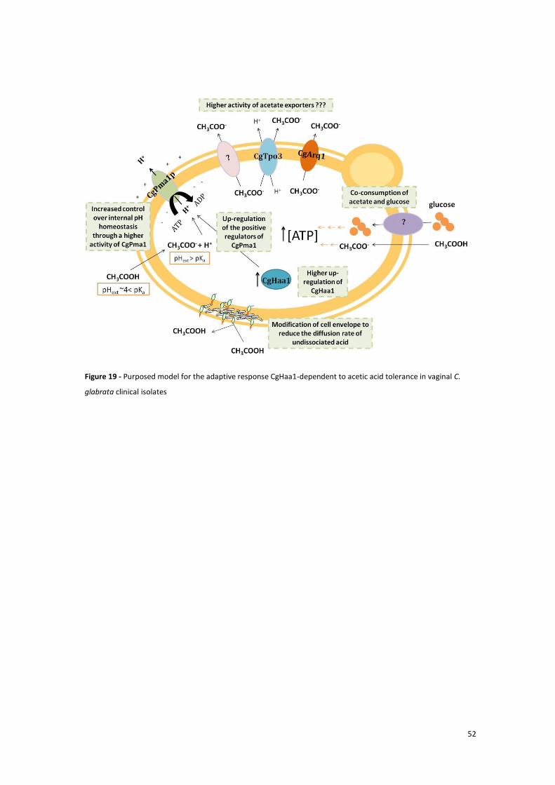
52
Figure 19 - Purposed model for the adaptive response CgHaa1-dependent to acetic acid tolerance in vaginal C.
glabrata clinical isolates

53
5. References
Abbott, D. A., E. Suir, A. J. van Maris and J. T. Pronk (2008). "Physiological and transcriptional responses to high concentrations of lactic acid in anaerobic chemostat cultures of Saccharomyces cerevisiae." Appl Environ Microbiol 74(18): 5759-5768.
Adam, C., E. Erdei, C. Casado, L. Kovacs, A. Gonzalez, L. Majoros, K. Petrenyi, P. Bagossi, I. Farkas, M. Molnar, I. Pocsi, J. Arino and V. Dombradi (2012). "Protein phosphatase CaPpz1 is involved in cation homeostasis, cell wall integrity and virulence of Candida albicans." Microbiology 158(Pt 5): 1258-1267.
Arendrup, M. C. (2010). "Epidemiology of invasive candidiasis." Curr Opin Crit Care 16(5): 445-452.
Bairwa, G. and R. Kaur (2011). "A novel role for a glycosylphosphatidylinositol-anchored aspartyl protease, CgYps1, in the regulation of pH homeostasis in Candida glabrata." Mol Microbiol 79(4): 900-913.
Bernardo, R. (2013a). Functional analysis of the Candida glabrata CgHaa1 (ORF CAGL0L09339g) transcription factor: role in acetic acid resistance and in biofilm formation, Instituto Superior Técnico.
Bernardo, R. (2013b). Functional analysis of the Candida glabrata CgHaa1 (ORF CAGL0L09339g) transcription factor: role in acetic acid resistance and in biofilm formation. Master Master Thesis, Instituto Superior Técnico.
Boskey, E. R., R. A. Cone, K. J. Whaley and T. R. Moench (2001). "Origins of vaginal acidity: high D/L lactate ratio is consistent with bacteria being the primary source." Hum Reprod 16(9): 1809-1813.
Boskey, E. R., K. M. Telsch, K. J. Whaley, T. R. Moench and R. A. Cone (1999). "Acid production by vaginal flora in vitro is consistent with the rate and extent of vaginal acidification." Infect Immun 67(10): 5170-5175.
Brunke, S. and B. Hube (2013). "Two unlike cousins: Candida albicans and C. glabrata infection strategies." Cell Microbiol 15(5): 701-708.
Carmelo, V., P. Bogaerts and I. Sa-Correia (1996). "Activity of plasma membrane H+-ATPase and expression of PMA1 and PMA2 genes in Saccharomyces cerevisiae cells grown at optimal and low pH." Arch Microbiol 166(5): 315-320.
Chaudry, A. N., P. J. Travers, J. Yuenger, L. Colletta, P. Evans, J. M. Zenilman and A. Tummon (2004). "Analysis of vaginal acetic acid in patients undergoing treatment for bacterial vaginosis." J Clin Microbiol 42(11): 5170-5175.
Costa, C., A. Henriques, C. Pires, J. Nunes, M. Ohno, H. Chibana, I. Sa-Correia and M. C. Teixeira (2013). "The dual role of candida glabrata drug:H+ antiporter CgAqr1 (ORF CAGL0J09944g) in antifungal drug and acetic acid resistance." Front Microbiol 4: 170.
Costa, C., J. Nunes, A. Henriques, N. P. Mira, H. Nakayama, H. Chibana and M. C. Teixeira (2014). "Candida glabrata drug:H+ antiporter CgTpo3 (ORF CAGL0I10384g): role in azole drug resistance and polyamine homeostasis." J Antimicrob Chemother 69(7): 1767-1776.
Ehrstrom, S., A. Yu and E. Rylander (2006). "Glucose in vaginal secretions before and after oral glucose tolerance testing in women with and without recurrent vulvovaginal candidiasis." Obstet Gynecol 108(6): 1432-1437.
Fallingborg, J. (1999). "Intraluminal pH of the human gastrointestinal tract." Dan Med Bull 46(3): 183-196.
Fernandes, A. R., N. P. Mira, R. C. Vargas, I. Canelhas and I. Sa-Correia (2005). "Saccharomyces cerevisiae adaptation to weak acids involves the transcription factor Haa1p and Haa1p-regulated genes." Biochem Biophys Res Commun 337(1): 95-103.
Fidel, P. L., Jr., J. A. Vazquez and J. D. Sobel (1999). "Candida glabrata: review of epidemiology, pathogenesis, and clinical disease with comparison to C. albicans." Clin Microbiol Rev 12(1): 80-96.

54
Gasch, A. P., P. T. Spellman, C. M. Kao, O. Carmel-Harel, M. B. Eisen, G. Storz, D. Botstein and P. O. Brown (2000). "Genomic expression programs in the response of yeast cells to environmental changes." Mol Biol Cell 11(12): 4241-4257.
Goossens, A., N. de La Fuente, J. Forment, R. Serrano and F. Portillo (2000). "Regulation of yeast H(+)-ATPase by protein kinases belonging to a family dedicated to activation of plasma membrane transporters." Mol Cell Biol 20(20): 7654-7661.
Goswami, D., R. Goswami, U. Banerjee, V. Dadhwal, S. Miglani, A. A. Lattif and N. Kochupillai (2006). "Pattern of Candida species isolated from patients with diabetes mellitus and vulvovaginal candidiasis and their response to single dose oral fluconazole therapy." J Infect 52(2): 111-117.
Gregori, C., C. Schuller, A. Roetzer, T. Schwarzmuller, G. Ammerer and K. Kuchler (2007). "The high-osmolarity glycerol response pathway in the human fungal pathogen Candida glabrata strain ATCC 2001 lacks a signaling branch that operates in baker's yeast." Eukaryot Cell 6(9): 1635-1645.
Guerreiro, J. F., N. P. Mira and I. Sa-Correia (2012). "Adaptive response to acetic acid in the highly resistant yeast species Zygosaccharomyces bailii revealed by quantitative proteomics." Proteomics 12(14): 2303-2318.
Hickey, R. J., X. Zhou, J. D. Pierson, J. Ravel and L. J. Forney (2012). "Understanding vaginal microbiome complexity from an ecological perspective." Transl Res 160(4): 267-282.
Holyoak, C. D., M. Stratford, Z. McMullin, M. B. Cole, K. Crimmins, A. J. Brown and P. J. Coote (1996). "Activity of the plasma membrane H(+)-ATPase and optimal glycolytic flux are required for rapid adaptation and growth of Saccharomyces cerevisiae in the presence of the weak-acid preservative sorbic acid." Appl Environ Microbiol 62(9): 3158-3164.
Huang, R., M. Li and R. L. Gregory (2011). "Bacterial interactions in dental biofilm." Virulence 2(5): 435-444.
Jandric, Z., C. Gregori, E. Klopf, M. Radolf and C. Schuller (2013). "Sorbic acid stress activates the Candida glabrata high osmolarity glycerol MAP kinase pathway." Front Microbiol 4: 350.
Kawahata, M., K. Masaki, T. Fujii and H. Iefuji (2006). "Yeast genes involved in response to lactic acid and acetic acid: acidic conditions caused by the organic acids in Saccharomyces cerevisiae cultures induce expression of intracellular metal metabolism genes regulated by Aft1p." FEMS Yeast Res 6(6): 924-936.
Kren, A., Y. M. Mamnun, B. E. Bauer, C. Schuller, H. Wolfger, K. Hatzixanthis, M. Mollapour, C. Gregori, P. Piper and K. Kuchler (2003). "War1p, a novel transcription factor controlling weak acid stress response in yeast." Mol Cell Biol 23(5): 1775-1785.
Lim, C. S., R. Rosli, H. F. Seow and P. P. Chong (2012). "Candida and invasive candidiasis: back to basics." Eur J Clin Microbiol Infect Dis 31(1): 21-31.
Mira, N. P., J. D. Becker and I. Sa-Correia (2010a). "Genomic expression program involving the Haa1p-regulon in Saccharomyces cerevisiae response to acetic acid." OMICS 14(5): 587-601.
Mira, N. P., S. F. Henriques, G. Keller, M. C. Teixeira, R. G. Matos, C. M. Arraiano, D. R. Winge and I. Sa-Correia (2011). "Identification of a DNA-binding site for the transcription factor Haa1, required for Saccharomyces cerevisiae response to acetic acid stress." Nucleic Acids Res 39(16): 6896-6907.
Mira, N. P., A. B. Lourenco, A. R. Fernandes, J. D. Becker and I. Sa-Correia (2009). "The RIM101 pathway has a role in Saccharomyces cerevisiae adaptive response and resistance to propionic acid and other weak acids." FEMS Yeast Res 9(2): 202-216.
Mira, N. P., M. Palma, J. F. Guerreiro and I. Sa-Correia (2010b). "Genome-wide identification of Saccharomyces cerevisiae genes required for tolerance to acetic acid." Microb Cell Fact 9: 79.
Mira, N. P., M. C. Teixeira and I. Sá-Correia (2010c). "Adaptive response and tolerance to weak acids in Saccharomyces cerevisiae: a genome-wide view." OMICS 14(5): 525-540.

55
Mollapour, M. and P. W. Piper (2006). "Hog1p mitogen-activated protein kinase determines acetic acid resistance in Saccharomyces cerevisiae." FEMS Yeast Res 6(8): 1274-1280.
Mollapour, M. and P. W. Piper (2007). "Hog1 mitogen-activated protein kinase phosphorylation targets the yeast Fps1 aquaglyceroporin for endocytosis, thereby rendering cells resistant to acetic acid." Mol Cell Biol 27(18): 6446-6456.
Moosa, M. Y., J. D. Sobel, H. Elhalis, W. Du and R. A. Akins (2004). "Fungicidal activity of fluconazole against Candida albicans in a synthetic vagina-simulative medium." Antimicrob Agents Chemother 48(1): 161-167.
Mundy, R. D. and B. Cormack (2009). "Expression of Candida glabrata adhesins after exposure to chemical preservatives." J Infect Dis 199(12): 1891-1898.
O'Hanlon, D. E., T. R. Moench and R. A. Cone (2011). "In vaginal fluid, bacteria associated with bacterial vaginosis can be suppressed with lactic acid but not hydrogen peroxide." BMC Infect Dis 11: 200.
Owen, D. H. and D. F. Katz (1999). "A vaginal fluid simulant." Contraception 59(2): 91-95. Petter, R. and K. J. Kwon-Chung (1996). "Disruption of the SNF1 gene abolishes trehalose
utilization in the pathogenic yeast Candida glabrata." Infect Immun 64(12): 5269-5273. Pfaller, M. A., M. Castanheira, S. R. Lockhart, A. M. Ahlquist, S. A. Messer and R. N. Jones
(2012). "Frequency of decreased susceptibility and resistance to echinocandins among fluconazole-resistant bloodstream isolates of Candida glabrata." J Clin Microbiol 50(4): 1199-1203.
Pfaller, M. A., D. J. Diekema, D. L. Gibbs, V. A. Newell, D. Ellis, V. Tullio, A. Rodloff, W. Fu, T. A. Ling and G. Global Antifungal Surveillance (2010). "Results from the ARTEMIS DISK Global Antifungal Surveillance Study, 1997 to 2007: a 10.5-year analysis of susceptibilities of Candida Species to fluconazole and voriconazole as determined by CLSI standardized disk diffusion." J Clin Microbiol 48(4): 1366-1377.
Pfaller, M. A., D. J. Diekema and G. International Fungal Surveillance Participant (2004). "Twelve years of fluconazole in clinical practice: global trends in species distribution and fluconazole susceptibility of bloodstream isolates of Candida." Clin Microbiol Infect 10 Suppl 1: 11-23.
Ravel, J., P. Gajer, Z. Abdo, G. M. Schneider, S. S. Koenig, S. L. McCulle, S. Karlebach, R. Gorle, J. Russell, C. O. Tacket, R. M. Brotman, C. C. Davis, K. Ault, L. Peralta and L. J. Forney (2011). "Vaginal microbiome of reproductive-age women." Proc Natl Acad Sci U S A 108 Suppl 1: 4680-4687.
Rodrigues, F., M. J. Sousa, P. Ludovico, H. Santos, M. Corte-Real and C. Leao (2012). "The fate of acetic acid during glucose co-metabolism by the spoilage yeast Zygosaccharomyces bailii." PLoS One 7(12): e52402.
Roetzer, A., T. Gabaldon and C. Schuller (2011). "From Saccharomyces cerevisiae to Candida glabratain a few easy steps: important adaptations for an opportunistic pathogen." FEMS Microbiol Lett 314(1): 1-9.
Roetzer, A., C. Gregori, A. M. Jennings, J. Quintin, D. Ferrandon, G. Butler, K. Kuchler, G. Ammerer and C. Schuller (2008). "Candida glabrata environmental stress response involves Saccharomyces cerevisiae Msn2/4 orthologous transcription factors." Mol Microbiol 69(3): 603-620.
Rosa, M. F. and I. Sa-Correia (1996). "Intracellular acidification does not account for inhibition of Saccharomyces cerevisiae growth in the presence of ethanol." FEMS Microbiol Lett 135(2-3): 271-274.
Sá-Correia, I., S. C. dos Santos, M. C. Teixeira, T. R. Cabrito and N. P. Mira (2009). "Drug:H+ antiporters in chemical stress response in yeast." Trends Microbiol 17(1): 22-31.
Sandai, D., Z. Yin, L. Selway, D. Stead, J. Walker, M. D. Leach, I. Bohovych, I. V. Ene, S. Kastora, S. Budge, C. A. Munro, F. C. Odds, N. A. Gow and A. J. Brown (2012). "The evolutionary rewiring of ubiquitination targets has reprogrammed the regulation of carbon assimilation in the pathogenic yeast Candida albicans." MBio 3(6).
56
Saporiti, A. M., D. Gomez, S. Levalle, M. Galeano, G. Davel, W. Vivot and L. Rodero (2001). "[Vaginal candidiasis: etiology and sensitivity profile to antifungal agents in clinical use]." Rev Argent Microbiol 33(4): 217-222.
Schuller, C., Y. M. Mamnun, M. Mollapour, G. Krapf, M. Schuster, B. E. Bauer, P. W. Piper and K. Kuchler (2004). "Global phenotypic analysis and transcriptional profiling defines the weak acid stress response regulon in Saccharomyces cerevisiae." Mol Biol Cell 15(2): 706-720.
Simões, T., N. P. Mira, A. R. Fernandes and I. Sa-Correia (2006). "The SPI1 gene, encoding a glycosylphosphatidylinositol-anchored cell wall protein, plays a prominent role in the development of yeast resistance to lipophilic weak-acid food preservatives." Appl Environ Microbiol 72(11): 7168-7175.
Simões, T., M. C. Teixeira, A. R. Fernandes and I. Sa-Correia (2003). "Adaptation of Saccharomyces cerevisiae to the herbicide 2,4-dichlorophenoxyacetic acid, mediated by Msn2p- and Msn4p-regulated genes: important role of SPI1." Appl Environ Microbiol 69(7): 4019-4028.
Sobel, J. D., S. Faro, R. W. Force, B. Foxman, W. J. Ledger, P. R. Nyirjesy, B. D. Reed and P. R. Summers (1998). "Vulvovaginal candidiasis: epidemiologic, diagnostic, and therapeutic considerations." Am J Obstet Gynecol 178(2): 203-211.
Sugiyama, M., S. P. Akase, R. Nakanishi, H. Horie, Y. Kaneko and S. Harashima (2014). "Nuclear localization of Haa1, which is linked to its phosphorylation status, mediates lactic acid tolerance in Saccharomyces cerevisiae." Appl Environ Microbiol 80(11): 3488-3495.
Sun, Y. and M. X. O'Riordan (2013). "Regulation of bacterial pathogenesis by intestinal short-chain Fatty acids." Adv Appl Microbiol 85: 93-118.
Tenreiro, S., P. A. Nunes, C. A. Viegas, M. S. Neves, M. C. Teixeira, M. G. Cabral and I. Sa-Correia (2002). "AQR1 gene (ORF YNL065w) encodes a plasma membrane transporter of the major facilitator superfamily that confers resistance to short-chain monocarboxylic acids and quinidine in Saccharomyces cerevisiae." Biochem Biophys Res Commun 292(3): 741-748.
Tenreiro, S., P. C. Rosa, C. A. Viegas and I. Sa-Correia (2000). "Expression of the AZR1 gene (ORF YGR224w), encoding a plasma membrane transporter of the major facilitator superfamily, is required for adaptation to acetic acid and resistance to azoles in Saccharomyces cerevisiae." Yeast 16(16): 1469-1481.
Trumbly, R. J. (1992). "Glucose repression in the yeast Saccharomyces cerevisiae." Mol Microbiol 6(1): 15-21.
Ueno, K., Y. Matsumoto, J. Uno, K. Sasamoto, K. Sekimizu, Y. Kinjo and H. Chibana (2011). "Intestinal resident yeast Candida glabrata requires Cyb2p-mediated lactate assimilation to adapt in mouse intestine." PLoS One 6(9): e24759.
Ullah, A., G. Chandrasekaran, S. Brul and G. J. Smits (2013a). "Yeast adaptation to weak acids prevents futile energy expenditure." Front Microbiol 4: 142.
Ullah, A., M. I. Lopes, S. Brul and G. J. Smits (2013b). "Intracellular pH homeostasis in Candida glabrata in infection-associated conditions." Microbiology 159(Pt 4): 803-813.
Yamaguchi, N., K. Sonoyama, H. Kikuchi, T. Nagura, T. Aritsuka and J. Kawabata (2005). "Gastric colonization of Candida albicans differs in mice fed commercial and purified diets." J Nutr 135(1): 109-115.
Yang, Y. L. (2003). "Virulence factors of Candida species." J Microbiol Immunol Infect 36(4): 223-228.
Yenush, L., J. M. Mulet, J. Arino and R. Serrano (2002). "The Ppz protein phosphatases are key regulators of K+ and pH homeostasis: implications for salt tolerance, cell wall integrity and cell cycle progression." EMBO J 21(5): 920-929.
Zhang, J. Y., J. H. Liu, F. D. Liu, Y. H. Xia, J. Wang, X. Liu, Z. Q. Zhang, N. Zhu, Y. Yan, Y. Ying and X. T. Huang (2014). "Vulvovaginal candidiasis: species distribution, fluconazole resistance and drug efflux pump gene overexpression." Mycoses.

57
Zhou, X., M. A. Hansmann, C. C. Davis, H. Suzuki, C. J. Brown, U. Schutte, J. D. Pierson and L. J. Forney (2010). "The vaginal bacterial communities of Japanese women resemble those of women in other racial groups." FEMS Immunol Med Microbiol 58(2): 169-181.