molecular mechanisms of inner ear...
TRANSCRIPT
Molecular Mechanisms of Inner EarDevelopment
Doris K. Wu and Matthew W. Kelley
National Institute on Deafness and Other Communication Disorders, Rockville, Maryland 20850
Correspondence: [email protected]
SUMMARY
The inner ear is a structurally complex vertebrate organ built to encode sound, motion, andorientation in space. Given its complexity, it is not surprising that inner ear dysfunction is arelatively common consequence of human genetic mutation. Studies in model organismssuggest that many genes currently known to be associated with human hearing impairmentare active during embryogenesis. Hence, the study of inner ear development provides a richcontext for understanding the functions of genes implicated in hearing loss. This chapterfocuses on molecular mechanisms of inner ear development derived from studies of modelorganisms.
Outline
1 Introduction
2 Axial specification
3 Specification of neural and sensory fates
4 Formation of semicircular canals and cristae
5 Formation of the cochlear duct
6 Future directions
References
Editors: Patrick P.L. Tam, W. James Nelson, and Janet Rossant
Additional Perspectives on Mammalian Development available at www.cshperspectives.org
Copyright # 2012 Cold Spring Harbor Laboratory Press; all rights reserved; doi: 10.1101/cshperspect.a008409
Cite this article as Cold Spring Harb Perspect Biol 2012;4:a008409
1
on February 7, 2019 - Published by Cold Spring Harbor Laboratory Press http://cshperspectives.cshlp.org/Downloaded from
1 INTRODUCTION
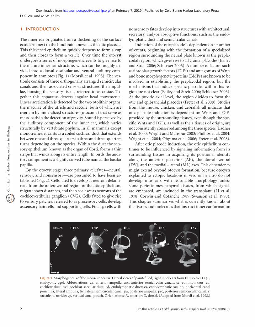
The inner ear originates from a thickening of the surfaceectoderm next to the hindbrain known as the otic placode.This thickened epithelium quickly deepens to form a cupand then closes to form a vesicle. Over time the otocystundergoes a series of morphogenetic events to give rise tothe mature inner ear structure, which can be roughly di-vided into a dorsal vestibular and ventral auditory com-ponent in amniotes (Fig. 1) (Morsli et al. 1998). The ves-tibule consists of three orthogonally arranged semicircularcanals and their associated sensory structures, the ampul-lae, housing the sensory tissue, referred to as cristae. To-gether this apparatus detects angular head movements.Linear acceleration is detected by the two otolithic organs,the maculae of the utricle and saccule, both of which areoverlain by mineralized structures (otoconia) that serve asmass loads in the detection of gravity. Sound is perceived bythe auditory component of the inner ear, which variesstructurally by vertebrate phylum. In all mammals exceptmonotremes, it exists as a coiled cochlear duct that extendsbetween one and three-quarters to three and three-quartersturns depending on the species. Within the duct the sen-sory epithelium, known as the organ of Corti, forms a thinstripe that winds along its entire length. In birds the audi-tory component is a slightly curved tube named the basilarpapilla.
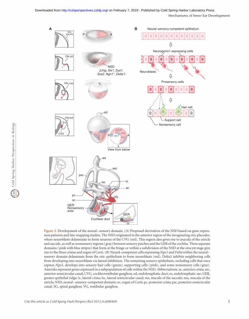
By the otocyst stage, three primary cell fates—neural,sensory, and nonsensory—are presumed to have been es-tablished (Fig. 2). Cells fated to develop as neurons delami-nate from the anteroventral region of the otic epithelium,migrate short distances, and then coalesce as neurons of thecochleovestibular ganglion (CVG). Cells fated to give riseto sensory patches, referred to as prosensory cells, developas sensory hair cells and supporting cells. Finally, cells with
nonsensory fates develop into structures with architectural,secretory, and/or absorptive functions, such as the endo-lymphatic duct and semicircular canals.
Induction of the otic placode is dependent on a numberof events, beginning with the formation of a specializedregion surrounding the neural plate known as the prepla-codal region, which gives rise to all cranial placodes (Baileyand Streit 2006; Schlosser 2006). A number of factors suchas fibroblast growth factors (FGFs) and antagonists of Wntsand bone morphogenetic proteins (BMPs) are known to beinvolved in establishing the preplacodal region, but themechanisms that induce specific placodes within this re-gion are not clear (Bailey and Streit 2006; Schlosser 2006).At the preotic axial level, the region divides to form theotic and epibranchial placodes (Freter et al. 2008). Studiesfrom the mouse, chicken, and zebrafish all indicate thatotic placode induction is dependent on Wnts and FGFsprovided by the surrounding tissues, even though the spe-cific Wnts and FGFs, as well as their tissues of origin, arenot consistently conserved among the three species (Ladheret al. 2000; Wright and Mansour 2003; Phillips et al. 2004;Wright et al. 2004; Ohyama et al. 2006; Freter et al. 2008).
After otic placode induction, the otic epithelium con-tinues to be influenced by signaling information from itssurrounding tissues in acquiring its positional identityalong the anterior–posterior (AP), the dorsal–ventral(DV), and the medial–lateral (ML) axes. This dependencymight extend beyond otocyst formation, because otocystsexplanted to ectopic locations in vivo or in vitro do notdevelop into ears with reasonable morphology unlesssome periotic mesenchymal tissues, from which signalsare emanated, are included in the transplant (Li et al.1978; Corwin and Cotanche 1989; Swanson et al. 1990).This chapter summarizes what is currently known aboutthe tissues and molecules that instruct inner ear formation
E10.75 E11.5
D
A
E13 E15 E17
cc edlsc
us
aa
ascla
csd
pa
psc
es
vp
ed
co hp
E12
Figure 1. Morphogenesis of the mouse inner ear. Lateral views of paint-filled, right inner ears from E10.75 to E17 (E,embryonic age). Abbreviations: aa, anterior ampulla; asc, anterior semicircular canals; cc, common crus; co,cochlear duct; csd, cochlear saccular duct; ed, endolymphatic duct; es, endolymphatic sac; hp, horizontal canalpouch; la, lateral ampulla; lsc, lateral semicircular canal; pa, posterior ampulla; psc, posterior semicircular canal; s,saccule; u, utricle; vp, vertical canal pouch. Orientations: A, anterior; D, dorsal. (Adapted from Morsli et al. 1998.)
D.K. Wu and M.W. Kelley
2 Cite this article as Cold Spring Harb Perspect Biol 2012;4:a008409
on February 7, 2019 - Published by Cold Spring Harbor Laboratory Press http://cshperspectives.cshlp.org/Downloaded from
Oticplacode
Otic pit
Otic cup
NSD(Lfng, Six1, Eya1,
Sox2, Ngn1*, Delta*)
Otocyst
CVG
A
Neural–sensory-competent epithelium
Neurogenin1 expressing cells
Neuroblasts
Prosensory cells
Hair cell
Support cell
Nonsensory cell
ed
AP
LView from below
ed
asc
lsc
psc
lc
ac
VG
GER
oc
mu
ms
SGoc
pc
GERNonsensoryarea
Cochlear duct
GERmsmuoc?
P
A B
Figure 2. Development of the neural–sensory domain. (A) Proposed derivatives of the NSD based on gene expres-sion patterns and fate-mapping studies. The NSD originated in the anterior region of the invaginating otic placodes,where neuroblasts delaminate to form neurons of the CVG (red). This region also gives rise to macula of the utricleand saccule, as well as nonsensory regions (gray) between sensory patches and the GER of the cochlea. Three separatedomains (pink with blue stripes) that form at the fringe or within a subdivision of the NSD at the otocyst stage giverise to the three cristae and organ of Corti. (B) Neural-competent cells expressing Ngn1 and Delta within the neural–sensory domain delaminate from the otic epithelium to form neuroblasts (red). Delta1 inhibits neighboring cellsfrom developing into neuroblasts via lateral inhibition. The remaining sensory epithelium, including cells that onceexpress Ngn1, develops into sensory hair cells (green), supporting cells (pink), and some nonsensory cells (gray).Asterisks represent genes expressed in a subpopulation of cells within the NSD. Abbreviations: ac, anterior crista; asc,anterior semicircular canal; CVG, cochleovestibular ganglion; ed, endolymphatic duct; es, endolymphatic sac; GER,greater epithelial ridge; lc, lateral crista; lsc, lateral semicircular canal; ms, macula of the saccule; mu, macula of theutricle; NSD, neural–sensory-competent domain; oc, organ of Corti; pc, posterior crista; psc, posterior semicircularcanal; SG, spiral ganglion; VG, vestibular ganglion.
Mechanisms of Inner Ear Development
Cite this article as Cold Spring Harb Perspect Biol 2012;4:a008409 3
on February 7, 2019 - Published by Cold Spring Harbor Laboratory Press http://cshperspectives.cshlp.org/Downloaded from

in amniotes, and illustrates how axial information drivescell-fate specification and formation of specific inner earstructures.
2 AXIAL SPECIFICATION
The axial identity of the inner ear is specified by signalsarising from surrounding tissues, including the neurecto-derm, mesoderm, endoderm, and neural crest. A particularaxis is specified when positional identity of ear tissues be-comes insensitive to changes in extrinsic signals. The tim-ing of this event in amniotes can be revealed using a surgicaltransplantation approach in chickens, whereby the conse-quences of inverting one specific axis of the ear rudimentrelative to the axes of the host body were assessed followingthe replacement of an ear rudiment with a contralateralcounterpart. The main conclusion from these experimentsis that axial specification of the inner ear is a progressiveevent. The first axis to be specified appears to be the APaxis,at least in terms of the sensory tissues, which occurs well inadvance of the DV axis (Wu et al. 1998), but axes for thenonsensory tissues are specified later and independentlyof the sensory tissues. These multiple steps of axial speci-fication imply a gradual restriction of cell fates over time.Therefore, identifying the timing of axial specification ofthe inner ear tissue will inform us of when extrinsic signalsmust be present, and may provide insights into the molec-ular cascades that accompany the formation of this com-plex organ.
2.1 Anterior–Posterior Axis
The first clear indication of AP asymmetry of the chickenand mouse ear rudiment is the regionalized expression ofgenes, such as Lunatic fringe (Lfng), Fgf10, Six1, and Sox2,strongly in the anterior but more weakly and diffusely inthe posterior region of the otic cup (Fig. 2) (Morsli et al.1998; Cole et al. 2000; Zheng et al. 2003; Alsina et al.2004; Kiernan et al. 2005). The anterior region is theneural–sensory-competent domain (NSD) (Fekete andWu 2002). The most anterior part of the NSD containsNeurogenin1-positive (Ngn1) cells that are fated to leavethe otic epithelium as neural precursors and form theCVG. The posterior otic region gives rise to mostly non-sensory tissues and only one sensory organ: the posteriorcrista.
Recent studies in both chicken and mouse suggest thatretinoic acid (RA), a critical morphogen for early embryo-genesis, is required for conferring anterior and posterioridentities to the inner ear (Bok et al. 2011). The expressionof an RA reporter, RARE-lacZ (retinoic acid response ele-ment driving lacZ), highlights different RA responsiveness
between anterior and posterior halves of the otic cup (Fig.3A). Moreover, this wave of RA responsiveness regressescaudally away from anterior regions when the otic cupcloses. This shift in RA responsiveness is attributed, inpart, to caudally shifting expression domains of the RAsynthetic and degradation enzymes, Raldh2 (retinaldehydedehydrogenase 2) and Cyp26 (cytochrome P450–associat-ed RA catabolizing enzyme), respectively (Fig. 3B) (Re-ijntjes et al. 2004; Sirbu et al. 2005). Gain- and loss-of-function studies in both chicken and mouse collectivelyindicate that cells in the anteriorotic region that are exposedonly briefly to RA give rise to neurons and most sensoryorgans of the innerear, whereas prolonged exposure and/orhigher levels of RA in the posterior otocyst promote forma-tion of nonsensory structures (Bok et al. 2011). Consistentwith this inference, local application of RA on the anteriorside of the developing otic cup in chicken resulted in aninner ear with two mirror-image posterior domains. Fur-thermore, one of the key effectors of high RA signaling inthe otic epithelium may be the T-box transcription factorTbx1 (Bok et al. 2011), which was shown to function as anegative regulator of otic neurogenesis (the anterior tissuefate) along the APaxis (Raft et al. 2004). These findings lendsupport to a compartment-boundary model of cell-fatespecification for the inner ear (Fekete 1996).
The RA synthesizing and degradation enzymes respon-sible for the differential RA response of the ear rudimentare likely to be Cyp26C1, which is expressed in ectodermanterior to the ear rudiment, and Raldh2, which is ex-pressed in the somites posterior to the otocyst (Fig. 3B)(Reijntjes et al. 2004). Although other RA degradation en-zymes are also expressed in rhombomeres adjacent to theear rudiment, hindbrain rotation experiments rule outtheir role in AP patterning of the chicken inner ear (Boket al. 2005). In mice the otic cup also responds differentiallyto RA along its AP axis (Fig. 3A), although no RA degra-dation enzyme has yet been identified in the ectodermadjacent to the ear primordium (MacLean et al. 2001;Tahayato et al. 2003). Therefore, although the requirementof RA in AP patterning seems to be conserved between miceand chicken, the tissues that elicit the differential RA re-sponse of the otic epithelium may be different. A gradientof RA signaling has also been implicated in patterning thehindbrain rhombomeres in mice (Sirbu et al. 2005). Thus,it is likely that dynamic RA signaling coordinates the APpatterning of multiple structures in this part of the embryo(Fig. 3B). In zebrafish RA has been shown to restrict theextent of the anterior neurogenic domain through the sup-pression of neurogenic genes by tbx1 and her9, a zebrafishortholog of Hes1 (Radosevic et al. 2011). Whether cellswithin the inner ears of zebrafish are responding differen-tially to RA along the AP axis is not clear.
D.K. Wu and M.W. Kelley
4 Cite this article as Cold Spring Harb Perspect Biol 2012;4:a008409
on February 7, 2019 - Published by Cold Spring Harbor Laboratory Press http://cshperspectives.cshlp.org/Downloaded from

Besides RA, FGF8, which is expressed in the ectodermanterior to the ear primordium in chicken, has also beenimplicated in the establishment of the neurogenic (anteri-or) fate via up-regulation of Sox3 (Abello et al. 2010). Onthe other hand, knocking down BMP signaling also suggestsa role for BMPs in mediating the nonneurogenic (posteri-or) fate (Abello et al. 2010). Because FGF and RA are shownin other systems to act antagonistically (Diez del Corral andStorey 2004; Marklund et al. 2004), FGF8 may diminish theresponsiveness of the anterior otic region to RA.
2.2 Dorsal–Ventral Patterning
In contrast to AP patterning, DV specification is dependenton ectoderm-derived signals from the hindbrain in bothchicken and mice (Giraldez 1998; Bok et al. 2005; Ricco-magno et al. 2005). DV inversion of a segment of the hind-brain adjacent to the ear rudiment in chicken is sufficientto override potential DV signaling from other tissues andactivate ventral genes in dorsal otic tissues (Bok et al. 2005).To date, Wnts secreted from the dorsal hindbrain and Sonic
hedgehog (Shh) secreted from the ventral floor plate andnotochord have been implicated in patterning of the innerear DVaxis (Fig. 3B). However, the weight of evidence sug-gests that other unidentified signals originating from thehindbrain are involved as well (Riccomagno et al. 2005; Boket al. 2007b). For example, BMPs are likely to act as dorsalsignaling molecules for the inner ear, although direct ex-perimental proof of this is lacking at present.
The Shh signaling pathway has been well studied (Ribesand Briscoe 2009; Murdoch and Copp 2010) and plays animportant role in ventral identities. One of its major ac-tions in target cells is believed to be regulation of the tran-scription factor Gli3. In the absence of Shh, the Gli3 proteinis cleaved and its amino-terminal portion functions as atranscription repressor. However, in the presence of Shh,Shh binds to its receptor Patched, which removes an inhib-itory effect on the transmembrane protein Smoothened.As a result, a number of intracellular signaling cascadesare initiated that maintain full-length Gli3 protein, whichthen functions as a transcription activator. Based on severalmutant mouse analyses and ablation studies in chicken, it
E8.75 E9A
B
DA
VV
P
RA
E9.5E9.5
100 μm100 μm50 μm
A
L100 μm
A
L
Figure 3. Anterior–posterior axial specification. (A) X-Gal histochemical staining in RARE-lacZ mouse embryosshows a transient gradient of RA responsiveness in the developing inner ear. At the otic cup stage (E8.75), theanterior border of the RA responsiveness is in the middle of the otic cup, and this border shifts caudally by the timethe otocyst is formed at E9.5. (B) Model of RA signaling in AP patterning of the chicken inner ear. Somites expressinghigh levels of the RA synthetic enzyme Raldh2 act as the main source of RA for patterning the inner ear. The RAdegradation enzyme Cyp26C1, expressed in the ectoderm rostral to the ear rudiment, modulates the level of RAsignaling perceived by the otic epithelium. Secreted molecules from the hindbrain such as Wnts (purple) and Shh(yellow) provide DV axial information to the inner ear.
Mechanisms of Inner Ear Development
Cite this article as Cold Spring Harb Perspect Biol 2012;4:a008409 5
on February 7, 2019 - Published by Cold Spring Harbor Laboratory Press http://cshperspectives.cshlp.org/Downloaded from

was proposed that Shh secreted from the ventral midlinegenerates opposing gradients of Gli3 repressor and Gli2 andGli3 activators in the otic epithelium, which pattern vari-ous inner ear structures along the DVaxis (Fig. 4) (Bok et al.2007c). The most apical region of the cochlear duct requiresthe highest level of Shh to induce Gli2 and Gli3 activators.The utricle, saccule, and proximal region of the cochlearduct require relatively lower levels of Shh to alleviate theGli3 repressor function. In contrast, dorsal structures suchas the semicircular canals require little Shh and instead aredependent on Gli3 repressor activity for proper formation(Bok et al. 2007b; Brown and Epstein 2011). The upstreamactivator of Gli3 transcription is not known. In the neuraltube, where similar Gli3 repressor and Gli activator gradi-ents are believed to pattern the DV axis of the neural tube,
there is evidence that transcription of Gli3 is activated byWnts (Alvarez-Medina et al. 2008). Whether Gli3 is in-duced by Wnts in the inner ear remains to be explored.
Wnts from the dorsal neural tube have been implicatedin otic DV patterning in addition to their earlier role inspecifying the otic placode fate (Ohyama et al. 2006; Jaya-sena et al. 2008). After otic placode specification, there isa continual requirement for Wnts in the DV patterning ofthe inner ear, based on studies using lithium chloride treat-ments in otic-hindbrain explant cultures (Riccomagno et al.2005). Lithium chloride, a Wnt/b-catenin pathwayagonist,is able to restore, in part, otocyst gene expression patternsaltered by ablation of the dorsal neural tube in otic-hind-brain explants.
Wnt1 and Wnt3a are expressed in the dorsal neural tubebut not in the otic placode or otocyst. Ears from Wnt1/Wnt3a double-knockout mice are rudimentary and cysticin structure, indicating that the affected areas are not lim-ited to the dorsal region (Fig. 4) (Riccomagno et al. 2005).These severe phenotypes may be caused by an earlier re-quirement of Wnts in placode specification, even thoughWnt1 or Wnt3a expression is low in the hindbrain duringthe time of otic placode induction (Parr et al. 1993). Nev-ertheless, results from genetic fate mapping of cells re-sponding to Wnt signaling in the mouse otocyst usingTopgal transgenic mice clearly indicate that the dorsal re-gion of the otic cup and otocyst are responsive to Wntsignaling (Fig. 5A, a,b). It is also possible to track the fatesof Wnt-responsive otocyst cells over time by crossing Rosa-lacZ, a Cre reporter strain, with the TopCreERT2 strain.Administration of tamoxifen at specific time points causesWnt-responsive cells harboring both alleles to permanentlyexpress lacZ. Administering tamoxifen between embryonicday (E) 8.5 and 8.75 followed by examination at E9.5 indi-cates that X-Gal-positive cells span most of the medial wallof the otocyst (Fig. 5A, d) (Riccomagno et al. 2005), cov-ering a much broader area than would be predicted by theTopCreERT2 mRNA or Topgal expression domains (Fig.5A, a–c). This broader X-Gal-positive domain in TopC-reER/Rosa-lacZ inner ears suggests that cells that receivedWnt signaling in the dorsal otic region at an earlier age havebecome displaced or have migrated ventrally by the otocyststage (Fig. 5A, d, arrows). Thus, the genetic fate-mappingresults are consistent with the inner ear phenotypes ofWnt1/Wnt3a double mutants, which involve more thanthe dorsal region.
In the zebrafish inner ear, hedgehog signaling has beenimplicated in AP rather than DV patterning (Hammondet al. 2003). Loss of hedgehog signaling resulted in innerears with a mirror-image duplication of two anteriorhalves, whereas gain of hedgehog signaling resulted in dou-ble posterior ears. More recent results show that in addition
Wild type
DorsalWnt
?
Gliactivators
ShhShh
Gli3repressor
GenotypesHindbrain
secreted molecules Gli activities Phenotypes
Ventral
Shh-/-
Wnt1-/-
Wnt3a-/-
Figure 4. Dorsal–ventral axial specification. The hindbrain is thesource of DV axial signaling for the inner ear. In the wild-type em-bryo, the developing inner ear receives Wnt signaling from the dorsalhindbrain (purple triangle) and Sonic hedgehog (Shh) from theventral midline structures of the floor plate and notochord. Thegraded Shh signaling (yellow triangle) results in various levels ofGli activator (blue triangle) and repressor (red triangle) activitiesin the developing inner ear. The distal region of the cochlear duct(blue region) requires a high level of Gli activators. The proximalregion of the cochlear duct and the saccule (pale red and blue region)require relatively lower levels of Shh signaling for counteracting theGli3 repressor function. The dorsal vestibular region of the inner ear(red region) requires Gli3 repressor function. In Shh2/2 ears onlythe dorsal vestibular region develops. In Wnt1/Wnt3a double mu-tants, the inner ear is rudimentary and cystic. However, Shh signalingappears normal based on the expression of putative Shh target genessuch as Pax2 and Otx2. Whether Gli3 repressor activities are stillpresent in the inner ear of the compound mutant is not clear.
D.K. Wu and M.W. Kelley
6 Cite this article as Cold Spring Harb Perspect Biol 2012;4:a008409
on February 7, 2019 - Published by Cold Spring Harbor Laboratory Press http://cshperspectives.cshlp.org/Downloaded from

to the double posterior phenotypes, too much hedgehogsignaling also affects DV and ML patterning, thus narrow-ing the differences between fish and amniotes (Hammondet al. 2010). For example, there is an increased prevalenceof a missing posterior canal phenotype in Shh2/2 ears(Brown and Epstein 2011), suggesting that the posteriorcanal is more sensitive to a lack of Shh signaling than theanterior canal. Consistently, in mutants that presumablyhave too much Shh signaling such as Extra-toes, in whichGli3 encodes a functional null protein, the anterior canal istruncated (Bok et al. 2007c). Therefore, these canal pheno-types in mice resemble those in the zebrafish, in that toomuch or too little hedgehog signaling affects anterior andposterior otic patterning, respectively.
2.3 Medial–Lateral Axis
Far less is known about how the ML axis of the ear is spec-ified. Hindbrain signaling is involved, and this signaling is
believed to be mediated, at least in part, through Gbx2 ex-pressed in the otic epithelium (Lin et al. 2005; Choo et al.2006). Also, it has been suggested that the medial otic regionmay be the first to be specified, followed by AP, DV, and thelateral axis (Bok et al. 2007b). This notion is largely based onearly initiation of genes such as Pax2 and Gbx2 at the oticplacode stage, which subsequently are associated with themedial half of the developing otocyst. However, there is nodirect experimental evidence that pinpoints the timing ofmedial or lateral axial specification. Rotating the ML axis ofan otocyst resulted in malformed ears with mixed expres-sion patterns of medial and lateral genes that were difficultto interpret (Wu et al. 1998). This is partially attributed tothe technical difficulty in generating a precise ML inversionowing to the geometry of the newly formed otocyst, which ismore like the shape of a covered bowl (flat on the lateralsurface) than a perfect sphere (Fig. 5A).
Further information about the origin of the cells in thelateral otic region comes from a fate-mapping study using
11
1 1
2 2
2 2
3 3
33
4 44 4
5 5 5
??
56 6
66
7 7
7
7
7
8 8
8
8 8
9 9
9
9
9M L10
10
1010
1011
11
11 1111
12
12
12 12
A/P boundary
M/L boundary
Stage 22(posterior view)
Stage 22(lateral view)
Stage 17(lateral view)
Stage 16.5(lateral view)
Stage 13.5(lateral view)
12
V
A
D
B
a
A
bE8.5
D
L
E9.5 E9.5 E9.5
Topgal Topgal TopCreERT2 TopCreERT2; Rosa-lacZ
c d
P
Figure 5. Fate-mapping studies of the mouse and chicken inner ear. (A) Genetic fate mapping of the Wnt-responsivecells in the mouse otic cup. X-Gal histochemical staining of a Wnt reporter strain, Topgal, shows that Wnt-responsivecells are restricted to the dorsal otic placode (a) and otocyst (b). However, tracing the progeny of the Wnt-responsivecells in TopCreERT2/Rosa-lacZ embryos after administration of tamoxifen at E8.75 shows these cells expandedventrally (d, arrows) beyond the initial TopCreERT2 mRNA domain (c). (B) Fate mapping of the rim of the chickenotic cup. Injection of lipophilic dyes to the indicated numerical positions at the rim of the otic cup shows that amajority of the cells that constitute the lateral wall of the otocyst (light blue) originated from the ventral posteriorregion of the otic cup at positions 6, 7, and 8. The most dorsal region of the otocyst, constituted by cells in positions1, 2, 10, 11, and 12, displaces medially to form the endolymphatic duct (tan). The lateral region of the otocyst givesrise to the vertical and horizontal canal pouches. (A adapted from Riccomagno et al. 2005; B adapted from Brigandeet al. 2000; Fig. 5 reprinted, with permission, # Elsevier.)
Mechanisms of Inner Ear Development
Cite this article as Cold Spring Harb Perspect Biol 2012;4:a008409 7
on February 7, 2019 - Published by Cold Spring Harbor Laboratory Press http://cshperspectives.cshlp.org/Downloaded from

lipophilic dyes to follow the fate of cells at the rim of thechicken otic cup (Brigande et al. 2000). This study showsthat as the otic cup closes to form a cyst, the lateral wall ofthe otocyst is primarily composed of cells that originatedfrom the ventral posterior rim of the otic cup at the 6, 7, and8 o’clock positions (Fig. 5B). After otic cup closure, themost dorsal region of the otocyst is displaced medially toform the endolymphatic duct (Fig. 5B, tan), whereas thelateral wall mainly gives rise to the vertical and horizontalpouches that form the three semicircular canals (Fig. 5B,light blue). Thus, it is conceivable that inner ears with anearly failure in dorsal or medial specification may haveadditional malformations beyond the primary defectiveregion.
Early ML specification of the inner ear may also beinvolved in the specification of vestibular and auditoryneuronal fates. After neuroblasts delaminate from theNSD of the otic cup and otocyst, they coalesce to formthe CVG, which subsequently splits to form the dorsalvestibular and ventral auditory ganglia (spiral ganglion inmammals). It is not clear when vestibular and auditoryneuronal fates are specified, and several lines of evidencesuggest that this specification may be an early event beforeneuroblasts leave the otic epithelium. Fate-mapping resultsfrom both chicken and mouse suggest that vestibular neu-roblasts are first to delaminate from the lateral region of theNSD, whereas auditory neuroblasts delaminate slightly lat-er from the medial region (Koundakjian et al. 2007; Bellet al. 2008). The two neurogenic regions within the oticepithelium where neuroblasts exit are also molecularly dis-tinct from each other; the vestibular region is Fgf3-positive,whereas the auditory region is Gata3- and Lmx1a-positive(Lawoko-Kerali et al. 2004; Koo et al. 2009). Furthermore,in the absence of Lmx1a, the vestibular neurogenic markerFgf3 is expanded medially, and there is an increase in theproduction of vestibular neurons as a result (Koo et al.2009). Therefore, the vestibular and auditory neuroblastsmay acquire their fates based on their position of originwithin the developing ML axis of the inner ear.
3 SPECIFICATION OF NEURAL ANDSENSORY FATES
3.1 Extrinsic Signals Regulating theNeural–Sensory Domain
Specification of the three primary axes dictates the forma-tion of all inner ear structures. In terms of the NSD, RAsignaling imparts AP differences and establishes the NSD inthe anterior otic region. Whereas ML axial signaling maydetermine the type of neurons generated in the NSD, oneof the roles of DV signaling is likely to restrict the NSD
domain to the ventral otic region, an effect that is mediatedin part by a balance of Wnts and Shh signaling (Figs. 2–4)(Riccomagno et al. 2005; Ohyama et al. 2006; Freyer andMorrow 2010; Brown and Epstein 2011).
3.2 Specification of Cell Fates
At the otic cup stage, the NSD is loosely defined by a num-ber of overlapping gene expression domains such as Lfng,Sox2, Eya1, and Six1 (Fig. 2). Whereas deletion of Lfng hasno obvious effect in the inner ear (Zhang et al. 2000), Sox2-,Eya1-, and Six1-null inner ears all have both neural andsensory defects (Xu et al. 1999; Zheng et al. 2003; Ozakiet al. 2004; Kiernan et al. 2005; Zou et al. 2008). Finally,deletion of the Notch ligand Jagged1 or the Notch effectorRbpj leads to significant sensory defects, although similaror reciprocal effects on neuronal development have notbeen confirmed (Brooker et al. 2006; Basch et al. 2011;Yamamoto et al. 2011).
Ngn1, which encodes a basic helix-loop-helix region(bHLH) transcription factor, is one of the first molecularmarkers associated with the neurogenic fate in the inner ear(Ma et al. 1998, 2000). Ngn1 expression is followed by theinitiation of NeuroD, which also encodes a bHLH tran-scription factor, and the neurogenic cells soon delaminatefrom the otic epithelium to form the neurons of the CVG.These neural-fated cells also express Delta1, a ligand of theNotch signaling pathway, which is postulated to functionby inhibiting the neighboring cells from developing intoneuroblasts (Adam et al. 1998; Brooker et al. 2006). Theremaining cells within the sensory epithelium are believedto gradually segregate over time to form various sensoryorgans consisting of sensory hair cells and supporting cells(Fig. 2).
3.3 Relationships between the Neural andSensory Domains
Fate-mapping studies using replication-incompetent retro-viruses in chicken provided evidence for a common lineagebetween neurons in the CVG and sensory cells in the utricle(Satoh and Fekete 2005). More recent genetic fate mappingof the Ngn1-expressing cells in mice indicate that sensoryorgans of both the utricle and saccule share a lineage rela-tionship with neurons of the CVG (Fig. 2) (Raft et al. 2007).In addition, some nonsensory regions between the two sen-sory organs and cells in the greater epithelial ridge of thecochlear duct also share the same lineage. Based on thevarious cell types that are derived from the Ngn1-positivelineage, Ngn1 fits the criteria of functioning as a proneuralgene in the inner ear, expressed broadly in a proneuraldomain initially and then subsequently restricted to a sub-set of neural precursor cells (Raft et al. 2007). Moreover,
D.K. Wu and M.W. Kelley
8 Cite this article as Cold Spring Harb Perspect Biol 2012;4:a008409
on February 7, 2019 - Published by Cold Spring Harbor Laboratory Press http://cshperspectives.cshlp.org/Downloaded from

enforced expression of Ngn1 in the greater epithelial ridge issufficient to induce a neuronal fate (Puligilla et al. 2010).
Despite the lineage relationship between the neuronsand the maculae, there is no direct evidence that the organof Corti and the three cristae are derived from the samelineage progenitors as the maculae. Taken at face value,these lineage results suggest that there could be an earlysubdivision of the NSD into a neural–sensory macula do-main and other sensory compartments comprising the or-gan of Corti and three cristae. The early expression of Bmp4only in the prospective cristae also supports the notion ofan early subdivision within the NSD (Morsli et al. 1998).
4 FORMATION OF SEMICIRCULAR CANALSAND CRISTAE
4.1 Extrinsic Signaling
The three sensory cristae and their associated semicircularcanals constitute the apparatus for detecting angular headmovements. Based on the Wnt1/Wnt3a double-mutantphenotypes, Wnts from the dorsal hindbrain are importantextrinsic signals for canal and crista formation (Fig. 4)(Riccomagno et al. 2005). Dlx5 is one of the downstreamgenes that respond to Wnt signaling, and the lack of Dlx5affects canal and crista formation (Acampora et al. 1999;Depew et al. 1999; Merlo et al. 2002; Riccomagno et al.2005). On the other hand, Hmx3, which is also requiredfor canal formation (Hadrys et al. 1998; Wang et al. 1998,2004), appears to be regulated by FGF rather than Wntsignaling (Riccomagno et al. 2005; Urness et al. 2010).
Shh from the ventral hindbrain and notochord alsoseems to be required for canal formation because the lateralcanal is missing and the shape of the anterior and posteriorcanals is abnormal in Shh2/2 mutants (Fig. 4). However,more recent data suggest that this requirement of Shh forcanal formation is a secondary effect, because inner ears ofmice with conditional knockout of Smoothened within theotic epithelium have normal canals (Brown and Epstein2011).
The mesenchyme surrounding the developing inner earis important for shaping the canals, although the mecha-nisms involved are largely unexplored. Surgical manipula-tions in ovo wherein posterior otic mesenchyme is replacedwith anteriorly derived mesenchyme resulted in inner earswith a posterior canal and cristae with anterior character-istics (Liang et al. 2010). Conversely, otic cups surroundedby only posterior otic mesenchyme sometimes develop asinner ears in which the anterior canal has posterior char-acteristics (Liang et al. 2010). Although the molecules in-volved are not known, studies in mice have implicatedseveral mesenchymal genes in canal formation, such as
Pou3f4 (also known as Brn4) and Prx (ten Berge et al.1998; Phippard et al. 1999; Sobol et al. 2005).
4.2 Patterning of the Canals and Cristae
The three canals and their cristae are derived from twoevaginations of the otocyst, the vertical and horizontal ca-nal pouches (Fig. 1). The vertical canal pouch gives rise tothe anterior and posterior canals, and the horizontal canalpouch forms the lateral canal. As the canal pouches increasein size, the opposing epithelia in the center portion of thestructures merge toward each other and form a fusion plate.Cells forming the fusion plate eventually are resorbed (Fig.6) (Martin and Swanson 1993), and as a result, the remain-ing edge of the pouch develops into a tube-shaped canal.The common crus, a connecting structure between the an-terior and posterior canals, also forms as a result of thisresorption process (Figs. 1 and 6).
Fate mapping the rim of the vertical canal pouch inchicken embryos using lipophilic dyes suggested that a ma-jority of the cells contributing to the canals originated in anarea adjacent to the presumptive cristae, thus named thecanal genesis zone (Fig. 6, dark blue) (Chang et al. 2004).Most of the cells in the canal pouch proper give rise to thecommon crus or disappear during resorption (Fig. 6, lightblue). This unusual pattern of growth may be the mecha-nism by which a sensory tissue, the crista, dictates forma-tion of its functionally associated nonsensory component,the canal. Although the hypothesis that sensory tissuesinduce formation of nonsensory structures is gaining sup-port (Cantos et al. 2000), understanding of the molecularpathways involved remains incomplete. It is believed toinvolve Fgf genes and Bmp4 emanating from the presump-tive cristae and Bmp2, which is expressed in the canalgenesis zone (Chang et al. 1999, 2004, 2008; Gerlach et al.2000). More recent data show that the vertical canal pouchresponds to BMP signaling by changing the cells on itsdorsolateral wall from a columnar to a squamous shape,thus expanding and increasing the size of the pouch (Ohtaet al. 2010). Whether these cells respond to BMP signalingsecreted from the hindbrain or within the otic epithelium,or both, is not clear. Furthermore, the molecular mecha-nism of the resorption process is also poorly understood,even though genes such as Netrin1 and Fgf9 are involved(Salminen et al. 2000; Pirvola et al. 2004).
Considering the complexity of this developmental pro-cess and the many genes that are involved in the regulationof canal formation, it is often difficult to pinpoint specificroles for those genes based on phenotypes alone (Changet al. 2003). For example, the lack of proper crista specifi-cation will certainly affect canal formation (Kiernan et al.2005; Chang et al. 2008). Failure to specify the canal pouch
Mechanisms of Inner Ear Development
Cite this article as Cold Spring Harb Perspect Biol 2012;4:a008409 9
on February 7, 2019 - Published by Cold Spring Harbor Laboratory Press http://cshperspectives.cshlp.org/Downloaded from

or, more specifically, the rim of the canal pouch will alsoresult in the absence of canals (Fig. 6). Furthermore, areciprocal inhibition between the prospective canal andresorption regions has been shown (Abraira et al. 2008).Lrig3, an immunoglobulin superfamily transmembraneprotein, has been shown to negatively regulate Netrin1 ex-pression in the resorption domain. In Netrin1 mutants,Lrig3 expression remains expanded and is not properlyrestricted to the rim of the canal pouch, suggesting thatNetrin1 also negatively regulates Lrig3. Based on thistype of reciprocal inhibitory relationship between the rimand center regions of the canal pouch, aberrant regulationof these domains could lead to a range of phenotypes,including no resorption, canal truncation (too much re-sorption), or canals with larger or smaller calibers. Mice
with truncations or thinning of canals commonly displaybehavioral deficits (Ponnio et al. 2002; Adams et al. 2007;Vervoort et al. 2010). In humans Goldenhar syndrome andsuperior canal dehiscence are examples of syndromes asso-ciated with canal defects of unknown etiology; furtherwork elucidating canal formation in model organismsmay begin to identify the causes of these and other humansyndromes.
5 FORMATION OF THE COCHLEAR DUCT
5.1 Extrinsic Factors
Multiple extrinsic factors govern proper outgrowth andextension of the cochlear duct. One clear example is Shhemanating from the notochord and floor plate. Similar
No resorptionNetrin1, Fgf9
Expansion of resorption domainLrig3
No rim specificationor identity
?
No canal formationCanal pouch
a b c
cba
Canal Canal
Common crus
Resorption and continued growth of canalFormation of fusion plateCanal pouch
Fusion plates
Canalgenesiszones
Normal
Mutantphenotypes
Figure 6. Model of semicircular canal formation. The growth of the canal pouch is promoted by a canal genesis zone(dark blue area), which is induced by the presumptive cristae (black circles). The canal genesis zone gives rise to mostof the cells in the canals (blue) and some cells within the common crus (blue dots). The center region of eachprospective canal (tan) forms a fusion plate that resorbs, leaving behind two canals joined by the common crus. Thebottom panel shows examples of a number of mutant phenotypes. Failure to induce proper growth of the canalpouch or in specification of the resorption domain can result in only a canal pouch, such as in Netrin1 and Fgf9mutants. Expansion of the resorption domain, as in the case of Lrig3 nulls, or failure to specify the rim of the canalpouch can lead to absence or truncation of the canal, or possibly a canal with smaller caliber.
D.K. Wu and M.W. Kelley
10 Cite this article as Cold Spring Harb Perspect Biol 2012;4:a008409
on February 7, 2019 - Published by Cold Spring Harbor Laboratory Press http://cshperspectives.cshlp.org/Downloaded from

ventral phenotypes occur in Shh2/2 ears and ears in chick-en embryos in which the ventral midline has been ablated,including agenesis of the cochlear duct (Riccomagno et al.2002; Bok et al. 2005). In addition, a later and closer sourceof Shh from the spiral ganglion has been implicated inrestricting the prosensory domain within the developingcochlea (Fig. 7) (Driver et al. 2008). These results alsoillustrate how primary axial information specification canbe subsequently reinforced by other tissue sources of thesame or different signaling molecules.
The proper alignment of the hindbrain with the earrudiment is important for cochlear duct formation (Boket al. 2007a; Liang et al. 2010). Additionally, it is well es-tablished that a mesenchymal contribution to cochlearduct formation is also important (Montcouquiol and Kel-ley 2003). Two transcription factors, Tbx1 and Pou3f4, ofwhich the latter is expressed only in the otic mesenchyme,have been implicated so far. Lack of Tbx1 or Pou3f4 in theotic mesenchyme can lead to abnormal coiling or shorten-ing of the cochlear duct, and these two pathways have beenshown to interact genetically (Phippard et al. 1999; Braun-stein et al. 2008, 2009). One possible mediator of theseeffects is RA, as both of these transcription factors are be-lieved to induce expression of the RA degradation enzymeCyp26 in the periotic mesenchyme (Braunstein et al. 2009).
5.2 Formation of the Organ of Corti
As the cochlear duct extends, it is regionalized into special-ized domains such as the organ or Corti, the stria vascula-ris, and Reissner’s membrane. In the mouse, outgrowth ofthe cochlear duct first becomes evident at approximatelyE11. Before E12.5 all of the epithelial cells that comprise thefloor of the duct have a similar morphology; however, evenat these early time points, a subset of cells express the pros-ensory markers Jag1, Sox2, and Lfng (Fig. 7). Factors thatlimit expression of prosensory markers to this subset ofcells are still not completely understood, but recent resultshave suggested that both BMP4 and Shh could act to inhibitprosensory formation outside of the longitudinal stripe(Fig. 7) (Driver et al. 2008; Ohyama et al. 2010). Beginningaround E13 prosensory cells begin to express p27kip1, acell cycle inhibitor, in a gradient that begins in the apexof the duct and extends toward the base over approximatelya 24-h period (Fig. 7) (Chen and Segil 1999). This wave ofexpression leads to terminal mitosis within the prosensorydomain. Deletion of p27kip1 results in several interestingchanges in the development of the cochlea, including anoverproduction of both hair cells and supporting cells. Sur-prisingly, this overproduction of cells leads to deafness,suggesting that more sensory cells do not make a bettercochlea.
Soon after expression of p27kip1 reaches the base of thecochlear duct, the first signs of cellular differentiation,which extends in an opposite gradient to the wave of cellcycle exit, begins in the base of the duct and extends towardthe apex (Anniko 1983).
5.3 Role of Atoh1 in Hair Cell Formation
The earliest known gene expressed in the prosensory do-main eventually associated with sensory hair cells is thebHLH transcription factor Atoh1, which is first detectedas a gradient that is strongest near the cochlear base andover time extends toward the apex (Anniko 1983; Lanfordet al. 2000). As development proceeds, Atoh1 expression isrestricted to cells that will develop as hair cells. Although thetargets of Atoh1 in developing hair cells are largely un-known, Atoh1 is known to bind to its own promoter regionto initiate a positive-feedback loop that will act to maintainor increase expression of the gene (Helms et al. 2000). Re-cent work using new techniques to assess gene expressionand Atoh1–DNA interactions in both the cerebellum anddorsal spinal cord has led to the generation of “Atoh1 targe-tomes” (Klisch et al. 2011; Lai et al. 2011) that include genessuch as Barhl1 and Hes6 that are expressed in hair cells;however, whether Atoh1 acts to directly regulate these geneswithin the inner ear remains to be determined. Consideringthe importance of regulating the number of cells that devel-op as hair cells, it is likely that multiple levels of regulatorycontrol determine how many cells will express Atoh1.
Several factors that positively or negatively regulateAtoh1 have been identified. Sox2, which is expressed in allprosensory regions, is required for Atoh1 expression. How-ever, Sox2 also seems to negatively regulate Atoh1, as pro-longed expression of Sox2 inhibits the ability of Atoh1 toinduce hair cell formation, whereas decreased expression ofSox2 leads to an increase in hair cell formation (Dabdoubet al. 2008). Recent studies in cell lines have suggested thatthe canonical Wnt signaling pathway can act to induceAtoh1 expression as a result of direct binding to a Tcf/Lef-binding site within the Atoh1 promoter region (Shiet al. 2010). Although not confirmed in the inner ear, theseresults would suggest that Wnt signaling acts as a positiveregulator of Atoh1 expression.
Other regulators of Atoh1 are the Ids (inhibitors ofdifferentiation), a family of bHLH-related genes that actas antagonists of other bHLH genes (Norton 2000). Threeof the four mammalian Id genes, Id1, Id2, and Id3, arebroadly expressed in the developing cochlear duct but be-come down-regulated in cells that will develop as hair cells(Jones et al. 2006). Moreover, forced persistent expressionof Id3 leads to an inhibition of hair cell formation, indicat-ing that Ids act to negatively regulate Atoh1.
Mechanisms of Inner Ear Development
Cite this article as Cold Spring Harb Perspect Biol 2012;4:a008409 11
on February 7, 2019 - Published by Cold Spring Harbor Laboratory Press http://cshperspectives.cshlp.org/Downloaded from

Jag1/Sox2/Lfng
Spiral ganglionBmp4
Jag1/Sox2/Lfng
Spiral ganglion - Shh
p27 kip1
Bmp4
Jag1/Sox2/Lfng
Spiral ganglion - Shh
p27 kip1
Atoh1
Bmp4
Jag1/Sox2/Lfng
Spiral ganglion - Shh
p27 kip1
Atoh1Fgfr3
Bmp4
Jag1/Sox2/Lfng
Spiral ganglion
p27 kip1
Atoh1 - Hair cellsFgfr3
KO OC
IP OP
P0
E16
E14
E13
E12
IHC 1 2 3
KO OCIHC
1 2 3
Bmp4
Figure 7. Development of the cochlear duct. Schematic cross sections through the base of the cochlear duct at theindicated ages. At E12 the prosensory domain, as defined by expression of Jag1/Sox2/Lfng, is already present, as is adomain of expression of Bmp4 located at the lateral edge of the duct. There is limited overlap between the twodomains. In addition, developing spiral ganglion neurons are located near the medial edge of the duct. At E13developing spiral ganglion neurons begin to express Sonic hedgehog (Shh), which is believed to diffuse into themedial side of the duct, and p27kip1 is expressed in a subset of the Jag1/Sox2/Lfng-positive cells. BMP4 and Shh arebelieved to restrict sensory formation to the middle of the duct. At E14 Atoh1 expression begins in a subset of thep27kip1-positive cells. By E16 Atoh1 expression is resolved to hair cells, and Fgfr3 is expressed in developing pillar andDeiters’ cells. At postnatal day 0 (P0), cellular patterning is essentially complete. Atoh1-positive hair cells aresurrounded by supporting cells and spiral ganglion neurons no longer express Shh. Abbreviations: IHC, innerhair cells; IP, inner pillar cell; KO, Kolliker’s organ; OC, organ of Corti; OP, outer pillar cell.
D.K. Wu and M.W. Kelley
12 Cite this article as Cold Spring Harb Perspect Biol 2012;4:a008409
on February 7, 2019 - Published by Cold Spring Harbor Laboratory Press http://cshperspectives.cshlp.org/Downloaded from
The best-characterized pathway involved in the regula-tion of hair cell development and Atoh1 expression is theNotch signaling pathway. Notch receptors and their ligandsare both transmembrane proteins and therefore can onlybind in cells that are directly in contact with one another.Classic work in Drosophila described a signaling loop inwhich the onset of expression of a proneural bHLH mole-cule leads to expression of a Notch ligand, which binds tothe Notch receptor in neighboring cells, leading to expres-sion of members of a family of inhibitory bHLHs, whichthen prevent expression of the original proneural bHLHwithin that cell (Fiuza and Arias 2007). Using this signalingpathway, equipotent progenitor cells can be sorted into themultiple cell types that are required to generate most organsor tissues.
Within the inner ear, localization studies showed ex-pression of Notch1 throughout the epithelium and expres-sion of two Notch ligands, Jagged2 and Delta-like1, indeveloping hair cells (Lanford et al. 1999; Morrison et al.1999). Moreover, several inhibitory bHLHs, including Hes5and Hey1, are expressed in developing supporting cells(Lanford et al. 2000; Zine et al. 2001; Hayashi et al. 2008;Doetzlhofer et al. 2009). Deletion of different members ofthe pathway results in varying increases in the number ofhair cells, an effect that is very consistent with classicNotch-mediated lateral inhibition (Lanford et al. 1999;Zine et al. 2001; Kiernan et al. 2006).
5.4 Development of Supporting Cells
All inner ear sensory epithelia contain both hair cells andsupporting cells. Whereas hair cells have very distinct mor-phologies and act in many ways like neurons, supportingcells are like glia: They surround and support the hair cells.However, the nature and developmental regulation of sup-porting cells is less well understood in comparison withhair cells. In vestibular structures supporting cells appearvery similar to unspecialized epithelial cells, whereas in thecochlea they take on several unique morphologies, includ-ing pillar cells and Deiters’ cells. Experiments in whichectopic hair cells are formed in nonsensory regions of theear show that hair cells generate signals, as yet unidentified,that induce support cell formation (Woods et al. 2004). Interms of the formation of the pillar cells, the unique celltypes that give rise to the tunnel of Corti, the FGF signalingpathway has been shown to regulate both the number andposition of these cells. Before pillar cell formation, Fgfr3 isexpressed in the population of cochlear progenitor cellsthat will develop as pillar cells, outer hair cells, and Deiters’cells (Fig. 7) (Mueller et al. 2002; Jacques et al. 2007). At thesame time, developing inner hair cells express FGF8, a li-gand with a strong binding affinity for FGFR3. Deletion of
either Fgf8 or Fgfr3 leads to a defect in pillar cell develop-ment, whereas deletion of Sprouty2, a molecule that hasbeen shown to act as an FGF antagonist and is expressed inthe cochlea, leads to an overproduction of pillar cells (Shimet al. 2005). These results are consistent with the hypothesisthat FGF8 secreted by inner hair cells binds to and activatesFGFR3 in adjacent cells, leading to the formation of pillarcells.
In addition to inducing prosensory cells to develop aspillar cells, FGF8/FGFR3 signaling also acts to preventthese same cells from developing as hair cells (Hayashiet al. 2007; Puligilla et al. 2007). This effect is mediatedthrough activation of the inhibitory bHLH Hey2, a genethat is normally regulated through the Notch pathway. In-terestingly, deletion of Hey2 alone does not lead to anychanges in cell fate, but when Hey2 is deleted along withinhibition of Notch signaling, pillar cells will convert intoouter hair cells (Doetzlhofer et al. 2009).
5.5 MicroRNAs and Cochlear Development
MicroRNAs (miRNAs), single-stranded RNAs with alength of approximately 21 nucleotides in their activeform, have recently been identified as important regulatorsof many developmental processes, including the inner ear(Soukup et al. 2009; Li and Fekete 2010). miRNAs hybrid-ize with specific RNA sequences in the 3′ untranslated re-gions (UTRs) of different mRNA transcripts, which leadsto inhibition of protein synthesis. Although the sequence ofeach miRNA can be used to predict which UTRs it will bindto, these predictions are imperfect, making it difficult toidentify which genes will be regulated by a particular miR-NA. At least 100 miRNAs are expressed in the developingcochlea (Elkan-Miller et al. 2011). Most experimental workhas been focused on the miR-183 family, which containsthree members, miR-96, miR-182, and miR-183 (Li andFekete 2010; Weston et al. 2011). Mutations in miR-96lead to deafness in both humans and mice, with analysisof the Diminuendo (A-to-T change in the seed region ofmiR-96) mouse indicating an arrest in the maturation ofhair cells at about the time of birth (Kuhn et al. 2011).Similar mutations in the seed region of human miR-96lead to progressive nonsyndromic deafness. Gain-of-func-tion experiments in zebrafish have shown that increasedexpression of miR-183 family members can lead to en-larged or duplicated otocysts as well as increased numbersof hair cells (Li and Fekete 2010). The specific genes withinthe inner ear that are regulated by miR-96 or miR-183 havenot been determined yet. Approximately 700 genes containUTR sequences that are a perfect match to the miR-96 seedregion, but only five, Aqp5, Celsr2, Myrip, Odf2, and Ryk,have been examined in either humans or mice. And only
Mechanisms of Inner Ear Development
Cite this article as Cold Spring Harb Perspect Biol 2012;4:a008409 13
on February 7, 2019 - Published by Cold Spring Harbor Laboratory Press http://cshperspectives.cshlp.org/Downloaded from

two of these genes, Aqp5 and Celsr2, were up-regulated incochlear tissue from mutant mice (Lewis et al. 2009). There-fore, although the mechanisms for these changes are not yetclear, the results clearly demonstrate important roles formiRNAs in regulating inner ear development.
5.6 Planar Cell Polarity in the Inner Ear
One of the most striking aspects of the mammalian cochlea,or any other hair cell epithelia, is the uniform orientationof stereociliary bundles. In the cochlea all stereociliarybundles are oriented toward the lateral edge of the epithe-lium, whereas vestibular epithelia typically contain a rever-sal zone in which the orientation of the bundles rotatesby 1808 (Fig. 8A,B). The uniform alignment of a cellularstructure within a plane of epithelial cells is referred to asplanar cell polarity (PCP). PCP has been studied extensive-ly in Drosophila development; however, the inner ear rep-resents one, if not the best, example of PCP within avertebrate system. All epithelial cells, including developinghair cells, have a single true cilium located on their lumenalsurface. As stereociliary bundle development begins, thiscilium, also known as the kinocilium, begins to migratefrom the center of the lumenal surface toward the cell’souter edge (Fig. 8C). The direction of this migration typ-ically approximates the final orientation of the bundle,usually within 458 of the final orientation (Cotanche andCorwin 1991; Denman-Johnson and Forge 1999). As thekinocilium migrates, adjacently located microvilli begin toextend and form the individual stereocilia within the bun-dle. By the time the kinocilium reaches the outer edge of thelumenal surface, an immature stereociliary bundle can alsobe identified.
As mentioned above, a set of core genes has been iden-tified that seem to regulate all instances of PCP in Droso-phila. Although mouse mutants for all of these genes are notyet available, data from the existing models clearly demon-strate a role for these factors in stereociliary bundle orien-tation. Vangl1 and Vangl2, Celsr1, and Fz3 and Fz6 are alltransmembrane proteins that when mutated lead to defectsin stereociliary bundle orientation (Curtin et al. 2003;Montcouquiol et al. 2003; Wang et al. 2006; Torban et al.2008); mutations in Dvl1 to -3, which are located in thecytoplasm, also disrupt bundle orientation (Etheridge et al.2008). Protein localization studies have shown asymmetricmembrane targeting of Vangl2, Fz3, and Fz6 to the medialmembranes of cochlear hair cells, whereas Dvl1 to -3 arelocalized to the lateral sides of the same hair cells (Fig. 8D).
Although the asymmetric localization of these proteinsis believed to be a crucial step in the determination of PCP,recent results regarding the localization of Prickle2, anadditional cytoplasmic core protein, in vestibular sensory
epithelia have confused this issue somewhat (Deans et al.2007). Based on the studies described above, a switch in theasymmetric localization of Prickle2 would be predicted tooccur at the reversal zone, but this is not the case. Prickle2 isrestricted to the same side of the cell across the entire sen-sory epithelia, even though the orientation of the stereo-ciliary bundles is rotated by 1808. These results show that
C
D
A B
Figure 8. Planar cell polarity in the inner ear. (A) Schematic surfaceview of the organ of Corti, illustrating the uniform orientation ofstereociliary bundles (green) in hair cells (blue). (B) Schematics ofthe saccule (upper) and utricle (lower) from a mouse. Arrows, ori-entation of the stereociliary bundles. The striolar reversal zones aremarked in green. (C) Development of a stereociliary bundle includesmovement of the developing kinocilium (red) from the center of thelumenal surface of the cell toward the outer edge. At the same time asubset of microvilli (green) grows and extends to develop as stereo-cilia, while other microvilli recede. (D) The PCP molecules Dvl1, -2,and -3, Vangl2, and Fz3 and -6 become asymmetrically localized todifferent sides of developing hair cells before the movement of thedeveloping kinocilium to the lateral edge of the lumenal surface. Thedirection of the kinocilium movement is toward the side of the cellwith Dvl1, -2, and -3, and away from the side of the cell that containsVangl2 and Fz3 and -6.
D.K. Wu and M.W. Kelley
14 Cite this article as Cold Spring Harb Perspect Biol 2012;4:a008409
on February 7, 2019 - Published by Cold Spring Harbor Laboratory Press http://cshperspectives.cshlp.org/Downloaded from

asymmetric localization of PCP proteins is not directlycorrelated with the location of the stereociliary bundle,and suggest that although localization of PCP proteinsindicates inherent cellular asymmetry, this localizationmay not directly regulate the location of the stereociliarybundle.
Frizzled and Disheveled gene products have beenshown to act as receptors (Frizzled) and intracellular me-diators (Disheveleds) of the Wnt/wingless (wg) ligands.This has led to the suggestion that Wnts could play a rolein regulating PCP in vertebrates, although mutations in wgdo not lead to PCP defects in Drosophila. In fact, severalrecent studies have provided growing evidence for a role forWnt in PCP. Mutations in Ror2, a Wnt coreceptor, lead tomild inner ear PCP defects, whereas inactivation of Cthrc1,a secreted glycoprotein that modulates Wnt function,genetically interacts with Vangl2 to modulate PCP (Yama-moto et al. 2008). Moreover, in the developing limb Wntsignaling has been shown to lead to the formation of aRor2/Vangl2 complex that induces a dose-dependentphosphorylation of Vangl2 (Gao et al. 2011). Surprisingly,modulation of Wnt function only leads to minor defects ininner ear PCP, although this may be a result of functionalredundancy, as multiple Wnts are expressed within the ear.
In addition to the core PCP genes, several genes that donot regulate PCP in Drosophila have been shown to play arole in PCP in vertebrates. For instance, mutations in Scrb1,a cytoplasmic PDZ protein, and Ptk7, a transmembranetyrosine kinase, lead to bundle orientation defects in ver-tebrates (Montcouquiol et al. 2003; Lu et al. 2004). Theseresults suggest that the molecular regulation of PCP is notcompletely conserved between Drosophila and vertebrates.
Finally, recent work in human genetics has led to thediscovery of a new class of syndromic mutations, all ofwhich occur in genes associated with cilia and/or basalbodies (Pan et al. 2005; Lee and Gleeson 2011). As a result,these syndromes are referred to as ciliopathies. Examplesinclude Bardet–Biedl syndrome and nephronophthisis.Analyses of mice or zebrafish with targeted mutations incilia-related genes have indicated variable defects in stereo-ciliary bundle formation and orientation and in hair celland auditory function (Ross et al. 2005; Jones et al. 2008).These results are consistent with a role for the kinocilium inthe formation of the stereociliary bundle, but the specificfunction of this structure remains to be determined.
6 FUTURE DIRECTIONS
Our understanding of the molecular pathways that regulatepatterning and cell-fate decisions in the inner ear has in-creased significantly in the last decade. Many of the devel-opmentally regulated genes described in this chapter are
known to be associated with human hearing loss and theyare summarized in table format (Table 1). Readers are re-ferred to more comprehensive reviews on the subject ofhuman deafness genes (Friedman et al. 2003; Leiboviciet al. 2008; Dror and Avraham 2009). Nevertheless, manyimportant and outstanding questions remain. How is axialinformation integrated at the cellular level? What are thecrucial molecules that act downstream from Wnt and Shhto specify particular cell fates? Although the initial gener-ation of the three primary axes is largely understood, innerear development does not proceed autonomously oncethese axes are established. Continual local signaling is
Table 1. Mammalian genes associated with hearing impairmentin humans
Genes
Expressiondomains in
mice Human disorders
Deficits or earstructuresaffected inhumans
Bbs1 to-12
Ubiquitous Bardet–Biedlsyndrome
Sensorineuralhearing loss
Eya1,Six1
Sensory,nonsensory,mesenchyme,
ganglion
Branchio-oto-renaland branchio-otosyndromes
Outer, middle,and inner earmalformations
Fgfr3 Cochlea Muenke syndrome Sensorineuralhearing loss
Gata3 Vestibule,cochlea
Hypoparathyroidism,sensorineural
deafness, renalanomaly (HDR)syndrome
Sensorineuralhearing loss
Gli3 Otic epithelium,periotic
mesenchyme
Pallister–Hallsyndrome
Sensorineuralhearing loss
Jagged1 Sensory organs Alagille syndrome Hearing loss andvestibularpathology
miR-96 Cochlea Progressivesensorineuralhearing loss
Sensorineuralhearing loss
Noggin Perioticmesenchyme,
cochlear duct
Proximalsymphalangism,
multiple synotosis
Conductivehearing loss
Pax2 Sensory organs,cochlea
Renal colobomasyndrome
Sensorineuralhearing loss
Sox2 Neural–sensorydomain,
sensoryorgans,ganglion
Anophthalamia,hearing loss, brain
anomalies
Sensorineuralhearing loss
Tbx1 Cristae andnonsensory
structures ofthe inner ear,perioticmesenchyme
DiGeorge syndrome Outer, middle,and inner ear
malformations
Mechanisms of Inner Ear Development
Cite this article as Cold Spring Harb Perspect Biol 2012;4:a008409 15
on February 7, 2019 - Published by Cold Spring Harbor Laboratory Press http://cshperspectives.cshlp.org/Downloaded from

required for reinforcement and refinement of positionalidentity and patterning. For example, Shh arising fromthe ventral midline is required for initial cochlear duct for-mation, whereas at later time points expression of Shh bythe developing spiral ganglion neurons acts to pattern dif-ferent aspects of the cochlear duct. However, the down-stream pathways that are mediated by Shh are largelyunknown.
Our understanding of how progenitors become speci-fied as hair cells or supporting cells is still limited, as are thefactors that generate the diversity of cell types within theinner ear. Within the cochlear duct, it is still not clear howthe organ of Corti is restricted to a narrow stripe of cells, orhow outgrowth of the duct correlates with cellular pattern.Finally, because of the directional nature of the stereociliarybundle, uniform orientation of these bundles (PCP) plays acrucial role in the function of the auditory and vestibularsystems. Although some of the genetic factors that regulatethis process have been identified, an understanding of howthe protein products of these genes act to physically directthe stereociliary bundle to specific locations on the surfaceof each hair cell remains to be determined.
ACKNOWLEDGMENTS
We thank Dr. Douglas Epstein for providing pictures forFigure 5 and sharing his accepted manuscript prior to pub-lication, and our laboratory members Drs. Steven Raft andElizabeth Driver for critical reading of the manuscript.
REFERENCES
Abello G, Khatri S, Radosevic M, Scotting PJ, Giraldez F, Alsina B. 2010.Independent regulation of Sox3 and Lmx1b by FGF and BMP signalinginfluences the neurogenic and non-neurogenic domains in the chickotic placode. Dev Biol 339: 166–178.
Abraira VE, Del Rio T, Tucker AF, Slonimsky J, Keirnes HL, GoodrichLV. 2008. Cross-repressive interactions between Lrig3 and netrin1 shape the architecture of the inner ear. Development 135: 4091–4099.
Acampora D, Merlo GR, Paleari L, Zerega B, Postiglione MP, Mantero S,Bober E, Barbieri O, Simeone A, Levi G. 1999. Craniofacial, vestibularand bone defects in mice lacking the Distal-less-related gene Dlx5.Development 126: 3795–3809.
Adam J, Myat A, Le Roux I, Eddison M, Henrique D, Ish-Horowicz D,Lewis J. 1998. Cell fate choices and the expression of Notch, Delta andSerrate homologues in the chick inner ear: Parallels with Drosophilasense-organ development. Development 125: 4645–4654.
Adams ME, Hurd EA, Beyer LA, Swiderski DL, Raphael Y, Martin DM.2007. Defects in vestibular sensory epithelia and innervation in micewith loss of Chd7 function: Implications for human CHARGE syn-drome. J Comp Neurol 504: 519–532.
Alsina B, Abello G, Ulloa E, Henrique D, Pujades C, Giraldez F. 2004. FGFsignaling is required for determination of otic neuroblasts in the chickembryo. Dev Biol 267: 119–134.
Alvarez-Medina R, Cayuso J, Okubo T, Takada S, Marti E. 2008. Wntcanonical pathway restricts graded Shh/Gli patterning activitythrough the regulation of Gli3 expression. Development 135: 237–247.
Anniko M. 1983. Cytodifferentiation of cochlear hair cells. Am J Otolar-yngol 4: 375–388.
Bailey AP, Streit A. 2006. Sensory organs: Making and breaking the pre-placodal region. Curr Top Dev Biol 72: 167–204.
Basch ML, Ohyama T, Segil N, Groves AK. 2011. Canonical Notch sig-naling is not necessary for prosensory induction in the mouse cochlea:Insights from a conditional mutant of RBPjk. J Neurosci 31: 8046–8058.
Bell D, Streit A, Gorospe I, Varela-Nieto I, Alsina B, Giraldez F. 2008.Spatial and temporal segregation of auditory and vestibular neurons inthe otic placode. Dev Biol 322: 109–120.
Bok J, Bronner-Fraser M, Wu DK. 2005. Role of the hindbrain in dorso-ventral but not anteroposterior axial specification of the inner ear.Development 132: 2115–2124.
Bok J, Brunet LJ, Howard O, Burton Q, Wu DK. 2007a. Role of hindbrainin inner ear morphogenesis: Analysis of Noggin knockout mice. DevBiol 311: 69–78.
Bok J, Chang W, Wu DK. 2007b. Patterning and morphogenesis of thevertebrate inner ear. Int J Dev Biol 51: 521–533.
Bok J, Dolson DK, Hill P, Ruther U, Epstein DJ, Wu DK. 2007c. Opposinggradients of Gli repressor and activators mediate Shh signaling alongthe dorsoventral axis of the inner ear. Development 134: 1713–1722.
Bok J, Raft S, Kong KA, Koo SK, Drager UC, Wu DK. 2011. Transientretinoic acid signaling confers anterior-posterior polarity to the innerear. Proc Natl Acad Sci 108: 161–166.
Braunstein EM, Crenshaw EBIII, Morrow BE, Adams JC. 2008. Cooper-ative function of Tbx1 and Brn4 in the periotic mesenchyme is nec-essary for cochlea formation. J Assoc Res Otolaryngol 9: 33–43.
Braunstein EM, Monks DC, Aggarwal VS, Arnold JS, Morrow BE. 2009.Tbx1 and Brn4 regulate retinoic acid metabolic genes during cochlearmorphogenesis. BMC Dev Biol 9: 31.
Brigande JV, Iten LE, Fekete DM. 2000. A fate map of chick otic cupclosure reveals lineage boundaries in the dorsal otocyst. Dev Biol227: 256–270.
Brooker R, Hozumi K, Lewis J. 2006. Notch ligands with contrastingfunctions: Jagged1 and Delta1 in the mouse inner ear. Development133: 1277–1286.
Brown AS, Epstein DJ. 2011. Otic ablation of smoothened reveals directand indirect requirement for Hedgehog signaling in inner ear devel-opment. Development 138: 3967–3976.
Cantos R, Cole LK, Acampora D, Simeone A, Wu DK. 2000. Patterning ofthe mammalian cochlea. Proc Natl Acad Sci 97: 11707–11713.
Chang W, Nunes FD, De Jesus-Escobar JM, Harland R, Wu DK. 1999.Ectopic noggin blocks sensory and nonsensory organ morphogenesisin the chicken inner ear. Dev Biol 216: 369–381.
Chang W, Cole LK, Cantos R, Wu DK. 2003. Molecular genetics ofvestibular organ development. In Springer handbook of auditory re-search: The vestibular system (ed. Highstein SM, et al.), Vol. 19. Spring-er-Verlag, New York.
Chang W, Brigande JV, Fekete DM, Wu DK. 2004. The development ofsemicircular canals in the inner ear: Role of FGFs in sensory cristae.Development 131: 4201–4211.
Chang W, Lin Z, Kulessa H, Hebert J, Hogan BL, Wu DK. 2008. Bmp4 isessential for the formation of the vestibular apparatus that detectsangular head movements. PLoS Genet 4: e1000050.
Chen P, Segil N. 1999. p27Kip1 links cell proliferation to morphogenesis inthe developing organ of Corti. Development 126: 1581–1590.
Choo D, Ward J, Reece A, Dou H, Lin Z, Greinwald J. 2006. Molecularmechanisms underlying inner ear patterning defects in kreisler mu-tants. Dev Biol 289: 308–317.
Cole LK, Le Roux I, Nunes F, Laufer E, Lewis J, Wu DK. 2000. Sensoryorgan generation in the chicken inner ear: Contributions of bonemorphogenetic protein 4, serrate 1, and lunatic fringe. J Comp Neurol424: 509–520.
Corwin JT, Cotanche DA. 1989. Development of location-specific haircell stereocilia in denervated embryonic ears. J Comp Neurol 288:529–537.
D.K. Wu and M.W. Kelley
16 Cite this article as Cold Spring Harb Perspect Biol 2012;4:a008409
on February 7, 2019 - Published by Cold Spring Harbor Laboratory Press http://cshperspectives.cshlp.org/Downloaded from
Cotanche DA, Corwin JT. 1991. Stereociliary bundles reorient duringhair cell development and regeneration in the chick cochlea. HearRes 52: 379–402.
Curtin JA, Quint E, Tsipouri V, Arkell RM, Cattanach B, Copp AJ, Hen-derson DJ, Spurr N, Stanier P, Fisher EM, et al. 2003. Mutation ofCelsr1 disrupts planar polarity of inner ear hair cells and causes severeneural tube defects in the mouse. Curr Biol 13: 1129–1133.
Dabdoub A, Puligilla C, Jones JM, Fritzsch B, Cheah KS, Pevny LH, KelleyMW. 2008. Sox2 signaling in prosensory domain specification andsubsequent hair cell differentiation in the developing cochlea. ProcNatl Acad Sci 105: 18396–18401.
Deans MR, Antic D, Suyama K, Scott MP, Axelrod JD, Goodrich LV. 2007.Asymmetric distribution of Prickle-like 2 reveals an early underlyingpolarization of vestibular sensory epithelia in the inner ear. J Neurosci27: 3139–3147.
Denman-Johnson K, Forge A. 1999. Establishment of hair bundle polar-ity and orientation in the developing vestibular system of the mouse. JNeurocytol 28: 821–835.
Depew MJ, Liu JK, Long JE, Presley R, Meneses JJ, Pedersen RA, Ruben-stein JL. 1999. Dlx5 regulates regional development of the branchialarches and sensory capsules. Development 126: 3831–3846.
Diez del Corral R, Storey KG. 2004. Opposing FGF and retinoid pathways:A signalling switch that controls differentiation and patterning onsetin the extending vertebrate body axis. Bioessays 26: 857–869.
Doetzlhofer A, Basch ML, Ohyama T, Gessler M, Groves AK, Segil N.2009. Hey2 regulation by FGF provides a Notch-independent mech-anism for maintaining pillar cell fate in the organ of Corti. Dev Cell 16:58–69.
Driver EC, Pryor SP, Hill P, Turner J, Ruther U, Biesecker LG, Griffith AJ,Kelley MW. 2008. Hedgehog signaling regulates sensory cell formationand auditory function in mice and humans. J Neurosci 28: 7350–7358.
Dror AA, Avraham KB. 2009. Hearing loss: Mechanisms revealed bygenetics and cell biology. Annu Rev Genet 43: 411–437.
Elkan-Miller T, Ulitsky I, Hertzano R, Rudnicki A, Dror AA, Lenz DR,Elkon R, Irmler M, Beckers J, Shamir R, et al. 2011. Integration oftranscriptomics, proteomics, and microRNA analyses reveals novelmicroRNA regulation of targets in the mammalian inner ear. PLoSONE 6: e18195.
Etheridge SL, Ray S, Li S, Hamblet NS, Lijam N, Tsang M, Greer J, KardosN, Wang J, Sussman DJ, et al. 2008. Murine Dishevelled 3 functionsin redundant pathways with Dishevelled 1 and 2 in normal cardiacoutflow tract, cochlea, and neural tube development. PLoS Genet 4:e1000259.
Fekete DM. 1996. Cell fate specification in the inner ear. Curr OpinNeurobiol 6: 533–541.
Fekete DM, Wu DK. 2002. Revisiting cell fate specification in the innerear. Curr Opin Neurobiol 12: 35–42.
Fiuza UM, Arias AM. 2007. Cell and molecular biology of Notch. JEndocrinol 194: 459–474.
Freter S, Muta Y, Mak SS, Rinkwitz S, Ladher RK. 2008. Progressiverestriction of otic fate: The role of FGF and Wnt in resolving innerear potential. Development 135: 3415–3424.
Freyer L, Morrow BE. 2010. Canonical Wnt signaling modulates Tbx1,Eya1, and Six1 expression, restricting neurogenesis in the otic vesicle.Dev Dyn 239: 1708–1722.
Friedman TB, Schultz JM, Ben-Yosef T, Pryor SP, Lagziel A, Fisher RA,Wilcox ER, Riazuddin S, Ahmed ZM, Belyantseva IA, et al. 2003.Recent advances in the understanding of syndromic forms of hearingloss. Ear Hear 24: 289–302.
Gao B, Song H, Bishop K, Elliot G, Garrett L, English MA, Andre P,Robinson J, Sood R, Minami Y, et al. 2011. Wnt signaling gradientsestablish planar cell polarity by inducing Vangl2 phosphorylationthrough Ror2. Dev Cell 20: 163–176.
Gerlach LM, Hutson MR, Germiller JA, Nguyen-Luu D, Victor JC, BaraldKF. 2000. Addition of the BMP4 antagonist, noggin, disrupts avianinner ear development. Development 127: 45–54.
Giraldez F. 1998. Regionalized organizing activity of the neural tuberevealed by the regulation of lmx1 in the otic vesicle. Dev Biol 203:189–200.
Hadrys T, Braun T, Rinkwitz-Brandt S, Arnold HH, Bober E. 1998. Nkx5-1 controls semicircular canal formation in the mouse inner ear. De-velopment 125: 33–39.
Hammond KL, Loynes HE, Folarin AA, Smith J, Whitfield TT. 2003.Hedgehog signalling is required for correct anteroposterior patterningof the zebrafish otic vesicle. Development 130: 1403–1417.
Hammond KL, van Eeden FJ, Whitfield TT. 2010. Repression of Hedge-hog signalling is required for the acquisition of dorsolateral cell fates inthe zebrafish otic vesicle. Development 137: 1361–1371.
Hayashi T, Cunningham D, Bermingham-McDonogh O. 2007. Loss ofFGFR3 leads to excess hair cell development in the mouse organ ofCorti. Dev Dyn 236: 525–533.
Hayashi T, Kokubo H, Hartman BH, Ray CA, Reh TA, Bermingham-McDonogh O. 2008. Hesr1 and Hesr2 may act as early effectors ofNotch signaling in the developing cochlea. Dev Biol 316: 87–99.
Helms AW, Abney AL, Ben-Arie N, Zoghbi HY, Johnson JE. 2000. Auto-regulation and multiple enhancers control Math1 expression in thedeveloping nervous system. Development 127: 1185–1196.
Jacques BE, Montcouquiol ME, Layman EM, Lewandoski M, Kelley MW.2007. Fgf8 induces pillar cell fate and regulates cellular patterning inthe mammalian cochlea. Development 134: 3021–3029.
Jayasena CS, Ohyama T, Segil N, Groves AK. 2008. Notch signaling aug-ments the canonical Wnt pathway to specify the size of the otic pla-code. Development 135: 2251–2261.
Jones JM, Montcouquiol M, Dabdoub A, Woods C, Kelley MW. 2006.Inhibitors of differentiation and DNA binding (Ids) regulate Math1and hair cell formation during the development of the organ of Corti. JNeurosci 26: 550–558.
Jones C, Roper VC, Foucher I, Qian D, Banizs B, Petit C, Yoder BK, ChenP. 2008. Ciliary proteins link basal body polarization to planar cellpolarity regulation. Nat Genet 40: 69–77.
Kiernan AE, Pelling AL, Leung KK, Tang AS, Bell DM, Tease C, Lovell-Badge R, Steel KP, Cheah KS. 2005. Sox2 is required for sensory organdevelopment in the mammalian inner ear. Nature 434: 1031–1035.
Kiernan AE, Xu J, Gridley T. 2006. The Notch ligand JAG1 is required forsensory progenitor development in the mammalian inner ear. PLoSGenet 2: e4.
Klisch TJ, Xi Y, Flora A, Wang L, Li W, Zoghbi HY. 2011. In vivo Atoh1targetome reveals how a proneural transcription factor regulates cer-ebellar development. Proc Natl Acad Sci 108: 3288–3293.
Koo SK, Hill JK, Hwang CH, Lin ZS, Millen KJ, Wu DK. 2009. Lmx1amaintains proper neurogenic, sensory, and non-sensory domains inthe mammalian inner ear. Dev Biol 333: 14–25.
Koundakjian EJ, Appler JL, Goodrich LV. 2007. Auditory neurons makestereotyped wiring decisions before maturation of their targets. J Neu-rosci 27: 14078–14088.
Kuhn S, Johnson SL, Furness DN, Chen J, Ingham N, Hilton JM, SteffesG, Lewis MA, Zampini V, Hackney CM, et al. 2011. miR-96 regulatesthe progression of differentiation in mammalian cochlear inner andouter hair cells. Proc Natl Acad Sci 108: 2355–2360.
Ladher RK, Anakwe KU, Gurney AL, Schoenwolf GC, Francis-West PH.2000. Identification of synergistic signals initiating inner ear develop-ment. Science 290: 1965–1967.
Lai HC, Klisch TJ, Roberts R, Zoghbi HY, Johnson JE. 2011. In vivoneuronal subtype-specific targets of Atoh1 (Math1) in dorsal spinalcord. J Neurosci 31: 10859–10871.
Lanford PJ, Lan Y, Jiang R, Lindsell C, Weinmaster G, Gridley T, KelleyMW. 1999. Notch signalling pathway mediates hair cell developmentin mammalian cochlea. Nat Genet 21: 289–292.
Lanford PJ, Shailam R, Norton CR, Gridley T, Kelley MW. 2000. Expres-sion of Math1 and HES5 in the cochleae of wildtype and Jag2 mutantmice. J Assoc Res Otolaryngol 1: 161–171.
Lawoko-Kerali G, Rivolta MN, Lawlor P, Cacciabue-Rivolta DI, Langton-Hewer C, van Doorninck JH, Holley MC. 2004. GATA3 and NeuroD
Mechanisms of Inner Ear Development
Cite this article as Cold Spring Harb Perspect Biol 2012;4:a008409 17
on February 7, 2019 - Published by Cold Spring Harbor Laboratory Press http://cshperspectives.cshlp.org/Downloaded from

distinguish auditory and vestibular neurons during development ofthe mammalian inner ear. Mech Dev 121: 287–299.
Lee JE, Gleeson JG. 2011. Cilia in the nervous system: Linking ciliafunction and neurodevelopmental disorders. Curr Opin Neurol 24:98–105.
Leibovici M, Safieddine S, Petit C. 2008. Mouse models for human he-reditary deafness. Curr Top Dev Biol 84: 385–429.
Lewis MA, Quint E, Glazier AM, Fuchs H, De Angelis MH, Langford C,van Dongen S, Abreu-Goodger C, Piipari M, Redshaw N, et al. 2009.An ENU-induced mutation of miR-96 associated with progressivehearing loss in mice. Nat Genet 41: 614–618.
Li H, Fekete DM. 2010. MicroRNAs in hair cell development and deaf-ness. Curr Opin Otolaryngol Head Neck Surg 18: 459–465.
Li CW, Van De Water TR, Ruben RJ. 1978. The fate mapping of theeleventh and twelfth day mouse otocyst: An in vitro study of the sitesof origin of the embryonic inner ear sensory structures. J Morphol 157:249–267.
Liang JK, Bok J, Wu DK. 2010. Distinct contributions from the hindbrainand mesenchyme to inner ear morphogenesis. Dev Biol 337: 324–334.
Lin Z, Cantos R, Patente M, Wu DK. 2005. Gbx2 is required for themorphogenesis of the mouse inner ear: A downstream candidate ofhindbrain signaling. Development 132: 2309–2318.
Lu X, Borchers AG, Jolicoeur C, Rayburn H, Baker JC, Tessier-Lavigne M.2004. PTK7/CCK-4 is a novel regulator of planar cell polarity in ver-tebrates. Nature 430: 93–98.
Ma Q, Chen Z, del Barco Barrantes I, de la Pompa JL, Anderson DJ. 1998.neurogenin1 is essential for the determination of neuronal precursorsfor proximal cranial sensory ganglia. Neuron 20: 469–482.
Ma Q, Anderson DJ, Fritzsch B. 2000. Neurogenin 1 null mutant earsdevelop fewer, morphologically normal hair cells in smaller sensoryepithelia devoid of innervation. J Assoc Res Otolaryngol 1: 129–143.
MacLean G, Abu-Abed S, Dolle P, Tahayato A, Chambon P, Petkovich M.2001. Cloning of a novel retinoic-acid metabolizing cytochrome P450,Cyp26B1, and comparative expression analysis with Cyp26A1 duringearly murine development. Mech Dev 107: 195–201.
Marklund M, Sjodal M, Beehler BC, Jessell TM, Edlund T, Gunhaga L.2004. Retinoic acid signalling specifies intermediate character in thedeveloping telencephalon. Development 131: 4323–4332.
Martin P, Swanson GJ. 1993. Descriptive and experimental analysis of theepithelial remodellings that control semicircular canal formation inthe developing mouse inner ear. Dev Biol 159: 549–558.
Merlo GR, Paleari L, Mantero S, Zerega B, Adamska M, Rinkwitz S, BoberE, Levi G. 2002. The Dlx5 homeobox gene is essential for vestibularmorphogenesis in the mouse embryo through a BMP4-mediatedpathway. Dev Biol 248: 157–169.
Montcouquiol M, Kelley MW. 2003. Planar and vertical signals controlcellular differentiation and patterning in the mammalian cochlea. JNeurosci 23: 9469–9478.
Montcouquiol M, Rachel RA, Lanford PJ, Copeland NG, Jenkins NA,Kelley MW. 2003. Identification of Vangl2 and Scrb1 as planar polaritygenes in mammals. Nature 423: 173–177.
Morrison A, Hodgetts C, Gossler A, Hrabe de Angelis M, Lewis J. 1999.Expression of Delta1 and Serrate1 (Jagged1) in the mouse inner ear.Mech Dev 84: 169–172.
Morsli H, Choo D, Ryan A, Johnson R, Wu DK. 1998. Development ofthe mouse inner ear and origin of its sensory organs. J Neurosci 18:3327–3335.
Mueller KL, Jacques BE, Kelley MW. 2002. Fibroblast growth factor sig-naling regulates pillar cell development in the organ of Corti. J Neuro-sci 22: 9368–9377.
Murdoch JN, Copp AJ. 2010. The relationship between sonic Hedgehogsignaling, cilia, and neural tube defects. Birth Defects Res A Clin MolTeratol 88: 633–652.
Norton JD. 2000. ID helix-loop-helix proteins in cell growth, differenti-ation and tumorigenesis. J Cell Sci 113: 3897–3905.
Ohta S, Mansour SL, Schoenwolf GC. 2010. BMP/SMAD signaling reg-ulates the cell behaviors that drive the initial dorsal-specific regionalmorphogenesis of the otocyst. Dev Biol 347: 369–381.
Ohyama T, Mohamed OA, Taketo MM, Dufort D, Groves AK. 2006. Wntsignals mediate a fate decision between otic placode and epidermis.Development 133: 865–875.
Ohyama T, Basch ML, Mishina Y, Lyons KM, Segil N, Groves AK. 2010.BMP signaling is necessary for patterning the sensory and nonsensoryregions of the developing mammalian cochlea. J Neurosci 30: 15044–15051.
Ozaki H, Nakamura K, Funahashi J, Ikeda K, Yamada G, Tokano H,Okamura HO, Kitamura K, Muto S, Kotaki H, et al. 2004. Six1 controlspatterning of the mouse otic vesicle. Development 131: 551–562.
Pan J, Wang Q, Snell WJ. 2005. Cilium-generated signaling and cilia-related disorders. Lab Invest 85: 452–463.
Parr BA, Shea MJ, Vassileva G, McMahon AP. 1993. Mouse Wnt genesexhibit discrete domains of expression in the early embryonic CNS andlimb buds. Development 119: 247–261.
Phillips BT, Storch EM, Lekven AC, Riley BB. 2004. A direct role for Fgfbut not Wnt in otic placode induction. Development 131: 923–931.
Phippard D, Lu L, Lee D, Saunders JC, Crenshaw EBIII. 1999. Targetedmutagenesis of the POU-domain gene Brn4/Pou3f4 causes develop-mental defects in the inner ear. J Neurosci 19: 5980–5989.
Pirvola U, Zhang X, Mantela J, Ornitz DM, Ylikoski J. 2004. Fgf9 signal-ing regulates inner ear morphogenesis through epithelial-mesenchy-mal interactions. Dev Biol 273: 350–360.
Ponnio T, Burton Q, Pereira FA, Wu DK, Conneely OM. 2002. Thenuclear receptor Nor-1 is essential for proliferation of the semicircularcanals of the mouse inner ear. Mol Cell Biol 22: 935–945.
Puligilla C, Feng F, Ishikawa K, Bertuzzi S, Dabdoub A, Griffith AJ,Fritzsch B, Kelley MW. 2007. Disruption of fibroblast growth factorreceptor 3 signaling results in defects in cellular differentiation, neu-ronal patterning, and hearing impairment. Dev Dyn 236: 1905–1917.
Puligilla C, Dabdoub A, Brenowitz SD, Kelley MW. 2010. Sox2 inducesneuronal formation in the developing mammalian cochlea. J Neurosci30: 714–722.
Radosevic M, Robert-Moreno A, Coolen M, Bally-Cuif L, Alsina B. 2011.Her9 represses neurogenic fate downstream of Tbx1 and retinoic acidsignaling in the inner ear. Development 138: 397–408.
Raft S, Nowotschin S, Liao J, Morrow BE. 2004. Suppression of neural fateand control of inner ear morphogenesis by Tbx1. Development 131:1801–1812.
Raft S, Koundakjian EJ, Quinones H, Jayasena CS, Goodrich LV, JohnsonJE, Segil N, Groves AK. 2007. Cross-regulation of Ngn1 and Math1coordinates the production of neurons and sensory hair cells duringinner ear development. Development 134: 4405–4415.
Reijntjes S, Gale E, Maden M. 2004. Generating gradients of retinoic acidin the chick embryo: Cyp26C1 expression and a comparative analysisof the Cyp26 enzymes. Dev Dyn 230: 509–517.
Ribes V, Briscoe J. 2009. Establishing and interpreting graded SonicHedgehog signaling during vertebrate neural tube patterning: Therole of negative feedback. Cold Spring Harb Perspect Biol 1: a002014.
Riccomagno MM, Martinu L, Mulheisen M, Wu DK, Epstein DJ. 2002.Specification of the mammalian cochlea is dependent on Sonic hedge-hog. Genes Dev 16: 2365–2378.
Riccomagno MM, Takada S, Epstein DJ. 2005. Wnt-dependent regula-tion of inner ear morphogenesis is balanced by the opposing andsupporting roles of Shh. Genes Dev 19: 1612–1623.
Ross AJ, May-Simera H, Eichers ER, Kai M, Hill J, Jagger DJ, Leitch CC,Chapple JP, Munro PM, Fisher S, et al. 2005. Disruption of Bardet-Biedl syndrome ciliary proteins perturbs planar cell polarity in verte-brates. Nat Genet 37: 1135–1140.
Salminen M, Meyer BI, Bober E, Gruss P. 2000. Netrin 1 is required forsemicircular canal formation in the mouse inner ear. Development127: 13–22.
D.K. Wu and M.W. Kelley
18 Cite this article as Cold Spring Harb Perspect Biol 2012;4:a008409
on February 7, 2019 - Published by Cold Spring Harbor Laboratory Press http://cshperspectives.cshlp.org/Downloaded from

Satoh T, Fekete DM. 2005. Clonal analysis of the relationships betweenmechanosensory cells and the neurons that innervate them in thechicken inner ear. Development 132: 1687–1697.
Schlosser G. 2006. Induction and specification of cranial placodes. DevBiol 294: 303–351.
Shi F, Cheng YF, Wang XL, Edge AS. 2010. b-Catenin up-regulates Atoh1expression in neural progenitor cells by interaction with an Atoh1 3′
enhancer. J Biol Chem 285: 392–400.Shim K, Minowada G, Coling DE, Martin GR. 2005. Sprouty2, a mouse
deafness gene, regulates cell fate decisions in the auditory sensoryepithelium by antagonizing FGF signaling. Dev Cell 8: 553–564.
Sirbu IO, Gresh L, Barra J, Duester G. 2005. Shifting boundaries ofretinoic acid activity control hindbrain segmental gene expression.Development 132: 2611–2622.
Sobol SE, Teng X, Crenshaw EBIII. 2005. Abnormal mesenchymal dif-ferentiation in the superior semicircular canal of Brn4/Pou3f4 knock-out mice. Arch Otolaryngol Head Neck Surg 131: 41–45.
Soukup GA, Fritzsch B, Pierce ML, Weston MD, Jahan I, McManus MT,Harfe BD. 2009. Residual microRNA expression dictates the extent ofinner ear development in conditional Dicer knockout mice. Dev Biol328: 328–341.
Swanson GJ, Howard M, Lewis J. 1990. Epithelial autonomy in the de-velopment of the inner ear of a bird embryo. Dev Biol 137: 243–257.
Tahayato A, Dolle P, Petkovich M. 2003. Cyp26C1 encodes a novel retinoicacid-metabolizing enzyme expressed in the hindbrain, inner ear, firstbranchial arch and tooth buds during murine development. Gene ExprPatterns 3: 449–454.
ten Berge D, Brouwer A, Korving J, Martin JF, Meijlink F. 1998. Prx1 andPrx2 in skeletogenesis: Roles in the craniofacial region, inner ear andlimbs. Development 125: 3831–3842.
Torban E, Patenaude AM, Leclerc S, Rakowiecki S, Gauthier S, Andel-finger G, Epstein DJ, Gros P. 2008. Genetic interaction between mem-bers of the Vangl family causes neural tube defects in mice. Proc NatlAcad Sci 105: 3449–3454.
Urness LD, Paxton CN, Wang X, Schoenwolf GC, Mansour SL. 2010. FGFsignaling regulates otic placode induction and refinement by control-ling both ectodermal target genes and hindbrain Wnt8a. Dev Biol 340:595–604.
Vervoort R, Ceulemans H, Van Aerschot L, D’Hooge R, David G. 2010.Genetic modification of the inner ear lateral semicircular canal phe-notype of the Bmp4 haplo-insufficient mouse. Biochem Biophys ResCommun 394: 780–785.
Wang W, Van De Water T, Lufkin T. 1998. Inner ear and maternal repro-ductive defects in mice lacking the Hmx3 homeobox gene. Develop-ment 125: 621–634.
Wang W, Grimmer JF, Van De Water TR, Lufkin T. 2004. Hmx2 and Hmx3homeobox genes direct development of the murine inner ear andhypothalamus and can be functionally replaced by Drosophila Hmx.Dev Cell 7: 439–453.
Wang Y, Guo N, Nathans J. 2006. The role of Frizzled3 and Frizzled6 inneural tube closure and in the planar polarity of inner-ear sensory haircells. J Neurosci 26: 2147–2156.
Weston MD, Pierce ML, Jensen-Smith HC, Fritzsch B, Rocha-Sanchez S,Beisel KW, Soukup GA. 2011. MicroRNA-183 family expression inhair cell development and requirement of microRNAs for hair cellmaintenance and survival. Dev Dyn 240: 808–819.
Woods C, Montcouquiol M, Kelley MW. 2004. Math1 regulates develop-ment of the sensory epithelium in the mammalian cochlea. Nat Neuro-sci 7: 1310–1318.
Wright TJ, Mansour SL. 2003. Fgf3 and Fgf10 are required for mouse oticplacode induction. Development 130: 3379–3390.
Wright TJ, Ladher R, McWhirter J, Murre C, Schoenwolf GC, MansourSL. 2004. Mouse FGF15 is the ortholog of human and chickFGF19, but is not uniquely required for otic induction. Dev Biol269: 264–275.
Wu DK, Nunes FD, Choo D. 1998. Axial specification for sensory organsversus non-sensory structures of the chicken inner ear. Development125: 11–20.
Xu PX, Adams J, Peters H, Brown MC, Heaney S, Maas R. 1999. Eya1-deficient mice lack ears and kidneys and show abnormal apoptosis oforgan primordia. Nat Genet 23: 113–117.
Yamamoto S, Nishimura O, Misaki K, Nishita M, Minami Y, Yonemura S,Tarui H, Sasaki H. 2008. Cthrc1 selectively activates the planar cellpolarity pathway of Wnt signaling by stabilizing the Wnt-receptorcomplex. Dev Cell 15: 23–36.
Yamamoto N, Chang W, Kelley MW. 2011. Rbpj regulates development ofprosensory cells in the mammalian inner ear. Dev Biol 353: 367–379.
Zhang N, Martin GV, Kelley MW, Gridley T. 2000. A mutation in theLunatic fringe gene suppresses the effects of a Jagged2 mutation oninner hair cell development in the cochlea. Curr Biol 10: 659–662.
Zheng W, Huang L, Wei ZB, Silvius D, Tang B, Xu PX. 2003. The role ofSix1 in mammalian auditory system development. Development 130:3989–4000.
Zine A, Aubert A, Qiu J, Therianos S, Guillemot F, Kageyama R, deRibaupierre F. 2001. Hes1 and Hes5 activities are required for thenormal development of the hair cells in the mammalian inner ear. JNeurosci 21: 4712–4720.
Zou D, Erickson C, Kim EH, Jin D, Fritzsch B, Xu PX. 2008. Eya1 genedosage critically affects the development of sensory epithelia in themammalian inner ear. Hum Mol Genet 17: 3340–3356.
Mechanisms of Inner Ear Development
Cite this article as Cold Spring Harb Perspect Biol 2012;4:a008409 19
on February 7, 2019 - Published by Cold Spring Harbor Laboratory Press http://cshperspectives.cshlp.org/Downloaded from

2012; doi: 10.1101/cshperspect.a008409Cold Spring Harb Perspect Biol Doris K. Wu and Matthew W. Kelley Molecular Mechanisms of Inner Ear Development
Subject Collection Mammalian Development
Mouse EmbryoThe Dynamics of Morphogenesis in the Early
HadjantonakisJaime A. Rivera-Pérez and Anna-Katerina Development
Neural Progenitors during Mammalian Cortical Cell Division Modes and Cleavage Planes of
Fumio Matsuzaki and Atsunori ShitamukaimicroRNAs as Developmental Regulators
Kathryn N. Ivey and Deepak SrivastavaBlood and Lymphatic Vessel Formation
Victoria L. Bautch and Kathleen M. CaronDevelopment of the Endochondral Skeleton
Fanxin Long and David M. Ornitz DevelopmentTranscriptional Networks in Liver and Intestinal
Karyn L. Sheaffer and Klaus H. KaestnerAdipogenesis
Kelesha Sarjeant and Jacqueline M. StephensPluripotency in the Embryo and in Culture
Jennifer Nichols and Austin SmithMolecular Mechanisms of Inner Ear Development
Doris K. Wu and Matthew W. Kelley Development and RegenerationSignaling and Transcriptional Networks in Heart
Benoit G. BruneauPolarity in Mammalian Epithelial Morphogenesis
Julie Roignot, Xiao Peng and Keith Mostov Cell DifferentiationSignals and Switches in Mammalian Neural Crest
Shachi Bhatt, Raul Diaz and Paul A. TrainorEye Development and Retinogenesis
Whitney Heavner and Larysa PevnyHematopoiesis
Michael A. Rieger and Timm SchroederPrimordial Germ Cells in Mice
Mitinori Saitou and Masashi YamajiEmbryonic AxesEstablishing Blastocyst Cell Lineages and Intercellular Interactions, Position, and Polarity in
P.L. TamRobert O. Stephenson, Janet Rossant and Patrick
http://cshperspectives.cshlp.org/cgi/collection/ For additional articles in this collection, see
Copyright © 2012 Cold Spring Harbor Laboratory Press; all rights reserved
on February 7, 2019 - Published by Cold Spring Harbor Laboratory Press http://cshperspectives.cshlp.org/Downloaded from