molecular diagnostics centre information for service users · manchester university hospital nhs...
TRANSCRIPT

Manchester University Hospital NHS Foundation Trust Molecular Diagnostics Centre (Haematology)
MDC User Guide Page 1 of 24 LI_HAEM_MD3 Edition 10
Central Manchester Haematology Service
Molecular Diagnostics Centre
Information for Service Users:
Heritable Disorders &
Haemato-Oncology Genetic diagnosis in: Heritable thrombophilia Hereditary haemochromatosis Heritable bleeding disorders Haemoglobinopathy Haematological Malignancies TPMT genotyping
Manchester University Hospital NHS Foundation Trust Molecular Diagnostics Centre (Haematology)
MDC User Guide Page 2 of 24 LI_HAEM_MD3 Edition 10
Contents Laboratory contact details and hours of opening Page 3
Diagnostic advice and results interpretation Page 3
Sample requirements/availability of laboratory results and reports Page 5
Specimen Acceptance Policy Page 8
Example Test Request Card Page 9
Quality statement Page 10
Patient Confidentiality Page 10
Complaints Procedure Page 10
Service Provision - Inherited disorders:
Heritable thrombophilia Page 11
Hereditary haemochromatosis Page 15
Heritable bleeding disorders Page 17
Haemoglobinopathy Page 19
TPMT genotyping Page 20 Service Provision - Haemato-Oncology:
Chronic Myeloid Leukaemia Page 21
Drug Resistance Page 21
Acute Lymphocytic Leukaemia Page 21
Acute Myeloid Leukaemia Page 21
Core-Binding Factor (CBF) Leukaemias Page 21
Acute Promyelocytic Leukaemia Page 22
Normal Karyotype AML testing Page 22
Myeloproliferative Disorders Page 22
Polycythaemia Vera (PV) Page 22
Essential Thrombocythaemia (ET) Page 22 and Idiopathic Myelofibrosis (IMF)
B and T cell lymphomas Page 23
Clinical Trials Samples Page 23 Tests under development Page 23
Manchester University Hospital NHS Foundation Trust Molecular Diagnostics Centre (Haematology)
MDC User Guide Page 3 of 24 LI_HAEM_MD3 Edition 10
Laboratory Contact Details and Hours of Opening The laboratory is open between 08.30 and 17.15, Monday to Friday.
For results enquiries or if you have a general laboratory query please contact the laboratory:
MDC Laboratory telephone line: 0161 276 4809
Molecular Oncology general enquiries: 0161 276 8039
MDC Fax: 0161 276 5989 Postal address: Molecular Diagnostics Centre CADET and MDC Building Manchester Royal Infirmary Oxford Road, Manchester M13 9WL Location The laboratory is part of the Manchester University Hospitals NHS Foundation Trust (MFT), situated in the CADET & MDC Building near the Clinical Science Building cluster, adjacent to Cobbett House. This is on the Oxford Road Campus (ORC) of MFT. For a full map of the hospital site please click on this link: or if you have a paper copy of this document type the following into your browser: http://intranet.cmht.nwest.nhs.uk/directorates/HSA/pdf/Manchester%20Hosps%20Inside%20Aug.09.pdf
Diagnostic Advice and Results Interpretation A key component of the service provided by the MDC is the availability of expert clinical scientific advice. Advice is provided on the genetics and molecular biology of haematological disorders, the investigation of these disorders and on the interpretation of test results. Advice concerning genetic investigation and results interpretation in heritable thrombophilia, hereditary haemochromatosis, heritable bleeding disorders, haemoglobinopathy, TPMT and molecular oncology is available from experienced Clinical Scientists or Clinicians as appropriate:
Molecular Haematology Service Lead: Dr Steve Keeney PhD Principal Clinical Scientist Tel: 0161 276 5990 E-mail: [email protected]
For all initial enquiries/advice/results interpretation contact: The MDC Duty Scientist Tel: 0161 276 4809 / 8039 E-mail: [email protected]
Manchester University Hospital NHS Foundation Trust Molecular Diagnostics Centre (Haematology)
MDC User Guide Page 4 of 24 LI_HAEM_MD3 Edition 10
Molecular Diagnostic Centre Service Provision
The Haematology Molecular Diagnostics Centre (MDC) provides specialist genetic diagnostic
services for both inherited disorders and haematological malignancies.
Service provision for inherited disorders include mutation identification, carrier diagnosis and
prenatal diagnosis in heritable bleeding disorders (including haemophilias A and B, von
Willebrand disease, coagulation FXI and FVII deficiencies) and in the haemoglobinopathies
(thalassaemias and haemoglobin variants such as sickle cell anaemia).
The MDC also provides genetic risk factor screening in hereditary haemochromatosis (HH) and
venous thrombosis (FV Leiden and prothrombin gene variant), and TPMT genotyping to inform
the potential risk for adverse reactions to thiopurine drugs. The latter is provided jointly with the
Department of Biochemistry at the Oxford Road Campus (ORC), MFT, which provides
phenotypic screening aspects of this service.
Please see page 5 for details of inherited disorders analysed, sample requirements and
turn-around times
Haemato-Oncology service provision contributes to the diagnosis of leukaemia and
myeloproliferative disorder patients using molecular genetics to monitor treatment responses,
detect early signs of relapse, and contribute to their risk stratification for therapy. These include
the number of chemotherapy courses administered, molecular targeted therapies and various
stem cell transplantation regimens.
Tests offered currently include screening and monitoring of genetic targets found in CML and
AML, and identification of mutations found in myeloproliferative neoplasms.
Please see page 6 for details of oncology analyses, sample requirements and turn-
around times
Scientific and Clinical advice
Advice on genetic investigation and interpretation of results in the above conditions is provided
by clinical scientists based in the MDC. These are specialist north-west of England regional
services.
Specific clinical advice, where required, is available from our team of Consultant
Haematologists with specialisms in the range of disorders we analyse.

Manchester University Hospital NHS Foundation Trust Molecular Diagnostics Centre (Haematology)
MDC User Guide Page 5 of 24 LI_HAEM_MD3 Edition 10
Sample Requirements and Availability of Laboratory Results and Reports – Heritable Disorders
The result / report availability times given below represent the time required to produce a diagnostic report following receipt of the sample in the laboratory. Sample and report transport times are not included. It is advisable not to post samples to the laboratory on a Friday or on the day preceding a bank holiday. Please store samples at 40C before sending, do not freeze, unless special arrangements have been made with the laboratory. Prenatal Diagnosis (PND) PND investigations are arranged in conjunction with the clinicans and obstetric service managing each individual, and vary depending on the nature of the disorder and the investigation required. Please contact the laboratory directly in order to discuss each referral episode.
FOR FURTHER INFORMATION TELEPHONE 0161 276 4809 Sample Requirements and Availability of Laboratory Results and Reports –
Molecular Oncology
Investigation Blood sample requirement
Result / report availability
Heritable Thrombophilia Factor V Leiden and PGV mutation identification
3 mls EDTA- or citrate-anticoagulated blood
Up to 10 working days
Hereditary Haemochromatosis HFE genotyping (C282Y, H63D)
3 mls EDTA- or citrate-anticoagulated blood
Up to 10 working days
Haemophilia A Haemophilia B von Willebrand Disease FXI deficiency FVII deficiency F8, F9, VWF, F11, F7 genes mutation detection
Please contact laboratory directly
Up to 6 weeks, depending on complexity of investigation Prenatal diagnosis: 3 calendar days
Haemoglobinopathy Alpha-and beta- thalassaemia, variant haemoglobin mutation detection
Please contact laboratory directly
Up to 6 weeks, depending on complexity of investigation Prenatal diagnosis: 3 calendar days
TPMT genotyping* Samples referred via ORC MFT Biochemistry (TPMT activity assay testing or triage for direct genotyping referral to the MDC)
Please contact laboratory directly
Up to 10 working days *from receipt in the MDC
Manchester University Hospital NHS Foundation Trust Molecular Diagnostics Centre (Haematology)
MDC User Guide Page 6 of 24 LI_HAEM_MD3 Edition 10
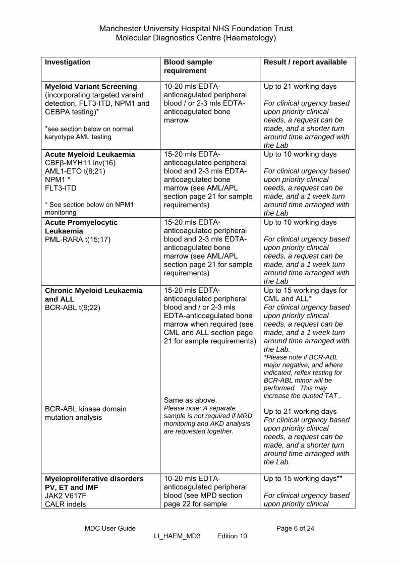
Investigation Blood sample
requirement Result / report available
Myeloid Variant Screening (incorporating targeted varaint detection, FLT3-ITD, NPM1 and CEBPA testing)* *see section below on normal karyotype AML testing
10-20 mls EDTA-anticoagulated peripheral blood / or 2-3 mls EDTA-anticoagulated bone marrow
Up to 21 working days For clinical urgency based upon priority clinical needs, a request can be made, and a shorter turn around time arranged with the Lab
Acute Myeloid Leukaemia CBFβ-MYH11 inv(16) AML1-ETO t(8;21) NPM1 * FLT3-ITD * See section below on NPM1 monitoring
15-20 mls EDTA-anticoagulated peripheral blood and 2-3 mls EDTA-anticoagulated bone marrow (see AML/APL section page 21 for sample requirements)
Up to 10 working days For clinical urgency based upon priority clinical needs, a request can be made, and a 1 week turn around time arranged with the Lab
Acute Promyelocytic Leukaemia PML-RARA t(15;17)
15-20 mls EDTA-anticoagulated peripheral blood and 2-3 mls EDTA-anticoagulated bone marrow (see AML/APL section page 21 for sample requirements)
Up to 10 working days For clinical urgency based upon priority clinical needs, a request can be made, and a 1 week turn around time arranged with the Lab
Chronic Myeloid Leukaemia and ALL BCR-ABL t(9;22) BCR-ABL kinase domain mutation analysis
15-20 mls EDTA-anticoagulated peripheral blood and / or 2-3 mls EDTA-anticoagulated bone marrow when required (see CML and ALL section page 21 for sample requirements) Same as above. Please note: A separate sample is not required if MRD monitoring and AKD analysis are requested together.
Up to 15 working days for CML and ALL* For clinical urgency based upon priority clinical needs, a request can be made, and a 1 week turn around time arranged with the Lab. *Please note if BCR-ABL major negative, and where indicated, reflex testing for BCR-ABL minor will be performed. This may increase the quoted TAT . Up to 21 working days For clinical urgency based upon priority clinical needs, a request can be made, and a shorter turn around time arranged with the Lab.
Myeloproliferative disorders PV, ET and IMF JAK2 V617F CALR indels
10-20 mls EDTA-anticoagulated peripheral blood (see MPD section page 22 for sample
Up to 15 working days** For clinical urgency based upon priority clinical
Manchester University Hospital NHS Foundation Trust Molecular Diagnostics Centre (Haematology)
MDC User Guide Page 7 of 24 LI_HAEM_MD3 Edition 10
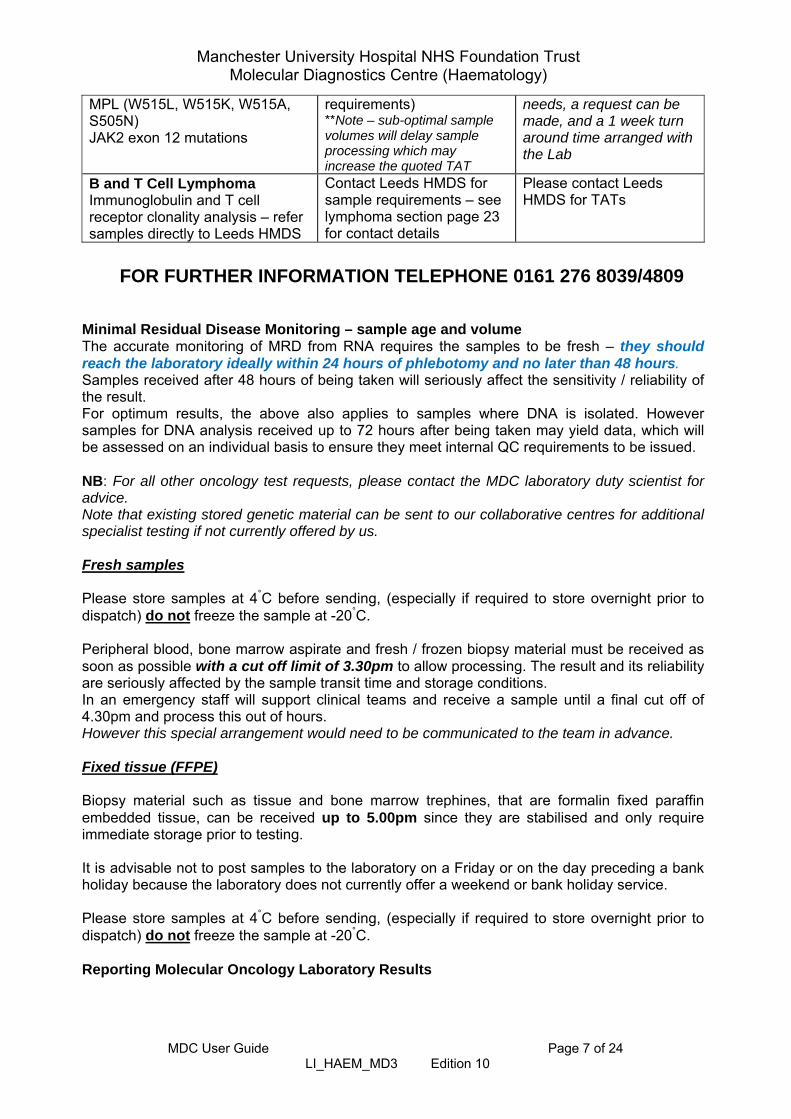
MPL (W515L, W515K, W515A, S505N) JAK2 exon 12 mutations
requirements) **Note – sub-optimal sample volumes will delay sample processing which may increase the quoted TAT
needs, a request can be made, and a 1 week turn around time arranged with the Lab
B and T Cell Lymphoma Immunoglobulin and T cell receptor clonality analysis – refer samples directly to Leeds HMDS
Contact Leeds HMDS for sample requirements – see lymphoma section page 23 for contact details
Please contact Leeds HMDS for TATs
FOR FURTHER INFORMATION TELEPHONE 0161 276 8039/4809
Minimal Residual Disease Monitoring – sample age and volume The accurate monitoring of MRD from RNA requires the samples to be fresh – they should reach the laboratory ideally within 24 hours of phlebotomy and no later than 48 hours. Samples received after 48 hours of being taken will seriously affect the sensitivity / reliability of the result. For optimum results, the above also applies to samples where DNA is isolated. However samples for DNA analysis received up to 72 hours after being taken may yield data, which will be assessed on an individual basis to ensure they meet internal QC requirements to be issued. NB: For all other oncology test requests, please contact the MDC laboratory duty scientist for advice. Note that existing stored genetic material can be sent to our collaborative centres for additional specialist testing if not currently offered by us.
Fresh samples Please store samples at 4°C before sending, (especially if required to store overnight prior to dispatch) do not freeze the sample at -20°C.
Peripheral blood, bone marrow aspirate and fresh / frozen biopsy material must be received as soon as possible with a cut off limit of 3.30pm to allow processing. The result and its reliability are seriously affected by the sample transit time and storage conditions. In an emergency staff will support clinical teams and receive a sample until a final cut off of 4.30pm and process this out of hours. However this special arrangement would need to be communicated to the team in advance. Fixed tissue (FFPE) Biopsy material such as tissue and bone marrow trephines, that are formalin fixed paraffin embedded tissue, can be received up to 5.00pm since they are stabilised and only require immediate storage prior to testing. It is advisable not to post samples to the laboratory on a Friday or on the day preceding a bank holiday because the laboratory does not currently offer a weekend or bank holiday service. Please store samples at 4°C before sending, (especially if required to store overnight prior to dispatch) do not freeze the sample at -20°C.
Reporting Molecular Oncology Laboratory Results
Manchester University Hospital NHS Foundation Trust Molecular Diagnostics Centre (Haematology)
MDC User Guide Page 8 of 24 LI_HAEM_MD3 Edition 10
Results are reported as an authorised report posted directly to the referrer. Results for ORC and Trafford MFT patients, other than MRD monitoring reports, can be accessed via the internal ICE system. Clinical Scientific Diagnostic Advice and Results Interpretation A key component of the service provided by the laboratory is the availability of expert clinical scientific advice. Advice is provided on the genetics and molecular biology of haematological malignant disorders, the investigation of these and the interpretation of test results. See page 3 for contact details.
Manchester University Hospital NHS Foundation Trust Molecular Diagnostics Centre (Haematology)
MDC User Guide Page 9 of 24 LI_HAEM_MD3 Edition 10
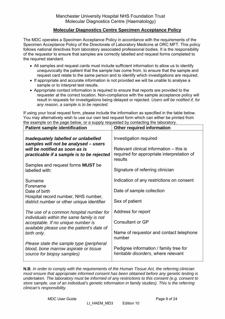
Molecular Diagnostics Centre Specimen Acceptance Policy The MDC operates a Specimen Acceptance Policy in accordance with the requirements of the Specimen Acceptance Policy of the Directorate of Laboratory Medicine at ORC MFT. This policy follows national directives from laboratory associated professional bodies. It is the responsibility of the requestor to ensure that samples are correctly labelled and request forms completed to the required standard.
All samples and request cards must include sufficient information to allow us to identify unequivocally the patient that the sample has come from, to ensure that the sample and request card relate to the same person and to identify which investigations are required.
If appropriate and accurate information is not provided we will be unable to analyse a sample or to interpret test results.
Appropriate contact information is required to ensure that reports are provided to the requester at the correct location. Non-compliance with the sample acceptance policy will result in requests for investigations being delayed or rejected. Users will be notified if, for any reason, a sample is to be rejected.
If using your local request form, please include the information as specified in the table below. You may alternatively wish to use our own test request form which can either be printed from the example on the page below, or a supply requested by contacting the laboratory. Patient sample identification Other required information Inadequately labelled or unlabelled samples will not be analysed – users will be notified as soon as is practicable if a sample is to be rejected Samples and request forms MUST be labelled with: Surname Forename Date of birth Hospital record number, NHS number, district number or other unique identifier The use of a common hospital number for individuals within the same family is not acceptable. If no unique number is available please use the patient’s date of birth only. Please state the sample type (peripheral blood, bone marrow aspirate or tissue source for biopsy samples)
Investigation required Relevant clinical information – this is required for appropriate interpretation of results Signature of referring clinician Indication of any restrictions on consent Date of sample collection Sex of patient Address for report Consultant or GP Name of requestor and contact telephone number Pedigree information / family tree for heritable disorders, where relevant
N.B. In order to comply with the requirements of the Human Tissue Act, the referring clinician must ensure that appropriate informed consent has been obtained before any genetic testing is undertaken. The laboratory must be informed of any restrictions to this consent (e.g. consent to store sample, use of an individual’s genetic information in family studies). This is the referring clinican’s responsibility.
Manchester University Hospital NHS Foundation Trust Molecular Diagnostics Centre (Haematology)
MDC User Guide Page 10 of 24 LI_HAEM_MD3 Edition 10
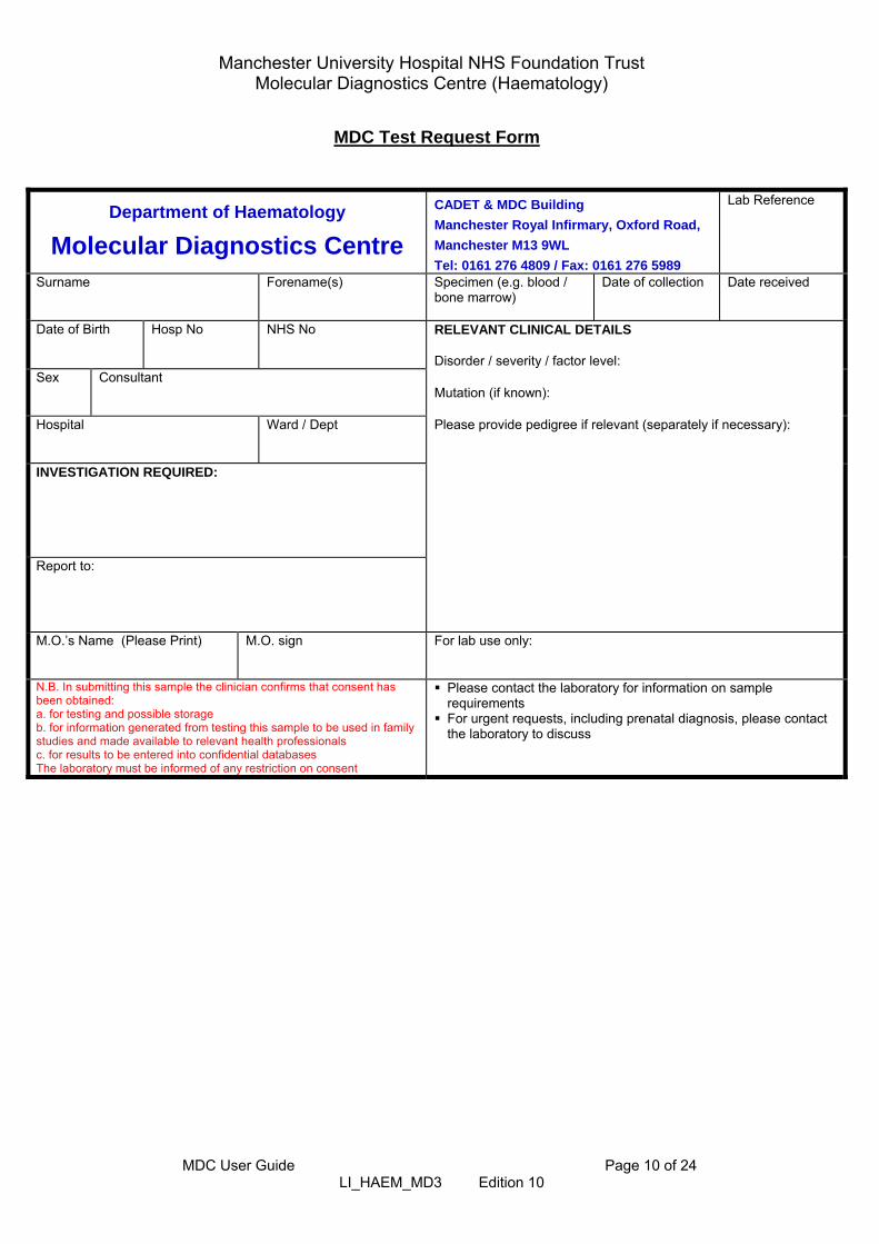
MDC Test Request Form
Department of Haematology
Molecular Diagnostics Centre
CADET & MDC Building
Manchester Royal Infirmary, Oxford Road,
Manchester M13 9WL
Tel: 0161 276 4809 / Fax: 0161 276 5989
Lab Reference
Surname
Forename(s)
Specimen (e.g. blood / bone marrow)
Date of collection
Date received
Date of Birth Hosp No NHS No
RELEVANT CLINICAL DETAILS Disorder / severity / factor level:
Sex
Consultant
Mutation (if known):
Hospital
Ward / Dept
Please provide pedigree if relevant (separately if necessary):
INVESTIGATION REQUIRED: Report to:
M.O.’s Name (Please Print)
M.O. sign
For lab use only:
N.B. In submitting this sample the clinician confirms that consent has been obtained: a. for testing and possible storage b. for information generated from testing this sample to be used in family studies and made available to relevant health professionals c. for results to be entered into confidential databases The laboratory must be informed of any restriction on consent
Please contact the laboratory for information on sample requirements
For urgent requests, including prenatal diagnosis, please contact the laboratory to discuss
Manchester University Hospital NHS Foundation Trust Molecular Diagnostics Centre (Haematology)
MDC User Guide Page 11 of 24 LI_HAEM_MD3 Edition 10
Quality Statement
The MDC is accredited with UKAS as part of the Department of Haematology at Manchester Royal Infirmary, UKAS accredited medical laboratory No. 8650. All investigations are subject to appropriate and rigorous Internal Quality Control (IQC) measures and the laboratory participates in appropriate External Quality Assurance (EQA) schemes, where available, for the disorders investigated.
Confidentiality The Molecular Diagnostics Centre follows local and national patient confidentiality standards. For further information please follow this link: http://www.cmft.nhs.uk/info-for-health-professionals/laboratory-medicine/policies-and-guidelines/confidentiality-of-patient-information
Complaints Procedure
If you have an issue you wish to raise with us, please contact the laboratory in the first instance on 0161 276 4809. If we cannot resolve your query directly, the MDC follows the Manchester Foundation Trust Directorate of Laboratory Medicine complaints procedure, by which means your complaint will be escalated as appropriate.
Manchester University Hospital NHS Foundation Trust Molecular Diagnostics Centre (Haematology)
MDC User Guide Page 12 of 24 LI_HAEM_MD3 Edition 10
Service Provision – Inherited Disorders
Heritable Thrombophilia The MDC provides a diagnostic referral service for genetic risk factors for venous thromboembolism (VTE), specifically for the identification of the FV Leiden mutation and the prothrombin gene variant (PGV). Phenotypic assays to measure antithrombin, protein C and protein S, rare deficiencies of which are associated with increased risk for VTE, and also lupus anticoagulant screening, are available from the Blood Coagulation Laboratory at ORC MFT. Note: There is convincing evidence that homozygosity for the methylenetetrahydrofolate reductase (MTHFR) genetic variant C677T is not a risk factor for VTE. There is no rationale for MTHFR C677T genotyping for clinical diagnostic purposes in VTE, hence this is not offered by the MDC. Background and general principles Thrombophilia testing is not appropriate and should not be carried out unless the
results may influence clinical management. Thrombophilia is a multifactorial condition with heritable, acquired and circumstantial
components which interact to give rise to an increased risk for VTE. VTE most commonly manifests as deep vein thrombosis (DVT) in the leg. This may progress to pulmonary embolism (PE) if the clot dislodges and travels to the lung. The incidence of venous thromboembolism (VTE) in the general population increases with age.
Up to 50% of individuals with VTE may have an identifiable heritable thrombophilia. This
has led to a large demand for thrombophilia testing, despite the fact that there is frequently a lack of evidence in the literature that this testing has clinical utility. In most cases patient management is unlikely to be altered by the presence or absence of a thrombophilia risk factor.
Heritable thrombophilias include factor V Leiden (F5 gene c.1601G>A) and a prothrombin
gene variant (F2 gene c.*97G>A) - PGV, both of which are common in Caucasians but generally rare in other ethnic groups, and deficiencies of antithrombin (AT), protein C (PC) and protein S (PS), all of which are rare.
The majority of the VTE events occurring in individuals with heritable thrombophilia are
provoked by one or more predisposing factors, eg surgery, immobility, increasing age, pregnancy, combined oral contraceptive pill, malignancy.
The magnitude of risk associated with a positive thrombophilia test result is often perceived
by the patient to be greater than it actually is, with associated negative psychological impact.
Testing for heritable thrombophilia is not indicated in unselected patients who present with
a first episode of VTE. Initiation and intensity of anticoagulant therapy following a diagnosis of acute VTE is not
generally influenced by the presence or absence of a heritable thrombophilia. Patients with PC or PS deficiency, however, should have initiation of anticoagulation with low molecular weight heparin for an adequate period of time alongside Warfarin.
Manchester University Hospital NHS Foundation Trust Molecular Diagnostics Centre (Haematology)
MDC User Guide Page 13 of 24 LI_HAEM_MD3 Edition 10
Decisions regarding duration of anticoagulation should be made taking into account
whether or not a first VTE was provoked and considering the risk of anticoagulant therapy related bleeding, regardless of whether or not a heritable thrombophilia is known to be present.
Finding a common heritable thrombophilia (heterozygosity for FV Leiden or PGV) does not
typically predict the likelihood of VTE recurrence. There is a higher recurrence risk in patients with AT, PC, or PS deficiency or with multiple defects.
Evidence suggests that thrombophilia testing does not reduce the risk of VTE recurrence in
clinical practice. Patients who have recurrently normal D-dimers after completion of anticoagulation therapy
have a comparatively low risk of VTE recurrence. Family testing Thrombophilia testing of asymptomatic relatives of patients with a history of VTE has not
been shown to reduce the incidence of VTE. The annual risk of unprovoked thrombosis in affected family members is low.
Case-finding of asymptomatic relatives with low risk thrombophilias such as heterozygosity
for FV Leiden or PGV is not indicated. Screening for AT deficiency is warranted in family members of individuals with AT deficiency.
If family history suggests a high degree of genetic penetrance it may be informative to test
symptomatic patients and their asymptomatic relatives with a view to informing clinical management at times of high thrombotic risk.
VTE has a clear familial component, independent of the presence or absence of a known
thrombophilia risk factor. In symptomatic families negative thrombophilia test results may be falsely reassuring.
Combined oral contraceptive pill (COCP) and HRT Personal and family clinical history is key to identify women at risk of VTE. Unselected
testing for heritable thrombophilia will provide an uncertain estimate of risk and is not warranted.
The absolute risk for VTE in previously asymptomatic women with FV Leiden or PGV using
COCP or HRT is not high. There is a substantial risk for VTE in women with AT, PC or PS deficiency, FV Leiden
homozygosity, PGV homozygosity, or combined thrombophilia defects in association with COCP or HRT use. In these cases COCP or HRT use is contraindicated.
The baseline risk for VTE in women using HRT is higher than in women using COCP
because the HRT-user population is older with a higher age-related risk for VTE. Testing for heritable thrombophilia in selected cases may assist counselling of women
considering COCP or HRT, for example if a high-risk thrombophilia has been identified in a symptomatic relative.

Manchester University Hospital NHS Foundation Trust Molecular Diagnostics Centre (Haematology)
MDC User Guide Page 14 of 24 LI_HAEM_MD3 Edition 10
Irrespective of the presence or absence of a detected heritable thrombophilia, a personal history of VTE is a contraindication to COCP or oral HRT. The use of COCP by women with a family history of VTE in a first-degree relative aged under 45 years is not recommended. A family history of VTE in a first-degree relative is a relative contraindication to HRT.
VTE in pregnancy There is a 5- to 10-fold increased risk for VTE during pregnancy. The risk is increased 100-
fold in women with previous thrombosis. In women with heterozygosity for FV Leiden or PGV, and no previous thrombotic history,
the absolute risk for pregnancy-associated VTE is low. Women with AT deficiency, PC deficiency, PS deficiency, homozygosity for FV Leiden, homozygosity for PGV, or compound heterozygosity are at higher risk.
Women should be clinically assessed for risk of pregnancy-associated VTE. Testing for
heritable thrombophilia is not generally required. Women with a previous unprovoked VTE should be tested for the presence of antiphospholipid antibodies.
In women with a VTE during pregnancy a screen for heritable thrombophilia (FV Leiden,
PGV, AT deficiency, PC deficiency, PS deficiency) should be carried out 6 to 8 weeks after discontinuation of antithrombotic therapy. Screening for antiphospholipid syndrome should also be performed.
It may be clinically informative to test asymptomatic women with a family history of VTE if a
thrombosis in a first-degree relative was unprovoked, or provoked by pregnancy, COCP use, or a minor risk factor, particularly if this is associated with a known thrombophilia.
Women with a family history of VTE and either AT deficiency or where a specific
thrombophilia has not been detected should be tested for antithrombin deficiency.
Recurrent pregnancy loss There is an association of FV Leiden and PGV with recurrent early and late pregnancy loss.
However these are weak contributors to risk rather than the sole cause of pregnancy loss. Testing for heritable thrombophilia is not recommended. Testing for the presence of
antiphospholipid antibodies should be undertaken. Recent large clinical trials of antithrombotic therapy have indicated no benefit of low
molecular weight heparin in preventing recurrent early pregnancy loss in the absence of thrombophilia. Further research is required to determine if LMWH may improve live birth rates in women with recurrent pregnancy loss in the presence of thrombophilia.
Arterial thrombosis There is no established causal relationship between heritable thrombophilia and arterial
thrombosis. Testing for heritable thrombophilia is not indicated in patients with arterial thrombosis.
Manchester University Hospital NHS Foundation Trust Molecular Diagnostics Centre (Haematology)
MDC User Guide Page 15 of 24 LI_HAEM_MD3 Edition 10
Children Thrombophilia testing in children is only available after discussion with a clinical
haematologist. The annual incidence of VTE in children is about 1/100,000 in the general population.
There is a bimodal distribution with peaks in the neonatal period and in adolescence. More than 90% of paediatric thrombotic events are related to underlying medical or surgical
risk factors, central venous lines in particular. D-dimers should not be used to diagnose or exclude VTE in children. Testing for heritable thrombophilia in unselected children presenting with a first VTE is not
indicated. Testing has uncertain predictive value for recurrence. Neonates and children with purpura fulminans should be tested urgently for PC and PS
deficiency. Children with early onset spontaneous thrombosis should be investigated for AT deficiency Children presenting with unprovoked VTE should be tested for anti-phospholipid antibodies.
Manchester University Hospital NHS Foundation Trust Molecular Diagnostics Centre (Haematology)
MDC User Guide Page 16 of 24 LI_HAEM_MD3 Edition 10
Hereditary Haemochromatosis (HH) A genetic diagnostic service is provided to identify DNA changes in the HFE gene which are
associated with an increased risk of developing HH. Identification of these changes facilitates the diagnosis and clinical management of at risk individuals, including family studies.
HH, an autosomal recessive adult onset disorder, is marked by increased plasma
transferrin saturation associated with defective synthesis and / or regulation of hepcidin. Genes involved include HFE, HFE2, TFR2 and HAMP. HFE-related HH is by far the most common form of the disorder.
Patients with suspected iron overload should have fasting transferrin saturation and serum
ferritin measurements. Normal serum ferritin essentially rules out iron overload, except in very rare cases of non HFE related HH. Raised serum ferritin levels have low specificity. HFE genotyping is indicated only if transferrin saturation is increased.
About 80% of Europeans with clinical iron overload are homozygous for the HFE C282Y
variant (p.Cys282Tyr). When iron stores are elevated, with or without clinical symptoms, C282Y homozygosity is required for a diagnosis of HFE-HH. Any other HFE genotype should be interpreted with caution. Homozygosity alone for C282Y is not sufficient to diagnose HH - about 1 in 250 Caucasians in the general population have this genotype.
C282Y homozygotes are likely to develop increased serum ferritin and transferrin
saturation, however only about 15 to 20% of individuals with this genotype go on to show clinical symptoms of iron overload. HH penetrance in C282Y homozygotes is higher in men than in women. Environmental factors such as alcohol misuse, steatosis and viral infections play key roles in the development of clinical symptoms.
The risk to develop iron overload in the future is increased in asymptomatic adult first
degree relatives of C282Y homozygous index cases. Genotyping for C282Y, in association with genetic counselling, is recommended in such relatives in order to inform their future risk to develop iron overload.
The predicted heterozygous carrier rate for C282Y in the general population is
approximately 10%. C282Y heterozygotes, including those in families where a diagnosis of iron overload has been made, are at very low risk of developing HH and genotyping of their first degree relatives is not recommended.
Hereditary haemochromatosis associated with the C282Y HFE gene mutation is a late-
onset disorder of low clinical penetrance. Our policy, in accordance with extant national guidance, is not to perform genetic testing in children until they are old enough to make their own informed decision about such testing, unless test results will have a direct bearing on current clinical management.
If a child has a close family history of clinical haemochromatosis parental testing may be
appropriate to establish the presence or absence of a HFE risk genotype for haemochromatosis. This would then inform the possibility of offering future testing, in adulthood.
In Europe, the HFE H63D (p.His63Asp) polymorphism is found in 20 to 25% of the general
population. C282Y / H63D compound heterozygosity is a risk factor for mildly elevated serum iron and hepatic iron stores but is considered to be insufficient independently to

Manchester University Hospital NHS Foundation Trust Molecular Diagnostics Centre (Haematology)
MDC User Guide Page 17 of 24 LI_HAEM_MD3 Edition 10
cause haemochromatosis. C282Y / H63D compound heterozygotes with iron overload should be investigated for additional contributory causes of hyperferritinaemia.
Homozygosity for H63D may be associated with increased transferrin saturation and serum
ferritin, however this genotype is considered unlikely independently to be sufficient cause for iron overload. H63D homozygotes with iron overload should be investigated for additional contributory causes of hyperferritinaemia. Heterozygosity for H63D is not a risk factor to develop haemochromatosis.
About 1 to 3% of individuals in northern European populations carry the HFE S65C
(p.Ser65Cys) polymorphism. There is no evidence that S65C has a role in clinically manifest iron overload and testing for S65C is not recommended for diagnostic purposes. We therefore do not carry out genotyping for this polymorphism.
About 5% of patients with haemochromatosis lack the above variants in the HFE gene.
Other rare genetic factors may be involved in some of these cases. If there is evidence of iron accumulation in the presence of a normal HFE genotype, such as persistently raised transferrin saturation, then further investigation may be justified after exclusion of secondary iron overload.
Manchester University Hospital NHS Foundation Trust Molecular Diagnostics Centre (Haematology)
MDC User Guide Page 18 of 24 LI_HAEM_MD3 Edition 10
Heritable Bleeding Disorders The MDC has close links with the Comprehensive Care Haemophilia Centres at Manchester
Royal Infirmary (MRI) and the Royal Manchester Children’s Hospital (RMCH). Genetic diagnosis in heritable bleeding disorders, including clinical scientific interpretation of individual cases, is provided to these haemophilia centres and also, as required, to other centres nationally and internationally.
Comprehensive genetic diagnosis is presently available for haemophilia A, haemophilia B,
von Willebrand disease (VWD), FXI deficiency, and FVII deficiency. Informed consent. It is the referring clinician’s responsibility to obtain and record informed
consent prior to genetic diagnosis in all heritable bleeding disorders. In this regard, it is also the referring clinician’s responsibility to inform the laboratory of any restrictions on consent (for example to store genetic information on confidential databases, or to apply an individual’s genetic information in the diagnosis of other family members). A standard consent form and patient information leaflet is in use within the MFT Comprehensive Care Haemophilia Centres.
Haemophilia A and Haemophilia B Haemophilia A is a heritable bleeding disorder resulting from low or, in severe cases, absent
functional levels of coagulation factor VIII. Inheritance is X-linked recessive. Haemophilia A is the most common of the severe bleeding disorders, with an approximate incidence of 1 new case per 5,000 male births. Haemophilia A is caused by mutations in the factor VIII (F8) gene.
Haemophilia B is a heritable bleeding disorder resulting from low or, in severe cases, absent
functional levels of coagulation factor IX. Inheritance is X-linked recessive. Haemophilia B has an approximate incidence of 1 new case per 25,000 male births. Haemophilia A is caused by mutations in the factor IX (F9) gene.
Investigations carried out include familial mutation identification and interpretation, carrier
diagnosis and prenatal diagnosis (PND). Genetic PND may be performed early in pregnancy, usually via chorionic villus sampling
(CVS) at 11 to 13 weeks of gestation. This allows first trimester diagnosis, avoiding late termination of pregnancy. The large majority of patients, following appropriate expert counselling, do not opt to terminate an affected pregnancy.
Genetic PND may also be performed in the third trimester of pregnancy, usually at about 36
weeks of gestation, by means of amniocentesis. This approach enables diagnosis of the presence or absence of a severe bleeding disorder in the fetus, facilitating important decisions about the clinical management of childbirth.
Genetic diagnosis of the causative mutation can provide important clinical information about
the risk for inhibitor development associated with clotting factor replacement therapy. This may particularly be valuable in mild or moderate haemophilia A.
Knowledge of the causative mutation may influence diagnosis and clinical management in
specific cases, for example genetic diagnosis of haemophilia B Leyden, where the onset of puberty leads to amelioration of the haemophilia phenotype, or the identification of mutations associated with a 1-stage / 2-stage clotting factor VIII assay discrepancy in haemophilia A.
Manchester University Hospital NHS Foundation Trust Molecular Diagnostics Centre (Haematology)
MDC User Guide Page 19 of 24 LI_HAEM_MD3 Edition 10
von Willebrand Disease (VWD) VWD, the most common of the heritable bleeding disorders, results from qualitative or
quantitative deficiencies of von Willebrand factor (VWF). VWF has essential roles in platelet-dependent primary haemostasis, and as a carrier for coagulation factor VIII in the blood circulation. Inheritance of VWD is autosomal. The molecular biology of VWD is complex.
There are three main types of VWD. Type 1 VWD is the most common form of the disorder,
resulting from a partial quantitative deficiency of VWF. Type 2 VWD results from VWF qualitative deficiencies, including type 2A, 2B, 2M and 2N VWD variants. Type 3 VWD is a rare, recessive, often severe bleeding disorder, the result of essentially complete quantitative deficiency of VWF.
Genetic diagnosis has an important role in type 3 VWD in carrier diagnosis, where this may
not be achievable by phenotypic means, and in PND in affected families (as above for haemophilia A and haemophilia B). Genetic diagnosis also has a role in selected cases of type 2 VWD. Generally speaking there is little application of genetic diagnosis in type 1 VWD.
FXI Deficiency Heritable FXI deficiency is a rare bleeding disorder resulting from low functional levels of
coagulation factor XI. Inheritance is autosomal. The estimated prevalence of major FXI deficiency (FXI:C ≤15 IU/dL) in most populations is about 1:1,000,000, however in Ashkenazi Jews it occurs in about 1:450 individuals.
Most bleeding in FXI deficiency is related to injury, although bleeding risk is unpredictable
and not related to genotype. Some patients with major FXI deficiency do not bleed, others with partial FXI deficiency (FXI:C 16 to 60 IU/dL) may bleed.
Heritable FXI deficiency is caused by mutations in the factor XI gene (F11). Major FXI
deficiency is generally associated with homozygosity or compound heterozygosity for F11 mutations. Partial FXI deficiency is usually associated with heterozygosity for mutations in F11. Genetic diagnosis has applications in family studies in FXI deficiency.
FVII Deficiency Heritable FVII deficiency is a rare bleeding disorder resulting from low functional levels of
coagulation factor VII. It is characterised by autosomal recessive inheritance, and has a prevalence of about 1:500,000.
FVII deficiency is clinically a very heterogeneous disorder. Bleeding symptoms range in
severity from asymptomatic, to mild (most cases), to lethal. A complete lack of FVII is considered to be incompatible with life.
F7 gene mutation(s) can be found in most cases of heritable FVII deficiency, however
genotype does not correlate well with FVII level or with bleeding risk. Genetic diagnosis has limited utility in the clinical management of FVII deficiency, although it can have a role in PND in families (frequently consanguineous) with severe FVII deficiency.
Manchester University Hospital NHS Foundation Trust Molecular Diagnostics Centre (Haematology)
MDC User Guide Page 20 of 24 LI_HAEM_MD3 Edition 10
Haemoglobinopathy
Haemoglobinopathy genetic diagnosis, including mutation identification, carrier and prenatal diagnosis, together with clinical interpretation, is provided from the MDC in collaboration with the Department of Haematology at the MRI, the Manchester Sickle Cell and Thalassaemia Centre.
The service is offered to pre-conceptual and ante-natal couples who are at risk of giving birth to children affected by a clinically significant haemoglobinopathy, as identified through phenotypic screening under the auspices of the NHS Sickle Cell and Thalassaemia Screening Programme: http://sct.screening.nhs.uk/
Our service has the ability to perform comprehensive genetic analysis of the alpha and beta
globin gene clusters by a combination of the following methods:
- Gap-PCR for common alpha gene deletions
- DNA sequencing of the alpha and beta gene loci for detection of point mutations and variants, with additional confirmatory tests as appropriate, e.g. ARMS PCR
- MLPA analysis of the alpha and beta globin gene clusters for rare or novel deletions/duplications
- HPFH investigation by a combination of HPFH specific gap PCR tests, DNA sequencing and MLPA (tests applied as required)
Genetic diagnosis referrals should meet GENETIC HAEMOGLOBINOPATHY TESTING
GUIDELINES - Referral guidance and associated paperwork can be accessed via the following link: https://mft.nhs.uk/the-trust/other-departments/laboratory-medicine/haematology/haemoglobinopathy/
Referrals will not be analysed until all appropriate referral information, including consent for
DNA analysis, has been received by the laboratory.
Where the local clinical service is managed by experts in these disorders, it will be at the discretion of your local referral team to decide which cases merit further investigation. These will be charged per investigation depending on the level of analysis required. For details of pricing please contact us to discuss individual referrals.
Manchester University Hospital NHS Foundation Trust Molecular Diagnostics Centre (Haematology)
MDC User Guide Page 21 of 24 LI_HAEM_MD3 Edition 10
TPMT genotyping (joint with the Biochemistry Department at ORC MFT)
TPMT (Thiopurine S-Methyltransferase) catalyses the rate-limiting enzymatic step in
the breakdown and excretion of immunosuppressive and chemotherapeutic drugs including Azathioprine and 6-Mercaptopurine. Drugs based on these compounds are used in the treatment of conditions including autoimmune disorders such as Crohn's disease and rheumatoid arthritis, organ transplant recipients, and in acute lymphoblastic leukaemia.
1 in 300 Caucasians are deficient in TPMT activity and are thus prone to life-
threatening myelosuppression if treated with standard thiopurine drug doses. A further 10% of the population have intermediate TPMT activity and are also prone to thiopurine drug side effects, requiring lower dosing.
Various clinical guidelines advocate determination of TPMT status of a patient prior
to administration of thiopurine drugs in order to limit the number of adverse drug reactions.
In the majority of cases, TPMT activity is determined for each referral by a
biochemical assay and only those with intermediate or deficient TPMT activity will subsequently be referred for TPMT genotyping analysis.
The majority of the variation in TPMT activity is accounted for by the presence of one
or more non-synonymous SNPs; in those with intermediate TPMT activity these are present on one allele only, whilst those with deficient TPMT activity are either homozygotes or compound heterozygotes for such changes.
Whilst many different TPMT genotypes have been described, 85-95% of TPMT
genetic variations are accounted for by the genotypes TPMT*3A, *3B, *3C and *2, characterized by SNPs within three exons of TPMT. The standard test for TPMT genotyping test detects these common variants only.
If deficient TPMT activity is not described by one of the common SNPs clinicians may
choose, at additional cost, for full TPMT gene analysis by DNA sequencing. Normally this approach should be applied after confirmatory TPMT activity testing has been performed.
Patients for which TPMT activity analysis is inappropriate, for example those that
have had a recent blood transfusion, can be genotyped as a front line test in order to exclude the presence of the common TPMT variants. Note that the referral route for TPMT genotyping is normally via Biochemistry so that the appropriate TPMT activity assay can be performed, and samples triaged for onward referral to the MDC for genotyping, where indicated.
Manchester University Hospital NHS Foundation Trust Molecular Diagnostics Centre (Haematology)
MDC User Guide Page 22 of 24 LI_HAEM_MD3 Edition 10
Service Provision – Haemato-Oncology
Chronic Myeloid Leukaemia The pre-treatment transcript level of t(9;22) [BCR-ABL] can inform prognosis and determine downstream log reduction levels. In addition, 3 monthly follow up minimal residual disease (MRD) monitoring is recommended as per national and international guidelines. Studies have indicated PB monitoring for most follow up time-points is sufficient, with BM monitoring at stipulated time points, according to national and ELN guidelines, where these will help to inform when cytogenetics testing is necessary. The use of donor lymphocyte infusion (DLI) can be indicated in post transplant patients. Therefore if DLI is being used, a clinically urgent test request can be made, so that a shorter turn around time is implemented in the laboratory to support therapeutic decision making. This would also be available when other clinical urgencies require this, such as loss of a response or disease progression. Drug Resistance The availability of 2nd and 3rd plus generation drugs, their appropriate usage, and the occurrence of resistance to therapy due to the presence of ABL kinase domain mutations, requires additional testing. We currently offer this test which is applied to mutations occurring in the kinase domain of the BCR-ABL fusion gene. This does not exclude other mechanisms of resistance. Acute Lymphocytic Leukaemia In ALL, paired PB and BM samples are required for MRD monitoring in acute phase disease. Refer to the AML sample criteria for PB and BM samples required below. We currently offer BCR-ABL testing for these patients. Acute Myeloid Leukaemia MRD monitoring is used to guide therapy and identify those patients at an increased risk of relapse. Paired PB and BM samples are required to accurately determine levels of MRD. This is because there can be large differences in the interpretation of the relapse risk if you are looking at leukaemic gene expression in PB or BM alone. For this reason it is important to identify the sample type (PB or BM) on the requisition form or request card. The accurate monitoring of MRD requires samples to be fresh, to reach us ideally within 24 hours of phlebotomy and no later than 48 hours for reliable result generation. When it is not possible to obtain both samples, it is advised to send one sample PB or BM rather than miss the time point of MRD monitoring and the interpretation of the results will take this into account.
Core-Binding Factor (CBF) Leukaemias Paired PB and BM samples are required for MRD as per the specimen requirements table Page 6. Use of DLI or monitoring patients pre and post transplant can be very informative for their clinical management. A repeat sample should always be tested if a
Manchester University Hospital NHS Foundation Trust Molecular Diagnostics Centre (Haematology)
MDC User Guide Page 23 of 24 LI_HAEM_MD3 Edition 10
significant change in transcript levels is observed to confirm this change, prior to any alteration in therapeutic decision making, which applies to all MRD monitoring. Acute Promyelocytic Leukaemia For APML, BM samples can indicate increased risk of relapse much earlier than PB. Residual transcript detection, following completion of treatment, is highly indicative of an increased risk of relapse. Paired PB and BM samples are the most informative specimens for MRD monitoring. As with all MRD, a pre-treatment diagnostic level is important to look at log reduction in levels over time. In APL, there is also the advantage of being able to detect the genetic lesion in molecular assays if cytogenetic testing is unable to detect the translocation due to technical issues with the sample. This helps to confirm the diagnosis and inform clinical management of the patient. Normal Karyotype AML testing (Myeloid variant screening) 40-50% of AMLs are cytogenetically normal (CN-AML) and molecular markers are becoming increasingly important for further classification of this subgroup. Variants of CEBPA, NPM1 and FLT3 are among the most common abnormalities found in CN-AML and bear important prognostic significance. Pathogenic variants in these genes are detected in our laboratory by a panel of screening tests. The Agena assay is designed to detect 58 variants associated with AML. The variants detected in the assay are located in the following genes: KIT, DNMT3a, FLT3, IDH1, IDH2, JAK2, KRAS, MPL, NPM1, NRAS, SF3B1, SRSF2, U2AF1 and WT1. Individual tests are performed for CEBPA (Sanger sequencing), FLT3-ITD and NPM1 (Fragment analysis). Testing on the diagnostic pre-treatment sample is recommended preferably bone marrow but peripheral blood is also suitable. Myeloproliferative Disorders In 2005, the JAK2 V617F mutation was found in almost all patients with PV and about half of those patients with ET and IMF. This test is now part of the clinical work up guidelines for diagnosing these patients. For JAK2 molecular tests, 10ml EDTA PB is required and not a BM aspirate sample. Polycythaemia Vera (PV) The JAK2 V617F mutation is found in ~95% of cases with PV, however approximately 5% of patients do not have this mutation. It has been reported that the majority of the JAK2 V617F negative PV patients have a mutation in an alternative region of the JAK2 gene, within exon 12. The laboratory currently performs testing for both JAK2 V617F and JAK2 exon 12 mutations.
Essential Thrombocythaemia (ET) and Idiopathic Myelofibrosis (IMF) These patients are tested for the JAK2 V617F mutation. In JAK2 V617F-negative ET and IMF cases, 3-4% and 4-8% of patients respectively have a mutation in exon 10 of the MPL gene. Mutations in exon 9 of the calreticulin gene (CALR) have been described in around one third of ET and IMF patients who are JAK2 V617F-negative and MPL-
Manchester University Hospital NHS Foundation Trust Molecular Diagnostics Centre (Haematology)
MDC User Guide Page 24 of 24 LI_HAEM_MD3 Edition 10
non-mutated. Screening of these mutations has been included as part of the BCSH criteria for the diagnosis of ET and IMF. CALR and MPL screening is performed by the laboratory.
B and T cell lymphomas At the present time please discuss sample requirements with Leeds HMDS. Contact details: HMDS St James’s Institute of Oncology Level 3 Bexley Wing St James’s University Hospital Leeds LS9 7TF Telephone for General Enquiries: 01132067851 Fax: 011332067883 Website: www.hmds.info
Clinical Trials Samples Clinical trial protocols will advise on the appropriateness of referral to designated centres and when it is appropriate to use regional centres. When the MDC laboratory participates in a clinical trial as the designated UK centre for testing, this will be indicated in the trial protocol. It is also appropriate to refer patients for relevant molecular testing that are not participants in a clinical trial. Therefore these can be referred directly to the MDC laboratory and any advice required can be sought by contacting the laboratory, with contact details indicated on page 3. Tests under development (will be referred on by us until in-house testing is validated) - Molecular targets as required for the molecular work up indicated by standard and
best practice relating to leukaemia and lymphoma. - Next generation sequencing to assess increased panels of genes for diagnostic
profiling to inform risk stratification of patients for specific treatment options The above gene panel tests will be influenced by NHS E Test Directory requirements