molecular characterization of the induction of cell cycle
TRANSCRIPT

Molecular Characterization of the Induction of Cell Cycle Inhibitor
p21 in Response to Inhibition of the Mitotic Kinase Aurora B
Untersuchungen zur Induktion des Zellzyklusinhibitors p21 nach
Inhibition der Mitotischen Kinase Aurora B
Doctoral thesis for a doctoral degree
at the Graduate School of Life Sciences, Julius-Maximilians-Universität Würzburg,
Section, Biomedicine
submitted by
Geeta Kumari
from
Kanpur, UP, India
Würzburg, 2014

Submitted on: ……………………………………………… Office stamp
Members of the Promotionskomitee:
Chairperson: Prof. Dr. Alexander Buchberger
Primary Supervisor: Prof. Dr. Stefan Gaubatz
Supervisor (Second): Prof. Dr. Peter Gallant
Supervisor (Third): Prof. Dr. Manfred Alsheimer
Supervisor (Fourth): ……………………………………………… (If applicable)
Date of Public Defence: …………………………………………….
Date of Receipt of Certificates: …………………………………………

Substantial parts of this thesis were published in following two articles:
1) Geeta Kumari, Tanja Ulrich, Michael Krause, Florian Finkernagel and Stefan Gaubatz.
“Induction of p21CIP1 and cell cycle arrest after inhibition of Aurora B kinase is attributed
to aneuploidy and reactive oxygen species.” J Biol Chem. 2014 April 29; doi:
10.1074/jbc.M114.555060.
2) Geeta Kumari*, Tanja Ulrich*, and Stefan Gaubatz. "A role for p38 in transcriptional
elongation of p21CIP1 in response to Aurora B inhibition." Cell Cycle. 2013 Jul
1;12(13):2051-60. doi: 10.4161/cc.25100. Epub 2013 Jun 6. *These authors contributed
equally to this work.

! I!
Table of Contents 1 Introduction ......................................................................................................... 1
1.1 The mammalian cell cycle and its regulation ....................................................... 1 1.2 Transcriptional regulation during cell cycle ......................................................... 3 1.3 Aurora kinases and there role in cancer ............................................................... 5
1.3.1 Role of Aurora B in cell cycle .............................................................................. 7 1.3.2 Regulation of Aurora B kinase function .............................................................. 9 1.3.3 Role of Aurora B kinase in cancer and inhibitors against Aurora B .................... 9
1.4 The cellular stress response and its relevance for cancer therapy ................. 10 1.4.1 The p53 tumor suppressor pathway ................................................................. 10 1.4.2 Mitogen activated protein kinase pathway (MAPK pathway) ............................ 11
1.5 Aneuploidy and cancer ......................................................................................... 13 1.6 Objectives of thesis ............................................................................................... 16
2 Materials and Methods ..................................................................................... 17 2.1 Materials ................................................................................................................. 17
2.1.1 Chemical stocks and reagents .......................................................................... 17 2.1.2 Antibiotics ......................................................................................................... 18 2.1.3 Enzymes ........................................................................................................... 18 2.1.4 Molecular kits and Protein/DNA markers .......................................................... 19 2.1.5 Devices ............................................................................................................. 19 2.1.6 Buffers .............................................................................................................. 19
2.1.6.1 General buffers ....................................................................................................... 19 2.1.6.2 Buffers for whole cell lysates ................................................................................... 20 2.1.6.3 Buffers for immunoblotting ...................................................................................... 21 2.1.6.4 Buffers for Chromatin Immunoprecipitation (ChIP) ................................................. 22 2.1.6.5 Buffers for flow cytometry (FACS) ........................................................................... 23 2.1.6.6 Buffers for immunofluorescence ............................................................................. 23 2.1.6.7 Buffers for centromere Fluorescence in-situ hybridization (FISH) .......................... 23 2.1.6.8 Staining solution ...................................................................................................... 23
2.1.7 Antibodies ......................................................................................................... 24 2.1.7.1 Primary antibodies .................................................................................................. 24 2.1.7.2 Secondary antibodies .............................................................................................. 26
2.1.8 Beads ................................................................................................................ 26 2.1.9 Plasmids ........................................................................................................... 27
2.1.9.1 Plasmids for overexpression ................................................................................... 27 2.1.9.2 Plasmids for RNA knockdown ................................................................................. 27

! II!
2.1.10 Primers ........................................................................................................... 27 2.1.10.1 Primers for cloning ................................................................................................ 27 2.1.10.2 Primers for quantitative real time PCR .................................................................. 28 2.1.10.3 Primers for Chromatin Immunoprecipitation .......................................................... 29
2.1.11 siRNA sequences ........................................................................................... 30 2.1.12 Cell lines, cell culture media and transfection reagents .................................. 30
2.1.12.1 Media and additives for mammalian cell culture ................................................... 30 2.1.12.2 Composition of media for soft agar assay ............................................................. 30 2.1.12.3 Human cell lines and media .................................................................................. 31 2.1.12.4 Transfection reagents and cell lines ...................................................................... 31 2.1.12.5 Bacterial strains ..................................................................................................... 31 2.1.12.6 Media for bacterial cell culture .............................................................................. 31
2.2 Methods .................................................................................................................. 32 2.2.1 Mammalian cell culture ..................................................................................... 32
2.2.1.1 Passaging of cells ................................................................................................... 32 2.2.1.2 Freezing and thawing of cells .................................................................................. 32 2.2.1.3 Counting cells .......................................................................................................... 32 2.2.1.4 Treatment of cells with reagents ............................................................................. 32 2.2.1.5 Synchronization of U2OS cells by thymidine .......................................................... 33 2.2.1.6 Determination of cell cycle phases by Flow Cytometry ........................................... 33 2.2.1.7 Transient transfection .............................................................................................. 33
2.2.1.7.1 Plasmid transfection with Calcium phosphate ................................................. 33 2.2.1.7.2 siRNA transfection with Lipofectamine RNAi Max ........................................... 34
2.2.1.8 Retroviral infection of cells ...................................................................................... 34 2.2.1.9 Immunofluorescence staining ................................................................................. 34 2.2.1.10 ROS detection ....................................................................................................... 35 2.2.1.11 Centromere fluorescence in situ hybridisation (FISH) ........................................... 35 2.2.1.12 Colony forming assay ............................................................................................ 35 2.2.1.13 Soft agar assay ..................................................................................................... 36
2.2.2 Molecular methods ........................................................................................... 36 2.2.2.1 RNA isolation .......................................................................................................... 36 2.2.2.2 Reverse transcription (RT) ...................................................................................... 36 2.2.2.3 Quantitative real-time PCR (qRT-PCR) .................................................................. 37
2.2.3 Biochemical methods ........................................................................................ 38 2.2.3.1 Whole cell lysates ................................................................................................... 38 2.2.3.2 Quantification of protein by Bradford method .......................................................... 38 2.2.3.3 Immunoprecipitation ................................................................................................ 38 2.2.3.4 SDS polyacrylamide gel electrophoresis (SDS-PAGE) ........................................... 38 2.2.3.5 Immunoblotting ........................................................................................................ 39 2.2.3.6 Chromatin immmunoprecipitation (ChIP) ................................................................ 39

! III!
2.2.4 Molecular biology ............................................................................................. 40 2.2.4.1 Isolation of plasmid DNA from bacteria ................................................................... 40
2.2.4.1.1 Mini preparation ............................................................................................... 40 2.2.4.1.2 Midi and Maxi preparation ................................................................................ 41
2.2.4.2 Isolation of plasmid DNA fragments from agarose gels .......................................... 41 2.2.4.3 Isolation of PCR products after restriction ............................................................... 41 2.2.4.4 Standard cloning methods ...................................................................................... 41
2.2.4.4.1 PCR for cloning of DNA fragments .................................................................. 41 2.2.4.4.2 Agarose gel electrophoresis ............................................................................ 42 2.2.4.4.3 Restriction digestion ......................................................................................... 42 2.2.4.4.4 Ligation ............................................................................................................ 43 2.2.4.4.5 Transformation of DH5α by heat shock ........................................................... 43 2.2.4.4.6 Sequencing ...................................................................................................... 43
3 Results ............................................................................................................... 44 3.1 Inhibition of Aurora B results in polyploidy and induction of the cell cycle
inhibitor p21 ................................................................................................................. 44 3.1.1 Inhibition of Aurora B in U2OS cells results in polyploidy and induction of p21 44 3.1.2 Inhibition of Aurora B in HCT116 cells results in polyploidy and induction of p21
................................................................................................................................... 45 3.2 Induction of p21 in response to Aurora B inhibition depends on p53 ............. 47 3.3 p38 MAPK is required for induction of p21 in response to Aurora B inhibition
....................................................................................................................................... 48 3.4 Co-inhibition of Aurora B and p38 inhibits cell proliferation in p53 dependent
manner .......................................................................................................................... 50 3.5 Cell cycle arrest after Aurora B inhibition requires p21 and is mediated by
inhibition of E2F-dependent transcription ................................................................ 51 3.6 p38 MAPK is required for transcriptional induction of p21 but not for its
protein stability ............................................................................................................ 54 3.7 p38 MAPK is not required for p53 binding to p21 promoter in response to
Aurora B inhibition ...................................................................................................... 55 3.8 p38 MAPK is required for transcriptional elongation of p21 in response to
Aurora B inhibition ...................................................................................................... 56 3.9 Transcriptional elongation of p21 in response to replication stress is
dependent on p38 MAPK ............................................................................................ 59 3.10 Inhibition of Aurora B activates both α and β isoforms of p38 MAPK without
affecting their subcellular localization ...................................................................... 61 3.11 Neither p38α nor p38β bind to the p21 gene upon Aurora B inhibition ......... 62

! IV!
3.12 Elongin A binding to the p21 gene locus is induced upon Aurora B inhibition
....................................................................................................................................... 64 3.13 Aurora B inhibition in interphase is not sufficient for induction of p21 ......... 66 3.14 Partial inhibition of Aurora B is sufficient to induce p21 without any
cytokinesis defects ..................................................................................................... 68 3.14.1 Partial inhibition of Aurora B by low doses of ZM447439 treatment in HCT116
cells results in p21 induction without binucleation ..................................................... 68 3.14.2 Partial inhibition of Aurora B by low doses of ZM447439 treatment in U2OS
cells results in p21 induction without binucleation ..................................................... 69 3.14.3 Partial inhibition of Aurora B by low doses of AZD1152-HQPA treatment in
U2OS cells results in p21 induction without binucleation .......................................... 70 3.15 Partial Aurora B inhibition results in increased aneuploidy ........................... 71 3.16 p21 induction after partial Aurora B inhibition does not involves DNA
damage ......................................................................................................................... 72 3.17 Partial inhibition of Aurora B results in proteotoxic stress but no autophagy
....................................................................................................................................... 74 3.18 Partial inhibition of Aurora B correlates with increased generation of reactive
oxygen species (ROS) ................................................................................................. 75 3.19 Drugs that target aneuploid cells synergize with inhibition of Aurora B ....... 77 3.20 The synergism of Aurora B inhibitor with AICAR/17AAG in decreasing cell
proliferation is due to a cooperative effect on induction of cell cycle inhibitor
proteins ......................................................................................................................... 79
4 Discussion ........................................................................................................ 81 4.1 p38 MAPK is necessary for p21 induction and is required for transcriptional
elongation stage of p21 gene regulation in response to Aurora B inhibition ........ 81 4.2 p38 is not recruited to p21 gene locus after Aurora B inhibition ...................... 82 4.3 Disruption of mitotic function of Aurora B is necessary for p21 induction, but
tetraploidy is not required .......................................................................................... 83 4.4 Partial Aurora B inhibition generates aneuploidy and subsequently
proteotoxic stress and oxidative stress .................................................................... 85 4.5 DNA damage pathway is not implicated in p21 induction after Aurora B
inhibition ....................................................................................................................... 88 4.6 Cell cycle arrest due to Aurora B inhibition depends on p21 and E2F target
genes ............................................................................................................................ 89

! V!
4.7 Drugs to which aneuploid cells are sensitive synergize with Aurora B
inhibitors, to inhibit cell proliferation via a cooperative effect on induction of cell
cycle inhibitor proteins ............................................................................................... 89 4.8 Hypothesis and working model ........................................................................... 91
5 Summary ........................................................................................................... 93
6 Zusammenfassung ........................................................................................... 94
7 References ........................................................................................................ 96
8 Appendix ......................................................................................................... 113
8.1 List of figures ....................................................................................................... 113 8.2 Abbreviations ....................................................................................................... 115 8.3 Own publications and conference contributions ............................................. 117 8.4 Curriculum vitae .................................................................................................. 119 8.5 Acknowledgements ............................................................................................. 120 8.6 Affidavit ................................................................................................................ 121

!
!
!Introduction
!! !
1!
1 Introduction 1.1 The mammalian cell cycle and its regulation The mammalian cell cycle consists of temporally distinct phases that include DNA
replication (S-phase) and cell division or mitosis (M-phase) separated by two gap phases
(G1 and G2-phase), which allow time for DNA repair and replication errors (Fig. 1). G1
phase (occurring between M-phase and S-phase) is a critical stage during cell cycle, as
during this phase the cell is responsive to various metabolic, stress and extracellular
signals and the critical decision to enter S-phase (in other words, to cross the restriction
point, R) is made, which commits the cell for the rest of the cell cycle (Pardee 1974). The
next gap phase G2 is between S-phase and M-phase, which monitors the completion of
DNA replication and genomic integrity before the cell starts dividing. The final phase of the
!Figure 1: The eukaryotic cell cycle and its regulation by cyclin-CDK complexes and CDK inhibitors (CKIs). Figure taken from (Dehay and Kennedy 2007).
cell cycle, M-phase consists of mitosis (division of nucleus) and cytokinesis (division of
cytoplasm). Mitosis is further divided into five distinct phases, prophase, prometaphase,
metaphase, anaphase and telophase. During prophase the chromosomes condense and
centrosomes move apart towards opposite spindle poles. Subsequently nuclear envelope
breakdown occurs. During prometaphase, kinetochores (molecular structures at the
centromeres of the chromosomes) capture the microtubules originating from both spindle
poles. By the time the cell enters metaphase, all the chromosomes are aligned at the
metaphase plate in equatorial plane. During anaphase the sister chromatids move
towards the opposite poles of the cell. Telophase is comprised of reformation of nuclear

!
!
!Introduction
!! !
2!
envelope around the daughter chromosomes at poles and chromosome decondensation.
Finally, formation of a contractile ring at the midbody separates the parent cell into two
daughter cells by the process of cytokinesis (Norbury and Nurse 1992).
The orderly progression of mammalian cell cycle is regulated by a family of
serine/threonine kinases known as cyclin dependent kinases (CDKs), which form active
hetrodimeric complexes with cyclins (Morgan 1997). For example during early G1, CDK4
and CDK6 form active complex with cyclin D, during late G1 CDK2 forms complex with
cyclin E1 and E2, during S phase CDK2 is activated by cyclin A1 and A2, CDK1 controls
entry into M-phase along with cyclin A and finally during M-phase CDK1 forms complex
with cyclin B (Fig. 1) (Malumbres and Barbacid 2009). Besides being controlled by the
fluctuating levels of cyclins during the cell cycle, CDK activity is also controlled by CDK
inhibitors (CKIs). There are two families of CKIs, the INK4 family (composed of p16INK4a,
p15INK4b, p18INK4c and p19INK4d) which inhibit CDK4 and CDK6 and hence only affect G1-S
transition and CIP/KIP family (composed of p21Cip1, p27Kip1, p57Kip2) which affect the
binding of cyclins D, E, A with their respective CDKs and hence affect both G1-S and G2-
M transitions (Sherr and Roberts 1999).
In addition to being regulated by CDKs, the coordinated progression of cell cycle is also
controlled by various other kinases, for example, checkpoint kinases (Chk), Polo like
kinases (Plk) and Aurora family of kinases. Checkpoint kinases (Chk1 and Chk2) are
activated in response to DNA damaging agents and provide cells time to repair the
damage by inducing cell cycle arrest (Bartek and Lukas 2003). Plk1 is required for
CDK1/Cyclin B activation, centrosome maturation, and spindle assembly as well as
cytokinesis (Petronczki et al. 2008). Aurora kinases are implicated in mitosis and meiosis
and play a key role in faithful segregation of the diploid content of genome into two
daughter cells (see section 1.3).
Genomic integrity is maintained by three major checkpoints during cell cycle, the G1-S
checkpoint, G2-M checkpoint and spindle assembly checkpoint. G1-S checkpoint is
activated due to DNA damage or DNA replication stress and is mediated via ATM/ATR-
Chk2(Chk1)-p53-p21 pathway, which arrests cells in G1 phase of the cell cycle. Any
unrepaired damage in previous S/G1 phase or DNA damage in G2 phase activates the
G2-M checkpoint, which inhibits the activity of cyclin B-CDK1 complex and hence
prevents entry into mitosis until the damage is repaired (Kastan and Bartek 2004). Spindle
assembly checkpoint (SAC) is activated in presence of unattached kinetochores during
metaphase to anaphase transition which targets the anaphase promoting complex
(APC/C) and prevents cell cycle progression until all the chromosomes are accurately

!
!
!Introduction
!! !
3!
bioriented which in turn ensures accurate segregation of genome (Musacchio and Salmon
2007).
1.2 Transcriptional regulation during cell cycle Transcription during cell cycle generates the molecular components required for various
essential processes (e.g. DNA replication, chromosome segregation etc.) and replenishes
the proteins degraded during cell cycle progression and cell division. To regulate the
expression of genes in a periodic manner, the process of transcription is intricately
regulated and coupled to post-translational regulation during cell cycle and this whole
mechanism is highly conserved across metazoans (Whitfield et al. 2002; Rustici et al.
2004; Jensen et al. 2006). Of all the cyclically regulated proteins in cell, the most
important ones are cyclins, which along with CDKs regulate the expression of a large
number of genes at critical transitions along with E2F transcription factors (Koepp et al.
1999; Murray 2004). Transcription mainly occurs during G1-to-S, G2-to-M and M-to-G1
transition, of which transcription during M-to-G1 phase transition is the least explored (in
humans) while during G1-to-S transition is the most studied. This is because of the
important role of G1-S transition in regulating the ‘restriction point’ during the G1 phase of
the cell cycle, which is deregulated in most of the cancers and is mainly regulated by
E2F/pRb pathway (Weinberg 1995; Sherr 1996). E2F are a family of transcription factors
whose target gene expression is regulated by pocket proteins (pRb, p130 and p107)
(Dimova and Dyson 2005; Heuvel and Dyson 2008). Some of the E2F family members
function as transcriptional activators (E2F1, E2F2 and E2F3A) whereas others function as
transcriptional repressors (E2F3B, E2F4-8). However recent evidences suggest that they
can switch their function from activation to repression and vice-versa (Chong et al. 2009;
Lee et al. 2011; Weijts et al. 2012).
!Figure 2: G1-S control by E2F-pocket protein complexes. Figure adapted from (Bertoli et al. 2013).

!
!
!Introduction
!! !
4!
pRb binds to activator E2Fs and repressor E2Fs (E2F4 and E2F5) are bound by p130 and
p107 to repress transcription during early G1 (Takahashi et al. 2000). Phosphorylation of
pocket proteins by G1 cyclin-CDKs dissociates them from their respective E2F partners
and this in turn causes dissociation of repressive E2Fs from promoters allowing the
activator E2Fs to bind to these promoters and hence activate target gene expression
required for G1-S transition (Fig. 2) (Takahashi et al. 2000; Balciunaite et al. 2005). Once
cells pass through this restriction-point, they initiate DNA replication and enter into S
phase.
RNA Polymerase II (RNAPII) performs transcription of all the coding genes in eukaryotes.
RNAPII catalytic core (composed of 12 subunits), associates with general transcription
factors (GTFs) such as TATA binding protein (TBP), TBP associated factors, TFIIB, TFIIE,
TFIIF and TFIIH at the promoters of genes to regulate their expression (Hahn 2004).
Transcription by RNAPII is regulated by phosphorylation of the C-terminal domain (CTD)
of the largest subunit of RNAPII (Phatnani and Greenleaf 2006). This phosphorylation of
CTD is in turn regulated during the cell cycle as various CTD kinases are members of the
cyclin-dependent kinase (cdk) superfamily, including p34cdc2 (cdk1), cdk7, cdk8, and cdk9.
Cdk7, a component of the general transcription factor TFIIH phosphorylates CTD at serine
5 and serine 7 and is required for promoter clearance (Akhtar et al. 2009). Cdk8 functions
as a part of mediator complex to phosphorylate CTD at serine 5 (Galbraith, Donner, and
Espinosa 2010). Cdk9 functions as a part of PTEFb (positive transcription elongation
factor) complex to phosphorylate CTD at serine 2 and converts it into elongating form
(Price 2000). p34cdc2 phosphorylates CTD to inhibit transcription in vitro in yeast (Gebara
et al. 1997).
!Figure 3: Differential phosphorylation of CTD of RNA Polymerase II during transcription cycle. Figure taken from (Sutherland and Bickmore 2009).

!
!
!Introduction
!! !
5!
Hence, cyclin-CDK complexes are the connecting link between cell cycle and RNAPII
transcription. Phosphorylation state of CTD dictates the transcriptional stage (pre-
initiation, initiation and elongation) of RNAPII (Fig. 3) (Egloff et al. 2012).
Besides phosphorylating RNAPII at CTD, the cyclin-CDK complexes can phosphorylate
transcription factors themselves. For example, cyclinA-Cdk2 phosphorylates E2Fs
decreasing their DNA binding ability and hence transcription (Krek et al. 1994). Thus the
transcription machinery and cell cycle are intricately related and regulated by cyclin-CDK
complexes through phosphorylation of pRb, E2F and CTD of RNAPII (Dynlacht 1997;
Bregman et al. 2000).
1.3 Aurora kinases and there role in cancer The Aurora family of kinases was initially discovered in Drosophila melanogaster, where
two family members are present. In yeast there is only one representative of this family
known as Ipl1p, whereas mammals have three family members, Aurora A, Aurora B and
Aurora C. In case of mammals, the three family members share around 70 % homology in
their C-terminal catalytic domains (Fig. 4). The mammalian family members are closely
related to AGC (cAMP-dependent, cGMP-dependent, protein kinase C) family of
serine/threonine kinases and share a common consensus phosphorylation motif
([R/K]x[S/T]Φ, in which x can be any amino acid and Φ is a hydrophobic residue) (Gold et
al. 2006; Alexander et al. 2011). Their expression levels peaks during mitosis, during
which each member has a distinct subcellular localization and function, ensuring that the
full complement of genome is divided equally to future generations.
!Figure 4: Domain structure of Aurora family of kinases. Schematic representation of domain structure of human Aurora A, B and C. There size is represented (in amino acid numbers) on right and the numbers in percentage indicate sequence identities. The kinase domains are in green while the activating T-loops are shown in red. The destruction box (D-box, blue) and the D-box activating domain (DAD, or A-box, red) are responsible for degradation of Aurora A but not for Aurora B and C. A-box is absent in Aurora B and Aurora C. Figure taken from (Keen and Taylor 2004).
!
!
!Introduction
!! !
6!
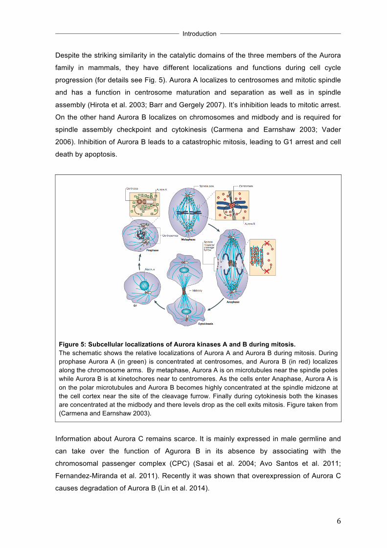
Despite the striking similarity in the catalytic domains of the three members of the Aurora
family in mammals, they have different localizations and functions during cell cycle
progression (for details see Fig. 5). Aurora A localizes to centrosomes and mitotic spindle
and has a function in centrosome maturation and separation as well as in spindle
assembly (Hirota et al. 2003; Barr and Gergely 2007). It’s inhibition leads to mitotic arrest.
On the other hand Aurora B localizes on chromosomes and midbody and is required for
spindle assembly checkpoint and cytokinesis (Carmena and Earnshaw 2003; Vader
2006). Inhibition of Aurora B leads to a catastrophic mitosis, leading to G1 arrest and cell
death by apoptosis.
!
Figure 5: Subcellular localizations of Aurora kinases A and B during mitosis. The schematic shows the relative localizations of Aurora A and Aurora B during mitosis. During prophase Aurora A (in green) is concentrated at centrosomes, and Aurora B (in red) localizes along the chromosome arms. By metaphase, Aurora A is on microtubules near the spindle poles while Aurora B is at kinetochores near to centromeres. As the cells enter Anaphase, Aurora A is on the polar microtubules and Aurora B becomes highly concentrated at the spindle midzone at the cell cortex near the site of the cleavage furrow. Finally during cytokinesis both the kinases are concentrated at the midbody and there levels drop as the cell exits mitosis. Figure taken from (Carmena and Earnshaw 2003).
Information about Aurora C remains scarce. It is mainly expressed in male germline and
can take over the function of Agurora B in its absence by associating with the
chromosomal passenger complex (CPC) (Sasai et al. 2004; Avo Santos et al. 2011;
Fernandez-Miranda et al. 2011). Recently it was shown that overexpression of Aurora C
causes degradation of Aurora B (Lin et al. 2014).

!
!
!Introduction
!! !
7!
1.3.1 Role of Aurora B in cell cycle Aurora B has various functions during the cell cycle as a part of the chromosomal
passenger complex (CPC) (Ruchaud et al. 2007; Carmena et al. 2012). CPC consists of
the enzymatic component Aurora B, a scaffold protein inner centromere protein (INCEP)
and two non-enzymatic subunits Survivin and Borealin. The protein stability of each
subunit of CPC depends on protein-protein interactions within the complex, hence
knockdown of any of the components or chemical inhibition results in a similar phenotype
in wide range of organisms (Adams et al. 2001; Biggins and Murray 2001; Lens et al.
2003; Gassmann 2004; Vader et al. 2006; Klein et al. 2006; Xu et al. 2009; Kelly et al.
2010). Aurora B has three important functions during the cell cycle:
1) Chromosome condensation - At the beginning of mitosis, in prophase Aurora B
mediates sister chromatid separation by causing cohesin dissociation from
chromosome arms (Giménez-Abián et al. 2004; Dai et al. 2006; Nishiyama et al.
2013), loading of Condensin I complex on the chromosome arms (Lipp et al.
2007), or by phosphorylating H3. Histone H3 is a substrate of Aurora B, which gets
phosphorylated at Ser10 during mitosis and is a widely used mitotic marker
(Crosio et al. 2002; Hirota et al. 2005). H3S10 is necessary for chromosome
condensation in Drosophila (Giet and Glover 2001), but in humans this
modification is implicated in chromosome condensation as well as dissociation of
HP1 from chromatin during mitosis (Hirota et al. 2005; Crosio et al. 2002). In
mammalian cells an additional phosphorylation at Ser28 is required for
chromosome condensation (Goto et al. 2002).
2) Spindle assembly checkpoint - The spindle assembly checkpoint (SAC) prevents
the metaphase to anaphase transition until all the chromosomes are accurately bi-
oriented (Musacchio and Salmon 2007). During pro-metaphase and metaphase
Aurora B becomes highly concentrated at inner centromeres and performs ‘error
correction’ by regulating microtubule-kinetochore attachments through
phosphorylation of a number of substrates like, MCAK (Lan et al. 2004),
KNL1/Mis12/Ndc80 complex (Welburn et al. 2010; Chan et al. 2012) and Mps1
(Biggins 2001; Santaguida et al. 2011; Saurin et al. 2011). Aurora B resolves
syntelic attachments and converts them to amphitelic attachments (for details see
Fig. 6). Aurora B generates unattached kinetochores which are sensed by SAC
and hence keeps this mitotic checkpoint active until all the incorrect attachments
are resolved (Ditchfield et al. 2003; Hauf et al. 2003; Nezi and Musacchio 2009;
Maldonado and Kapoor 2011).

!
!
!Introduction
!! !
8!
3) Cytokinesis – The final stage of cell cycle is cytokinesis, which generates two
daughter cells by formation of actomyosin ring at the cell equator. Cytokinesis
requires proper functioning of the centralspindlin complex [composed of GTPase
activating protein (GAP) MgcRacGAP and a kinesin, MKLP1] (Zhao and Fang
2005; Neef et al. 2006) whose activity in turn is regulated by Aurora B (Minoshima
et al. 2003; Touré et al. 2008; Guse et al. 2005; Douglas et al. 2010). Besides this,
Aurora B also phosphorylates other cytoskeletal proteins, like vimentin (Goto et al.
2003), myosin II regulatory light chain (Murata-Hori et al. 2000), desmin and GFAP
(Kawajiri et al. 2003) required for abscission of the cell at correct time (Norden et
al. 2006; Ozlu et al. 2010).
Figure 6: Chromosome bi-orientation at metaphase plate. Schematic representation of different types of kinetochore-microtubule attachments during metaphase. Correctly bi-oriented chromosomes show amphitelic attachment (with the sister kinetochores attached to opposite poles, lower center). Mal-oriented chromosomes show monotelic/mono-oriented attachment (only one kinetochore is attached to one pole, lower left), merotelic attachment (one kinetochore is attached to both poles, upper center) or syntelic attachments (both kinetochores are attached to the same pole, lower right). Figure taken from (Keen and Taylor 2004).
Although Aurora B mainly functions in mitosis, a recent study reported it’s function in
interphase where Aurora B phosphorylates and mediates degradation of p53 (Gully et al.
2012). Mice in which one allele of Aurora B is disrupted develop tumors, signifying the
importance of Aurora B in maintaining genome integrity (Fernandez-Miranda et al. 2011).
!
!
!Introduction
!! !
9!
1.3.2 Regulation of Aurora B kinase function Aurora B is highly regulated during the cell cycle to execute an orderly and timely
phosphorylation of its substrates. The mechanisms that regulate Aurora B are discussed
below:
• Phosphorylation- In order to have kinase activity, Aurora B must be
phosphorylated at a key threonine residue (T232) in its T-loop. This is
accomplished by auto-phosphorylation induced by a conformational change upon
interaction of Aurora B with the C-terminal IN-box of INCEP (Yasui et al. 2004;
Sessa et al. 2005). Further the phosphorylation of substrates by Aurora B is
counteracted by antagonistic phosphatases protein phosphatase 1 (PP1) and
protein phosphatase 2A during mitosis (Liu et al. 2010; Foley et al. 2011). • Localization- Substrate specificity of Aurora B during mitosis is determined by its
differential localization which is dictated by its interaction with various protein
partners. For example, two non-enzymatic proteins Survivin and Borealin along
with INCEP, target Aurora B to different sites such as chromosome arms and inner
centromere by docking to these sites and hence function as ‘passenger proteins’
during cell division (Vader et al. 2006). • Ubiquitin mediated proteolysis- Aurora B is targeted for degradation by the E3
ubiquitin ligase, anaphase-promoting complex/cyclosome (APC/C). APC/C
mediates proteasomal degradation of Aurora B by ubiquitylation in conjunction with
cdc20 homolog 1 (Cdh1) as the cells exit mitosis to ensure G1 cells have very low
Aurora B protein levels (Stewart and Fang 2005).
1.3.3 Role of Aurora B kinase in cancer and inhibitors against Aurora B Cancer is a disease whose characteristic features include chromosomal rearrangements
and aneuploidy. As Aurora B has key functions in spindle assembly, mitotic checkpoint
and chromosome segregation, it is reported to be deregulated in a wide range of cancers
for example, NSCLC (non small cell lung cancer), colon and pancreatic cancer to name a
few (Bischoff et al. 1998; Adams et al. 2001). Both the activity as well expression levels of
Aurora B are increased in cancer and this is associated with poor prognosis as well. Also
ectopic expression of Aurora B has been reported to cause transformation of cells in
culture (Ota et al. 2002). This extensive correlation of Aurora B with cancer provides the
basis for its importance as a target for chemotherapy (Keen and Taylor 2004; Girdler
2006; Gully et al. 2010). A variety of chemical inhibitors against Aurora B have been
developed so far, some of the examples being ZM447439 (Ditchfield et al. 2003),
Hesperadin (Hauf et al. 2003), VX680 (Harrington et al. 2004) and AZD1152 (Mortlock et

!
!
!Introduction
!! !
10!
al. 2007). These chemical compounds are competitive inhibitors of the ATP binding site of
Aurora B.
ZM447439 is a quinazoline derivative and was initially thought to inhibit both Aurora A and
B. But further in vitro studies revealed that it had 20 times more specificity towards Aurora
B and the cellular phenotypes were consistent with Aurora B inhibition (Ditchfield et al.
2003).
AZD1152, a member of 5-acetanilide-substituted 3-aminopyrazoles series is a highly
selective Aurora B kinase chemical inhibitor (IC50 of 0.37 nM for Aurora B in contrast to
1368 nM for Aurora A). It is a dihydrogen prodrug which is highly soluble in pH adjusted
aqueous solutions and undergoes rapid conversion to active form AZD1152-HQPA in vivo
(Mortlock et al. 2007; Wilkinson et al. 2007).
1.4 The cellular stress response and its relevance for cancer therapy Cellular stress response is a defense mechanism elicited when cells are challenged with
adverse conditions such as damage to DNA/protein, hypoxia, metabolic constraints,
oxidative stress or oncogene activation. The severity and duration of stress determine the
output, which could be either cell survival or cell death (by apoptosis, necrosis or
autophagy) (Kültz 2005; Fulda et al. 2010). In addition to integrating the extracellular and
intracellular stimuli, the cellular stress response also determines the efficacy and outcome
of a chemotherapeutic regimen. The p53 tumor suppressor pathway and mitogen
activated protein kinase pathway (MAPK) standout in this respect.
1.4.1 The p53 tumor suppressor pathway Central to regulation of most of the stress signaling in a cell and mutated/deregulated in
more than 50 % human tumors is p53, a tumor suppressor protein also known as “the
guardian of genome” (Lane 1992; Vogelstein et al. 2000). p53 is a transcription factor
which prevents tumor development by causing G1 cell cycle arrest, apoptosis and
senescence under adverse stressful conditions by activation of a number of target genes
including p21, GADD45, BAX and 53BP1 (Fig. 7) (Vousden and Prives 2009; Bieging et
al. 2014). Some of the p53 functions are also independent of its transcriptional activity as
summarized in Fig. 7 (Moll et al. 2005; Sengupta and Harris 2005; Suzuki et al. 2009).
Elegant recent findings demonstrate that other functions of p53 in DNA repair, regulation
of metabolism and oxidative stress also contribute to its tumor suppressive functions
(Brady et al. 2011; Li et al. 2012; Valente et al. 2013) besides its classical functions in
preventing tumor growth by cell cycle arrest, apoptosis and senescence known so far.

!
!
!Introduction
!! !
11!
!Figure 7: p53 signaling. Figure taken from (Brown et al. 2009).
The half-life of p53 protein is very short under unstressed conditions as it is continuously
degraded by the E3 ubiquitin ligase MDM2 through proteasome-mediated degradation
(Momand et al. 1992; Kubbutat et al. 1997). In response to a wide range of stress (DNA
damage, hypoxia, damage to mitotic spindle, heat shock, oncogenes or unfolded
proteins), various signaling proteins phosphorylate p53, preventing its interaction with
MDM2 and hence its degradation. This is followed by other modifications (acetylation and
methylation) of p53 and it’s binding as a homotetramer to its binding site (p53BS1 and 2)
on the target gene promoters resulting in their induction (Vousden and Lane 2007; Riley
et al. 2008; Kruse and Gu 2009).
The p53 signaling is deregulated in tumors by three common mechanisms, (a) mutations
in DNA binding domain of p53 and hence preventing its binding to DNA, (b) mutations that
prevent the proper folding and oligomerization of p53 and (c) overexpression of p53
regulatory proteins such as MDM2. Hence, not surprisingly all these mechanisms are
being currently explored in clinic for effective cancer therapy (Muller and Vousden 2013;
Hoe et al. 2014).
1.4.2 Mitogen activated protein kinase pathway (MAPK pathway) The mitogen-activated protein kinases (MAPKs) are a family of stress kinases that serve
to integrate the signals from a number of environmental and cellular stimuli to activate
cellular responses. Of all the kinases in the eukaryotic genome (around 518 in humans)
MAPKs are involved in most of the signaling, which is highly conserved from yeast to
mammals (Qi and Elion 2005). Each MAPK cascade is composed of three tiers of kinases
MAPKKK (MAP3K, MAPK-kinase-kinase), MAPKK (MAP2K, MAPK-kinase) and MAPK
and kinases in each tier phosphorylate and activate the members of next tier (Fig. 8). In

!
!
!Introduction
!! !
12!
humans, the MAPKs are composed of four subfamilies; (1) ERKs (extracellular signal-
regulated kinases), (2) JNK/SAPK (c-Jun N-terminal related kinases or stress activated
protein kinases), (3) p38-MAPK, (4) ERK5/big MAPK-1 (BMK1), classified by the MAPK at
the end of the phosphorylation cascade (Raman et al. 2007). Signaling by ERK is
activated by growth factors and results in cell growth and differentiation (Shaul and Seger
2007). JNK and p38-MAPK signaling is mainly activated by inflammatory cytokines,
environmental stress as well as genotoxic stress and they contribute to cell cycle
regulation, cell differentiation, apoptosis and inflammation (Wagner and Nebreda 2009).
Growth factors as well as cellular stress activate the ERK5 cascade leading to
angiogenesis, anti-apoptosis, cell proliferation and differentiation (Wang and Tournier
2006).
!Figure 8: General cascade of MAPK pathways. Figure taken from (Kumar et al. 2003).
The p38 MAPK family is composed of four family members: MAPK11 (p38α), MAPK12
(p38β), MAPK13 (p38γ) and MAPK14 (p38δ), which differ in their expression profiles,
substrate specificity and sensitivity towards chemical inhibitors such as SB202190 and
BIRB796. p38α is ubiquitously expressed in most cell types, whereas the expression of
other isoforms is more restricted to certain tissues (e.g. p38β in brain, p38γ skeletal
muscle, p38δ in endocrine glands). p38α, the mammalian MAPK orthologue of Hog1 (the
osmosensing MAPK of Saccharomyces cerevisiae) is the most extensively characterized
!
!
!Introduction
!! !
13!
isoform among all (Cuadrado and Nebreda 2010).
p38 MAPK, a serine/threonine kinase can function both as a tumor suppressor as well
oncogene depending on the intensity and duration of stress, the cell type and cross talk
with other signaling pathways. For example it halts cell cycle progression in response to
DNA damage and other environmental insults (Bulavin et al. 2001), but can also induce
angiogenesis under hypoxic conditions (Pages 2000). p38 MAPK has various
physiological functions such as myogenic differentiation, keratinocyte differentiation and
cell migration (Wu et al. 2000; Efimova 2003; Rousseau et al. 1997). p38 MAPK regulates
cell cycle checkpoints at G0, G1/S and G2/M transitions during the cell cycle. By
regulating the cyclin levels (cyclin A or D1), phosphorylation of retinoblastoma protein
(pRb) and phosphorylation of p53 (Ser33 and Ser46), p38 has and effect on G1/S
transition (Ambrosino and Nebreda 2001; Bulavin et al. 1999; Sanchez-Prieto et al. 2000).
G2/M checkpoint activated by various stress stimuli is controlled by p38 MAPK through
activation of MAPKAP-K2, which phosphorylates Cdc25B and Cdc25C causing their
translocation into cytoplasm (Mikhailov et al. 2005; Manke et al. 2005). Besides this, p38
MAPK also plays an important role in gene expression control (Nadal et al. 2011).
Regulation of p38 MAPK is mainly by dual phosphorylation, auto-phosphorylation,
phosphatases, and scaffold proteins (Kyriakis and Avruch 2001; Ge 2002; Keyse 2000;
Owens and Keyse 2007). Due to the critical relevance of p38 MAPK pathway in
proliferation control and apoptosis, it is deregulated in array of cancer types and is a
attractive clinical target (Wagner and Nebreda 2009).
1.5 Aneuploidy and cancer Chromosomal instability (CIN), a term assigned jointly for aneuploidy (numerical/whole-
chromosome alterations) and structural chromosome alterations
(translocations/deletions/insertions) is a hallmark of cancer (Mertens et al. 1994; Mertens
et al. 1997; Lengauer et al. 1998; Weaver and Cleveland 2007). There are different
mechanisms, which can generate aneuploidy as summarized in Fig. 9; (a) Mitotic
checkpoint defects - Compromised SAC (due to loss or gain of individual components
such as BUBR1), can cause abrupt entry into anaphase despite the presence of
unattached kinetochores resulting in daughter cells with a gain or loss of chromosome(s).
In fact a number of cancers have been reported to have mutated or altered expression
and gene silencing (by methylation) of SAC components (Wang et al. 2004; Kops et al.
2005; Park et al. 2007; Haruta et al. 2008). (b) Cohesion defects - Inability to separate
sister chromatids during mitosis due to defects in components of the molecular machinery
that keeps the sister chromatids attached (e.g. separase, cohesin, securin), also
contributes to aneuploidy. Aneuploid cancers show a high correlation with somatic

!
!
!Introduction
!! !
14!
mutations in the protein components required for attachment of sister chromatids (Barber
et al. 2008; Zhang et al. 2008). (c) Merotelic attachments - Defects to resolve merotelic
attachments during mitosis often result in misseggregations and lagging chromosomes
resulting in aneuploidy (Cimini 2008). Merotelic attachments arise due to an increase in
number of centrosomes or and increased stability of kinetochore-microtubule attachments
(Ganem et al. 2009; Bakhoum et al. 2009). Cancer cells also show high frequency of
merotelic attachments (Cimini et al. 2001; Cimini 2008).
!Figure 9: Different mechanisms that generate aneuploidy during mitosis. For details see text. Figure taken from (Holland and Cleveland 2009).
(d) Multiploar mitotic spindles – Multipolar spindles arising due to multiple centrosomes
allow the cells to undergo division, but often result in merotelic attachments and hence
aneuploidy (Brinkley 2001; Nigg 2002; Silkworth et al. 2009). Centrosome amplification
occurs in primary human tumors and is highly correlated with CIN (Pihan et al. 2003; Nigg
2006).
Extensive studies in yeast and mammalian studies implicate that aneuploidy reduces the
fitness and generates a stressed state (Torres et al. 2007; Williams et al. 2008). The
numerical change in chromosome number reflects into transcriptome and proteome of the
aneuploid cells. Aneuploid yeast cells display a specific gene signature known as
‘’Environmental Stress Response’’ (ESR) (Torres et al. 2007; Pavelka et al. 2010).
Disturbed protein balance due to aneuploidy activates the ubiquitin-proteasome pathway
and chaperone pathways, which relives the protein burden on the cell by causing their
degradation. This also generates a metabolic and energetic stress on the cell (Torres et

!
!
!Introduction
!! !
15!
al. 2010; Stingele et al. 2012; Oromendia and Amon 2014). As aneuploidy is associated
with a specific stress response and cancer cells have developed adaptations to tolerate it,
aneuploidy might be an interesting target for clinic.
Aneuploidy is one of the ‘hallmarks’ of cancer as it is associated with around 90 % solid
tumors and more than 50 % hematopoietic cancers in humans (Mitelman Database of
Chromosome Aberrations and Gene Fusions in Cancer, 2014). But aneuploidy is not an
accurate predictor of tumor susceptibility in mice models of mitotic checkpoint dysfunction
and it is also associated with reduced proliferation rates of yeast and mammalian cells
under in vitro conditions. For instance, mice which are prone to aneuploidy (due to
mutations of various components of SAC), develop spontaneous tumors very late (>18
months) and only a fraction of aneuploid mice develop spontaneous tumors (Holland and
Cleveland 2009), trisomic MEFs have proliferation defects and do not immortalize or
undergo immortalization quite late (Williams et al. 2008) and haploid yeast strains with an
extra chromosome do not proliferate (Torres et al. 2007). This discrepancy between
observed strong association of aneuploidy with cancer (which grow rapidly) and adverse
effects of aneuploidy on growth rate and a poor correlation with tumor susceptibility in
mice is termed as ‘aneuploidy paradox’ (Sheltzer and Amon 2011). The most suitable
explanation for this is the differences in the extracellular environments and genetic context
of the tumor cells in comparison to the cells grown in culture. Under culture conditions,
mammalian and yeast cells are selected for growing fast, whereas tumor cells are
continuously adapting to the varying intracellular and extracellular conditions resulting in
slower growth rate, which might provide additional advantages, such as acquisition of
additional mutations that help them to ameliorate the imbalances in proteome due the
aneuploid karyotype resulting in a more aggressive phenotype (Araujo et al. 2007;
Anjomshoaa et al. 2009; Torres et al. 2008; Torres et al. 2010). In summary, aneuploidy
can suppress or promote tumorigenesis depending on the cell type and genetic
background (Weaver and Cleveland 2007; Holland and Cleveland 2009; Gordon et al.
2012; Holland and Cleveland 2012).
!

!
!
!Introduction
!! !
16!
1.6 Objectives of thesis The fact that Aurora B kinase is overexpressed in a variety of cancers and has enzymatic
kinase activity (enabling it to be inhibited by chemical compounds) makes it an attractive
target for cancer therapy. Currently two Aurora B inhibitors AZD1152 and BI811283 are in
phase III and phase II clinical trials respectively (Marzo and Naval 2013). More detailed
understanding of the cellular signaling pathways regulated by Aurora B is required to
explain the side effects as well as to provide biomarkers of response, for better evaluation
of these chemical inhibitors in clinic. Aims of this thesis were:
1) To study the mitotic stress signaling pathways activated due to Aurora B inhibition
using two small molecule inhibitors of Aurora B (ZM447439 and AZD1152-HQPA)
as chemical tools.
2) To further study the therapeutic implications of Aurora B inhibitors in combination
therapy for treatment of cancer.

Materials and Methods
17!
2 Materials and Methods 2.1 Materials
2.1.1 Chemical stocks and reagents Unless specified, commonly used chemicals were purchased from AppliChem, Roth,
Invitrogen, Invivogen or Sigma with analysis quality.
Chemical Stock concentration Agarose Ready to use AICAR (AMPK activator) (Biomol) 20 mM in DMSO Ammonium persulfate (APS) 10 % in H2O AZD1152-HQPA (Aurora B kinase inhibitor) (Selleckchem) 10 mM in DMSO
BIRB796 (p38 MAP kinase inhibitor) (Selleckchem) 10 mM in DMSO
Bovine serum albumin (BSA) 20 mg/ml in H2O BrdU 10 mg/ml in 1X PBS Cycloheximide 10 mg/ml in H2O Doxorubicin 1.7 mM DMSO Ready to use dNTPs 2 mM dATP, dCTP, dGTP, dTTP each DTT 1 M in H2O Ethidium bromide 10 mg/ml in H2O H2DCF-DA (Molecular Probes) 20 mM in anhydrous DMSO Hoechst 33258 10 mg/ml in H2O Hoechst 33342 Ready to use ImmuMount (Shandon) Ready to use KU5593 (ATM kinase inhibitor) (Selleck) 10 mM in DMSO Low melting agarose Ready to use Luminol 250 mM in DMSO MitoSox Red (Life Technologies) 5 mM in DMSO p-Coumaric acid 90 mM in DMSO PMSF (Phenylmethylsulphonyl- fluoride) (Roche) 10 mg/ml in isopropanol
Polybrene (Hexadimethrine bromide) 4 mg/ml in H2O Ponceau S solution 0.1 % Ponceau S in 5 % acetic acid Propidium iodide (PI) 1 mg/ml in H2O

Materials and Methods
18!
2.1.2 Antibiotics Antibiotic Stock
concentration Final Concentration Use for cell line
Ampicillin 100 mg/ml 100 µg/ml in LB-medium DH5α (E-coli) Blasticidin 10 mg/ml 10 µg/ml in DMEM U2OS-EcoR-Neo Puromycin 10 mg/ml 2 µg/ml in DMEM U2OS-EcoR-Neo
2.1.3 Enzymes Enzymes Company Absolute QPCR SYBER Green Mix ThermoFisher DNase I, RNase free Roche Fast Alkaline Phosphatase (1 U/µl) Fermentas M-MLV-RT Transcriptase (200 U/µl) ThermoFisher Pfu DNA Polymerase (2.5 U/µl) Promega Phusion High Fidelity DNA Polymerase (2 U/µl) Finnzymes
Restriction Endonucleases New England Biolabs (NEB), Fermentas
RiboLock RNase-Inhibitor (40 U/µl) Fermentas T4-DNA Ligase (400 U/µl) New England Biolabs (NEB)
Protease Inhibitor Cocktail Sigma ready to use
Proteinase K 10 mg/ml in 50 mM Tris pH 8.0/ 1 mM CaCl2
Protogel 30 % (Biozym) Ready to use Random Primer (Roche) 500 mg/ml in H2O
RNase A 10 mg/ml in 10 mM Tris-HCl pH 7.4, 150 mM NaCl
Sodium dodecyl sulfate (SDS) 20 % (w/v) in H2O SB202190 (p38 MAP kinase inhibitor) 10 mM in DMSO Tetramethylethylenediamine (Temed) 99 % Ready to use Trizol/Trifast (total RNA isolation reagent) (Peqlab/Thermo) Ready to use
Thymidine 200 mM in H2O VE821 (ATR kinase inhibitor) (Tinib-Tools) 10 mM in DMSO ZM447439 (Aurora kinase Inhibitor) (Enzo) 10 mM in DMSO 4,5-Dihydroxy-1,3-benzenedisulfonic acid disodium salt (Tiron) (superoxide anion scavenger)
100 mM in H2O
17AAG (HSP90 inhibitor) (Selleckchem) 20 mM in DMSO

Materials and Methods
19!
2.1.4 Molecular kits and Protein/DNA markers !Kits Company Jetstar Gel Extraction kit Genomed GeneRulerTM DNA Ladder Fermentas Plasmid Midi-/Maxi-preps kit Invitrogen PageRulerTM Prestained Protein Ladder Fermentas QIAquick PCR purification kit Qiagen
2.1.5 Devices Device Company Agarose gel electrophoresis system Peqlab Bioruptor Diagenode Centrifuges Eppendorf (5417R and 5415D) Heraeus (Megafuge 1.0R) FACS Beckman Coulter (Cytomics FC500) Incubators Heraeus Nunc Microscopes Confocal (Nikon Eclipse Ti) Fluorescence (Leica DMI 6000B) Mx3000 qPCR Agilent technologies Nanodrop Theromo Scientific (Nanodrop 2000) SDS-PAGE Gel Electrophoresis system BIO-RAD 2.1.6 Buffers 2.1.6.1 General buffers !5X DNA Loading buffer 15 % Ficoll
0.05 % Bromophenol blue 0.05 % Xylene cyanol 0.05 M EDTA
0.5 M EDTA pH 8.0 0.5 M EDTA adjust pH to 8.0 with NaOH pellets
2X HBS 280 mM NaCl 1.5 mM Na2HPO4 50 mM HEPES-KOH, pH 7.05

Materials and Methods
20!
Miniprep Solution S1 50 mM Tris-HCl, pH 8.0 10 mM EDTA 100 µg/ml RNase A
Miniprep Solution S2
200 mM NaOH 1 % SDS
Miniprep Solution S3
3.1 mM Potassium Acetate adjust pH to 8.0 with glacial acetic acid
10X PBS 130 mM NaCl 3 mM KCl 64 mM Na2HPO4
15 mM KH2PO4 adjust pH to 7.4 with HCl
50X TAE buffer
200 mM Tris base 250 mM glacial acetic acid 500 mM EDTA, pH 8.0
10X TE 100 mM Tris-HCl, pH 7.5 10 mM EDTA
20X SSC
3 M NaCl 0.3 M Na-Citrate adjust pH to 7.0 with NaOH
2.1.6.2 Buffers for whole cell lysates TNN buffer
50 mM Tris-HCl, pH 7.5 120 mM NaCl 5 mM EDTA 0.5 % NP-40 10 mM Na4H2PO7 2 mM Na3VO4
100 mM NaF PIC (Sigma) 1:500 (added freshly)

Materials and Methods
21!
Bradford Solution
50 mg Coomassie Brilliant Blue G 23.75 ml ethanol 50 ml 85 % (v/v) ortho-phosphoric acid add to 500 ml H2O filter twice
2.1.6.3 Buffers for immunoblotting 4X Upper stock for SDS gels
33 g Tris 10 ml SDS (20 %) add to 500 ml H2O, adjust to pH 6.8
4X Lower stock for SDS gels 90.85 g Tris 10 ml SDS (20 %) add to 500 ml H2O, adjust to pH 8.8
Acrylamide buffer for SDS-gels (Protogel) 30 % (w/v) acrylamide 0.8 % (w/v) N,N’-methylenbisacrylamide
Blotting buffer (1X)
0.6 g Tris 2.258 g Glycin 150 ml methanol add to 1 l H2O
Blocking solution 3 % (w/v) milk powder in 0.05 % TBST, or 5 % (w/v) milk powder in 0.1 % TBST (for cell signaling antibodies)
Electrophoresis sample buffer (ESB) (3X) 300 mM Tris-HCl, pH 6.8 15 mM EDTA 150 mM DTT 12 % (w/v) SDS 15 % (w/v) glycerol 0.03 % (w/v) bromophenol blue
0.15 M NaCl
Ponceau S 0.1 % Ponceau S 5 % glacial acetic acid

Materials and Methods
22!
SDS running buffer (10X)
144 g Glycin 30 g Tris 10 g SDS add to 1 l H2O
Substrate solution 10 ml 100 mM Tris-HCl, pH 8.5 50 µl 250 mM luminol 22 µl 90 mM p-coumaric acid 3 µl 30 % H2O2
TBST 0.05 % Tween 20 in 1X TBS, or 0.1 % Tween 20 in 1X TBS
2.1.6.4 Buffers for Chromatin Immunoprecipitation (ChIP) Cell lysis buffer
5 mM PIPES, pH 8.0 85 mM KCl 0.5 % NP-40 PIC 1:500 (added freshly) PMSF 1mM (added freshly)
Nuclei lysis buffer
50 mM Tris-HCl, pH 8.1 10 mM EDTA 1 % SDS PIC 1:500 (added freshly) PMSF 1mM (added freshly)
IP Dilution buffer 0.01 % SDS 1.1 % Triton 1.2 mM EDTA 16.7 mM Tris-HCl, pH 8.2 167 mM NaCl PIC 1:500 (added freshly) PMSF 1mM (added freshly)
LiCl wash buffer 0.25 M LiCl 0.5 % NP-40 0.5 % DOC 1 mM EDTA
Materials and Methods
23!
10 mM Tris-HCl, pH 8.0 PIC 1:500 (added freshly) PMSF 1mM (added freshly)
Elution buffer
50 mM Tris-HCl, pH 8.0 1 % SDS 10 mM EDTA
2.1.6.5 Buffers for flow cytometry (FACS) Sodium citrate
38 mM in 1X PBS
2.1.6.6 Buffers for immunofluorescence PSP
15 g paraformaldehyde 10 g sucrose add to 500 ml in 1X PBS, stored at -20ºC
PBST
0.1 % Triton-X-100 500 ml 1X PBS, stored at 4ºC and 0.2 % Triton-X-100 500 ml 1X PBS, stored at 4ºC
Blocking solution
5 % BSA in 1X PBS
2.1.6.7 Buffers for centromere Fluorescence in-situ hybridization (FISH) Wash buffer I
0.4X SSC 0.3 % NP-40
Wash buffer II 2X SSC 0.1 % NP-40
2.1.6.8 Staining solution Crystal violet
0.1 % crystal violet in 20 % ethanol

Materials and Methods
24!
2.1.7 Antibodies
2.1.7.1 Primary antibodies
!!
Antibody against
Catalog number Origin Application
and dilution Company
α-tubulin T6074 Mouse monoclonal
WB 1:10,000
IF 1:200 Sigma
Aurora B ab2254 Rabbit polyclonal
WB 1:1000 Abcam
β-actin sc-47778 Mouse monoclonal
WB 1:5000 Santa Cruz
B-Myb (LX015.1) none Mouse
monoclonal WB 1:5 (Tavner et al.,
2007)
BrdU-FITC 347583 Mouse monoclonal
IF 1:10 BD Bioscience
Cyclin-A (BF683) sc-239 Mouse
monoclonal WB 1:1000 Santa Cruz
E2F-1 (C-20) sc-193 Rabbit polyclonal WB 1:1000 Santa Cruz
HA MMA-101P Mouse monoclonal
WB 1:1000
IF 1:100 HISS
IgG I5006 Mouse monoclonal
ChIP 2 µg Sigma
p21 (C-19) sc-397 Rabbit polyclonal
WB 1:1000 Santa Cruz
p27 610241 Mouse monoclonal
WB 1:1000 BD
Transduction LaboratoriesTM
p38 #9212 Rabbit monoclonal
WB 1:1000 Cell signaling
p53 (DO-1) sc-126 Mouse monoclonal
WB 1:5000
ChIP 3 µg Santa Cruz

Materials and Methods
25!
pH3 06-570 Rabbit polyclonal
WB 1:1000 Millipore
Phospho-ATM/ATR substrates
#2851
Rabbit monoclonal WB 1:1000 Cell signaling
Phospho-Chk1 (Ser345) #2348
Rabbit monoclonal WB 1:1000 Cell signaling
Phospho-Chk2 (Thr68) #2661
Rabbit monoclonal WB 1:1000 Cell signaling
Phosho-Histone H2A.X (Ser139) #2577
Rabbit monoclonal
WB 1:1000 Cell signaling
Phospho-p38 #4511
Rabbit monoclonal
WB 1:1000 Cell signaling
Phospho-Ser2-RNA
Polymerase II
ab5095
Rabbit polyclonal ChIP 3 µg Abcam
pRb sc-50
Rabbit polyclonal
WB 1:1000 Santa Cruz
RNA Polymerase II
sc-899
Rabbit polyclonal
ChIP 3 µg Santa Cruz
SV40 Large T (Pab 108)
sc-148
Mouse monoclonal WB 1:1000 Santa Cruz

Materials and Methods
26!
2.1.7.2 Secondary antibodies
Antibody Company Application and dilution
anti-mouse HRP conjugated GE Healthcare WB 1:5000
anti-Protein A HRP conjugated BD Biosciences WB 1:5000
anti-rabbit HRP conjugated Invitrogen WB 1:5000
anti-mouse Alexa 488 Invitrogen IF 1:500
anti-rabbit Alexa 594 Invitrogen IF 1:500
2.1.8 Beads Dynabeads Protein G Life Technologies Monoclonal Anti-HA Agarose Conjugate Clone HA-7 Sigma

Materials and Methods
27!
2.1.9 Plasmids
2.1.9.1 Plasmids for overexpression
Internal number Plasmid name Description
210 pBabe-puro Empty vector control for retroviral transfections
746 pBabe-H2B-GFP GFP control for retroviral transfections
934 pBabe-puro-LargeT antigen-WT
Retroviral expression of Large T antigen (wild type)
1277 pBabe-puro-HA-p38alpha Retroviral expression of HA-p38alpha
1279 pBabe-puro-HA-p38beta Retroviral expression of HA-p38beta
1399 pBabe-puro-HA-mElongin A Retroviral expression of mouse Elongin A
1400 pBabe-puro-LargeT-K1 mutant
Retroviral expression of Large T K1 mutant
1401 pBabe-puro-LargeT-Δ434-444 mutant
Retroviral expression of Large T Δ434-444 mutant
2.1.9.2 Plasmids for RNA knockdown
Internal number Plasmid name Description
652 pMSCV480-Blasticidin Empty vector control for retroviral transfections
679 pMSCV480-shp53-Blasticidin Retroviral expression of shp53
2.1.10 Primers Primer oligonucleotides were purchased from Metabion or MWG.
2.1.10.1 Primers for cloning !
Internal number Sequence (5’ to 3’) Target gene Directionality
SG1785 ggggatccATGGCGGCGGAGTC Mouse Elongin A
Forward
SG1786 gggctcgagTTATCGCCGGGAGAATC Reverse Restriction sites (BamHI GGATCC / XhoI CTCGAG) are underlined.

Materials and Methods
28!
2.1.10.2 Primers for quantitative real time PCR All primers are for human sequences, unless indicated.
Internal number Sequence (5’ to 3’) Target gene Directionality
SG572 GGTACTGAAGTCCGGGAACC CCNA2
Forward
SG573 GAAGATCCTTAAGGGGTGCAA Reverse
SG628 TCACTGTCTTGTACCCTTGTGC p21
Forward
SG629 GGCGTTTGGAGTGGTAGAAA Reverse
SG645 GCCCAATACGACCAAATCC GAPDH
Forward
SG646 AGCCACATCGCTCAGACAC Reverse
SG771 AGGCCTTGGAACTCAAGGAT p53
Forward
SG772 CCCTTTTTGGACTTCAGGTG Reverse
SG1511 GACTCCAAGCGCGAAAAC MDM2
Forward
SG1512 GGTGGTTACAGCACCATCAGT Reverse
SG1630 GATGGCCCAGAAGGAGAACT Aurora B
Forward
SG1631 AGGCTCTTTCCGGAGGACT Reverse
SG1632 CAGTTCTGCTCTAGGTGGAAGTC TNFSF7
Forward
SG1633 AGGAAGAAGCGTTCGAGAGA Reverse
SG1634 TTTGCCATCCAGAACAAGC ATF3
Forward
SG1635 CATCTTCTTCAGGGGCTACCT Reverse
SG1636 AGAGGAGGAAAGGCAATGAAG SORC3
Forward
SG1637 TTGGTTGAGAGCATTAAACAGTG Reverse
SG1638 GGGCCGTTACCCCTACATTA SESN1
Forward
SG1639 TTCACTAAGTAGGAGCACTG Reverse
SG1648 TACTGACCCCACCTGAGCA FDXR
Forward
SG1649 TCGACTCTGCCTCAGTACACC Reverse
SG1650 AAGGCACCTCTGAGAACTTCA SERPINE1
Forward
SG1651 CCCAGGACTAGGCAGGTG Reverse
SG1652 TTCACCCAAGTGGTGCAG ANK1
Forward
SG1653 CTCATCCGTGAATTGCTCCT Reverse
SG1656 CCGGATACTCACGCCAGA GDF15
Forward
SG1657 AGAGATACGCAGGTGCAGGT Reverse

Materials and Methods
29!
SG1662 TTCCGTCCGCTAGGAGTCT BLM
Forward
SG1663 GACGTTCTAGTTGCTCCTGTAGATT Reverse
SG1666 CGACGTTATTCTGATCTCACCA MCM3
Forward
SG1667 CAAGGGGATTGTTCTCCTCA Reverse
SG1668 AGTAGGTGCTTGGCGGTTC RFC3
Forward
SG1669 CACAGTAGATAACACGTGGCAAA Reverse
!2.1.10.3 Primers for Chromatin Immunoprecipitation
!! !
Internal number Sequence (5’ to 3’) Target Directionality
SG540 GGCAGCAAGAGTCACTCCA GAPDH2 promoter
Forward
SG541 TGTCTCTTGAAGCACACAGGTT Reverse
SG1585 CTGTGGCTCTGATTGGCTTT p53 binding site 1 (p21 promoter)
Forward
SG1586 CTCCTACCATCCCCTTCCTC Reverse
SG1670 TATATCAGGGCCGCGCTG p21 gene (-20)
Forward
SG1671 GGCTCCACAAGGAACTGACTTC Reverse
SG1672 CCAGGAAGGGCGAGGAAA p21 gene (+507), p21
primary transcript
Forward
SG1673 GGGACCGATCCTAGACGAACTT Reverse
SG1675 CGTGTTCGCGGGTGTGT p21 gene (+182)
Forward
SG1676 CATTCACCTGCCGCAGAAA Reverse
SG1677 CCTCCCACAATGCTGAATATACAG p21 gene (+8566)
Forward
SG1678 AGTCACTAAGAATCATTTATTGAGCACC Reverse
SG1679 CCTGGCTGACTTCTGCTGTCT p21 gene (+7011), p21
primary transcript
Forward
SG1680 CGGCGTTTGGAGTGGTAGA Reverse
SG1683 TCTGTCTCGGCAGCTGACAT p21 gene (+11443)
Forward
SG1684 ACCACAAAAGATCAAGGTGAGTGA Reverse

Materials and Methods
30!
2.1.11 siRNA sequences siRNA oligos were purchased from MWG.
!siRNA against Sequence (5’ to 3’) Target/Reference
ctrl UGGUUUACAUGUCGACUAA non targeting
Aurora B AACGCGGCACUUCACAAUUGA Human Aurora B, Lampson et al., 2005
pRb Dharmacon smart pool Human Retinoblastoma protein 2.1.12 Cell lines, cell culture media and transfection reagents
2.1.12.1 Media and additives for mammalian cell culture DMEM (4.5 g Glucose/L-Glutamine) Gibco®, Life Technologies Fetal calf serum (FCS) Gibco®, Life Technologies OptimeM Gibco®, Life Technologies Penicillin/Streptomycin (10 U/µl each) Cambrex/ Lonza TrpLETM Express Gibco®, Life Technologies Trypsin EDTA (200 mg/ml) Gibco®, Life Technologies
2.1.12.2 Composition of media for soft agar assay 10X DMEM (20 % FCS) (50 ml)
10X DMEM 10ml 1 M Sodium bicarbonate, autoclaved 1.85 ml (3.7 %) FCS 10 ml (20 %) 200 mM Glutamax 5 ml (20 mM) D-gluc (dehydrated) 450 mg Penstrep 0.5 ml H2O 22.65 ml
1.4 % low melting agarose, autoclaved for base layer 0.7 % low melting agarose, autoclaved for top layer

Materials and Methods
31!
2.1.12.3 Human cell lines and media All cell lines were cultured in DMEM media with 10 % FCS and 1 % Penstrep.
Cell line Description Reference
HCT116-WT
Human colorectal carcinoma tumor cell line (wild type p53 and p21) (Brattain et al. 1981)
HCT116-p21-/-
Human colorectal carcinoma tumor cell line (p21 null) (Waldman et al. 1995)
HCT116-p53-/-
Human colorectal carcinoma tumor cell line (p53 null) (Bunz, 1998)
PlatE Retroviral packaging cell line for generating stable cell lines by
retroviral infection (Morita et al. 2000)
U2OS Human osteosarcoma tumor cell line (Ponten and Saksela 1967)
U2OS-EcoR-neo
U2OS cells with ecotropic receptor for retroviral infection (neomycin
resistance cassette)
Created in lab by stably expressing ecotropic receptor
(neomycin resistance) in U2OS cells
2.1.12.4 Transfection reagents and cell lines
Transfection reagent Cell line Purpose
Lipofectamine RNAi Max U2OS siRNA transfection
Calcium phosphate U2OS Plasmid transfection
Calcium phosphate PlatE Plasmid transfection
2.1.12.5 Bacterial strains E.coli DH5α- competent cells for transformation of plasmid DNA
2.1.12.6 Media for bacterial cell culture Luria Bertani (LB) Agar 40 g powder in 1 l H2O, autoclaved Luria Bertani (LB) Medium 25 g powder in 1 l H2O, autoclaved

Materials and Methods
32!
2.2 Methods
2.2.1 Mammalian cell culture
2.2.1.1 Passaging of cells
!Eukaryotic cells were cultivated in a tissue culture incubator at 37ºC with 5 % CO2. For
passaging, cells were washed once with PBS and incubated with TrypLE Express
(HCT116-WT, p21-/-, p53-/- cells) or Trypsin/EDTA (U2OS and PlatE cells) for a few
minutes at 37ºC. The detached cells were resuspended in media and plated on new cell
culture dishes.
2.2.1.2 Freezing and thawing of cells !To freeze cells, cells on 10 cm dishes were trypsinized and transferred into a 15 ml falcon
tube with 10 ml fresh media. Cells were then pelleted by centrifugation for 3 min at 1200
rpm, the supernatant was discarded and the cells were resuspended in 1 ml ice-cold
freeze medium (DMEM media containing 10 % DMSO) and transferred into cryotubes.
Cells were stored at -80ºC for short term or in liquid nitrogen for long term.
For thawing cells, cells were quickly thawed in a 37ºC water bath. The cell suspension
was mixed with 9 ml fresh medium and centrifuged for 3 min at 1200rpm. The supernatant
was discarded and the pellet was resuspended in 10 ml fresh medium and seeded into 10
cm dishes.
2.2.1.3 Counting cells !Cell counting was performed using a Neubauer Chamber. The number of cells per ml in
suspension was calculated using the following formula:
Cells/ml = (Cells counted/ number of counted large squares) x 104
2.2.1.4 Treatment of cells with reagents !All treatments were done 24 h after seeding the cells. Before treatment, the cells were fed
with fresh media.
AMPK activator Cells were treated with 200 µM AICAR for various time
points.
ATM kinase inhibitor Cells were pretreated with 5 µM or 10 µM KU5593 for 2 h
before any further treatments.
ATR kinase inhibitor Cells were pretreated with 0.1 µM or 1 µM VE821 for 2 h

Materials and Methods
33!
before any further treatments.
Aurora kinase inhibitors Cells were treated with various concentrations of ZM447439
or AZD1152-HQPA for different time points.
BrdU To label cells in S-phase cells were treated with 15 µg/ml
BrdU for 2 h before fixation.
Doxorubicin To induce DNA damage cells were treated with 1 µM
Doxorubicin for 6 h or 24 h.
Hsp90 inhibitor Cells were treated with 8 nM 17AAG for various time points.
p38 MAP kinase inhibitors Cells were pretreated with 10 µM SB202190 or 1 µM
BIRB796 for 2 h before any further treatments.
Thymidine For cell synchronization in G1/S phase, cells were treated
with 2.5 mM thymidine for 24 h.
2.2.1.5 Synchronization of U2OS cells by thymidine !For synchronization at the G1/S border, U2OS cells at 50 % confluency (seeded 24 h
before) were treated with 2.5 mM thymidine for 24 h. Then, the cells were released into
cell cycle by washing three times with PBS and feeding with fresh media.
2.2.1.6 Determination of cell cycle phases by Flow Cytometry !Cells in different cell cycle phases were measured by propidium iodide FACS (PI FACS).
For this, cells were harvested by trypsinization and centrifuged at 1200 rpm for 5 minutes
at 4ºC. Then, the pellet was washed once with ice cold PBS and the cells were fixed over
night in 1 ml 80 % ethanol at -20ºC. Before measurement, cells were pelleted by
centrifuging for 10 minutes at 1000 rpm followed by washing once with PBS at 4ºC. The
cells were then resuspended in 500 µl 38 mM sodium citrate and 25 µl RNAse A (10
mg/ml) for 1 h at 37ºC. After this, the cells were stained with 15 µl PI (1 mg/ml) and then
measured by FACS.
2.2.1.7 Transient transfection !2.2.1.7.1 Plasmid transfection with Calcium phosphate !PlatE cells were transfected using calcium phosphate. 30 µg of plasmid DNA was mixed
with 50 µl of 2.5 M CaCl2 and with H2O to a final volume of 500 µl. In a 15 ml falcon tube,
DNA/CaCl2 mixture was added drop wise to 500 µl of 2X HBS. This solution was added
slowly to the cells. After 18-24 h of incubation, cells were washed once with PBS and fed
with fresh medium. 24 hours later, the virus supernatant produced was harvested for cell

Materials and Methods
34!
infection (see section 2.2.1.6).
2.2.1.7.2 siRNA transfection with Lipofectamine RNAi Max !U2OS cells were transfected with 10-45 nM siRNA using Lipofectamine RNAi Max (Life
Technologies). Before starting, cells (seeded 24 h before) were fed with fresh media
without penicillin and streptomycin. siRNA was diluted in Optimem to a final volume of 250
µl (for 6 well) or 500 µl (for 6 cm dishes), mixed gently by pipetting once up and down and
incubated for 5 minutes. In a separate tube, 2.5 µl Lipofecatime RNAi Max was diluted in
OptimeM medium to a final volume of 250 µl (for 6 well), mixed gently by pipetting once
up and down and incubated for 5 minutes (for 6 cm dishes 5 µl Lipofectamine RNAi Max
was diluted to a final volume of 500 µl). Then, the siRNA/OptimeM mix was added gently
to Lipofectamine RNAi Max/OptimeM mix, and mixed gently by pipetting once up and
down. This complex was incubated for 20 minutes and then added gently dropwise to
cells. After 24 h, cells were fed with fresh media. The cells were harvested after 48-72 h of
transfection, and processed for RNA or protein analysis.
2.2.1.8 Retroviral infection of cells !For production of ecotropic viral supernatant, platE cells were transiently transfected with
the plasmid of interest using calcium phosphate (see section 2.2.1.5.1). 36-48 h after
transfection, the virus supernatants were harvested, filtered (0.45 µm pore size), mixed
with 10 µg/ml polybrene and added to the cells (U2OS-EcoR cells seeded 18-20 h
before). 24 h after infection, the cells were washed once with PBS and fed with fresh
medium and selection was started 48 h after infection.
2.2.1.9 Immunofluorescence staining !For immunofluorescence staining, cells were plated on cover slips in 6-well plates. After
washing once with PBS cells were fixed with PSP for 10 min at RT. Cells were then
washed twice with PBS, permeabilized in PBS/0.2 % Triton-X-100 for 5 min at RT and
washed once in PBS/0.1 % Triton-X-100 (PBST). Unspecific staining was minimized by
blocking for 30 min with 3 % BSA (in PBS). The cells were washed 3 times in PBS (p21-
staining) or 5 mM MgSO4 (in PBS for BrdU-staining) and incubated with the primary
antibody diluted in PBS or 5mM MgSO4 (in PBS with DNAase for BrdU staining) for 1 h in
a humidified chamber. The coverslips were then washed three times (3 minutes each)
with PBS and incubated with the secondary antibody for 30 min in a humidified chamber.
After washing with PBS for 3 times (3 minutes each) cells were stained with Hoechst
33258 (1:1000 in PBS) for 1 minute, washed once with PBS and mounted on glass slides

Materials and Methods
35!
with ImmuMount.
2.2.1.10 ROS detection !For the detection of ROS (reactive oxygen species), cells grown on coverslips were
washed once with warm PBS and incubated with 10 µM 2'-7'-dichlorodihydrofluorescein
diacetate (H2DCF-DA, Molecular Probes) in PBS. After 10 minutes at 37°C, the H2DCF-
DA solution was removed and the cells were incubated for 10 minutes with complete
medium at 37°C. Cells were washed again with warm PBS and fixed in 4 % formalin.
Nuclei were counterstained with Hoechst 33258. For detection with MitoSox Red, cells
grown on coverslips were washed once with warm PBS and incubated for 10 minutes with
5 µM MitoSox Red (Molecular Probes) in PBS at 37°C. Cells were washed three times
with PBS and nuclei were counterstained with Hoechst 33342 for 20 minutes. The
coverslips were then washed once with PBS and mounted on glass slides. Intracellular
ROS levels were visualized using an inverted microscope.
2.2.1.11 Centromere fluorescence in situ hybridisation (FISH) !Cells were trypsinized and centrifuged for 3 minutes at 1000 rpm at room temperature.
The cell pellet was resuspended in 5 ml 0.8 % sodium citrate and incubated at 37ºC for 30
min. After a further centrifugation step of 3 minutes at 1000 rpm at room temperature,
cells were fixed in 5 ml 3:1 methanol:glacial acetic acid solution (freshly prepared) and
stored O/N at -20ºC. The fixed cells were dropped onto a slide and air-dried for 3 days (or
at least for 24 h). After this, the sample slides were pretreated in 2X SSC/0.5 % NP-40 pH
7.0 at 37ºC for minutes and then dehydrated in 70 %, 85 %, and 100 % ethanol for 1
minute each. Then the slides were left to air dry at least for 30 minutes. After this 5µl of
probe [5X, SE7(D7Z1)/8(D8Z1), Kreatech] against chromosome 7 (red) and chromosome
8 (green) was applied, covered with glass coverslip, sealed with fixogum and left in dark
for 2 minutes. Then the sample and probe were denatured on a hot plate at 75ºC for 5
minutes in dark followed by overnight incubation at 37ºC in a humidified chamber in dark.
The next day, after removing the coverslips the slides were first washed in preheated
wash buffer I at 72ºC for 2 min, then washed in wash buffer II at room temperature for 1
minute. After a dehydration series (70 %, 85 %, 100 % ethanol) for 1 min each, slides
were air dried and then counterstained with DAPI, and covered with coverslip.
2.2.1.12 Colony forming assay !U2OS and HCT116-WT cells were plated in a very low density on 10 cm dishes (6000
cells) or 6 well plates (700 cells/well) and treated with different drugs (ZM447439,

Materials and Methods
36!
SB202190, AICAR, 17AAG). The cells were fed with fresh media and drug every 3 days.
After 14 days, cells were fixed for 10 min with 4 % PFA, washed with tap water and air-
dried. Then the cells were stained with 0.1 % crystal violet staining solution for 20 min,
washed with tap water and air dried again.
For quantification, the crystal violet stain was extracted with 5 ml 10 % acetic acid by
shaking for 20 minutes at room temperature. Then the extracted dye was diluted 1:4 in
dH2O and absorbance was measured at 590 nm (using 10 % acetic acid as reference).
2.2.1.13 Soft agar assay !HCT116 cells seeded in 6 cm dishes were treated with ZM447439 and AICAR. Three
days later, 1x10⁴ cells were transferred to 2 ml DMEM containing 0.35 % low-gelling
agarose (containing the respective drugs) and seeded in triplicate into six-well plates
containing a 2-ml layer of solidified 0.7 % agarose in complete medium. After 13 days, the
number of foci was scored.
2.2.2 Molecular methods !2.2.2.1 RNA isolation
!Total RNA was isolated from cells by using the RNA isolation reagent Trizol/Trifast
(Peqlab). After removing the medium, 1 ml Trifast was added onto the cell culture plate
and cells were collected into an eppendorf tube by pippeting up and down. After 5 minutes
of incubation at room temperature, 200 µl chloroform was added and thoroughly vortexed
for 15 sec and further incubated for 3 minutes at room temperature. Then the tubes were
centrifuged at 12000 g and 4ºC for 10 min and the upper aqueous phase was transferred
to a new reaction tube. RNA was precipitated with 500 µl isopropanol at -20ºC for 1 h and
then centrifuged for 10 min at 12000 g and 4ºC. The pellet was washed with 75 % ethanol
(in DEPC water) and resuspended in 25 µl DEPC water.
2.2.2.2 Reverse transcription (RT) !
To transcribe RNA into cDNA, 2-2.5 µg RNA was mixed with 0.5 µg random primer (0.5
mg/ ml) and brought to 10 µl with DEPC water. After incubation at 70ºC for 5 minutes, the
samples were left for 1 min at 4ºC and then mixed with 5 µl M-MLV 5X reaction buffer,
6.25 µl dNTPs (2 mM), 0.5 µl Ribolock RNase inhibitor (40 U/µl), 0.5 µl M-MLV-RT (200 U
/µl) and 2.75 µl H2O. For cDNA synthesis, the samples were incubated at 37ºC for 60 min
and then inactivated for 15 min at 70ºC.
Materials and Methods
37!
2.2.2.3 Quantitative real-time PCR (qRT-PCR) !To determine the amount of a specific mRNA compared to a housekeeping gene, the
following reaction was prepared:
Standard reaction mix:
12.5 µl absolute qRT-PCR SYBER Green Mix
10.5 µl H2O
1 µl fw / rev primer mix (10 pmol/µl each)
1 µl cDNA
Standard PCR program (40 cycles):
95 °C 15 min
95 °C 15 s
60 °C 1 min
The relative expression of a gene compared to a housekeeping gene was calculated with
this formula: 2-ΔΔCt
where ΔΔCt = ΔCt (sample) – ΔCt (reference)
and ΔCt = Ct (gene of interest) – Ct (housekeeping gene)
The standard deviation of ΔΔCt was calculated with:
s=√(s12+s22)
where s1 = standard deviation (gene of interest)
and s2 = standard deviation (housekeeping gene)
The margin of error for 2-ΔΔCt was determined by this formula: 2-ΔΔCt+/-s and the error used
for the error bars was calculated with: 2-ΔΔCt+/-s - 2-ΔΔCt
Materials and Methods
38!
2.2.3 Biochemical methods !2.2.3.1 Whole cell lysates
!Cells were scraped with cold PBS and centrifuged for 5 min at 3000 rpm and 4°C. The
pellet was resuspended with 10 times its amount of TNN buffer (with freshly added
protease inhbitor cocktail in 1:1000 ratio) by vortexing and incubating on ice for 20
minutes. Then the lysates were centrifuged at 14000 rpm for 10 min at 4ºC to remove cell
debris. The supernatant was transferred in a new reaction tube and protein was quantified
by Bradford method (see section 2.2.3.2). The required amount of protein was
immediately used for immunoprecipitation (see section 2.2.3.3) or boiled with 3X ESB and
loaded on SDS-polyacrylamide gel for SDS-PAGE (see section 2.2.3.4). Rest of the lysate
was stored at -80ºC.
2.2.3.2 Quantification of protein by Bradford method !
The protein concentration was determined with the method described by Bradford
(Bradford, 1976). 1 µl of whole cell lysate was mixed with 100 µl 0.15 M NaCl and 1 ml of
Bradford solution. Extinction at 595 nm was measured and compared to a standard BSA
dilution series.
2.2.3.3 Immunoprecipitation !
For immunoprecipitation, between 0.5 and 1 mg of whole cell lysate was incubated for 4
hours or overnight, with the desired antibodies on a rotating wheel at 4°C. 40 µl of protein
A- (polyclonal antibodies) or protein G-sepharose (monoclonal antibodies) were added
and incubated for additional 1 h at 4 °C on the roating wheel. The beads were washed 5
times with TNN and centrifuged between the washing steps for 1 minute at 3000 rpm at
4°C. After the last wash, the supernatant was removed completely with a Hamilton syringe
and the beads were resuspended in 40 µl 3X ESB and heated for 5 minutes at 95°C.
Samples were stored at -20°C or directly used for electrophoresis.
In parallel, 5-10 % of the protein amount used for immunoprecipitation was heated with 3X
ESB at 95 °C for 5 min and used as input.
2.2.3.4 SDS polyacrylamide gel electrophoresis (SDS-PAGE)
!SDS-PAGE analysis was performed using the discontinuous method (Laemmli, 1970). 8 -
14 % resolving gel was prepared and after polymerization, the stacking gel was poured on
the top. The gel compositions were as follows:

Materials and Methods
39!
Separating gel (10 %) Stacking gel
6.1 ml H2O 6.9 ml H2O
3.7 ml 1.5 M Tris pH 8.8 1.4 ml 0.5 MTris pH 6.8
5 ml Acrylamid/Bisacrylamid 1.6 ml Acrylamid/Bisacrylamid
75 µl 20 % SDS 50µl 20 % SDS
100 µl 10 % APS 50µl 10 % APS
10 µl TEMED 10 µl TEMED
Electrophoresis was carried out in 1X SDS running buffer for about 1.5 h at constant
current of 35 mA/gel. The gels were then used for immunoblotting.
2.2.3.5 Immunoblotting !The transfer of proteins onto PVDF membranes was done via electroblotting using a
BioRad Wet Blot gadget. The PVDF membrane was preincubated for 1 min with 100 %
methanol and rinsed with blotting buffer. The membrane was laid onto a layer of Whatman
filter paper and the SDS-polyacrylamide gel was placed on the membrane, followed by a
second layer of filter paper. This “sandwich” was clasped on both sides by sponges and
placed in a cooled wet blotting tank (Biorad). The transfer was done for 1-2 h (depending
on the size of the protein to be detected) at constant voltage of 100 V in 1X Blotting
Buffer. Successful and equal transfer of proteins was visualized by staining of the
membrane with a Ponceau S solution and destaining with H2O.
To detect specific proteins with their respective antibodies, the membranes were blocked
with 3 % milk powder in TBST (blocking solution) for 1 h, and then incubated overnight at
4°C with the primary antibody diluted in blocking solution. Afterwards, the membrane was
washed 3 times for 5 minutes in TBST and incubated with the secondary HRP-
conjugated antibody (diluted in blocking solution) for 1 h at room temperature. After 3
wash steps of 5 minutes in TBST, specific bands were detected using a Luminol-
substrate-solution. The membrane was wrapped in plastic foil and exposed to an ECL-
film.
2.2.3.6 Chromatin immmunoprecipitation (ChIP) !For ChIP, 4.5×10⁶ cells were seeded in 15 cm dishes in 20 ml medium and 24 h later
treated with various inhibitors. After 24 h, the cellular protein-DNA complexes were cross-
linked by adding 540 µl 37 % formaldehyde to the dishes and incubating at room
temperature (slowly shaking) for 10 minutes. The reaction was stopped by adding 2.5 ml 1

Materials and Methods
40!
M glycine and incubated for 5 minutes at room temperature. Afterwards cells were
washed two times with ice cold PBS and scraped into 15 ml falcons and then pelleted by
centrifugation at 1200 rpm for 5 minutes at 4ºC. The cells were lysed in 10 times lysis
buffer (depending on the pellet) for 10 min. After centrifugation for 5 minutes, at 2800 rpm
at 4ºC, the nuclei were lysed in 800 µl nuclei lysis buffer for 10 min. Lysed nuclei were
sonicated for 10 min using the Bioruptor with 30 sec on/ 30 sec off cycle with high
intensity. Thereby chromatin was fragmented into 250-1000 bp fragments. To remove cell
debris chromatin was centrifuged at 14000 rpm for 10 minutes at 4ºC and 50 µl of the
chromatin was used to check the chromatin size. For this, 2 µl of 5 M NaCl and 1 µl
RNase A was added to the chromatin and incubated O/N at 65ºC. After 2 h treatment with
2 µl Proteinase K (10 mg/ml) the chromatin size was analyzed on a 1.2 % agarose gel.
The remaining chromatin was diluted 1:10 with dilution buffer and 2 ml chromatin was use
per IP. 20 µl of the diluted chromatin was removed as input and stored overnight at -20ºC.
After addition of the antibodies the samples were incubated at 4ºC overnight on a rotating
wheel. The following day immunoprecipitations were collected by adding 50 µl magnetic
Protein-G Dynabeads beads to the chromatin for 1-2 hours at 4ºC. The beads were
washed 7 times with 1 ml LiCl-washing buffer and eluted with 100 µl elution buffer for 15
min. A second elution step was performed with additional 150 µl elution buffer for 15 min
and both supernatants were combined. To reverse the crosslinking, 10 µl 5 M NaCl and 5
µl RNase A was added to the eluted chromatin as well to the input samples and incubated
overnight at 65ºC. Proteins were degraded by incubation with 2 µl proteinase K (10
mg/ml) for 2 h at 55ºC. Thereafter the DNA was purified using Qiagen PCR purification kit
according to the manufacturers manual. The chromatin was eluted in 50 µl elution buffer
(Qiagen).
1 µl of the purified chromatin was used for quantitative PCR analysis and precipitated
samples were compared to input chromatin.
2.2.4 Molecular biology
!2.2.4.1 Isolation of plasmid DNA from bacteria
!2.2.4.1.1 Mini preparation
!Single colonies were picked from an LB agar plate after transformation and incubated in 3
ml LB medium containing ampicillin over night in a shaker at 37°C. 1.5 ml bacterial culture
was pelleted and resuspended in 200 µl S1. The bacteria were lysed by adding 200 µl S2
for 5 minutes. This reaction was neutralized with 200 µl S3. The bacterial debris was
pelleted for 10 minutes at full speed at 4ºC and plasmid DNA in the supernatant was

Materials and Methods
41!
precipitated with 500 µl isopropanol. After centrifugation for 20 minutes at 4ºC, the pellet
was washed with 1 ml 70 % ethanol at 4ºC. The pellet was air dried and resuspended in
50 µl H2O. Positive bacterial clones were identified by restriction digestion (see section
2.2.4.4.4).
2.2.4.1.2 Midi and Maxi preparation !A single colony was picked from a LB agar plate after transformation and cultured in 3 ml
LB medium containing ampicillin at 37 °C for 6-8 h. 500 µl of this culture was transferred
into either 100 ml (Midi preparation) or 200 ml (Maxi preparation) LB medium containing
ampicillin and left growing over night in a shaker at 37°C. Plasmids were purified with
Midi- or Maxi-Kits from Invitrogen according to the manufacturer’s instructions.
2.2.4.2 Isolation of plasmid DNA fragments from agarose gels !Plasmid DNA was digested with the desired restriction enzymes and incubated at 37°C for
1-2 h. The restriction digest was loaded on a 0.8 – 1.4 % agarose gel and fragments were
separated by electrophoresis (see section 2.2.4.4.2) at 110 V for 1-2 h. The desired bands
were cut out and isolated with the JetStar gel extraction kit (Genomed) according to the
manufacturer’s instructions.
2.2.4.3 Isolation of PCR products after restriction !To purify PCR products after restriction digestion, the QIAquick PCR purification kit from
Qiagen was used according to the manufacturer’s protocol.
2.2.4.4 Standard cloning methods !2.2.4.4.1 PCR for cloning of DNA fragments !To amplify DNA fragments for cloning, the PhusionTM High Fidelity polymerase
(Finnzymes) was used, which is a special proof reading polymerase and has a 3’- 5’ proof
reading / exonuclease activity.
Standard reaction mix:
Template DNA 100 ng
Polymerase buffer 10 µl
Phusion polymerase 0.5 µl
dNTPs (2mM) 5µl

Materials and Methods
42!
Forward primer (10µM) 3µl
Reverse primer (10µM) 3µl
ddH2O add to 50µl
Standard PCR conditions:
Initial denaturation 30 sec 98ºC
Denaturation 10 sec 98ºC
Annealing 30 sec 45-59ºC (primer dependent)
Elongation 30 sec/kb 72°C → back to step 2, 30 cycles 72°C
Final elongation 10 min 72ºC
For further cloning, PCR products were separated on a 0.8-1.4 % agarose gel by
electrophoresis (see section 2.2.4.4.2), excised and then eluted from the gel. Afterwards
they were digested with the appropriate restriction enzymes (see section 2.2.4.4.3).
2.2.4.4.2 Agarose gel electrophoresis !DNA fragments from restriction digests (see section 2.2.4.4.3) or cloning PCRs (see
section 2.2.4.4.1) were separated for analytical or preparative purpose via agarose gel
electrophoresis. The desired amount of agarose (0.8 – 1.4 %) was added to 1X TAE
buffer and then heated in a microwave until it was completely dissolved. Ethidium bromide
was added in the concentration of 1 µg/100 ml to TAE to enable the visibility of the DNA
fragments under UV light. DNA samples were mixed with 6X DNA-Loading buffer and
loaded into the pockets of the gel. As a marker, 1 kb DNA ladder (Fermentas) was used.
Electrophoresis was performed at 110 V for about 1-2 h. DNA bands were visualized
under UV light and then photographed and/or excised.
2.2.4.4.3 Restriction digestion !Restriction digestion of plasmid DNA and PCR fragments was performed with an
adequate restriction endonuclease and the recommended buffer for approximately 3 h at
37°C.
Standard reaction mix:
DNA 0.5-5 µg

Materials and Methods
43!
10X buffer 5 µl
Enzyme 0.5 µl
ddH2O add to 50µl
Digested DNA fragments from a vector were separated and analyzed by agarose gel
electrophoresis. Digested PCR products were purified with the QIAquick PCR purification
kit.
2.2.4.4.4 Ligation !Ligation was performed with T4-DNA-ligase (NEB) in a molar ratio of 1:3 and 1:5 (vector
to insert). The mixture was set up in a 15 µl volume, with 1 unit T4-DNA-ligase and ~50 ng
vector DNA and incubated at room temperature for 1 h. 5 µl of ligation mix was used for
transformation (see section 2.2.4.4.5).
2.2.4.4.5 Transformation of DH5α by heat shock !For transformation, chemical competent bacteria cells (DH5α) were used. These cells
(stored at -80°C) were first thawed on ice for 10 min. Then 100 ng of plasmid DNA or the
5 µl ligation reaction were mixed with 60 µl of the bacteria in a reaction tube and the
mixture was set on ice for 10 min. Next, the bacteria were given heat shock for 90
seconds at 42°C and then cooled on ice for 3 minutes. After addition of 400 µl warm LB
media (without antibiotics), the sample was incubated for 15-30 min at 37°C. The bacterial
cells were centrifuged for 2 minutes at 1000 g and the supernatant was discarded. The
pellet was resuspended in 50 µl LB medium and plated on LB agar plates (with required
antibiotic) and incubated over night at 37°C. The colonies were picked for plasmid
isolation.
2.2.4.4.6 Sequencing !All sequencing was done by LGC genomics

!
!
!Results
!! !
44!
3 Results 3.1 Inhibition of Aurora B results in polyploidy and induction of the cell cycle
inhibitor p21 To investigate the effect of Aurora B inhibition on cell cycle progression, two chemical
inhibitors of Aurora B kinase, ZM447439 (Ditchfield et al. 2003) and AZD1152-HQPA
(Yang et al. 2007; Wilkinson et al. 2007) were used. In addition, Aurora B was also
depleted by a specific siRNA. Further, two cell lines U2OS and HCT116 were employed
for the study.
3.1.1 Inhibition of Aurora B in U2OS cells results in polyploidy and induction of p21
Given the critical requirement of Aurora B during cell cycle, impairment of its function is
associated with polyploidy (Hauf 2003; Wilkinson et al. 2007; Ditchfield et al. 2003) and
cell cycle arrest due to induction of the CDK inhibitor p21(Cip1) (Gizatullin et al. 2006;
Trakala et al. 2013). Indeed these phenotypes were observed when Aurora B was
inhibited in U2OS cells by ZM447439 (Fig. 10A and B). U2OS cells were treated with
ZM447439 for 24 h and then processed for immunostaining to visualize nuclei and p21
(Fig. 10A), which indicated that a large fraction of cells become multinucleated (indicating
failure of cytokinesis) and show very strong p21 induction upon Aurora B inhibition. DNA
content analysis of ZM447439 treated U2OS cells by flow cytometry (PI FACS) further
confirmed the polyploidy status of these cells as compared to normal cell cycle profile of
DMSO treated control cells (Fig. 10B). Absence of sub-G1 population in the PI FACS
profile of ZM447439 treated cells (Fig. 10B) indicated that there was no apoptosis in
U2OS cells after Aurora B inhibition. Also treatment of U2OS cells with a more specific
Aurora B inhibitor AZD1152-HQPA (Mortlock et al. 2007) resulted in induction of p21 in
time and concentration dependent manner, as shown by immunoblotting for p21 (Fig
10C). To rule out the possibility that the observed cellular response is an artifact of
pharmacological inhibition, Aurora B was depleted by a specific siRNA, which also
resulted in strong induction of p21 protein (Fig. 10D).

!
!
!Results
!! !
45!
!!
Figure 10: Inhibition of Aurora B in U2OS cells results in polyploidy and induction of p21. (A) U2OS cells were treated with DMSO or 1 µM ZM447439 for 24 h. p21 (in red) and α-tubulin (in green) were detected by immunostaining. Nuclei (in blue) were stained with Hoechst 33528. (B) U2OS cells were treated as in (A) and analyzed by PI FACS. (C) U2OS cells were treated with DMSO or indicated concentrations of AZD1152-HQPA for 24 h or 48 h and p21 levels were determined by immunoblotting. β-actin served as loading control. (D) U2OS cells were transfected with indicated concentrations of control siRNA or Aurora B specific siRNA for 48 h. p21 and Aurora B levels were determined by immunoblotting. α-tubulin served as loading control. Parts of this figure were published in similar form in Kumari et al. 2013.
3.1.2 Inhibition of Aurora B in HCT116 cells results in polyploidy and induction of p21
To further confirm that the effects of Aurora B inhibition are not specific for one cell line,
another cell line HCT116 was used. Treatment of HCT116 cells with ZM447439 or
AZD1152-HQPA also resulted in induction of p21 protein in a time and concentration
dependent manner (Fig. 11A). The flow cytometry profiles of HCT116 cells treated with
two different Aurora B inhibitors also indicated generation of multinucleated cells (increase
in proportion of cells with DNA content of 4N or more) (Fig 11B). The fraction of cells with
4N DNA content increased after prolonged treatment for 48 h (as compared to 24 h) (Fig
11B), indicating that some cells escaped arrest at 4N and continued through additional
cell cycles, a phenotype of Aurora B inhibition shown previously (Ditchfield 2003;
Gizatullin et al. 2006). These findings are in line with previous studies (Tao et al. 2008;
Kaestner at al. 2009; Nair et al. 2009) and imply that p21 induction and generation of
polyploidy is a general response to Aurora B inhibition and is not restricted to only one
cancer cell type, confirming Aurora B as a key regulator of cell division.

!
!
!Results
!! !
46!
!!Figure 11: Inhibition of Aurora B in HCT116 cells results in polyploidy and induction of p21. (A) HCT116 cells were treated with DMSO or with the indicated concentrations of ZM447439 or AZD1152-HQPA for 24 h or 48 h. p21 levels were determined by immunoblotting. β-actin served as loading control. (B) FACS analysis of HCT116 cells treated with DMSO, ZM447439 (0.5 µM or 1 µM) or AZD1152-HQPA (50 nM or 100 nM) for 24 h or 48 h.!!This figure was published in similar form in Kumari et al. 2013.!

!
!
!Results
!! !
47!
3.2 Induction of p21 in response to Aurora B inhibition depends on p53 Since p21 is a well-known p53 target gene (El-Deiry et al. 1993) and recent studies
showed a role of Aurora B in mediating p53 degradation (Wu et al. 2011; Gully et al.
2012), it was of considerable importance to test the requirement of p53 for p21 induction
after Aurora B inhibition. Indeed strong induction of p53 was observed upon treatment of
U2OS cells with Aurora B inhibitor ZM447439 (Fig. 12A). To test whether induction of p21
is p53 dependent, HCT116 p53 null cells (Bunz, 1998) were used. Treatment of HCT116
!!
Figure 12: Induction of p21 in response to Aurora B inhibition depends on p53. (A) U2OS cells were treated with DMSO or 1 µM ZM447439 for 24 h. p53 and p21 levels were determined by immunoblotting. β-actin served as loading control. (B) HCT116 wild type (WT) and p53 negative cells were treated with DMSO, 1 µM ZM447439 for 48 h or with 1 µM doxorubicin (Doxo) for 24 h. p53 and p21 levels were determined by immunoblotting. β-actin served as loading control. (C) U2OS-EcoR cells stably expressing empty vector (E.V.) or SV40-Large T antigen (wild type, K1 or ∆434-444) were treated with DMSO or 1 µM ZM447439 for 24 h. Large T and p21 levels were analyzed by immunoblotting. β-actin served as loading control. (D) U2OS cells were transfected with 12 nM control siRNA or pRb specific siRNA for 48 h and then treated with DMSO or 1 µM ZM447439 for 24 h. pRb and p21 protein levels were determined by immunoblotting. β-actin was used as loading control. Parts of this figure were published in similar form in Kumari et al. 2014.
wild-type (WT) cells with ZM447439 or doxorubicin (a DNA damaging agent, used as
positive control) activated p53 and induced p21, whereas ZM447439 and doxorubicin
failed to induce p21 in isogenic HCT116 p53 null cells (Fig. 12B), indicating that p53 is

!
!
!Results
!! !
48!
required for p21 induction in response to Aurora B inhibition. To further verify the
requirement of p53 for p21 induction, U2OS-EcoR cells stably expressing the SV40 large
T (LT) antigen that binds and inhibits pRb family members and p53 (Ali and DeCaprio
2001), were generated. Two mutants of LT, LT-K1 and LT-Δ434-444, were also used to
test which pathway (pRb or p53) is involved in p21 induction. The LT-K1 mutant can no
longer bind to and inactivate pRb and thus only targets p53 (Stubdal et al. 1997). In
contrast, LT-Δ434-444 cannot bind to p53 and only targets pRb and the related ‘pocket
proteins’ p107 and p130 (Kierstead and Tevethia 1993). Expression of LT wild-type and
its mutants was verified by immunoblotting (Fig. 12C). Expression of all three LT antigens
was detectable and not changed after Aurora B inhibition, although expression of LT-
Δ434-444 was weaker as compared to LT-wild-type and LT-K1, as has been
demonstrated previously (Ye et al. 2007) (Fig. 12C). Induction of p21 after inhibition of
Aurora B was blocked by wild-type LT (Fig. 12C). The K1 mutant was also able to block
p21 induction, while LT-Δ434-444 did not prevent induction of p21 after Aurora B inhibition
by ZM447439 treatment. However, since the LT-Δ434-444 mutant was expressed at low
levels as compared to wild-type and K1 mutant, to directly test the requirement of pRb for
p21 induction after Aurora B inhibition, pRb was knocked down by pRb specific siRNA
(Fig. 12D). Knockdown of pRb had no effect on p21 induction after Aurora B inhibition
(Fig. 12D). Taken together, these findings indicate that p53 but not pRb-proteins are
required for induction of p21 in response to Aurora B inhibition.
3.3 p38 MAPK is required for induction of p21 in response to Aurora B inhibition
Cell cycle arrest by p53-p21 activation due to impairment of cytokinesis and resulting
tetraploidy has been reported to involve the stress-activated kinase, p38 MAPK (Ganem
and Pellman 2007; Mikule et al. 2007; Thompson and Compton 2010). Hence, it was of
interest to investigate if p38 MAPK is involved in p21 activation in response to Aurora B
inhibition. To test this, an antibody against the active phosphorylated form of p38 MAPK
was used. Strikingly, p38 MAPK was phosphorylated and hence activated in a time
dependent manner after Aurora B inhibition (Fig. 13A). The total p38 protein levels
remained unchanged after treatment with ZM447439. There was a correlation in activation
of p38, induction of p53 and activation of p21 in a time dependent manner after Aurora B
inhibition, suggesting that inhibition of Aurora B could result in activation of p38-p53-p21
pathway that blocks the proliferation of cells (Fig. 13A). The next important question to be
addressed was whether p38 is required for activation of p53 and p21 in response to

!
!
!Results
!! !
49!
!!
Figure 13: p38 MAPK is required for induction of p21 in response to Aurora B inhibition. (A) U2OS cells were treated for the indicated timepoints with 1 µM ZM447439. Levels of p53, p21, phosphorylated p38 (p-p38), total p38 and α-tubulin (control) were determined by immunoblotting. (B) U2OS cells were treated with DMSO, 1 µM ZM447439 alone, 10 µM SB202190 alone or pretreated with 10 µM SB202190 for 2 h and then treated with 1 µM ZM447439 for 24 h. Levels of p21 and p53 were determined by immunoblotting. β-actin served as loading control. (C) U2OS cells were treated with DMSO, 50 nM AZD1152-HQPA alone, 1 µM BIRB796 alone or pretreated with 1 µM BIRB796 for 2h and then treated with 50 nM AZD1152-HQPA for 24 h. Levels of p21 and p53 were determined by immunoblotting. β-actin served as loading control. (D) HCT116 cells were treated with DMSO, 1 µM ZM447439 alone, 1 µM

!
!
!Results
!! !
50!
BIRB79 alone or pretreated with 1 µM BIRB796 for 2 h and then treated with ZM447439 (1 µM) for 24 h. p21 and p53 protein levels were determined by immunoblotting. β-actin served as loading control. (E) HCT116 cells were treated as in (D), but with a different Aurora B inhibitor, AZD1152-HQPA (50 nM). α-tubulin served as loading control. (F) U2OS cells were treated with DMSO, 1 µM ZM447439 alone, 10 µM SB202190 alone, 1 µM doxorubicin (Doxo) alone or pre-treated with 10 µM SB202190 for 2 h, and then treated either with 1 µM ZM447439 or 1 µM doxorubicin (Doxo) for 24 h. Levels of p21 were determined by immunoblotting. β-actin served as loading control. This figure was published in similar form in Kumari et al. 2013.
!Aurora B inhibition. For this, two chemical inhibitors of p38 MAPK, SB202190 (inhibits all
the four isoforms p38α, p38β, p38 γ and p38δ) (Lee et al. 1994) and BIRB796 (inhibits
specifically p38α and p38β isoforms) (Kuma et al. 2005) were used. Co-treatment with
SB202190 and ZM447439 blocked the p21 induction mediated by ZM447439 alone,
indicating that indeed p38 is required for p21 induction after inhibition of Aurora B (Fig.
13B). The other more specific p38 inhibitor BIRB796 also blocked the p21 induction
mediated by AZD1152-HQPA (Fig. 13C) as well as ZM447439 (Fig. 13D and Fig 13E) in
both U2OS and HCT116 cells, confirming that p38 signaling contributes to p21 induction.
Interestingly inhibition of p38 (by SB202190 or BIRB796) did not affect p53 activation after
Aurora B inhibition, suggesting that p53 induction does not require activation of p38 (Fig.
13B, C, D, E). To test whether requirement of p38 MAPK was specific for the p21
induction observed after Aurora B inhibition, cells were treated with the chemotherapeutic
drug doxorubicin (a DNA damaging agent), which also induces p21 (Fig. 13E, lane 5).
However, this p21 induction was not blocked by SB202190 co-treatment, indicating that
p38 is not required for p21 activation following a different type of cellular stress, such as
DNA damage. These results emphasize that p38 MAPK is specifically required for
activation of p21 after impairment of Aurora B function.
3.4 Co-inhibition of Aurora B and p38 inhibits cell proliferation in p53 dependent manner
Previous data from our group indicated that long-term co-treatment of cells with
ZM447439 and SB202190 resulted in reduced colony formation due to apoptosis (data
from Tanja Ulrich, published in Kumari et al. 2013). To investigate whether this
cooperation requires p53, U2OS-EcoR cells stably expressing shRNA against p53 were
generated. The knock down of p53 was confirmed by RT-qPCR of the RNA isolated from
cells stably expressing shp53 (Fig. 14A). As a control, cells expressing empty vector were
used. These cells were seeded at very low density (6000 cells/10 cm dish) and treated
with DMSO, ZM447439 alone, SB202190 alone or a combination of ZM447439 and
SB202190 for 14 days and then the colonies were visualized by crystal violet staining.

!
!
!Results
!! !
51!
While a strong reduction in colony formation was observed in cells expressing empty
vector upon combination of ZM447439 and SB202190 treatment, this co-operation was
weaker in cells in which p53 was knocked down (Fig. 14B). In light of the previous finding
from our group that SB2020190 co-operates with ZM447439 to inhibit colony formation by
inducing apoptosis, this result supports that functional p53 is required for the induction of
apoptosis when p38 is inhibited along with Aurora B.
!Figure 14: Co-inhibition of Aurora B and p38 inhibits cell proliferation in p53 dependent manner. (A) Quantification of p53 mRNA levels in U2OS-EcoR cells stably expressing empty vector (EV) or shRNA against p53 (shp53). (B) U2OS-EcoR cells stably expressing empty vector (EV) or shRNA against p53 (shp53) were treated with DMSO, 0.5 µM ZM447439 alone, 10 µM SB202190 alone or a combination of 0.5 µM ZM447439 and 10 µM SB202190 for 14 days. The colonies were fixed and stained with crystal violet.
3.5 Cell cycle arrest after Aurora B inhibition requires p21 and is mediated
by inhibition of E2F-dependent transcription To directly test the requirement of p21 in cell cycle arrest and hence preventing
endoreduplication following Aurora B inhibition, HCT116 p21 null cells (Waldman et al.
1995) were used. Upon treatment with ZM447439, the proportion of cells with 4N DNA
content was significantly increased in HCT116 p21 null cells as compared to isogenic
HCT116 wild-type (WT) cells, indicating that p21 prevents polyploidization after inhibition
of Aurora B (Fig. 15A).
To investigate if impairment of Aurora B function also regulates other genes besides p21,
a genome-wide microarray analysis was performed in our group (performed by Tanja
Ulrich and published in Kumari et al. 2014). For this, U2OS cells were treated with DMSO,
ZM447439 alone, SB202190 alone or pretreated with SB202190 for 2 h and then treated
with ZM447439 for 24 h. The results of this analysis indicated that in addition to a large
number of genes, which were up regulated by 2-fold on ZM447439 treatment (730 genes)
some genes were also down regulated (430 genes) by 2-fold. The expression of 53 %

!
!
!Results
!! !
52!
!
Figure 15: Cell cycle arrest after Aurora B inhibition requires p21 and is mediated by inhibition of E2F-dependent transcription. (A) HCT116 wild type (WT) and HCT116 p21 null cells were treated with 1 µM ZM447439 for 24 h and 48 h. Cell cycle distribution was analyzed by FACS. (B) U2OS cells were treated with DMSO, 1 µM ZM447439 alone, 10 µM SB202190 alone or pretreated with 10 µM SB202190 for 2 h and then treated with 1 µM ZM447439 for 24 h. Levels of p21, hyperphosphorylated Rb (p-
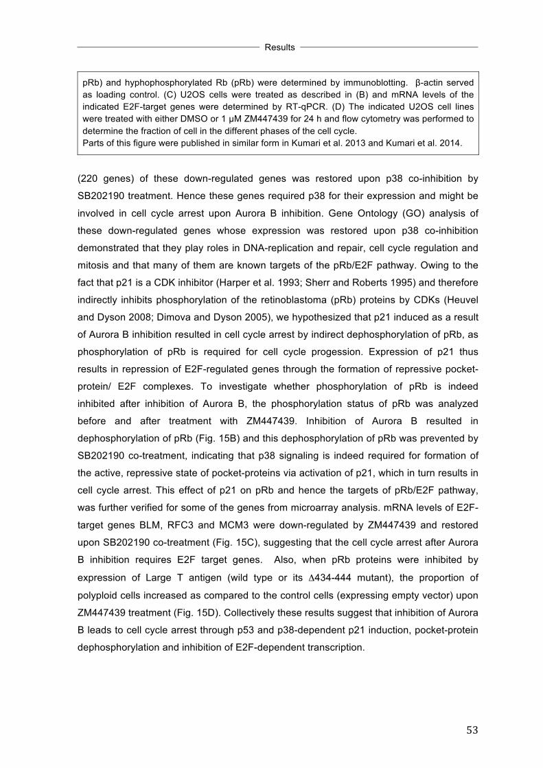
!
!
!Results
!! !
53!
pRb) and hyphophosphorylated Rb (pRb) were determined by immunoblotting. β-actin served as loading control. (C) U2OS cells were treated as described in (B) and mRNA levels of the indicated E2F-target genes were determined by RT-qPCR. (D) The indicated U2OS cell lines were treated with either DMSO or 1 µM ZM447439 for 24 h and flow cytometry was performed to determine the fraction of cell in the different phases of the cell cycle. Parts of this figure were published in similar form in Kumari et al. 2013 and Kumari et al. 2014.
(220 genes) of these down-regulated genes was restored upon p38 co-inhibition by
SB202190 treatment. Hence these genes required p38 for their expression and might be
involved in cell cycle arrest upon Aurora B inhibition. Gene Ontology (GO) analysis of
these down-regulated genes whose expression was restored upon p38 co-inhibition
demonstrated that they play roles in DNA-replication and repair, cell cycle regulation and
mitosis and that many of them are known targets of the pRb/E2F pathway. Owing to the
fact that p21 is a CDK inhibitor (Harper et al. 1993; Sherr and Roberts 1995) and therefore
indirectly inhibits phosphorylation of the retinoblastoma (pRb) proteins by CDKs (Heuvel
and Dyson 2008; Dimova and Dyson 2005), we hypothesized that p21 induced as a result
of Aurora B inhibition resulted in cell cycle arrest by indirect dephosphorylation of pRb, as
phosphorylation of pRb is required for cell cycle progession. Expression of p21 thus
results in repression of E2F-regulated genes through the formation of repressive pocket-
protein/ E2F complexes. To investigate whether phosphorylation of pRb is indeed
inhibited after inhibition of Aurora B, the phosphorylation status of pRb was analyzed
before and after treatment with ZM447439. Inhibition of Aurora B resulted in
dephosphorylation of pRb (Fig. 15B) and this dephosphorylation of pRb was prevented by
SB202190 co-treatment, indicating that p38 signaling is indeed required for formation of
the active, repressive state of pocket-proteins via activation of p21, which in turn results in
cell cycle arrest. This effect of p21 on pRb and hence the targets of pRb/E2F pathway,
was further verified for some of the genes from microarray analysis. mRNA levels of E2F-
target genes BLM, RFC3 and MCM3 were down-regulated by ZM447439 and restored
upon SB202190 co-treatment (Fig. 15C), suggesting that the cell cycle arrest after Aurora
B inhibition requires E2F target genes. Also, when pRb proteins were inhibited by
expression of Large T antigen (wild type or its Δ434-444 mutant), the proportion of
polyploid cells increased as compared to the control cells (expressing empty vector) upon
ZM447439 treatment (Fig. 15D). Collectively these results suggest that inhibition of Aurora
B leads to cell cycle arrest through p53 and p38-dependent p21 induction, pocket-protein
dephosphorylation and inhibition of E2F-dependent transcription.
!

!
!
!Results
!! !
54!
3.6 p38 MAPK is required for transcriptional induction of p21 but not for its protein stability
To investigate if p38 has a role in transcription of the p21 gene in response to Aurora B
inhibition, mRNA levels of p21 were analyzed by real time RT-qPCR. Treatment of cells
with ZM447439 resulted in induction of p21 mRNA levels and this induction was blocked
by SB202190 co-treatment (Fig. 16A), indicating that p21 transcriptional induction after
Aurora B inhibition depends on p38 signaling. p38 has been reported to stabilize p21 by
!!
Figure 16: p38 is required for transcriptional induction of p21 but not for its protein stability. (A) U2OS cells were treated with DMSO, 1 µM ZM447439 alone, 10 µM SB202190 alone or pretreated with 10 µM SB202190 for 2 h and then treated with 1 µM ZM447439 for 24 h. mRNA level of p21 was determined by RT-qPCR. (B) U2OS cells were treated with DMSO, 1 µM ZM447439 or pretreated with 10 µM SB202190 for 2 h followed by treatment with 1 µM ZM447439 for 22 h and then with 20 µg/ml cycloheximide (CHX) for the indicated time points to block protein synthesis. p21 levels were determined by immunoblotting. β-actin served as loading control. (C) The protein bands of p21 and β-actin of the blot shown in (B) were quantified by ImageJ. The amount of p21 protein (relative to β-actin) was plotted against time. Parts of this figure were published in similar form in Kumari et al. 2013.
phosphorylating it at Ser-130 in response to stress signals induced by TGFβ (Kim et al.
2002). To examine if p38 activated as a consequence of Aurora B inhibition has an effect
on p21 protein stability, the half-life of p21 protein induced after Aurora B inhibition (in

!
!
!Results
!! !
55!
absence and presence of p38 co-inhibition) was determined by using cycloheximide (a
protein synthesis inhibitor). U2OS cells were treated with ZM447439 or pretreated with
SB202190 for 2 hours followed by ZM447439 treatment for 22 hours and then either left
untreated or were treated with cycloheximide for 0.5, 1 and 2 hours. The stability of p21
protein induced after Aurora B inhibition was not affected by co-inhibition of p38,
indicating that p38 does not influence p21 protein stability under these conditions (Fig.
3.6B).
3.7 p38 MAPK is not required for p53 binding to p21 promoter in response to
Aurora B inhibition Having shown that p38 is required for transcriptional induction of p21 without affecting its
protein stability and total p53 protein levels; it was tempting to study the transcriptional
regulation of p21 gene in more detail. For this, first the binding of p53 to the p21 promoter
was studied by chromatin immunoprecipitation (ChIP) assay at p21 gene locus using an
antibody specific for p53. Cells were treated with DMSO, ZM447439 or with a combination
of SB202190 (pretreatment for 2 h) and ZM447439. Chromatin was isolated and
immunoprecipitated with a p53 specific antibody. As a control, nonspecific IgG was used.
Promoter regions were detected by quantitative real time PCR. p53 binding was robustly
induced at the high affinity p53 binding site 1 (p53BS1) upon treatment with ZM447439
(Fig. 17B) or AZD1152-HQPA (Fig. 17D). No significant binding of p53 was observed at
the distal regions of the p21 gene, as expected (Fig. 18B). In addition, no significant
binding was observed at the GAPDH2 promoter used as a control. Interestingly, p53
binding was not affected by p38 inhibition with SB202190 (Fig. 17B) or BIRB796 (Fig.
17D). Thus, p38 is not required for binding of p53 to its binding site on p21 promoter,
implicating that p38 acts at a step subsequent to p53 binding at the p21 gene locus.

!
!
!Results
!! !
56!
! Figure 17: p38 MAPK is not required for p53 binding to p21 promoter in response to Aurora B inhibition. (A) and (B) U2OS cells were treated with DMSO, 1 µM ZM447439 alone, or pretreated with 10 µM SB202190 for 2 h and then treated with 1 µM ZM447439 for 24 h. Chromatin was isolated and precipitated with antibody specific for p53 or nonspecific IgG. ChIP enriched DNA was amplified by qPCR using the indicated amplicons. GAPDH2 promoter was analyzed as a control. (C) and (D) HCT116 cells were treated with DMSO, 50 nM AZD1152-HQPA alone, or pretreated with 1 µM BIRB796 for 2h and then treated with 50 nM AZD1152-HQPA for 24 h. Chromatin was isolated and precipitated with antibody specific for p53 or nonspecific IgG. ChIP enriched DNA was amplified by qPCR using the indicated amplicons. GAPDH2 promoter was analyzed as a control.
3.8 p38 MAPK is required for transcriptional elongation of p21 in response to Aurora B inhibition
Regulation of p21 expression has been shown to occur at the level of transcriptional
elongation (Gomes et al. 2006; Valin et al. 2013). To explore this possibility, ChIP assay
with antibody against RNA Polymerase II (RNA Pol II) was performed at the p21 gene
locus. Significant amount of RNA Pol II was observed at the core promoter (-20 region) of
p21 gene even before stimulation by inhibition of Aurora B (Fig. 18D), consistent with
previous reports showing that RNA Polymerase II is paused at the promoters of stress
responsive genes (Adelman and Lis 2012). Very low binding of RNA Pol II was observed
in the transcribed region of the p21 gene, indicating that the polymerase is paused at the
p21 promoter. Upon inhibition of Aurora B by ZM447439 treatment, loading of RNA Pol II

!
!
!Results
!! !
57!
!
Figure 18: p38 MAPK is required for transcriptional elongation of p21 in response to Aurora B inhibition. (A) Schematic diagram of the p21 gene locus and position of the amplicons used for ChIP analysis and primary transcript analysis. U2OS cells were treated with DMSO, 1 µM ZM447439 alone, or pretreated with 10 µM SB202190 for 2 h and then treated with 1 µM ZM447439 for 24 h. Chromatin was isolated and precipitated with nonspecific IgG (B), antibody specific for p53 (C), total RNA polymerase II (D) and phospho-ser2 RNA Pol II (E). ChIP enriched DNA was

!
!
!Results
!! !
58!
amplified by qPCR using the indicated amplicons. The GAPDH2 promoter was analyzed as a control. (F) U2OS cells were treated with DMSO, 1 µM ZM447439 alone, or pretreated with 10 µM SB202190 for 2 h and then treated with 1 µM ZM447439 for 24 h. Total RNA was isolated and subjected to RT-qPCR using primers specific for shorter (+507) and longer region (+7011) primary p21 transcripts. This figure was published in similar form in Kumari et al. 2013.
moderately increased at the core promoter (less than 2-fold), whereas it’s loading in the
distal regions of the p21 gene was strongly induced (3-4 fold) (Fig. 18D). The loading of
RNA Pol II at the core promoter (-20 region) was not affected by p38 inhibition by
SB202190 co-treatment (instead there was a slight increase binding), whereas its
recruitment to the distal regions of the p21 gene was strongly blocked upon p38 inhibition
(Fig. 18D). These findings indicate a role for p38 MAPK in mediating transcriptional
elongation of p21 gene after recruitment of RNA Pol II, in response to Aurora B inhibition.
To confirm the requirement of p38 in elongation stage of the transcriptional induction of
p21, ChIP assay was performed with antibody against RNA polymerase II phosphorylated
at serine 2 (Ser 2P RNA Pol II), which represents the elongating form of RNA Polymerase
II (Phatnani and Greenleaf 2006; Egloff et al. 2012). Interestingly the elongating form of
RNA polymerase II was strongly enriched towards the 3’ region of the p21 gene after
Aurora B inhibition by ZM447439 and this enrichment was impaired by p38 inhibition by
SB202190 co-treatment (Fig. 18E), emphasizing the notion that p38 is required for
transcriptional elongation of p21 gene. To corroborate the finding that p38 is required for
the elongation of the p21 gene and hence is required to generate the full-length p21
mRNA, nascent p21 transcripts were analyzed by RT-qPCR. Short p21 transcripts were
analyzed by with primers specific for an amplicon in the first intron of the gene (+507)
whereas long p21 transcripts were analyzed with primers specific for the second intron
just before the third exon (+7011). Inhibition of Aurora B by ZM447439 treatment induced
short transcripts, but their induction was not prevented by co-inhibition of p38. Longer p21
transcripts were also induced after Aurora B inhibition. Importantly, accumulation of the
longer transcript was significantly reduced when p38 was inhibited by SB202190 co-
treatment (Fig. 18F). Together these data indicate that p38 signaling is required for
transcriptional elongation of p21 after Aurora B inhibition.

!
!
!Results
!! !
59!
3.9 Transcriptional elongation of p21 in response to replication stress is dependent on p38 MAPK
Several studies have investigated the transcriptional elongation control of p21 in response
to stress (Espinosa et al. 2003; Mattia et al. 2007; Beckerman et al. 2009; Gomes 2006;
Valin et al. 2013). Beckerman et al. (2009) demonstrated that blocking DNA replication by
hydroyxurea (HU, induces S-phase arrest by replication block) induced p53 but did not
activate a subset of p53 targets including p21 due to marked reduction in its
transcriptional elongation. The authors further showed that, Chk1 is employed in sensing
the replication block caused due to stalled replication forks in presence of HU and
inhibiting Chk1 by caffeine co-treatment relieved the elongation block of p21 gene leading
to significant accumulation of p21 mRNA and protein. In order to test the requirement of
p38 MAPK in this elongation pathway, levels of phosphorylated p38 were analyzed under
conditions where elongation block was relieved (i.e. under caffeine and HU co-treatment
conditions). Indeed p38 was phosphorylated and hence activated under these conditions
when p21 was induced, whereas total p38 levels remained unchanged (Fig. 19A). As
reported previously (Gottifredi et al. 2001; Beckerman et al. 2009) HU treatment induced
p53 and co-treatment with caffeine and HU induced p21, without affecting p53 protein
levels induced by HU alone (Fig. 19A). To further confirm the requirement of p38 in this
pathway, the p38 MAPK specific inhibitor BIRB796 was used to inhibit p38. Inhibition of
p38 partially blocked the p21 induction upon caffeine and HU co-treatment, without
affecting the p53 protein levels (Fig. 19B). Interestingly, the enrichment of elongating form
of RNA polymerase II in the distal regions of the p21 gene observed upon caffeine and
HU co-treatment conditions was also partially blocked by p38 co-inhibition by BIRB796
(Fig. 19C). Taken together these findings strongly support the importance of p38 signaling
in mediating transcriptional elongation p21 gene under stress conditions.

!
!
!Results
!! !
60!
!
! Figure 19: Transcriptional elongation of p21 in response to replication stress is dependent on p38 MAPK. (A) U2OS cells were untreated (unt.) or pretreated with 4 mM caffeine for 1 h and then left untreated or were treated with 1.7 mM HU for 24 h. The levels of respective proteins were determined by immunoblotting. α-tubulin served as loading control. (B) U2OS cells were pretreated with DMSO, 4 mM caffeine, 1 µM BIRB796 or a combination of 4 mM caffeine and 1 µM BIRB796 and then left untreated or were treated with 1.7 mM HU for 24 h. p53 and p21 protein levels were determined by immunoblotting. β-actin served as loading control. (C) U2OS cells were treated as in (B). Chromatin was isolated and precipitated with antibody specific for phospho-ser2 RNA Pol II. ChIP enriched DNA was amplified by RT-qPCR using the indicated amplicons. GAPDH2 promoter was analyzed as a control.
!!
!!

!
!
!Results
!! !
61!
3.10 Inhibition of Aurora B activates both α and β isoforms of p38 MAPK without affecting their subcellular localization
Phosphorylation of p38 triggers a conformational change leading to its translocation into
the nucleus (Wood et al. 2009). Subcellular localization is an important mechanism by
which substrate specificity of p38 MAPK is regulated (Roux and Blenis 2004; Raman et al.
2007). Hence, I was prompted to study the subcellular localization of p38 after Aurora B
inhibition. Since SB202190 inhibits all the four isoforms of p38 (p38α, p38β, p38γ and
p38δ), whereas BIRB796 specifically inhibits only the α and β isoforms, the findings so far
suggest the preferential activation of p38α and/or p38β (as compared to p38γ and p38δ)
due to Aurora B kinase inhibition. So, future experiments designed to explore the
mechanism by which p38 was mediating the activation of p21, were concentrated mainly
on p38α and p38β isoforms.
To study subcellular localization of p38α and p38β, U2OS-EcoR cells stably expressing
haemagglutinin (HA) tagged versions of p38α and p38β (HA-p38α and HA-p38β) were
generated. The expression of HA-p38α and HA-p38β was verified by immunoblotting (Fig.
20A). Further, these cells showed stabilization of p53 and induction of p21 upon Aurora B
inhibition by ZM4447439 treatment, indicating that overexpression of these constructs did
not had any off target effects and did not alter the outcome of Aurora B inhibition. Aurora
B was inhibited by ZM447439 treatment and the lysates were immunoprecipitated with a
HA-tag specific antibody and then immunoblotted with an antibody against
phosphorylated p38. Both α and β isoforms of p38 were phosphorylated upon Aurora B
inhibition (Fig. 20A). To test whether inhibition of Aurora B or co-inhibition of Aurora B and
p38 affects the subcellular localization of these isoforms of p38, cells expressing either
HA-p38α or HA-p38β were treated with ZM447439 alone, SB202190 alone or pretreated
with SB202190 for 2 h and then treated with ZM447439 for 24 h, fixed and analyzed by
immunostaining using HA-tag specific antibody. Both HA-p38α and HA-p38β were
localized in cytoplasm as well in nucleus under control conditions (DMSO panel in Fig.
20B and C) in accordance with published data (Raingeaud et al. 1995). The subcellular
localizations of both the isoforms remained unchanged on ZM447439 treatment or
ZM447439 and SB202190 co-treatment (Fig. 20B and C). From these data it is clear that
the subcellular localization of p38α and p38β is not influenced by Aurora B inhibition.

!
!
!Results
!! !
62!
! Figure 20: Inhibition of Aurora B activates both α and β isoforms of p38 MAPK without affecting their subcellular localization. (A) U2OS-EcoR cells stably expressing either HA-p38α or HA-p38β were treated with 1 µM ZM447439 for 24 h and lysates were immunoprecipitated with HA-tag specific antibody. Immunoprecipitated proteins were analyzed by blotting with antibodies against HA and phosphorylated p38 (p-p38). The input sample was analyzed for with antibodies against HA, p-p38, p38, p53 and p21. β-actin served as loading control. (B) and (C) U2OS-EcoR cells stably expressing either HA-p38α or HA-p38β were treated with DMSO, 1 µM ZM447439, 10 µM SB202190 or first pretreated with 10 µM SB202190 for 2 h and then treated with 1 µM ZM447439 for 24 h. HA-tagged proteins (in green) and p21 (in red) were detected by immunostaining. Nuclei (in blue) were stained with Hoechst 33528.!
3.11 Neither p38α nor p38β bind to the p21 gene upon Aurora B inhibition It is known that the yeast p38-related MAP kinase, Hog1 associates with the transcribed
region of osmoresponsive genes and promotes their transcriptional elongation in
response to osmotic stress (Proft et al. 2006). To investigate, if this is true for human p38
MAPK as well, the binding of HA-p38α and HA-p38β to the p21 gene locus was studied by
ChIP. U2OS-EcoR cells stably expressing either HA-p38α or HA-p38β were treated with
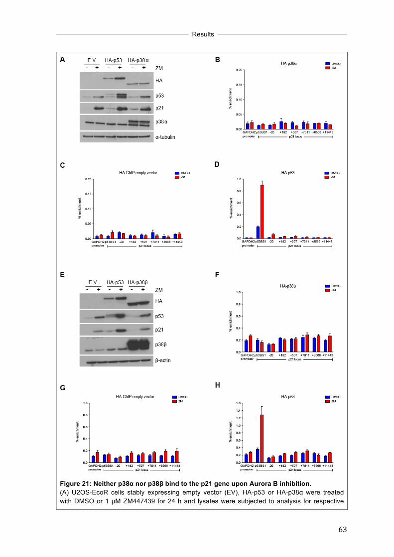
!
!
!Results
!! !
63!
Figure 21: Neither p38α nor p38β bind to the p21 gene upon Aurora B inhibition. (A) U2OS-EcoR cells stably expressing empty vector (EV), HA-p53 or HA-p38α were treated with DMSO or 1 µM ZM447439 for 24 h and lysates were subjected to analysis for respective

!
!
!Results
!! !
64!
proteins indicated. (B) U2OS-EcoR cells stably expressing HA-p38α, empty vector (C) or HA-p53 (D) were treated with DMSO or 1 µM ZM447439 for 24 h and chromatin was immunoprecipitated with a HA-tag specific antibody. ChIP enriched DNA was amplified by qPCR using the indicated amplicons. The GAPDH2 promoter was analyzed as a control. (E) U2OS-EcoR cells stably expressing empty vector (EV), HA-p53 or HA-p38 β were treated with DMSO or 1 µM ZM447439 for 24 h and lysates were subjected to analysis for respective proteins indicated. (F) U2OS-EcoR cells stably expressing HA-p38β, empty vector (G) or HA-p53 (H) were treated with DMSO or 1 µM ZM447439 for 24 h and chromatin was immunoprecipitaed by HA-tag specific antibody. ChIP enriched DNA was amplified by qPCR using the indicated amplicons. GAPDH2 promoter was analyzed as a control.
ZM447439 and chromatin was isolated and immunoprecipitated with an antibody against
the HA-tag. U2OS-EcoR cells stably expressing empty vector were used as negative
control, whereas for positive control U2OS-EcoR cells stably expressing HA-p53 were
used. Expression of HA-p38α and HA-p38β and induction of p53 and p21 upon Aurora B
inhibition was verified by immunoblotting (Fig. 21A and E). In ChIP analysis, HA-p38α did
not show any significant binding to the complete p21 gene locus (similar to empty vector
expressing cells) either in untreated conditions or upon ZM447439 treatment (Fig. 21B
and C). Similarly HA-p38β showed the same background binding to the p21 gene locus as
empty vector (Fig. 21F and G). On the other hand HA-p53 binding was strongly induced at
the p53 binding site 1 (p53BS1) in the p21 promoter upon Aurora B inhibition by
ZM447439 treatment as expected (Fig. 21D and H). These results suggest that p38α and
p38β are not recruited to p21 gene locus after Aurora B inhibition.
!3.12 Elongin A binding to the p21 gene locus is induced upon Aurora B
inhibition Elongin A has been reported to bind p21 gene and mediate its transcriptional elongation
in response to various cellular stresses (Kawauchi et al. 2013). To investigate the
involvement of Elongin A in p21 transcriptional elongation, U2OS-EcoR cells stably
expressing HA-tagged mouse Elongin A (HA-mEloA) were generated. The expression of
HA-mEloA was verified by immunobloting (Fig. 22A). Further, treatment of these cells with
ZM447439 resulted in p21 induction and this induction was blocked by p38 inhibition by
SB202190 co-treatment, indicating that over expression of mouse Elongin A does not
interfere with the activation of p21 upon Aurora B inhibition (Fig. 22A). U2OS-EcoR cells
stably expressing HA-mEloA were treated with DMSO, ZM447439 or pretreated with
SB202190 and then treated with ZM447439. Chromatin was isolated and
immunoprecipitated with antibody against HA-tag. The binding of HA-mElongin A was
induced at the core promoter as well as in the coding region the p21 gene towards the
distal regions on Aurora B inhibition by ZM447439 treatment (Fig. 22B). Interestingly this

!
!
!Results
!! !
65!
enrichment was blocked when p38 was inhibited by SB202190 co-treatment. This
suggests that Elongin A recruitment to p21 gene locus might be dependent on signaling
from p38. To confirm this possibility the effect of knockdown of Elongin A on p21 induction
upon Aurora B inhibition needs to be investigated.
!
Figure 22: Binding of Elongin A to the p21 gene locus is induced upon Aurora B inhibition. (A) U2OS-EcoR cells stably expressing empty vector (EV) or HA-mEloA were treated with DMSO, 1 µM ZM447439 alone, 10 µM SB202190 alone or pretreated with 10 µM SB202190 for 2 h and then treated with 1 µM ZM447439 for 24 h. Levels of HA-mEloA and p21 were determined by immunoblotting. β-actin served as loading control. (B) U2OS-EcoR cells stably expressing HA-mEloA, were treated as in (A) and chromatin was immunoprecipitated by HA-tag specific antibody. ChIP enriched DNA was amplified by qPCR using the indicated amplicons. GAPDH2 promoter was analyzed as a control.
!!!

!
!
!Results
!! !
66!
3.13 Aurora B inhibition in interphase is not sufficient for induction of p21 Recently it has been proposed that Aurora B has a function outside mitosis, during the
interphase where it mediates degradation of p53 by phosphorylation (Gully et al. 2012). In
order to test whether the interphase function or the mitotic function of Aurora B is
necessary for p21 induction, cells were arrested at the G1/S border by thymidine. The
G1/S arrest was confirmed by flow cytometry (Fig. 23B). Inhibition of Aurora B in
interphase (in G1/S arrested cells) by ZM447439 treatment did not induce p21 (Fig. 23A,
lane 3), whereas p21 was strongly induced (Fig. 23A, lane 5) when the cells were first
released from G1/S block and then treated with ZM447439 (inhibition of Aurora B outside
interphase). As control, asynchronous cells were treated with ZM447439, which showed
p21 induction (Fig. 23A, lane 6). Although G1/S arrested cells already showed p53
stabilization, this stabilization increased when cells were released from G1/S block and
then treated with ZM443439. Taken together these findings support the conclusion that
the induction of p53 and p21 is not due to inhibition of interphase function of Aurora B, but
due to inhibition of its function during mitosis.

!
!
!Results
!! !
67!
! Figure 23: Aurora B inhibition in interphase is not sufficient for induction of p21. (A) U2OS cells were synchronized in G1/S by treatment with 2.5 mM thymidine for 24 h (lanes 2-5). Thymidine-blocked cells were treated with DMSO or with 1 µM ZM447439 for 24 h either without release (lane 2 and 3) or after a 2 h release from the thymidine block (lane 4 and 5). Asynchronous cells (lane 1) and asynchronous cells treated with 1 µM ZM447439 for 24 h (lane 6) served as controls. Levels of p21 were determined by immunoblotting. β-actin served as a loading control. (B) Cell cycle synchronization of the experiment shown in (A) was confirmed by flow cytometry. This figure was published in similar form in Kumari et al. 2014.
!
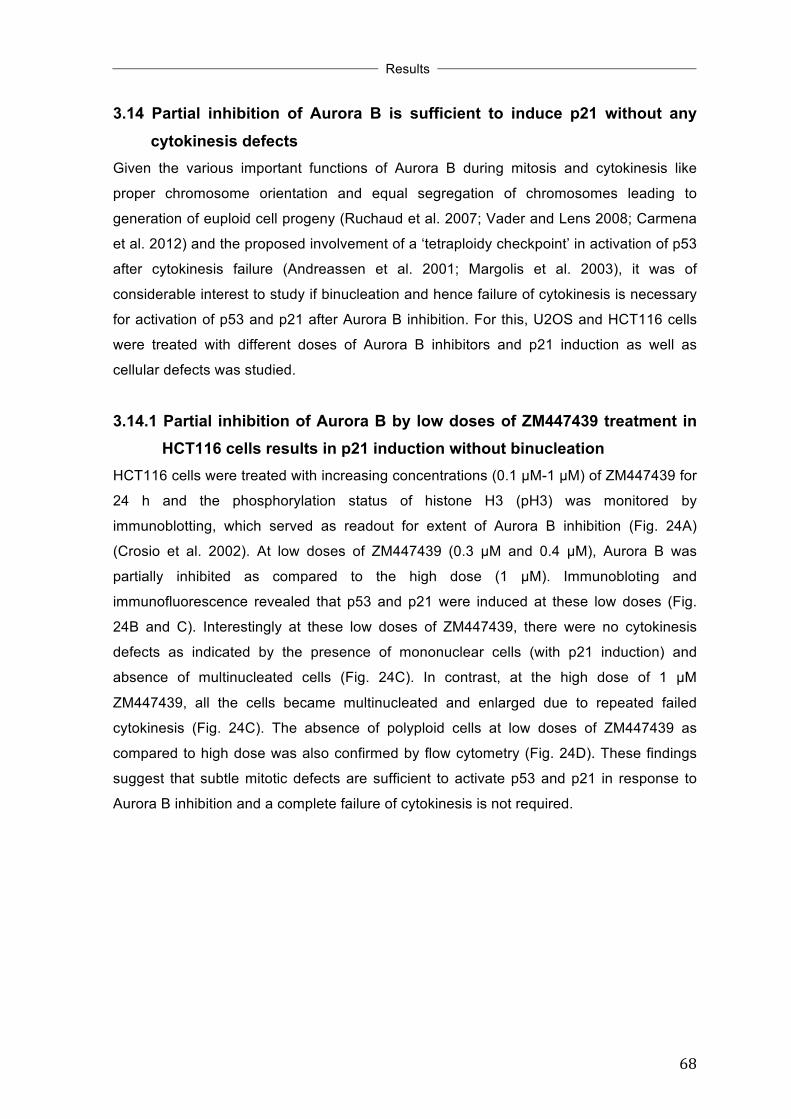
!
!
!Results
!! !
68!
3.14 Partial inhibition of Aurora B is sufficient to induce p21 without any cytokinesis defects
Given the various important functions of Aurora B during mitosis and cytokinesis like
proper chromosome orientation and equal segregation of chromosomes leading to
generation of euploid cell progeny (Ruchaud et al. 2007; Vader and Lens 2008; Carmena
et al. 2012) and the proposed involvement of a ‘tetraploidy checkpoint’ in activation of p53
after cytokinesis failure (Andreassen et al. 2001; Margolis et al. 2003), it was of
considerable interest to study if binucleation and hence failure of cytokinesis is necessary
for activation of p53 and p21 after Aurora B inhibition. For this, U2OS and HCT116 cells
were treated with different doses of Aurora B inhibitors and p21 induction as well as
cellular defects was studied.
3.14.1 Partial inhibition of Aurora B by low doses of ZM447439 treatment in HCT116 cells results in p21 induction without binucleation
HCT116 cells were treated with increasing concentrations (0.1 µM-1 µM) of ZM447439 for
24 h and the phosphorylation status of histone H3 (pH3) was monitored by
immunoblotting, which served as readout for extent of Aurora B inhibition (Fig. 24A)
(Crosio et al. 2002). At low doses of ZM447439 (0.3 µM and 0.4 µM), Aurora B was
partially inhibited as compared to the high dose (1 µM). Immunobloting and
immunofluorescence revealed that p53 and p21 were induced at these low doses (Fig.
24B and C). Interestingly at these low doses of ZM447439, there were no cytokinesis
defects as indicated by the presence of mononuclear cells (with p21 induction) and
absence of multinucleated cells (Fig. 24C). In contrast, at the high dose of 1 µM
ZM447439, all the cells became multinucleated and enlarged due to repeated failed
cytokinesis (Fig. 24C). The absence of polyploid cells at low doses of ZM447439 as
compared to high dose was also confirmed by flow cytometry (Fig. 24D). These findings
suggest that subtle mitotic defects are sufficient to activate p53 and p21 in response to
Aurora B inhibition and a complete failure of cytokinesis is not required.

!
!
!Results
!! !
69!
! Figure 24: Partial inhibition of Aurora B by low doses of ZM447439 treatment in HCT116 cells results in p21 induction without binucleation. (A) HCT116 cells were treated with various concentrations of ZM447439 for 24 h. Histone H3 phosphorylated at serine 10 (pH3) was analyzed by immunoblotting. β-actin served as a loading control. (B) HCT116 cells were treated with 0.3 µM, 0.4 µM or 1 µM ZM447439 for 24 h. Levels of p53 and p21 proteins were determined by immunoblotting. α-tubulin served as loading control. (C) HCT116 cells were treated as in (B) and level of p21 was determined by immunostaining. DNA was counterstained with Hoechst 33258. (D) HCT116 cells were treated as in (B) and FACS was done to determine the fraction of cells in different phases of the cell cycle. Parts of this figure were published in similar form in Kumari et al. 2014.
!
3.14.2 Partial inhibition of Aurora B by low doses of ZM447439 treatment in U2OS cells results in p21 induction without binucleation
To confirm that the effects seen at low doses of ZM447439 are not specific for HCT116
cells, another cell line U2OS was also treated with different doses (0.5 µM and 1 µM) of
ZM447439. Treatment of U2OS cells with 0.5 µM ZM447439 did not lead to cytokinesis
defects, but induced p21 at comparable levels to the higher dose of 1 µM (Fig. 25A). Also,
p53 and p-p38 were activated at these low doses of Aurora B inhibitor (Fig. 25B),

!
!
!Results
!! !
70!
indicating that the p38-p53-p21 pathway activation in response to Aurora B inhibition is
sensitive to defects in mitosis and not to inhibition of cytokinesis. The cell cycle profiles of
U2OS cells treated with 0.5 µM and 1 µM ZM447439, further confirmed that there was no
cell division failure at the low doses (Fig. 25C).
!
! Figure 25: Partial inhibition of Aurora B by low doses of ZM447439 treatment in U2OS cells results in p21 induction without binucleation. (A) U2OS cells were treated with DMSO, 0.5 µM or 1 µM ZM447439 for 24 h. p21 (in red) and α-tubulin (in green) was detected by immunostaining. Nuclei (in blue) were stained with Hoechst 33528. (B) U2OS cells were treated as in (A) and respective protein levels were determined by immunoblotting. (C) U2OS cells were treated as in (A) and analyzed by FACS to determine cells in different phases of cell cycle. Parts of this figure were published in similar form in Kumari et al. 2014.
!!3.14.3 Partial inhibition of Aurora B by low doses of AZD1152-HQPA
treatment in U2OS cells results in p21 induction without binucleation To exclude the possibility that the phenotypes observed in U2OS and HCT116 cells by
low doses of ZM447439 treatment are artifact, another inhibitor of Aurora B, AZD1152-
HQPA was used. U2OS cells were treated with 50 nM and 100 nM AZD1152-HQPA.
Although both concentrations of AZD1152-HQPA induced comparable levels of p53, p-
p38 and p21 (Fig. 26A), there were no binucleated or multinucleated cells at the lower

!
!
!Results
!! !
71!
dose of 50 nM as compared to higher dose of 100 nM AZD1152-HQPA. Taken together,
all these data with low doses of two different Aurora B inhibitors in two different cell lines
indicate that tetraploidy and hence cytokinesis failure is not required for p38-p53-p21
activation in response to Aurora B inhibition.
!
! Figure 26: Partial inhibition of Aurora B by low doses of AZD1152-HQPA treatment in U2OS cells results in p21 induction without binucleation. (A) U2OS cells were treated with DMSO, 50 nM or 100 nM AZD1152-HQPA for 24 h. Respective protein levels were determined by immunoblotting. (B) U2OS cells were treated as in (A) and p21 levels (in red) were determined by immunostaining. Nuclei (in blue) were stained with Hoechst 33528. This figure was published in similar form in Kumari et al. 2014.
!!3.15 Partial Aurora B inhibition results in increased aneuploidy The activation of p38-p53-p21 pathway in absence of any cytokinesis defects upon partial
Aurora B inhibition suggests that chromosomal segregation errors due to Aurora B
inhibition might be responsible for activation of this pathway. To answer this possibility the
frequency of aneuploidy was determined by carrying out interphase FISH assays using
fluorescent probes specific for chromosome 7 and 8. Partial inhibition of Aurora B by low
doses of (0.3 µM or 0.4 µM) ZM447439 treatment (for 48 h) in HCT116 cells indeed
caused increased aneuploidy as indicated by an increase in the deviation from mode for
chromosome 7 from 5.01 % to 6.47 % (with 0.3 µM ZM447439) and to 11.84 % (with 0.4
µM ZM447439) and for chromosome 8 an increase from 3.34 % to 6.47 % (with 0.3 µM
ZM447439) and to 14.11 % (with 0.4 µM ZM447439) (Fig. 27A and B). Hence p53 and

!
!
!Results
!! !
72!
p21 activation on partial Aurora B inhibition correlates with increased chromosome
segregation errors in absence of tetraploidy.
!
! Figure 27: Partial Aurora B inhibition results in increased aneuploidy. (A) Example microphotographs of interphase FISH assays of HCT116 cells treated with DMSO, 0.3 µM or 0.4 µM ZM447439 for 48 h. Chromosome 7: red; chromosome 8: green (B) Quantification of interphase FISH signals for chromosomes 7 and 8 in DMSO and ZM447349 treated HCT116 cells shown in (A). The difference in the percentage of cell off the mode after treatment with 400 nM ZM447439 was statistically significant (chromosome 7 or 8 p-values <0.001). With 300 nM ZM447439 the difference in percentage of cells of the mode was not significant for chromosome 7 but was significant for chromosome 8 (p=0.009). Chi-squared test was used to analyze the data. This figure was published in similar form in Kumari et al. 2014.
!!3.16 p21 induction after partial Aurora B inhibition does not involves DNA
damage A plausible cause of p21 activation due to partial Aurora B inhibition might be
chromosomal segregation errors causing DNA damage, which has been reported to
activate p53 (Thompson and Compton 2010b; Janssen et al. 2011). Presence of DNA
damage can be detected by classical markers like presence of phosphorylated H2A.X (a
widely used marker for DNA damage response as a result of double strand breaks) or
presence of activated ATM (ataxia-telangiectasia mutated) and ATR (ATM and Rad3-
related) kinases (Zhou and Elledge 2000; Falck et al. 2005). All these possibilities were

!
!
!Results
!! !
73!
! Figure 28: p21 induction after partial Aurora B inhibition does not involve DNA damage. (A) U2OS cells were treated with DMSO, 0.5 µM ZM447439, 1 µM ZM447439 for 24 h or with 1 µM doxorubicin (Dox.) for 6 h. p21, p-H2A.X and β-actin protein levels were determined by immunoblotting. (B) U2OS cells were treated as in (A) and protein levels of phosphorylated ATM/ATR substrates and p21 were detected by immunoblotting. β-actin served as a control. (C) U2OS cells were pretreated with the indicated concentration of VE821 for 2 h and then treated with 1 µM ZM447439 for 24 h. p21 levels were determined by immunoblotting. β-actin served as a control. (D) U2OS cells were pretreated with the indicated concentration of KU5593 for 2 h and then treated with 1 µM ZM447439 for 24 h. p21 levels were determined by immunoblotting. β-actin served as a control. (E) U2OS cells were pretreated with DMSO, 1 µM VE821, 10 µM KU5593 or a combination of 1 µM VE821 and 10 µM KU5593 for 2 h and then treated with 1 µM doxorubicin or left untreated for 6 h. p-Chk1(Ser345), p-Chk2(Thr68) and p-ATM/p-ATR substrate protein levels were determined by immunoblotting. β-actin served as a control. This figure was published in similar form in Kumari et al. 2014.
!explored. However there was no evidence of DNA damage upon partial Aurora B
inhibition as evident by absence of p-H2A.X in U2OS cells upon ZM443439 treatment,

!
!
!Results
!! !
74!
whereas p-H2A.X was detected by treatment with the DNA damaging agent, doxorubicin
(used as positive control) (Fig. 28A). Also, phosphorylation of ATM/ATR kinase substrates
was not detected in response to ZM447439 treatment (at both low and high doses) (Fig.
28B) while a positive result was obtained with doxorubicin, which induced the
phosphorylation of ATM/ATR substrates (Fig. 28B). To further confirm that ATM and ATR
kinases are not involved in p21 induction in response to Aurora B inhibition, the effects of
ATM inhibitor KU5593 (Hickson et al. 2004) and ATR inhibitor VE821 (Reaper et al. 2011)
on p21 induction mediated by ZM447439 treatment was investigated. However, none of
these inhibitors were able to prevent the p21 induction upon ZM447439 treatment,
indicating that they are not involved in the activation of the p53-p21 pathway after Aurora
B inhibition (Fig. 28B and C). In order to test if the inhibitors were working, the effect of
ATM inhibitor KU5593 on p-Chk2(Thr68) and of ATR inhibitor VE821 on p-Chk1(Ser345)
induction upon doxorubicin treatment was studied. As expected, the phosphorylation of
Chk1 at Ser-345 was efficiently blocked by VE821 and the phosphorylation of Chk2 at
Thr-68 was efficiently blocked by KU5593, while the combined inhibitor treatment
efficiently blocked the phosphorylation of ATM/ATR substrates (Fig. 28E). Overall, these
results indicate that the DNA damage does not contributes to p21 activation after
impairment of Aurora B function.
!3.17 Partial inhibition of Aurora B results in proteotoxic stress but no
autophagy Aneuploidy has been linked with increased proteotoxic stress due to protein imbalance, in
humans as well yeast (Stingele et al. 2012; Oromendia et al. 2012). Proteotoxic stress
leads to formation of ubiquitinated protein aggregates, which can be detected by
immunoblotting. Indeed increased poly-ubiquitin aggregates were detected in both U2OS
and HCT116 cells upon partial Aurora B inhibition by ZM447439 treatment (Fig. 29A).
Partial Aurora B inhibition by AZD1152-HQPA treatment in U2OS cells also resulted in
generation of poly-ubiquitin aggregates (Fig. 29B). Since aneuploidy has also been linked
to increased autophagy (Stingele et al. 2013), the effect of partial Aurora B inhibition on
two autophagy markers (p62 and LC3) was investigated. However the protein levels of
these markers (p62 and LC3) remained unchanged upon partial Aurora B inhibition by low
dose ZM447439 treatment in both U2OS and HCT116 cells (Fig. 29C). These findings
demonstrate that aneuploidy caused due to partial Aurora B inhibition generates
proteotoxic stress.
!

!
!
!Results
!! !
75!
! Figure 29: Partial inhibition of Aurora B results in proteotoxic stress but no autophagy. (A) U2OS and HCT116 cells were treated with 0.5 µM (for 24 h) or 0.3 µM (for 48 h) ZM447439, respectively. As a control, cells were treated with DMSO. Ubiquitin conjugates were detected by immunoblotting. β-actin served as a control. (B) U2OS cells were treated with DMSO, 50 nM or 100 nM AZD1152-HQPA for 24 h. Ubiquitin conjugates were detected by immunoblotting. β-actin served as a control. (C) U2OS and HCT116 cells were treated as in (A) and protein levels of p62 and LC3 were determined by immunoblotting. β-actin served as a control.
!!3.18 Partial inhibition of Aurora B correlates with increased generation of
reactive oxygen species (ROS) The metabolic imbalance resulting due to aneuploidy results in generation of reactive
oxygen species (ROS) (Li et al. 2010). Hence it was speculated that ROS might be
generated due to aneuploidy after partial Aurora B inhibition. To test the production of
ROS, the dye H2DCF-DA was used, which is a general oxidative stress indicator and
gives green fluorescence in presence of ROS. Interestingly partial Aurora B inhibition by
ZM447439 treatment resulted in generation of significant amount of ROS (Fig. 30A).

!
!
!Results
!! !
76!
! Figure 30: Partial inhibition of Aurora B correlates with increased generation of reactive oxygen species (ROS). (A) Detection of ROS with the fluorescent dye H2DFC-DA in U2OS cells treated with DMSO or 0.5 µM ZM447439 for 24 h. (B) ROS generation was analyzed with MitoSox Red in U2OS cells treated with DMSO, 0.5 µM ZM447439 or co-treated with 0.5 µM ZM447439 and 5 mM Tiron for 24 h. Bar 10 µm. (C) U2OS cells were treated as in (B). Levels of p53, p21, phosphorylated p38 (p-p38) and β-actin were determined by immunoblotting. (D) U2OS cells were treated for 48 h with DMSO, 0.5 µM ZM447439, 5 mM Tiron or 0.5 µM ZM447439 and 5 mM Tiron. Cells were pulse labeled with 15 µg/ml BrdU for 2 h. The percentage of BrdU-positive cells was determined by immunofluorescence. (E) Quantification of the BrdU positive cells shown in (D). Error bars represent standard deviation of four independent experiments. The differences between DMSO and ZM447439 (p=0.005) and between ZM447439 and ZM447439+Tiron (p=0.0019) were statistically significant (student's t-test). (F) U2OS cells were treated as in (B) and p21 mRNA levels were determined by qRT-PCR. (G) U2OS cells were treated with DMSO, 0.5 µM ZM447439 or co-treated with 0.5 µM ZM447439 and 5 mM Tiron for 72 h. Protein levels of p21 and the E2F target genes (E2F1, B-Myb, Cyclin A) were determined by immunoblotting. β-actin served as loading control. Parts of this figure were published in similar form in Kumari et al. 2014.
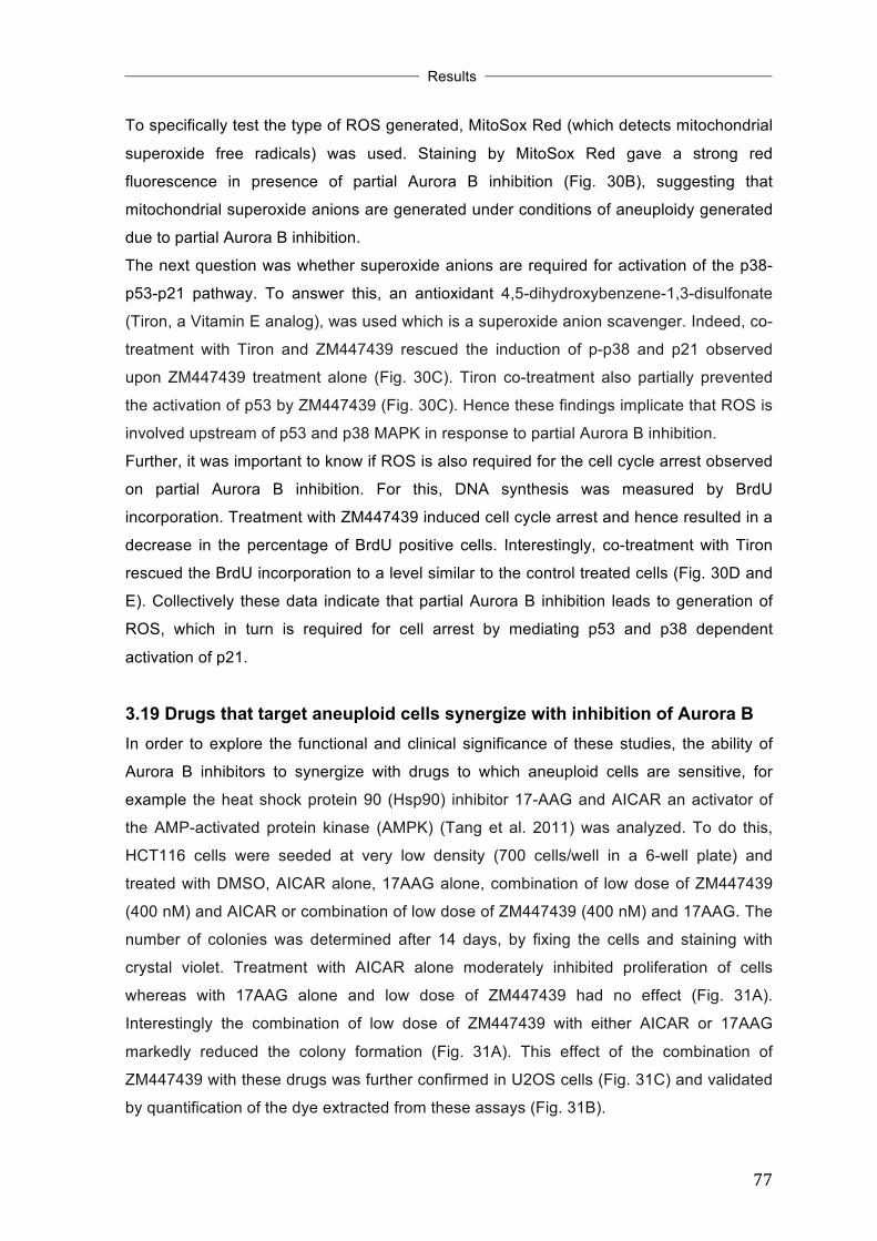
!
!
!Results
!! !
77!
To specifically test the type of ROS generated, MitoSox Red (which detects mitochondrial
superoxide free radicals) was used. Staining by MitoSox Red gave a strong red
fluorescence in presence of partial Aurora B inhibition (Fig. 30B), suggesting that
mitochondrial superoxide anions are generated under conditions of aneuploidy generated
due to partial Aurora B inhibition.
The next question was whether superoxide anions are required for activation of the p38-
p53-p21 pathway. To answer this, an antioxidant 4,5-dihydroxybenzene-1,3-disulfonate
(Tiron, a Vitamin E analog), was used which is a superoxide anion scavenger. Indeed, co-
treatment with Tiron and ZM447439 rescued the induction of p-p38 and p21 observed
upon ZM447439 treatment alone (Fig. 30C). Tiron co-treatment also partially prevented
the activation of p53 by ZM447439 (Fig. 30C). Hence these findings implicate that ROS is
involved upstream of p53 and p38 MAPK in response to partial Aurora B inhibition.
Further, it was important to know if ROS is also required for the cell cycle arrest observed
on partial Aurora B inhibition. For this, DNA synthesis was measured by BrdU
incorporation. Treatment with ZM447439 induced cell cycle arrest and hence resulted in a
decrease in the percentage of BrdU positive cells. Interestingly, co-treatment with Tiron
rescued the BrdU incorporation to a level similar to the control treated cells (Fig. 30D and
E). Collectively these data indicate that partial Aurora B inhibition leads to generation of
ROS, which in turn is required for cell arrest by mediating p53 and p38 dependent
activation of p21.
3.19 Drugs that target aneuploid cells synergize with inhibition of Aurora B In order to explore the functional and clinical significance of these studies, the ability of
Aurora B inhibitors to synergize with drugs to which aneuploid cells are sensitive, for
example the heat shock protein 90 (Hsp90) inhibitor 17-AAG and AICAR an activator of
the AMP-activated protein kinase (AMPK) (Tang et al. 2011) was analyzed. To do this,
HCT116 cells were seeded at very low density (700 cells/well in a 6-well plate) and
treated with DMSO, AICAR alone, 17AAG alone, combination of low dose of ZM447439
(400 nM) and AICAR or combination of low dose of ZM447439 (400 nM) and 17AAG. The
number of colonies was determined after 14 days, by fixing the cells and staining with
crystal violet. Treatment with AICAR alone moderately inhibited proliferation of cells
whereas with 17AAG alone and low dose of ZM447439 had no effect (Fig. 31A).
Interestingly the combination of low dose of ZM447439 with either AICAR or 17AAG
markedly reduced the colony formation (Fig. 31A). This effect of the combination of
ZM447439 with these drugs was further confirmed in U2OS cells (Fig. 31C) and validated
by quantification of the dye extracted from these assays (Fig. 31B).

!
!
!Results
!! !
78!
!
Figure 31: Drugs that target aneuploid cells synergize with inhibition of Aurora B. (A) HCT116 and U2OS cells were treated with DMSO, AICAR (200 µM), 17-AAG (8nM), ZM447439 (0.4 µM for HCT116, 0.5 µM for U2OS), a combination of ZM447439 and AICAR or a combination of ZM447439 and 17AAG. 14 days later, colonies were fixed and stained with crystal violet. (B) Quantification of the colonies shown in (A). The dye was extracted and absorbance was determined at 590 nm. The experiment was performed in triplicates. Error bars

!
!
!Results
!! !
79!
represent standard deviation. (*P<0.05, ***P<0.01, student’s t-test). (C) Soft-agar assays were performed to analyze anchorage independent growth of HCT116 cells treated with DMSO, AICAR (200 µM), ZM447439 (0.4 µM) or a combination of 0.4 µM ZM447439 and 200 µM AICAR. This figure was published in similar form in Kumari et al. 2014.
Next, the ability of the combination of ZM447439 and AICAR to inhibit oncogenic
transformation was explored by determining the ability of HCT116 cells to grow
independently of anchorage in presence of these drugs. Low dose ZM447439 or AICAR
treatment had no effect on colony formation, whereas the combined treatment strongly
prevented the ability of HCT116 cells to grow independent of anchorage (Fig. 31C).
Together these results support the notion that low doses of Aurora B inhibitor ZM447439
synergize with drugs (AICAR and 17AAG), to which aneuploid cells are sensitive, to inhibit
cell proliferation and transformation.
3.20 The synergism of Aurora B inhibitor with AICAR/17AAG in decreasing cell proliferation is due to a cooperative effect on induction of cell cycle inhibitor proteins
The decrease in proliferation of cells upon co-treatment with ZM447439 and AICAR (and
17AAG) might be due to an additive effect on activation of cell cycle inhibitor p21 (Chae et
al. 2012; Vaseva et al. 2011). In order to explore this possibility U2OS cells were treated
with DMSO, ZM447439 (0.5 µM), AICAR (200 µM), 17AAG (8 nM), a combination of
ZM447439 and AICAR or a combination of ZM447439 and 17AAG for 12 h or 24 h.
Interestingly, AICAR cooperated with ZM447439 to induce p21 and p53 at 12 h and also
to induce p27 after 24 h (Fig. 32A and B). 17AAG also augmented p21 induction after 12
h co-treatment with ZM447439 (Fig. 32C). These results indicate that AICAR and 17AAG
act together with ZM447439 at an early time-point to cause growth inhibition by increasing
the expression of well-known cell cycle inhibitors p53, p21 and p27.

!
!
!Results
!! !
80!
Figure 32: AICAR and 17AAG cooperate with ZM447439 to induce p21, p53 and p27. (A) U2OS cells were treated with DMSO, 0.5 µM ZM447439, 200 µM AICAR or a combination of ZM447439 (0.5 µM) and AICAR (200 µM) for 12 h and protein levels of p53, p21, and β-actin were determined by immunoblotting. (B) U2OS cells were treated with DMSO, 0.5 µM ZM447439, 200 µM AICAR or a combination of ZM447439 (0.5 µM) and AICAR (200 µM) for 24 h and protein levels of p21 and p27 were determined by immunoblotting. β-actin served as loading control. (C) U2OS cells were treated with DMSO, 0.5 µM ZM447439, 8 nM 17AAG or a combination of ZM447439 (0.5 µM) and 17AAG (8 nM) for 12 h and protein levels of p21 and β-actin were determined by immunoblotting. Parts of this figure were published in similar form in Kumari et al. 2014.
!
!
!Discussion
!! !
81!
4 Discussion It has been reported that the tumor suppressor p53 and its target gene p21 (a cell cycle
inhibitor) are activated as a consequence of Aurora B inhibition. However the detailed
molecular requirements for p21 induction have not been established. In this study I
investigated the pathways leading to p21 activation due to Aurora B inhibition in detail.
Using chemical inhibitors against Aurora B (ZM447439 and AZD1152-HQPA) and against
p38 MAPK (SB202190 and BIRB796), I demonstrate here that, induction of p21 in
response to Aurora B inhibition requires activation of p38 MAPK, while p53 induction is
independent of p38 MAPK activity. p38 mediates the activation of p21 by stimulating the
transcriptional elongation of p21 gene by RNA Polymerase II. Further I show that partial
inhibition of Aurora B by low doses of Aurora B inhibitors is sufficient for activation of p38,
p53 and p21 in the absence of any cytokinesis defects. Instead, aneuploidy and
subsequent generation of oxidative stress accomplish the activation of p38-p53-p21
pathway under partial Aurora B inhibition conditions. Finally I provide evidence that cancer
cells treated with low doses of Aurora B inhibitor are sensitive to drugs that specifically
target aneuploid cells such as AICAR and 17AAG.
4.1 p38 MAPK is necessary for p21 induction and is required for transcriptional elongation stage of p21 gene regulation in response to Aurora B inhibition
Inhibition of Aurora B activates p38, a stress activated mitogen kinase by inducing
phosphorylation of p38 (active form of p38 MAPK) without changing the total p38 protein
levels (Fig. 13A). Both isoforms of p38, p38α and p38β are phosphorylated upon Aurora
B inhibition (Fig. 20A). Inhibition of p38 MAPK (by two chemical inhibitors SB202190 and
BIRB796) prevents the p21 induction mediated by two different Aurora B inhibitors
(ZM447439 and AZD1152-HQPA) in two different cell lines (Fig. 13B-E). Conversely, p38
inhibition does not block the p21 activation in response to DNA damage induced by
doxorubicin (Fig. 13F). These data strongly point towards the specific requirement of p38
in p21 induction in context of Aurora B inhibition. In fact a number of other studies have
reported the involvement of p38 MAPK in cell cycle arrest and activation of p53 and p21
due to mitotic stress such as disruption of centriole function (Mikule et al. 2007; Uetake et
al. 2007; Thompson and Compton 2010).
Inhibition of p38 does not influence p53 induction, indicating that it is regulating p21
without affecting the protein levels of p53 (Fig.13B-E). Also p38 does not modulate the
stability of the p21 mRNA (as observed by Tanja Ulrich and published in Kumari et al.

!
!
!Discussion
!! !
82!
2013) or protein (Fig. 16B and C). Chromatin immunoprecipitation analysis at the p21
gene locus revealed that p53 binding is strongly induced at its binding site (p53BS1) in the
p21 promoter upon Aurora B inhibition but this binding remains unaltered upon p38 co-
inhibition (Fig. 17B and D, Fig. 18C). This indicates that p53 binding to the p21 gene locus
does not require p38 signaling. The loading of RNA Polymerase II to the promoter of p21
gene also occurs independently of p38 activity (Fig. 18D). Interestingly, the enrichment of
the elongating form of RNA Polymerase II in the p21 coding region and formation of full-
length p21 primary transcripts requires p38 signaling (Fig. 18E and F). Collectively, these
data identify p38 MAPK as a downstream mediator of Aurora B in regulating p21 gene
expression through its transcriptional elongation. Regulation of p21 gene expression by
modulation of transcriptional elongation by RNA Polymerase II has been demonstrated by
several other studies (Gomes et al. 2006; Kawauchi et al. 2013; Valin et al. 2013).
Regulation at the transcriptional elongation stage occurs at the promoters of stress
responsive genes, which are characterized by the presence of paused RNA Polymerase II
at the promoter in the uninduced state. This ensures rapid induction of gene transcription
once the cell receives a stress signal (Muse et al. 2007; Price 2008; Adelman and Lis
2012). In fact I also found that the RNA Polymerase II was paused at the p21 gene
promoter in absence of any stimulation (Fig. 18D). Collectively these findings demonstrate
a yet unknown role of p38 MAPK in mediating transcriptional elongation of p21 gene and
hence its importance in fine-tuning the stress signaling activated due to Aurora B
inhibition.
4.2 p38 is not recruited to p21 gene locus after Aurora B inhibition In light of the published findings that the yeast counterpart of human p38, Hog1 binds to
the coding regions of genes and has a role in transcriptional elongation (Proft et al. 2006),
it was of interest to study if the mammalian counterpart also has the same function. As the
commercially available antibodies for p38 and p-p38 do not work in ChIP, I performed HA-
ChIP assays with cells stably expressing HA-p38α and HA-p38β. However, none of the
p38 isoforms bound to p21 gene locus upon Aurora B inhibition (Fig. 21B and F).
Nevertheless, the possibility that the endogenous p38 is recruited to the p21 gene locus
after Aurora B inhibition cannot be excluded. It is possible that p38 is binding to the p21
gene locus through interaction with the RNA Polymerase II transcription machinery via
transient interactions, and that these interactions are too weak to be detected by the
crosslinking conditions used in the ChIP assay. There is evidence that mammalian p38
MAPK is recruited to the promoter as well as to the coding regions of stress responsive
genes such as c-Fos, IL8, Cox2 via the respective transcription factors Elk1, NFκB and
!
!
!Discussion
!! !
83!
AP1, in response to osmostress (Ferreiro et al. 2010). Besides the possibility of direct
binding of p38 to the p21 gene locus, another possibility is that p38 is recruiting (or
phosphorylating resulting in differential activity) other mediators of the transcriptional
elongation machinery (transcription factors and other DNA-binding proteins such as
chromatin modifiers) to the p21 gene locus to activate its elongation. For instance, p38
has an essential role in muscle differentiation by phosphorylating BAF60c, a subunit of
SWI/SNF complex, which promotes the recruitment of the chromatin remodeling complex
SWI/SNF along with the muscle transcription factor MyoD (Simone et al. 2004; Forcales et
al. 2012). p38 also phosphorylates several transcription factors including myocyte
enhancer factors 2C and 2A (MEF2C and 2A) (Han et al. 1997; Zhao et al. 1999), p53
(Bulavin et al. 1999) and C/EBP (c-AMP response element binding protein) family of
transcription factors (Wang and Ron 1996).
Regarding transcriptional elongation control by p38, one interesting candidate is Elongin
A. Kawauchi et al. (2013) recently demonstrated that Elongin A is necessary for
transcriptional elongation of stress responsive genes including p21 and ATF3 under stress
conditions. In fact I could also demonstrate that recruitment of Elongin A to the p21 gene
locus is induced by Aurora B inhibition and also that this recruitment is blocked upon p38
co-inhibition (Fig. 22B). Requirement of Elongin A for p21 induction requires further
validation by studying the effect of Elongin A knockdown (by siRNA/shRNA) on p21
activation after Aurora B inhibition. Another relevant mediator in transcriptional elongation
of p21 after Aurora B inhibition is CDK8, a component of the Mediator complex. Recently
it was shown that under hypoxia conditions, HIF1A recruits CDK8 to stimulate
transcriptional elongation by RNA Polymerase II (Galbraith et al. 2013). It will be
interesting to study if CDK8 is also recruited to p21 gene locus upon impairment of Aurora
B function, and if yes, then whether its recruitment is p38 dependent. Exploring the
mechanism by which p38 is mediating transcriptional elongation of p21 gene upon
disruption of Aurora B function will be of foremost importance in future experiments.
4.3 Disruption of mitotic function of Aurora B is necessary for p21 induction, but tetraploidy is not required
Chemical inhibition of Aurora B by ZM447439 and AZD1152-HQPA generates polyploidy
and induces p21 at both mRNA and protein levels (Fig. 10, Fig. 11 and Fig. 16A).
Knockdown of Aurora B also induces p21 (Fig. 10D). This induction of p21 is p53
dependent as absence of p53 or disruption of its function (by SV40 large T antigen) failed
to induce p21 upon Aurora B inhibition (Fig. 12B and C). These findings are in line with a
number of reported studies (Gizatullin et al. 2006; Tao et al. 2008; Kaestner et al. 2009).

!
!
!Discussion
!! !
84!
p53 is stabilized due to prevention of its degradation by Aurora B when Aurora B is
inhibited as reported by Gully et al. (2012). p53 is a tumor suppressor and guardian of
genome, which prevents cell cycle progression under stress conditions by regulating the
induction of cell cycle inhibitor protein p21. After defects in mitosis, p53 causes a pseudo-
G1 arrest (cells arrested in G2/M phase with 4N DNA content) (Lanni and Jacks 1998).
This is evident by the accumulation of cells with 4N DNA content (Fig. 10B and 11B), p21
induction (Fig. 10C) and presence of dephosphorylated pRb upon ZM447439 treatment
(Fig. 15B). This ensures that the cells with abnormal DNA content and altered ploidy
status are eliminated by apoptosis after prolonged cell cycle arrest or undergo
senescence. An interesting question to be answered is that, when during the cell cycle, is
p53 activated after Aurora B inhibition. For example, in mitosis, as proposed by
(Blagosklonny 2006) or in the subsequent G1 phase. In support of the second possibility,
a recently published study reported that, Aurora B also plays a role during interphase in
mediating p53 degradation by phosphorylating it, in addition to it’s functions during mitosis
(Gully et al. 2012). Hence, it might be possible that p53 stabilization resulting from Aurora
B inhibition during interphase is responsible for p21 induction. Alternatively, it is also
possible that p21 induction is due to signaling pathways activated due to failed mitosis. In
order to tease apart these alternate hypotheses, Aurora B was inhibited in interphase or
during mitosis. When cells are treated with ZM447439 during interphase, p21 is not
induced, whereas a strong p21 induction occurs when cells progress through mitosis after
treatment with ZM447439 (Fig. 23). Hence, in order for p21 to be induced, inhibition of
Aurora B during mitosis is necessary. The requirement of mitotic entry for Aurora B
inhibition mediated p53 induction has been shown by a previous study which
demonstrated that inhibition of mitotic entry by CDK1 inhibitor prevented the p53 induction
by Aurora B inhibitor (Dreier et al. 2009). Hence, these findings imply that defects during
mitosis are responsible for activation of p53-p21 pathway. This result is not in agreement
with the recently published findings, where it was shown that knocking out Aurora B
(using conditional MEFs) during quiescence (G0) phase induces p21 (Trakala et al. 2013).
The observed disparity between my study and by Trakala et al. might be due to the
different cell systems used (human cells vs. primary mouse cells respectively) and hence
point towards different requirements for p21 induction after Aurora B inhibition in these
two systems, which certainly requires further investigation.
Treatment of U2OS and HCT116 cells with doses of Aurora B inhibitors, which are half (or
below) of the dose at which severe cytokinesis defects are observed, still induces p53,
p38 and p21 (Fig. 24B and C, Fig. 25A and B, Fig. 26A). Interestingly, under conditions of
partial Aurora B inhibition there is no cytokinesis failure as indicated by the presence of

!
!
!Discussion
!! !
85!
mononuclear cells with low doses of Aurora B inhibitors as compared to a large number of
multinucleated cells observed with higher doses (Fig. 24C, Fig. 25A, Fig. 26B). Also the
cell cycle profiles of cells with partial Aurora B inhibition resemble with that of control cells,
showing no obvious changes in their profile as compared to cells with complete Aurora B
inhibition, which have a highly distorted profile with a large polyploid fraction (Fig. 24D and
Fig. 25C). The protein levels of phosphorylated histone H3 (pH3, a Aurora B substrate)
decreased with increasing concentrations of ZM447439, indicating the extent of Aurora B
inhibition (Fig. 24A). Since the effect of partial Aurora B inhibition on cell division, cell
cycle profile and the p38-p53-p21 pathway was confirmed using two chemical inhibitors
against Aurora B and in two cell lines, the possibility that these effects were artifact is
highly unlikely. This finding implies that complete inhibition of cytokinesis and subsequent
generation of a tetraploid state is not necessary for induction of the p38-p53-p21 pathway,
hence supporting the notion that the cell cycle arrest does not requires a ‘tetraploidy
checkpoint’. It has been proposed that the tetraploidy resulting due to cytokinesis failure
activates a ‘tetraploidy checkpoint’ which counts the number of chromosomes and
centrosomes resulting in cell-cycle arrest in subsequent G1 phase by activation of p53
and p21 (Andreassen et al. 2001; Margolis et al. 2003). In addition to the evidence
presented here, other studies (Uetake 2004; Wong and Stearns 2005) also support that a
tetraploidy checkpoint does not exists.
4.4 Partial Aurora B inhibition generates aneuploidy and subsequently proteotoxic stress and oxidative stress
When Aurora B is inhibited partially, although no tetraploidy was observed, an increase in
chromosome misseggregation (as quantified by FISH assay) and hence aneuploidy is
observed (Fig. 27). This could be due to inhibition of the ability of Aurora B to resolve
merotelic attachments (where one kinetochore is attached to microtubules emanating from
opposite spindle poles). Indeed it has been reported previously that at low doses of
Aurora B inhibitor ZM447439, the frequency of merotelic attachments increased from 8 to
50% in Ptk1 cells (Cimini et al. 2006). Cimini et al. (2006) proposed that this is due to
increased stabilization of microtubule-kinetochore interactions upon partial Aurora B
inhibition resulting in anaphase lagging chromosomes. The same study by Cimini et al.
shows that at low doses of ZM447439, there is only a partial inhibition of histone H3
phosphorylation, consistent with my findings (Fig. 24A). Induction of p53 and p21 due to
aneuploidy is also consistent with the findings of Thompson and Compton (2010), where
they show that chromosomal missegregations caused either by monastrol washout or by
siRNA mediated knockdown of the mitotic kinesin MCAK (mitotic centromere-associated

!
!
!Discussion
!! !
86!
kinesin) induces p53 and p21. Aneuploidy generated by other means such as due to
compromised SAC components also induces p21 (Li et al. 2010).
The findings of the present study suggest that when Aurora B activity is partially blocked,
its functions in regulating kinetochore-microtubule turnover (required for faithful
chromosome segregation) are preferentially affected, while its functions at the midbody
(required for proper execution of cytokinesis) remain unperturbed resulting in only
chromosome segregation defects (causing aneuploidy) in absence of any cytokinesis
defects. This may be due to abundance of PP1 and PP2A phosphatases near the
centromeres at the kinetochores as compared to other regions in the cell such as
chromosome arms and midbody, due to preferential targeting of these phosphatases to
the centromere (Kitajima et al. 2006; Posch et al. 2010). In future experiments it will be
interesting to study the inhibition of kinetochore substrates (Hec1, MCAK) vs. midbody
substrates (MgcRacGAP, MKLP1) of Aurora B under conditions of partial and complete
Aurora B inhibition.
In a state of aneuploidy, the chromosome complement is not an exact multiple of the
haploid karyotype, as a result of which there is a disturbance in the stoichiometric ratio of
the proteins due to absence/presence of extra chromosomes in the cell. This in turn
causes burden on the mechanisms for protein quality control (chaperone system) and
protein clearance/degradation (proteasome, autophagy), causing proteotoxic stress (Fig.
33).
!Figure 33: Proteotoxic stress in response to aneuploidy. Figure taken from (Gordon et al. 2012).
Indeed ubiquitin aggregates are accumulated in the cell as a result of proteotoxic stress
caused by partial Aurora B inhibition (Fig. 29A and B). A similar correlation between

!
!
!Discussion
!! !
87!
aneuploidy and proteotoxic stress has been demonstrated in yeast (Oromendia et al.
2012) as well human cell lines (Stingele et al. 2012). Although proteotoxic stress causes
activation of autophagy (Stingele et al. 2013), there was no evidence of autophagy under
conditions of partial Aurora B inhibition (Fig. 29C). However involvement of proteasomal
pathway for degradation of the accumulated ubiquitinated protein aggregates remains to
be explored. This could be addressed by using proteasomal inhibitors.
Partial Aurora B inhibition also causes generation of oxidative stress as indicated by
production of reactive oxygen species (ROS). Additionally ROS is required induction of p-
p38 and p21 as well as for cell-cycle arrest under partial Aurora B conditions (Fig. 30).
Specifically superoxide anions (O2•−) are generated by partial Aurora B function
impairment as indicated by positive MitoSox Red staining upon low dose ZM447439
treatment (Fig. 30B). Scavanging of superoxide anions by antioxidant Tiron, prevents the
activation of p-p38 and p21, as well rescues the cell cycle arrest and expression of E2F
target genes caused by partial Aurora B inhibition (Fig. 30C-G). These data demonstrate
that ROS is required for activation of the p38-p53-p21 signaling and for mounting
subsequent cell cycle arrest after Aurora B inhibition. ROS might be generated due to the
disturbed metabolic profile of the cell caused by the protein imbalance as a result of an
altered chromosome complement (Li et al. 2010; Fang and Zhang 2011). Aneuploidy has
been shown to activate p53 through ATM and ROS (Li et al. 2010). In summary,
aneuploidy and resulting proteotoxic stress caused by Aurora B inhibition generates ROS
which acts upstream of p38 to activate p21 and this in turn cause cell cycle arrest. One
mechanism by which ROS could activate p38 is by activating the upstream kinase of p38,
ASK1 by causing its dissociation from glutathione-S-transferase Mu and Thoiredoxin (Trx)
(Fig. 34) (Dorion 2002; Liu 2002). Alternatively, ROS may act by preventing the activation
of p38 MAPK phosphatases (PPM1D, Wip1, DUSP26), implicated in inactivation of p38 by
oxidation of the active site cysteine residue (Fig. 34) (Bulavin et al. 2002; Bulavin et al.
2004; Yu et al. 2007). Unveiling the requirement of the above-mentioned MAPK
phosphatases and kinases upstream of p38 in future will provide more details for the p38-
p53-p21 signaling axis activated due to Aurora B inhibition.

!
!
!Discussion
!! !
88!
!Figure 34: Regulation of p38 MAPK pathway by ROS. ROS affects the p38 MAPK cascade by regulating its upstream activators (ASK1 and MKK3/4/6) through regulating Trx, GSTm, PP5 and PP2C, or by having an effect on phosphatases (MKP, Wip1), which in turn regulate p38. Figure taken from (Kennedy et al. 2007).
4.5 DNA damage pathway is not implicated in p21 induction after Aurora B
inhibition Aurora B inhibition did not induce any of the well known DNA damage markers such as p-
H2A.X and phosphorylation of ATM/ATR substrates (Fig. 28A and B). Further, the finding
that neither the ATM (KU5593) inhibitor nor the ATR inhibitor (VE821) have any effect on
p21 induction by Aurora B inhibition, confirms that DNA damage response is dispensable
for p21 activation (Fig. 28C and D). However it remains to be tested whether ROS
generated due to aneuploidy is causing oxidative damage to DNA. This could be done by
for example, measuring the 8-hydroxy-2′-deoxyguanosine (8-OHdG) levels in genomic
DNA. DNA damage is a well-known contributor towards activation of p53 through
activation of ATM pathway (Shiloh and Ziv 2013) and opposing forces at a merotelicaly
attached centromere from the spindle poles or randomly segregating chromosomes stuck
at the cleavage furrow might cause DNA damage (Janssen et al. 2011). However similar
to findings in this study, Thompson and Compton (2010) also did not observe generation
of p-H2A.X foci after chromosome missegregations (after monastrol washout). Although Li
et al. (2010) demonstrate that ATM is required for activation of p53 and p21 in aneuploid
cells, they also failed to depict activation of p-Chk1/Chk2 or presence of p-H2A.X in
aneuploid HCT116 cells and propose a noncanonical pathway for activation of ATM via
oxidative stress. Thus interfering with Aurora B function does not activate the classical
DNA damage response (which activates ATM/ATR kinases resulting in generation of p-
H2A.X foci).
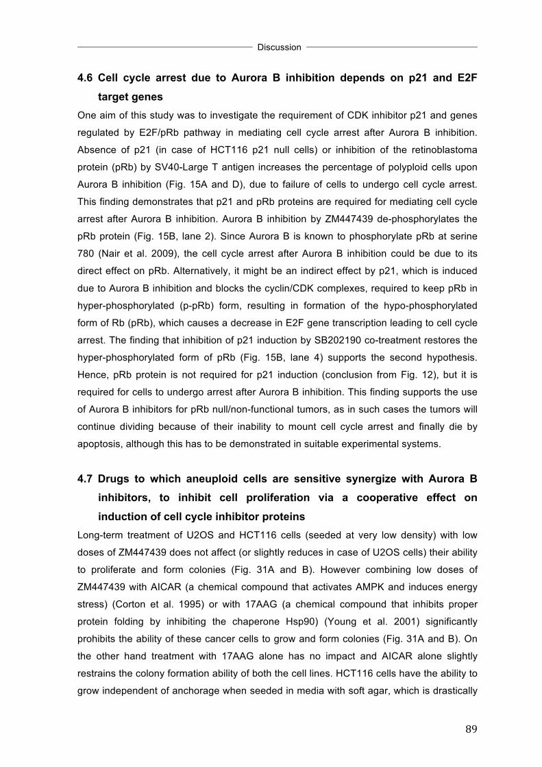
!
!
!Discussion
!! !
89!
4.6 Cell cycle arrest due to Aurora B inhibition depends on p21 and E2F target genes
One aim of this study was to investigate the requirement of CDK inhibitor p21 and genes
regulated by E2F/pRb pathway in mediating cell cycle arrest after Aurora B inhibition.
Absence of p21 (in case of HCT116 p21 null cells) or inhibition of the retinoblastoma
protein (pRb) by SV40-Large T antigen increases the percentage of polyploid cells upon
Aurora B inhibition (Fig. 15A and D), due to failure of cells to undergo cell cycle arrest.
This finding demonstrates that p21 and pRb proteins are required for mediating cell cycle
arrest after Aurora B inhibition. Aurora B inhibition by ZM447439 de-phosphorylates the
pRb protein (Fig. 15B, lane 2). Since Aurora B is known to phosphorylate pRb at serine
780 (Nair et al. 2009), the cell cycle arrest after Aurora B inhibition could be due to its
direct effect on pRb. Alternatively, it might be an indirect effect by p21, which is induced
due to Aurora B inhibition and blocks the cyclin/CDK complexes, required to keep pRb in
hyper-phosphorylated (p-pRb) form, resulting in formation of the hypo-phosphorylated
form of Rb (pRb), which causes a decrease in E2F gene transcription leading to cell cycle
arrest. The finding that inhibition of p21 induction by SB202190 co-treatment restores the
hyper-phosphorylated form of pRb (Fig. 15B, lane 4) supports the second hypothesis.
Hence, pRb protein is not required for p21 induction (conclusion from Fig. 12), but it is
required for cells to undergo arrest after Aurora B inhibition. This finding supports the use
of Aurora B inhibitors for pRb null/non-functional tumors, as in such cases the tumors will
continue dividing because of their inability to mount cell cycle arrest and finally die by
apoptosis, although this has to be demonstrated in suitable experimental systems.
4.7 Drugs to which aneuploid cells are sensitive synergize with Aurora B inhibitors, to inhibit cell proliferation via a cooperative effect on induction of cell cycle inhibitor proteins
Long-term treatment of U2OS and HCT116 cells (seeded at very low density) with low
doses of ZM447439 does not affect (or slightly reduces in case of U2OS cells) their ability
to proliferate and form colonies (Fig. 31A and B). However combining low doses of
ZM447439 with AICAR (a chemical compound that activates AMPK and induces energy
stress) (Corton et al. 1995) or with 17AAG (a chemical compound that inhibits proper
protein folding by inhibiting the chaperone Hsp90) (Young et al. 2001) significantly
prohibits the ability of these cancer cells to grow and form colonies (Fig. 31A and B). On
the other hand treatment with 17AAG alone has no impact and AICAR alone slightly
restrains the colony formation ability of both the cell lines. HCT116 cells have the ability to
grow independent of anchorage when seeded in media with soft agar, which is drastically

!
!
!Discussion
!! !
90!
reduced in the combined presence of AICAR and low doses of ZM447439 (Fig. 31C).
AICAR and 17AAG selectively kill aneuploid cells without having any impact on euploid
cells due the disturbed energy balance and proteotoxic stress in aneuploid cells (Tang et
al. 2011). Requirement of extra energy for transcription, translation and degradation of
proteins produced from the extra copies of genome imposes energy stress, metabolic
stress and proteotoxic stress on aneuploid cells (Stingele et al. 2012; Oromendia and
Amon 2014). One possible molecular explanation for this co-operative ability of Aurora B
inhibitor ZM447439 and AICAR/17AAG in impeding cell-growth is augmentation of
induction of cell cycle inhibitors p21, p53 and p27 (Fig. 32A, B and C). The combined
treatment induces more p21, p53 and p27 at very early time points as compared to
treatment with AICAR, 17AAG or ZM447439 alone. Co-treatment of ZM447439 with
AICAR was more effective in colorectal carcinoma cell line (HCT116) and with 17AAG
was more effective in osteosarcoma cell line (U2OS). These different sensitivities might
be due to different set of mutations in these two types of cancers and detailed
understanding of genetic background of these cell lines will provide information about the
mechanism of action of these drugs as well as help develop targeted therapies for
subgroups of tumors with common markers/mutations. AICAR and 17AAG had no
cooperative affect on phosphorylation of p38 MAPK caused by ZM447439 alone (data not
shown), indicating that they induce p21 and p53 through an independent pathway that
does not requires p38. Also the combination of ZM447439 and AICAR/17AAG did not
alter the apoptotic pathway (as determined with apoptotic markers such as cleaved-PARP
and cleaved-caspase) and the activation of autophagy markers (LC3, p62 and Hsp70)
(data not shown).
In support of these findings are data from yeast, where presence of one extra
chromosome sensitizes them towards proteasome inhibitors, due increased burden on
proteasome system to clear poly-ubiquitinated proteins for degradation (Torres et al.
2007). Primary MEFs which become spontaneously aneuploid due to a compromised
spindle assembly checkpoint (SAC) (as a result of impaired BubR1 allele or by checkpoint
resistant Cdc20 allele) are also sensitive to AICAR and 17AAG (Tang et al. 2011). In the
future it will be important to validate the effects of combined Aurora B inhibitor and
AICAR/17AAG in in-vivo xenograft mouse models. These results suggest that
combinations of low doses of Aurora B inhibitor with certain drugs to which aneuploid cell
are sensitive might be effective for cancer therapy. This will not only increase the efficacy
of Aurora B inhibitors, but also lead to fewer side effects due to usage of low doses of
Aurora B inhibitors. Currently both AICAR and 17AAG are in clinical trials (Gordon et al.
2012). The combination of Aurora B inhibitors with the classes of drugs that interfere with

!
!
!Discussion
!! !
91!
the metabolic balance and proteostasis of cells certainly needs to be tested in trials going
on clinic.
4.8 Hypothesis and working model Taken together, the findings of this study provide evidence for a yet unknown role of the
p38 MAPK in transcriptional elongation of the p21 gene and present sufficient details of
the signaling pathways downstream of Aurora B which support the use of combination
Aurora B inhibitors with drugs to which aneuploid cells are sensitive, for treatment of
cancer. We propose the following working model (Fig. 35): Impairment of Aurora B kinase
function by chemical inhibitors (ZM447439/AZD1152-HQPA) or by siRNA mediated
knockdown results in stabilization of p53 as proposed by (Gully et al. 2012) and induces
its binding to the p53 binding site (p53BS1) in the p21 gene promoter. However this
binding is not sufficient for p21 activation because p38 MAPK, which is also activated by
Aurora B inhibition is required for mediating the transcriptional elongation of p21 gene by
inducing the recruitment of the elongating form of RNA Polymerase II (P-Ser2-RNA
Polymerase II) to the p21 coding region, resulting in transcriptional induction of p21 gene.
Activation of p38 MAPK in turn correlates with generation of aneuploidy and depends on
generation of reactive oxygen species (ROS). Further, the induced p21 leads to
dephosphorylation of retinoblastoma protein (pRb) owing to the ability of p21 to inhibit
cyclin-CDK complexes, which are required to maintain pRb in phosphorylated state
(inactive state). The resulting inactive pRb-E2F complexes prevent expression of E2F
target genes and hence block G1-to-S transition causing G1 arrest.

!
!
!Discussion
!! !
92!
!Figure 35: Model for p21 induction and cell cycle arrest due to Aurora B inhibition. See text for details. Parts of this figure were published in similar form in Kumari et al. 2013 and Kumari et al. 2014.
Aurora BZM447439AZD1152siRNA
p53
p38
p53 RNA POLII
Chromosome missegregation
ROS
Aneuploidy
elongation
p21
p21 mRNA
ȋ phosphorylation
stablized p53
Cyclin/CDK
G1 arrest
pRb
p-pRb E2F target genes
p21 gene locus

!
!
!Summary
!! !
93!
5 Summary !Aurora B is a mitotic kinase that is essential for cell division. Because it is mutated or
overexpressed in a range of cancer types, it has been suggested as a novel therapeutic
target. Currently chemical inhibitors against Aurora B are in various phases of clinical
trials for treatment of solid tumors and leukemia. Information regarding the molecular
requirements for the reported phenotypes of Aurora B inhibition such as cell cycle arrest,
activation of the tumor suppressor p53 and its target p21 are not well understood.
In this study, I investigated the requirements for p21 induction after Aurora B inhibition. I
found that p38 is phosphorylated and activated when Aurora B is inhibited. Experiments
with chemical inhibitors against p38 indicate that p38 is required for p21 induction and cell
cycle arrest in response to Aurora B inhibition. p53 induction after impairment of Aurora B
function and the recruitment of p53 to its binding site in the p21 gene promoter occur
independently of p38 signaling. Instead, I found that p38 is required for the enrichment of
the elongating RNA Polymerase II in the coding region of the p21 gene. Furthermore, p38
is required for formation of the full-length p21 mRNA transcript. These data indicate that
p38 promotes the transcriptional elongation of p21 gene in response to Aurora B
inhibition. In further experiments I could show that the p21 causes cell cycle arrest due to
a decrease in E2F-dependent transcription by promoting the dephosphorylation of the
retinoblastoma protein.
Using synchronized cells I could show that the induction of p21 in response to Aurora B
inhibition requires transition through an aberrant mitosis and does not occur in cells that
are arrested in interphase. Interestingly, p38, p53 and p21 are already induced by partial
inhibition of Aurora B, which results in aneuploidy but not in cytokinesis failure and in
tetraploidy. This supports the notion that activation of p38-p53-p21 signaling correlates
with aneuploidy but not with tetraploidy or binucleation. Partial inhibition of Aurora B also
leads to increased generation of reactive oxygen species (ROS), which are required for
the activation of p38, p21 and cell cycle arrest. Based on these observations I propose the
following model: Inhibition of Aurora B leads to chromosome missegregation resulting in
aneuploidy. This results in increased generation of ROS (reactive oxygen species)
possibly through proteotoxic stress caused by an imbalance of protein synthesis in
aneuploid cells. ROS triggers the activation of p38, which then stimulates the
transcriptional elongation of p21 resulting in cell cycle arrest.
Aneuploidy, proteotoxic stress and oxidative stress are hallmarks of cancer cells. Based
on my results reported in this study, I suggest that the combination of Aurora B inhibitors
with drugs that specifically target aneuploid cells might be a novel strategy for cancer
therapy, as this is a lethal combination for proliferation of cancer cells.

!
!
!Zusammenfassung
!! !
94!
6 Zusammenfassung Aurora B ist eine mitotische Kinase, die entscheidende Funktionen in der Zellteilung
ausübt. Aurora B ist außerdem in einer Vielzahl von Krebsarten mutiert oder
überexprimiert. Daher ist die Aurora B Kinase ein attraktives Ziel für die Tumortherapie.
Gegenwärtig werden Aurora B-Inhibitoren zur Behandlung von soliden Tumoren und
Leukämien in verschiedenen klinischen Studien getestet. Es fehlen jedoch Informationen,
welche molekularen Mechanismen den beschriebenen Phänotypen wie Zellzyklusarrest,
Aktivierung des Tumorsuppressors p53 und seines Zielgens p21 nach Aurora B-
Hemmung zugrunde liegen.
Hauptziel dieser Arbeit war es die Mechanismen der p21-Induktion nach Hemmung von
Aurora B zu untersuchen. Es konnte gezeigt werden, dass nach Hemmung von Aurora B
die p38 MAPK phosphoryliert und somit aktiviert wird. Experimente mit p38-Inhbitoren
belegen, dass p38 für die Induktion von p21 und den Zellzyklusarrest benötigt wird. Die
Stabilisierung von p53 nach Aurora B-Inhibition und die Rekrutierung von p53 an den p21-
Genpromotor erfolgen jedoch unabhängig vom p38-Signalweg. Stattdessen ist p38 für die
Anreicherung der elongierenden RNA-Polymerase II in der kodierenden Region des p21-
Gens und für die Bildung des p21 mRNA Transkripts notwendig. Diese Daten zeigen,
dass p38 transkriptionelle Elongation des p21-Gens nach Aurora B Hemmung fördert. In
weiteren Untersuchungen konnte ich zeigen, dass die Aurora B-Hemmung zu einer
Dephosphorylierung des Retinoblastoma-Proteins führt und dadurch eine Abnahme der
E2F-abhängigen Transkription bewirkt. Dies löst indirekt einen Zellzyklusarrest aus.
Weiterhin konnte mit Hilfe von synchronisierten Zellen gezeigt werden, dass p21 nach
Durchlaufen einer abnormalen Mitose induziert wird, jedoch nicht nach Aurora B-
Hemmung in der Interphase. Interessanterweise werden p38, p53 und p21 schon bei
partieller Inhibition von Aurora B aktiviert. Die partielle Inhibition von Aurora B führt zu
chromosomaler Instabilität aber nicht zum Versagen der Zytokinese und zur Bildung
polyploider Zellen. Damit korreliert die Aktivierung des p38-p53-p21-Signalweges nicht mit
Tetraploidie sondern mit vermehrter Aneuploidie. Die partielle Hemmung von Aurora B
führt außerdem zur vermehrten Entstehung von reaktive Sauerstoffspezies (ROS), welche
für die Aktivierung von p38, p21 und für den Zellzyklusarrest benötigt werden. Basierend
auf diesen Beobachtungen kann folgendes Modell postuliert werden: Die Hemmung von
Aurora B führt zu Fehlern in der Chromosomenverteilung in der Mitose und damit zu
Aneuploidie. Dies führt zu vermehrter Produktion von ROS, möglicherweise durch
proteotoxischer Stress, hervorgerufen durch die Imbalanz der Proteinbiosynthese in

!
!
!Zusammenfassung
!! !
95!
aneuploiden Zellen. ROS bewirkt eine Aktivierung der p38 MAPK und trägt damit zur
Induktion von p21 und dem resultierenden Zellzyklusarrest bei.
Aneuploidie, proteotoxischer und oxidativer Stress stellen Schlüsselmerkmale von
Tumorkrankungen dar. Anhand der Ergebnisse dieser Arbeit könnte die Kombination von
Aurora B-Hemmstoffen mit Medikamenten, die gezielt aneuploide Zellen angreifen, in
Tumorerkrankungen therapeutisch wirksam sein.

!
!
!References
!! !
96!
7 References Adams, Richard R., Mark D. Eckley, Paola Vagnarelli, Sally P. Wheatley, Dietlind L.
Gerloff, Alastair M. Mackay, Phyllis A. Svingen, Scott H. Kaufmann, and William C. Earnshaw. 2001. “Human INCENP Colocalizes with the Aurora-B/AIRK2 Kinase on Chromosomes and Is Overexpressed in Tumour Cells.” Chromosoma 110 (2): 65–74. doi:10.1007/s004120100130.
Adams, Richard R., Helder Maiato, William C. Earnshaw, and Mar Carmena. 2001. “Essential Roles of Drosophila Inner Centromere Protein (INCENP) and Aurora B in Histone H3 Phosphorylation, Metaphase Chromosome Alignment, Kinetochore Disjunction, and Chromosome Segregation”, May.
Adelman, Karen, and John T. Lis. 2012. “Promoter-Proximal Pausing of RNA Polymerase II: Emerging Roles in Metazoans.” Nature Reviews Genetics 13 (10): 720–31. doi:10.1038/nrg3293.
Akhtar, Md. Sohail, Martin Heidemann, Joshua R. Tietjen, David W. Zhang, Rob D. Chapman, Dirk Eick, and Aseem Z. Ansari. 2009. “TFIIH Kinase Places Bivalent Marks on the Carboxy-Terminal Domain of RNA Polymerase II.” Molecular Cell 34 (3): 387–93. doi:10.1016/j.molcel.2009.04.016.
Alexander, Jes, Daniel Lim, Brian A. Joughin, Bjorn Hegemann, James RA Hutchins, Tobias Ehrenberger, Frank Ivins, Fabio Sessa, Otto Hudecz, and Erich A. Nigg. 2011. “Spatial Exclusivity Combined with Positive and Negative Selection of Phosphorylation Motifs Is the Basis for Context-Dependent Mitotic Signaling.” Science Signaling 4 (179): ra42.
Ali, Syed Hamid, and James A. DeCaprio. 2001. “Cellular Transformation by SV40 Large T Antigen: Interaction with Host Proteins.” Seminars in Cancer Biology 11 (1): 15–23. doi:10.1006/scbi.2000.0342.
Ambrosino, Concetta, and Angel R. Nebreda. 2001. “Cell Cycle Regulation by p38 MAP Kinases.” Biology of the Cell 93 (1-2): 47–51.
Andreassen, Paul R., Olivier D. Lohez, Françoise B. Lacroix, and Robert L. Margolis. 2001. “Tetraploid State Induces p53-Dependent Arrest of Nontransformed Mammalian Cells in G1.” Molecular Biology of the Cell 12 (5): 1315–28.
Anjomshoaa, A, S Nasri, B Humar, J L McCall, A Chatterjee, H-S Yoon, L McNoe, M A Black, and A E Reeve. 2009. “Slow Proliferation as a Biological Feature of Colorectal Cancer Metastasis.” British Journal of Cancer 101 (5): 822–28. doi:10.1038/sj.bjc.6605229.
Araujo, Sergio E. A., Wanderley M. Bernardo, Angelita Habr-Gama, Desiderio R. Kiss, and Ivan Cecconello. 2007. “DNA Ploidy Status and Prognosis in Colorectal Cancer: A Meta-Analysis of Published Data:” Diseases of the Colon & Rectum 50 (11): 1800–1810. doi:10.1007/s10350-007-9013-6.
Avo Santos, M., C. van de Werken, M. de Vries, H. Jahr, M. J. M. Vromans, J. S. E. Laven, B. C. Fauser, G. J. Kops, S. M. Lens, and E. B. Baart. 2011. “A Role for Aurora C in the Chromosomal Passenger Complex during Human Preimplantation Embryo Development.” Human Reproduction 26 (7): 1868–81. doi:10.1093/humrep/der111.
Bakhoum, Samuel F., Sarah L. Thompson, Amity L. Manning, and Duane A. Compton. 2009. “Genome Stability Is Ensured by Temporal Control of Kinetochore–microtubule Dynamics.” Nature Cell Biology 11 (1): 27–35. doi:10.1038/ncb1809.
Balciunaite, E., A. Spektor, N. H. Lents, H. Cam, H. te Riele, A. Scime, M. A. Rudnicki, R. Young, and B. D. Dynlacht. 2005. “Pocket Protein Complexes Are Recruited to Distinct Targets in Quiescent and Proliferating Cells.” Molecular and Cellular Biology 25 (18): 8166–78. doi:10.1128/MCB.25.18.8166-8178.2005.
Barber, Thomas D., Kirk McManus, Karen WY Yuen, Marcelo Reis, Giovanni Parmigiani, Dong Shen, Irene Barrett, Yasaman Nouhi, Forrest Spencer, and Sanford

!
!
!References
!! !
97!
Markowitz. 2008. “Chromatid Cohesion Defects May Underlie Chromosome Instability in Human Colorectal Cancers.” Proceedings of the National Academy of Sciences 105 (9): 3443–48.
Barr, A. R., and F. Gergely. 2007. “Aurora-A: The Maker and Breaker of Spindle Poles.” Journal of Cell Science 120 (17): 2987–96. doi:10.1242/jcs.013136.
Bartek, Jiri, and Jiri Lukas. 2003. “Chk1 and Chk2 Kinases in Checkpoint Control and Cancer.” Cancer Cell 3 (5): 421–29.
Beckerman, R., A. J. Donner, M. Mattia, M. J. Peart, J. L. Manley, J. M. Espinosa, and C. Prives. 2009. “A Role for Chk1 in Blocking Transcriptional Elongation of p21 RNA during the S-Phase Checkpoint.” Genes & Development 23 (11): 1364–77. doi:10.1101/gad.1795709.
Bertoli, Cosetta, Jan M. Skotheim, and Robertus A. M. de Bruin. 2013. “Control of Cell Cycle Transcription during G1 and S Phases.” Nature Reviews Molecular Cell Biology 14 (8): 518–28. doi:10.1038/nrm3629.
Bieging, Kathryn T., Stephano Spano Mello, and Laura D. Attardi. 2014. “Unravelling Mechanisms of p53-Mediated Tumour Suppression.” Nature Reviews Cancer 14 (5): 359–70.
Biggins, S. and W. Murray, A.2001. “The Budding Yeast Protein Kinase Ipl1/Aurora Allows the Absence of Tension to Activate the Spindle Checkpoint.” Genes & Development 15 (23): 3118–29. doi:10.1101/gad.934801.
Bischoff, James R., Lee Anderson, Yingfang Zhu, Kevin Mossie, Lelia Ng, Brian Souza, Brian Schryver, Peter Flanagan, Felix Clairvoyant, and Charles Ginther. 1998. “A Homologue of Drosophila Aurora Kinase Is Oncogenic and Amplified in Human Colorectal Cancers.” The EMBO Journal 17 (11): 3052–65.
Blagosklonny, Mikhail V. 2006. “Analytical Report Prolonged Mitosis Versus Tetraploid Checkpoint.” Cell Cycle 5 (9): 971–75.
Bradford, M (1976). "A rapid and sensitive method for the quantitation of microgram quantities of protein using the principle of protein dye binding." Analytical Biochemistry 72, 248–254.
Brady, Colleen A., Dadi Jiang, Stephano S. Mello, Thomas M. Johnson, Lesley A. Jarvis, Margaret M. Kozak, Daniela Kenzelmann Broz, et al. 2011. “Distinct p53 Transcriptional Programs Dictate Acute DNA-Damage Responses and Tumor Suppression.” Cell 145 (4): 571–83. doi:10.1016/j.cell.2011.03.035.
Brattain, Michael G., W. David Fine, F. Mahnaz Khaled, Jerry Thompson, and Diane E. Brattain. 1981. “Heterogeneity of Malignant Cells from a Human Colonic Carcinoma.” Cancer Research 41 (5): 1751–56.
Bregman, David B., Richard G. Pestell, and Vincent J. Kidd. 2000. “Cell Cycle Regulation and RNA Polymerase II.” Front Biosci 5 (5): D244–257.
Brinkley, Bill R. 2001. “Managing the Centrosome Numbers Game: From Chaos to Stability in Cancer Cell Division.” Trends in Cell Biology 11 (1): 18–21.
Brown, Christopher J., Sonia Lain, Chandra S. Verma, Alan R. Fersht, and David P. Lane. 2009. “Awakening Guardian Angels: Drugging the p53 Pathway.” Nature Reviews Cancer 9 (12): 862–73. doi:10.1038/nrc2763.
Bulavin, D V, Y Higashimoto, I J Popoff, W A Gaarde, V Basrur, O Potapova, E Appella, and A J Fornace Jr. 2001. “Initiation of a G2/M Checkpoint after Ultraviolet Radiation Requires p38 Kinase.” Nature 411 (6833): 102–7. doi:10.1038/35075107.
Bulavin, Dmitry V, Crissy Phillips, Bonnie Nannenga, Oleg Timofeev, Larry A Donehower, Carl W Anderson, Ettore Appella, and Albert J Fornace. 2004. “Inactivation of the Wip1 Phosphatase Inhibits Mammary Tumorigenesis through p38 MAPK–mediated Activation of the p16Ink4a-p19Arf Pathway.” Nature Genetics 36 (4): 343–50. doi:10.1038/ng1317.
Bulavin, Dmitry V., Oleg N. Demidov, Shin’ichi Saito, Paivikki Kauraniemi, Crissy Phillips, Sally A. Amundson, Concetta Ambrosino, et al. 2002. “Amplification of PPM1D in
!
!
!References
!! !
98!
Human Tumors Abrogates p53 Tumor-Suppressor Activity.” Nature Genetics 31 (2): 210–15. doi:10.1038/ng894.
Bulavin, Dmitry V., Shińichi Saito, M. Christine Hollander, Kazuyasu Sakaguchi, Carl W. Anderson, Ettore Appella, and Albert J. Fornace. 1999. “Phosphorylation of Human p53 by p38 Kinase Coordinates N-Terminal Phosphorylation and Apoptosis in Response to UV Radiation.” The EMBO Journal 18 (23): 6845–54.
Bulavin, Dmitry V., Natalia D. Tararova, Nikolai D. Aksenov, Valery A. Pospelov, and Tatiana V. Pospelova. 1999. “Deregulation of p53/p21 Cip1/Waf1 Pathway Contributes to Polyploidy and Apoptosis of E1A+ cHa-Ras Transformed Cells after Γ-Irradiation.” Oncogene 18 (41).
Bunz, F. 1998. “Requirement for p53 and p21 to Sustain G2 Arrest After DNA Damage.” Science 282 (5393): 1497–1501. doi:10.1126/science.282.5393.1497.
Carmena, Mar, and William C. Earnshaw. 2003. “The Cellular Geography of Aurora Kinases.” Nature Reviews Molecular Cell Biology 4 (11): 842–54. doi:10.1038/nrm1245.
Carmena, Mar, Michael Wheelock, Hironori Funabiki, and William C. Earnshaw. 2012. “The Chromosomal Passenger Complex (CPC): From Easy Rider to the Godfather of Mitosis.” Nature Reviews Molecular Cell Biology 13 (12): 789–803. doi:10.1038/nrm3474.
Chae, Hee-Don, Man-Ryul Lee, and Hal E. Broxmeyer. 2012. “5-Aminoimidazole-4-Carboxyamide Ribonucleoside Induces G1/S Arrest and Nanog Downregulation via p53 and Enhances Erythroid Differentiation.” STEM CELLS 30 (2): 140–49. doi:10.1002/stem.778.
Chan, Y. W., A. A. Jeyaprakash, E. A. Nigg, and A. Santamaria. 2012. “Aurora B Controls Kinetochore-Microtubule Attachments by Inhibiting Ska Complex-KMN Network Interaction.” The Journal of Cell Biology 196 (5): 563–71. doi:10.1083/jcb.201109001.
Chong, Jean-Leon, Pamela L. Wenzel, M. Teresa Sáenz-Robles, Vivek Nair, Antoney Ferrey, John P. Hagan, Yorman M. Gomez, et al. 2009. “E2f1–3 Switch from Activators in Progenitor Cells to Repressors in Differentiating Cells.” Nature 462 (7275): 930–34. doi:10.1038/nature08677.
Cimini, Daniela. 2008. “Merotelic Kinetochore Orientation, Aneuploidy, and Cancer.” Biochimica et Biophysica Acta (BBA) - Reviews on Cancer 1786 (1): 32–40. doi:10.1016/j.bbcan.2008.05.003.
Cimini, Daniela, Bonnie Howell, Paul Maddox, Alexey Khodjakov, Francesca Degrassi, and E. D. Salmon. 2001. “Merotelic Kinetochore Orientation Is a Major Mechanism of Aneuploidy in Mitotic Mammalian Tissue Cells.” The Journal of Cell Biology 153 (3): 517–28.
Cimini, Daniela, Xiaohu Wan, Christophe B. Hirel, and E.D. Salmon. 2006. “Aurora Kinase Promotes Turnover of Kinetochore Microtubules to Reduce Chromosome Segregation Errors.” Current Biology 16 (17): 1711–18. doi:10.1016/j.cub.2006.07.022.
Corton, Julia M., John G. Gillespie, Simon A. Hawley, and D. Grahame Hardie. 1995. “5-Aminoimidazole-4-Carboxamide Ribonucleoside.” European Journal of Biochemistry 229 (2): 558–65.
Crosio, C., G. M. Fimia, R. Loury, M. Kimura, Y. Okano, H. Zhou, S. Sen, C. D. Allis, and P. Sassone-Corsi. 2002. “Mitotic Phosphorylation of Histone H3: Spatio-Temporal Regulation by Mammalian Aurora Kinases.” Molecular and Cellular Biology 22 (3): 874–85. doi:10.1128/MCB.22.3.874-885.2002.
Cuadrado, Ana, and Angel R. Nebreda. 2010. “Mechanisms and Functions of p38 MAPK Signalling.” Biochemical Journal 429 (3): 403–17. doi:10.1042/BJ20100323.
Dai, Jun, Beth A. Sullivan, and Jonathan M.G. Higgins. 2006. “Regulation of Mitotic Chromosome Cohesion by Haspin and Aurora B.” Developmental Cell 11 (5): 741–50. doi:10.1016/j.devcel.2006.09.018.

!
!
!References
!! !
99!
De Nadal, Eulàlia, Gustav Ammerer, and Francesc Posas. 2011. “Controlling Gene Expression in Response to Stress.” Nature Reviews Genetics, November. doi:10.1038/nrg3055. http://www.nature.com/doifinder/10.1038/nrg3055.
Dehay, Colette, and Henry Kennedy. 2007. “Cell-Cycle Control and Cortical Development.” Nature Reviews Neuroscience 8 (6): 438–50. doi:10.1038/nrn2097.
Dimova, Desssislava K, and Nicholas J Dyson. 2005. “The E2F Transcriptional Network: Old Acquaintances with New Faces.” Oncogene 24 (17): 2810–26. doi:10.1038/sj.onc.1208612.
Ditchfield, C. 2003. “Aurora B Couples Chromosome Alignment with Anaphase by Targeting BubR1, Mad2, and Cenp-E to Kinetochores.” The Journal of Cell Biology 161 (2): 267–80. doi:10.1083/jcb.200208091.
Dorion, S. 2002. “Activation of the p38 Signaling Pathway by Heat Shock Involves the Dissociation of Glutathione S-Transferase Mu from Ask1.” Journal of Biological Chemistry 277 (34): 30792–97. doi:10.1074/jbc.M203642200.
Douglas, Max E., Tim Davies, Nimesh Joseph, and Masanori Mishima. 2010. “Aurora B and 14-3-3 Coordinately Regulate Clustering of Centralspindlin during Cytokinesis.” Current Biology 20 (10): 927–33. doi:10.1016/j.cub.2010.03.055.
Dreier, Megan R., Aaron Z. Grabovich, Jamie D. Katusin, and William R. Taylor. 2009. “Short and Long-Term Tumor Cell Responses to Aurora Kinase Inhibitors.” Experimental Cell Research 315 (7): 1085–99. doi:10.1016/j.yexcr.2009.02.008.
Dynlacht, Brian D. 1997. “Regulation of Transcription by Proteins That Control the Cell Cycle.” Nature 389 (6647): 149–52.
Efimova, T. 2003. “A Regulatory Role for p38 MAPK in Keratinocyte Differentiation: evidence for p38-erk1/2 complex formation.” Journal of Biological Chemistry 278 (36): 34277–85. doi:10.1074/jbc.M302759200.
Egloff, Sylvain, Martin Dienstbier, and Shona Murphy. 2012. “Updating the RNA Polymerase CTD Code: Adding Gene-Specific Layers.” Trends in Genetics 28 (7): 333–41. doi:10.1016/j.tig.2012.03.007.
El-Deiry, W. 1993. “WAF1, a Potential Mediator of p53 Tumor Suppression.” Cell 75 (4): 817–25. doi:10.1016/0092-8674(93)90500-P.
Espinosa, Joaquın M., Ramiro E. Verdun, and Beverly M. Emerson. 2003. “p53 Functions through Stress-and Promoter-Specific Recruitment of Transcription Initiation Components before and after DNA Damage.” Molecular Cell 12 (4): 1015–27.
Falck, Jacob, Julia Coates, and Stephen P. Jackson. 2005. “Conserved Modes of Recruitment of ATM, ATR and DNA-PKcs to Sites of DNA Damage.” Nature 434 (7033): 605–11.
Fang, Xiao, and Pumin Zhang. 2011. “Aneuploidy and Tumorigenesis.” Seminars in Cell & Developmental Biology 22 (6): 595–601. doi:10.1016/j.semcdb.2011.03.002.
Fernandez-Miranda, G., M. Trakala, J. Martin, B. Escobar, A. Gonzalez, N. B. Ghyselinck, S. Ortega, M. Canamero, I. P. de Castro, and M. Malumbres. 2011. “Genetic Disruption of Aurora B Uncovers an Essential Role for Aurora C during Early Mammalian Development.” Development 138 (13): 2661–72. doi:10.1242/dev.066381.
Ferreiro, I., M. Barragan, A. Gubern, E. Ballestar, M. Joaquin, and F. Posas. 2010. “The p38 SAPK Is Recruited to Chromatin via Its Interaction with Transcription Factors.” Journal of Biological Chemistry 285 (41): 31819–28. doi:10.1074/jbc.M110.155846.
Foley, Emily A., Maria Maldonado, and Tarun M. Kapoor. 2011. “Formation of Stable Attachments between Kinetochores and Microtubules Depends on the B56-PP2A Phosphatase.” Nature Cell Biology 13 (10): 1265–71. doi:10.1038/ncb2327.
Forcales, Sonia V., Sonia Albini, Lorenzo Giordani, Barbora Malecova, Luca Cignolo, Andrei Chernov, Paula Coutinho, Valentina Saccone, Silvia Consalvi, and Roy Williams. 2012. “Signal-Dependent Incorporation of MyoD–BAF60c into Brg1-

!
!
!References
!! !
100!
Based SWI/SNF Chromatin-Remodelling Complex.” The EMBO Journal 31 (2): 301–16.
Fulda, Simone, Adrienne M. Gorman, Osamu Hori, and Afshin Samali. 2010. “Cellular Stress Responses: Cell Survival and Cell Death.” International Journal of Cell Biology 2010: 1–23. doi:10.1155/2010/214074.
Galbraith, Matthew D, Aaron J Donner, and Joaquín M Espinosa. 2010. “CDK8: A Positive Regulator of Transcription.” Transcription 1 (1): 4–12. doi:10.4161/trns.1.1.12373.
Galbraith, Matthew D., Mary A. Allen, Claire L. Bensard, Xiaoxing Wang, Marie K. Schwinn, Bo Qin, Henry W. Long, et al. 2013. “HIF1A Employs CDK8-Mediator to Stimulate RNAPII Elongation in Response to Hypoxia.” Cell 153 (6): 1327–39. doi:10.1016/j.cell.2013.04.048.
Ganem, Neil J., Susana A. Godinho, and David Pellman. 2009. “A Mechanism Linking Extra Centrosomes to Chromosomal Instability.” Nature 460 (7252): 278–82. doi:10.1038/nature08136.
Ganem, Neil J., and David Pellman. 2007. “Limiting the Proliferation of Polyploid Cells.” Cell 131 (3): 437–40. doi:10.1016/j.cell.2007.10.024.
Gassmann, R. 2004. “Borealin: A Novel Chromosomal Passenger Required for Stability of the Bipolar Mitotic Spindle.” The Journal of Cell Biology 166 (2): 179–91. doi:10.1083/jcb.200404001.
Ge, B. 2002. “MAPKK-Independent Activation of p38alpha Mediated by TAB1-Dependent Autophosphorylation of p38alpha.” Science 295 (5558): 1291–94. doi:10.1126/science.1067289.
Gebara, M M, M H Sayre, and J L Corden. 1997. “Phosphorylation of the Carboxy-Terminal Repeat Domain in RNA Polymerase II by Cyclin-Dependent Kinases Is Sufficient to Inhibit Transcription.” Journal of Cellular Biochemistry 64 (3): 390–402.
Giet, Régis, and David M. Glover. 2001. “Drosophila Aurora B Kinase Is Required for Histone H3 Phosphorylation and Condensin Recruitment during Chromosome Condensation and to Organize the Central Spindle during Cytokinesis.” The Journal of Cell Biology 152 (4): 669–82.
Giménez-Abián, Juan F., Izabela Sumara, Toru Hirota, Silke Hauf, Daniel Gerlich, Consuelo de la Torre, Jan Ellenberg, and Jan-Michael Peters. 2004. “Regulation of Sister Chromatid Cohesion between Chromosome Arms.” Current Biology 14 (13): 1187–93.
Girdler, F. 2006. “Validating Aurora B as an Anti-Cancer Drug Target.” Journal of Cell Science 119 (17): 3664–75. doi:10.1242/jcs.03145.
Gizatullin, Farid, Yao Yao, Victor Kung, Matthew W. Harding, Massimo Loda, and Geoffrey I. Shapiro. 2006. “The Aurora Kinase Inhibitor VX-680 Induces Endoreduplication and Apoptosis Preferentially in Cells with Compromised p53-Dependent Postmitotic Checkpoint Function.” Cancer Research 66 (15): 7668–77.
Gold, Matthew G, David Barford, and David Komander. 2006. “Lining the Pockets of Kinases and Phosphatases.” Current Opinion in Structural Biology 16 (6): 693–701. doi:10.1016/j.sbi.2006.10.006.
Gomes, N. P. 2006. “Gene-Specific Requirement for P-TEFb Activity and RNA Polymerase II Phosphorylation within the p53 Transcriptional Program.” Genes & Development 20 (5): 601–12. doi:10.1101/gad.1398206.
Gordon, David J., Benjamin Resio, and David Pellman. 2012. “Causes and Consequences of Aneuploidy in Cancer.” Nature Reviews Genetics, January. doi:10.1038/nrg3123. http://www.nature.com/doifinder/10.1038/nrg3123.
Goto, H. 2003. “Aurora-B Regulates the Cleavage Furrow-Specific Vimentin Phosphorylation in the Cytokinetic Process.” Journal of Biological Chemistry 278 (10): 8526–30. doi:10.1074/jbc.M210892200.
!
!
!References
!! !
101!
Goto, Hidemasa, Yoshihiro Yasui, Erich A. Nigg, and Masaki Inagaki. 2002. “Aurora-B Phosphorylates Histone H3 at serine28 with Regard to the Mitotic Chromosome Condensation.” Genes to Cells 7 (1): 11–17.
Gottifredi, Vanesa, Sheau-Yann Shieh, Yoichi Taya, and Carol Prives. 2001. “p53 Accumulates but Is Functionally Impaired When DNA Synthesis Is Blocked.” Proceedings of the National Academy of Sciences 98 (3): 1036–41.
Gully, C. P., G. Velazquez-Torres, J.-H. Shin, E. Fuentes-Mattei, E. Wang, C. Carlock, J. Chen, et al. 2012. “Aurora B Kinase Phosphorylates and Instigates Degradation of p53.” Proceedings of the National Academy of Sciences 109 (24): E1513–E1522. doi:10.1073/pnas.1110287109.
Gully, Christopher P., Fanmao Zhang, Jian Chen, James A. Yeung, Guermarie Velazquez-Torres, Edward Wang, S. C. Yeung, and Mong-Hong Lee. 2010. “Antineoplastic Effects of an Aurora B Kinase Inhibitor in Breast Cancer.” Mol Cancer 9 (42): 10–1186.
Guse, Annika, Masanori Mishima, and Michael Glotzer. 2005. “Phosphorylation of ZEN-4/MKLP1 by Aurora B Regulates Completion of Cytokinesis.” Current Biology 15 (8): 778–86. doi:10.1016/j.cub.2005.03.041.
Hahn, Steven. 2004. “Structure and Mechanism of the RNA Polymerase II Transcription Machinery.” Nature Structural & Molecular Biology 11 (5): 394.
Han, J, Y Jiang, Z Li, V V Kravchenko, and R J Ulevitch. 1997. “Activation of the Transcription Factor MEF2C by the MAP Kinase p38 in Inflammation.” Nature 386 (6622): 296–99. doi:10.1038/386296a0.
Harrington, Elizabeth A, David Bebbington, Jeff Moore, Richele K Rasmussen, Abi O Ajose-Adeogun, Tomoko Nakayama, Joanne A Graham, et al. 2004. “VX-680, a Potent and Selective Small-Molecule Inhibitor of the Aurora Kinases, Suppresses Tumor Growth in Vivo.” Nature Medicine 10 (3): 262–67. doi:10.1038/nm1003.
Haruta, Masayuki, Yoshiyuki Matsumoto, Hideki Izumi, Naoki Watanabe, Masahiro Fukuzawa, Shinya Matsuura, and Yasuhiko Kaneko. 2008. “Combined BubR1 Protein down-Regulation and RASSF1A Hypermethylation in Wilms Tumors with Diverse Cytogenetic Changes.” Molecular Carcinogenesis 47 (9): 660–66. doi:10.1002/mc.20412.
Hauf, S. 2003. “The Small Molecule Hesperadin Reveals a Role for Aurora B in Correcting Kinetochore-Microtubule Attachment and in Maintaining the Spindle Assembly Checkpoint.” The Journal of Cell Biology 161 (2): 281–94. doi:10.1083/jcb.200208092.
Hickson, Ian, Yan Zhao, Caroline J. Richardson, Sharon J. Green, Niall MB Martin, Alisdair I. Orr, Philip M. Reaper, Stephen P. Jackson, Nicola J. Curtin, and Graeme CM Smith. 2004. “Identification and Characterization of a Novel and Specific Inhibitor of the Ataxia-Telangiectasia Mutated Kinase ATM.” Cancer Research 64 (24): 9152–59.
Hirota, Toru, Naoko Kunitoku, Takashi Sasayama, Tomotoshi Marumoto, Dongwei Zhang, Masayuki Nitta, Katsuyoshi Hatakeyama, and Hideyuki Saya. 2003. “Aurora-A and an Interacting Activator, the LIM Protein Ajuba, Are Required for Mitotic Commitment in Human Cells.” Cell 114 (5): 585–98.
Hirota, Toru, Jesse J. Lipp, Ban-Hock Toh, and Jan-Michael Peters. 2005. “Histone H3 Serine 10 Phosphorylation by Aurora B Causes HP1 Dissociation from Heterochromatin.” Nature 438 (7071): 1176–80. doi:10.1038/nature04254.
Hoe, Khoo Kian, Chandra S. Verma, and David P. Lane. 2014. “Drugging the p53 Pathway: Understanding the Route to Clinical Efficacy.” Nature Reviews Drug Discovery 13 (3): 217–36. doi:10.1038/nrd4236.
Holland, Andrew J, and Don W Cleveland. 2012. “Losing Balance: The Origin and Impact of Aneuploidy in Cancer.” EMBO Reports 13 (6): 501–14. doi:10.1038/embor.2012.55.

!
!
!References
!! !
102!
Holland, Andrew J., and Don W. Cleveland. 2009. “Boveri Revisited: Chromosomal Instability, Aneuploidy and Tumorigenesis.” Nature Reviews Molecular Cell Biology 10 (7): 478–87. doi:10.1038/nrm2718.
Janssen, A., M. van der Burg, K. Szuhai, G. J. P. L. Kops, and R. H. Medema. 2011. “Chromosome Segregation Errors as a Cause of DNA Damage and Structural Chromosome Aberrations.” Science 333 (6051): 1895–98. doi:10.1126/science.1210214.
Jensen, Lars Juhl, Thomas Skøt Jensen, Ulrik de Lichtenberg, Søren Brunak, and Peer Bork. 2006. “Co-Evolution of Transcriptional and Post-Translational Cell-Cycle Regulation.” Nature, September. doi:10.1038/nature05186. http://www.nature.com/doifinder/10.1038/nature05186.
Kaestner, P., A. Stolz, and H. Bastians. 2009. “Determinants for the Efficiency of Anticancer Drugs Targeting Either Aurora-A or Aurora-B Kinases in Human Colon Carcinoma Cells.” Molecular Cancer Therapeutics 8 (7): 2046–56. doi:10.1158/1535-7163.MCT-09-0323.
Kastan, Michael B., and Jiri Bartek. 2004. “Cell-Cycle Checkpoints and Cancer.” Nature 432 (7015): 316–23.
Kawajiri, Aie, Yoshihiro Yasui, Hidemasa Goto, Masaaki Tatsuka, Masahide Takahashi, Koh-ichi Nagata, and Masaki Inagaki. 2003. “Functional Significance of the Specific Sites Phosphorylated in Desmin at Cleavage Furrow: Aurora-B May Phosphorylate and Regulate Type III Intermediate Filaments during Cytokinesis Coordinatedly with Rho-Kinase.” Molecular Biology of the Cell 14 (4): 1489–1500.
Kawauchi, J., M. Inoue, M. Fukuda, Y. Uchida, T. Yasukawa, R. C. Conaway, J. W. Conaway, T. Aso, and S. Kitajima. 2013. “Transcriptional Properties of Mammalian Elongin A and Its Role in Stress Response.” Journal of Biological Chemistry 288 (34): 24302–15. doi:10.1074/jbc.M113.496703.
Keen, Nicholas, and Stephen Taylor. 2004. “Aurora-Kinase Inhibitors as Anticancer Agents.” Nature Reviews Cancer 4 (12): 927–36. doi:10.1038/nrc1502.
Kelly, A. E., C. Ghenoiu, J. Z. Xue, C. Zierhut, H. Kimura, and H. Funabiki. 2010. “Survivin Reads Phosphorylated Histone H3 Threonine 3 to Activate the Mitotic Kinase Aurora B.” Science 330 (6001): 235–39. doi:10.1126/science.1189505.
Kennedy, Norman J., Cristina Cellurale, and Roger J. Davis. 2007. “A Radical Role for p38 MAPK in Tumor Initiation.” Cancer Cell 11 (2): 101–3. doi:10.1016/j.ccr.2007.01.009.
Keyse, Stephen M. 2000. “Protein Phosphatases and the Regulation of Mitogen-Activated Protein Kinase Signalling.” Current Opinion in Cell Biology 12 (2): 186–92.
Kierstead, T. D., and M. J. Tevethia. 1993. “Association of p53 Binding and Immortalization of Primary C57BL/6 Mouse Embryo Fibroblasts by Using Simian Virus 40 T-Antigen Mutants Bearing Internal Overlapping Deletion Mutations.” Journal of Virology 67 (4): 1817–29.
Kim, G.-Y. 2002. “The Stress-Activated Protein Kinases p38alpha and JNK1 Stabilize p21Cip1 by Phosphorylation.” Journal of Biological Chemistry 277 (33): 29792–802. doi:10.1074/jbc.M201299200.
Kitajima, Tomoya S., Takeshi Sakuno, Kei-ichiro Ishiguro, Shun-ichiro Iemura, Tohru Natsume, Shigehiro A. Kawashima, and Yoshinori Watanabe. 2006. “Shugoshin Collaborates with Protein Phosphatase 2A to Protect Cohesin.” Nature 441 (7089): 46–52. doi:10.1038/nature04663.
Klein, Ulf R., Erich A. Nigg, and Ulrike Gruneberg. 2006. “Centromere Targeting of the Chromosomal Passenger Complex Requires a Ternary Subcomplex of Borealin, Survivin, and the N-Terminal Domain of INCENP.” Molecular Biology of the Cell 17 (6): 2547–58.
Koepp, Deanna M., J. Wade Harper, and Stephen J. Elledge. 1999. “How the Cyclin Became a Cyclin: Regulated Proteolysis in the Cell Cycle.” Cell 97 (4): 431–34.

!
!
!References
!! !
103!
Kops, Geert J. P. L., Beth A. A. Weaver, and Don W. Cleveland. 2005. “On the Road to Cancer: Aneuploidy and the Mitotic Checkpoint.” Nature Reviews Cancer 5 (10): 773–85. doi:10.1038/nrc1714.
Krek, W. 1994. “Negative Regulation of the Growth-Promoting Transcription Factor E2F-1 by a Stably Bound Cyclin A-Dependent Protein Kinase.” Cell 78 (1): 161–72. doi:10.1016/0092-8674(94)90582-7.
Kruse, Jan-Philipp, and Wei Gu. 2009. “Modes of p53 Regulation.” Cell 137 (4): 609–22. doi:10.1016/j.cell.2009.04.050.
Kubbutat, M H, S N Jones, and K H Vousden. 1997. “Regulation of p53 Stability by Mdm2.” Nature 387 (6630): 299–303. doi:10.1038/387299a0.
Kültz, Dietmar. 2005. “MOLECULAR AND EVOLUTIONARY BASIS OF THE CELLULAR STRESS RESPONSE.” Annual Review of Physiology 67 (1): 225–57. doi:10.1146/annurev.physiol.67.040403.103635.
Kuma, Y. 2005. “BIRB796 Inhibits All p38 MAPK Isoforms in Vitro and in Vivo.” Journal of Biological Chemistry 280 (20): 19472–79. doi:10.1074/jbc.M414221200.
Kumar, Sanjay, Jeffrey Boehm, and John C. Lee. 2003. “p38 MAP Kinases: Key Signalling Molecules as Therapeutic Targets for Inflammatory Diseases.” Nature Reviews Drug Discovery 2 (9): 717–26. doi:10.1038/nrd1177.
Kyriakis, J M, and J Avruch. 2001. “Mammalian Mitogen-Activated Protein Kinase Signal Transduction Pathways Activated by Stress and Inflammation.” Physiological Reviews 81 (2): 807–69.
Laemmli, U (1970). ‘Cleavage of structural proteins during the assembly of the head of bacteriophage T4." Nature, 227, 680–685. doi:10.1038/227680a0.
Lan, Weijie, Xin Zhang, Susan L Kline-Smith, Sara E Rosasco, Gregory A Barrett-Wilt, Jeffrey Shabanowitz, Donald F Hunt, Claire E Walczak, and P.Todd Stukenberg. 2004. “Aurora B Phosphorylates Centromeric MCAK and Regulates Its Localization and Microtubule Depolymerization Activity.” Current Biology 14 (4): 273–86. doi:10.1016/j.cub.2004.01.055.
Lampson MA, Kapoor TM. "The human mitotic checkpoint protein BubR1 regulates chromosome- spindle attachments." Nature Cell Biology 2005; 7:93-8; PMID:15592459; http://dx.doi.org/10.1038/ncb1208
Lane, D P. 1992. “Cancer. p53, Guardian of the Genome.” Nature 358 (6381): 15–16. doi:10.1038/358015a0.
Lanni, Jennifer S., and Tyler Jacks. 1998. “Characterization of the p53-Dependent Postmitotic Checkpoint Following Spindle Disruption.” Molecular and Cellular Biology 18 (2): 1055–64.
Lee, B.-K., A. A. Bhinge, and V. R. Iyer. 2011. “Wide-Ranging Functions of E2F4 in Transcriptional Activation and Repression Revealed by Genome-Wide Analysis.” Nucleic Acids Research 39 (9): 3558–73. doi:10.1093/nar/gkq1313.
Lee, J C, J T Laydon, P C McDonnell, T F Gallagher, S Kumar, D Green, D McNulty, M J Blumenthal, J R Heys, and S W Landvatter. 1994. “A Protein Kinase Involved in the Regulation of Inflammatory Cytokine Biosynthesis.” Nature 372 (6508): 739–46. doi:10.1038/372739a0.
Lengauer, C, K W Kinzler, and B Vogelstein. 1998. “Genetic Instabilities in Human Cancers.” Nature 396 (6712): 643–49. doi:10.1038/25292.
Lens, Susanne, Rob MF Wolthuis, Rob Klompmaker, Jos Kauw, Reuven Agami, Thijn Brummelkamp, Geert Kops, and René H. Medema. 2003. “Survivin Is Required for a Sustained Spindle Checkpoint Arrest in Response to Lack of Tension.” The EMBO Journal 22 (12): 2934–47.
Li, M., X. Fang, D. J. Baker, L. Guo, X. Gao, Z. Wei, S. Han, J. M. van Deursen, and P. Zhang. 2010. “The ATM-p53 Pathway Suppresses Aneuploidy-Induced Tumorigenesis.” Proceedings of the National Academy of Sciences 107 (32): 14188–93. doi:10.1073/pnas.1005960107.

!
!
!References
!! !
104!
Li, Tongyuan, Ning Kon, Le Jiang, Minjia Tan, Thomas Ludwig, Yingming Zhao, Richard Baer, and Wei Gu. 2012. “Tumor Suppression in the Absence of p53-Mediated Cell-Cycle Arrest, Apoptosis, and Senescence.” Cell 149 (6): 1269–83. doi:10.1016/j.cell.2012.04.026.
Lin, B-W, Y-C Wang, P-Y Chang-Liao, Y-J Lin, S-T Yang, J-H Tsou, K-C Chang, et al. 2014. “Overexpression of Aurora-C Interferes with the Spindle Checkpoint by Promoting the Degradation of Aurora-B.” Cell Death and Disease 5 (3): e1106. doi:10.1038/cddis.2014.37.
Lipp, J. J., T. Hirota, I. Poser, and J.-M. Peters. 2007. “Aurora B Controls the Association of Condensin I but Not Condensin II with Mitotic Chromosomes.” Journal of Cell Science 120 (7): 1245–55. doi:10.1242/jcs.03425.
Liu, D., M. Vleugel, C. B. Backer, T. Hori, T. Fukagawa, I. M. Cheeseman, and M. A. Lampson. 2010. “Regulated Targeting of Protein Phosphatase 1 to the Outer Kinetochore by KNL1 Opposes Aurora B Kinase.” The Journal of Cell Biology 188 (6): 809–20. doi:10.1083/jcb.201001006.
Liu, Y. 2002. “Thioredoxin Promotes ASK1 Ubiquitination and Degradation to Inhibit ASK1-Mediated Apoptosis in a Redox Activity-Independent Manner.” Circulation Research 90 (12): 1259–66. doi:10.1161/01.RES.0000022160.64355.62.
Maldonado, Maria, and Tarun M. Kapoor. 2011. “Constitutive Mad1 Targeting to Kinetochores Uncouples Checkpoint Signalling from Chromosome Biorientation.” Nature Cell Biology 13 (4): 475–82. doi:10.1038/ncb2223.
Malumbres, Marcos, and Mariano Barbacid. 2009. “Cell Cycle, CDKs and Cancer: A Changing Paradigm.” Nature Reviews Cancer 9 (3): 153–66. doi:10.1038/nrc2602.
Manke, Isaac A., Anhco Nguyen, Daniel Lim, Mary Q. Stewart, Andrew E.H. Elia, and Michael B. Yaffe. 2005. “MAPKAP Kinase-2 Is a Cell Cycle Checkpoint Kinase That Regulates the G2/M Transition and S Phase Progression in Response to UV Irradiation.” Molecular Cell 17 (1): 37–48. doi:10.1016/j.molcel.2004.11.021.
Margolis, Robert L., Olivier D. Lohez, and Paul R. Andreassen. 2003. “G1 Tetraploidy Checkpoint and the Suppression of Tumorigenesis.” Journal of Cellular Biochemistry 88 (4): 673–83. doi:10.1002/jcb.10411.
Marzo, Isabel, and Javier Naval. 2013. “Antimitotic Drugs in Cancer Chemotherapy: Promises and Pitfalls.” Biochemical Pharmacology 86 (6): 703–10. doi:10.1016/j.bcp.2013.07.010.
Mattia, M., V. Gottifredi, K. McKinney, and C. Prives. 2007. “p53-Dependent p21 mRNA Elongation Is Impaired When DNA Replication Is Stalled.” Molecular and Cellular Biology 27 (4): 1309–20. doi:10.1128/MCB.01520-06.
Mertens, Fredrik, Bertil Johansson, Mattias Höglund, and Felix Mitelman. 1997. “Chromosomal Imbalance Maps of Malignant Solid Tumors: A Cytogenetic Survey of 3185 Neoplasms.” Cancer Research 57 (13): 2765–80.
Mertens, Fredrik, Bertil Johansson, and Felix Mitelman. 1994. “Isochromosomes in Neoplasia.” Genes, Chromosomes and Cancer 10 (4): 221–30.
Mikhailov, Alexei, Mio Shinohara, and Conly L Rieder. 2005. “The p38-Mediated Stress-Activated Checkpoint. A Rapid Response System for Delaying Progression through Antephase and Entry into Mitosis.” Cell Cycle (Georgetown, Tex.) 4 (1): 57–62.
Mikule, Keith, Benedicte Delaval, Philipp Kaldis, Agata Jurcyzk, Polla Hergert, and Stephen Doxsey. 2007. “Loss of Centrosome Integrity Induces p38—p53—p21-Dependent G1—S Arrest.” Nature Cell Biology 9 (2): 160–70. doi:10.1038/ncb1529.
Minoshima, Yukinori, Toshiyuki Kawashima, Koichi Hirose, Yukio Tonozuka, Aie Kawajiri, Ying Chun Bao, Xingming Deng, Masaaki Tatsuka, Shuh Narumiya, and W. Stratford May Jr. 2003. “Phosphorylation by Aurora B Converts MgcRacGAP to a RhoGAP during Cytokinesis.” Developmental Cell 4 (4): 549–60.

!
!
!References
!! !
105!
Mitelman Database of Chromosome Aberrations and Gene Fusions in Cancer (2014). Mitelman F, Johansson B and Mertens F (Eds.), http://cgap.nci.nih.gov/Chromosomes/Mitelman"
Moll, Ute M, Sonja Wolff, Daniel Speidel, and Wolfgang Deppert. 2005. “Transcription-Independent pro-Apoptotic Functions of p53.” Current Opinion in Cell Biology 17 (6): 631–36. doi:10.1016/j.ceb.2005.09.007.
Momand, Jamil, Gerard P. Zambetti, David C. Olson, Donna George, and Arnold J. Levine. 1992. “The Mdm-2 Oncogene Product Forms a Complex with the p53 Protein and Inhibits p53-Mediated Transactivation.” Cell 69 (7): 1237–45. doi:10.1016/0092-8674(92)90644-R.
Morgan, David O. 1997. “Cyclin-Dependent Kinases: Engines, Clocks, and Microprocessors.” Annual Review of Cell and Developmental Biology 13 (1): 261–91.
Morita, S., T. Kojima, and T. Kitamura. 2000. “Plat-E: An Efficient and Stable System for Transient Packaging of Retroviruses.” Gene Therapy 7 (12). http://search.ebscohost.com/login.aspx?direct=true&profile=ehost&scope=site&authtype=crawler&jrnl=09697128&AN=8852816&h=qitdV%2BrxkHnrhpUOlZ9%2B72O4G0slNYIJJGshzO6ZgLZpiqjXlFl2iAq8cp9eXDy0tAIMcU5u8JXEZeIJRFIxNg%3D%3D&crl=c.
Mortlock, Andrew A., Kevin M. Foote, Nicola M. Heron, Frédéric H. Jung, Georges Pasquet, Jean-Jacques M. Lohmann, Nicolas Warin, et al. 2007. “Discovery, Synthesis, and in Vivo Activity of a New Class of Pyrazoloquinazolines as Selective Inhibitors of Aurora B Kinase.” Journal of Medicinal Chemistry 50 (9): 2213–24. doi:10.1021/jm061335f.
Muller, Patricia A. J., and Karen H. Vousden. 2013. “p53 Mutations in Cancer.” Nature Cell Biology 15 (1): 2–8. doi:10.1038/ncb2641.
Murata-Hori, Maki, Katsumi Fumoto, Yasuaki Fukuta, Takahiro Iwasaki, Asako Kikuchi, Masaaki Tatsuka, and Hiroshi Hosoya. 2000. “Myosin II Regulatory Light Chain as a Novel Substrate for AIM-1, an aurora/Ipl1p-Related Kinase from Rat.” Journal of Biochemistry 128 (6): 903–7.
Murray, Andrew W. 2004. “Recycling the Cell Cycle: Cyclins Revisited.” Cell 116 (2): 221–34.
Musacchio, Andrea, and Edward D. Salmon. 2007. “The Spindle-Assembly Checkpoint in Space and Time.” Nature Reviews Molecular Cell Biology 8 (5): 379–93. doi:10.1038/nrm2163.
Muse, Ginger W, Daniel A Gilchrist, Sergei Nechaev, Ruchir Shah, Joel S Parker, Sherry F Grissom, Julia Zeitlinger, and Karen Adelman. 2007. “RNA Polymerase Is Poised for Activation across the Genome.” Nature Genetics 39 (12): 1507–11. doi:10.1038/ng.2007.21.
Nair, Jayasree S., Alan L. Ho, N. Tse Archie, Jesse Coward, Haider Cheema, Grazia Ambrosini, Nicholas Keen, and Gary K. Schwartz. 2009. “Aurora B Kinase Regulates the Postmitotic Endoreduplication Checkpoint via Phosphorylation of the Retinoblastoma Protein at Serine 780.” Molecular Biology of the Cell 20 (8): 2218–28.
Neef, Rüdiger, Ulf R. Klein, Robert Kopajtich, and Francis A. Barr. 2006. “Cooperation between Mitotic Kinesins Controls the Late Stages of Cytokinesis.” Current Biology 16 (3): 301–7. doi:10.1016/j.cub.2005.12.030.
Nezi, Luigi, and Andrea Musacchio. 2009. “Sister Chromatid Tension and the Spindle Assembly Checkpoint.” Current Opinion in Cell Biology 21 (6): 785–95. doi:10.1016/j.ceb.2009.09.007.
Nigg, Erich A. 2002. “Centrosome Aberrations: Cause or Consequence of Cancer Progression?” Nature Reviews. Cancer 2 (11): 815–25. doi:10.1038/nrc924.

!
!
!References
!! !
106!
Nigg, Erich A. 2006. “Origins and Consequences of Centrosome Aberrations in Human Cancers.” International Journal of Cancer 119 (12): 2717–23. doi:10.1002/ijc.22245.
Nishiyama, T., M. M. Sykora, P. J. Huis in ’t Veld, K. Mechtler, and J.-M. Peters. 2013. “Aurora B and Cdk1 Mediate Wapl Activation and Release of Acetylated Cohesin from Chromosomes by Phosphorylating Sororin.” Proceedings of the National Academy of Sciences 110 (33): 13404–9. doi:10.1073/pnas.1305020110.
Norbury, Chris, and Paul Nurse. 1992. “Animal Cell Cycles and Their Control.” Annual Review of Biochemistry 61 (1): 441–68.
Norden, Caren, Manuel Mendoza, Jeroen Dobbelaere, Chitra V. Kotwaliwale, Sue Biggins, and Yves Barral. 2006. “The NoCut Pathway Links Completion of Cytokinesis to Spindle Midzone Function to Prevent Chromosome Breakage.” Cell 125 (1): 85–98. doi:10.1016/j.cell.2006.01.045.
Oromendia, A. B., and A. Amon. 2014. “Aneuploidy: Implications for Protein Homeostasis and Disease.” Disease Models & Mechanisms 7 (1): 15–20. doi:10.1242/dmm.013391.
Oromendia, A. B., S. E. Dodgson, and A. Amon. 2012. “Aneuploidy Causes Proteotoxic Stress in Yeast.” Genes & Development 26 (24): 2696–2708. doi:10.1101/gad.207407.112.
Ota, Takahide, Shiho Suto, Hiroshi Katayama, Zhen-Bo Han, Fumio Suzuki, Masayo Maeda, Mikio Tanino, Yasuhiko Terada, and Masaaki Tatsuka. 2002. “Increased Mitotic Phosphorylation of Histone H3 Attributable to AIM-1/Aurora-B Overexpression Contributes to Chromosome Number Instability.” Cancer Research 62 (18): 5168–77.
Owens, D M, and S M Keyse. 2007. “Differential Regulation of MAP Kinase Signalling by Dual-Specificity Protein Phosphatases.” Oncogene 26 (22): 3203–13. doi:10.1038/sj.onc.1210412.
Ozlu, N., F. Monigatti, B. Y. Renard, C. M. Field, H. Steen, T. J. Mitchison, and J. J. Steen. 2010. “Binding Partner Switching on Microtubules and Aurora-B in the Mitosis to Cytokinesis Transition.” Molecular & Cellular Proteomics 9 (2): 336–50. doi:10.1074/mcp.M900308-MCP200.
Pages, G. 2000. “Stress-Activated Protein Kinases (JNK and p38/HOG) Are Essential for Vascular Endothelial Growth Factor mRNA Stability.” Journal of Biological Chemistry 275 (34): 26484–91. doi:10.1074/jbc.M002104200.
Pardee, Arthur B. 1974. “A Restriction Point for Control of Normal Animal Cell Proliferation.” Proceedings of the National Academy of Sciences 71 (4): 1286–90.
Park, H., Y. Jeon, H. Shin, I. Kim, H. Kang, S. Jeong, D. Chung, and C. Lee. 2007. “Differential Promoter Methylation May Be a Key Molecular Mechanism in Regulating BubR1 Expression in Cancer Cells.” Experimental and Molecular Medicine 39 (2): 195.
Pavelka, Norman, Giulia Rancati, Jin Zhu, William D. Bradford, Anita Saraf, Laurence Florens, Brian W. Sanderson, Gaye L. Hattem, and Rong Li. 2010. “Aneuploidy Confers Quantitative Proteome Changes and Phenotypic Variation in Budding Yeast.” Nature 468 (7321): 321–25. doi:10.1038/nature09529.
Petronczki, Mark, Péter Lénárt, and Jan-Michael Peters. 2008. “Polo on the Rise—from Mitotic Entry to Cytokinesis with Plk1.” Developmental Cell 14 (5): 646–59. doi:10.1016/j.devcel.2008.04.014.
Phatnani, H. P., and A. L. Greenleaf. 2006. “Phosphorylation and Functions of the RNA Polymerase II CTD.” Genes & Development 20 (21): 2922–36. doi:10.1101/gad.1477006.
Pihan, German A., Jan Wallace, Yening Zhou, and Stephen J. Doxsey. 2003. “Centrosome Abnormalities and Chromosome Instability Occur Together in Pre-Invasive Carcinomas.” Cancer Research 63 (6): 1398–1404.

!
!
!References
!! !
107!
Ponten, Jan, and Eero Saksela. 1967. “Two Established in Vitro Cell Lines from Human Mesenchymal Tumours.” International Journal of Cancer 2 (5): 434–47.
Posch, M., G. A. Khoudoli, S. Swift, E. M. King, J. G. DeLuca, and J. R. Swedlow. 2010. “Sds22 Regulates Aurora B Activity and Microtubule-Kinetochore Interactions at Mitosis.” The Journal of Cell Biology 191 (1): 61–74. doi:10.1083/jcb.200912046.
Price, D. H. 2000. “P-TEFb, a Cyclin-Dependent Kinase Controlling Elongation by RNA Polymerase II.” Molecular and Cellular Biology 20 (8): 2629–34. doi:10.1128/MCB.20.8.2629-2634.2000.
Price, David H. 2008. “Poised Polymerases: On Your Mark…Get Set…Go!” Molecular Cell 30 (1): 7–10. doi:10.1016/j.molcel.2008.03.001.
Proft, M, G Mas, E Denadal, A Vendrell, N Noriega, K Struhl, and F Posas. 2006. “The Stress-Activated Hog1 Kinase Is a Selective Transcriptional Elongation Factor for Genes Responding to Osmotic Stress.” Molecular Cell 23 (2): 241–50. doi:10.1016/j.molcel.2006.05.031.
Qi, M. 2005. “MAP Kinase Pathways.” Journal of Cell Science 118 (16): 3569–72. doi:10.1242/jcs.02470.
Raingeaud, Joël. 1995. “Pro-Inflammatory Cytokines and Environmental Stress Cause p38 Mitogen-Activated Protein Kinase Activation by Dual Phosphorylation on Tyrosine and Threonine.” Journal of Biological Chemistry 270 (13): 7420–26. doi:10.1074/jbc.270.13.7420.
Raman, M, W Chen, and M H Cobb. 2007. “Differential Regulation and Properties of MAPKs.” Oncogene 26 (22): 3100–3112. doi:10.1038/sj.onc.1210392.
Reaper, Philip M, Matthew R Griffiths, Joanna M Long, Jean-Damien Charrier, Somhairle MacCormick, Peter A Charlton, Julian M C Golec, and John R Pollard. 2011. “Selective Killing of ATM- or p53-Deficient Cancer Cells through Inhibition of ATR.” Nature Chemical Biology 7 (7): 428–30. doi:10.1038/nchembio.573.
Riley, Todd, Eduardo Sontag, Patricia Chen, and Arnold Levine. 2008. “Transcriptional Control of Human p53-Regulated Genes.” Nature Reviews Molecular Cell Biology 9 (5): 402–12. doi:10.1038/nrm2395.
Rousseau, Simon, Francois Houle, Jacques Landry, and Jacques Huot. 1997. “p38 MAP Kinase Activation by Vascular Endothelial Growth Factor Mediates Actin Reorganization and Cell Migration in Human Endothelial Cells.” Oncogene 15 (18). http://search.ebscohost.com/login.aspx?direct=true&profile=ehost&scope=site&authtype=crawler&jrnl=09509232&AN=8919896&h=BN%2BFtd2qGBWZD5F3OqkixGdA5S1LGdwEUcadOs7dIJMxgNcSwTjmQNwFDDaKoo42y5ThcUuu%2BnUh5Tk9zpUkVg%3D%3D&crl=c.
Roux, P. P., and J. Blenis. 2004. “ERK and p38 MAPK-Activated Protein Kinases: A Family of Protein Kinases with Diverse Biological Functions.” Microbiology and Molecular Biology Reviews 68 (2): 320–44. doi:10.1128/MMBR.68.2.320-344.2004.
Ruchaud, Sandrine, Mar Carmena, and William C. Earnshaw. 2007. “Chromosomal Passengers: Conducting Cell Division.” Nature Reviews Molecular Cell Biology 8 (10): 798–812. doi:10.1038/nrm2257.
Rustici, Gabriella, Juan Mata, Katja Kivinen, Pietro Lió, Christopher J Penkett, Gavin Burns, Jacqueline Hayles, Alvis Brazma, Paul Nurse, and Jürg Bähler. 2004. “Periodic Gene Expression Program of the Fission Yeast Cell Cycle.” Nature Genetics 36 (8): 809–17. doi:10.1038/ng1377.
Sanchez-Prieto, Ricardo, Jose M. Rojas, Yoichi Taya, and J. Silvio Gutkind. 2000. “A Role for the p38 Mitogen-Activated Protein Kinase Pathway in the Transcriptional Activation of p53 on Genotoxic Stress by Chemotherapeutic Agents.” Cancer Research 60 (9): 2464–72.
Santaguida, Stefano, Claudio Vernieri, Fabrizio Villa, Andrea Ciliberto, and Andrea Musacchio. 2011. “Evidence That Aurora B Is Implicated in Spindle Checkpoint Signalling Independently of Error Correction.” The EMBO Journal 30 (8): 1508–19.

!
!
!References
!! !
108!
Sasai, Kaori, Hiroshi Katayama, David L. Stenoien, Satoshi Fujii, Reiko Honda, Masashi Kimura, Yukio Okano, et al. 2004. “Aurora-C Kinase Is a Novel Chromosomal Passenger Protein That Can Complement Aurora-B Kinase Function in Mitotic Cells.” Cell Motility and the Cytoskeleton 59 (4): 249–63. doi:10.1002/cm.20039.
Saurin, Adrian T., Maike S. van der Waal, René H. Medema, Susanne M.A. Lens, and Geert J.P.L. Kops. 2011. “Aurora B Potentiates Mps1 Activation to Ensure Rapid Checkpoint Establishment at the Onset of Mitosis.” Nature Communications 2 (May): 316. doi:10.1038/ncomms1319.
Sengupta, Sagar, and Curtis C. Harris. 2005. “p53: Traffic Cop at the Crossroads of DNA Repair and Recombination.” Nature Reviews Molecular Cell Biology 6 (1): 44–55. doi:10.1038/nrm1546.
Sessa, Fabio, Marina Mapelli, Claudio Ciferri, Cataldo Tarricone, Liliana B. Areces, Thomas R. Schneider, P. Todd Stukenberg, and Andrea Musacchio. 2005. “Mechanism of Aurora B Activation by INCENP and Inhibition by Hesperadin.” Molecular Cell 18 (3): 379–91. doi:10.1016/j.molcel.2005.03.031.
Shaul, Yoav D., and Rony Seger. 2007. “The MEK/ERK Cascade: From Signaling Specificity to Diverse Functions.” Biochimica et Biophysica Acta (BBA) - Molecular Cell Research 1773 (8): 1213–26. doi:10.1016/j.bbamcr.2006.10.005.
Sheltzer, Jason M., and Angelika Amon. 2011. “The Aneuploidy Paradox: Costs and Benefits of an Incorrect Karyotype.” Trends in Genetics 27 (11): 446–53. doi:10.1016/j.tig.2011.07.003.
Sherr, C J, and J M Roberts. 1995. “Inhibitors of Mammalian G1 Cyclin-Dependent Kinases.” Genes & Development 9 (10): 1149–63. doi:10.1101/gad.9.10.1149.
Sherr, Charles J. 1996. “Cancer Cell Cycles.” Science 274 (5293): 1672–77. Sherr, Charles J., and James M. Roberts. 1999. “CDK Inhibitors: Positive and Negative
Regulators of G1-Phase Progression.” Genes & Development 13 (12): 1501–12. Shiloh, Yosef, and Yael Ziv. 2013. “The ATM Protein Kinase: Regulating the Cellular
Response to Genotoxic Stress, and More.” Nature Reviews Molecular Cell Biology 14 (4): 197–210. doi:10.1038/nrm3546.
Silkworth, William T., Isaac K. Nardi, Lindsey M. Scholl, and Daniela Cimini. 2009. “Multipolar Spindle Pole Coalescence Is a Major Source of Kinetochore Mis-Attachment and Chromosome Mis-Segregation in Cancer Cells.” Edited by Kevin G. Hardwick. PLoS ONE 4 (8): e6564. doi:10.1371/journal.pone.0006564.
Simone, Cristiano, Sonia Vanina Forcales, David A Hill, Anthony N Imbalzano, Lucia Latella, and Pier Lorenzo Puri. 2004. “p38 Pathway Targets SWI-SNF Chromatin-Remodeling Complex to Muscle-Specific Loci.” Nature Genetics 36 (7): 738–43. doi:10.1038/ng1378.
Stewart, Scott, and Guowei Fang. 2005. “Destruction Box–Dependent Degradation of Aurora B Is Mediated by the Anaphase-Promoting Complex/Cyclosome and Cdh1.” Cancer Research 65 (19): 8730–35.
Stingele, Silvia, Gabriele Stoehr, Karolina Peplowska, Jürgen Cox, Matthias Mann, and Zuzana Storchova. 2012. “Global Analysis of Genome, Transcriptome and Proteome Reveals the Response to Aneuploidy in Human Cells.” Molecular Systems Biology 8 (September). doi:10.1038/msb.2012.40. http://msb.embopress.org/cgi/doi/10.1038/msb.2012.40.
Stingele, Silvia, Gabriele Stoehr, and Zuzana Storchova. 2013. “Activation of Autophagy in Cells with Abnormal Karyotype.” Autophagy 9 (2): 246–48. doi:10.4161/auto.22558.
Stubdal, Hilde, Juan Zalvide, Kathryn S. Campbell, Colleen Schweitzer, Thomas M. Roberts, and James A. DeCaprio. 1997. “Inactivation of pRB-Related Proteins p130 and p107 Mediated by the J Domain of Simian Virus 40 Large T Antigen.” Molecular and Cellular Biology 17 (9): 4979–90.

!
!
!References
!! !
109!
Sutherland, Heidi, and Wendy A. Bickmore. 2009. “Transcription Factories: Gene Expression in Unions?” Nature Reviews Genetics 10 (7): 457–66. doi:10.1038/nrg2592.
Suzuki, Hiroshi I., Kaoru Yamagata, Koichi Sugimoto, Takashi Iwamoto, Shigeaki Kato, and Kohei Miyazono. 2009. “Modulation of microRNA Processing by p53.” Nature 460 (7254): 529–33. doi:10.1038/nature08199.
Takahashi, Yasuhiko, Joseph B. Rayman, and Brian David Dynlacht. 2000. “Analysis of Promoter Binding by the E2F and pRB Families in Vivo: Distinct E2F Proteins Mediate Activation and Repression.” Genes & Development 14 (7): 804–16.
Tang, Yun-Chi, Bret R. Williams, Jake J. Siegel, and Angelika Amon. 2011. “Identification of Aneuploidy-Selective Antiproliferation Compounds.” Cell 144 (4): 499–512. doi:10.1016/j.cell.2011.01.017.
Tao, Yungan, Ping Zhang, F. Girdler, V. Frascogna, M. Castedo, J. Bourhis, G. Kroemer, and E. Deutsch. 2008. “Enhancement of Radiation Response in p53-Deficient Cancer Cells by the Aurora-B Kinase Inhibitor AZD1152.” Oncogene 27 (23): 3244–55.
Tavner, F., J. Frampton, and R. J. Watson. 2006. “Targeting an E2F Site in the Mouse Genome Prevents Promoter Silencing in Quiescent and Post-Mitotic Cells.” Oncogene 26 (19): 2727–35.
Thompson, S. L., and D. A. Compton. 2010. “Proliferation of Aneuploid Human Cells Is Limited by a p53-Dependent Mechanism.” The Journal of Cell Biology 188 (3): 369–81. doi:10.1083/jcb.200905057.
Torres, E. M., B. R. Williams, and A. Amon. 2008. “Aneuploidy: Cells Losing Their Balance.” Genetics 179 (2): 737–46. doi:10.1534/genetics.108.090878.
Torres, Eduardo M., Noah Dephoure, Amudha Panneerselvam, Cheryl M. Tucker, Charles A. Whittaker, Steven P. Gygi, Maitreya J. Dunham, and Angelika Amon. 2010. “Identification of Aneuploidy-Tolerating Mutations.” Cell 143 (1): 71–83. doi:10.1016/j.cell.2010.08.038.
Torres, Eduardo M., Tanya Sokolsky, Cheryl M. Tucker, Leon Y. Chan, Monica Boselli, Maitreya J. Dunham, and Angelika Amon. 2007. “Effects of Aneuploidy on Cellular Physiology and Cell Division in Haploid Yeast.” Science 317 (5840): 916–24.
Touré, Aminata, Rym Mzali, Caroline Liot, Laetitia Seguin, Laurence Morin, Catherine Crouin, Ilin Chen-Yang, et al. 2008. “Phosphoregulation of MgcRacGAP in Mitosis Involves Aurora B and Cdk1 Protein Kinases and the PP2A Phosphatase.” FEBS Letters 582 (8): 1182–88. doi:10.1016/j.febslet.2007.12.036.
Trakala, Marianna, Gonzalo Fernández-Miranda, Ignacio Pérez de Castro, Christopher Heeschen, and Marcos Malumbres. 2013. “Aurora B Prevents Delayed DNA Replication and Premature Mitotic Exit by Repressing p21Cip1.” Cell Cycle 12 (7): 1030–41. doi:10.4161/cc.24004.
Uetake, Y. 2004. “Cell Cycle Progression after Cleavage Failure: Mammalian Somatic Cells Do Not Possess a ‘Tetraploidy Checkpoint.’” The Journal of Cell Biology 165 (5): 609–15. doi:10.1083/jcb.200403014.
Uetake, Y., J. Loncarek, J. J. Nordberg, C. N. English, S. La Terra, A. Khodjakov, and G. Sluder. 2007. “Cell Cycle Progression and de Novo Centriole Assembly after Centrosomal Removal in Untransformed Human Cells.” The Journal of Cell Biology 176 (2): 173–82. doi:10.1083/jcb.200607073.
Vader, G. 2006. “The Chromosomal Passenger Complex: Guiding Aurora-B through Mitosis.” The Journal of Cell Biology 173 (6): 833–37. doi:10.1083/jcb.200604032.
Vader, Gerben, Jos J W Kauw, René H Medema, and Susanne M A Lens. 2006. “Survivin Mediates Targeting of the Chromosomal Passenger Complex to the Centromere and Midbody.” EMBO Reports 7 (1): 85–92. doi:10.1038/sj.embor.7400562.
Vader, Gerben, and Susanne M.A. Lens. 2008. “The Aurora Kinase Family in Cell Division and Cancer.” Biochimica et Biophysica Acta (BBA) - Reviews on Cancer 1786 (1): 60–72. doi:10.1016/j.bbcan.2008.07.003.

!
!
!References
!! !
110!
Valente, Liz J., Daniel H.D. Gray, Ewa M. Michalak, Josefina Pinon-Hofbauer, Alex Egle, Clare L. Scott, Ana Janic, and Andreas Strasser. 2013. “p53 Efficiently Suppresses Tumor Development in the Complete Absence of Its Cell-Cycle Inhibitory and Proapoptotic Effectors p21, Puma, and Noxa.” Cell Reports 3 (5): 1339–45. doi:10.1016/j.celrep.2013.04.012.
Valin, A., J. Ouyang, and G. Gill. 2013. “Transcription Factor Sp3 Represses Expression of p21CIP1 via Inhibition of Productive Elongation by RNA Polymerase II.” Molecular and Cellular Biology 33 (8): 1582–93. doi:10.1128/MCB.00323-12.
Van den Heuvel, Sander, and Nicholas J. Dyson. 2008. “Conserved Functions of the pRB and E2F Families.” Nature Reviews Molecular Cell Biology 9 (9): 713–24. doi:10.1038/nrm2469.
Vaseva, A V, A R Yallowitz, N D Marchenko, S Xu, and U M Moll. 2011. “Blockade of Hsp90 by 17AAG Antagonizes MDMX and Synergizes with Nutlin to Induce p53-Mediated Apoptosis in Solid Tumors.” Cell Death and Disease 2 (5): e156. doi:10.1038/cddis.2011.39.
Vogelstein, B, D Lane, and A J Levine. 2000. “Surfing the p53 Network.” Nature 408 (6810): 307–10. doi:10.1038/35042675.
Vousden, Karen H., and David P. Lane. 2007. “p53 in Health and Disease.” Nature Reviews Molecular Cell Biology 8 (4): 275–83. doi:10.1038/nrm2147.
Vousden, Karen H., and Carol Prives. 2009. “Blinded by the Light: The Growing Complexity of p53.” Cell 137 (3): 413–31. doi:10.1016/j.cell.2009.04.037.
Wade Harper, J. 1993. “The p21 Cdk-Interacting Protein Cip1 Is a Potent Inhibitor of G1 Cyclin-Dependent Kinases.” Cell 75 (4): 805–16. doi:10.1016/0092-8674(93)90499-G.
Wagner, Erwin F., and Ángel R. Nebreda. 2009. “Signal Integration by JNK and p38 MAPK Pathways in Cancer Development.” Nature Reviews Cancer 9 (8): 537–49. doi:10.1038/nrc2694.
Waldman, Todd, Kenneth W. Kinzler, and Bert Vogelstein. 1995. “p21 Is Necessary for the p53-Mediated G1 Arrest in Human Cancer Cells.” Cancer Research 55 (22): 5187–90.
Wang, XiaoZhong, and David Ron. 1996. “Stress-Induced Phosphorylation and Activation of the Transcription Factor CHOP (GADD153) by p38 MAP Kinase.” Science 272 (5266): 1347–49.
Wang, Xin, and Cathy Tournier. 2006. “Regulation of Cellular Functions by the ERK5 Signalling Pathway.” Cellular Signalling 18 (6): 753–60. doi:10.1016/j.cellsig.2005.11.003.
Wang, Zhenghe, Jordan M. Cummins, Dong Shen, Daniel P. Cahill, Prasad V. Jallepalli, Tian-Li Wang, D. Williams Parsons, Giovanni Traverso, Mark Awad, and Natalie Silliman. 2004. “Three Classes of Genes Mutated in Colorectal Cancers with Chromosomal Instability.” Cancer Research 64 (9): 2998–3001.
Weaver, Beth AA, and Don W. Cleveland. 2007. “Aneuploidy: Instigator and Inhibitor of Tumorigenesis.” Cancer Research 67 (21): 10103–5.
Weijts, Bart GMW, Walbert J. Bakker, Peter WA Cornelissen, Kuo-Hsuan Liang, Frank H. Schaftenaar, Bart Westendorp, Charlotte ACMT de Wolf, Maya Paciejewska, Colinda LGJ Scheele, and Lindsey Kent. 2012. “E2F7 and E2F8 Promote Angiogenesis through Transcriptional Activation of VEGFA in Cooperation with HIF1.” The EMBO Journal 31 (19): 3871–84.
Weinberg, Robert A. 1995. “The Retinoblastoma Protein and Cell Cycle Control.” Cell 81 (3): 323–30.
Welburn, Julie P.I., Mathijs Vleugel, Dan Liu, John R. Yates III, Michael A. Lampson, Tatsuo Fukagawa, and Iain M. Cheeseman. 2010. “Aurora B Phosphorylates Spatially Distinct Targets to Differentially Regulate the Kinetochore-Microtubule Interface.” Molecular Cell 38 (3): 383–92. doi:10.1016/j.molcel.2010.02.034.

!
!
!References
!! !
111!
Whitfield, Michael L., Gavin Sherlock, Alok J. Saldanha, John I. Murray, Catherine A. Ball, Karen E. Alexander, John C. Matese, Charles M. Perou, Myra M. Hurt, and Patrick O. Brown. 2002. “Identification of Genes Periodically Expressed in the Human Cell Cycle and Their Expression in Tumors.” Molecular Biology of the Cell 13 (6): 1977–2000.
Wilkinson, R. W., R. Odedra, S. P. Heaton, S. R. Wedge, N. J. Keen, C. Crafter, J. R. Foster, et al. 2007. “AZD1152, a Selective Inhibitor of Aurora B Kinase, Inhibits Human Tumor Xenograft Growth by Inducing Apoptosis.” Clinical Cancer Research 13 (12): 3682–88. doi:10.1158/1078-0432.CCR-06-2979.
Williams, Bret R., Vineet R. Prabhu, Karen E. Hunter, Christina M. Glazier, Charles A. Whittaker, David E. Housman, and Angelika Amon. 2008. “Aneuploidy Affects Proliferation and Spontaneous Immortalization in Mammalian Cells.” Science 322 (5902): 703–9.
Wong, Connie, and Tim Stearns. 2005. “Mammalian Cells Lack Checkpoints for Tetraploidy, Aberrant Centrosome Number, and Cytokinesis Failure.” BMC Cell Biology 6 (1): 6.
Wood, C. David, Tina M. Thornton, Guadalupe Sabio, Roger A. Davis, and Mercedes Rincon. 2009. “Nuclear Localization of p38 MAPK in Response to DNA Damage.” International Journal of Biological Sciences 5 (5): 428.
Wu, L., C. A. Ma, Y. Zhao, and A. Jain. 2011. “Aurora B Interacts with NIR-p53, Leading to p53 Phosphorylation in Its DNA-Binding Domain and Subsequent Functional Suppression.” Journal of Biological Chemistry 286 (3): 2236–44. doi:10.1074/jbc.M110.174755.
Wu, Z., P. J. Woodring, K. S. Bhakta, K. Tamura, F. Wen, J. R. Feramisco, M. Karin, J. Y. J. Wang, and P. L. Puri. 2000. “p38 and Extracellular Signal-Regulated Kinases Regulate the Myogenic Program at Multiple Steps.” Molecular and Cellular Biology 20 (11): 3951–64. doi:10.1128/MCB.20.11.3951-3964.2000.
Xu, Z., H. Ogawa, P. Vagnarelli, J. H. Bergmann, D. F. Hudson, S. Ruchaud, T. Fukagawa, W. C. Earnshaw, and K. Samejima. 2009. “INCENP-Aurora B Interactions Modulate Kinase Activity and Chromosome Passenger Complex Localization.” The Journal of Cell Biology 187 (5): 637–53. doi:10.1083/jcb.200906053.
Yang, J., T. Ikezoe, C. Nishioka, T. Tasaka, A. Taniguchi, Y. Kuwayama, N. Komatsu, et al. 2007. “AZD1152, a Novel and Selective Aurora B Kinase Inhibitor, Induces Growth Arrest, Apoptosis, and Sensitization for Tubulin Depolymerizing Agent or Topoisomerase II Inhibitor in Human Acute Leukemia Cells in Vitro and in Vivo.” Blood 110 (6): 2034–40. doi:10.1182/blood-2007-02-073700.
Yasui, Y. 2003. “Autophosphorylation of a Newly Identified Site of Aurora-B Is Indispensable for Cytokinesis.” Journal of Biological Chemistry 279 (13): 12997–3. doi:10.1074/jbc.M311128200.
Ye, X., B. Zerlanko, R. Zhang, N. Somaiah, M. Lipinski, P. Salomoni, and P. D. Adams. 2007. “Definition of pRB- and p53-Dependent and -Independent Steps in HIRA/ASF1a-Mediated Formation of Senescence-Associated Heterochromatin Foci.” Molecular and Cellular Biology 27 (7): 2452–65. doi:10.1128/MCB.01592-06.
Young, J. C. 2001. “Hsp90: A Specialized but Essential Protein-Folding Tool.” The Journal of Cell Biology 154 (2): 267–74. doi:10.1083/jcb.200104079.
Yu, W., I. Imoto, J. Inoue, M. Onda, M. Emi, and J. Inazawa. 2007. “A Novel Amplification Target, DUSP26, Promotes Anaplastic Thyroid Cancer Cell Growth by Inhibiting p38 MAPK Activity.” Oncogene 26 (8): 1178–87.
Zhang, Nenggang, Gouquing Ge, Rene Meyer, Sumita Sethi, Dipanjan Basu, Subhashree Pradhan, Yi-Jue Zhao, Xiao-Nan Li, Wei-Wen Cai, and Adel K. El-Naggar. 2008. “Overexpression of Separase Induces Aneuploidy and Mammary Tumorigenesis.” Proceedings of the National Academy of Sciences 105 (35): 13033–38.

!
!
!References
!! !
112!
Zhao, Ming, Liguo New, Vladimir V. Kravchenko, Yutaka Kato, Hermann Gram, Franco di Padova, Eric N. Olson, Richard J. Ulevitch, and Jiahuai Han. 1999. “Regulation of the MEF2 Family of Transcription Factors by p38.” Molecular and Cellular Biology 19 (1): 21–30.
Zhao, Wei-meng, and Guowei Fang. 2005. “MgcRacGAP Controls the Assembly of the Contractile Ring and the Initiation of Cytokinesis.” Proceedings of the National Academy of Sciences of the United States of America 102 (37): 13158–63.
Zhou, Bin-Bing S., and Stephen J. Elledge. 2000. “The DNA Damage Response: Putting Checkpoints in Perspective.” Nature 408 (6811): 433–39.

!
!
!Appendix
!! !
113!
8 Appendix 8.1 List of figures Figure 1: The eukaryotic cell cycle and its regulation by cyclin-CDK complexes and CDK
inhibitors (CKIs). ............................................................................................... 1
Figure 2: G1-S control by E2F-pocket protein complexes. .............................................. 3
Figure 3: Differential phosphorylation of CTD of RNA Polymerase II during transcription
cycle. ................................................................................................................. 4
Figure 4: Domain structure of Aurora family of kinases. .................................................. 5
Figure 5: Subcellular localizations of Aurora kinases A and B during mitosis. ................. 6
Figure 6: Chromosome bi-orientation at metaphase plate. .............................................. 8
Figure 7: p53 signaling. .................................................................................................. 11
Figure 8: General cascade of MAPK pathways. ............................................................ 12
Figure 9: Different mechanisms that generate aneuploidy during mitosis.. ................... 14
Figure 10: Inhibition of Aurora B in U2OS cells results in polyploidy and induction of p21.
........................................................................................................................ 45
Figure 11: Inhibition of Aurora B in HCT116 cells results in polyploidy and induction of
p21. ................................................................................................................. 46
Figure 12: Induction of p21 in response to Aurora B inhibition depends on p53. ............. 47
Figure 13: p38 MAPK is required for induction of p21 in response to Aurora B inhibition.
........................................................................................................................ 49
Figure 14: Co-inhibition of Aurora B and p38 inhibits cell proliferation in p53 dependent
manner. ........................................................................................................... 51
Figure 15: Cell cycle arrest after Aurora B inhibition requires p21 and is mediated by
inhibition of E2F-dependent transcription. ...................................................... 52
Figure 16: p38 is required for transcriptional induction of p21 but not for its protein
stability. ........................................................................................................... 54
Figure 17: p38 MAPK is not required for p53 binding to p21 promoter in response to
Aurora B inhibition. .......................................................................................... 56
Figure 18: p38 MAPK is required for transcriptional elongation of p21 in response to
Aurora B inhibition. .......................................................................................... 57
Figure 19: Transcriptional elongation of p21 in response to replication stress is
dependent on p38 MAPK. ............................................................................... 60
Figure 20: Inhibition of Aurora B activates both α and β isoforms of p38 MAPK without
affecting their subcellular localization. ............................................................ 62
Figure 21: Neither p38α nor p38β bind to the p21 gene upon Aurora B inhibition. .......... 63

!
!
!Appendix
!! !
114!
Figure 22: Binding of Elongin A to the p21 gene locus is induced upon Aurora B
inhibition. ......................................................................................................... 65
Figure 23: Aurora B inhibition in interphase is not sufficient for induction of p21. ............ 67
Figure 24: Partial inhibition of Aurora B by low doses of ZM447439 treatment in HCT116
cells results in p21 induction without binucleation. ......................................... 69
Figure 25: Partial inhibition of Aurora B by low doses of ZM447439 treatment in U2OS
cells results in p21 induction without binucleation. ......................................... 70
Figure 26: Partial inhibition of Aurora B by low doses of AZD1152-HQPA treatment in
U2OS cells results in p21 induction without binucleation. ............................... 71
Figure 27: Partial Aurora B inhibition results in increased aneuploidy. ............................ 72
Figure 28: p21 induction after partial Aurora B inhibition does not involve DNA damage.
........................................................................................................................ 73
Figure 29: Partial inhibition of Aurora B results in proteotoxic stress but no autophagy. . 75
Figure 30: Partial inhibition of Aurora B correlates with increased generation of reactive
oxygen species (ROS). ................................................................................... 76
Figure 31: Drugs that target aneuploid cells synergize with inhibition of Aurora B. ......... 78
Figure 32: AICAR and 17AAG cooperate with ZM447439 to induce p21, p53 and p27. . 80
Figure 33: Proteotoxic stress in response to aneuploidy. ................................................ 86
Figure 34: Regulation of p38 MAPK pathway by ROS. .................................................... 88
Figure 35: Model for p21 induction and cell cycle arrest due to Aurora B inhibition. ....... 92

!
!
!Appendix
!! !
115!
8.2 Abbreviations
AICAR 5-Aminoimidazole-4-carboxamide ribonucleotide
AMPK 5' adenosine monophosphate-activated protein kinase
APC/C Anaphase promoting complex/Cyclosome
APS Ammonium persulfate
ATM Ataxia telangiectasia mutated
ATR Ataxia telangiectasia and Rad3-related protein
BrdU Bromodeoxyuridine
BSA Bovine serum albumin
CaCl2 Calcium chloride
CDK Cyclin-dependent kinase
ChIP Chromatin immunoprecipitation
CHX Cycloheximide
Co-IP Co-immunoprecipitation
Ctrl. Control
DEPC Diethylpyrocarbonate
DMEM Dulbecco’s modified eagle medium
DMSO Dimethyl sulfoxide
DNA Deoxyribonucleic acid
DOC Deoxycholate
dNTP Deoxyribonucleotide triphosphate
DTT Dithiothreitol
ECL Enhanced chemiluminescence
EDTA Ethylenediaminetetraacetic acid
e.g. Example
ESB Electrophoresis sample buffer
FACS Flourescence activated cell sorting
FCS Fetal calf seum
Fig. Figure
G0, G1, G2 Gap phases
GAPDH Glyceraldehyde-3-phosphate dehydrogenase
h Hours
HBS Hepes buffered saline
HDAC Histone deacetylase
HRP Horseradish peroxidase

!
!
!Appendix
!! !
116!
Hsp90 Heat shock protein 90
HU Hydroxyurea
IF Immunofluorescence
IP Immunoprecipitation
kDa kilodalton
LB Luria broth
LiCl Lithium chloride
MAPK Mitogen-activated protein kinase
MgSO4 Magnesium sulphate
M phase Mitosis and cytokinesis
NP-40 Nonylphenoxylpolyethoxyl-ethanol
O/N Over night
PBS Phosphate buffer saline
PCR Ploymerase chain reaction
PFA Paraformaldehyde
pH3 phospho histone H3 (Ser10)
PI Propidium iodode
PMSF Phenylmethanesulfonylflouride
pRb Retinoblastoma protein
qPCR Quantitative real time PCR
RNA Ribonucleic acid
ROS Reactive oxygen species
rpm Revolutions per minute
SDS Sodium dodecyl sulfate
shRNA short hairpin RNA
siRNA small interfering RNA
S-phase Synthesis phase
TAE Tris-acetate-EDTA
TBS Tris-buffered saline
TBST Tris buffered saline-Tween
TEMED N’N’N’N’-tetramethylenediamine
Tris Tris(hydroxymethyl)aminomethane
UV Ultra violet light
WT Wild type
17AAG 17-N-allylamino-17-demethoxygeldanamycin

!
!
!Appendix
!! !
117!
8.3 Own publications and conference contributions
2014 Geeta Kumari, Tanja Ulrich, Michael Krause, Florian Finkernagel and
Stefan Gaubatz. “Induction of p21CIP1 and cell cycle arrest after
inhibition of Aurora B kinase is attributed to aneuploidy and reactive
oxygen species.” J Biol Chem. 2014 April 29; doi:
10.1074/jbc.M114.555060.
2013 Geeta Kumari*, Tanja Ulrich*, and Stefan Gaubatz. "A role for p38 in
transcriptional elongation of p21CIP1 in response to Aurora B
inhibition". Cell Cycle. 2013 Jul 1;12(13):2051-60. doi:
10.4161/cc.25100. Epub 2013 Jun 6. *These authors contributed
equally to this work.
Conference contributions (Talks and Posters)
04-09.02.2014 Cancer Epigenetics and Transcriptional Regulation joint meeting.
Keystone Symposia held at Santa Fe Community and Convention
Center, New Mexico, USA.
Poster: A role for p38 in transcriptional elongation of p21CIP1 in
response to Aurora B inhibition. Geeta Kumari, Tanja Ulrich and Stefan
Gaubatz.
09-10.10.2013 8th International GSLS Symposium. “Scientific Crosstalk” held at RVZ,
Wuerzburg, Germany.
Poster: Aneuploidy due to inhibition of Aurora B induces p21 through
activation of p38 by proteotoxic stress and ROS. Geeta Kumari, Tanja
Ulrich and Stefan Gaubatz.
25-27.09.2013 Retreat “Schloss Pommersfelden 2013” by the Integrated Graduate
College (IGC), “Ras-Dependent pathways in human Cancer” held at
Pommersfelden, Germany.
Talk: A role for p38 in transcriptional elongation of p21CIP1 in
response to Aurora B inhibition.
13-15.02.2012 Retreat “Kloster Schöntal 2012” by the Integrated Graduate College
(IGC), “Ras-Dependent pathways in human Cancer” held at Schöntal,
Germany.

!
!
!Appendix
!! !
118!
Talk: Role of human DREAM complex in tumor suppression.
19-20.10.2011 6th International GSLS Symposium. “Bio Bang” held at RVZ,
Wuerzburg, Germany.
Poster: Role of human DREAM complex in tumor suppression. Geeta
Kumari, Nina Reichert, Stefanie Hauser and Stefan Gaubatz.
29-31.07.2011 GSLS Annual Retreat, 2011 held at Nürnberg, Germany.
Talk: Role of DREAM complex in Quiescence and Differentiation.

!
!
!Appendix
!! !
119!
8.4 Curriculum vitae
!

!
!
!Appendix
!! !
120!
8.5 Acknowledgements
!First and foremost, I convey my gratitude and sincere thanks to my supervisor Prof. Dr.
Stefan Gaubatz for his supervision of my PhD thesis, advice and guidance during my PhD
project. His positive attitude and keen interest in research is highly motivating and always
inspired me to advance during my PhD work. It has been an invaluable experience so far
and I learned a lot since the time I arrived here in Wuerzburg.
Besides my supervisor, I extend my gratitude and thanks to other members of my thesis
committee: Prof. Dr. Peter Gallant and Prof. Dr. Manfred Alsheimer for their
encouragement and insightful comments during our meetings.
I thank the Graduate School of Life Sciences (GSLS), University of Wuerzburg for funding
my PhD project and providing this great opportunity to pursue research here in Germany.
The opportunity to attend an international conference and various courses with GSLS
funding was a great experience and is an additional asset for my scientific career.
I thank all the former lab members, Steffi, Tanja, Jasmina and Piero for their advice and
support. I am extremely thankful to Steffi for her help both in the lab as well outside the
lab, during the initial days when I started my PhD project. A special thanks to Andrea our
former lab secretory, for her support and help. I thank all the present lab colleagues
Sabine, Patrick, Fabian, Marc and Katja for all their help and support as well for the
various fun moments in the lab. Special thanks to Sabine, for translating the summary of
this work from English to German. Thanks to Adelgunde and Susi for their technical
assistance and for the fun chats we had despite their difficulties in speaking English.
I thank all my friends here in Wuerzburg, specially Priya, Aruna and Prema for making my
life easy and full of fun and joy when I was outside the lab.
Most importantly I am grateful my family members, specially my father and mother for
their continuous unconditional love and support throughout my life and career.

!
!
!Appendix
!! !
121!
8.6 Affidavit
!I hereby confirm that my thesis entitled "Molecular Characterization of the Induction of Cell Cycle
Inhibitor p21 in Response to Inhibition of the Mitotic Kinase Aurora B" is the result of my own
work. I did not receive any help or support from commercial consultants. All sources and / or
materials applied are listed and specified in the thesis.
Furthermore, I confirm that this thesis has not yet been submitted as part of another examination
process neither in identical nor in similar form.
Wuerzburg, Date
Eidesstattliche Erklärung
Hiermit erkläre ich an Eides statt, die Dissertation “Untersuchungen zur Induktion des
Zellzyklusinhibitors p21 nach Inhibition der mitotischen Kinase Aurora B” eigenständig, d.h.
insbesondere selbständig und ohne Hilfe eines kommerziellen Promotionsberaters, angefertigt und
keine anderen als die von mir angegebenen Quellen und Hilfsmittel verwendet zu haben.
Ich erkläre außerdem, dass die Dissertation weder in gleicher noch in ähnlicher Form bereits in
einem anderen Prüfungsverfahren vorgelegen hat.
Würzburg, Date