molecular cell biology (bio 5068) cell cycle imcb5068.wustl.edu/mcb/lecturers/bose/lecture...
TRANSCRIPT
Molecular Cell Biology (Bio 5068)Cell Cycle I
Ron Bose, MD PhDNovember 13, 2018
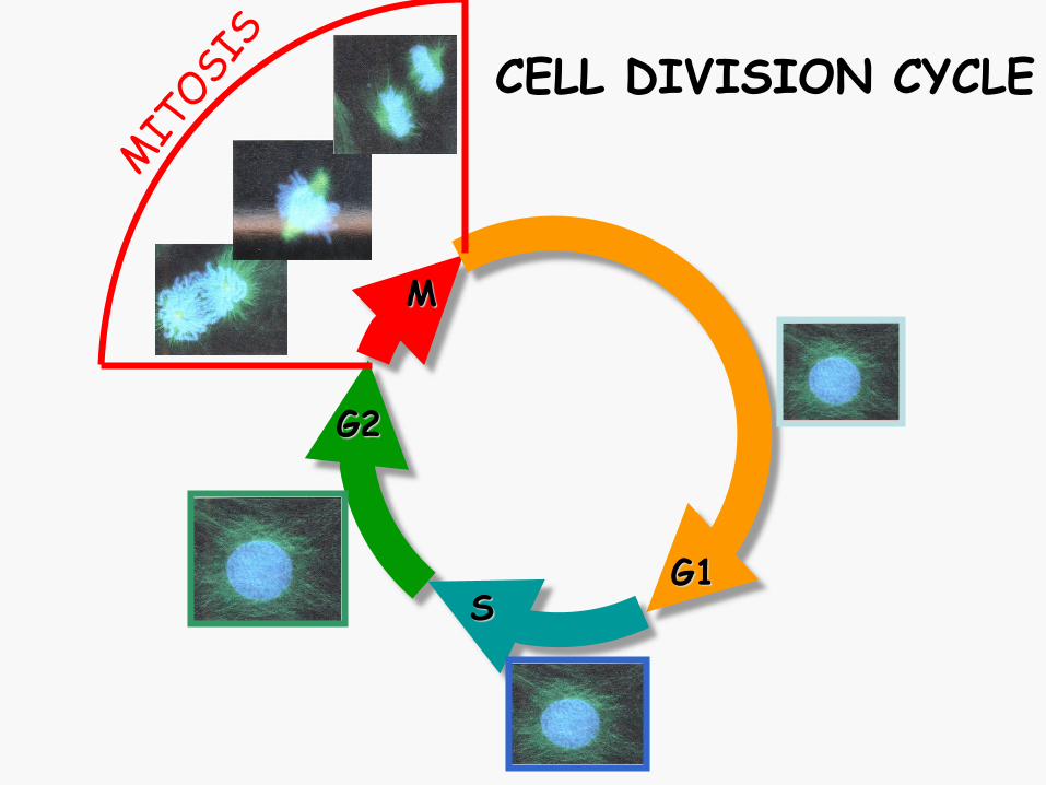
G1
M
G2
S
CELL DIVISION CYCLE
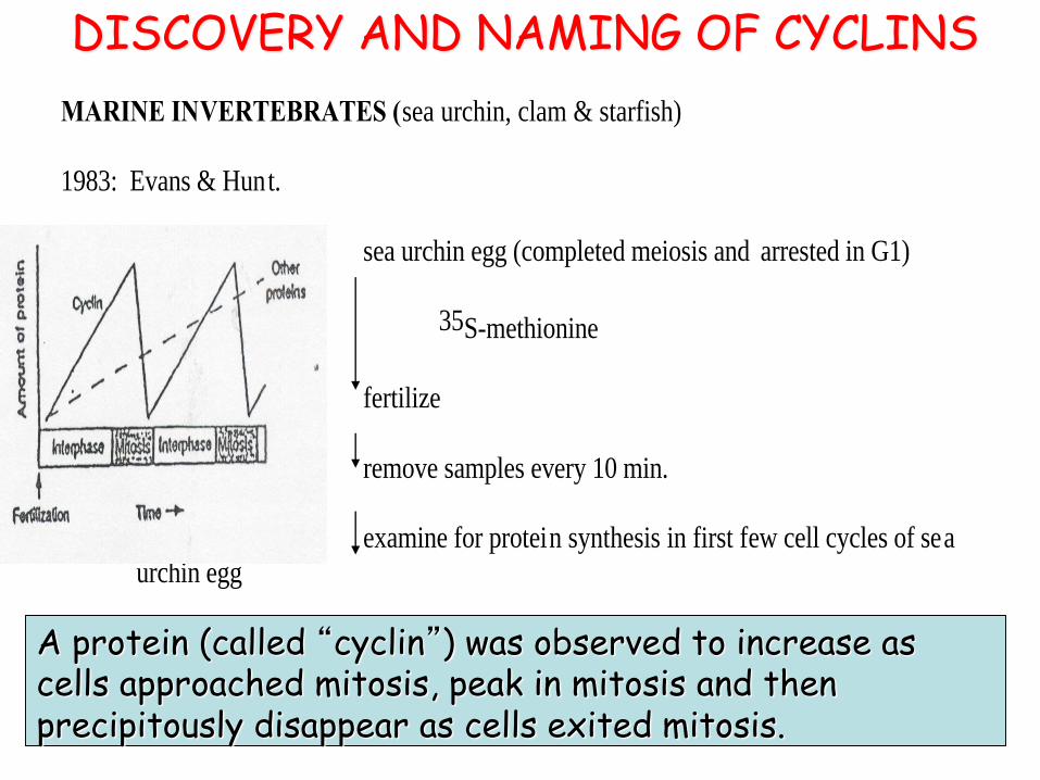
MARINE INVERTEBRATES (sea urchin, clam & starfish)
1983: Evans & Hunt.
sea urchin egg (completed meiosis and arrested in G1)
35S-methionine
fertilize
remove samples every 10 min.
examine for protein synthesis in first few cell cycles of sea
urchin egg
Saw 55kDa protein= sea urchin cyclin (periodic protein synthesized throughout the cell
cycle but degraded at the end of mitosis:
DISCOVERY AND NAMING OF CYCLINS
A protein (called “cyclin”) was observed to increase as cells approached mitosis, peak in mitosis and then precipitously disappear as cells exited mitosis.
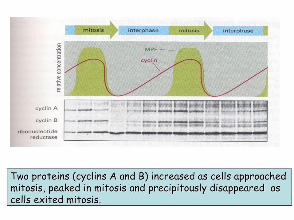
Two proteins (cyclins A and B) increased as cells approached mitosis, peaked in mitosis and precipitously disappeared as cells exited mitosis.
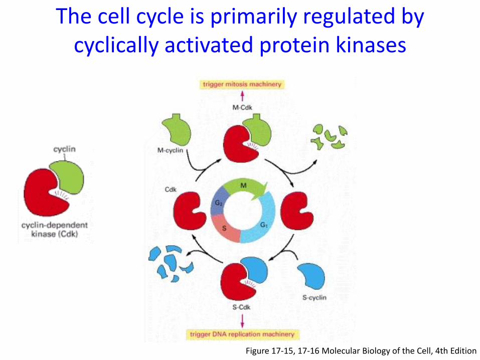
The cell cycle is primarily regulated by cyclically activated protein kinases
Figure 17-15, 17-16 Molecular Biology of the Cell, 4th Edition
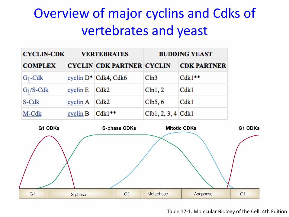
Table 17-1. Molecular Biology of the Cell, 4th Edition
Overview of major cyclins and Cdks of vertebrates and yeast
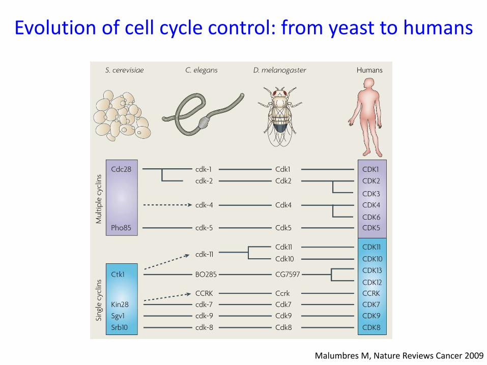
Malumbres M, Nature Reviews Cancer 2009
Evolution of cell cycle control: from yeast to humans
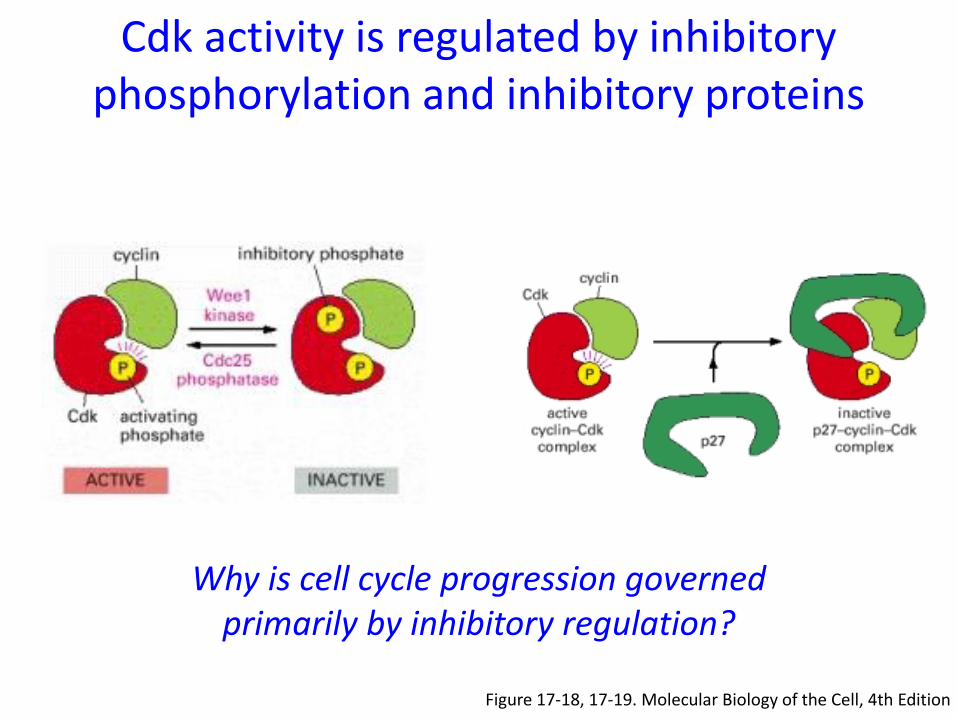
Cdk activity is regulated by inhibitory phosphorylation and inhibitory proteins
Figure 17-18, 17-19. Molecular Biology of the Cell, 4th Edition
Why is cell cycle progression governed primarily by inhibitory regulation?
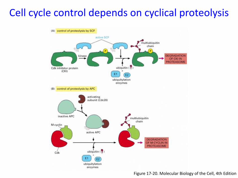
Figure 17-20. Molecular Biology of the Cell, 4th Edition
Cell cycle control depends on cyclical proteolysis
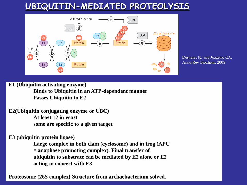
UBIQUITIN-MEDIATED PROTEOLYIS
E1 (Ubiquitin activating enzyme)
Binds to Ubiquitin in an ATP-dependent manner
Passes Ubiquitin to E2
E2(Ubiquitin conjugating enzyme or UBC)
At least 12 in yeast
some are specific to a given target
E3 (ubiquitin protein ligase)
Large complex in both clam (cyclosome) and in frog (APC
= anaphase promoting complex). Final transfer of
ubiquitin to substrate can be mediated by E2 alone or E2
acting in concert with E3
Proteosome (26S complex) Structure from archaebacterium solved.
Deshaies RJ and Joazeiro CA.
Annu Rev Biochem. 2009
UBIQUITIN-MEDIATED PROTEOLYSIS
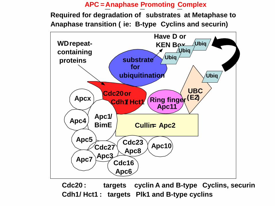
APC =Anaphase Promoting Complex
Required for degradation of substrates at Metaphase to
Anaphase transition ( ie: B-type Cyclins and securin)
Cdc20 : targets cyclin A and B-type Cyclins, securin
Cdh1/ Hct1 : targets Plk1 and B-type cyclins
substrate
Ring finger
UBC(E2)
for
ubiquitination
Apc10
Cdc20Apcx
Apc4
Apc5
Apc7
Cdc27
Apc3
Apc1/
BimE
Cdc23
Apc8
Cdc16
Apc6
Have D or
KEN Box
Cullin= Apc2
Apc11
or
Cdh1/ Hct1
WDrepeat-
containing
proteins
Ubiq
Ubiq
Ubiq
Ubiq
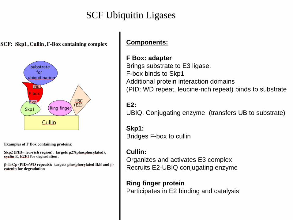
Components:
F Box: adapter
Brings substrate to E3 ligase.
F-box binds to Skp1
Additional protein interaction domains
(PID: WD repeat, leucine-rich repeat) binds to substrate
E2:
UBIQ. Conjugating enzyme (transfers UB to substrate)
Skp1:
Bridges F-box to cullin
Cullin:
Organizes and activates E3 complex
Recruits E2-UBIQ conjugating enzyme
Ring finger protein
Participates in E2 binding and catalysis
SCF Ubiquitin Ligases
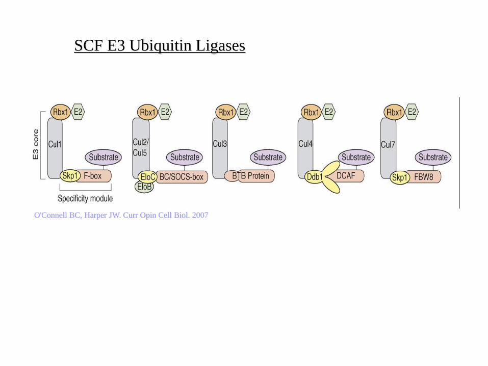
O'Connell BC, Harper JW. Curr Opin Cell Biol. 2007
SCF E3 Ubiquitin Ligases
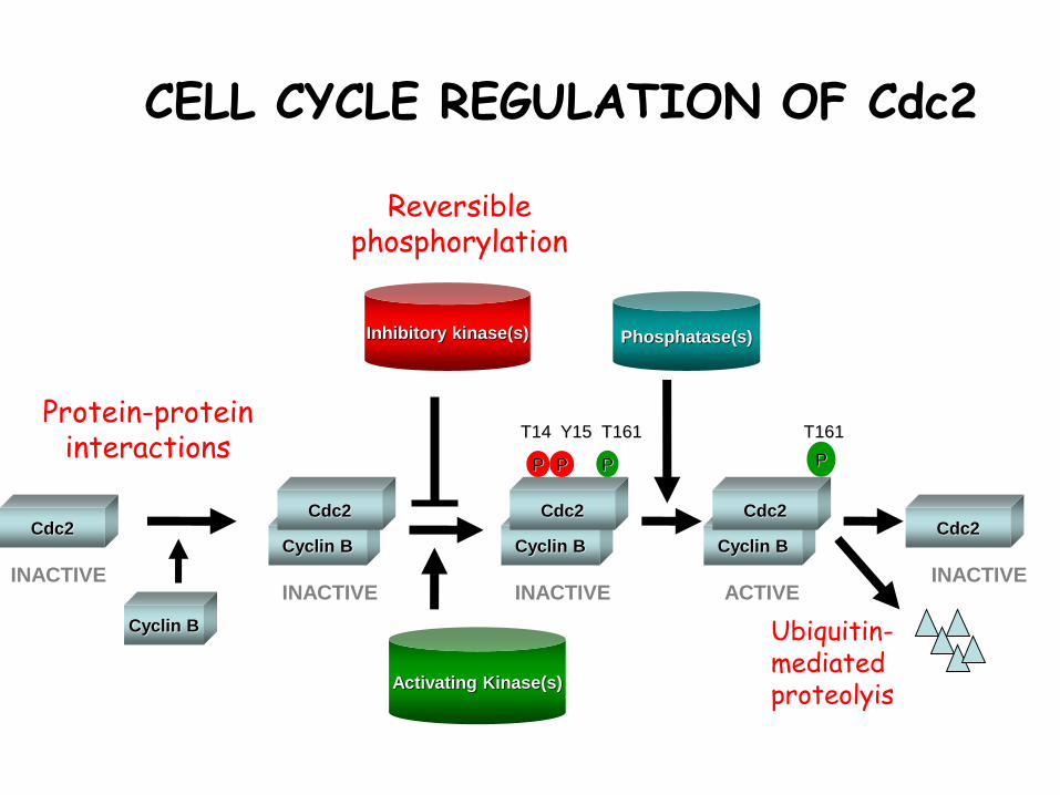
CELL CYCLE REGULATION OF Cdc2
Phosphatase(s)
P
T161
P P P
T14 Y15 T161
Cyclin B
Cdc2
Inhibitory kinase(s)
Activating Kinase(s)
Cyclin B
Cdc2
Cyclin B
Cdc2Cdc2
Cyclin B
Cdc2
INACTIVEINACTIVE INACTIVE
INACTIVEACTIVE
Protein-protein interactions
Reversible phosphorylation
Ubiquitin-mediated proteolyis
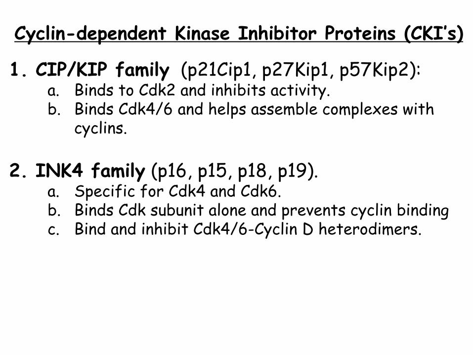
Cyclin-dependent Kinase Inhibitor Proteins (CKI’s)
1. CIP/KIP family (p21Cip1, p27Kip1, p57Kip2):a. Binds to Cdk2 and inhibits activity.b. Binds Cdk4/6 and helps assemble complexes with
cyclins.
2. INK4 family (p16, p15, p18, p19). a. Specific for Cdk4 and Cdk6. b. Binds Cdk subunit alone and prevents cyclin binding c. Bind and inhibit Cdk4/6-Cyclin D heterodimers.
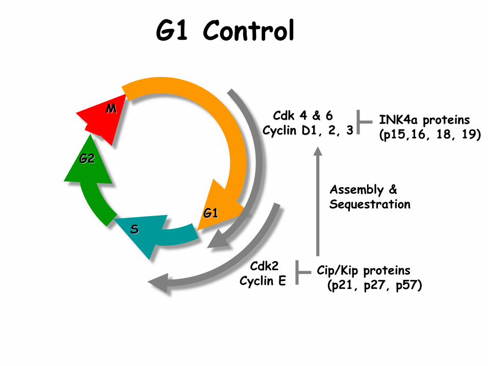
G1 Control
Cdk 4 & 6 Cyclin D1, 2, 3
Cdk2Cyclin E
G1
M
G2
S
Cip/Kip proteins(p21, p27, p57)
INK4a proteins(p15,16, 18, 19)
Assembly & Sequestration
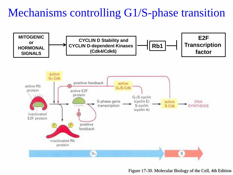
Mechanisms controlling G1/S-phase transition
Figure 17-30. Molecular Biology of the Cell, 4th Edition
MITOGENIC
or
HORMONAL
SIGNALS
CYCLIN D Stability and
CYCLIN D-dependent Kinases
(Cdk4/Cdk6)Rb1
E2F
Transcription
factor
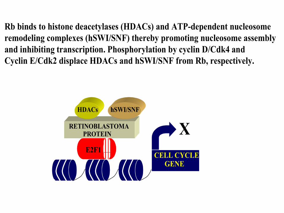
CELL CYCLE GENE
XRETINOBLASTOMA PROTEIN
E2F1
HDACs hSWI/SNF
Rb binds to histone deacetylases (HDACs) and ATP-dependent nucleosome
remodeling complexes (hSWI/SNF) thereby promoting nucleosome assembly
and inhibiting transcription. Phosphorylation by cyclin D/Cdk4 and
Cyclin E/Cdk2 displace HDACs and hSWI/SNF from Rb, respectively.
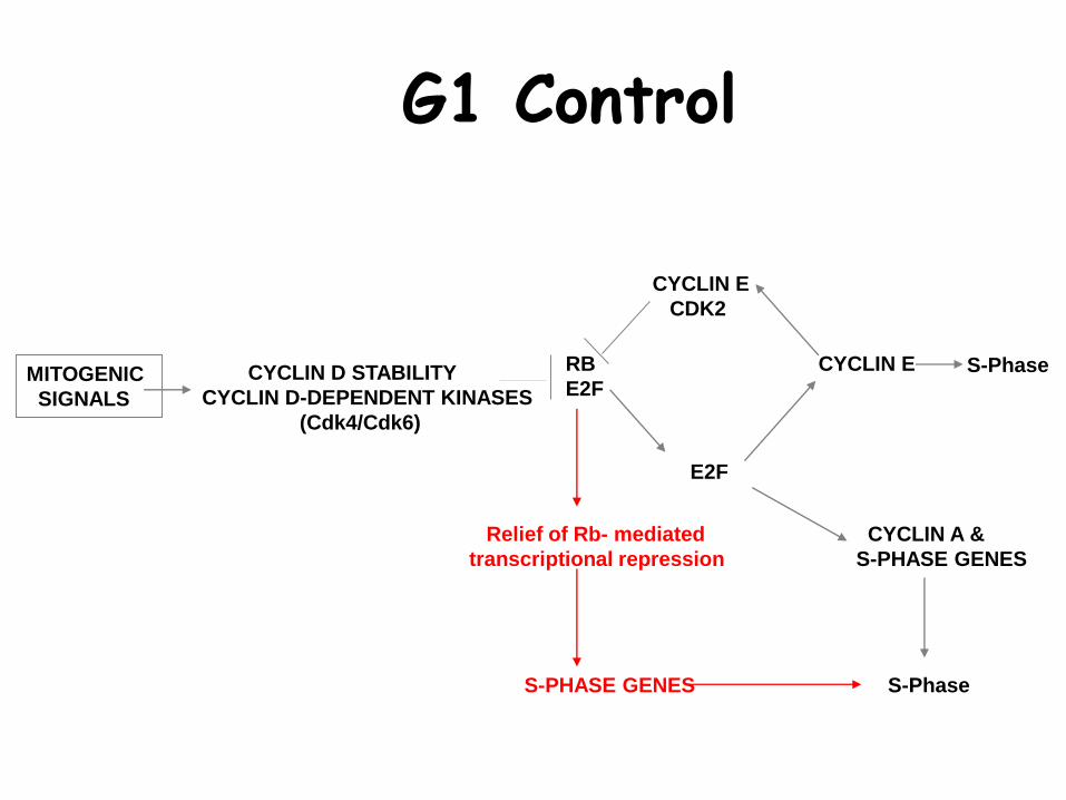
MITOGENIC
SIGNALS
CYCLIN D STABILITY
CYCLIN D-DEPENDENT KINASES
(Cdk4/Cdk6)
RB
E2F
E2F
CYCLIN E
CYCLIN E
CDK2
CYCLIN A &
S-PHASE GENES
S-PhaseS-PHASE GENES
Relief of Rb- mediated
transcriptional repression
S-Phase
G1 Control
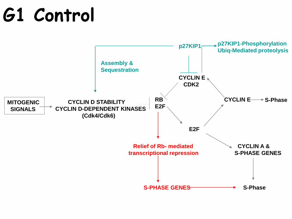
MITOGENIC
SIGNALS
CYCLIN D STABILITY
CYCLIN D-DEPENDENT KINASES
(Cdk4/Cdk6)
RB
E2F
E2F
CYCLIN E
CYCLIN E
CDK2
CYCLIN A &
S-PHASE GENES
p27KIP1 p27KIP1-Phosphorylation
Ubiq-Mediated proteolysis
Assembly &
Sequestration
S-PhaseS-PHASE GENES
Relief of Rb- mediated
transcriptional repression
S-Phase
G1 Control
CheckpointsWhat are they?
How were they defined?
How does their derailment contribute to cancer?
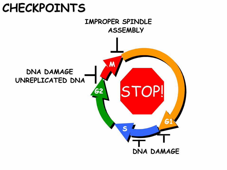
CHECKPOINTS
G1G1
MM
G2G2
SS
DNA DAMAGE
DNA DAMAGEUNREPLICATED DNA
IMPROPER SPINDLE ASSEMBLY
STOP!
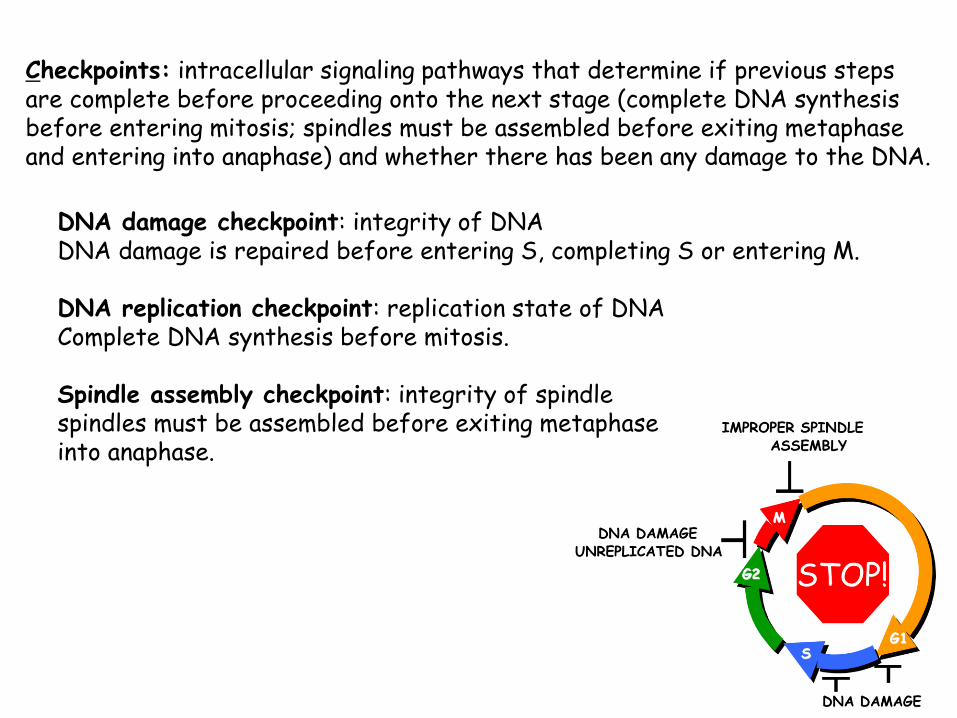
Checkpoints: intracellular signaling pathways that determine if previous stepsare complete before proceeding onto the next stage (complete DNA synthesis before entering mitosis; spindles must be assembled before exiting metaphase and entering into anaphase) and whether there has been any damage to the DNA.
DNA damage checkpoint: integrity of DNA DNA damage is repaired before entering S, completing S or entering M.
DNA replication checkpoint: replication state of DNA Complete DNA synthesis before mitosis.
Spindle assembly checkpoint: integrity of spindle spindles must be assembled before exiting metaphase into anaphase.
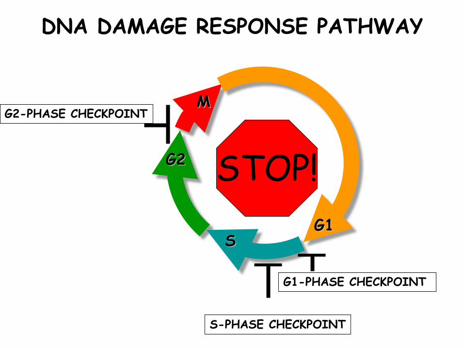
G1
M
G2
S
G1-PHASE CHECKPOINT
S-PHASE CHECKPOINT
G2-PHASE CHECKPOINT
DNA DAMAGE RESPONSE PATHWAY
STOP!
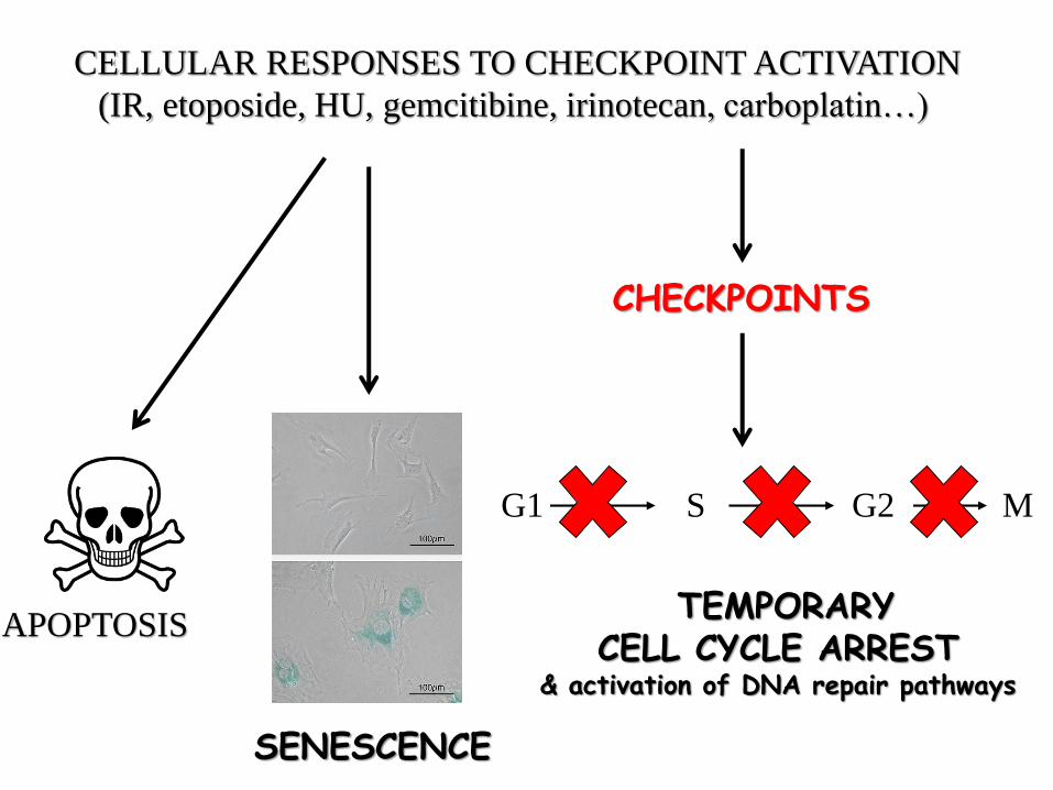
CELLULAR RESPONSES TO CHECKPOINT ACTIVATION
(IR, etoposide, HU, gemcitibine, irinotecan, carboplatin…)
G1 S G2 M
CHECKPOINTS
APOPTOSIS
SENESCENCE
TEMPORARY CELL CYCLE ARREST
& activation of DNA repair pathways
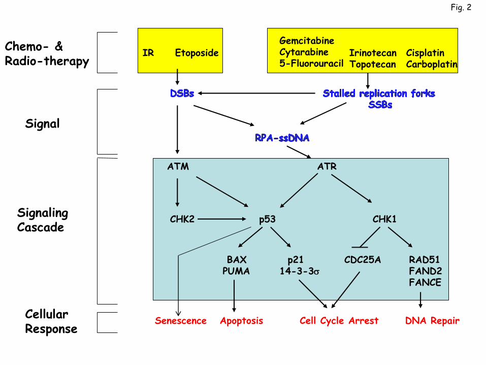
IR EtoposideGemcitabineCytarabine5-Fluorouracil
IrinotecanTopotecan
CisplatinCarboplatin
ATM ATR
CHK2 p53 CHK1
RAD51FAND2FANCE
p2114-3-3s
BAXPUMA
CDC25A
Apoptosis Cell Cycle Arrest DNA Repair
Signaling Cascade
CellularResponse
Chemo- &Radio-therapy
Signal
Fig. 2
Senescence
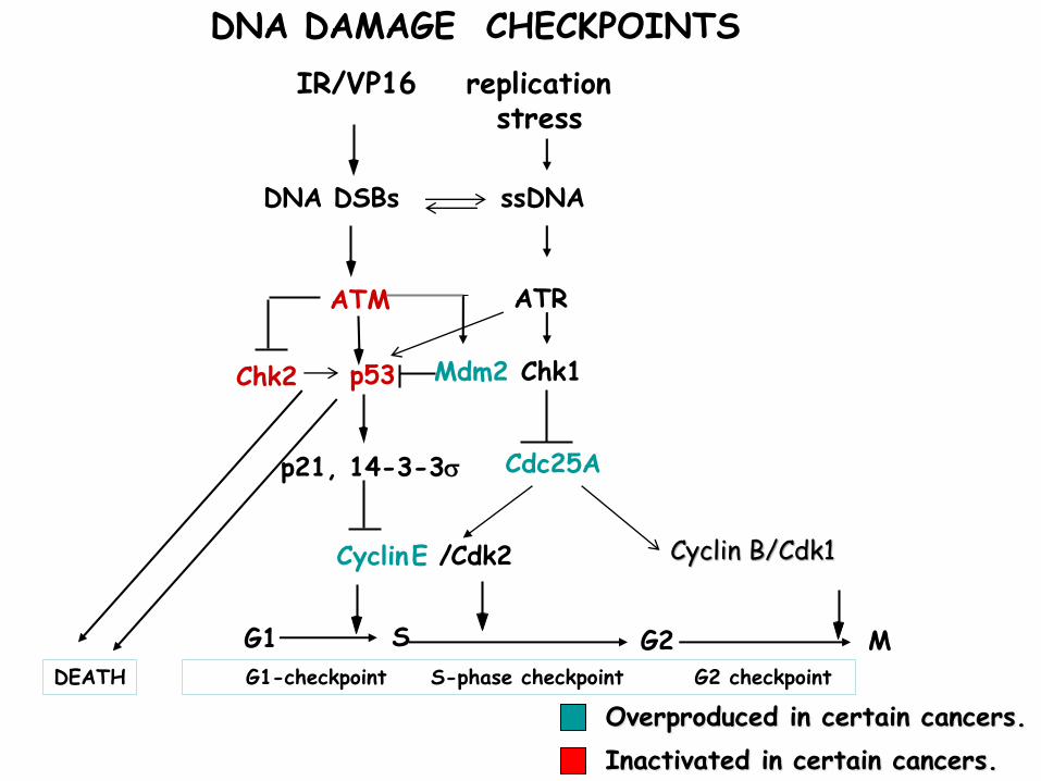
DNA DAMAGE CHECKPOINTS
Chk1
CyclinE / Cdk2
G1 S G2G1-checkpoint S-phase checkpoint G2 checkpoint
MDEATH
Cdc25A
DNA DSBs ssDNA
ATM
Mdm2p53
p21, 14-3-3s
IR/VP16 replication stress
ATR
Chk2
Cyclin B/Cdk1
Overproduced in certain cancers.
Inactivated in certain cancers.
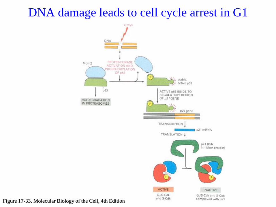
DNA damage leads to cell cycle arrest in G1
Figure 17-33. Molecular Biology of the Cell, 4th Edition
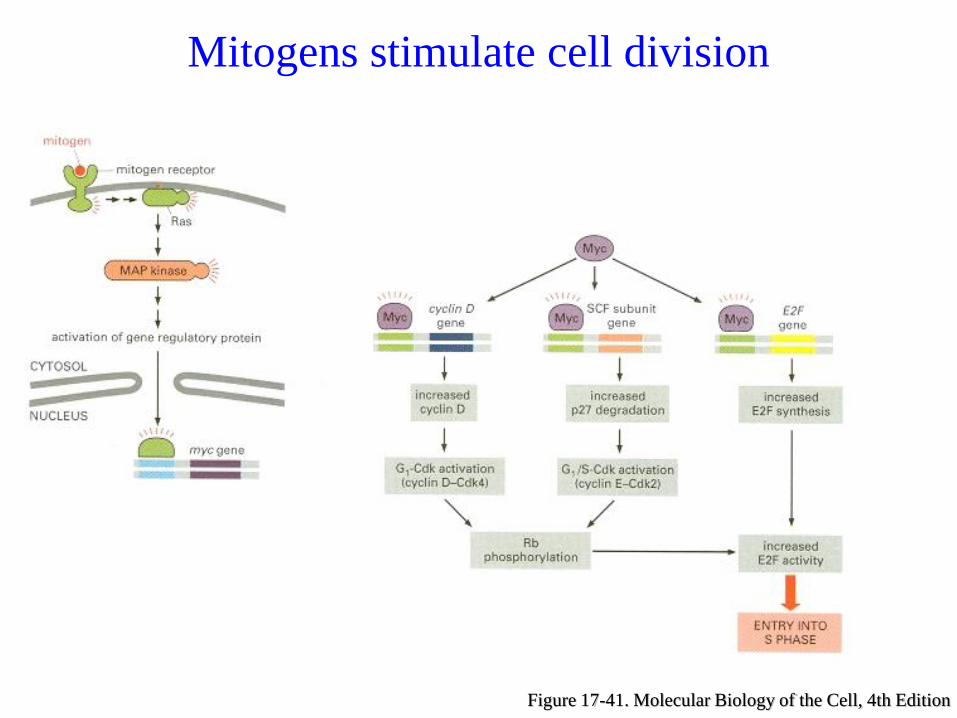
Figure 17-41. Molecular Biology of the Cell, 4th Edition
Mitogens stimulate cell division
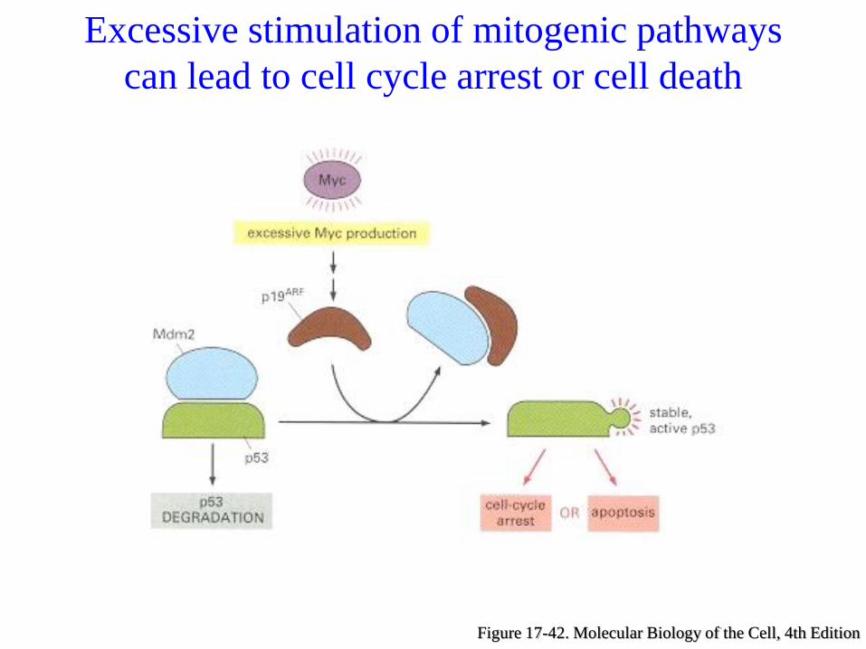
Figure 17-42. Molecular Biology of the Cell, 4th Edition
Excessive stimulation of mitogenic pathways
can lead to cell cycle arrest or cell death
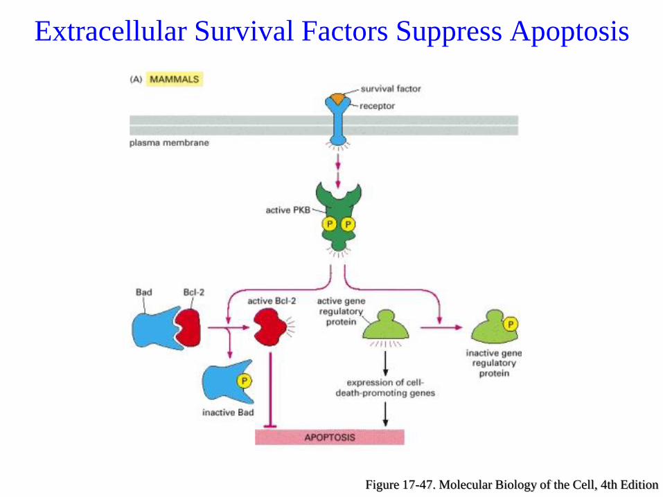
Figure 17-47. Molecular Biology of the Cell, 4th Edition
Extracellular Survival Factors Suppress Apoptosis
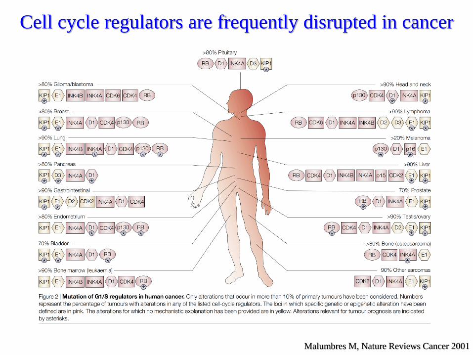
Malumbres M, Nature Reviews Cancer 2001
Cell cycle regulators are frequently disrupted in cancer
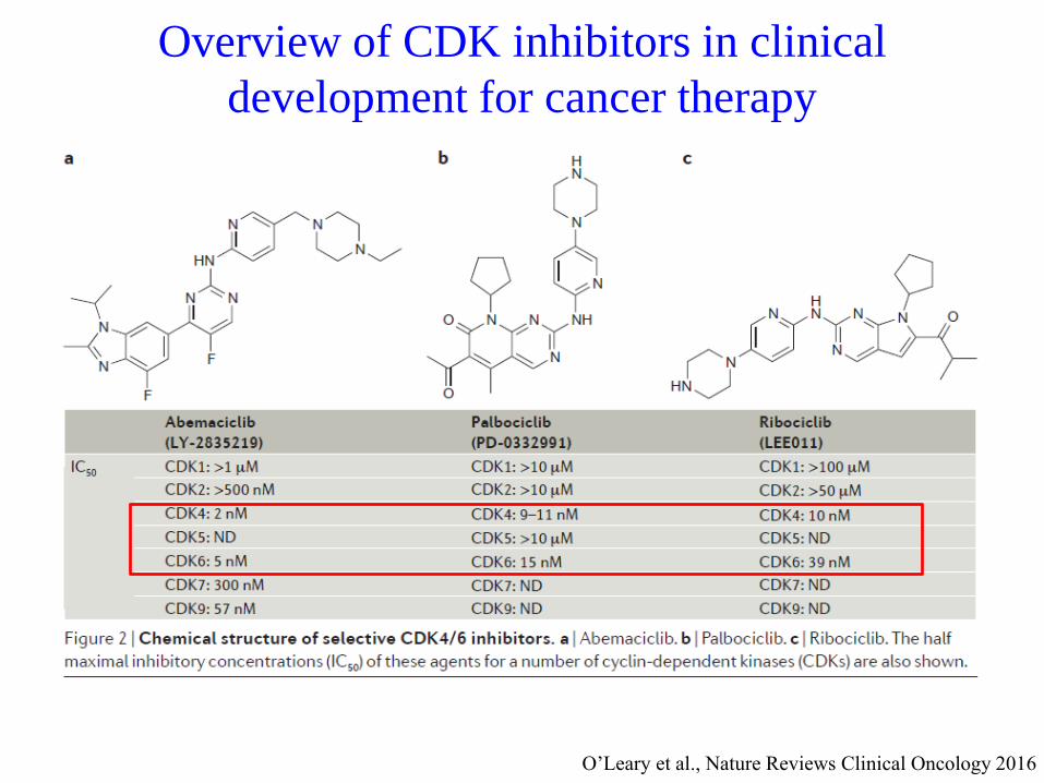
O’Leary et al., Nature Reviews Clinical Oncology 2016
Overview of CDK inhibitors in clinical
development for cancer therapy
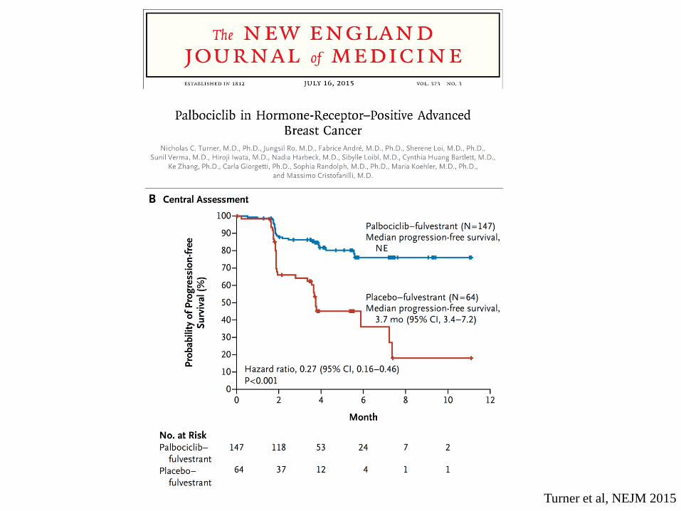
Turner et al, NEJM 2015
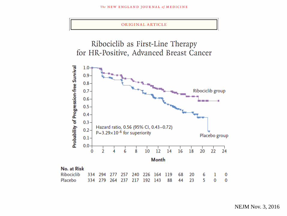
NEJM Nov. 3, 2016
Conclusions
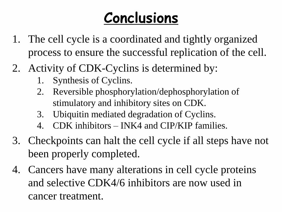
1. The cell cycle is a coordinated and tightly organized
process to ensure the successful replication of the cell.
2. Activity of CDK-Cyclins is determined by:1. Synthesis of Cyclins.
2. Reversible phosphorylation/dephosphorylation of
stimulatory and inhibitory sites on CDK.
3. Ubiquitin mediated degradation of Cyclins.
4. CDK inhibitors – INK4 and CIP/KIP families.
3. Checkpoints can halt the cell cycle if all steps have not
been properly completed.
4. Cancers have many alterations in cell cycle proteins
and selective CDK4/6 inhibitors are now used in
cancer treatment.
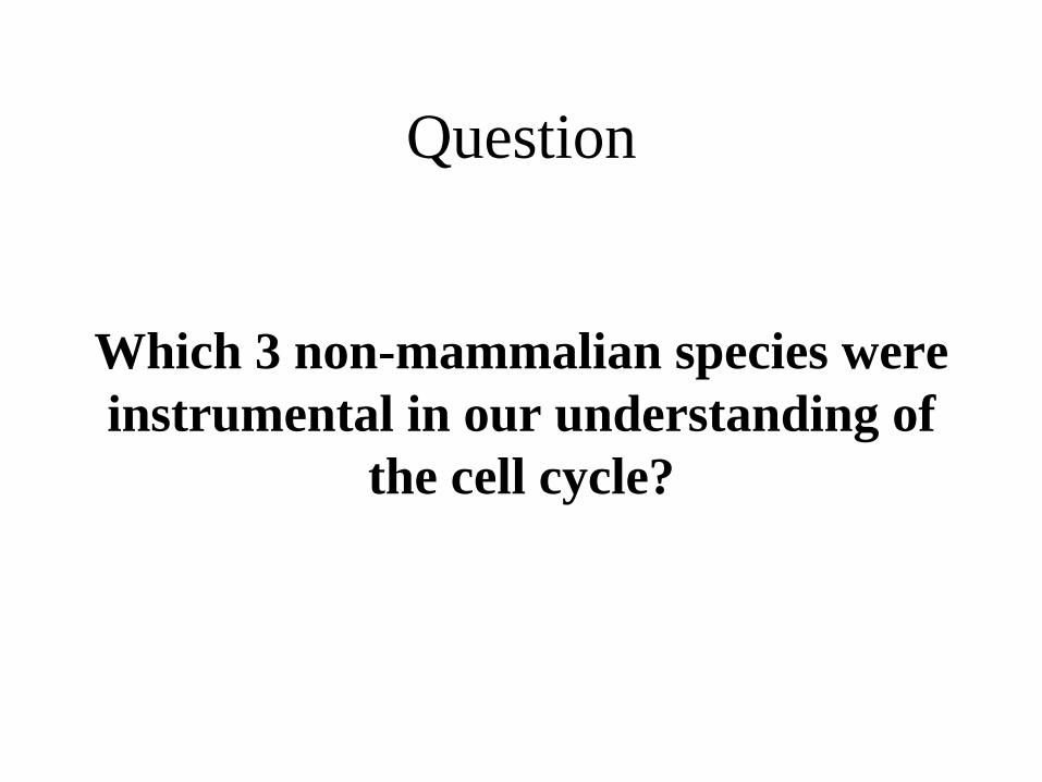
Question
Which 3 non-mammalian species were
instrumental in our understanding of
the cell cycle?

Prize and Question