molecular biology of pasteurella multocida toxin · pasteurella multocida is an opportunistic...
TRANSCRIPT
Molecular Biology of Pasteurellamultocida Toxin
Joachim H. C. Orth and Klaus Aktories
Abstract Pasteurella multocida toxin (PMT) is the causative agent of progressiveatrophic rhinitis in swine. The 146 kDa single-chain toxin harbours discretedomains important for receptor binding, internalisation and biological activity. Themolecular basis of the toxin’s activity is the deamidation of a specific glutamineresidue in the a-subunit of heterotrimeric G proteins. This results in an inhibitionof the inherent GTPase activity leading to a constitutively active phenotype of theG protein. Due to the ability of the toxin to act on various families of heterotri-meric G proteins, a large subset of signal transduction pathways is stimulated.
Contents
1 Structure of PMT................................................................................................................. 742 Uptake into Eukaryotic Cells.............................................................................................. 773 Activation of Heterotrimeric G Proteins ............................................................................ 794 Substrate Specificity............................................................................................................ 825 G Protein Prerequisites for PMT Action............................................................................ 84
5.1 Role of Gbc ................................................................................................................ 845.2 PMT-Induced Activation of G Proteins is Independent of GPCR Interaction ........ 85
6 PMT and Cytotoxic Necrotizing Factors............................................................................ 867 Outlook ................................................................................................................................ 87References.................................................................................................................................. 87
J. H. C. Orth (&) � K. AktoriesInstitut für Experimentelle und Klinische Pharmakologie und Toxikologie,Albert-Ludwigs-Universität Freiburg, Albertstr. 25, 79104 Freiburg, Germanye-mail: [email protected]
K. Aktoriese-mail: [email protected]
Current Topics in Microbiology and Immunology (2012) 361: 73–92 73DOI: 10.1007/82_2012_201� Springer-Verlag Berlin Heidelberg 2012Published Online: 28 February 2012
Pasteurella multocida is an opportunistic pathogenic bacterium living in the nasalpharyngeal space of animals. Infections of humans normally arise from scratchesand bites by domesticated animals such as cats and dogs. Under special conditionsinfection of pigs with P. multocida leads to an atrophic rhinitis, which is char-acterised by the atrophy of nasal turbinate bones accompanied by a shortening andtwisting of the snout. The causative agent of the atrophic rhinitis was found to bethe bacterial protein toxin PMT. The toxin is produced by two (A and D) of fiveserogroups of P. multocida (Frandsen et al. 1991; Williamson 1994).
After entering the cell, the 146 kDa toxin activates various signal transductionpathways by stimulating heterotrimeric G proteins of the Gaq/11, Ga12/13 and Gai
family. After a discussion of the structure of PMT, the mechanisms by which PMTenters eukaryotic cells and produces biological effects will be described in detail.
1 Structure of PMT
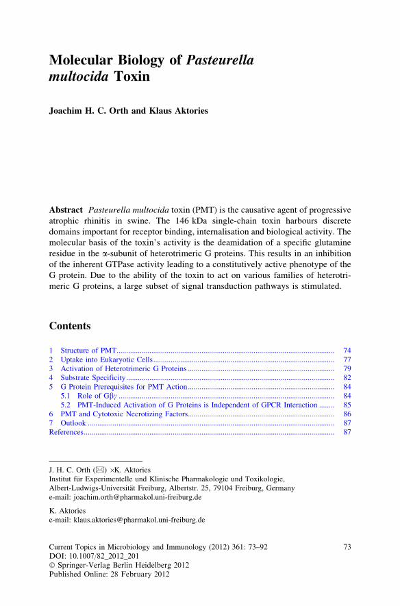
Similar to many bacterial protein toxins, Pasteurella multocida toxin (PMT) is amultifunctional protein comprising different domains with specific functions. Thetoxin contains a receptor binding and translocation domain (B) and a biologicallyactive (A) domain. Therefore, it can be described as a typical AB-type proteintoxin (Fig. 1).
PMT consists of 1285 amino acid residues resulting in a mass of 146 kDa(Buys et al. 1990; Petersen 1990). The receptor binding domain is located in theN-terminal part of the protein, including the amino acid residues 1–580. Withinthis domain a putative translocation domain was found between residues 402–457.The N-terminus of PMT has homology (*20%) to another family of bacterialprotein toxins named cytotoxic necrotizing factors found in uropathogenic E. coli(CNF1, CNF2, CNF3) and Yersinia pseudotuberculosis (CNFY) (Lemichez et al.1997; Pullinger et al. 2001). CNFs are deamidases, which act on small GTPaseslike Rho, Rac or Cdc42 (Flatau et al. 1997; Schmidt et al. 1997). Consistent withthis homology, the receptor binding domains of CNF and PMT are located in theN-terminal part and the active domains in the C-terminus (Busch et al. 2001;Pullinger et al. 2001).
The crystal structure of the C-terminal part of PMT revealed three domainsdesignated C1, C2 and C3 (PDB ID 2EBF). The C3 domain is of major importancebecause it harbours the catalytic activity of the toxin to modify intracellular tar-gets. The overall structure of the crystallised portion was described as Trojanhorse-like shape with feet (C1), a body (C2) and a head (C3) (Kitadokoro et al.2007; Miyazawa et al. 2006).
The feet C1-domain, encompassing amino acid residues 575–719, consists ofseven helices. The tertiary structure of the first four helices shows similarity to theN-terminal portion of Clostridium difficile toxin B (25% identity) (Kamitani et al.2010). In toxin B, these four helices serve as a plasma membrane targeting signal.An ectopically expressed GFP-fusion of a C1 fragment of PMT (amino acid
74 J. H. C. Orth and K. Aktories
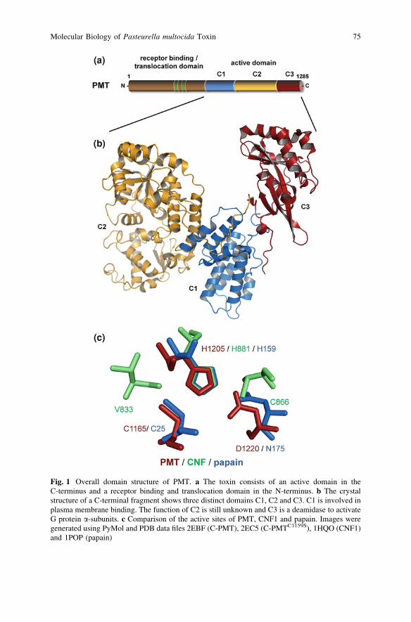
Fig. 1 Overall domain structure of PMT. a The toxin consists of an active domain in theC-terminus and a receptor binding and translocation domain in the N-terminus. b The crystalstructure of a C-terminal fragment shows three distinct domains C1, C2 and C3. C1 is involved inplasma membrane binding. The function of C2 is still unknown and C3 is a deamidase to activateG protein a-subunits. c Comparison of the active sites of PMT, CNF1 and papain. Images weregenerated using PyMol and PDB data files 2EBF (C-PMT), 2EC5 (C-PMTC1159S), 1HQO (CNF1)and 1POP (papain)
Molecular Biology of Pasteurella multocida Toxin 75
residues 569–671) shows strong localisation to the plasma membrane. Deletion ofeach of the four helices impaired localisation to the membrane. Additionally, aGFP-fusion of a C-terminal fragment of PMT (amino acid residues 569–1285)localised to the plasma membrane, whereas a deletion mutant of the first fourhelices of C1 (amino acid residues 671–1285) did not localise in the membrane.Because the primary target proteins of PMT are plasma membrane-bound het-erotrimeric G proteins, impaired localisation to the substrate would diminish PMTtoxicity. Congruently, expression of PMT fragments which are impaired in plasmamembrane binding, showed no biological activity. Interestingly, an N-myris-toylation peptide tag compensated for the deleted helices and restored PMTactivity (Kamitani et al. 2010).
Moreover, this domain was identified as a conserved membrane localisationdomain in clostridial glucosyltransferase toxins from C. difficile, C. novyi, C. sordelliior C. perfringens and the Multifunctional Autoprocessing RTX toxins (MARTX)from Vibrio cholerae, V. vulnificus or V. anguillarum (Geissler et al. 2009). GFPfusion proteins of these homologue domains are also membrane associated. Threeamino acid residues were identified, which are 100% identical between membranelocalisation domains. For PMT, these residues are Tyr-611, Ser-651 and Arg-653.Consequences of site-directed mutagenesis of these residues were only tested inMARTX-derived domains, but not in the correspondent PMT domain. The resultsshowed that only the Ser and the Arg are essential for appropriate membrane tar-geting. Their function could be the maintenance of the overall structure of the four-helix bundle (Geissler et al. 2010).
The largest domain in the C-terminal part of PMT is the so-called body orC2 domain (amino acid residues 720–1,104). C2 consists of 18 helices and nineb-strands and can be divided into two subdomains. Both subdomains exhibit astructure typical of nucleotide-binding proteins. Folylpolyglutamate synthetaseand cdc14bs show structural homology to the second subdomain. Because bothenzymes interact with a phosphate group it was supposed that this could be a hintto the still unknown function of the C2 domain (Kitadokoro et al. 2007).
Intracellularly expressed PMT and toxin truncations confirmed that the bio-logically active domain, i.e. the G protein-activating domain is located within C3(Aminova et al. 2008). This C3 domain (amino acid residues 1,105–1,285) isconnected by a long loop (1,087–1,104) to the C2 domain. C3 is separated into twosubdomains and provides the catalytic cleft for the enzymatic function of the toxin.Interestingly, a disulfide bond was found in C3 between Cys-1159 and Cys-1165.Mutational studies of these residues revealed that the toxic activity of PMT strictlydepends on Cys-1165 but not Cys-1159 (Busch et al. 2001; Kitadokoro et al. 2007;Ward et al. 1998). In addition, His-1205, His-1223, Asp-1220 and Gln-1225 areessential for PMT activity (Kitadokoro et al. 2007; Orth et al. 2003; Pullinger andLax 2007). Structural analysis of PMT mutants in which the disulfide bonds wereablated by replacing the respective Cys-1159 or Cys-1165 residues by Ser (PDBID 2EC5/2EBH) displayed different structures compared to wt-PMT (PDB ID2EBF). Most striking is a reorientation of Cys-1165 when Cys-1159 is replaced bySer. Cys-1165 is displaced towards the catalytic cleft of the C3 domain and forms
76 J. H. C. Orth and K. Aktories
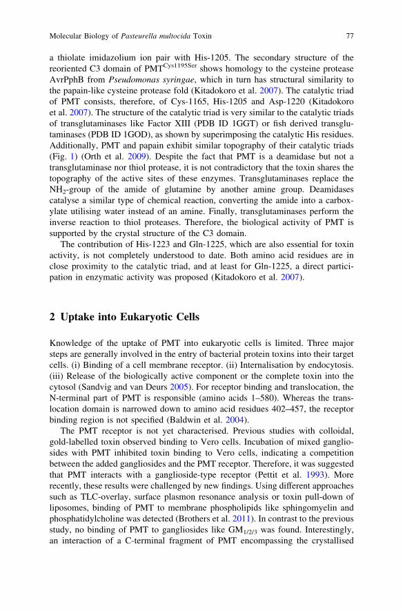
a thiolate imidazolium ion pair with His-1205. The secondary structure of thereoriented C3 domain of PMTCys1195Ser shows homology to the cysteine proteaseAvrPphB from Pseudomonas syringae, which in turn has structural similarity tothe papain-like cysteine protease fold (Kitadokoro et al. 2007). The catalytic triadof PMT consists, therefore, of Cys-1165, His-1205 and Asp-1220 (Kitadokoroet al. 2007). The structure of the catalytic triad is very similar to the catalytic triadsof transglutaminases like Factor XIII (PDB ID 1GGT) or fish derived transglu-taminases (PDB ID 1GOD), as shown by superimposing the catalytic His residues.Additionally, PMT and papain exhibit similar topography of their catalytic triads(Fig. 1) (Orth et al. 2009). Despite the fact that PMT is a deamidase but not atransglutaminase nor thiol protease, it is not contradictory that the toxin shares thetopography of the active sites of these enzymes. Transglutaminases replace theNH2-group of the amide of glutamine by another amine group. Deamidasescatalyse a similar type of chemical reaction, converting the amide into a carbox-ylate utilising water instead of an amine. Finally, transglutaminases perform theinverse reaction to thiol proteases. Therefore, the biological activity of PMT issupported by the crystal structure of the C3 domain.
The contribution of His-1223 and Gln-1225, which are also essential for toxinactivity, is not completely understood to date. Both amino acid residues are inclose proximity to the catalytic triad, and at least for Gln-1225, a direct partici-pation in enzymatic activity was proposed (Kitadokoro et al. 2007).
2 Uptake into Eukaryotic Cells
Knowledge of the uptake of PMT into eukaryotic cells is limited. Three majorsteps are generally involved in the entry of bacterial protein toxins into their targetcells. (i) Binding of a cell membrane receptor. (ii) Internalisation by endocytosis.(iii) Release of the biologically active component or the complete toxin into thecytosol (Sandvig and van Deurs 2005). For receptor binding and translocation, theN-terminal part of PMT is responsible (amino acids 1–580). Whereas the trans-location domain is narrowed down to amino acid residues 402–457, the receptorbinding region is not specified (Baldwin et al. 2004).
The PMT receptor is not yet characterised. Previous studies with colloidal,gold-labelled toxin observed binding to Vero cells. Incubation of mixed ganglio-sides with PMT inhibited toxin binding to Vero cells, indicating a competitionbetween the added gangliosides and the PMT receptor. Therefore, it was suggestedthat PMT interacts with a ganglioside-type receptor (Pettit et al. 1993). Morerecently, these results were challenged by new findings. Using different approachessuch as TLC-overlay, surface plasmon resonance analysis or toxin pull-down ofliposomes, binding of PMT to membrane phospholipids like sphingomyelin andphosphatidylcholine was detected (Brothers et al. 2011). In contrast to the previousstudy, no binding of PMT to gangliosides like GM1/2/3 was found. Interestingly,an interaction of a C-terminal fragment of PMT encompassing the crystallised
Molecular Biology of Pasteurella multocida Toxin 77
domains C1, C2 and C3 with GM1 was discovered. The authors suggested thatremoval of the PMT N-terminus unmasks the membrane binding site of the C1domain.
Surface plasmon resonance analysis leads to an at least bi-phasic binding ofPMT to cells. After initial binding with low affinity to an abundant membranecomponent, a more specific binding to sphingomyelin could follow. Theinvolvement of an additional proteinaceous receptor, which would induce endo-cytosis, was proposed (Brothers et al. 2011).
After binding to the receptor at the plasma membrane, the toxin enters the cellby endocytosis. Utilising various inhibitors of vesicle trafficking and GFP-fusionsof PMT fragments, the intracellular pathway of endocytosed toxin was followed.GFP-fusions of the N-terminal portion of PMT harbouring the putative receptorbinding and translocation domain were found to colocalize with transferrinreceptor and at early time points with cholera toxin B subunit, indicating locali-sation in early endosomes (Repella et al. 2011). After passing early endosomes,transferrin receptor traffics to recycling endosomes and cholera toxin via the Golgiapparatus to the endoplasmic reticulum. Conversely, PMT is supposed to trans-locate to late endosomes where the translocation to the cytosol occurs (Repellaet al. 2011). In line with this model, inhibitors of trafficking between Golgiapparatus and endoplasmic reticulum, like brefeldin A, do not inhibit the bio-logical activity of PMT. However, cell entry depends on Arf6. The small GTPaseArf6 binds to endosomes and is important for the trafficking of recycling endo-somes (Peters et al. 1995). Both a dominant-negative form and a constitutivelycycling mutant of Arf6 inhibited PMT intoxication, implicating the involvement ofthis GTPase in toxin uptake (Repella et al. 2011).
It is thought that PMT translocates on the way from early to late endosomes tothe cytosol. The acidification of the endosome plays a pivotal role for translocation(Baldwin et al. 2004; Rozengurt et al. 1990). Consequently, inhibition of endo-some acidification by blockade of the vacuolar H+ ATPases using bafilomycin A1represses toxin activity (Baldwin et al. 2004). In addition, the direct transfer ofplasma membrane bound toxin to the cytosol is inducible by mimicking theendocytic conditions, e.g. applying acidic medium to cells (Baldwin et al. 2004).The translocation depends on a putative translocation T-domain, consisting of twopredicted hydrophobic helices (residues 402–423 and 437–457) linked by a pep-tide loop (residues 424–436). It is proposed that acidification induces a structuralchange in the toxin, which was previously observed utilising circular dichroismand measuring susceptibility to proteases (Smyth et al. 1995, 1999). This structuralchange exposes the T-domain, allowing it to insert into the vesicular membrane.Mutational studies of amino acid residues in the peptide loop between thehydrophobic helices suggest that acidic residues in this region are of majorimportance for membrane insertion (Baldwin et al. 2004).
So far, our knowledge of the membrane translocation process itself is scant. It issuggested that the toxin at least partly unfolds. Whether the toxin spontaneouslyrefolds in the cytosol or whether chaperons support this process is not known.
78 J. H. C. Orth and K. Aktories
3 Activation of Heterotrimeric G Proteins
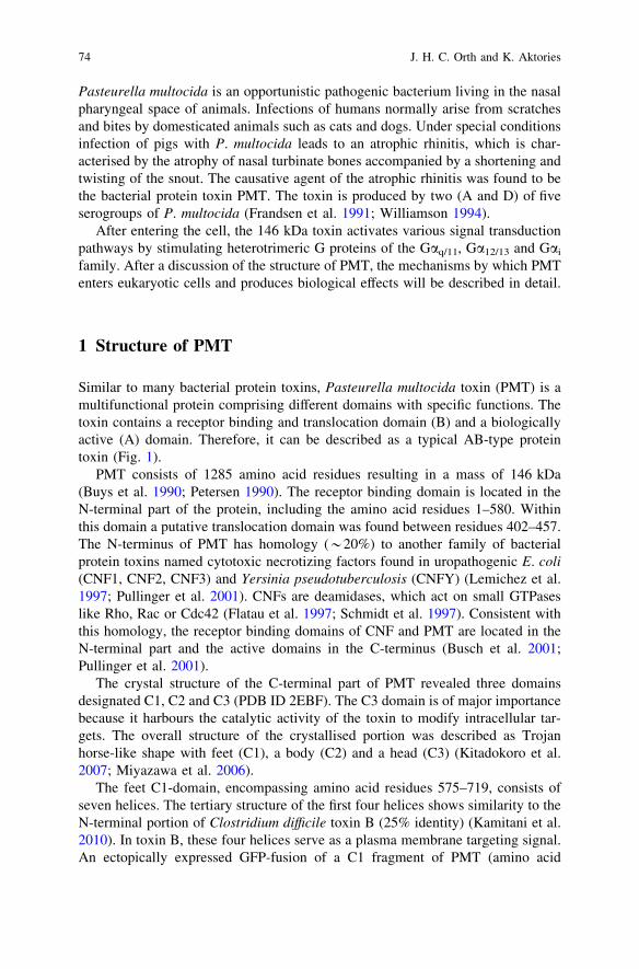
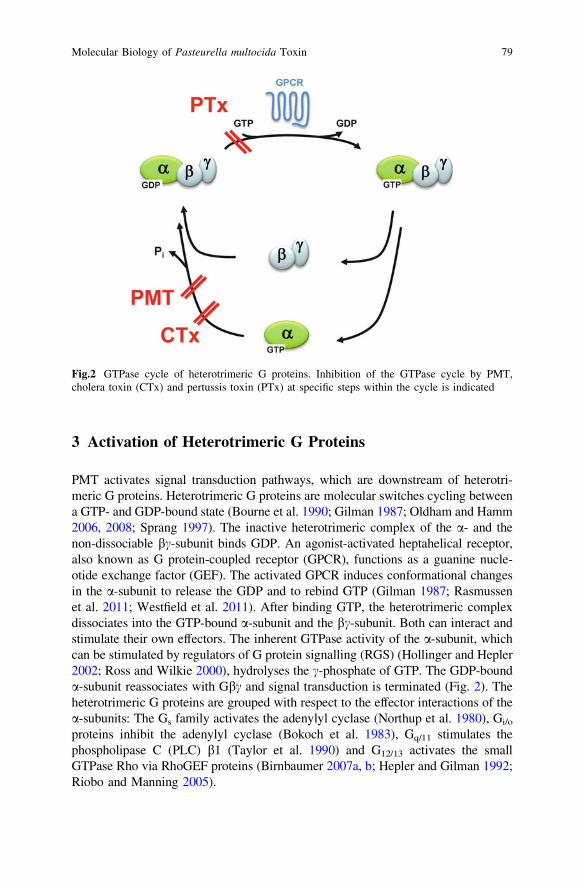
PMT activates signal transduction pathways, which are downstream of heterotri-meric G proteins. Heterotrimeric G proteins are molecular switches cycling betweena GTP- and GDP-bound state (Bourne et al. 1990; Gilman 1987; Oldham and Hamm2006, 2008; Sprang 1997). The inactive heterotrimeric complex of the a- and thenon-dissociable bc-subunit binds GDP. An agonist-activated heptahelical receptor,also known as G protein-coupled receptor (GPCR), functions as a guanine nucle-otide exchange factor (GEF). The activated GPCR induces conformational changesin the a-subunit to release the GDP and to rebind GTP (Gilman 1987; Rasmussenet al. 2011; Westfield et al. 2011). After binding GTP, the heterotrimeric complexdissociates into the GTP-bound a-subunit and the bc-subunit. Both can interact andstimulate their own effectors. The inherent GTPase activity of the a-subunit, whichcan be stimulated by regulators of G protein signalling (RGS) (Hollinger and Hepler2002; Ross and Wilkie 2000), hydrolyses the c-phosphate of GTP. The GDP-bounda-subunit reassociates with Gbc and signal transduction is terminated (Fig. 2). Theheterotrimeric G proteins are grouped with respect to the effector interactions of thea-subunits: The Gs family activates the adenylyl cyclase (Northup et al. 1980), Gi/o
proteins inhibit the adenylyl cyclase (Bokoch et al. 1983), Gq/11 stimulates thephospholipase C (PLC) b1 (Taylor et al. 1990) and G12/13 activates the smallGTPase Rho via RhoGEF proteins (Birnbaumer 2007a, b; Hepler and Gilman 1992;Riobo and Manning 2005).
Fig.2 GTPase cycle of heterotrimeric G proteins. Inhibition of the GTPase cycle by PMT,cholera toxin (CTx) and pertussis toxin (PTx) at specific steps within the cycle is indicated
Molecular Biology of Pasteurella multocida Toxin 79
PMT activates diverse family members of heterotrimeric G proteins. Via Gaq
the toxin leads to activation of PLCb1, resulting in increased levels of dia-cylglycerol, inositoltrisphosphate and Ca2+ (Rozengurt et al. 1990; Staddon et al.1991; Wilson et al. 1997; Zywietz et al. 2001). PMT-activated Ga13 leads tostimulation of the small GTPase RhoA (Orth et al. 2005; Zywietz et al. 2001) andvia Gai the adenylyl cyclase is inhibited (Orth et al. 2008). A comprehensiveoverview of PMT-induced signalling is lined out in Cellular effects of Pasteurellamultocida toxin by Wilson.
The molecular mechanism by which PMT activates signalling via heterotri-meric G proteins was elucidated on the basis of Gai. One advantage of Gai is thepossibility to easily determine the GTPase activity, which is the key in terminatingG protein signalling. A so-called multiple cycle GTPase assay (Aktories andJakobs 1981) in membrane preparations was utilised to measure the effect of PMTon heterotrimeric G proteins (Orth et al. 2008). An agonist of the Gi-couplingEDG-receptor was used to induce G protein cycling and the released c-phosphatewas measured. The receptor agonist LPA exhibited a strong induction of GTPaseactivity of Gai. Interestingly, in membranes of PMT-intoxicated cells the basalGTPase activity was diminished and no increase of GTPase activity was inducedby receptor agonists (Orth et al. 2008). The inhibition of GTPase activity inducedby PMT suggested that the toxin activated G protein signalling by blocking theterminating GTP hydrolysis. However, the multiple cycle GTPase assay was notappropriate to determine unequivocally the step of GTP hydrolysis. Besidesinhibition of the GTPase activity, the toxin caused uncoupling of Gai from itsreceptor. This was measured by receptor-induced GTPcS-binding, which wasblocked by PMT treatment (Orth et al. 2007).
The effect of PMT was compared to that of pertussis toxin (PTx). PTx ADP-ribosylates Gai proteins and inhibits the interaction of the heterotrimeric G proteinwith the GPCR (Gierschik 1992; Katada and Ui 1982; Murayama and Ui 1983;Nürnberg 1997; Ui 1984). Thereby the activation and the cycling of the G proteinare blocked. The observed outcome is the inhibition of the GTPase activity andreceptor-induced GTPcS-binding. In respect to the effects measured (e.g., inhibi-tion of steady-state GTP hydrolysis and blockade of agonist-induced GTPcS-binding), PMT and PTx caused similar results, however, PTx blocked Gai sig-nalling and PMT stimulated Gai signalling as revealed by inhibition of theadenylyl cyclase. This indicated that PMT disrupts GTPase cycling at a differentstep as PTx.
A major experimental advantage of using Gai compared to other G protein a-subunits is the possibility of expressing the functional recombinant protein in highamounts. Therefore, Gai2 was coexpressed with the toxin in E. coli. Subsequently,the purified G protein was utilised for a single turnover GTPase assay. This kind ofassay enables the determination of the GTPase reaction itself and not only thecomplete cycle (e.g., nucleotide-binding, hydrolysis and release). The intrinsicGTPase activity of Gai2, which was coexpressed with the inactive PMT mutantCys-1165-Ser, was stimulated by addition of regulator of G protein signalling(RGS)3s or RGS16. Both RGS proteins function as GTPase activating proteins
80 J. H. C. Orth and K. Aktories
(GAP) to facilitate the intrinsic GTPase reaction of the a-subunit. Gai2 coex-pressed with active PMT showed decreased basal GTP-hydrolysing activity andRGS proteins did not increase this activity over the basal level (Orth et al. 2009).These results defined the hydrolysis of GTP as the critical step inhibited by thetoxin’s action.
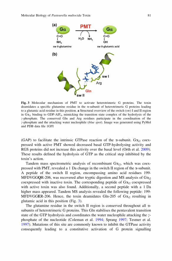
Tandem mass spectrometric analysis of recombinant Gai2, which was coex-pressed with PMT, revealed a 1 Da change in the switch II region of the a-subunit.A peptide of the switch II region, encompassing amino acid residues 199-MFDVGGQR-206, was recovered after tryptic digestion and MS analysis of Gai2
coexpressed with inactive toxin. The corresponding peptide of Gai2 coexpressedwith active toxin was also found. Additionally, a second peptide with a 1 Dahigher mass appeared. Tandem MS analysis revealed the following peptide: 199-MFDVGGER-206. Hence, the toxin deamidates Gln-205 of Gai2 resulting inglutamic acid in this position (Fig. 3).
The glutamine residue in the switch II region is conserved throughout all a-subunits of heterotrimeric G proteins. This Gln stabilises the pentavalent transitionstate of the GTP hydrolysis and coordinates the water nucleophile attacking the c-phosphate of the nucleotide (Coleman et al. 1994; Sprang 1997; Tesmer et al.1997). Mutations of this site are commonly known to inhibit the GTPase activityconsequently leading to a constitutive activation of G protein signalling
Fig. 3 Molecular mechanism of PMT to activate heterotrimeric G proteins. The toxindeamidates a specific glutamine residue in the a-subunit of heterotrimeric G proteins leadingto a glutamic acid residue in this position. a Structural overview of the switch (sw) I and II regionin Gai1 binding to GDP-AlF4
- , mimicking the transition state complex of the hydrolysis of thec-phosphate. The conserved Gln and Arg residues participate in the coordination of thec-phosphate and the attacking water nucleophile (blue spot). Image was generated using PyMoland PDB data file 1GFI
Molecular Biology of Pasteurella multocida Toxin 81
(De Vivo et al. 1992). This is true not only for the a-subunits of heterotrimeric Gproteins, but also for small GTP-binding proteins of the Ras superfamily (Bourneet al. 1989). Another important aspect of this type of mutation of G proteins is theirfrequent finding in mammalian tumours. As known for the transforming Rasmutation at position Gln-61, mutations of the a-subunit of heterotrimeric G pro-teins, causing inhibition of the GTPase activity, were described as transformingoncogenes and were found in diverse types of tumours (Kalinec et al. 1992;Radhika and Dhanasekaran 2001). For example, mutations of Gai2 at position Gln-205 have been observed in pituitary adenomas and mutations of Gln-209 of Gaq
were found in melanoma of the uvea and blue naevi (Van Raamsdonk et al. 2008;Williamson et al. 1995).
To further verify that an exchange of the conserved Gln to Glu, as catalysed byPMT, blocks GTP hydrolysis, the mutation was introduced in Gai2 (Gln-205) andGaq (Gln-209). Both mutations constitutively activated the G proteins leading toan inhibition of adenylyl cyclase or stimulation of PLCb1, respectively (Orth et al.2009). All these data show that PMT deamidates an essential Gln residue in theswitch II region of the a-subunit of heterotrimeric G proteins. The resulting Gluresidue is not capable of hydrolysing the bound nucleotide, leading to a consti-tutive active phenotype of the G protein.
Another amino acid residue contributing to the c-phosphate coordination ofGTP is the Arg in the switch I region. Interestingly, this Arg in the switch I regionof Gas is the target site for ADP-ribosylation by cholera toxin (Dop Van et al.1984; Freissmuth and Gilman 1989), a key feature of cholera pathogenesis.Accordingly, the GTPase activity of CTx-modified Gas is blocked and the aden-ylyl cyclase is stimulated by constitutively active Gas (Cassel and Selinger 1977).
4 Substrate Specificity
As mentioned above, the PMT-targeted Gln in the switch II region of heterotri-meric G protein a-subunits is conserved throughout all members of G proteins.However, the toxin-induced activation has been verified for only a subset ofheterotrimeric G proteins. Initially, the activation of heterotrimeric G proteins byPMT was studied indirectly by measurement of the specific downstream signalling
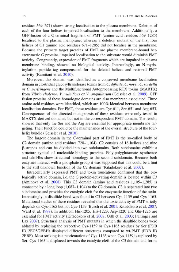
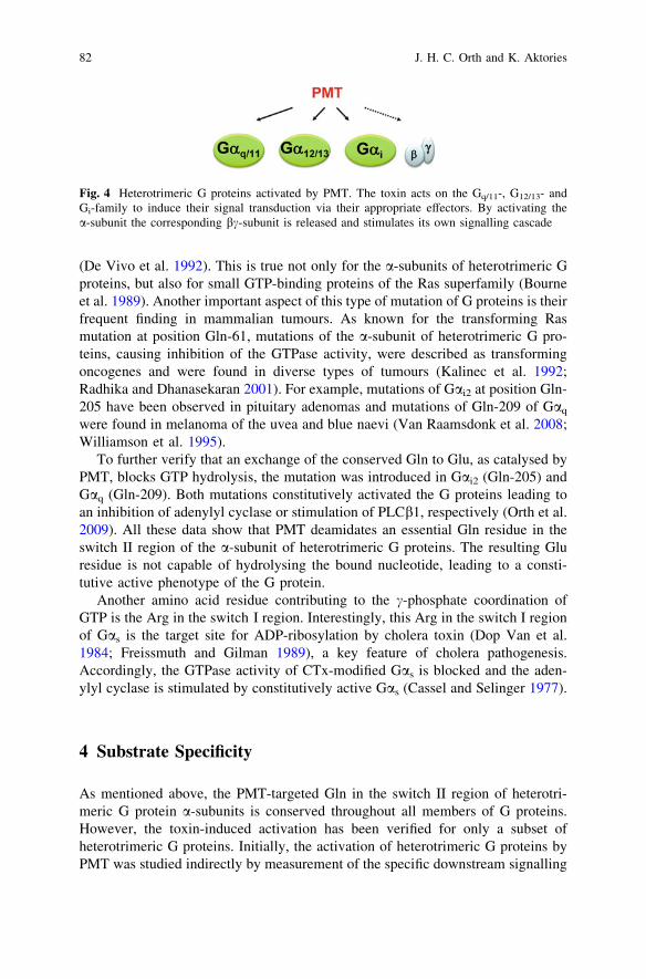
Fig. 4 Heterotrimeric G proteins activated by PMT. The toxin acts on the Gq/11-, G12/13- andGi-family to induce their signal transduction via their appropriate effectors. By activating thea-subunit the corresponding bc-subunit is released and stimulates its own signalling cascade
82 J. H. C. Orth and K. Aktories
of the respective G proteins (Higgins et al. 1992; Rozengurt et al. 1990; Staddonet al. 1990, 1991). By different methods, members of the Gaq-, Ga13- and Gai-family have been identified as substrates of the toxin. Identification of the primarymolecular mode of action of PMT as a deamidation reaction allows the directstudy of the covalent modification of toxin-targeted G proteins (Fig. 4).
Most importantly, the deamidation can be verified by mass spectrometricanalysis of recombinant protein for Gai2. Moreover, the shift of the isoelectricpoint (pI), resulting from the deamidation of the Gln to the more acidic Glu, isdetectable by 2D-gel electrophoresis. For example, a change of 0.07 units wascalculated and verified for Gai2 and Gai1, which were recovered from PMT-treatedcells (Orth et al. 2009).
Gaq is a well-known target of PMT (Wilson et al. 1997). Several biochemicaldata support this view. First, as mentioned above, native gel electrophoresisdemonstrates a shift of Gaq after PMT intoxication, indicating a change of the pIinduced by deamidation (Orth et al. 2009). More directly, deamidation of Gaq wasverified by a monoclonal antibody, which specifically detects the switch II regionof Gaq covering the deamidated Gln-209 (i.e. Glu-209) (Kamitani et al. 2011).Because the monoclonal antibody discriminates perfectly between unaffected Gproteins and PMT-deamidated G proteins, it is a useful tool to verify the toxin’saction at the G protein level. This antibody was employed to answer the puzzle ofPMT substrate specificity, which mystified studies with the toxin for years. Thus,several previous studies using mouse embryonic fibroblasts deficient for Gaq, Ga11
or both, showed that PMT-induced Gaq/11-dependent signalling to the PLCb1exclusively via Gaq but not via Ga11 (Orth et al. 2004; Zywietz et al. 2001). Thesefindings suggested that Ga11 was not modified by PMT, although the switch IIregion shares high homology between all heterotrimeric G proteins and is evenidentical between Gaq and Ga11 (Orth et al. 2004; Zywietz et al. 2001). However,the new findings obtained by deamidation-specific antibody and now confirmed byMS analysis in our laboratory (J.H.C. Orth and K. Aktories, unpublished data)indicate that both Gaq and Ga11 are deamidated by PMT. These results suggestthat differences in toxin-activated Gaq/11-signalling are not based on the toxin-substrate interaction per se, but possibly on the interaction of the deamidated Gproteins (Gaq and Ga11) with their effectors.
As for Gaq/11, cellular effects or signal transduction events were utilised todemonstrate PMT-induced activation of the Ga12/13 family. A common effector ofGa12/13 and Gaq/11 is the small GTPase RhoA (Vogt et al. 2003). RhoGEF pro-teins, which can be exclusively activated by Ga12/13 (p115RhoGEF), Gaq/11
(p63RhoGEF) or by both (LARG) connect the heterotrimeric G proteins with thesmall G protein RhoA (Booden et al. 2002; Hart et al. 1998; Kozasa et al. 1998;Lutz et al. 2005). Consequently, PMT treatment of cells leads to RhoA-dependentreorganisation of the actin cytoskeleton, stress fibre formation and downstream, toan increase in endothelial permeability (Dudet et al. 1996; Essler et al. 1998;Lacerda et al. 1996; Zywietz et al. 2001). Utilising mouse embryonic fibroblastsdeficient for Gaq/11 or Ga12/13, the involvement of Ga12/13 besides Gaq/11 in PMT-induced RhoA activation was demonstrated (Zywietz et al. 2001). Another method
Molecular Biology of Pasteurella multocida Toxin 83
of differentiating between Gaq/11- and Ga12/13-mediated signalling is the cyclicpeptide YM-254890, which is a specific inhibitor of Gaq/11 signalling (Takasakiet al. 2004). Using both approaches (e.g., genetic knock-out and YM-254890),the dissection of PMT-induced G protein signalling is possible. Most convincingly,the rescue of Ga13 in Ga12/13-deficient cells treated with the Gaq/11 inhibitorreconstitutes toxin-induced RhoA activation (Orth et al. 2005).
Taken together, the activation of heterotrimeric G proteins by PMT is dem-onstrated for Gai, Gaq and Ga13 by measuring G protein signalling. Studies on theG protein itself show deamidation of Gai1, Gai2 and Gaq by a deamidation-triggered change of the pI or Gaq and Ga11 by recognition by a specific antibody.The most stringent evidence for PMT-induced activation is MS analysis. Unfor-tunately, up to now this has been described only for Gai2.
5 G Protein Prerequisites for PMT Action
5.1 Role of Gbc
Heterotrimeric G proteins consist of the nucleotide-binding a-subunit and thenon-dissociable bc-subunit (Gilman 1987; Oldham et al. 2006; Sprang 1997).After GPCR-induced activation and subsequent dissociation of Ga from Gbc,both parts induce downstream signalling. Therefore, various questions ariseconcerning PMT action and Gbc: (i) Does PMT induce dissociation of the het-erotrimeric complex? (ii) Is Gbc signalling stimulated by PMT? (iii) Does thecycling between heterotrimeric complex and the dissociated form influence theactivation by PMT?
Classical studies have shown that the heterotrimeric complex and not the freea-subunit of Gaibc is the preferred substrate of PTx, allowing the determination ofthe formation of the heterotrimeric complex by PTx-catalysed ADP-ribosylation(Katada et al. 1986). Treatment of mammalian cells with PMT as well as coex-pression of Gai2 with PMT leads to an inhibition of PTx-induced ADP-ribosyla-tion of Gai. Additionally, the binding of Gbc to Gai2, coexpressed with activetoxin, is reduced to the same extent as Gai2 is deamidated by PMT (*50%) (Orthet al. 2008, 2009). Therefore, a dissociation of the heterotrimeric complex by PMTis likely.
PMT-induced activation of Gbc signalling is most convincingly demonstratedby determination of the activity of PI3Kc (Preuss et al. 2009). PI3Kc is anestablished effector of bc-subunits leading to PIP3 formation (Maier et al. 1999;Stephens et al. 1997). Therefore, PMT induces the translocation of a GFP sensorprotein (GFP-Grp1PH), which interacts with PIP3, to the plasma membrane (Preusset al. 2009). Moreover, using scavengers of Gbc, like GRK2-CT (G-protein-coupled-receptor kinase 2 C-terminus) (Wu et al. 1998), the direct involvement ofGbc in PMT-induced PI3Kc activation can be demonstrated.
84 J. H. C. Orth and K. Aktories
Sequestration of bc-subunits not only blocks Gbc downstream signalling, butalso activation of Ga-dependent signalling by PMT. Overexpression of GRK2-CTor phosducin, both proteins that sequester Gbc (Hawes et al. 1994; Wu et al. 1998),strongly reduces PMT activation of Ga-dependent signalling (Preuss et al. 2009).Moreover, two mutants of Gaq (I25A/E26A and G208A) were utilised to study therole of Gbc in more detail. The mutants manipulate the interaction of Ga withGbc. The double mutant of Gaq (I25A/E26A) does not bind Gbc, while Gaq
G208A
binds with increased affinity to Gbc (Jetzt et al. 2003; Lee et al. 1992). Becauseboth mutants inhibit PMT-induced Gaq activation, it is likely that the bindingcapability of Ga to Gbc itself is not important, but rather the cycling of theheterotrimeric complex, e.g. the association and dissociation of Gbc. Therefore, ithas been suggested that during cycling of the G protein a favourable structureserves as target for the toxin (Preuss et al. 2009). Furthermore, Kamitani et al.tested in an in vitro system whether PMT prefers the monomeric Ga or the het-erotrimeric complex Gabc for deamidation (Kamitani et al. 2011). These studiesshowed that both states are recognised by the toxin, but the monomeric a-subunitis two orders of magnitude less sensitive than the heterotrimeric complex. Thus,the presence of Gbc at least enhances PMT action. On the other hand, toxin-induced deamidation during coexpression in E. coli occurs without Gbc (Orth et al.2009), indicating that Gbc is not absolutely required for modification of the a-subunit by PMT.
5.2 PMT-Induced Activation of G Proteins is Independentof GPCR Interaction
The interaction, i.e. activation of heterotrimeric G proteins by GPCR, dependsmainly on the C-terminal amino acid residues of the Ga protein. The last fiveamino acids are essential for interaction and determine the specificity of G protein-GPCR coupling (Conklin et al. 1993; Hamm 1998; Parekh 2006). On the basis ofGaq it was studied whether PMT-induced activation depends on any receptorinteraction (Orth et al. 2007). Therefore, G protein constructs, which cannot coupleto GPCRs or G protein-receptor fusion proteins, were tested for PMT-inducedactivation. As expected, the C-terminal deletion mutant of Gaq was not stimulatedby a Gq-coupling receptor. In contrast, the toxin was still able to stimulate thisdeletion mutant, indicating that the function of PMT is independent of the couplingof the G protein with GPCRs. Vice versa, it was tested whether the toxin alsoactivates an a1b-adrenoceptor-Gaq chimera. This chimera was also stimulated bythe toxin, supporting the view that PMT acts completely independently of anyreceptor interaction (Orth et al. 2007).
In agreement with these results, it was shown that PTx does not block PMT-induced Gai activation (Orth et al. 2008). PTx ADP-ribosylates a Cys residue inthe C-terminus of Gai/o proteins and inhibits interaction of the G protein with the
Molecular Biology of Pasteurella multocida Toxin 85
receptor (Katada et al. 1982; Murayama et al. 1983). Even after PTx intoxication,PMT is able to stimulate Gai leading to an inhibition of the adenylyl cyclase.
6 PMT and Cytotoxic Necrotizing Factors
PMT belongs to a large group of deamidating toxins and/effectors, which causemajor pathophysiological alterations of target cells by removing the amide func-tional group from a specific glutamine residue of the targeted protein substrate. Whilethe targets of PMT are the a-subunits of heterotrimeric G proteins, the cytotoxicnecrotizing factors (CNF) 1–3 from E. coli and CNFY from Y. pseudotuberculosisdeamidate small GTPases of the Rho family (Flatau et al. 1997; Hoffmann et al. 2004;Hoffmann and Schmidt 2004; Schmidt et al. 1997; Stoll et al. 2009). It is fascinatingthat CNFs target the functionally equivalent glutamine residue (e.g., Gln-61 of Racand Cdc42 or Gln-63 of Rho) of GTPases, resulting in inhibition GTP hydrolysis andconstitutive activation of the small G proteins as known for PMT and heterotrimericG proteins (Aktories 2011).
PMT and CNFs share significant amino acid sequence homology in theN-terminal receptor binding and translocation domain (Lemichez et al. 1997;
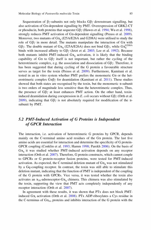
Fig. 5 Comparison of thecatalytic domains of PMTand CNF1. Crystal structuresof the catalytic activedomains of PMT and CNF1.Catalytic triads (blue sticks)of PMT and CNF1 areindicated in the folds. Imageswere generated using PyMoland PDB data files 2EC5(C-PMTC1159S) and 1HQO(CNF1)
86 J. H. C. Orth and K. Aktories
Pullinger et al. 2001). However, in contrast to the functionally related eukaryotictargets and type of reaction, no homology is observed in the primary sequence orthe tertiary structure of the catalytic active deamidase domain. Moreover,the topography of the catalytic centres of PMT and CNF1 are different (Fig. 5).The catalytic triads of PMT and CNF share the catalytic active Cys and Hisresidues. In the case of PMT, the third catalytic active residue is an Asp and forCNF it is a Val. Superimposing the active His residues revealed that the catalytictriads of PMT and CNF do not match (Fig. 1c). All structural data together indicatethat the molecular mechanism of PMT and CNFs to activate G proteins wasindependently developed during evolution.
7 Outlook
For more than 20 years after the discovery of PMT, its molecular mode of actionremained enigmatic. With the elucidation of the toxin as a deamidase, a new era ofPMT research has developed. There are still important questions to answer. Theprecise structural determinants of substrate (G protein) recognition by the toxin arenot known. The uptake of the toxin is still not well understood. Are domains 1 and/or domain 2 of the Trojan horse of the C-terminal part of PMT involved in uptakeor processing? The answers to these important questions will help to furtherunderstand the molecular biology of the toxin. Another important area which hasstill to be studied is the functional consequence of the actions of the toxin,especially with respect to its pathophysiological role and mechanism in disease.What are the precise pathophysiological steps finally leading to the sequelae ofbone destruction of the nose? Thus, we have much to learn about PMT.
Acknowledgments Work of the authors was supported by the German Research Foundation(DFG) through Collaborative Research Centre (SFB) 746 and the Cluster of Excellence 294BIOSS.
References
Aktories K (2011) Bacterial protein toxins that modify host regulatory GTPases. Nat RevMicrobiol 9:487–498
Aktories K, Jakobs KH (1981) Epinephrine inhibits adenylate cyclase and stimulates a GTPase inhuman platelet membranes via a-adrenoceptors. FEBS Lett 130(2):235–238
Aminova LR, Luo S, Bannai Y, Ho M, Wilson BA (2008) The C3 domain of Pasteurellamultocida toxin is the minimal domain responsible for activation of Gq-dependent calciumand mitogenic signaling. Protein Sci 17:1–5
Baldwin MR, Lakey JH, Lax AJ (2004) Identification and characterization of the Pasteurellamultocida toxin translocation domain. Mol Microbiol 54:239–250
Birnbaumer L (2007a) Expansion of signal transduction by G proteins. The second 15 years or so:from 3 to 16 alpha subunits plus betagamma dimers. Biochem Biophys Acta 1768:772–793
Molecular Biology of Pasteurella multocida Toxin 87

Birnbaumer L (2007b) The discovery of signal transduction by G proteins: a personal account andan overview of the initial findings and contributions that led to our present understanding.Biochem Biophys Acta 1768:756–771
Bokoch GM, Katada T, Northup JK, Hewlett EL, Gilman AG (1983) Identification of thepredominant substrate for ADP-ribosylation by islet activating protein. J Biol Chem258:2072–2075
Booden MA, Siderovski DP, Der CJ (2002) Leukemia-associated Rho guanine nucleotideexchange factor promotes G alpha q-coupled activation of RhoA. Mol Cell Biol 22:4053–4061
Bourne HR, Landis CA, Masters SB (1989) Hydrolysis of GTP by the alpha-chain of Gs andother GTP binding proteins. Proteins 6:222–230
Bourne HR, Sanders DA, McCormick F (1990) The GTPase superfamily: a conserved switch fordiverse cell functions. Nature 348:125–132
Brothers MC, Ho M, Maharjan R, Clemons NC, Bannai Y, Waites MA, Faulkner MJ,Kuhlenschmidt TB, Kuhlenschmidt MS, Blanke SR, Rienstra CM, Wilson BA (2011)Membrane interaction of Pasteurella multocida toxin involves sphingomyelin. FEBS J278(23):4633–4648
Busch C, Orth J, Djouder N, Aktories K (2001) Biological activity of a C-terminal fragment ofPasteurella multocida toxin. Infect Immun 69:3628–3634
Buys WEC, Smith HE, Kamps AMIE, Smits MA (1990) Sequence of the dermonecrotic toxin ofPasteurella multocida ssp. multocida. Nucl Acids Res 18:2815–2816
Cassel D, Selinger Z (1977) Mechanism of adenylate cyclase activation by cholera toxin:inhibition of GTP hydrolysis at the regulatory site. Proc Natl Acad Sci U S A 74:3307–3311
Coleman DE, Berghuis AM, Lee E, Linder ME, Gilman AG, Sprang SR (1994) Structuresof active conformations of Gi alpha 1 and the mechanism of GTP hydrolysis. Science 265:1405–1412
Conklin BR, Farfel Z, Lustig KD, Julius D, Bourne HR (1993) Substitution of three amino acidsswitches receptor specificity of Gq alpha to that of Gi alpha. Nature 363:274–276
De Vivo M, Chen J, Codina J, Iyengar R (1992) Enhanced phospholipase C stimulation andtransformation in NIH-3T3 cells expressing Q209LGq-alpha-subunits. J Biol Chem 267:18263–18266
Dop Van C, Tsubokawa M, Bourne HR, Ramachandran J (1984) Amino acid sequence of retinaltransducin at the site ADP- ribosylated by cholera toxin. J Biol Chem 259:696–698
Dudet LI, Chailler P, Dubreuil D, Martineau-Doize B (1996) Pasteurella multocida toxinstimulates mitogenesis and cytoskeleton reorganization in swiss 3T3 fibroblasts. J CellPhysiol 168:173–182
Essler M, Hermann K, Amano M, Kaibuchi K, Heesemann J, Weber PC, Aepfelbacher M (1998)Pasteurella multocida toxin increases endothelial permeability via rho kinase and myosinlight chain phosphatase. J Immunol 161:5640–5646
Flatau G, Lemichez E, Gauthier M, Chardin P, Paris S, Fiorentini C, Boquet P (1997)Toxin-induced activation of the G protein p21 Rho by deamidation of glutamine. Nature387:729–733
Frandsen PL, Foged NT, Petersen SK, Bording A (1991) Characterization of toxin from differentstrains of Pasteurella multocida serotype A and D. Zentralbl Veterinarmed B 38:345–352
Freissmuth M, Gilman AG (1989) Mutations of GS alpha designed to alter the reactivity of theprotein with bacterial toxins. Substitutions at ARG187 result in loss of GTPase activity. J BiolChem 264:21907–21914
Geissler B, Bonebrake A, Sheahan KL, Walker ME, Satchell KJ (2009) Genetic determination ofessential residues of the Vibrio cholerae actin cross-linking domain reveals functionalsimilarity with glutamine synthetases. Mol Microbiol 73:858–868
Geissler B, Tungekar R, Satchell KJ (2010) Identification of a conserved membrane localizationdomain within numerous large bacterial protein toxins. Proc Natl Acad Sci U S A 107:5581–5586
88 J. H. C. Orth and K. Aktories

Gierschik P (1992) ADP-ribosylation of signal-transducing guanine nucleotide-binding proteinsby pertussis toxin. Curr Top Microbiol Immunol 175:69–98
Gilman AG (1987) G proteins: transducers of receptor-generated signals. Annu Rev Biochem56:615–649
Hamm HE (1998) The many faces of G protein signaling. J Biol Chem 273:669–672Hart MJ, Jiang X, Kozasa T, Roscoe W, Singer WD, Gilman AG, Sternweis PC, Bollag G (1998)
Direct stimulation of the guanine nucleotide exchange activity of p115 RhoGEF by Ga13.Science 280:2112–2114
Hawes BE, Touhara K, Kurose H, Lefkowitz RJ, Inglese J (1994) Determination of the G betagamma-binding domain of phosducin. A regulatable modulator of G beta gamma signaling.J Biol Chem 269:29825–29830
Hepler JR, Gilman AG (1992) G proteins. Trends Biochem Sci 17:383–387Higgins TE, Murphy AC, Staddon JM, Lax AJ, Rozengurt E (1992) Pasteurella multocida toxin
is a potent inducer of anchorage-independent cell growth. Proc Natl Acad Sci U S A 89:4240–4244
Hoffmann C, Schmidt G (2004) CNF and DNT. Rev Physiol Biochem Pharmacol 152:49–63Hoffmann C, Pop M, Leemhuis J, Schirmer J, Aktories K, Schmidt G (2004) The Yersinia
pseudotuberculosis cytotoxic necrotizing factor (CNFY) selectively activates RhoA. J BiolChem 279:16026–16032
Hollinger S, Hepler JR (2002) Cellular regulation of RGS proteins: modulators and integrators ofG protein signaling. Pharmacol Rev 54:527–559
Jetzt A, Howe JA, Horn MT, Maxwell E, Yin Z, Johnson D, Kumar CC (2003) Adenoviral-mediated expression of a kinase-dead mutant of Akt induces apoptosis selectively in tumorcells and suppresses tumor growth in mice. Cancer Res 63:6697–6706
Kalinec G, Nazarali AJ, Hermouet S, Xu N, Gutkind JS (1992) Mutated alpha subunit of the Gqprotein induces malignant transformation in NIH 3T3 cells. Mol Cell Biol 12:4687–4693
Kamitani S, Kitadokoro K, Miyazawa M, Toshima H, Fukui A, Abe H, Miyake M, Horiguchi Y(2010) Characterization of the membrane-targeting C1 domain in Pasteurella multocidatoxin. J Biol Chem 285:25467–25475
Kamitani S, Ao S, Toshima H, Tachibana T, Hashimoto M, Kitadokoro K, Fukui-Miyazaki A,Abe H, Horiguchi Y (2011) Enzymatic actions of Pasteurella multocida toxin detected bymonoclonal antibodies recognizing the deamidated alpha subunit of the heterotrimericGTPase G(q). FEBS J. 278:2702–2712
Katada T, Ui M (1982) Direct modification of the membrane adenylate cyclase system by islet-activating protein due to ADP-ribosylation of a membrane protein. Proc Natl Acad Sci U S A79:3129–3133
Katada T, Oinuma M, Ui M (1986) Two guanine nucleotide-binding proteins in rat brain servingas the specific substrate of islet-activating protein, pertussis toxin. Interaction of the alpha-subunits with beta gamma-subunits in development of their biological activities. J Biol Chem261:8182–8191
Kitadokoro K, Kamitani S, Miyazawa M, Hanajima-Ozawa M, Fukui A, Miyake M, Horiguchi Y(2007) Crystal structures reveal a thiol protease-like catalytic triad in the C-terminal region ofPasteurella multocida toxin. Proc Natl Acad Sci U S A 104:5139–5144
Kozasa T, Jiang X, Hart MJ, Sternweis PM, Singer WD, Gilman AG, Bollag G, Sternweis PC(1998) p115 RhoGEF, a GTPase activating protein for Galpha12 and Galpha13. Science280:2109–2111
Lacerda HM, Lax AJ, Rozengurt E (1996) Pasteurella multocida toxin, a potent intracellularlyacting mitogen, induces p125FAK and paxillin tyrosine phosphorylation, actin stress fiberformation, and focal contact assembly in Swiss 3T3 cells. J Biol Chem 271:439–445
Lee E, Taussig R, Gilman AG (1992) The G226A mutant of Gsa highlights the requirement fordissociation of G protein subunits. J Biol Chem 267:1212–1218
Lemichez E, Flatau G, Bruzzone M, Boquet P, Gauthier M (1997) Molecular localization of theEscherichia coli cytotoxic necrotizing factor CNF1 cell-binding and catalytic domains.Mol Microbiol 24:1061–1070
Molecular Biology of Pasteurella multocida Toxin 89
Lutz S, Freichel-Blomquist A, Yang Y, Rumenapp U, Jakobs KH, Schmidt M, Wieland T (2005)The guanine nucleotide exchange factor p63RhoGEF, a specific link between Gq/11-coupledreceptor signaling and RhoA. J Biol Chem 280:11134–11139
Maier U, Babich A, Nurnberg B (1999) Roles of non-catalytic subunits in gbetagamma-inducedactivation of class I phosphoinositide 3-kinase isoforms beta and gamma. J Biol Chem 274:29311–29317
Miyazawa M, Kitadokoro K, Kamitani S, Shime H, Horiguchi Y (2006) Crystallization andpreliminary crystallographic studies of the Pasteurella multocida toxin catalytic domain. ActaCrystallograph Sect F Struct Biol Cryst Commun 62:906–908
Murayama T, Ui M (1983) Loss of the inhibitory function of the guanine nucleotide regulatorycomponent of adenylate cyclase due to its ADP ribosylation by islet-activating protein,pertussis toxin, in adipocyte membranes. J Biol Chem 258(5):3319–3326
Northup JK, Sternweis PC, Smigel MDC, Ross EM, Gilman AG (1980) Purification of theregulatory component of adenylate cyclase. Proc Natl Acad Sci U S A 77:6516–6520
Nürnberg B (1997) Pertussis toxin as a cell biological tool. In: Aktories K (ed) Bacterial toxins—tools in cell biology and pharmacology. Chapman & Hall, Weinheim
Oldham WM, Hamm HE (2006) Structural basis of function in heterotrimeric G proteins. Q RevBiophys 39:117–166
Oldham WM, Hamm HE (2008) Heterotrimeric G protein activation by G-protein-coupledreceptors. Nat Rev Mol Cell Biol 9:60–71
Orth JH, Blöcker D, Aktories K (2003) His1205 and His 1223 are essential for the activity of themitogenic Pasteurella multocida toxin. Biochemistry 42:4971–4977
Orth JH, Lang S, Aktories K (2004) Action of Pasteurella multocida toxin depends on the helicaldomain of Galphaq. J Biol Chem 279:34150–34155
Orth JH, Lang S, Taniguchi M, Aktories K (2005) Pasteurella multocida toxin-induced activationof RhoA is mediated via two families of G{alpha} proteins, G{alpha}q and G{alpha}12/13.J Biol Chem 280:36701–36707
Orth JH, Lang S, Preuss I, Milligan G, Aktories K (2007) Action of Pasteurella multocida toxinon Galpha(q) is persistent and independent of interaction with G-protein-coupled receptors.Cell Signal 19:2174–2182
Orth JH, Fester I, Preuss I, Agnoletto L, Wilson BA, Aktories K (2008) Activation of Galphai andsubsequent uncoupling of receptor-Galphai signaling by Pasteurella multocida toxin. J BiolChem 283:23288–23294
Orth JH, Preuss I, Fester I, Schlosser A, Wilson BA, Aktories K (2009) Pasteurella multocidatoxin activation of heterotrimeric G proteins by deamidation. Proc Natl Acad Sci U S A106:7179–7184
Parekh AB (2006) On the activation mechanism of store-operated calcium channels. PflugersArch 453:303–311
Peters PJ, Hsu VW, Ooi CE, Finazzi D, Teal SB, Oorschot V, Donaldson JG, Klausner RD (1995)Overexpression of wild-type and mutant ARF1 and ARF6: distinct perturbations ofnonoverlapping membrane compartments. J Cell Biol 128:1003–1017
Petersen SK (1990) The complete nucleotide sequence of the Pasteurella multocida toxin geneand evidence for a transcriptional repressor. TxaR Mol Microbiol 4:821–830
Pettit RK, Ackermann MR, Rimler RB (1993) Receptor-mediated binding of Pasteurellamultocida dermonecrotic toxin to canine osteosarcoma and monkey kidney (vero) cells. LaborInvest 69:94–100
Preuss I, Kurig B, Nürnberg B, Orth JH, Aktories K (2009) Pasteurella multocida toxin activatesGbetagamma dimers of heterotrimeric G proteins. Cell Signal 21:551–558
Pullinger GD, Lax AJ (2007) Histidine residues at the active site of the Pasteurella multocidatoxin. Open Biochem J 1:7–11
Pullinger GD, Sowdhamini R, Lax AJ (2001) Localization of functional domains of the mitogenictoxin of Pasteurella multocida. Infect Immun 69:7839–7850
Radhika V, Dhanasekaran N (2001) Transforming G proteins. Oncogene 20:1607–1614
90 J. H. C. Orth and K. Aktories
Rasmussen SG, DeVree BT, Zou Y, Kruse AC, Chung KY, Kobilka TS, Thian FS, Chae PS,Pardon E, Calinski D, Mathiesen JM, Shah ST, Lyons JA, Caffrey M, Gellman SH, Steyaert J,Skiniotis G, Weis WI, Sunahara RK, Kobilka BK (2011) Crystal structure of the beta2adrenergic receptor-Gs protein complex. Nature 477:549–555
Repella TL, Ho M, Chong TPM, Bannai Y, Wilson BA (2011) Arf6-dependent intracellulartrafficking of Pasteurella multocida toxin and pH-dependent translocation from lateendosomes. Toxins 3:218–241
Riobo NA, Manning DR (2005) Receptors coupled to heterotrimeric G proteins of the G12family. Trends Pharmacol Sci 26:146–154
Ross EM, Wilkie TM (2000) GTPase-activating proteins for heterotrimeric G proteins: regulatorsof G protein signaling (RGS) and RGS-like proteins. Annu Rev Biochem 69:795–827
Rozengurt E, Higgins T, Chanter N, Lax AJ, Staddon JM (1990) Pasteurella multocida toxin:potent mitogen for cultured fibroblasts. Proc Natl Acad Sci U S A 87:123–127
Schmidt G, Sehr P, Wilm M, Selzer J, Mann M, Aktories K (1997) Gln63 of Rho is deamidatedby Escherichia coli cytotoxic necrotizing factor 1. Nature 387:725–729
Smyth MG, Pickersgill RW, Lax AJ (1995) The potent mitogen Pasteurella multocida toxin ishighly resistant to proteolysis but becomes susceptible at lysosomal pH. FEBS Lett 360:62–66
Smyth MG, Sumner IG, Lax AJ (1999) Reduced pH causes structural changes in the potentmitogenic toxin of Pasteurella multocida. FEMS Microbiol Lett 180:15–20
Sprang SR (1997) G protein mechanisms: insights from structural analysis. Annu Rev Biochem66:639–678
Staddon JM, Chanter N, Lax AJ, Higgins TE, Rozengurt E (1990) Pasteurella Multocida toxin, apotent mitogen, stimulates protein kinase C-dependent and -independent protein phosphor-ylation in swiss 3T3 cells. J Biol Chem 265:11841–11848
Staddon JM, Barker CJ, Murphy AC, Chanter N, Lax AJ, Michell RH, Rozengurt E (1991)Pasteurella multocida toxin, a potent mitogen, increases inositol 1,4,5-triphosphate andmobilizes Ca2+ in swiss 3T3 cells. J Biol Chem 266:4840–4847
Stephens LR, Eguinoa A, Erdjument-Bromage H, Lui M, Cooke F, Coadwell J, Smrcka AS,Thelen M, Cadwallader K, Tempst P, Hawkins PT (1997) The G beta gamma sensitivity of aPI3 K is dependent upon a tightly associated adaptor, p101. Cell 89:105–114
Stoll T, Markwirth G, Reipschlager S, Schmidt G (2009) A new member of a growing toxinfamily – Escherichia coli cytotoxic necrotizing factor 3 (CNF3). Toxicon 54:745–753
Takasaki J, Saito T, Taniguchi M, Kawasaki T, Moritani Y, Hayashi K, Kobori M (2004) A novelGalphaq/11-selective inhibitor. J Biol Chem 279:47438–47445
Taylor SJ, Smith JA, Exton JH (1990) Purification from bovine liver membranes of a guaninenucleotide-dependent activator of phosphoinositide-specific phospholipase C. Immunologicidentification as a novel G-protein alpha subunit. J Biol Chem 265:17150–17156
Tesmer JJ, Berman DM, Gilman AG, Sprang SR (1997) Structure of RGS4 bound to AlF4-activated G(i alpha1): stabilization of the transition state for GTP hydrolysis. Cell 89:251–261
Ui M (1984) Islet-activating protein, pertussis toxin: a probe for functions of the inhibitoryguanine nucleotide regulatory component of adenylate cyclase. Trendsss Pharmacol Sci5:277–279
Van Raamsdonk CD, Bezrookove V, Green G, Bauer J, Gaugler L, O’Brien JM, Simpson EM,Barsh GS, Bastian BC (2008) Frequent somatic mutations of GNAQ in uveal melanoma andblue naevi. Nature 457:599–602
Van Sandvig K, Deurs B (2005) Delivery into cells: lessons learned from plant and bacterialtoxins. Gene Ther 12:865–872
Vogt S, Grosse R, Schultz G, Offermanns S (2003) Receptor-dependent RhoA activation inG12/G13-deficient cells. J Biol Chem 278:28743–28749
Ward PN, Miles AJ, Sumner IG, Thomas LH, Lax AJ (1998) Activity of the mitogenicPasteurella multocida toxin requires an essential C-terminal residue. Infect Immun 66:5636–5642
Westfield GH, Rasmussen SG, Su M, Dutta S, DeVree BT, Chung KY, Calinski D, Velez-Ruiz G,Oleskie AN, Pardon E, Chae PS, Liu T, Li S, Woods VL Jr, Steyaert J, Kobilka BK, Sunahara
Molecular Biology of Pasteurella multocida Toxin 91
RK, Skiniotis G (2011) Structural flexibility of the G alpha s alpha-helical domain in thebeta2-adrenoceptor Gs complex. Proc Natl Acad Sci U S A 108:16086–16091
Williamson MP (1994) The structure and function of proline-rich regions in proteins. Biochem J297(Pt 2):249–260
Williamson EA, Ince PG, Harrison D, Kendall-Taylor P, Harris PE (1995) G-protein mutationsin human pituitary adrenocorticotrophic hormone-secreting adenomas. Eur J Clin Invest25:128–131
Wilson BA, Zhu X, Ho M, Lu L (1997) Pasteurella multocida toxin activates the inositoltriphosphate signaling pathway in Xenopus oocytes via Gqa-coupled phospholipase C-b1.J Biol Chem 272:1268–1275
Wu G, Benovic JL, Hildebrandt JD, Lanier SM (1998) Receptor docking sites for G-proteinbetagamma subunits. implications for signal regulation. J Biol Chem 273:7197–7200
Zywietz A, Gohla A, Schmelz M, Schultz G, Offermanns S (2001) Pleiotropic effects ofPasteurella multocida toxin are mediated by Gq-dependent and -independent mechanisms.Involvement of Gq but not G11. J Biol Chem 276:3840–3845
92 J. H. C. Orth and K. Aktories