molecular biology lecture 3 chapter 4 molecular cloning methods copyright © the mcgraw-hill...
TRANSCRIPT

Molecular Biology
Lecture 3
Chapter 4
Molecular Cloning Methods
Copyright © The McGraw-Hill Companies, Inc. Permission required for reproduction or display.
4-2
Lecture outline
• Gene cloning
• Restriction endonucleases
• Plasmids
pBR322
pUC
• Selection

4-3
Gene Cloning
• Introduce a foreign gene or piece of DNA into a suitable vector and inserting the recombinant molecule into a bacterial host
• Cloning can also be done in eukaryotic cells such as yeast
• One can then produce large quantities of the gene or piece of DNA in pure form

4-4
The Role of Restriction Endonucleases• Restriction endonucleases, first
discovered in the late 1960s, are named for preventing invasion by foreign DNA by cutting it into pieces
• These enzymes cut at sites within the foreign DNA instead of chewing from the ends
• By cutting DNA at specific sites they function as finely honed molecular knives

4-5
Restriction-Modification System• What prevents these enzymes
from cutting up the host DNA?– They are paired with methylases– Theses enzymes recognize, and
methylate the same site
• The sequence specific methylase and restriction endonuclease are called a restriction-modification system, R-M system
• Methylation protects DNA, after replication (the parental strand is already methylated)

4-6
Restriction Endonuclease Specificity
• A 6-bp cutter will yield DNA fragments averaging 4000-bp or 4 kilobases (4kb) in length
e.g. EcoR1 recognize the sequenceGAATTCCTTAAG
Probability of finding the sequence1/.25 X .25 X .25 X .25 X .25 X .25 = 4096

4-7
Restriction Endonuclease SpecificityRestriction endonucleases recognize a specific DNA sequence, cutting ONLY at that sequence
– These enzymes can recognize 4-bp, 6-bp, 8-bp sequences
– The frequency of cuts lessens when the recognition sequence is longer
4-8
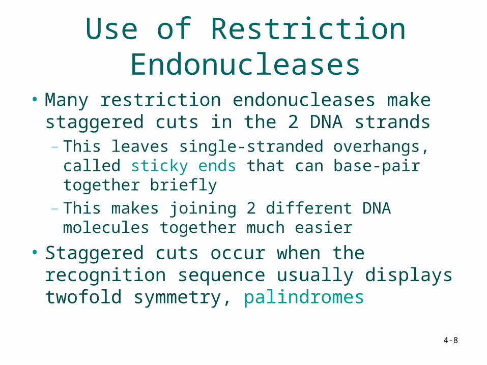
Use of Restriction Endonucleases
• Many restriction endonucleases make staggered cuts in the 2 DNA strands– This leaves single-stranded overhangs, called
sticky ends that can base-pair together briefly– This makes joining 2 different DNA molecules
together much easier
• Staggered cuts occur when the recognition sequence usually displays twofold symmetry, palindromes

4-9
Use of Restriction Endonucleases
• Example EcoR1– This enzyme leaves single-stranded
overhangs, called sticky ends that can base-pair together
GAATTC GAATTC
CTTAAG CTTAA G
+ EcoR1 = and

4-10
Use of Restriction Endonucleases
• Making a recombinant DNA molecule
in presence of DNA ligase and ATP
+
GAATTCCTTAAG
DNA ligase will synthesize the phosphodiester bonds

4-11
Summary
• Restriction endonucleases recognize specific sequences in DNA molecules and make cuts in both strands
• This allows very specific cutting of DNAs• The cuts in the two strands are frequently
staggered, so restriction enzymes can create sticky ends that help to link together 2 DNAs to form a recombinant DNA in vitro

4-12
Vectors• Vectors function as DNA carriers to allow
replication of recombinant DNAs
• Typical experiment uses 1 vector plus a piece of foreign DNA – Foreign DNA has no origin of replication, the
site where DNA replication begins– Depends on the vector for its replication
• There are 2 major classes of vectors:– Plasmids – Phages (not covered in this course)

4-13
First cloning Experiment Using Restriction Endonuclease
• An early experiment used EcoRI to cut 2 plasmidsSmall circular pieces of DNA independent of the host chromosome
• Each plasmid had 1 site for EcoRI– Cutting converted circular plasmids
into linear DNA with the same sticky ends
– The ends base pair• Some ends re-close• Others join the 2 pieces
• DNA ligase joins 2 pieces with covalent bonds

4-14
Plasmids As Vectors
• pBR plasmids were developed early but are rarely used today
• pUC series is similar to pBR– 40% of the DNA, including tetracycline
resistance has been deleted– Cloning sites are clustered together into one
area called the multiple cloning site (MCS)

4-15
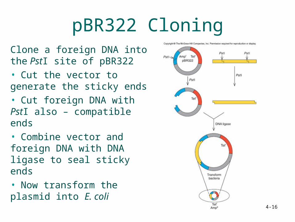
pBR322 Plasmid
• pBR322 illustrates cloning methods simply– Resistance for 2
antibiotics• Tetracycline• Ampicillin
– Origin of replication between the 2 resistance genes
– Only 1 site for several restriction enzymes
4-16
pBR322 CloningClone a foreign DNA into the PstI site of pBR322• Cut the vector to generate the sticky ends• Cut foreign DNA with PstI also – compatible ends• Combine vector and foreign DNA with DNA ligase to seal sticky ends• Now transform the plasmid into E. coli

4-17
Bacterial Transformation
• Traditional method involves incubating bacterial cells in concentrated calcium salt solution– The solution makes the cell membrane leaky,
permeable to the plasmid DNA
• Newer method uses high voltage to drive the DNA into the cells in process called electroporation

4-18
Screening Transformants
• Transformation produces bacteria with:– Religated plasmid– Religated insert– Recombinants
• Identify the recombinants using the antibiotic resistance– Grow cells with tetracycline so only cells with plasmid
grow, not foreign DNA only– Next, grow copies of the original colonies with
ampicillin which kills cells with plasmid including foreign DNA

4-19
Screening With Replica Plating• Replica plating transfers
clone copies from original tetracycline plate to a plate containing ampicillin
• A sterile velvet transfer tool can be used to transfer copies of the original colonies
• Desired colonies are those that do NOT grow on the new ampicillin plate

4-20
Directional Cloning
• Cut a plasmid with 2 restriction enzymes
• Clone in a piece of foreign DNA with 1 sticky end recognizing each enzyme
• The insert DNA is placed into the vector in only 1 orientation
• Vector religation is also prevented as the two restriction sites are incompatible
4-21
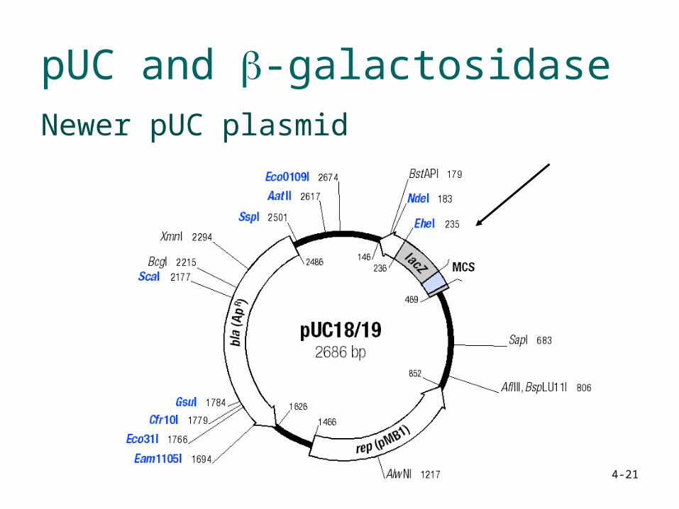
pUC and -galactosidaseNewer pUC plasmid

4-22
pUC and -galactosidaseNewer pUC plasmid
lacZ (-peptide)O
lac promoter
MCS
-Induced by lactose or IPTG-Under the control of the lac repressor-The MCS is a cluster of sequences recognized by restriction
endonucleases

4-23
pUC and -galactosidaseClones with foreign DNA in the MCS disrupt the
ability of the cells to make -galactosidase
-galactosidase is encoded by the lacZ gene of the lac operon
-galactosidase cleaves lactose and can also cleave the synthetic substrate X-gal.
Cleaved X-gal gives a blue coloration. You can monitor the activity of -galactosidase by looking at the blue coloration

4-24
pUC and -galactosidase-complementation
Plasmid contains part of the lacZ gene coding for the N-terminal extremity of the -galactosidase enzyme.
When expressed in E. coli lacZ- strain = no activity
Host bacterial strain contains a truncated lacZ gene encoding a polypeptide missing the N-terminal extremity
When expressed in E. coli = no activity

4-25
pUC and -galactosidase-complementation
When the plasmid is introduced in the bacterial strain containing the truncated enzyme, activity is recovered.
The two partial gene products can cooperate to form an active enzyme
(model on the blackboard)
4-26
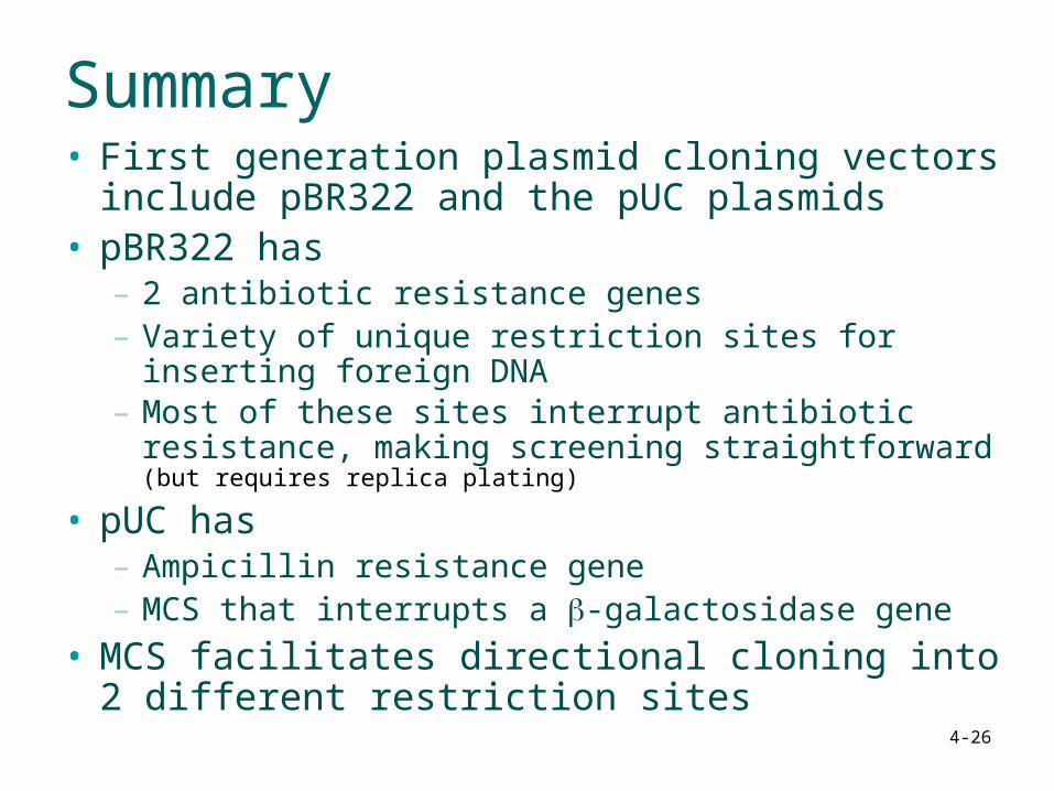
Summary• First generation plasmid cloning vectors include
pBR322 and the pUC plasmids• pBR322 has
– 2 antibiotic resistance genes – Variety of unique restriction sites for inserting foreign
DNA– Most of these sites interrupt antibiotic resistance,
making screening straightforward (but requires replica plating)
• pUC has– Ampicillin resistance gene– MCS that interrupts a -galactosidase gene
• MCS facilitates directional cloning into 2 different restriction sites

4-27