molecular bases of dna packaging in bacteria revealed by
TRANSCRIPT

HAL Id: hal-02352618https://hal.archives-ouvertes.fr/hal-02352618
Submitted on 23 Nov 2020
HAL is a multi-disciplinary open accessarchive for the deposit and dissemination of sci-entific research documents, whether they are pub-lished or not. The documents may come fromteaching and research institutions in France orabroad, or from public or private research centers.
L’archive ouverte pluridisciplinaire HAL, estdestinée au dépôt et à la diffusion de documentsscientifiques de niveau recherche, publiés ou non,émanant des établissements d’enseignement et derecherche français ou étrangers, des laboratoirespublics ou privés.
Molecular Basis of DNA Packaging in Bacteria Revealedby All-Atoms Molecular Dynamic Simulations: The case
of histone like proteins in Borrelia burgdoferi.Cécilia Hognon, Simon Garaude, Joanna Timmins, Christophe Chipot,
Francois Dehez, Antonio Monari
To cite this version:Cécilia Hognon, Simon Garaude, Joanna Timmins, Christophe Chipot, Francois Dehez, et al.. Molec-ular Basis of DNA Packaging in Bacteria Revealed by All-Atoms Molecular Dynamic Simulations: Thecase of histone like proteins in Borrelia burgdoferi.. Journal of Physical Chemistry Letters, AmericanChemical Society, 2019, �10.1021/acs.jpclett.9b02978�. �hal-02352618�

Molecular Bases of DNA Packaging in Bacteria Revealed by All-AtomMolecular Dynamics Simulations: The Case of Histone-Like Proteinsin Borrelia burgdorferiCecilia Hognon,† Simon Garaude,† Joanna Timmins,‡ Christophe Chipot,†,§,∥ Francois Dehez,*,†,∥
and Antonio Monari*,†
†Universite de Lorraine and CNRS, LPCT UMR 7019, F-54000 Nancy, France‡Universite Grenoble Alpes, CNRS, CEA, IBS, F-38000 Grenoble, France§Department of Physics, University of Illinois at UrbanaChampaign, 1110 West Green Street, Urbana, Illinois 61801, UnitedStates∥Laboratoire International Associe Centre National de la Recherche Scientifique et University of Illinois at Urbana−Champaign,54506 Vandoeuvre-les-Nancy Cedex, France
*S Supporting Information
ABSTRACT: DNA compaction is essential to ensure the packaging of the geneticmaterial in living cells and also plays a key role in the epigenetic regulation of geneexpression. In both humans and bacteria, DNA packaging is achieved by specific well-conserved proteins. Here, by means of all-atom molecular dynamics simulations,including the determination of relevant free-energy profiles, we rationalize the molecularbases for this remarkable process in bacteria, illustrating the crucial role played bypositively charged amino acids of a small histone-like protein. We also presentcompelling evidence that this histone-like protein alone can induce strong bending of aDNA duplex around its core domain, a process that requires overcoming a major free-energy barrier.
DNA packaging is essential for all living organisms to allowthe compaction of the long nucleic acid polymers into
the confined environment of bacterial cells or nuclei. Thecompaction of DNA is achieved by the action of two types ofproteins: (i) large enzymes that alter the topology of the DNA(relax or supercoil the DNA), including DNA topoisomerasesor DNA gyrases, and (ii) small, basic proteins such as histonesthat possess a high-density of positive charges on their surfaceto engage in strong electrostatic interactions with thenegatively charged DNA backbone.1−3 Interestingly, evenlimited mutations in such proteins are usually lethal to thecell, pointing out the crucial importance of DNA compaction.4
In addition to its space-saving role, compaction of DNA is alsoimportant to enhance the global stability of DNA5,6 and toregulate gene expression.7−11 Indeed, the transition from themore compact heterochromatin to euchromatin is known toincrease the accessibility of the genes to the promoting factorsand hence enhance gene expression.12−16 Chromatin remodel-ing is also finely tuned by complex cross-talks between DNAand histone epigenetic marks, such as methylation oracetylation.17,18
In eukaryotes, DNA packaging is largely achieved byhistones, whose complex of eight monomers, called nucleo-some, constitutes the basic unit of chromatin, and
chromosomes.19,20 Conversely, in bacteria in which genomeorganization is much simpler than in eukaryotes, a set ofproteins known as nucleoid-associated proteins (NAPs) orhistone-like proteins play a similar role in DNA pack-aging.21−26 There are different classes of histone-like proteins,the most common ones being HU27−32 and the integrationhost factor (IHF),33−39 whose combined action is necessary toinduce DNA compaction in Escherichia coli.40−45
In this Letter, we specifically focus on the behavior of thehistone-like protein, Hbb, from the pathogenic bacteriumBorrelia burgdorferi,46−48 known to be the causative agent ofLyme disease.49−51 Unlike E. coli, this organism does notpossess HU and IHF encoding genes but instead encodes foran HU variant, Hbb. Although the crystal structure of acomplex between Hbb and a 35-nucleotide double-strandedDNA oligomer was solved by Mouw and Rice46 and severalkey amino acids necessary to ensure Hbb’s biological functionshave been identified,46,47 a global picture, at atomistic scaleresolution, of its mechanism of action is still lacking andconstitutes the object of the present contribution.
Received: October 10, 2019Accepted: November 6, 2019Published: November 6, 2019
Letter
pubs.acs.org/JPCLCite This: J. Phys. Chem. Lett. 2019, 10, 7200−7207
© 2019 American Chemical Society 7200 DOI: 10.1021/acs.jpclett.9b02978J. Phys. Chem. Lett. 2019, 10, 7200−7207
Dow
nloa
ded
via
INIS
T-C
NR
S on
Dec
embe
r 5,
201
9 at
14:
42:4
4 (U
TC
).Se
e ht
tps:
//pub
s.ac
s.or
g/sh
arin
ggui
delin
es f
or o
ptio
ns o
n ho
w to
legi
timat
ely
shar
e pu
blis
hed
artic
les.
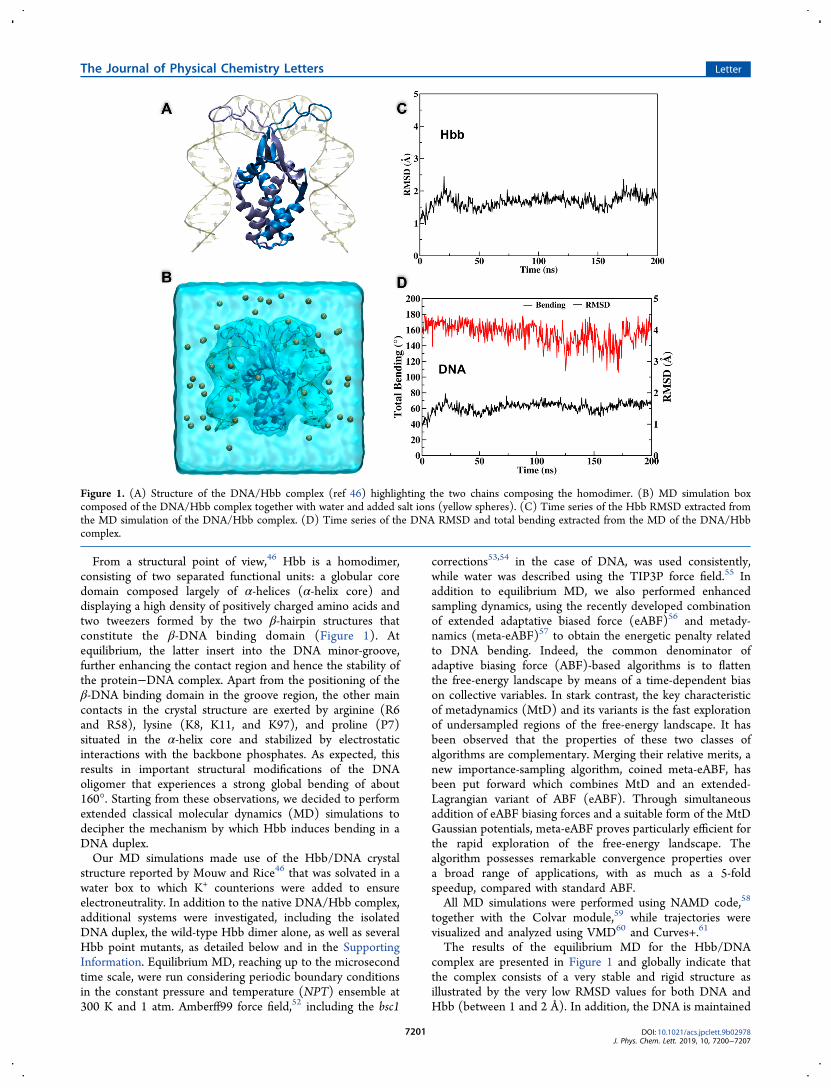
From a structural point of view,46 Hbb is a homodimer,consisting of two separated functional units: a globular coredomain composed largely of α-helices (α-helix core) anddisplaying a high density of positively charged amino acids andtwo tweezers formed by the two β-hairpin structures thatconstitute the β-DNA binding domain (Figure 1). Atequilibrium, the latter insert into the DNA minor-groove,further enhancing the contact region and hence the stability ofthe protein−DNA complex. Apart from the positioning of theβ-DNA binding domain in the groove region, the other maincontacts in the crystal structure are exerted by arginine (R6and R58), lysine (K8, K11, and K97), and proline (P7)situated in the α-helix core and stabilized by electrostaticinteractions with the backbone phosphates. As expected, thisresults in important structural modifications of the DNAoligomer that experiences a strong global bending of about160°. Starting from these observations, we decided to performextended classical molecular dynamics (MD) simulations todecipher the mechanism by which Hbb induces bending in aDNA duplex.Our MD simulations made use of the Hbb/DNA crystal
structure reported by Mouw and Rice46 that was solvated in awater box to which K+ counterions were added to ensureelectroneutrality. In addition to the native DNA/Hbb complex,additional systems were investigated, including the isolatedDNA duplex, the wild-type Hbb dimer alone, as well as severalHbb point mutants, as detailed below and in the SupportingInformation. Equilibrium MD, reaching up to the microsecondtime scale, were run considering periodic boundary conditionsin the constant pressure and temperature (NPT) ensemble at300 K and 1 atm. Amberff99 force field,52 including the bsc1
corrections53,54 in the case of DNA, was used consistently,while water was described using the TIP3P force field.55 Inaddition to equilibrium MD, we also performed enhancedsampling dynamics, using the recently developed combinationof extended adaptative biased force (eABF)56 and metady-namics (meta-eABF)57 to obtain the energetic penalty relatedto DNA bending. Indeed, the common denominator ofadaptive biasing force (ABF)-based algorithms is to flattenthe free-energy landscape by means of a time-dependent biason collective variables. In stark contrast, the key characteristicof metadynamics (MtD) and its variants is the fast explorationof undersampled regions of the free-energy landscape. It hasbeen observed that the properties of these two classes ofalgorithms are complementary. Merging their relative merits, anew importance-sampling algorithm, coined meta-eABF, hasbeen put forward which combines MtD and an extended-Lagrangian variant of ABF (eABF). Through simultaneousaddition of eABF biasing forces and a suitable form of the MtDGaussian potentials, meta-eABF proves particularly efficient forthe rapid exploration of the free-energy landscape. Thealgorithm possesses remarkable convergence properties overa broad range of applications, with as much as a 5-foldspeedup, compared with standard ABF.All MD simulations were performed using NAMD code,58
together with the Colvar module,59 while trajectories werevisualized and analyzed using VMD60 and Curves+.61
The results of the equilibrium MD for the Hbb/DNAcomplex are presented in Figure 1 and globally indicate thatthe complex consists of a very stable and rigid structure asillustrated by the very low RMSD values for both DNA andHbb (between 1 and 2 Å). In addition, the DNA is maintained
Figure 1. (A) Structure of the DNA/Hbb complex (ref 46) highlighting the two chains composing the homodimer. (B) MD simulation boxcomposed of the DNA/Hbb complex together with water and added salt ions (yellow spheres). (C) Time series of the Hbb RMSD extracted fromthe MD simulation of the DNA/Hbb complex. (D) Time series of the DNA RMSD and total bending extracted from the MD of the DNA/Hbbcomplex.
The Journal of Physical Chemistry Letters Letter
DOI: 10.1021/acs.jpclett.9b02978J. Phys. Chem. Lett. 2019, 10, 7200−7207
7201

in a highly bent configuration characterized by an averagebending value of ∼155°, oscillating marginally on the timescale of our MD simulations. In agreement with the literature,DNA bending is mainly maintained by salt bridges between thenegatively charged backbone of the DNA and a series ofpositively charged amino acids in Hbb (see the SupportingInformation). As for Hbb, the oscillation of the RMSD is alsovery limited for both the core and the β-DNA bindingdomains, with the latter being engaged permanently in stronginteractions with the DNA minor groove, thereby adding tothe stability of the complex. Interestingly, and as reported inthe Supporting Information (Figure S1), the Hbb/DNAcomplex remains stable even when shortening the DNAoligomers up to 4 nucleobases at each end (3′- and 5′)The dynamics of the isolated Hbb is strikingly different, as
illustrated in Figure 2. In the absence of interactions with theDNA oligomer, while the protein core remains extremely rigid(illustrated by the small RMSD values), the β-DNA bindingdomain exhibits a much larger flexibility with RMSD valuesranging between 5 and 20 Å resulting from a polymorphismcharacterized by the coexistence of open and closed structuresand their rapid interconversion. This high flexibility and
apparent absence of significant free-energy barrier for theopening of the β-DNA binding domain may be critical tofacilitate DNA recognition during the first steps of the bendingprocess.As expected, the same instability is also observed for the
bent isolated DNA oligomers that very rapidly adopt a straightB-DNA conformation, as evidenced by the sharp increase ofthe RMSD at the start of the MD simulations (Figure 3).Although this transition requires a significant structuralrearrangement of the DNA, it takes place very rapidly inabout 20 ns. By performing meta-eABF simulations along theΔRMSD global variable, we determined the free-energypenalty necessary to bend the DNA oligomer in the absenceof Hbb. Note that the ΔRMSD variable was chosen because itallows us to follow the global variation of the DNA structurewith respect to an arbitrary initial condition. In particular,values of the ΔRMSD close to −20 Å are indicative of aglobally linear DNA oligomer while ΔRMSD of +20 Åindicates a bent conformation equivalent to the one observedin the DNA/Hbb crystal complex. As inferred from Figure 3B,this penalty is estimated to be around 35−40 kcal/mol; therather flat free-energy potential observed for a large interval of
Figure 2. MD simulations of the isolated Hbb dimer. (A) Time evolution of the RMSD for the whole Hbb protein, the α-helical core, and the β-DNA binding domain during the course of the MD simulation of the isolated Hbb dimer. (B) Representative snapshots retracing the evolution ofthe Hbb structure and showing the coexistence and the rapid interconversion between open and closed conformations for the β-DNA bindingdomain. The snapshots are colored as a function of time going from blue (0 ns) to red (200 ns).
Figure 3. Equilibrium MD simulations of the 35-nucleotide DNA duplex. (A) Time evolution of the RMSD and total bending of the solvatedDNA. (B) Free-energy profile showing the cost of DNA bending. The statistical errors are also reported. Representative snapshots for the straightand bent conformations are also shown at their corresponding ΔRMSD values.
The Journal of Physical Chemistry Letters Letter
DOI: 10.1021/acs.jpclett.9b02978J. Phys. Chem. Lett. 2019, 10, 7200−7207
7202

the ΔRMSD is due to the known relatively high flexibility ofDNA oligomers, while the sharp increase around 15 Å is due tothe sampling of a highly bent conformation that is normallynot accessible for solvated DNA and requires the presence ofcompaction proteins. Note that the modeling of such anunusual conformation by a simpler elastic model wouldconstitute a methodological challenge because of the difficultyin sampling highly distorted conformations. Together, theseobservations strongly suggest that Hbb is able to convertelectrostatic interactions into mechanical work on the DNAduplex that exceeds 40 kcal/mol.Next, in order to better characterize the molecular bases
underlying this remarkably efficient energy conversion, wemutated several residues in the Hbb core to the neutral alanineamino acid (see Figure 4 and the Supporting Information) toidentify the key residues responsible for the electrostaticinteractions with the DNA.As shown in Figure 4, the mutation of only one residue
(R58) on each of the Hbb monomers induces a significantdecrease of the average bending as compared to the wild-type,while it has little influence on the standard deviation, i.e. on thespread of the distribution of the bending angles. Hence, whilethe mutation of R58 shifts the equilibrium toward a less bentDNA configuration it seems to affect only marginally thestability of the complex, as reflected by the width of thedistribution. The cumulative mutation of the subsequentresidue, R6, while not impacting the average bending, inducesa significant increase in the distribution width that can becorrelated with a more pronounced destabilization of thecomplex. This is also supported by the establishment of anequilibrium between bent and straight DNA forms, as can beseen in the representative snapshots reported in the SupportingInformation (Figures S2−S7). Interestingly, the furthermutation of K11 residue has almost no effect on the averagebending value and on the distribution, and the equilibriumbetween bent and straighter DNA forms is still preserved. Theadditional mutations of residues constituting the α-helical coretriad K97, P8, and K7 shifts the average bending towardstraighter forms closer to the ones typical of linear B-DNA,while preserving, however, an equilibrium with bent structures,as evidenced by the analysis of the distribution of the bendingangles and the representative snapshots reported in the
Supporting Information. The analysis of these snapshots(Figures S2−S7) also indicates that the β-DNA bindingdomain remains stable and interacting with the DNA minorgroove in all cases. As a result, because of the constraintsimposed by this β-DNA binding domain on the DNA, asignificant extent of bending of the oligomer is observed in allcases (Figure 4).From these results, it is evident that both the electrostatic
interactions in the core and the constraints exerted by the β-DNA binding domain of Hbb are necessary to overcome thesignificant free-energy penalty associated with DNA bendingby Hbb. However, although it is clear that Hbb can maintainthe constrained configuration of the DNA, it is still unclearwhether Hbb alone is able to induce this major bending of theDNA. To address this issue, we thus performed equilibriumMD simulations in which a straight DNA duplex was manuallyplaced in contact with an Hbb dimer in an open conformationin which the β-DNA binding domains were out of the DNAmajor groove and no longer contacting the DNA (Figure 5).Although a fully bent structure as seen in the crystal structurewas not observed during the course of our MD simulations, wedid evidence the occurrence, and the persistence, of metastablestates in which a consistent though partial bending wasobserved, in particular for one-half of the DNA duplex, asreported in Figure 5 and as can be appreciated from the movieof the MD trajectory presented in the Supporting Information.These results represent, to the best of our knowledge, the firstobservation of DNA bending induced by a bacterial histone-like protein, although the spontaneous coiling of DNA arounda nanoparticle has been previously reported,62 and definitivelypoint toward the capability of Hbb alone to induce this majorDNA rearrangement, in agreement with the apparent absenceof other histone-like proteins in Borrelia burgdorferi.Even if we did not observe persistent intermediates that
could allow discriminating between productive and abortivetrajectories, we have also evidenced a complex interplaybetween the position of the β-DNA binding domain and theextent of DNA bending. Indeed, strong bending of the DNA,reproducing the one observed in the crystal structure, wasobserved only for the region of the DNA oligomer in which theβ-hairpin tweezers were correctly positioned in the minorgroove (right of Figure 5 C−F). The interaction between the
Figure 4. (A) Box plot reporting the extent of DNA bending induced by the wild-type (WT) and mutated Hbb dimers. Values were extracted fromthe equilibrium MD averaged over the entire trajectory (N > 700; boxes represent mean ± standard deviation); some of the correspondingrepresentative snapshots are also reported in the Supporting Information. For the different Hbb mutants, the mutated amino acids are indicatedbelow the y axis. All amino acids were substituted with alanine. (B) Cartoon representation of the Hbb dimer highlighting the position of themutated amino acids.
The Journal of Physical Chemistry Letters Letter
DOI: 10.1021/acs.jpclett.9b02978J. Phys. Chem. Lett. 2019, 10, 7200−7207
7203

concerted motion of some well-conserved histone residues andAT-rich DNA sequences has also been recently evidenced innucleosomal DNA unwrapping by MD simulations.63
Furthermore, we also observed a partial unfolding of theamino-terminal residues of Hbb during the course of the MDsimulations. These largely basic residues were seen to moveaway from the α-helical core of Hbb and to approach the DNAduplex to engage in energetically favorable electrostaticinteractions with the DNA backbone during the initial stepsof the bending process. Extensive DNA bending thus appearsto be achieved by the combined action of the kinking inducedby the β-DNA binding domain and the tethering of the DNAends by positively charged residues from the amino-terminusof Hbb in order to pull the DNA toward the α-helical core ofHbb where the bent conformation of the DNA is stabilized byadditional electrostatic contacts. Hence, it is obvious that thepeculiar structure and properties of Hbb play a fundamental
role in the coordinated and controlled compaction of the DNAgenome for its correct packaging within the cell. This complexprocess cannot be described simply by the bending of DNAoligomers around a positively charged rigid and globularprotein core.In this work, thanks to high-level full atom MD simulations,
we have characterized the behavior of the histone-like proteinHbb in the presence and absence of DNA and have inparticular highlighted the stability of the complex formed withDNA. Furthermore, we estimated the mechanical worknecessary to maintain the highly bent DNA structure toamount to at least 35−40 kcal/mol, i.e. a value largelyexceeding the free-energy barrier of many chemical reactions.The extremely high mechanical constraints exerted by both thecharged Hbb core and the flexible β-DNA binding domainhave also been confirmed by point mutations and in particularby the necessity to disrupt almost all the salt bridges to
Figure 5. Microsecond MD simulation of the Hbb/DNA recognition process. (A) Time series of the RMSD of the full Hbb dimer, the α-helicalcore, and the β-DNA binding domain. (B) Time series of the RMSD and global bending of the DNA. The average values of the bending for thesolvated DNA (29°) and DNA complexed with Hbb (155°) are reported as magenta lines. (C−F) Representative snapshots extracted from the MDsimulation showing the Hbb/DNA recognition and the induction of the DNA bending.
The Journal of Physical Chemistry Letters Letter
DOI: 10.1021/acs.jpclett.9b02978J. Phys. Chem. Lett. 2019, 10, 7200−7207
7204

persistently switch the equilibrium toward a straighter DNAform. Finally, we have provided the first, although partial,observation of DNA bending by Hbb. These data stronglysupport the hypothesis that Hbb alone not only maintains butalso promotes DNA bending. Our results are also coherentwith the observations of Rubio-Cosials et al.64 who haveidentified the strong mechanical constraints induced by thehuman mitochondrial transcription factor A, inducing a strongbending and U-turning of DNA oligomers. Finally, even if thestandard force field may overestimate the interactions betweencharged amino acids and phosphate65,66 we believe that ourresults, and in particular the free-energy profile providing thepenalty for bending the DNA in absence of the Hbb protein,are strong enough to provide a consistent picture of thebending process.In the future, we plan to provide a full free-energy profile of
the Hbb-assisted DNA packaging through the use of biasedMD simulations and the definition of proper collectivevariables able to take into account the interplay between theβ-DNA binding domain positioning and the DNA bending. Inaddition, the full DNA bending process will also becharacterized experimentally, also in the presence of pointmutations in the Hbb sequence via suitable techniques such asForster resonance energy transfer.
■ ASSOCIATED CONTENT*S Supporting InformationThe Supporting Information is available free of charge on theACS Publications website at DOI: 10.1021/acs.jp-clett.9b02978.
Analysis of the behavior of the Hbb/DNA complexwhen shortening the DNA strands; indication of thedifferent point mutations and analysis of the time seriesof the DNA RMSD and the reproduction ofrepresentative snapshots for each case; extendedcomputational and methodological details for theequilibrium MD simulations and the free-energycalculations (PDF)
Visualization of the MD trajectory for the DNA partialsupercoiling around the Hbb complex (MP4)
■ AUTHOR INFORMATIONCorresponding Authors*E-mail: [email protected].*E-mail: [email protected] Timmins: 0000-0002-9066-9095Christophe Chipot: 0000-0002-9122-1698Antonio Monari: 0000-0001-9464-1463NotesThe authors declare no competing financial interest.
■ ACKNOWLEDGMENTSSupport from the Universite de Lorraine and French CNRS isgratefully acknowledged. Most of the MD simulations wereperformed on the LPCT local computing clusters. Somecalculations were achieved on the Explor computing centerunder the grant “Dancing under the light”. The State-RegionPlan ‘Technological Innovations, Modeling and PersonalizedMedical Support’ (IT2MP)”, and the European Regional
Development Funds (ERDF) is acknowledged for generoussupport.
■ REFERENCES(1) Dame, R. T. The Role of Nucleoid-Associated Proteins in theOrganization and Compaction of Bacterial Chromatin. Mol. Microbiol.2005, 56 (4), 858−870.(2) Patrick Higgins, N.; Vologodskii, A. V. Topological Behavior ofPlasmid DNA. Plasmid Biology 2014, 193−202.(3) Chesterton, C. J. The Structure and Function of Chromatin.FEBS Lett. 1975, 55 (1−2), 296−297.(4) von Holt, C. Histones in Perspective. BioEssays 1985, 3 (3),120−124.(5) Downs, J. A.; Jackson, S. P. Protective Packaging for DNA.Nature 2003, 424 (6950), 732−734.(6) Turro, N. J. Damage Control of DNA in Nucleosome CoreParticles: When a Histone’s Loving, Protective Embrace Is Just NotGood Enough. Chem. Biol. 2002, 9 (4), 399.(7) Zentner, G. E.; Henikoff, S. Regulation of NucleosomeDynamics by Histone Modifications. Nat. Struct. Mol. Biol. 2013,20, 259−266.(8) Henikoff, S. Nucleosome Destabilization in the EpigeneticRegulation of Gene Expression. Nat. Rev. Genet. 2008, 9 (1), 15−26.(9) Schones, D. E.; Cui, K.; Cuddapah, S.; Roh, T. Y.; Barski, A.;Wang, Z.; Wei, G.; Zhao, K. Dynamic Regulation of NucleosomePositioning in the Human Genome. Cell 2008, 132 (5), 887−898.(10) Tirosh, I.; Barkai, N. Two Strategies for Gene Regulation byPromoter Nucleosomes. Genome Res. 2008, 18 (7), 1084−1091.(11) Weake, V. M.; Workman, J. L. Inducible Gene Expression:Diverse Regulatory Mechanisms. Nat. Rev. Genet. 2010, 11 (6), 426−437.(12) Du Toit, A. Chromatin: Defining Heterochromatin. Nat. Rev.Mol. Cell Biol. 2012, 13 (11), 684−685.(13) Strålfors, A.; Ekwall, K. Heterochromatin and Euchromatin-Organization, Boundaries, and Gene Regulation. In Encyclopedia ofMolecular Cell Biology and Molecular Medicine; Meyers, R. A., Ed.;Wiley-VCH Verlag GmbH & Co, 2011.(14) Amoils, S. Chromatin: The Road to Silence. Nat. Rev. Mol. CellBiol. 2005, 6 (8), 593.(15) Richards, E. J.; Elgin, S. C. Epigenetic Codes forHeterochromatin Formation and Silencing. Cell 2002, 108 (4),489−500.(16) Wang, J.; Jia, S. T.; Jia, S. New Insights into the Regulation ofHeterochromatin. Trends Genet. 2016, 32 (5), 284−294.(17) Rice, J. C.; Allis, C. D. Histone Methylation versus HistoneAcetylation: New Insights into Epigenetic Regulation. Curr. Opin. CellBiol. 2001, 13, 263.(18) Ausio, J.; van Holde, K. E. Histone Hyperacetylation: Its Effectson Nucleosome Conformation and Stability. Biochemistry 1986, 25(6), 1421−1428.(19) Luger, K.; Dechassa, M. L.; Tremethick, D. J. New Insights intoNucleosome and Chromatin Structure: An Ordered State or aDisordered Affair? Nat. Rev. Mol. Cell Biol. 2012, 13 (7), 436−447.(20) Tessarz, P.; Kouzarides, T. Histone Core ModificationsRegulating Nucleosome Structure and Dynamics. Nat. Rev. Mol. CellBiol. 2014, 15 (11), 703−708.(21) Dillon, S. C.; Dorman, C. J. Bacterial Nucleoid-AssociatedProteins, Nucleoid Structure and Gene Expression. Nat. Rev.Microbiol. 2010, 8 (3), 185−195.(22) Martínez-Antonio, A.; Medina-Rivera, A.; Collado-Vides, J.Structural and Functional Map of a Bacterial Nucleoid. Genome Biol.2009, 10 (12), 247.(23) Dame, R. T.; Noom, M. C.; Wuite, G. J. L. Bacterial ChromatinOrganization by H-NS Protein Unravelled Using Dual DNAManipulation. Nature 2006, 444 (7117), 387−390.(24) Lee, S. Y.; Lim, C. J.; Droge, P.; Yan, J. Regulation of BacterialDNA Packaging in Early Stationary Phase by Competitive DNABinding of Dps and IHF. Sci. Rep. 2016, 5, 18146.
The Journal of Physical Chemistry Letters Letter
DOI: 10.1021/acs.jpclett.9b02978J. Phys. Chem. Lett. 2019, 10, 7200−7207
7205

(25) Chen, Y.; Wang, F.; Xu, J.; Mehmood, M. A.; Xiao, X.Physiological and Evolutionary Studies of NAP Systems in ShewanellaPiezotolerans WP3. ISME J. 2011, 5 (5), 843−855.(26) Dame, R. T.; Dorman, C. J. Bacterial Chromatin; 2010.(27) Drlica, K.; Rouviere-Yaniv, J. Histonelike Proteins of Bacteria.Microbiol. Rev. 1987, 51 (3), 301−319.(28) Flashner, Y.; Gralla, J. D. DNA Dynamic Flexibility and ProteinRecognition: Differential Stimulation by Bacterial Histone-likeProtein HU. Cell 1988, 54 (5), 713−721.(29) Boubrik, F.; Rouviere-Yaniv, J. Increased Sensitivity to GammaIrradiation in Bacteria Lacking Protein HU. Proc. Natl. Acad. Sci. U. S.A. 1995, 92 (9), 3958−3962.(30) Berger, M.; Farcas, A.; Geertz, M.; Zhelyazkova, P.; Brix, K.;Travers, A.; Muskhelishvili, G. Coordination of Genomic Structureand Transcription by the Main Bacterial Nucleoid-Associated ProteinHU. EMBO Rep. 2010, 11 (1), 59−64.(31) Balandina, A.; Kamashev, D.; Rouviere-Yaniv, J. The BacterialHistone-like Protein HU Specifically Recognizes Similar Structures inAll Nucleic Acids. DNA, RNA, and Their Hybrids. J. Biol. Chem.2002, 277 (31), 27622−27628.(32) Grove, A.; Grove, A. Functional Evolution of Bacterial Histone-Like HU Proteins Functional Evolution of Bacterial Histone-Like HUProteins. Curr. Issues Mol. Biol. 2016, 12, 1−12.(33) Friedman, D. I. Integration Host Factor: A Protein for AllReasons. Cell 1988, 55 (4), 545−554.(34) Lin, E. C. C.; Lynch, A. S.; Nash, H. A. The HU and IHFProteins: Accessory Factors for Complex Protein-DNA Assemblies. InRegulation of Gene Expression in Escherichia coli; 1996; pp 149−179.(35) Hoover, T. R.; Santero, E.; Porter, S.; Kustu, S. The IntegrationHost Factor Stimulates Interaction of RNA Polymerase with NIFA,the Transcriptional Activator for Nitrogen Fixation Operons. Cell1990, 63 (1), 11−22.(36) Mangan, M. W.; Lucchini, S.; Danino, V.; Croinín, T. O.;Hinton, J. C. D.; Dorman, C. J. The Integration Host Factor (IHF)Integrates Stationary-Phase and Virulence Gene Expression inSalmonella Enterica Serovar Typhimurium. Mol. Microbiol. 2006, 59(6), 1831−1847.(37) Goosen, N.; van de Putte, P. The Regulation of TranscriptionInitiation by Integration Host Factor.Mol. Microbiol. 1995, 16 (1), 1−7.(38) Dorman, C. J. DNA Supercoiling and EnvironmentalRegulation of Gene Expression in Pathogenic Bacteria. Infect.Immun. 1991, 59 (3), 745−749.(39) Ali, B. M. J.; Amit, R.; Braslavsky, I.; Oppenheim, A. B.; Gileadi,O.; Stavans, J. Compaction of Single DNA Molecules Induced byBinding of Integration Host Factor (IHF). Proc. Natl. Acad. Sci. U. S.A. 2001, 98 (19), 10658−10663.(40) Bonnefoy, E.; Rouviere-Yaniv, J. HU and IHF, TwoHomologous Histone-like Proteins of Escherichia Coli, FormDifferent Protein-DNA Complexes with Short DNA Fragments.EMBO J. 1991, 10 (3), 687−696.(41) Swinger, K. K.; Rice, P. A. IHF and HU: Flexible Architects ofBent DNA. Curr. Opin. Struct. Biol. 2004, 14 (1), 28−35.(42) Freundlich, M.; Ramani, N.; Mathew, E.; Sirko, A.; Tsui, P. TheRole of Integration Host Factor In Gene Expression in EscherichiaColi. Mol. Microbiol. 1992, 6 (18), 2557−2563.(43) Kano, Y.; Imamoto, F. Requirement of Integration Host Factor(IHF) for Growth of Escherichia Coli Deficient in HU Protein. Gene1990, 89 (1), 133−137.(44) Yasunobu, K.; Tohru, O.; Teru, O.; Sota, H.; Tuneko, O.;Fumio, I. Participation of the Histone-like Protein HU and of IHF inMinichromosomal Maintenance in Escherichia Coli. Gene 1991, 103(1), 25−30.(45) Eisenstein, B. I.; Sweet, D. S.; Vaughn, V.; Friedman, D. I.Integration Host Factor Is Required for the DNA Inversion ThatControls Phase Variation in Escherichia Coli. Proc. Natl. Acad. Sci. U.S. A. 1987, 84 (18), 6506−6510.
(46) Mouw, K. W.; Rice, P. A. Shaping the Borrelia BurgdorferiGenome: Crystal Structure and Binding Properties of the DNA-Bending Protein Hbb. Mol. Microbiol. 2007, 63 (5), 1319−1330.(47) Kobryn, K.; Naigamwalla, D. Z.; Chaconas, G. Site-SpecificDNA Binding and Bending by the Borrelia Burgdorferi Hbb Protein.Mol. Microbiol. 2000, 37 (1), 145−155.(48) Samuels, D. S. Gene Regulation in Borrelia Burgdorferi. Annu.Rev. Microbiol. 2011, 65 (1), 479−499.(49) Casjens, S.; Palmer, N.; Van Vugt, R.; Huang, W. M.;Stevenson, B.; Rosa, P.; Lathigra, R.; Sutton, G.; Peterson, J.; Dodson,R. J.; et al. A Bacterial Genome in Flux: The Twelve Linear and NineCircular Extrachromosomal DNAs in an Infectious Isolate of theLyme Disease Spirochete Borrelia Burgdorferi. Mol. Microbiol. 2000,35 (3), 490−516.(50) Johnson, R. C.; Schmid, G. P.; Hyde, F. W.; Steigerwalt, A. G.;Brenner, D. J. Borrelia Burgdorferi Sp. Nov.: Etiologic Agent of LymeDisease. Int. J. Syst. Bacteriol. 1984, 34 (4), 496−497.(51) Fraser, C. M.; Casjens, S.; Huang, W. M.; Sutton, G. G.;Clayton, R.; Lathigra, R.; White, O.; Ketchum, K. A.; Dodson, R.;Hickey, E. K.; et al. Genomic Sequence of a Lyme DiseaseSpirochaete, Borrelia Burgdorferi. Nature 1997, 390 (6660), 580−586.(52) Hornak, V.; Abel, R.; Okur, A.; Strockbine, B.; Roitberg, A.;Simmerling, C. Comparison of Multiple Amber Force Fields andDevelopment of Improved Protein Backbone Parameters. Proteins:Struct., Funct., Genet. 2006, 65 (3), 712−725.(53) Perez, A.; Marchan, I.; Svozil, D.; Sponer, J.; Cheatham, T. E.;Laughton, C. A.; Orozco, M. Refinement of the AMBER Force Fieldfor Nucleic Acids: Improving the Description of AlphaγConformers.Biophys. J. 2007, 92 (11), 3817−3829.(54) Ivani, I.; Dans, P. D.; Noy, A.; Perez, A.; Faustino, I.; Hospital,A.; Walther, J.; Andrio, P.; Goni, R.; Balaceanu, A.; et al. PARMBSC1:A REFINED FORCE-FIELD FOR DNA SIMULATIONS. Nat.Methods 2016, 13 (1), 55−58.(55) Mark, P.; Nilsson, L. Structure and Dynamics of the TIP3P,SPC, and SPC/E Water Models at 298 K. J. Phys. Chem. A 2001, 105(43), 9954−9960.(56) Zhao, T.; Fu, H.; Lelievre, T.; Shao, X.; Chipot, C.; Cai, W.The Extended Generalized Adaptive Biasing Force Algorithm forMultidimensional Free-Energy Calculations. J. Chem. Theory Comput.2017, 13 (4), 1566−1576.(57) Fu, H.; Zhang, H.; Chen, H.; Shao, X.; Chipot, C.; Cai, W.Zooming across the Free-Energy Landscape: Shaving Barriers, andFlooding Valleys. J. Phys. Chem. Lett. 2018, 9 (16), 4738−4745.(58) Phillips, J. C.; Braun, R.; Wang, W.; Gumbart, J.; Tajkhorshid,E.; Villa, E.; Chipot, C.; Skeel, R. D.; Kale, L.; Schulten, K. ScalableMolecular Dynamics with NAMD. J. Comput. Chem. 2005, 26 (16),1781−1802.(59) Fiorin, G.; Klein, M. L.; Henin, J. Using Collective Variables toDrive Molecular Dynamics Simulations. Mol. Phys. 2013, 111 (22−23), 3345−3362.(60) Humphrey, W.; Dalke, A.; Schulten, K. VMD: Visual MolecularDynamics. J. Mol. Graphics 1996, 14 (1), 33−38.(61) Lavery, R.; Moakher, M.; Maddocks, J. H.; Petkeviciute, D.;Zakrzewska, K. Conformational Analysis of Nucleic Acids Revisited:Curves+. Nucleic Acids Res. 2009, 37 (17), 5917−5929.(62) Nash, J. A.; Singh, A.; Li, N. K.; Yingling, Y. G. Characterizationof Nucleic Acid Compaction with Histone-Mimic Nanoparticlesthrough All-Atom Molecular Dynamics. ACS Nano 2015, 9 (12),12374−12382.(63) Winogradoff, D.; Aksimentiev, A. Molecular Mechanism ofSpontaneous Nucleosome Unraveling. J. Mol. Biol. 2019, 431 (2),323−335.(64) Rubio-Cosials, A.; Battistini, F.; Gansen, A.; Cuppari, A.;Bernado, P.; Orozco, M.; Langowski, J.; Toth, K.; Sola, M. ProteinFlexibility and Synergy of HMG Domains Underlie U-Turn Bendingof DNA by TFAM in Solution. Biophys. J. 2018, 114 (10), 2386−2396.
The Journal of Physical Chemistry Letters Letter
DOI: 10.1021/acs.jpclett.9b02978J. Phys. Chem. Lett. 2019, 10, 7200−7207
7206

(65) Yoo, J.; Aksimentiev, A. New Tricks for Old Dogs: Improvingthe Accuracy of Biomolecular Force Fields by Pair-SpecificCorrections to Non-Bonded Interactions. Phys. Chem. Chem. Phys.2018, 20 (13), 8432−8449.(66) Yoo, J.; Aksimentiev, A. Improved Parameterization of Amine-Carboxylate and Amine-Phosphate Interactions for MolecularDynamics Simulations Using the CHARMM and AMBER ForceFields. J. Chem. Theory Comput. 2016, 12 (1), 430−443.
The Journal of Physical Chemistry Letters Letter
DOI: 10.1021/acs.jpclett.9b02978J. Phys. Chem. Lett. 2019, 10, 7200−7207
7207