molecular analysis of the circadian clock system in...
TRANSCRIPT

Molecular analysis of the circadian clock system in the cricket,
Gryllus bimaculatus
2014, March
Outa Uryu
Graduate School of Natural Science and Technology
Doctor Course
OKAYAMA UNIVERSITY
Okayama, JAPAN

I
Table of contents
Acknowledgement ············································································ ⅳ
Abstract ·························································································· ⅴ
List of abbreviations ········································································· ⅶ
Chapter 1: General introduction
1.1. Observations on circadian rhythms of insects ··································· 2
1.2. Localization of the circadian pacemaker in insects ····························· 3
1.3. Molecular oscillatory mechanism of the Drosophila circadian clock ····· 4
1.4. The function of clock genes in other insects ····································· 6
1.5. The circadian rhythms of peripheral tissues and their relationship to the
central clock ······················································································· 6
1.6. The cricket behavior and neurobiology ··········································· 8
1.7. The objective of this study ···························································· 9
References ····················································································· 10
Figures ·························································································· 16
Chapter 2: Long-term effect of systemic RNA interference on circadian clock
genes in hemimetabolous insects
2.1. Abstract ···················································································· 19
2.2. Introduction ·············································································· 19
2.3. Materials and methods ······························································· 22
Animals
Systemic RNAi
Quantitative real-time PCR
Behavior analysis
2.4. Results ····················································································· 24
RNAi efficiency with different dsRNA concentrations
The effective period of the systemic RNAi
Persistence of the administered dsRNA
2.5. Discussion ················································································ 28
References ···················································································· 30
Table ··························································································· 34
Figures ························································································· 35

II
Chapter 3: The clock gene cycle plays an important role in the circadian clock
of the cricket Gryllus bimaculatus
3.1. Abstract ···················································································· 40
3.2. Introduction ·············································································· 41
3.3. Materials and methods ······························································· 43
Animals
Cloning and structural analysis of the clock gene cyc
Measurement of mRNA levels
RNAi
Behavioral analysis
3.4. Results ····················································································· 47
Cloning and structural analysis of Gb’cyc
Gb’cyc expression in the optic lobe under LD and DD
Gb’cyc dsRNA suppresses levels of Gb’cyc transcripts
Gb’cyc RNAi lengthened free-running period of the locomotor rhythm
Effects of Gb’cyc dsRNA on Gb’per, Gb’tim and Gb’Clk transcripts
Effects of dsper and dsClk on Gb’cyc mRNA levels
3.5. Discussion ················································································ 51
References ···················································································· 55
Table ··························································································· 59
Figures ························································································· 61
Chapter 4: Circadian oscillations outside the optic lobe in the cricket Gryllus
bimaculatus
4.1. Abstract ···················································································· 67
4.2. Introduction ·············································································· 67
4.3. Materials and methods ······························································· 69
Animals
RNA preparation and reverse transcription
Detection of mRNAs in the neural and peripheral tissues
Measurement of mRNA levels
Surgical operation
Statistical analysis
4.4. Results ····················································································· 72
Expression of Gb’per and Gb’tim mRNA in tissues outside the optic lobe
Effects of optic lobe removal on mRNA rhythms outside the optic lobe

III
4.5. Discussion ················································································ 75
References ···················································································· 80
Figures ························································································· 83
Chapter 5: Post-embryonic development of the circadian oscillations within
and outside the optic lobe in the cricket, Gryllus bimaculatus
5.1. Abstract ···················································································· 89
5.2. Introduction ·············································································· 89
5.3. Materials and methods ······························································· 90
Animals
RNA preparation and reverse transcription
Measurement of mRNA levels
Surgical operation
Statistics
5.4. Results ····················································································· 92
Expression profile of clock genes in nymphal optic lobes
Expression profiles of clock genes in nymphal extra-optic lobe tissues
Effects of optic lobe removal on mRNA rhythms of nymphal extra-optic
lobe tissues
5.5. Discussion ················································································ 95
References ···················································································· 98
Table ··························································································102
Figures ························································································104
Chapter 6: General discussion
6.1. Advantage of RNAi in non-model insects ·····································110
6.2. Possible evolutional position of the cricket circadian clock ··············111
6.3. The central and peripheral circadian clock system in the cricket ·······113
6.4. Coordination of the central and peripheral circadian clock system in the
cricket ····························································································115
6.5. Conclusion ··············································································118
References ····················································································119
Figures ·························································································123

IV
Acknowledgement
I am grateful to my supervisor Prof. Kenji Tomioka, Graduate School of
Natural Science and Technology, Okayama University, for his kind guidance,
continuous help and constant inspiration throughout the progress of this work
and in preparation of the thesis.
I thank Dr. Taishi Yoshii, Okayama University, for technical advice,
helpful discussion and excellent comments especially about chapter 2.
I am thankful to all professors of the Graduate School of Natural
Science and Technology, Okayama University, especially, Dr. Hideki
Nakagoshi and Dr. Hitoshi Ueda for valuable suggestions and technical advice.
I should acknowledge present and previous members of the
Chronobiology Laboratory for their support for my research activities,
especially, Dr. Yuichi Kamae for technical advice and helpful discussion.

V
Abstract
Circadian clocks are an important regulator of daily behavioral and
physiological timing of animals. The molecular machinery of the circadian clock
has been extensively studied in the fruit fly, Drosophila melanogaster. The
oscillation of the Drosophila clock is thought to be generated by a molecular
mechanism that is composed of transcriptional-translational autoregulatory
feedback loops. Recent studies suggest that in other insect species, the clock
mechanism may somewhat differ from that of Drosophila. The present study
showed that knocking-down of clock-genes by RNAi persisted for a long period
in two insect species, Gryllus bimaculatus and Thermobia domestica. The
long-lasting effect of RNAi is convenient for chronobiological studies that
require monitoring of physiological functions over long periods of time. Then,
through molecular cloning, full length cDNA sequence of the clock gene cycle
(Gb’cyc) was obtained. Gb’cyc was rhythmically expressed in the optic lobe with
a peak similar to that of the Gb’period (Gb’per) and Gb’timeless (Gb’tim) around
mid-night under LD. The cyc RNAi resulted in an oscillation of Gb’Clock
(Gb’Clk) that was rather constitutively expressed in intact crickets. The circadian
clock in G. bimaculatus might have a unique molecular oscillatory mechanism
that has features of both Drosophila and mammalian clocks. The extra-optic lobe
tissues also show rhythms of clock gene expression in nymphal and adult
crickets. In nymphal crickets, the mRNA levels of clock genes were significantly
lower than those in adults. Unlike in adults, the nymphal brain and mid-gut
exhibited no rhythms of the clock gene expression in DD or when the optic
lobes were bilaterally removed.

VI
These results clearly provided fundamental information for utilizing
RNAi technique in any long-running experiment and revealed that Gb’cyc is
involved in the central machinery of the cricket circadian clock. More
importantly, the results on measurement of Gb’cyc mRNA and Gb’Clk mRNA in
Gb’cyc RNAi crickets suggest that the circadian clock in G. bimaculatus has a
unique molecular oscillatory mechanism that has features of both Drosophila
and mammalian clocks. Furthermore, the results of the clock gene expression
outside the optic lobe suggest that the rhythms outside the optic lobe are weak
in nymphs, become robust after the imaginal molt, and receive a control from
the central clock.

VII
List of abbreviations
ANOVA, analysis of variance
bHLH, basic helix-loop-helix
BCTR, BMAL1 C-terminal region
CK1, CASEIN KINASE1
CK2, CASEIN KINASE2
Clk, Clock
CLK, CLOCK
CODEHOP, consensus-degenerate hybrid oligonucleotide primers
cry, cryptochrome
CRY, CRYPTOCHROME
Cry2, cryptochrome2
CRY2, CRYPTOCHROME2
CT, circadian time
cwo, clockwork orange
cyc, cycle
CYC, CYCLE
DBT, DOUBLETIME
DD, constant darkness
dsRNA, double-stranded RNA
EAG, electroanntenographic
LD, light-dark cycle
MTs, Malpighian tubules
OLX, optic lobe removal
pdf, pigment-dispersing factor
PDF, pigment-dispersing factor
PDH, pigment-dispersing hormone
Pdp1ε, PAR domain protein 1 ε
PDP1ε, PAR DOMAIN PROTEIN 1ε
per, period
PER, PERIOD
RdRp, RNA-dependent RNA polymerase
RISC, RNA-induced silencing complex
ROR , retinoic acid receptor-related orphan receptor
SCN, suprachiasmatic nuleus
SGG, SHAGGY

VIII
siRNA, small interfering RNA
TAG, terminal abdominal ganglion
tim, timeless
TIM, TIMELESS
vri, vrille
VRI, VRILLE
UTR, untranslated region
ZT, zeitgeber time

- 1 -
Chapter 1.
General introduction

- 2 -
1.1. Observations on circadian rhythms of insects
The majority of insects show daily activity cycles. They are nocturnal,
diurnal or crepuscular. For example, cockroaches show almost nocturnal
activity rhythms in light-dark (LD) conditions. Under constant conditions of
light and temperature the locomotor activity rhythm has been shown to persist
for several months in cockroaches (Roberts, 1960). The mosquito also shows
daily rhythms in their flight activities that could be recorded by automatic
devices, with which flight noise is amplified (Jones, 1964; Nayar and Sauerman,
1971). In LD 12:12 the mosquito Anopheles gambiae shows a bimodal activity
pattern with an intense activity lasting 20 to 30 minutes following both
light-off and light-on (Jones et al., 1966, 1967). The fruit fly also shows bimodal
locomotor activity rhythms with peaks at around dawn and before dusk.
When the flies are transferred to DD, they show free-running rhythms with a
period of approximately 24 hr (Konopka and Benzer, 1971). The cricket Gryllus
bimaculatus shows locomotor activity at night and this rhythm persists in
isolated individuals in constant conditions in the laboratory (Tomioka and
Chiba, 1982).
Once-in-a-lifetime events, such as adult emergence or larval hatching of
insects, often occur at a particular time of day and the rhythm can be detected
in a population consisting of individuals with different developmental stages.
For example, the hatching of the cricket G. bimaculatus occurs rhythmically
during the night, persisting in constant conditions (Tomioka et al., 1991). Egg
hatching rhythms have been described in the corn borer Diatraea grandiosella

- 3 -
and the silk moth Antheraea pernyi. In both cases the rhythm free-ran in
darkness (Takeda, 1983; Sauman et al., 1996).
These overt rhythms serve as an indirect marker for the state of the
circadian clock. Daily locomotor activity rhythms are commonly observed in
many insects and have been studied in holometabolous insects such as flies,
beetles and moths as well as in hemimetabolous insects including cockroaches
and crickets (Konopka and Benzer, 1971; Truman, 1972; Page and Barrett, 1989).
1.2. Localization of the circadian pacemaker in insects
The pacemakers regulating the circadian rhythm have been studied and
localized to discrete regions of the brain. The brain of insect can be divided into
two major areas, i.e. the optic lobe and the central brain. The optic lobes are
paired bilateral structures that recieve input from the compound eyes. The
visual imformation is then transmitted to the central brain.
Crickets and cockroaches were used to search for the clock location
because their large size and ease of handling made them suitable experimental
subjects. The optic lobes were found to be involved in generation of circadian
oscillations in these insects. Removal of the two optic lobes resulted in a loss of
locomotor activity rhythms in the cockroach Leucophaea maderae and the cricket
G. bimaculatus (Page et al., 1977; Tomioka and Chiba, 1984, 1989). The fact
suggests that the optic lobes either contain the circadian clock or are a part of
the output pathway of the clock.
In contrast to cockroaches and crickets, results from flies and moths
indicate importance of the central brain as the site of the relevant pacemaker. In

- 4 -
silk moths (A. pernyi and H. cecropia) extirpation of the optic lobes had no effect
on the persistence of the flight activity rhythm, but removal of the cerebral
lobes led to arrhythmicity (Truman, 1974). Locomotor activity rhythms of the
house fly Musca domestica continued after surgical lesions of the optic lobes but
disappeared after lesions of the cerebral brain (Helfrich et al., 1985). Similarly,
circadian rhythms of locomotor activity in the fruit fly persisted in a variety of
mutants with largely reduced optic lobes (Helfrich and Engelmann, 1987). The
importance of the cerebral lobe in the rhythm generation has also been shown
by a transplantation experiment in the fruit fly D. melanogaster (Handler and
Konopka, 1979).
1.3. Molecular oscillatory mechanism of the Drosophila circadian clock
The molecular machinery of the circadian clock has been extensively
studied in the fruit fly, D. melanogaster. The oscillation of the Drosophila clock is
thought to be generated by a molecular mechanism that is composed of
transcriptional-translational autoregulatory feedback loops (Dunlap, 1999). At
least three interdependent feedback loops (Hardin, 2006; Sandrelli et al., 2008),
in which so-called clock genes play a significant role, are thought to constitute
the rhythm-generating machinery (Fig. 1-1).
One major loop is formed by period (per), timeless (tim), Clock (Clk), and
cycle (cyc) (Hardin, 2006; Stanewsky, 2002). per and tim mRNA abundance
oscillates in tandem with a maximum in the early night. Their transcription is
activated by transcriptional activator CLK and CYC that are encoded by Clk
(Allada et al., 1998) and cyc genes (Rutila et al., 1998). CLK and CYC proteins

- 5 -
contain a basic helix-loop-helix (bHLH) region allowing them to bind to a short
DNA sequence called E-box in the promotor region of per and tim (Kyriacou
and Rosato, 2000). PER and TIM proteins increase during the night and
heterodimerize in the cytoplasm. The heterodimerization is mediated by PAS
(stands for PER, ARNT and SINGLEMINDED) domains in PER. The PER/TIM
heterodimer is then translocated to the nucleus to repress per and tim
transcription through its inhibitory action to CLK/CYC (Williams and Sehgal,
2001). PER and TIM are posttranslationally regulated by DOUBLETIME (DBT),
CASEIN KINASE 2 (CK2) and SHAGGY (SGG), and through this regulation
their stability and the timing of nuclear transport are controlled (Akten et al.,
2003; Martinek et al., 2001; Price et al., 1998).
Other genes involved as elements within the proposed loop include
vrille (vri) and PAR domain protein 1ε (Pdp1ε) that regulate the rhythmic expression
of Clk. The CLK-CYC heterodimer activates the transcription of Pdp1ε and vri
during late day to early night. The vri mRNA is soon translated to its product
protein VRI, which enters the nucleus, binds to a V/P-box in the promoter
region of Clk, and inhibits its transcription. Thus the Clk mRNA is reduced
during the night. PDP1ε is thought to bind to the V/P-box competitively with
VRI and activates transcription of Clk. Thus, the Clk transcripts increase during
the day, also leading to a subsequent increase of CLK protein (Cyran et al.,
2003; Glossop et al., 2003).
The third loop includes clockwork orange (cwo), which is a transcriptional
repressor belonging to the basic helix-loop-helix ORANGE family. cwo is
rhythmically expressed to peak under the regulation by CLK-CYC and forms its

- 6 -
own negative feedback loop. CWO represses the expression of other clock genes,
such as per and tim, through E-box elements (Kadener et al., 2007; Matsumoto et
al., 2007).
1.4. The function of clock genes in other insects
The machinery of the Drosophila circadian clock has been understood in
detail as described above. However, this hypothesis is not fully supported by
recent studies using other insect species. For example, in the firebrat Thermobia
domestica, Td’Clk transcripts show no rhythmic change both under light-dark
cycles and constant darkness, in contrast to Drosophila Clk that shows rhythmic
expression with antiphase against timeless (Kamae et al., 2010; Kamae et al.,
2012). The honeybee Apis mellifera lacks timeless gene in its genome and has a
mammalian-type cryptochrome (cry2) gene, leading to a conclusion that A.
mellifera has a mammalian-type circadian clock (Rubin et al., 2006). In the
silkmoth Antheraea pernyi, PER shows an oscillation in its abundance in the
cytoplasm but not in the nucleus (Sauman and Reppert, 1996). In the monarch
butterfly Danaus plexippus, cry2 works together with per as a transcriptional
repressor of the negative feedback loop similar to the mammalian clock (Zhu et
al., 2008). Thus there might be a considerable diversification of the circadian
clock in insects.
1.5. The circadian rhythms of peripheral tissues and their relationship to the
central clock
Besides the central clock localized in the nervous system, there are

- 7 -
clocks in various peripheral tissues such as the compound eyes, antennae,
prothoracic glands, Malpighian tubules (MTs), and testes. They are called
‘‘peripheral clocks’’. In Drosophila, many organs show circadian rhythms that
maintain oscillations in an isolated and cultured condition (Giebultowicz and
Hege, 1997; Plautz et al., 1997). Those include legs, proboscis, antennae, wings
and MTs. These tissues can be entrained to light cycles and temperature cycles
in vitro (Levine et al., 2002; Glaser and Stanewsky, 2005). Thus, they have a
complete set of circadian clock including the entrainment mechanism. The
central and peripheral relationship has been tested in MTs by transplanting
them to an abdomen of host flies which had been entrained to the reversed LD
cycle (Giebultowicz et al., 2000). The transplanted MTs maintained their
original phase for several cycles, indicating that the clock in the MTs can
oscillate independently of the central clock of the host. It is now generally
accepted that in Drosophila the peripheral tissues have a tissue autonomous
clock independent of the central clock and their proper phase is regulated by
direct entrainment to environmental cycles.
In some cases, however, the peripheral clocks are apparently dependent
on the central clock. In cockroaches, the antennal odor sensitivity rhythm
measured by EAG is driven by the central clock, since it is lost when the optic
tracts are bilaterally severed (Page and Koelling, 2003). The receptor cells in
each sensillum, however, still maintain the sensitivity rhythm in those operated
cockroaches (Saifullah and Page, 2009). Thus, the central clock organizes the
temporal structure of the antenna. Severance of the optic nerves also prevents
the ERG rhythms in the compound eye (Wills et al., 1985) unlike in Drosophila,

- 8 -
where rhythmic expression of PER protein in the compound eye persisted at
least for a few days in disconnected mutant flies that lack neurons located in the
lateral protocerebrum (Zerr et al., 1990). The molecular mechanisms and
functions of the circadian clock vary in a tissue dependent and a species
dependent manner (Fig. 1-2). This is probably because of different life-styles
among insects.
1.6. The cricket behavior and neurobiology
The cricket displays elaborate behaviors that can be easily studied in the
laboratory. Some of their behavioral patterns can be measured with high
resolution, even in partially restrained animals. The cricket, G. bimaculatus has
been used as a model insect in behavior and neurobiology fields for a long time.
For example, the cricket has been used to investigate a fascinating acoustic
communication system involved in calling, courtship, and fighting behaviors
(Hedwig, 2006; Loher and Dambach, 1989), a discriminatory olfactory learning
system (Matsumoto and Mizunami, 2000), and embryonic development and
appendage regeneration (Nakamura et al., 2010; Mito et al., 2002). There have
been many studies of the acoustic, tactile, visual and mating behavior of crickets
(Kutsch and Huber, 1989; Schildberger et al., 1989; Gnatzy and Hustert, 1989;
Honegger and Campan, 1989; Matsumoto and Sakai, 2000a,b). The activities of
cerebral neurons have been studied during phonotactic orientation (Böhm and
Schildberger, 1992; Staudacher and Schildberger, 1998) and of neurons in the
terminal abdominal ganglion during escape responses evoked by air currents
(Hörner, 1992; Kohstall-Schnell and Gras, 1994).

- 9 -
1.7. The objective of this study
The cricket, G. bimaculatus shows punctual diurnal activity in nymphal
stage but becomes nocturnal after imaginal molt. Its central circadian clock has
been localized in the optic lobe and the functions of clock genes could be
investigated by molecular method such as RNA interference (Tomioka and
Abdelsalam, 2004; Danbara et al., 2010; Moriyama et al., 2008 and 2012). Thus,
the cricket is the best insect model for studying the phase setting controlled by
the circadian clock at a molecular level.
In this study, I addressed the following three issues: (1) fundamental
information for utilizing RNAi technique for effective knock-down of cricket’s
gene; (2) molecular oscillatory mechanism of the circadian clock in the cricket;
(3) relationships between central and peripheral clocks in the circadian
organization. The clock gene cycle was cloned from the cricket G. bimaculatus
and its function in circadian rhythm generation was analyzed. Since it is very
difficult to isolate mutants in the cricket, RNA interference mediated gene
silencing was used as a major tool to dissect the molecular mechanism. I first
attempted to determine the optimal concentration of double-stranded RNA
(dsRNA) for systemic RNAi and the period of persistence of the RNAi effect in
two insect species, the cricket G. bimaculatus and the firebrat T. domestica. To
investigate the central and peripheral relationships in the circadian
organization of nymphal and adult crickets, circadian rhythms of tissues
outside the optic lobes were examined by measuring mRNA levels of clock
genes in nymphal and adult crickets before and after the optic lobe removal.
The goals of this study were twofold: establishing the fundamental knowledge
- 10 -
of the clock in the cricket and development of the cricket G. bimaculatus as a
good model insect for molecular study of the circadian clock system.
References
Akten, B., Jauch, E., Genova, G.K., Kim, E.Y., Edery, I., Raabe, T., Jackson, F.R.
(2003) A role for CK2 in the Drosophila circadian oscillator. Nat Neurosci
6:251-257.
Allada, R., White, N.E., So, W.V., Hall, J.C., Rosbash, M. (1998) A mutant
Drosophila homolog of mammalian Clock disrupts circadian rhythms
and transcription of period and timeless. Cell 93, 791–804.
Böhm, H., Schildberger, K. (1992) Brain neurones involved in the control of
walking in the cricket Gryllus bimaculatus. J. Exp. Biol. 166, 113–130.
Cyran, S.A., Buchsbaum, A.M., Reddy, K.L., Lin, M.C., Glossop, N.R.J., Hardin,
P.E., Young, M.W., Stori, R.V., Blau, J. (2003) vrille, Pdp1 and dClock
form a second feedback loop in the Drosophila circadian clock. Cell
112:329-341.
Danbara, Y., Sakamoto, T., Uryu, O., Tomioka, K. (2010) RNA interference of
timeless gene does not disrupt circadian locomotor rhythms in the
cricket Gryllus bimaculatus. J Insect Physiol 56:1738-1745.
Dunlap, J.C. (1999) Molecular bases for circadian clocks. Cell 96:271-290.
Giebultowicz, J.M., Hege, D.M. (1997) Circadian clock in Malpighian tubules.
Nature 386:664.
Giebultowicz, J.M., Riemann, J.G., Raina, A.K., Ridgway, R.L. (1989) Circadian
system controlling release of sperm in the insect testes. Science 245:
1098–1100.
Giebultowicz, J.W., Stanewsky, R., Hall, J.C., Hege, D.M. (2000) Transplanted
Drosophila excretory tubules maintain circadian clock cycling out of
phase with the host. Current Biology 10: 107–110.
Gnatzy, W., Hustert, R. (1989) Mechanoreceptors in behavior. In Cricket
Behavior and Neuroethology (ed. F. Huber, T. E. Moore and W. Loher),
pp. 198–226. New York: Cornell University Press.
Glaser, F.T., Stanewsky, R. (2005) Temperature synchronization of the
Drosophila circadian clock. Curr Biol 15:1352–1363

- 11 -
Glossop, N.R., Houl, J.H., Zheng, H., Ng, F.S., Dudek, S.M., Hardin, P.E. (2003)
VRILLE feeds back to control circadian transcription of Clock in the
Drosophila circadian oscillator. Neuron 37:249-261.
Hardin, P.E. (2006) Essential and expendable features of the circadian
timekeeping mechanism. Curr Opin Neurobiol 16:686–692.
Hedwig, B. (2006) Pulses, patterns and paths: neurobiology of acoustic
behaviour in crickets. J Comp Physiol A Neuroethol Sens Neural
Behav Physiol 192:677-689.
Helfrich, C., Cymborowski, B., Engelmann, W. (1985) Circadian activity rhythm
of the house fly continues after optic tract severance and lobectomy.
Chronobiology International 2:19-32.
Helfrich, C., Engelmann, W. (1987) Evidences for circadian rhythmicity in the
per0 mutant of Drosophila melanogaster. Z Naturforsch C. 42:1335-1338.
ind-evoked escape running of the cricket Gryllus bimaculatus. II.
Neurophysiological analysis. J. Exp. Biol. 171: 215–245.
Honneger, H.W., Campan, R. (1989) Vision and visually guided behavior. In
Cricket Behavior and Neuroethology (ed. F. Huber, T. E. Moore and W.
Loher), pp. 147–177. New York: Cornell University Press.
Ito, C., Goto, S.G., Shiga, S., Tomioka, K., Numata, H. (2008) Peripheral
circadian clock for the cuticle deposition rhythm in Drosophila
melanogaster. Proc Natl Acad Sci U S A America 105, 8446–8451.
Jones, M.D.R. (1964) The automatic recording of mosquito activity. J Insect
Physiol 10:343-351.
Jones, M.D.R., Ford, M.G., Gillett, J.D. (1966) Light-on and light-off effects on
the circadian flight activity in the mosquito Anopheles gambiae. Nature,
Lond 211:871-872.
Jones, M.D.R., Hill, M. and Hope, A.M. (1967) The circadian flight activity of the
mosquito Anopheles gambiae: phase setting by the light regime. J exp Biol
47:503-511.
Kadener, S., Stoleru, D., McDonald, M., Nawathean, P., Rosbash, M. (2007)
Clockwork orange is a transcriptional repressor and a new Drosophila
circadian pacemaker component. Genes Dev 21:1675-1686.
Kamae, Y., Tanaka, F., Tomioka, K. (2010) Molecular cloning and functional
analysis of the clock genes, clock and cycle, in the firebrat Thermobia
domestica. J Insect Physiol 56: 1291–1299.

- 12 -
Kamae, Y., Tomioka, K. (2012) timeless is an essential component of the
circadian clock in a primitive insect, the firebrat Thermobia domestica. J
Biol Rhythms 27:126-134.
Kohstall-Schnell, D., Gras, H. (1994) Activity of giant interneurones and other
wind-sensitive elements of the terminal ganglion in the walking cricket.
J Exp Biol. 193: 157–181.
Konopka, R.J. and Benzer, S. (1971) Clock mutants of Drosophila melanogaster.
Proc. Natl. Acad. Sci. USA 68:2112-2116.
Krishnan, B., Levine, J.D., Lynch. M.K., Dowse, H.B., Funes, P., Hall, J.C.,
Hardin, P.E., Dryer, S.E. (2001) A new role for cryptochrome in a
Drosophila circadian oscillator. Nature 411, 313–317.
Kutsch, W., Huber, F. (1989) Neural basis of song production. In Cricket
Behavior and Neuroethology (ed. F. Huber, T. E. Moore and W. Loher),
pp. 262–309. New York: Cornell University Press.
Kyriacou, C.P., Rosato, E. (2000) Squaring up the E-box. J Biol Rhythms
15:483-90.
Levine, J.D., Funes, P., Dowse, H.B., Hall, J.C. (2002) Advanced analysis of a
cryptochrome mutation’s effects on the robustness and phase of
molecular cycles in isolated peripheral tissues of Drosophila. BMC
Neurosci 3:5
Loher, W., Dambach, M. (1989) Reproductive Behavior. In Cricket Behavior and
Neuroethology (ed. F. Huber, T. E. Moore and W. Loher), pp. 43–82.
New York: Cornell University Press.
Martinek, S., Inonog, S., Manoukian, A.S., Young, M.W. (2001) A role for the
segment polarity gene shaggy/GSK-3 in the Drosophila circadian clock.
Cell 105:769-779.
Matsumoto, A., Ukai-Tadenuma, M., Yamada, R.G., Houl, J., Uno, K.D.,
Kasukawa, T., Dauwalder, B., Itoh, T.Q., Takahashi, K., Ueda, R.,
Hardin, P.E., Tanimura, T., Ueda, H.R. (2007) A functional genomics
strategy reveals clockwork orange as a transcriptional regulator in the
Drosophila circadian clock. Genes Dev 21:1687-1700.
Matsumoto, Y., Sakai, M. (2000a). Brain control of mating behaviour in the male
cricket Gryllus bimaculatus DeGeer: the center for inhibition of
copulation actions. J. Insect Physiol. 46: 527–538.

- 13 -
Matsumoto, Y., Sakai. M. (2000b). Brain control of mating behaviour in the male
cricket Gryllus bimaculatus DeGeer: brain neurons responsible for
inhibition of copulation actions. J. Insect Physiol. 46: 539–552.
Matsumoto, Y., Mizunami, M. (2000) Olfactory learning in the
cricket Gryllus bimaculatus. J Exp Biol. 203:2581-2588.
Mito, T., Inoue, Y., Kimura, S., Miyawaki, K., Niwa, N., Shinmyo, Y., Ohuchi,
H., Noji, S. (2002) Involvement of hedgehog, wingless, and dpp in the
initiation of proximodistal axis formation during theregeneration of
insect legs, a verification of the modified boundary model. Mech
Dev. 114:27-35.
Moriyama, Y., Sakamoto, T., Karpova, S.G., Matsumoto, A., Noji, S., Tomioka, K.
(2008) RNA interference of the clock gene period disrupts circadian
rhythms in the cricket Gryllus bimaculatus. Journal of Biological
Rhythms 23: 308–318.
Moriyama, Y., Kamae, Y., Uryu, O., Tomioka, K. (2012) Gb’Clock Is Expressed in
the Optic Lobe and Required for the Circadian Clock in the Cricket
Gryllus bimaculatus. J Biol Rhythms 27:467-477.
Nakamura, T., Yoshizaki, M., Ogawa, S., Okamoto, H., Shinmyo, Y., Bando,
T., Ohuchi, H., Noji, S., Mito, T. (2010) Imaging of transgenic cricket
embryos reveals cell movements consistent with a syncytial patterning
mechanism. Curr Biol. 20:1641-1647.
Nayar, J. K., Sauerman, D. M. (1971) The effect of light regimes on the circadian
rhythm of flight activity in the mosqito Aedes taeniorhynchus. J exp Biol
54:745-756.
Page, T.L., Caldarola, P.C., Pittendrigh, C.S. (1977)
Mutual entrainment of bilaterally distributed circadian pacemaker. Proc
Natl Acad Sci U S A. 74:1277-1281.
Page, T.L. (1982) Transplantation of the cockroach circadian pacemaker. Science
216:73-75.
Page, T.L., Barrett, R.K. (1989) Effects of light on circadian pacemaker
development. II. Responses to light. J Comp Phisiol A. 165:51-59.
Page, T.L., Koelling, E. (2003) Circadian rhythm in olfactory response in the
antennae controlled by the optic lobe in the cockroach. J Insect Physiol
49: 697–707.
Plautz, J.D., Kaneko, M., Hall, J.C., Kay, S.A. (1997) Independent photoreceptive

- 14 -
circadian clocks throughout Drosophila. Science 278: 1632–1635.
Price, J.L., Blau, J., Rothenfluh, A., Abodeely, M., Kloss, B., Young, M.W. (1998)
double-time is a novel Drosophila clock gene that regulates PERIOD
protein accumulation. Cell 94:83-95.
Roberts, S.K. de F. (1960) Circadian activity in cockroaches. I. The free-running
rhythm in steady-state. J cell Comp Physiol 55:99-110
Rubin, E.B., Shemesh, Y., Cohen, M., Elgavish, S., Robertson, H.M., Bloch, G.
(2006) Molecular and phylogenetic analyses reveal mammalian-like
clockwork in the honeybee (Apis mellifera) and shed new light on the
molecular evolution of the circadian clock. Genome Res 16: 1352-1365.
Rutila, J.E., Suri, V., Le, M., So, W.V., Rosbash, M., Hall, JC. (1998) CYCLE is a
second bHLH-PAS clock protein essential for circadian rhythmicity and
transcription of Drosophila period and timeless. Cell 93:805-814.
Sauman, I. and Reppert, S.M. (1996) Circadian clock neurons in the silkmoth
Antheraea pernyi: Novel mechanisms of period protein regulation.
Neuron 17:889-900.
Sandrelli, F., Costa, R., Kyriacou, C.P., Rosato, E. (2008) Comparative analysis of
circadian clock genes in insects. Insect Mol Biol 17:447–463
Saifullah, ASM., Page, T.L. (2009) Circadian regulation of olfactory receptor
neurons in the cockroach antenna. J Biol Rhythms 24:144–152
Schildberger, K., Huber, F., Wohker, W. (1989) Central auditory pathway:
Neural correlates of phonotactic behavior. In Cricket Behavior and
Neuroethology (ed. F. Huber, T. E. Moore and W. Loher), pp. 423–458.
New York: Cornell University Press.
Staudacher, E., Schildberger, K. (1998) Gating of sensory responses of
descending brain neurones during walking in crickets. J. Exp. Biol. 201,
559–572.
Stanewsky, R. (2002) Clock mechanisms in Drosophila. Cell Tissue Res 309:11–26
Takeda, M. (1983) Ontogeny of the circadian system governing ecdysial
rhythms in a holometabolous insect, Diatraea grandiosella (Pyralidae).
Physiol Entomol 8: 321-331.
Tomioka, K., Chiba, Y. (1982) Persistence of circadian ERG rhythms in the
cricket with optic tract severed. Naturwissenschaften 69: 355–356.
Tomioka, K., Chiba, Y. (1984) Effects of Nymphal Stage Optic Nerve Severance
or Optic Lobe Removal on the Circadian Locomotor Rhythm of the
Cricket, Gryllus bimaculatus. Zool Sci 1:375-382.

- 15 -
Tomioka, K. and Chiba, Y. (1989) Photoperiodic entrainment of locomotor
activity in crickets (Gryllus bimaculatus) lacking the optic lobe
pacemaker. J Insect Physiol 35:827-835.
Tomioka, K., Wakatsuki, T., Shimono, K., Chiba, Y. (1991) Circadian control of
hatching in the cricket, Gryllus bimaculatus. J Insect Physiol 37:365-371.
Tomioka, K. and Abdelsalam, S. (2004) Circadian organization in
hemimetabolous insects. Zool Sci 21:1153-1162.
Truman, J.W. (1972) Physiology of insect rhythms. I. Circadian organization of
the encorine events underlying the moulting cycle of larval tobacco
hornworms. J exp Biol 57:805-820.
Truman, J.W. (1974) Physiology of insect rhythms IV. Role of the brain in the
regulation of the flight rhythm of the giant silkmoths. J Comp Physiol
95:281-296.
Williams, J.A., Sehgal. A. (2001) Molecular components of
the circadian system in Drosophila. Annu Rev Physiol 63:729-55.
Wills, S.A., Page, T.L., Colwell, C.S. (1985) Circadian rhythms in the
electroretinogram of the cockroach. J Biol Rhythms 1: 25–37.
Wiedenmann, G., Lukat, R., Weber, F. (1986) Cyclic layer deposition in the
cockroach endocuticle: a circadian rhythm? J Insect Physiol 32:1019–
1027.
Zerr, D.M., Hall, J.C., Rosbash, M., Siwicki, K.K. (1990) Circadian fluctuations of
period protein immunoreactivity in the CNS and the visual system of
Drosophila. J Neurosci 10:2749–2762
Zhu, H., Sauman, I., Yuan, Q., Casselman, A., Emery-Le, M., Emery, P., Reppert,
S.M. (2008) Cryptochromes define a novel circadian clock mechanism in
monarch butterflies that may underlie sun compass navigation. PLoS
Biol 6:138-155.

- 16 -
Fig. 1-1. The molecular oscillatory mechanism of the Drosophila clock.
CLK and CYC form a heterodimer that promotes transcription of per, tim, vri
and Pdp1ε through E-box during late day to early night. Thus levels of per and
tim transcripts begin to rise at late day. During the late day, translated TIM
proteins are degraded by light-activated CRY: a light-dependent reset
mechanism of the clock. As PER and TIM levels increase during midnight, the
proteins form a stable complex that is capable of moving into the nucleus, the
PER-TIM complex represses transcription of per and tim through inhibitory
action to CLK-CYC. In the late night, phosphorylated PER no longer bind with
TIM, and TIM is degraded by the proteasome system. The CLK-CYC
heterodimer is thus released from suppression to reactivate per and tim
transcription, starting the next cycle. Meanwhile, VRI accumulates during early
night and represses Clk transcription through its binding to VRI/PDP1-box
(V/P-box). Later accumulating PDP1ε activates Clk transcription by competitive
binding to V/P-box with VRI, leading to a rhythmic expression of CLK with a
peak at early day. cwo is rhythmically expressed to peak under the regulation
by dCLK-CYC and forms its own negative feed-back loop. White and black bars
indicate light and dark phase, respectively.

- 17 -
Fig. 1-2. The Central and peripheral clock structure
The relationship of the central and peripheral clocks can be assumed. All clocks
individually perceive light directly or indirectly through photoreceptors, e.g.,
the compound eye for the optic lobe clock (top, right) and the thoracic
photoreceptive elements for the epithelial cuticle deposition clock in Drosophila,
so that they synchronize each other under light–dark cycle. Some peripheral
tissues of Drosophila have tissue autonomous clocks independent of the central
clock and their proper phase is regulated by direct entrainment to
environmental cycles. The molecular mechanisms and functions of the circadian
clock vary in a tissue dependent and a species dependent manner.

- 18 -
Chapter 2.
Long-term effect of systemic RNA interference on circadian clock
genes in hemimetabolous insects

- 19 -
2.1. Abstract
RNA interference (RNAi) strategy, which enables gene-specific
knock-down of transcripts, has been spread across a wide area of insect studies
for investigating gene function without regard to model and non-model insects.
This technique is of particular benefit to promote molecular studies on
non-model insects. However, the optimal conditions for RNAi are still not well
understood because of its variable efficiency depending on the species, target
genes, and experimental conditions. To apply RNAi technique to long-running
experiments such as chronobiological studies, the effects of RNAi have to
persist throughout the experiment. In this study, it was attempted to determine
the optimal concentration of double-stranded RNA (dsRNA) for systemic RNAi
and its effective period in two different insect species, the cricket Gryllus
bimaculatus and the firebrat Thermobia domestica. In both species, higher
concentrations of dsRNA principally yielded a more efficient knock-down of
mRNA levels of tested clock genes, although the effect depended on the gene
and the species. Surprisingly, the effect of the RNAi reached its maximum effect
1–2 weeks and 1 month after the injection of dsRNA in the crickets and the
firebrats, respectively, suggesting a slow but long-term effect of RNAi. This
study provides fundamental information for utilizing RNAi technique in any
long-running experiment.
2.2. Introduction
RNA interference (RNAi) is a highly conserved mechanism in
eukaryotes that protects organisms from invasive/parasitic nucleic acids such
- 20 -
as viruses and transposons (Belles, 2010; Lozano et al., 2012). Eukaryotic cells
can be stimulated by double-stranded RNA (dsRNA) and thereupon destroy
mRNAs that share sequences with the dsRNA, resulting in the inhibition of
virus/transposon activities. To achieve this process, the introduced long
dsRNA is first digested by Dicer, a dsRNA-specific endonuclease, to short
double stranded RNA fragments called small interfering RNAs (siRNA;~20–23
nucleotides; reviewed in e.g. Burand and Hunter (2012) and Hutvagner and
Zamore (2002)). Next, the siRNAs are unwound, separated in single strands,
and loaded into the RNA-induced silencing complex (RISC). With guide of the
single strand siRNAs (called guide-strands), RISC targets mRNA that has a
complementary sequence and cleaves the mRNA or interrupts its transcription,
eventually leading to silencing of target mRNA. After the discovery of RNAi by
Fire et al. (1998) in Caenorhabditis elegans (C. elegans), RNAi has been used as a
tool to study gene function. In insects, dsRNA is usually introduced either by
injection of dsRNA into the body or a tissue (systemic RNAi) or by feeding
(Huvenne and Smagghe, 2010). Both methods need a mechanism by which cells
take up exogenous dsRNA to initiate RNAi. In C. elegans, it is known that a
transmembrane protein, SID-1, plays an important role in this dsRNA uptake
(Winston et al., 2002), whereas the function of insect orthologs of the sid-1 gene
have been under dispute (reviewed in Huvenne and Smagghe (2010)). The best
known model insect, D. melanogaster, is less successful in systemic RNAi and
lacks sid orthologs (e.g. Miller et al., 2008; Roignant et al., 2003), while Saleh et al.
(2006) showed that scavenger receptors, which play a role in endocytosis, are
involved in dsRNA uptake in both Drosophila melanogaster and C. elegans.

- 21 -
The most common way to introduce dsRNA into non-model insects is
injection of dsRNA into their bodies because the feeding method is less effective
and requires much higher concentrations of dsRNA. Surprisingly, it appears
that the effect of the systemic RNAi can persist for a long time (Tomioka et al.,
2009). When RNAi was performed for the first time in the cricket G. bimaculatus
for the period (Gb’per) gene, which is a circadian clock gene in animals,
locomotor rhythms were almost completely disrupted for more than 50 days
after dsperiod RNA (dsper RNA) injection (Moriyama et al., 2008). Similar
long-lasting effects of RNAi were observed for other clock genes (Danbara et al.,
2010; Moriyama et al., 2012) and also in other insect species, such as the German
cockroach Blattella germanica and the firebrat Thermobia domestica (Lee et al.,
2009; Kamae et al., 2010; Kamae and Tomioka, 2012). These studies suggested
that a single injection of dsRNA is sufficient for gene silencing over a long
period, while almost nothing is known about a time effect on systemic RNAi in
insects.
Before investigating the functions of many genes by systemic RNAi in
the future, it is worthwhile to determine optimal conditions for RNAi and its
effective period. In this study, it was thus tested different concentrations of
dsRNA for several clock genes and investigated the long-term effect of RNAi in
the cricket and the firebrat. This study found that the best gene knock-down
occurs 1–2 weeks (crickets) and 1 month (firebrats) after the dsRNA injection,
whereas the dsRNA level rapidly decreases before the strongest effect of RNAi
is observed. This study will discuss this mismatch between the RNAi effect and
the level of exogenous dsRNA.

- 22 -
2.3. Materials and methods
Animals
Adult male crickets, G. bimaculatus, and adult male and female firebrats,
T. domestica, were used. They were obtained from a laboratory colony
maintained under a light-dark (LD) cycle of 12 h of light and 12 h of darkness at
a constant temperature of 25 ºC (crickets) and 30 ºC (firebrats).
Systemic RNAi
Target gene dsRNAs were synthesized using Megascript High Yield
Transcription kit (Ambion, Austin, TX) as previously described (Kamae et al.,
2010; Kamae and Tomioka, 2012; Moriyama et al., 2008, 2012; Chapter 3). The
specificity of the dsRNAs was already confirmed in the previous studies using
non-specific dsRNA, thereby demonstrating that injection of the non-specific
dsRNA does not have any effect on the target RNA levels. The obtained dsRNA
was adjusted to a final concentration of 10–20 μM with ultrapure water
(Invitrogen, Carlsbad, CA), depending on the viscosity of the dsRNA solution.
The dsRNA solution was stored at -80 ºC until use. 1520 nl (Gb’per), 760 nl
(other cricket genes) and 70 nl (all firebrat genes) of dsRNA solutions were
injected into the abdomen of the crickets and the firebrats with the nanoliter
injector (WPI, Sarasota, FL) after anesthesia with CO2 at Zeitgeber time 7–10
(ZT; ZT0 = lights-on, ZT12 = lights-off).
Quantitative real-time PCR
mRNA and dsRNA levels were measured using quantitative real-time
RT-PCR (qPCR) as described previously (Kamae and Tomioka, 2012; Moriyama

- 23 -
et al., 2012). Total RNAs were extracted with TRIzol (Invitrogen) from 6 optic
lobes (crickets) and from 5 whole bodies (firebrats), and the obtained RNA was
treated with DNase I to remove contaminating genomic DNA. Reverse
transcription was conducted with random 6mers and Primescript™ RT reagent
kit (Takara, Otsu, Japan). Universal SYBR Green Master (Roche Diagnostics,
Tokyo, Japan) containing SYBR green was used for DNA polymerase. All
primers used in this study are listed in Table 2-1. qPCR was performed using
the Mx3000P Real-Time PCR system (Stratagene, La Jolla, CA). All values were
normalized to the values for Gb’rpl18a (GenBank/EMBL/DDBJ Accession No.
DC448653) and Td’rp49 (GenBank/EMBL/DDBJ Accession No. AB550830),
which are housekeeping genes serving as an internal control for the cricket and
the firebrat, respectively. Results of 3–4 independent experiments were pooled
to calculate the mean ± SEM. Statistics was performed using Tukey–Kramer
multiple comparison test.
Behavior analysis
Locomotor activities were recorded as described previously (Moriyama
et al., 2008). Briefly, adult crickets were individually housed in transparent
plastic boxes (18 × 9 × 4.5 cm) with a plastic plate that seesaws by the
movement of the cricket. The number of movements of the seesaw plate was
recorded by a magnetic sensor during consecutive 6 min intervals. Food and
water were provided ad libitum. The recording apparatus was placed in an
incubator (MIR-153, Sanyo Biomedica, Osaka, Japan) to control temperature
and light. Temperature in the incubator was kept at 25 ºC throughout the
recoding. The dsRNA injection was performed at ZT7~10 on the first day of the

- 24 -
recording. The control crickets were administrated non-specific dsRNA that
derives from a coral gene (DsRed2) as described in Moriyama et al., (2012). The
raw data were displayed as double-plotted actograms to judge activity patterns
using ActogramJ (http://actogramj.neurofly.de/) (Schmid et al., 2011).
2.4. Results
RNAi efficiency with different dsRNA concentrations
In previous studies, 10 and 20 μM concentrations of dsRNAs were
routinely used for injection into G. bimaculatus (e.g. Moriyama et al., 2008).
These concentrations were determined by finding a practicable viscosity of the
dsRNA solution which does not interfere with injection using the nanoliter
injector. To investigate sufficient amounts of dsRNA required for RNAi in the
cricket, I diluted the dsRNA solutions for the clock genes, Gb’per (GenBank/
EMBL/DDBJ Accession No. BAG48878), Clock (Gb’Clk; GenBank/EMBL/ DDBJ
Accession No. AB738083) and cycle (Gb’cyc; GenBank/EMBL/ DDBJ Accession
No. AB762416) which have been used in previous studies (Moriyama et al., 2008,
2012; Chapter 3) and injected these into the adult crickets. The optic lobes, in
which the cricket central circadian clock resides, were sampled at the time-point
of the maximum expression of each gene: at ZT6 for Gb’Clk and ZT18 for Gb’per
and Gb’cyc under LD 12:12 and total RNA was extracted to measure mRNA
levels of each gene with qPCR. In all tested clock genes, the highest
concentrations of dsRNA caused the most efficient gene knock-down (Fig.
2-1A), while the sensitivity to dsRNAs depended on the gene. DsRNA
concentrations of 1 and 2 μM were, for instance, sufficient to reduce mRNA

- 25 -
levels of Gb’per and Gb’Clk to significant levels, respectively, whereas a higher
concentration (20 μM) was required for Gb’cyc to achieve a significant
reduction.
The same approach was taken for another insect species, the firebrat
Thermobia domestica. Since the Td’per gene has not successfully been cloned in
the firebrat, the timeless gene was examined (Td’tim; GenBank/EMBL/DDBJ
Accession No. AB644410) instead of Td’per, whereas Td’Clk
(GenBank/EMBL/DDBJ Accession No. AB550828) and Td’cyc
(GenBank/EMBL/DDBJ Accession No. AB550829) were the same as in the
cricket experiment. In the firebrat, all clock genes were significantly
knocked-down even with the lowest concentration (1 μM) of dsRNA (Fig. 2-1B).
Moreover, the RNAi effect was not further enhanced with increasing
concentration, suggesting that 1 μM of dsRNA is already sufficient for the gene
knock-down in firebrats. This study took the different body size of the cricket
and the firebrat into account and re-calculated the injected dsRNA amount per
weight (Fig. 2-1). For each of the three firebrat genes, 1 μM of dsRNA solutions
corresponded to 1.12–1.21 μg/g. This amount was much less than the 5–10 μM
(2.33–4.33 μg/g) dsRNA concentrations used in the crickets. Nevertheless, the
efficiency of the RNAi response was not higher in the crickets, suggesting that
RNAi sensitivity also depends on the species. Particularly the cyc gene seems to
be more difficult to knock-down in the cricket than in the firebrat. The Gb’cyc
knock-down was improved step by step with increasing concentrations of dscyc
RNA (Fig. 2-1A). Thus, the amount of dsRNA may be increased for a more
efficient knock-down.

- 26 -
The effective period of the systemic RNAi
Next, this study examined how long this RNAi effect lasts. The Clk gene
was selected for this experiment because this gene showed an efficient RNAi
response in both species. 20 μM of dsClk RNA solution was injected into the
adult crickets and the time-course profile of Gb’Clk mRNA levels was examined
over 2 weeks. Compared to the untreated crickets, Gb’Clk mRNA levels were
barely reduced 24 h after the injection (P < 0.05), and the reduction at 48 and 96
h was not significantly different (Fig. 2-2A). Surprisingly, the most efficient
knock-down for Gb’Clk mRNA occurred 1–2 weeks after the injection. Thus, the
systemic RNAi knocks down gene expression within a short period to a certain
extent, but a longer period is required to obtain the most efficient RNAi effect.
The same experiment was repeated in the firebrats and injected 10 μM
of dsClk RNA. This species has an advantage for this kind of experiment
because their lifespan is longer than those crickets. Therefore, the time-course
observation was extended up to 2 months. On average, the firebrats molted ca.
two times within this 2 months. Like in the crickets, Td’Clk mRNA levels were
not significantly reduced until 96 h (4 days), and 1 week was required to detect
a significant reduction (P < 0.05; Fig. 2-2B), although the reduction in this
experiment was somehow less compared to the previous experiment (see Fig.
2-1B). The most efficient reduction was observed 1 month after the dsRNA
injection (P < 0.01), and the mRNA level recovered to control levels in the next
month. Thus, the effect of RNAi is time-limited.
The slow RNAi effect was also observed at the behavioral level in the
crickets. After the injection of dsClk RNA solution, the crickets exhibited a

- 27 -
free-running rhythm with a period of ca. 24 h for first 4 days in constant dark
conditions (DD), and then the free-running period started to lengthen as was
observed in a previous study (Fig. 2-3B; Moriyama et al., 2012). The control
crickets did not change the period during the experiment (Fig. 2-3A).
Persistence of the administered dsRNA
It was wondered whether the injected dsRNA survived in the body for
such a long period as more than 2 weeks. Therefore, this study designed a pair
of primers that recognizes dsClk cDNA: this primer set binds to a sequence
within the dsClk cDNA. Since discrimination between dsClk RNA and
endogenous Clk mRNA was difficult in this protocol, Clk mRNA levels of
untreated crickets and untreated firebrats were used as a control. 24 h after the
injection, qPCR detected about six times higher expression compared to the
average Gb’Clk mRNA expression levels in the untreated crickets (Fig. 2-4A),
thereby demonstrating that the primers detected dsClk RNA together with the
endogenous Clk mRNA. However, this high level of RNA rapidly declined and
was no longer significantly different from the untreated animals 1 week after
the injection, although additionally the reduced level of endogenous Clk mRNA
had to be taken into account which is caused by RNAi. In the firebrats, Td’Clk
RNA levels (dsClk RNA and endogenous Clk mRNA) were about 80 times
higher than the intact levels 24 h after the dsClk RNA injection, but the level
declined rapidly and 1 week after the injection there was already no significant
difference in the RNA levels between the untreated and the treated animals (Fig.
2-4B). Altogether, this dsRNA degradation kinetics suggests that dsRNA would
mostly be digested within 1 week, whereas the RNAi effect occurs later,

- 28 -
showing that the long-term effect of RNAi is not simply due to a long lifespan
of the injected dsRNA within the insect body.
2.5. Discussion
Systemic RNAi is a powerful tool to investigate gene function in
non-model insects. However, the range of its application is not fully understood
in insect science. Inspired by a great publication studying the experimental
conditions for RNAi conducted by a group of Lepidoptera researchers
(Terenius et al., 2011), it was decided to investigate the experimental conditions
of RNAi for the experimental insects. The main aim in this study was to identify
the most efficient period of the RNAi effect in the cricket and the firebrat
because the long-lasting effects of RNAi on locomotor rhythms were previously
discovered (reviewed in Tomioka et al., (2009)). This was confirmed at the level
of mRNA and it was shown that the clock-gene knock-down persists for a long
period in both insect species (Fig. 4-2). This long-lasting RNAi effect is
fortunately convenient for the chronobiological studies which require
monitoring of physiological functions over long periods of time.
A big question remaining to be answered is why the most efficient gene
knock-down was observed when the level of dsRNA was low. One hypothesis
is that small fragments of dsRNA (siRNAs) derived from dsRNA digested by
the Dicer endonuclease have a long lifespan in the body and that siRNAs work
as the guide-strands for RISC over a month, at least in the case of the firebrats.
This dsRNA digestion and the accumulation of siRNA may take a long time,
leading to the slow RNAi effect. An alternative explanation would be that these

- 29 -
insects might have a mechanism to endogenously amplify the siRNAs by an
RNA polymerase. In C. elegans and plants, exogenous siRNA is used as a
template for RNA-dependent RNA polymerase (RdRp) that produces
secondary siRNAs, thereby enhancing and extending the RNAi effect (Pak and
Fire, 2007; Sijen et al., 2007; reviewed in Rother and Meister (2011)). This
multiplicative effect is called ‘‘transitive property’’ of the RNAi and is
apparently important for the RNAi effect in C. elegans (reviewed in May and
Plasterk (2005)). Unfortunately, a homolog of RdRp has not been identified in
insect genomes so far (reviewed in Rother and Meister (2011)). However, a
recent study in D. melanogaster has shown that the Drosophila elongator subunit
1 gene, Delp1, has RdRp activity and that this gene is well conserved in all
eukaryotes (Lipardi and Paterson, 2009). Therefore, it is possible that crickets
and firebrats also possess a homolog of the elp1 gene and can replicate siRNA
by its RdRp activity, leading to the longlasting RNAi effects. However, it must
be noted here that the RNAi effect does not last forever, as the knock-down of
Td’Clk could not be observed in the firebrats after 2 months (Fig. 2-2B).
During this study, it was realized that tests of different concentrations
of dsRNA and observation of the time-course profile of gene knock-down
provide useful information. Since extremely high concentrations of dsRNA may
cause non-specific off-target effect, it may be necessary to determine sufficient
amounts of dsRNA for the injection. The experiment investigating the
time-course profile helps to estimate the right time after the dsRNA
administration to conduct the experiment. The injection of dsRNA into the
maternal body to perform parental RNAi may also be a possibility to control

- 30 -
the long-term effect of RNAi if a researcher needs to test the phenotype in an
early developmental stage (Bucher et al., 2002; Liu and Kaufman, 2004; Mito et
al., 2008; Ronco et al., 2008; Sakamoto et al., 2009).
In summary, this study provides fundamental information for
conducting systemic RNAi for long-running experiments in insects. Since RNAi
became a common tool to investigate the function of clock genes in insect
species (e.g. Ikeno et al., 2010; Kotwica et al., 2009; Tobback et al., 2012), it is
interesting to study interspecies differences in RNAi efficiency for clock genes.
References
Belles, X. (2010) Beyond Drosophila: RNAi in vivo and functional genomics in
insects. Annual Review of Entomology 55, 111–128.
Bucher, G., Scholten, J., Klingler, M. (2002) Parental RNAi in Tribolium
(Coleoptera). Current Biology: CB 12, R85–R86.
Burand, J.P., Hunter, W.B. (2012) RNAi: future in insect management. Journal of
Invertebrate Pathology.
Danbara, Y., Sakamoto, T., Uryu, O., Tomioka, K. (2010) RNA interference of
timeless gene does not disrupt circadian locomotor rhythms in the
cricket Gryllus bimaculatus. Journal of Insect Physiology 56, 1738–1745.
Fire, A., Xu, S., Montgomery, M.K., Kostas, S.A., Driver, S.E., Mello, C.C. (1998)
Potent and specific genetic interference by double-stranded RNA in
Caenorhabditis elegans. Nature 391, 806–811.
Hutvagner, G., Zamore, P.D. (2002) RNAi: nature abhors a double-strand.
Current Opinion in Genetics & Development 12, 225–232.
Huvenne, H., Smagghe, G. (2010) Mechanisms of dsRNA uptake in insects and
potential of RNAi for pest control: a review. Journal of Insect
Physiology 56, 227–235.
Ikeno, T., Tanaka, S.I., Numata, H., Goto, S.G. (2010) Photoperiodic diapause
under the control of circadian clock genes in an insect. BMC Biology 8,
116.
Kamae, Y., Tanaka, F., Tomioka, K. (2010) Molecular cloning and functional

- 31 -
analysis of the clock genes, Clock and cycle, in the firebrat Thermobia
domestica. Journal of Insect Physiology 56, 1291–1299.
Kamae, Y., Tomioka, K. (2012) Timeless is an essential component of the
circadian clock in a primitive insect, the firebrat Thermobia domestica.
Journal of Biological Rhythms 27, 126–134.
Kotwica, J., Bebas, P., Gvakharia, B.O., Giebultowicz, J.M. (2009) RNA
interference of the period gene affects the rhythm of sperm release in
moths. Journal of Biological Rhythms 24, 25–34.
Lee, C.M., Su, M.T., Lee, H.J. (2009) Pigment dispersing factor: an output
regulator of the circadian clock in the German cockroach. Journal of
Biological Rhythms 24, 35–43.
Lipardi, C., Paterson, B.M. (2009) Identification of an RNA-dependent RNA
polymerase in Drosophila involved in RNAi and transposon suppression.
Proceedings of the National Academy of Sciences of the United States of
America 106, 15645–15650.
Liu, P.Z., Kaufman, T.C. (2004) Kruppel is a gap gene in the intermediate
germband insect Oncopeltus fasciatus and is required for development of
both blastoderm and germ band-derived segments. Development 131,
4567–4579.
Lozano, J., Gomez-Orte, E., Lee, H.J., Belles, X. (2012) Super-induction of Dicer-2
expression by alien double-stranded RNAs: an evolutionary ancient
response to viral infection? Development Genes and Evolution 222,
229–235.
May, R.C., Plasterk, R.H. (2005) RNA interference spreading in C. elegans.
Methods in Enzymology 392, 308–315.
Miller, S.C., Brown, S.J., Tomoyasu, Y. (2008) Larval RNAi in Drosophila?
Development Genes and Evolution 218, 505–510.
Mito, T., Ronco, M., Uda, T., Nakamura, T., Ohuchi, H., Noji, S. (2008)
Divergent and conserved roles of extradenticle in body segmentation
and appendage formation, respectively, in the cricket Gryllus
bimaculatus. Developmental Biology 313, 67–79.
Moriyama, Y., Kamae, Y., Uryu, O., Tomioka, K. (2012) Gb’Clock is expressed in
the optic lobe and is required for the circadian clock in the cricket
Gryllus bimaculatus. Journal of Biological Rhythms 27, 467–477.
Moriyama, Y., Sakamoto, T., Karpova, S.G., Matsumoto, A., Noji, S., Tomioka, K.
(2008) RNA interference of the clock gene period disrupts circadian

- 32 -
rhythms in the cricket Gryllus bimaculatus. Journal of Biological
Rhythms 23, 308–318.
Pak, J., Fire, A. (2007) Distinct populations of primary and secondary effectors
during RNAi in C. elegans. Science 315, 241–244.
Roignant, J.Y., Carre, C., Mugat, B., Szymczak, D., Lepesant, J.A., Antoniewski,
C. (2003) Absence of transitive and systemic pathways allows
cell-specific and isoformspecific RNAi in Drosophila. RNA 9, 299–308.
Ronco, M., Uda, T., Mito, T., Minelli, A., Noji, S., Klingler, M. (2008) Antenna
and all gnathal appendages are similarly transformed by homothorax
knock-down in the cricket Gryllus bimaculatus. Developmental Biology
313, 80–92.
Rother, S., Meister, G. (2011) Small RNAs derived from longer non-coding
RNAs. Biochimie 93, 1905–1915.
Sakamoto, T., Uryu, O., Tomioka, K. (2009) The clock gene period plays an
essential role in photoperiodic control of nymphal development in the
cricket Modicogryllus siamensis. Journal of Biological Rhythms 24, 379–
390.
Saleh, M.C., van Rij, R.P., Hekele, A., Gillis, A., Foley, E., O’Farrell, P.H.,
Andino, R. (2006) The endocytic pathway mediates cell entry of
dsRNA to induce RNAi silencing. Nature Cell Biology 8, 793–802.
Schmid, B., Helfrich-Forster, C., Yoshii, T. (2011) A new ImageJ plug-in
‘‘ActogramJ’’ for chronobiological analyses. Journal of Biological
Rhythms 26, 464–467.
Sijen, T., Steiner, F.A., Thijssen, K.L., Plasterk, R.H. (2007) Secondary siRNAs
result from unprimed RNA synthesis and form a distinct class. Science
315, 244–247.
Terenius, O., Papanicolaou, A., Garbutt, J.S., Eleftherianos, I., Huvenne, H.,
Kanginakudru, S., Albrechtsen, M., An, C., Aymeric, J.L., Barthel, A.,
Bebas, P., Bitra, K., Bravo, A., Chevalier, F., Collinge, D.P., Crava, C.M.,
de Maagd, R.A., Duvic, B., Erlandson, M., Faye, I., Felfoldi, G.,
Fujiwara, H., Futahashi, R., Gandhe, A.S., Gatehouse, H.S., Gatehouse,
L.N., Giebultowicz, J.M., Gomez, I., Grimmelikhuijzen, C.J., Groot,
A.T., Hauser, F., Heckel, D.G., Hegedus, D.D., Hrycaj, S., Huang, L.,
Hull, J.J., Iatrou, K., Iga, M., Kanost, M.R., Kotwica, J., Li, C., Li, J., Liu,
J., Lundmark, M., Matsumoto, S., Meyering-Vos, M., Millichap, P.J.,
Monteiro, A., Mrinal, N., Niimi, T., Nowara, D., Ohnishi, A., Oostra, V.,

- 33 -
Ozaki, K., Papakonstantinou, M., Popadic, A., Rajam, M.V., Saenko, S.,
Simpson, R.M., Soberon, M., Strand, M.R., Tomita, S., Toprak, U.,
Wang, P., Wee, C.W., Whyard, S., Zhang, W., Nagaraju, J.,
Ffrench-Constant, R.H., Herrero, S., Gordon, K., Swevers, L., Smagghe,
G. (2011) RNA interference in Lepidoptera: an overview of successful
and unsuccessful studies and implications for experimental design.
Journal of Insect Physiology 57, 231–245.
Tobback, J., Vuerinckx, K., Boerjan, B., Huybrechts, R. (2012) RNA interference
mortality points to noncircadian functions for the clock gene in the
desert locust Schistocerca gregaria. Insect Molecular Biology 21, 369–
381.
Tomioka, K., Sakamoto, T., Moriyama, T. (2009) RNA interference is a powerful
tool for chronobiological study in the cricket. Sleep and Biological
Rhythms 7, 144–151.
Uryu, O., Karpova, S.G., Tomioka, K. (2013) The clock gene cycle plays an important role in the circadian clock of the cricket Gryllus bimaculatus. J Insect Physiol 59: 697-704
Winston, W.M., Molodowitch, C., Hunter, C.P. (2002) Systemic RNAi in C.
elegans requires the putative transmembrane protein SID-1. Science 295,
2456–2459.

- 34 -
Table 2-1. Primer sequences used for qPCR
Forward Reverse
G. bimaculatus
per 5’-AAGCAAGCAAGCATCCTCAT-3’ 5’-CTGAGAAAGGAGGCCACAAG-3’
Clk 5’-AATGACCGTAGTCGAGAAAGTGAAG-3’ 5’-TTGCGATGATTGAGGTTGTTG-3’
Clk for dsRNAa 5’-GCATCACTTCTCTTCTGGGTCA-3’ 5’-CATACTGCGCACCATCAACAC-3’
cyc 5´- GGCCGAAGCTCATAAAGTGG -3´ 5´- AACCGCACAAAGGAACCATC -3´
rpl18a 5’-GCTCCGGATTACATCGTTGC-3’ 5’-GCCAAATGCCGAAGTTCTTG-3’
T. domestica
tim 5’-TACAAGCCAGGTCCATCACA-3’ 5’-TCAAGCGTCAATTCAGCATC-3’
Clk 5´-ATCGCAAGGGTCTGGAAGTG-3´ 5´-GGAAAACTCGCCAAGACAGG-3´
Clk for dsRNAa 5´-CCGCAAGTGGATAAGGCAAG-3´ 5´-CCCAGTTCCCACGAAAACTAA-3´
cyc 5´-CGTGTAATCTGTCGTGTTTGGTG-3´ 5´-GAATCGTCCGCCTTTCCTC-3´
rp49 5´-AGTCCGAAGGCGGTTTAAGG-3´ 5´-TACAGCGTGTGCGATCTCTG-3´
a primer sets used for detecting exogenous dsRNA.

- 35 -
Fig. 2-1. Dose-dependent effects of dsRNAs on clock genes in G. bimaculatus
(A) and in T. domestica (B).
Three different concentrations of dsRNA were tested for per, Clk and cyc genes
for the crickets, and for tim, Clk and cyc genes for the firebrats. In both species
dsRNA was injected at ZT7~10. (A) Total RNA was extracted from 6 optic-lobes
of adult crickets that were sampled at ZT6 for Clk and ZT18 for per and cyc
under LD 12:12 conditions 7 days after the dsRNA injection. Mean (± SEM)
mRNA levels were normalized to the average values of untreated, non-injected
animals. The intact level is set to 1.0. For dsper RNA 1520 nl and for dsClk and
dscyc RNA 760 nl were injected into the abdomens. The concentration and
amount of dsRNA are indicated by mol concentration (M), the total amount of
injected dsRNA (in μg) and the total amount of dsRNA by the average weight
of the cricket (in μg/g). The average weight of 20 adult crickets was 946 mg. (B)
Total RNA was extracted from 5 whole firebrats that were sampled at ZT10
under LD12:12 conditions 7 days after the dsRNA injection. The average weight
of 20 adult firebrats was 21.5 mg. Asterisks indicate significant differences from
the level of untreated animals (* P < 0.05; ** P < 0.01, Tukey–Kramer multiple
comparison test).

- 36 -
Fig. 2-2. Time-course profiles of the RNAi effect on Clk gene expression in G.
bimaculatus and in T. domestica.
20μM (crickets) and 10 μM (firebrats) of dsRNA were used for injection. Total
RNA was extracted from 6 optic-lobes of injected crickets and 5 bodies of
injected firebrats that were sampled at ZT10 in LD 12:12 conditions. (A) Clk
mRNA levels in crickets were examined 24, 48, 96 h, 1 week (w), and 2 w after
the dsRNA injection. The maximum knock-down occurred 1–2 weeks after the
dsRNA injection. (B) Clk mRNA levels of firebrats were examined 24, 48, 96 h, 1
w, 1 month (mo), and 2 mo after the dsRNA injection. The maximum
knockdown occurred 1 month after the dsRNA injection. The data are shown as
mean ± SEM after normalization to the mean value of the untreated animals.
The intact level is set to 1.0. Asterisks indicate significant differences from the
level of untreated animals (* P < 0.05; ** P < 0.01, Tukey–Kramer multiple
comparison test).
- 37 -
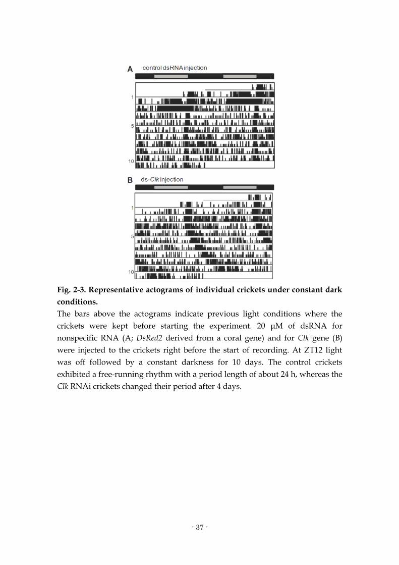
Fig. 2-3. Representative actograms of individual crickets under constant dark
conditions.
The bars above the actograms indicate previous light conditions where the
crickets were kept before starting the experiment. 20 μM of dsRNA for
nonspecific RNA (A; DsRed2 derived from a coral gene) and for Clk gene (B)
were injected to the crickets right before the start of recording. At ZT12 light
was off followed by a constant darkness for 10 days. The control crickets
exhibited a free-running rhythm with a period length of about 24 h, whereas the
Clk RNAi crickets changed their period after 4 days.

- 38 -
Fig. 2-4. The dsRNA levels in crickets (A) and firebrats (B) were investigated
in a time series using a pair of primers that binds within the dsRNA
sequence and also within the endogenous Clk.
The data are shown as mean ± SEM after normalization to the mean value of the
untreated animals. The intact level is set to 1.0. The value of the untreated
animals virtually indicates the mRNA level of the endogenous Clk gene
expression. Asterisks indicate significant differences from the level of untreated
animals (* P < 0.05; ** P < 0.01, Tukey–Kramer multiple comparison test). See
Fig. 2- 3 for more details.

- 39 -
Chapter 3.
The clock gene cycle plays an important role in the circadian clock
of the cricket Gryllus bimaculatus

- 40 -
3.1. Abstract
To dissect the molecular oscillatory mechanism of the circadian clock in
the cricket Gryllus bimaculatus, a cDNA of the clock gene cycle (Gb’cyc) was
cloned and its structure and function were analyzed. Gb’cyc contains four
functional domains, i.e. bHLH, PAS-A, PAS-B and BCTR domains, and is
expressed rhythmically in light dark cycles, peaking at mid night. The RNA
interference (RNAi) of Clock (Gb’Clk) and period (Gb’per) reduced the Gb’cyc
mRNA levels and abolished the rhythmic expression, suggesting that the
rhythmic expression of Gb’cyc is regulated by a mechanism including Gb’Clk
and Gb’per. These features are more similar to those of mammalian orthologue
of cyc (Bmal1) than those of Drosophila cyc. A single treatment with
double-stranded RNA (dsRNA) of Gb’cyc effectively knocked down the Gb’cyc
mRNA level and abolished its rhythmic expression. The cyc RNAi failed to
disrupt the locomotor rhythm, but lengthened its free-running period in
constant darkness (DD). It is thus likely that Gb’cyc is involved in the circadian
clock machinery of the cricket. The cyc RNAi crickets showed a rhythmic
expression of Gb’per and timeless (Gb’tim) in the optic lobe in DD, explaining the
persistence of the locomotor rhythm. Surprisingly, cyc RNAi revealed a
rhythmic expression of Gb’Clk in DD which is otherwise rather constitutively
expressed in the optic lobe. These facts suggest that the cricket might have a
unique clock oscillatory mechanism in which both Gb’cyc and Gb’Clk are
rhythmically controlled and that under abundant expression of Gb’cyc the
rhythmic expression of Gb’Clk may be concealed.

- 41 -
3.2. Introduction
Most insects live in harmony with the natural daily cycle to show a
daily rhythm in their behavior. The rhythm is regulated by an endogenous
mechanism called circadian clock which generates a 24 h oscillation. The
oscillatory mechanism of the circadian clock is most extensively studied in the
fruit fly Drosophila melanogaster, and thought to consist of interlocked
autoregulatory transcriptional/ translational feedback loops involving a set of
clock genes such as period (per), timeless (tim), Clock (Clk) and cycle (cyc) (Hardin,
2009; Tomioka and Matsumoto, 2010). In brief, following transcription of per
and tim, their product proteins PER and TIM accumulate in the cytoplasm
during the night, form PER/TIM heterodimer to enter the nucleus, and
inactivate heterodimeric transcription factors CLK/CYC to repress their own
transcription (Sehgal et al., 1994; Lee et al., 1999). This negative feedback
generates the rhythmic expression of per and tim. The Clk gene is also
rhythmically expressed by a loop including vrille (vri) and Par domain protein 1ε
(Pdp1ε) (Cyran et al., 2003; Glossop et al., 2003). CLK/CYC activates the
transcription of both vri and Pdp1ε but vri mRNA peaks prior to the peak of
Pdp1ε mRNA. The VRI protein thus accumulating earlier represses the Clk
transcription, while PDP1ε later stimulates the transcription of Clk by
competitive binding to VRI/PDP1ε box with VRI, making Clk to peak during
the early day (Cyran et al., 2003; Glossop et al., 2003; Hardin, 2005). The third
loop produces a rhythmic expression of clockwork orange (cwo) (Kadener et al.,
2007; Lim et al., 2007; Matsumoto et al., 2007). These three loops are thought to
be coupled to produce a stable circadian oscillation.

- 42 -
However, immunohistochemical and molecular studies have yielded
noticeably different results among insects, suggesting that insect clocks have
considerably diversified. For example, there is no evidence that PER enters the
nucleus in moths, bugs and cockroaches (Sauman and Reppert, 1996;
Vafopoulou et al., 2007; Wen and Lee, 2008; Zavodska et al., 2008), and tim is
absent in the genome of hymenopteran species (Rubin et al., 2006; Zhan et al.,
2011). In addition, some insects show rhythmic expression of cyc instead of Clk
(Rubin et al., 2006; Kamae et al., 2010). To understand how the variety of clock
mechanisms derived from an ancestral one, further comparative study is
necessary.
In the cricket Gryllus bimaculatus, per and Clk are essential components
of the clock because their RNA interference (RNAi) stops the molecular
oscillation as well as the locomotor rhythm (Moriyama et al., 2008, 2012). In
contrast to Drosophila, however, Clk is rather constitutively expressed in the
optic lobe, suggesting a cyclic expression of its partner, cyc.
In this study, cDNA of the clock gene cycle (Gb’cyc) was cloned from the
cricket G. bimaculatus, examined its daily expression pattern using quantitative
real-time RT-PCR (qPCR), and investigated its role in circadian oscillatory
mechanism with RNAi. The results showed that the sequence of Gb’cyc showed
a similarity to that of its mammalian orthologue, Bmal1 and that its expression
was rhythmic in light dark (LD) cycles. Interestingly, its knockingdown by
RNAi resulted in a lengthening of free-running period of locomotor rhythms
and revealed a rhythmic expression of Gb’Clk. These results suggest not only
that Gb’cyc is an important component of the cricket’s clock but also that the

- 43 -
cricket might possess a unique circadian clock including a mechanism for
rhythmic expression of both cyc and Clk in certain conditions.
3.3. Materials and methods
Animals
Adult male crickets, G. bimaculatus, were used. They were obtained
from a laboratory colony maintained under standard environmental conditions
with a light dark (LD) cycle of 12 h light to 12 h dark (light: 06:00–18:00 h;
Japanese standard time, JST) at a constant temperature of 25.0 ± 0.5 ºC. They
were fed laboratory chow and water.
Cloning and structural analysis of the clock gene cyc
Total RNA was extracted with TRIzol (Invitrogen, Carlsbad, CA) from 6
adult male optic lobes collected at ZT 14 (ZT stands for zeitgeber time and ZT0
corresponds to lights-on and ZT12 to lights-off). 5 μg of total RNA was used for
reverse transcription to obtain cDNA, using SuperScript II (Invitrogen,
Carlsbad, CA). Using the single-stranded cDNA as a template, PCR was
performed with degenerate primers deduced from the conserved amino acid
sequences among insect cyc orthologues. The primers used were
5’-CAAGAGATGGCGAGATAAGATGAAYACNTA-3’ for forward, and
5’-TCATNCGRCARAARAA-3’ for reverse. The PCR conditions employed
were: 30 s for denaturation at 95 ºC, 30 s for annealing at 55 ºC, and 90 s for
extension at 72 ºC for 35 cycles with ExTaq DNA polymerase (Takara, Ohtsu,
Japan). The purified fragment was cloned into TOPO-pCR II vector (Invitrogen)

- 44 -
and sequenced with BigDye Terminator v3.1 Cycle Sequencing Kit (Applied
Biosystems, Foster City, CA). 5’ and 3’ RACEs were done with GeneRacer™ kit
(Invitrogen) and SMARTer™ RACE cDNA Amplification kit (Takara) with gene
specific primers, 5’-ACATGCTCCGTCAAAACAAGAG-3’ and 5’-
GTCTGTATCGCAGGTGCTCA -3’, respectively. RACE fragments were
purified, cloned and sequenced as mentioned above. Sequences were analyzed
by Genetyx ver. 6 (Genetic Information Processing Software, Tokyo, Japan) and
BioEdit ver. 7.0.5 (Biological Sequence Alignment Editor, Ibis Therapeutic,
Carlsbad, CA). CYC’s amino acid sequences were analyzed with ClustalW
(http://clustalw.ddbj.nig.ac.jp/top-j.html). Sequences of known insect cycs
were obtained from GenBank.
Measurement of mRNA levels
Quantitative real-time RT-PCR (qPCR) was used to measure mRNA
levels. Total RNA was extracted and purified from 6 adult male optic lobes with
TRIzol® Reagent (Invitrogen). To remove contaminating genomic DNA, the
total RNA was treated with DNase I. About 500 ng total RNA of each sample
was reverse transcribed with random 6mers using PrimeScript™ RT reagent Kit
(Takara). Real-time PCR was performed by Mx3000P™ Real-Time PCR System
(Stratagene, La Jolla, CA) using FastStart Universal SYBR Green Master (Roche,
Tokyo, Japan) including SYBER Green with primers
5’-GGCCGAAGCTCATAAAGTGG-3’ and
5’-AACCGCACAAAGGAACCATC-3’ for Gb’cyc,
5’-GATTATGAAGTCTGTGATGATTGG-3’ and
5’-AGCATTGGAGAGAACTGAAGAGGT-3’ for Gb’tim, and with those

- 45 -
described previously for Gb’Clk, Gb’per, and Gb’rpl18a (Moriyama et al., 2012).
The results were analyzed using the software associated with the instrument.
The values were normalized with the values of Gb’rpl18a at each time point.
Results of three to eight independent experiments were used to calculate the
mean ± SEM.
RNAi
Double stranded RNA (dsRNA) for Gb’cyc, Gb’Clk, Gb’per and DsRed2,
derived from a coral species (Discosoma sp.), were synthesized using
MEGAscript® High Yield Transcription Kit (Ambion, Austin, TX, USA). For
Gb’cyc, Gb’Clk and Gb’per, using cDNA, prepared as described above as a
template, PCR was performed with ExTaq DNA polymerase (Takara). The
primers used were 5’-
TAATACGACTCACTATAGGGCGTGCACTCGTACACTGAGG-3’ for forward,
and 5’-AATTAACCCTCACTAAAGGGAGGTTCTGCTGCTTCT TTCG-3’ for
reverse for Gb’cyc (dscyc #1),
5’-TAATACGACTCACTATAGGGATGGAAATCCTTCCGAAACC-3’ for
forward, and
5’-AATTAACCCTCACTAAAGGGATCCCACCTCTTTGATGCAC-3’ for
reverse for another region of the Gb’cyc (dscyc #2). The primers for Gb’per,
Gb’Clk and DsRed2 were those described in Moriyama et al., (2012). Amplified
Gb’cyc (#1 450 bp and #2 529 bp) fragments were linearized and precipitated
with ethanol. For DsRed2 dsRNA, DsRed2 was linearized from pDsRed2-N1
(Clontech, Mountain View, CA, USA), amplified with the forward and reverse
primers. With each of these linearized fragments as a template, RNA was

- 46 -
synthesized and dsRNAs were prepared and stored at -80 ºC as described
previously (Moriyama et al., 2012). 760 nl of dsRNA solution was injected with
the nanoliter injector (WPI, Sarasota, FL, USA) into the abdomen of adults
anesthetized with CO2. Otherwise noted, the injection was made within 3 days
after the imaginal molt, and the optic lobes were collected seven days after the
injection for the measurement of mRNA levels.
Behavioral analysis
Locomotor activities were recorded in the same way as described
previously (Moriyama et al., 2008). Briefly, adult crickets were individually
housed in a transparent plastic box (18 × 9 × 4.5 cm) with a rocking substratum.
The number of substratum rocking was recorded every 6 min by a
computerized system. Food and water were provided ad libitum. The
locomotor activity was monitored in an incubator (MIR-153, Sanyo Biomedica,
Osaka, Japan), in which temperature was kept at 25 ºC and lighting conditions
were given by a cool white fluorescent lamp connected to an electric timer. The
light intensity was 600–1000 lux at the animal’s level, varying with the
proximity to the lamp. The raw data were displayed as conventional
double-plotted actograms to judge activity patterns, and statistically analyzed
by the chi-square periodogram (Sokolove and Bushell, 1978) with Actogram J
(http://actogramj.neurofly.de/) (Schmid et al., 2011). If a peak of the
periodogram appeared above the 0.05 confidence level (alpha = 0.005), the
power value (height of the peak above the confidence level) was greater than or
equal to 10, and the width of the peak was greater than or equal to 2, the period
for the peak was designated as statistically significant (Kaneko et al., 2000).

- 47 -
3.4. Results
Cloning and structural analysis of Gb’cyc
A cyc was cloned homologue from the optic lobe of G. bimaculatus by
degenerate PCR strategy with primers designed from conserved amino acid
sequences of known insect’s cyc orthologues. A 516 bp of cyc fragment
including a part of bHLH and PAS-A domain was first obtained. Then 3’ and 5’
RACEs were performed with gene specific primers designed from the obtained
sequence. The obtained full length cDNA was of 1967 bp encoding a protein of
599 aa residues with 100 and 67 bp of 3’- and 5’-UTR, respectively. A BLAST
database search indicated that the amino acid sequence has 49–78% identities
along the entire length of the protein with those of known insects’ cyc
orthologues (Table 3-1), including the firebrat Thermobia domestica
(GenBank/EMBL/DDBJ accession No. BAJ16354), the honeybee Apis mellifera
(XP_001121441), the beetle Tribolium castaneum (NP_001107795), the monarch
butterfly Danaus plexippus (AAR13012) and the fruit fly D. melanogaster
(NP_524168). The deduced protein had four highly conserved regions that are
characteristic of known CYC proteins (Fig. 3-1A and B): (i) bHLH domain, (ii)
PAS-A domain, (iii) PAS-B domain, (iv) BMAL1 C-terminal region (BCTR) in
C-termini. The BCTR, which shows potent transcriptional activity in vitro
(Takahata et al., 2000; Chang et al., 2003; Chang and Reppert, 2003), has a
particularly high similarity (83–90% identity) to those of known insect CYC
(Table 3-1). Based on these identities and similarities, this gene was concluded
to encode a cricket’s CYC (Gb’cyc; Gen- Bank/EMBL/DDBJ accession No.

- 48 -
AB762416). A phylogenetic tree based on the amino acid sequences of CYCs
from known insect species revealed that Gb’cyc forms a separate bifurcation (Fig.
3-1C).
Gb’cyc expression in the optic lobe under LD and DD
To determine if Gb’cyc transcript oscillated in a circadian manner, the
levels of Gb’cyc mRNA in the optic lobe were examined under LD12:12 and in
the second day after transfer to constant darkness (DD) by qPCR (Fig. 3-2). The
Gb’cyc mRNA level showed a significant fluctuation peaking at ZT18 in LD
(ANOVA, P < 0.05). A slight increase was observed during the midday but was
not significant (Tukey-test, P > 0.05). In DD, however, there was no significant
daily fluctuation (ANOVA, P > 0.05; Fig. 3-2B).
Gb’cyc dsRNA suppresses levels of Gb’cyc transcripts
To examine the effects of Gb’cyc dsRNA on Gb’cyc expression, the Gb’cyc
mRNA levels were measured by qPCR in the optic lobe of the adult male
crickets injected with dscyc#1 (Fig. 3-2). In the control crickets treated with
DsRed2 dsRNA (dsDsRed2), the Gb’cyc mRNA levels were slightly reduced but
no significant difference was observed when compared with those of intact
crickets in LD (Fig. 3-2A). Gb’cyc RNAi significantly reduced Gb’cyc mRNA
levels in LD to nearly 25% of the peak level of intact crickets throughout the day
(Fig. 3-2A, t-test, P < 0.05). Similar significant reduction was observed in DD
(t-test, P < 0.05): the Gb’cyc mRNA levels stayed below the trough level of intact
crickets throughout the day (Fig. 3-2B). Although slight fluctuations in Gb’cyc
mRNA levels were observed in both LD and DD, these were statistically not

- 49 -
significant (ANOVA, P > 0.1). To exclude off-target effects of dsRNA, the Gb’cyc
mRNA levels were measured after injection with dscyc#2 synthesized from a
different region of the Gb’cyc cDNA. The results were similar to those treated
with dscyc#1: cyc mRNA levels were significantly reduced (Fig. 3-2A, t-test, P <
0.01). It is thus evident that dscyc successfully suppressed the expression of
Gb’cyc mRNA through RNAi.
Gb’cyc RNAi lengthened free-running period of the locomotor rhythm
To examine the role of Gb’cyc in the circadian rhythm generation, the
effects of dscyc on locomotor rhythms were examined in male crickets. Effects
of dscyc#1 and dscyc#2 were examined in 55 and 20 adult crickets, respectively.
As a control DsRed2 dsRNA was injected in the abdomen of 17 adult crickets.
Locomotor rhythms were also measured in 12 intact males. Both the dscyc
injected and the dsDsRed2 injected control crickets exhibited an activity rhythm
under LD12:12, with peaks at lights-on and sometimes in mid light phase in the
early adult stage, thereafter an intense activity peak appeared at lights-off (Fig.
3-3A, B and C). Similar locomotor rhythms were observed in intact crickets
(data not shown), as previously reported (Tomioka and Chiba, 1982). On
transfer to DD, all the crickets showed a free-running rhythm persisting from
the light-off component (Fig. 3-3). The average free-running periods of dscyc
injected crickets were 24.9 ± 0.4 h for both dscyc#1 (n = 31) and dscyc#2 (n = 14),
which were significantly longer than that of intact (23.6 ± 0.2, n = 12) and
control crickets injected with DsRed2 dsRNA (23.8 ± 0.4 h, n = 14) (t-test, P <
0.01, Table 3-2). There was no marked difference in power of the rhythm
between control and Gb’cyc RNAi crickets both under LD and DD (Table3- 2).

- 50 -
Effects of Gb’cyc dsRNA on Gb’per, Gb’tim and Gb’Clk transcripts
In Drosophila, CYC is known to form a heterodimer with CLK to activate
the transcription of per and tim (Rutila et al., 1998). To examine whether Gb’cyc
is involved in the regulation of these genes, the levels of Gb’per, Gb’tim and
Gb’Clk mRNA in the optic lobe were measured by qPCR in adult male crickets
seven days after injection of dscyc#1. In intact crickets, the Gb’per and Gb’tim
showed a clear rhythm peaking in the mid night phase in LD as have been
reported previously (Moriyama et al., 2008; Danbara et al., 2010) and their
mRNA levels were significantly reduced by Gb’cyc RNAi at ZT2, 10–18 (t-test, P
< 0.01) for Gb’per and at ZT14 (t-test, P < 0.01) for Gb’tim (Fig. 3-4A and B). No
clear rhythms were observed in both per and tim mRNA. The Gb’Clk mRNA
showed no significant rhythm in intact crickets kept in LD as reported by
Moriyama et al., (2012) and the dscyc treatment significantly reduced Gb’Clk
mRNA levels at most time points (ZT6, 14, 18; t-test, P < 0.05, Fig. 3-4C). In
constant darkness (DD), however, these clock genes showed a rhythmic
expression, even after the dscyc treatment. The Gb’per and Gb’tim peaked at
CT18 in intact crickets and similar rhythms were observed in cyc RNAi crickets
for both Gb’per and Gb’tim (Fig. 3-4D and E, ANOVA, P < 0.05). These molecular
rhythms are consistent with the free-running locomotor rhythm in DD.
Surprisingly, the Gb’Clk also showed a significantly rhythmic expression with a
peak at CT14 (Fig. 3-4F, ANOVA, P < 0.05), although it showed no significant
rhythm in intact crickets (Fig. 3-4F, Moriyama et al., 2012).

- 51 -
Effects of dsper and dsClk on Gb’cyc mRNA levels
The effects of dsper and dsClk on expression of Gb’cyc were then
examined. The knock-down of respective mRNA by dsper and dsClk through
RNAi has been confirmed previously (Moriyama et al., 2008, 2012). When
Gb’per was knocked-down, the Gb’cyc mRNA level was significantly
downregulated (t-test, P < 0.05) except for ZT22 and stayed below the basal
level of intact animals throughout the day, showing no statistically significant
rhythm (ANOVA, P > 0.05, Fig. 3-5). In Clk RNAi, Gb’cyc mRNA levels were
again significantly reduced at ZT 6, 10 and 18 (t-test, P < 0.05) and showed no
statistically significant rhythm (ANOVA, P > 0.05, Fig. 3-5).
3.5. Discussion
In the present study, the structure, expression profile, and function of
the clock gene cyc were investigated in a hemimetabolous insect, G. bimaculatus.
The deduced Gb’CYC protein has 4 functional domains, i.e. bHLH, PAS-A,
PAS-B and BCTR domains (Fig. 3-1), which are commonly observed among
known insect CYC orthologues (Bembenek et al., 2007). The BCTR domain,
which is missing in Drosophila CYC, has particularly high homology with CYC
orthologues from other insects, supporting the view that the BCTR is
evolutionarily conserved in insects, but it might have been somehow lost in
Drosophila (Chang et al., 2003).
The BCTR domain has been shown to have potent transcriptional
activity in vitro (Takahata et al., 2000; Chang et al., 2003; Chang and Reppert,

- 52 -
2003). The result that Gb’cyc RNAi suppressed the mRNA levels of Gb’per (Fig.
3-4) is consistent with this hypothesis and also in good agreement with what is
expected from the Drosophila clock model. It is most probable that Gb’cyc acts as
a positive element in the clock machinery and Gb’cyc may directly transactivate
Gb’per. Interestingly, however, in the Gb’cyc RNAi crickets the mRNA level of
Gb’tim was knocked down in LD but maintained a clear rhythm in DD (Fig. 3-4).
A similar rhythm was also seen in Gb’per in DD (Fig. 3-4). These facts suggest
that there is a mechanism maintaining a rhythmic transcription of Gb’per and
Gb’tim even after Gb’cyc is substantially knocked down and that the rhythmic
expression of Gb’per and Gb’tim may be responsible for the locomotor rhythm
with a lengthened free-running period (Fig. 3-3). Gb’Clk might be a candidate
element responsible for this rhythmic transcription of Gb’per and Gb’tim because
Gb’Clk was rhythmically expressed in the Gb’cyc RNAi crickets (Fig. 3-4) and
Gb’Clk has poly-Q region which is likely involved in transcriptional activation
(Allada et al., 1998; Moriyama et al., 2012). Another explanation for the
free-running locomotor rhythms in DD might be that there is enough amount of
residual Gb’cyc mRNA surviving the RNAi treatment for generation of the
rhythm. An important issue remains to be answered is why clear rhythms of
Gb’per and Gb’tim in the cyc RNAi treated crickets were observed only in DD
(Fig. 3-4).
The Gb’cyc was rhythmically expressed in the optic lobe in LD (Fig. 3-2)
and a similar and clear oscillation has been observed in the adult brain (Chapter
5). The rhythm in the optic lobe was abolished in Gb’per RNAi and Gb’Clk RNAi
crickets (Fig. 3-5). These facts suggest that the rhythmic expression of Gb’cyc is

- 53 -
not a simple response to LD but regulated by a mechanism involving Gb’per
and Gb’Clk. This hypothesis is conceivable given that rhythmic expression of cyc
is also known for the firebrat (T. domestica), one of the most primitive insects,
and for those insects phylogenetically higher than crickets such as the honeybee
(A. mellifera) and the sandfly (Lutzomyia longipalpis) (Meireles-Filho et al., 2006a;
Rubin et al., 2006; Kamae et al., 2010). Since the rhythmic expression of Gb’cyc
disappeared in DD (Fig. 3-2B), the rhythm seems highly dependent on light
cycles, however. It may be also possible that Gb’cyc is rhythmically expressed
only in a subset of neurons that controls behavior and the rhythm is masked by
constitutive expression in other non-pacemaker cells in DD. Further critical
study is necessary to clarify the reason why the rhythmic expression became
obscure in DD.
Interestingly, the Gb’cyc mRNA peaked in the mid night similar to those
of Gb’per and Gb’tim (Figs. 3-2A, 3-4A and B). Coherence of the peak phase was
also found in Gb’Clk mRNA in cyc RNAi crickets (Fig. 3-4F). A similar situation
is observed in the sandfly L. longipalpis, in which Clk and cyc transcripts peak
simultaneously with or slightly earlier than per and tim (Meireles-Filho et al.,
2006a, 2009b) and also in some mammalian peripheral tissues where Bmal1 and
per are expressed almost in phase (Tong et al., 2004; Bebas et al., 2009). This
feature is opposed to the Drosophila clock hypothesis, where peaks of per/tim
and Clk transcription are about 12 h apart from each other (Hardin, 2009). The
reason and mechanism underlying this phase coherence should be addressed in
future studies.
The present study revealed that the cricket clock possesses a unique
- 54 -
feature in that Gb’cyc shows a clear rhythmic expression in LD and that Gb’Clk
is expressed in a rhythmic fashion only when Gb’cyc is knocked-down (Figs. 3-2
and 3-4). In both vertebrates and insects, either cyc (Bmal1) or Clk is reportedly
expressed in a rhythmic manner (Honma et al., 1998; Stanewsky, 2002; Hardin,
2009). In insects, Clk is known to oscillate in Drosophila while cyc does in other
species (Cyran et al., 2003; Glossop et al., 2003; Meireles- Filho et al., 2006a;
Rubin et al., 2006; Kamae et al., 2010). Although further experiments are
necessary to confirm the rhythmic expression of Gb’Clk in cyc RNAi crickets, it
should be worth examining whether the two mechanisms for rhythmic
expression of cyc and Clk might coexist in the cricket. Since a more primitive
insect, the firebrat, exhibits a rhythm in cyc but not in Clk expression (Kamae et
al., 2010), it might provide a view on the evolutionary history of the insect
circadian clock.
The data indicate that Gb’per is required for the sufficient expression of
Gb’cyc at least in LD (Fig. 3-5). Because it is also required for Gb’Clk (Moriyama
et al., 2012), Gb’per seems to play a common role in regulation of these two
genes. One may explain that a reduced level of PER by per RNAi may increase
the transcriptional activity of CLK/CYC since the CLK/CYC transcriptional
activity is suppressed by PER/TIM (Stanewsky, 2002; Tomioka and Matsumoto,
2010). The increased activity of CLK/CYC would result in an increased level of
transcriptional repressor like vri for Clk, which may eventually reduce the cyc
and Clk transcript levels. While the transcriptional regulations of Gb’Clk and
Gb’cyc in crickets should be elucidated in future, the results strongly suggest
that both regulations are interlocked in the circadian feedback loop. It is

- 55 -
interesting to know the mechanism by which the two positive elements are
regulated in transcription. Future studies would reveal a unique molecular
feedback system that may be similar to neither Drosophila nor mammalian
system.
References
Allada, R., White, N.E., So, W.V., Hall, J.C., Rosbash, M. (1998) A mutant
Drosophila homolog of mammalian Clock disrupts circadian rhythms
and transcription of period and timeless. Cell 93, 791–804.
Bebas, P., Goodall, C.P., Majewska, M., Neumann, A., Giebultowicz, J.M.,
Chappell, P.E. (2009) Circadian clock and output genes are rhythmically
expressed in extratesticular ducts and accessory organs of mice. The
FASEB Journal 23, 523– 533.
Bembenek, J., Itokawa, K., Hiragaki, S., Qi-Miao, S., Tufail, M., Takeda, M.
(2007) Molecular characterization and distribution of CYCLE protein
from Athalia rosae. Journal of Insect Physiology 53, 418–427.
Chang, D.C., McWatters, H.G., Williams, J.A., Gotter, A.L., Levine, J.D., Reppert,
S.M. (2003) Constructing a feedback loop with circadian clock molecules
from the silkmoth, Antheraea pernyi. Journal of Biological Chemistry 278,
38149–38158.
Chang, D.C., Reppert, S.M. (2003) A novel C-terminal domain of Drosophila
PERIOD inhibits dCLOCK:CYCLE-mediated transcription. Current
Biology 13, 758–762.
Cyran, S.A., Buchsbaum, A.M., Reddy, K.L., Lin, M.-C., Glossop, N.R.J., Hardin,
P.E., Young, M.W., Storti, R.V., Blau, J. (2003) Vrille, Pdp1 and dClock
form a second feedback loop in the Drosophila circadian clock. Cell 112,
329–341.
Danbara, Y., Sakamoto, T., Uryu, O., Tomioka, K. (2010) RNA interference of
timeless gene does not disrupt circadian locomotor rhythms in the
cricket Gryllus bimaculatus. Journal of Insect Physiology 56, 1738–1745.
Glossop, N.R., Houl, J.H., Zheng, H., Ng, F.S., Dudek, S.M., Hardin, P.E. (2003)
VRILLE feeds back to control circadian transcription of clock in the
Drosophila circadian oscillator. Neuron 37, 249–261.

- 56 -
Hardin, P. (2005) The circadian timekeeping system of Drosophila. Current
Biology 15, R714–R722.
Hardin, P. (2009) Molecular mechanisms of circadian timekeeping in Drosophila.
Sleep and Biological Rhythms 7, 235–242.
Honma, S., Ikeda, M., Abe, H., Tanahashi, Y., Namihira, M., Honma, K.,
Nomura, M. (1998) Circadian oscillation of BMAL1, a partner of a
mammalian clock gene clock, in rat suprachiasmatic nucleus.
Biochemical Biophysical Research Communications 250, 83–87.
Kadener, S., Stoleru, D., McDonald, M., Nawathean, P., Rosbash, M. (2007)
Clockwork orange is a transcriptional repressor and a new Drosophila
circadian pacemaker component. Genes and Development 21, 1675–
1686.
Kamae, Y., Tanaka, F., Tomioka, K. (2010) Molecular cloning and functional
analysis of the clock genes, clock and cycle, in the firebrat Thermobia
domestica. Journal of Insect Physiology 56, 1291–1299.
Kaneko, M., Park, J.H., Cheng, Y., Hardin, P.E., Hall, J.C. (2000) Disruption of
synaptic transmission or clock-gene-product oscillations in circadian
pacemaker cells of Drosophila cause abnormal behavioral rhythms.
Journal of Neurobiology 43, 207–233.
Lee, C., Bae, K., Edery, I. (1999) PER and TIM inhibit the DNA binding activity
of a Drosophila CLOCK-CYC/dBMAL1 Heterodimer without disrupting
formation of the Heterodimer: a Basis for circadian transcription.
Molecular and Cellular Biology 19, 5316–5325.
Lim, C., Chung, B.Y., Pitman, J.L., McGill, J.J., Pradhan, S., Lee, J., Keegan, K.P.,
Choe, J., Allada, R. (2007) Clockwork orange encodes a transcriptional
repressor important for circadian-clock amplitude in Drosophila. Current
Biology 17, 1082–1089.
Matsumoto, A., Ukai-Tadenuma, M., Yamada, R.G., Houl, J., Uno, K.D.,
Kasukawa, T., Dauwalder, B., Itoh, T.Q., Takahashi, K., Ueda, R.,
Hardin, P.E., Tanimura, T., Ueda, H.R. (2007) A functional genomics
strategy reveals clockwork orange as a transcriptional regulator in the
Drosophila circadian clock. Genes and Development 21, 1687–1700.
Meireles-Filho, A.C.A., Amoretty, P.R., Souza, N.A., Kyriacou, C.P., Peixoto,
A.A. (2006a) Rhythmic expression of the cycle gene in a hematophagous
insect vector. BMC Molecular Biology 7, 38.
Meireles-Filho, A.C.A., Rivas, G.B.S., Gesto, J.S.M., Machado, R.C., Britto, C., de

- 57 -
Souza, N.A., Peixoto, A.A. (2006b) The biological clock of an
hematophagous insect: locomotor activity rhythms, circadian
expression and downregulation after a blood meal. FEBS Letters 580, 2–
8.
Moriyama, Y., Kamae, Y., Uryu, O., Tomioka, K. (2012) Gb’Clock is expressed in
the optic lobe and required for the circadian clock in the cricket Gryllus
bimaculatus. Journal of Biological Rhythms 27, 467–477.
Moriyama, Y., Sakamoto, T., Karpova, S.G., Matsumoto, A., Noji, S., Tomioka, K.
(2008) RNA interference of the clock gene period disrupts circadian
rhythms in the cricket Gryllus bimaculatus. Journal of Biological
Rhythms 23, 308–318.
Rubin, E.B., Shemesh, Y., Cohen, M., Elgavish, S., Robertson, H.M., Bloch, G.
(2006) Molecular and phylogenetic analyses reveal mammalian-like
clockwork in the honey bee (Apis mellifera) and shed new light on the
molecular evolution of the circadian clock. Genome Research 16, 1352–
1365.
Rutila, J.E., Suri, V., Le, M., So, W.V., Rosbash, M., Hall, J.C. (1998) CYCLE is a
second bHLH-PAS clock protein essential for circadian rhythmicity
and transcription of Drosophila period and timeless. Cell 93, 805–814.
Sauman, I., Reppert, S.M. (1996) Circadian clock neurons in the silkmoth
Antheraea pernyi: novel mechanisms of period protein regulation.
Neuron 17, 889–900.
Schmid, B., Helfrich-Forster, C., Yoshii, T. (2011) A new ImageJ plug-in
‘‘ActogramJ’’ for chronobiological analyses. Journal of Biological
Rhythms 26, 464–467.
Sehgal, A., Price, J.L., Man, B., Young, M.W. (1994) Loss of circadian behavioral
rhythms and per RNA oscillations in the Drosophila mutant timeless.
Science 263, 1603–1606.
Sokolove, P.G., Bushell, W.N. (1978) The chi square periodogram: its utility for
analysis of circadian rhythm. Journal of Theoretical Biology 72, 131–
160.
Stanewsky, R. (2002) Clock mechanisms in Drosophila. Cell and Tissue Research
309, 11–26.
Takahata, S., Ozaki, T., Mimura, J., Kikuchi, Y., Sogawa, K., Fujii-Kuriyama, Y.
(2000) Transactivation mechanisms of mouse clock transcription
factors, mClock and mArnt3. Genes to Cells 5, 739–747.

- 58 -
Tomioka, K., Chiba, Y. (1982) Post-embryonic development of circadian rhythm
in the cricket, Gryllus bimaculatus. Journal of Comparative Physiology
A 147, 299– 304.
Tomioka, K., Matsumoto, A. (2010) A comparative view of insect circadian
clocks. Cellular and Molecular Life Sciences 67, 1397–1406.
Tong, Y., Guo, H., Brewer, J.M., Lee, H., Lehman, M.N., Bittman, E.L. (2004)
Expression of haPer1 and haBmal1 in Syrian hamsters: heterogeneity
of transcripts and oscillations in the periphery. Journal of Biological
Rhythms 19, 113–125.
Vafopoulou, X., Steel, C.G.H., Terry, K.L. (2007) Neuroanatomical relations of
prothoracicotropic hormone neurons with the circadian timekeeping
system in the brain of larval anxd adult Rhodnius prolixus (Hemiptera).
Journal of Comparative Neurology 503, 511–524.
Wen, C.-J., Lee, H.-J. (2008) Mapping the cellular network of the circadian clock
in two cockroach species. Archives of Insect Biochemistry and
Physiology 68, 215– 231.
Zavodska, R., Wen, C.-J., Hrdy, I., Sauman, I., Lee, H.-J., Sehnal, F. (2008)
Distribution of corazonin and pigment-dispersing factor in the
cephalic ganglia of termites. Arthropod Structure Development 37,
273–286.
Zhan, S., Merlin, C., Boore, J.L., Reppert, S.M. (2011) The monarch butterfly
genome yields insights into long-distance migration. Cell 147, 1171–
1185.

- 59 -
Table 3-1. Overall amino acid identity (%) of whole sequence and functional
domains of Gryllus bimaculatus CYCLE with those insect orthologues.
Species Whole
sequence bHLH PAS-A PAS-B BCTR
Thermobia domestica 78 74 94 87 90
Tribolium castaneum 63 72 89 79 88
Danaus plexippus 49 63 70 64 83
Drosophila melanogaster 62 61 81 69 ―
Apis mellifera 78 74 92 89 90

- 60 -
Table 3-2. Effects of dscyc and dsDsRed2 on the locomotor rhythm of the
cricket Gryllus bimaculatus.
Treatment n Period/Free-running
period (mean±SD) h
Power
(mean±SEM)
LD
intact 12 24.0±0.14 485.1±19.4
DsRed2 dsRNA 17 24.0±0.07 150.6±19.4
cyc dsRNA(#1) 55 24.0±0.1 207.7±18.2
cyc dsRNA(#2) 20 24.0±0.06 181.4±25.9
DD
intact 12 23.6±0.2 342.0±40.0
DsRed2 dsRNA 14 23.8±0.4 380.9±84.1
cyc dsRNA(#1) 31 24.9±0.4** 290.8±32.8
cyc dsRNA(#2) 14 24.9±0.4** 304.9±46.5
** P < 0.01, t-test vs DsRed2 dsRNA treatment.
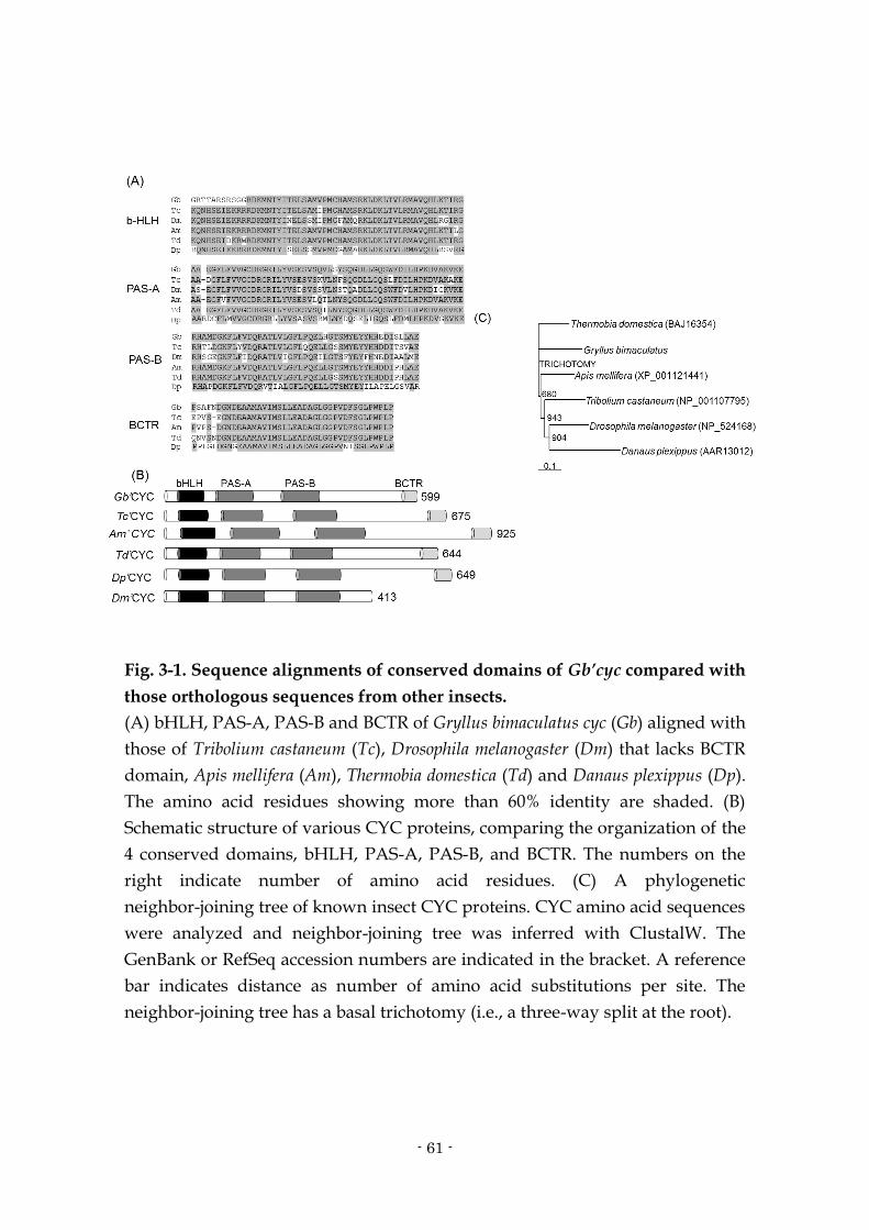
- 61 -
Fig. 3-1. Sequence alignments of conserved domains of Gb’cyc compared with
those orthologous sequences from other insects.
(A) bHLH, PAS-A, PAS-B and BCTR of Gryllus bimaculatus cyc (Gb) aligned with
those of Tribolium castaneum (Tc), Drosophila melanogaster (Dm) that lacks BCTR
domain, Apis mellifera (Am), Thermobia domestica (Td) and Danaus plexippus (Dp).
The amino acid residues showing more than 60% identity are shaded. (B)
Schematic structure of various CYC proteins, comparing the organization of the
4 conserved domains, bHLH, PAS-A, PAS-B, and BCTR. The numbers on the
right indicate number of amino acid residues. (C) A phylogenetic
neighbor-joining tree of known insect CYC proteins. CYC amino acid sequences
were analyzed and neighbor-joining tree was inferred with ClustalW. The
GenBank or RefSeq accession numbers are indicated in the bracket. A reference
bar indicates distance as number of amino acid substitutions per site. The
neighbor-joining tree has a basal trichotomy (i.e., a three-way split at the root).

- 62 -
Fig. 3-2. Expression profiles of Gb’cyc mRNA in the Gryllus bimaculatus
optic lobes and effects of Gb’cyc dsRNA under LD 12:12 (A) and DD (B).
Filled circles, filled squares, open circles and squares indicate the mRNA
levels of intact, DsRed2 dsRNA, cyc#1 dsRNA (dscyc#1) and dscyc#2
injected crickets, respectively. The abundance of Gb’cyc mRNA was
measured by quantitative real-time RT-PCR. Total RNA was extracted from
the optic lobes that were collected at 4 h interval starting at 2 h after light-on
(Zeitgeber time 2) or 2 h after projected light-on (Circadian time 2) in the
second day of DD. White, black and gray bars indicate light phase,
night/subjective night and subjective day, respectively. The data collected
from 3~8 independent experiments were averaged and plotted as mean ±
SEM. The abundance of Gb’rpl18a mRNA was used as an internal reference.
In intact crickets, Gb’cyc mRNA levels showed significant daily changes
(ANOVA, P < 0.05) to peak at ZT18 (A), while no significant rhythm was
detected in DD (B) (ANOVA, P>0.05). Note that both dscyc#1 and dscyc#2
significantly reduced cyc mRNA levels and abolished the rhythm both in
LD and DD (ANOVA, P > 0.05). *P < 0.05, **P < 0.01, t-test, vs intact
crickets.

- 63 -
Fig. 3-3. Double-plotted actograms (left) and χ2 periodograms (right) of
locomotor rhythms of crickets Gryllus bimaculatus injected with Gb’cyc
dsRNA [dscyc#1 (A) or dscyc#2 (B)], or with DsRed2 dsRNA (C) under
LD12:12 and DD at a constant temperature of 25 ºC.
Arrowheads indicate the day when the crickets were transferred from LD to
DD. a and b indicated in the periodogram correspond to the analyzed time
span, a and b, indicated in the actogram. An oblique line in the
periodogram indicates significance level of P < 0.05; peak value above the
line was designated as significant. The cyc RNAi crickets showed locomotor
rhythm free-running with a period longer than 24 h in DD (A, B), but the
control cricket injected with DsRed2 dsRNA showed a rhythm with a period
shorter than 24 h (C). For further explanations see text.

- 64 -
Fig. 3-4. Relative abundance of Gb’per (A, D), Gb’tim (B, E) or Gb’Clk mRNA
(C, F) in the optic lobes of intact and of cyc double-stranded RNA
(dsRNA)-injected adult male crickets Gryllus bimaculatus under LD (A-C) or
DD (D-F).
Filled and open circles indicate the mRNA levels of intact and Gb’cyc dsRNA
injected crickets, respectively. In Gb’cyc RNAi crickets, the optic lobes were
collected 7 days after the dsRNA injection. The abundance of mRNA was
measured by qPCR with total RNA extracted from the optic lobes. The data
collected from 3~8 independent experiments were averaged and plotted as
mean ± SEM for dsRNA-injected and intact crickets, respectively. The
abundance of Gb’rpl18a mRNA was used as an internal reference. *P < 0.05, **P
< 0.01, t-test vs intact crickets. Note that Gb’cyc dsRNA down-regulated Gb’per,
Gb’tim and Gb’Clk mRNA and prevents the rhythmic expression of Gb’per and
Gb’tim while revealed a rhythmic expression in Gb’Clk in LD (ANOVA, P <
0.05). In DD, however, Gb’per, Gb’tim and Gb’Clk mRNAs oscillate clearly
(ANOVA, P < 0.05 for Gb’per and Gb’Clk, P < 0.01 for Gb’tim). For further
explanations see text.

- 65 -
Fig. 3-5. Relative abundance of cyc mRNA in the optic lobes of intact
crickets and those injected with dsRNA of per or Clk in LD. Filled circle,
open square and triangle indicate the mRNA levels of intact,
dsper-injected and dsClk-injected crickets, respectively.
The data for intact crickets are replotted from Fig. 2A. *P < 0.05, **P < 0.01,
t-test vs intact crickets. Note that Gb’per and Gb’Clk dsRNAs down-regulate
the Gb’cyc mRNA levels and abolished the daily expression rhythm
(ANOVA, P > 0.05). For further explanations see text.
- 66 -
Chapter 4.
Circadian oscillations outside the optic lobe in the cricket Gryllus
bimaculatus

- 67 -
4.1. Abstract
Although circadian rhythms are found in many peripheral tissues in
insects, the control mechanism is still to be elucidated. To investigate the central
and peripheral relationships in the circadian organization, circadian rhythms
outside the optic lobes were examined in the cricket Gryllus bimaculatus by
measuring mRNA levels of period (Gb’per) and timeless (Gb’tim) genes in the
brain, terminal abdominal ganglion (TAG), anterior stomach, mid-gut, testis,
and MTs. Except for MTs and testis, the tissues showed a daily rhythmic
expression in either both Gb’per and Gb’tim or Gb’tim alone in LD. Under
constant darkness, however, the tested tissues exhibited rhythmic expression of
Gb’per and Gb’tim mRNAs, suggesting that they include a circadian oscillator.
The amplitude and the levels of the mRNA rhythms varied among those
rhythmic tissues. Removal of the optic lobe, the central clock tissue,
differentially affected the rhythms: the anterior stomach lost the rhythm of both
Gb’per and Gb’tim; in the mid-gut and TAG, Gb’tim expression became
arrhythmic but Gb’per maintained rhythmic expression; a persistent rhythm
with a shifted phase was observed for both Gb’per and Gb’tim mRNA rhythms
in the brain. These data suggest that rhythms outside the optic lobe receive
control from the optic lobe to different degrees, and that the oscillatory
mechanism may be different from that of Drosophila.
4.2. Introduction
Insects show rhythmic lives to adapt daily cyclic environment

- 68 -
(Saunders, 2002). Those daily rhythms are controlled by an endogenous
mechanism called the circadian clock. The locus of the clock has been identified
in some insects. For example, it is the optic lobe in crickets (Tomioka and Chiba,
1992), cockroaches (Page, 1982), and beetles (Balkenohl and Weber, 1981), but it
is within the central brain in flies (Handler and Konopka, 1979), mosquitoes
(Kasai and Chiba, 1987) and moths (Truman, 1974). In addition to these central
clock tissues regulating activity rhythms, peripheral tissues also show some
rhythms. The compound eyes have a circadian rhythm in their responsiveness
to light in crickets (Tomioka and Chiba, 1982a) and cockroaches (Wills et al.,
1985), and sperm release from testis to the vas deferens in moths occurs in a
circadian rhythmic manner (Giebultowicz et al., 1989). Antennal odor
sensitivity is also under regulation of the circadian clock in cockroaches and
flies (Krishnan et al., 2001; Page and Koelling, 2003). Cuticle secretion also
occurs in a rhythmic manner in cockroaches (Wiedenmann et al., 1986) and flies
(Ito et al., 2008).
The relationship between the central and peripheral clocks has been
investigated in only limited species, however. In Drosophila, the peripheral
oscillators are almost independent from the central clock, and they show a
persistent rhythm even after being isolated and cultured in vitro (Plautz et al.,
1997). Drosophila MTs transplanted to a host, which has a rhythm with an
antiphase relationship to the donor, maintained its own phase for a long period
(Giebultowicz et al., 2000). On the other hand, the cockroach antennal response
to odors and compound eye sensitivity to light are apparently controlled by the
central clock (Wills et al., 1985; Page and Koelling, 2003). However, the

- 69 -
mechanisms through which the peripheral rhythm is regulated by the central
clock are still largely unknown.
In this study, circadian rhythms were investigated in tissues beyond the
optic lobe in the cricket using mRNA expression of the clock genes, period
(Gryllus bimaculatus period, Gb’per) and timeless (G. bimaculatus timeless, Gb’tim),
which are major players in the oscillatory mechanism of Drosophila (cf.
Stanewsky, 2002; Hardin, 2006). The relationship between rhythms outside the
optic lobe and the central clock in the optic lobe was also examined. It was
found that some tissues show rhythmic expression of the clock genes both
under a light–dark cycle and under constant darkness. After optic lobe removal,
the brain showed a persistent rhythm of both Gb’per and Gb’tim mRNAs, while
mid-gut and terminal abdominal ganglion (TAG) maintained only Gb’per
mRNA rhythm and anterior stomach lost both rhythms. These data suggest that
the rhythms outside the optic lobe are controlled by the optic lobe to different
degrees, and that the oscillatory mechanism may be different from that of
Drosophila.
4.3. Materials and methods
Animals
Adult male crickets, G. bimaculatus, were used. They were purchased or
obtained from a laboratory colony maintained under standard environmental
conditions with a light dark (LD) cycle of 12 h light to12 h dark (light: 06:00–
18:00 h; Japanese standard time) at a constant temperature of 25 ± 0.5 ºC. They
were fed laboratory chow and water.

- 70 -
RNA preparation and reverse transcription
Total RNA was extracted and purified from 5 adult male brains,
anterior stomachs, mid-guts, MTs, testes and 10 adult male terminal abdominal
ganglia (TAG) with ISOGEN (Nippon Gene, Tokyo, Japan) or TRIzol
(Invitrogen, Carlsbad, CA). To remove contaminating genomic DNA, total RNA
was treated with DNase I. About 500 ng of total RNA of each sample was
reverse transcribed with random 6mers using Primescript™ RT reagent kit
(Takara, Ohtsu, Japan).
Detection of mRNAs in the neural and peripheral tissues
RT-PCR was used to investigate whether clock genes were expressed in
the male brain, anterior stomach, mid-gut, Malpighian tubule, testis and TAG.
PCR was performed with Gb-per-F1 (5’-AAGCAAGCAAGCATCCTCAT-3’)
and Gb-per-R1 (5’-CTGAGAAAGGAGGCCACAAG-3’) primers for Gb’per
(GenBank/EMBL/DDBJ accession no. AB375516), Gb-tim-F1
(5’-GCGGTATTTAGAAGGCCACACAGTAA-3’) and Gb-tim-A2048
(5’-CAGCTCCAATTCCAGTTGTG-3’) for Gb’tim (GenBank/EMBL/DDBJ
accession no. AB548625; Danbara et al., 2010), and Gb-rpl18a-195F
(5’-GCTCCGGATTACATCGTTGC-3’) and Gb- rpl18a-339R
(5’-GCCAAATGCCGAAGTTCTTG-3’) for Gb’rpl18a (Gen- Bank/EMBL/DDBJ
accession no. DC448653), a housekeeping gene. PCR amplifications of the
synthesized cDNA were performed for 30 cycles according to the following
schedule: 94 ºC for 30 s, 63.8 ºC for 30 s, and 72 ºC for 1 min for Gb’per, 94 ºC for
30 s, 64 ºC for 30 s, and 72 ºC for 2 min for Gb’tim, and 94 ºC for 30 s, 66 ºC for 30

- 71 -
s, and 72 ºC for 1 min for Gb’rpl18a. The PCR products were analyzed by
electrophoresis. The amounts of PCR products detected were quantified by
ImageJ (http://rsb.info.nih.gov/nih-image/).
Measurement of mRNA levels
Quantitative real-timeRT-PCR (qPCR) was performed to measure
mRNA levels byMx3000P real-time PCR System (Stratagene, La Jolla, CA) using
FastStart Universal SYBR Green Master (Roche, Tokyo, Japan) including SYBR
Green with Gb-per-F1 and Gb-per-R1 primers for Gb’per, Gb-tim-A1
(5’-CATCCGTTGACCCTATTTGC-3’) and Gb-tim-S1
(5’-GACGAATGCATTTGGTTGTG-3’) for Gb’tim, Gb-rpl18a- 195F and
Gb-rpl18a-339R for Gb’rpl18a. Standard curves for the transcripts were
generated by serial (10×) dilutions of amplified cDNAs and included in each
real-time PCR run. After 40 cycles, samples were run for melting curve analysis,
and in every case, a single expected amplicon was confirmed. The results were
analyzed using software associated with the instrument. The values were
normalized with the values of Gb’rpl18a at each time point as described
previously (Moriyama et al., 2008, 2009). Four independent experiments were
used to calculate the mean ± SEM.
Surgical operation
Animals were collected from the colony chamber between 10:00
(Zeitgeber time (ZT) 4; ZT0 corresponds to light-on and ZT12 corresponds to
light-off) and 14:00 (ZT8) for operation. The outer two neuropils of the optic
lobe, the lamina and medulla (hereafter referred to as the ‘‘optic lobe’’), were

- 72 -
surgically removed as follows. An incision was made along the margin of the
compound eye, and the eye capsule was pried open so that the optic lobe and
optic nerves were exposed. Then the optic stalk and optic nerves were cut with
a pair of fine scissors to remove the optic lobe. The eye capsule was then put
back into the original position. The operation was made under CO2 anesthesia.
The whole procedure took about 5min. The post-operative animal was kept in a
plastic container and placed in the rearing room under LD 12:12 at 25 ºC until
use.
Statistical analysis
Significance of the daily and circadian rhythmicity in mRNA expression
was examined and the acrophase was determined by the single cosiner method
(Nelson et al., 1979) fitting a cosine curve of 24 h period using Time Series
Analysis Serial Cosiner 6.3 (Expert Soft Technologie Inc.). For DD experiments,
data starting at CT2 on the first day of DD were used for analysis.
4.4. Results
Expression of Gb’per and Gb’tim mRNA in tissues outside the optic lobe
Expression of Gb’per and Gb’tim mRNA was first examined in tissues
outside the optic lobe. Total RNAs were extracted from the brain, TAG, testis,
mid-gut, anterior stomach, and MTs, reverse transcribed and used for RT-PCR.
As shown in Fig. 4-1, mRNAs of both Gb’per and Gb’tim were detected in all the
examined tissues. For Gb’per, a relatively high amount of mRNA was detected
in the brain, TAG, mid-gut and MTs and the least was detected in testis; the

- 73 -
amount of Gb’tim mRNA was greatest in the MTs and least in the testis.
To investigate whether Gb’per and Gb’tim transcripts oscillated in
nervous tissues, levels of Gb’per and Gb’tim mRNAs were examined in the brain
and TAG under 12 h light to 12 h dark (LD) cycle and the ensuing DD by qPCR
(Fig. 4-2). Both Gb’per and Gb’tim showed a daily rhythm of mRNA expression
with a peak during the late day to early night under LD in the brain (Cosiner, P
< 0.001). In TAG, although significant rhythmicity was detected only in Gb’tim
(Cosiner, P < 0.001), both Gb’per and Gb’tim showed a rhythmic profile in LD.
The amplitude was nearly 2-fold for both Gb’per and Gb’tim in the brain, while it
was about 1.9-fold for Gb’tim in TAG. The mRNA expression rhythm persisted
in the ensuing DD with a peak during the early subjective night for at least two
cycles (Cosiner, P < 0.01), suggesting that it is driven by an endogenous
mechanism. In the brain, however, the mRNA expression of Gb’tim was rather
quickly dampened in DD (Fig. 4-2A). The rhythmic profiles were basically
similar to those in the optic lobe (Moriyama et al., 2008), the central clock tissue
of the cricket (Tomioka and Chiba, 1984, 1992).
Then expression patterns of Gb’per and Gb’tim mRNAs were examined
in peripheral tissues by qPCR with total RNAs extracted from 4 different
peripheral tissues: anterior stomach, mid-gut, testis, and MTs (Fig. 4-3). In the
anterior stomach, although both Gb’per and Gb’tim showed a weakly rhythmic
expression profile with a peak during mid- to late-night in LD, single cosiner
analysis revealed a statistically significant rhythm only for Gb’tim (Cosiner, P <
0.01; Fig. 4-3C). In the ensuing DD, however, they both showed robust rhythms
with a peak during the subjective midnight (Cosiner, P < 0.001; Fig. 4-3C). The

- 74 -
amplitude for Gb’per and Gb’tim was about 1.3-fold and about 1.4-fold,
respectively, in LD and about 1.6-fold and about 2.2-fold, respectively, in DD. In
the mid-gut, significant rhythmicity was detected in Gb’tim mRNA expression
in both LD and DD (Cosiner, P < 0.01) although its level was gradually reduced
(Fig. 4-3D); the Gb’per mRNA also showed a weakly rhythmic profile in both LD
and DD, but it was revealed to be significant only in DD (Cosiner, P < 0.03; Fig.
4-3D). The amplitude for Gb’per and Gb’tim was about 1.2-fold and about
1.4-fold, respectively, in LD and about 1.2-fold and about 1.5-fold, respectively,
in DD. No clear rhythms of Gb’per and Gb’tim expression were found in the
testis and MTs in LD (Cosiner, P > 0.05; Fig. 4-3A and B). In the testis, however,
both Gb’per and Gb’tim showed a rhythmic profile peaking in the early
subjective night in DD (Cosiner, P < 0.05 for Gb’per and P < 0.01 for Gb’tim). The
amplitudes of Gb’per and Gb’tim were approximately 1.5-fold and 1.4-fold,
respectively.
Effects of optic lobe removal on mRNA rhythms outside the optic lobe
To examine whether the mRNA rhythms were driven by the central
pacemaker in the optic lobe, levels of Gb’per and Gb’tim mRNA were measured
by qPCR in the brain, TAG, anterior stomach and mid-gut of the adult male
crickets with the optic lobes bilaterally removed. The tissue sampling was
performed every 4 h under LD, 25 ºC, starting at ZT14, at least 48 h after the
operation. In the brain, both Gb’per and Gb’tim showed a robust rhythmic
profile of mRNA expression (Cosiner, P < 0.001 for Gb’per and P < 0.002 for
Gb’tim; Fig. 4-4A). The peak of Gb’tim mRNAs occurred approximately 4 h
earlier than that in intact crickets, however. This was confirmed by analysis

- 75 -
with the single cosiner method; the acrophase was ZT12.9 and ZT16.2 for the
optic lobeless and intact crickets, respectively. Analysis with the single cosiner
method also revealed that the phase of Gb’per mRNA expression was advanced
by nearly 2 h (ZT13.3 and ZT15.4 for optic lobeless and intact crickets,
respectively). In the TAG and mid-gut, Gb’per clearly showed rhythmic mRNA
expression (Cosiner, P < 0.05 for TAG and P < 0.001 for mid-gut), while
surprisingly Gb’tim mRNA showed no clear oscillation (Figs. 4-4B and 4-5B,
Cosiner, P > 0.4); the Gb’tim levels were up-regulated and down-regulated,
respectively, in the TAG and mid-gut. No phase changes associated with the
optic lobe removal in Gb’per rhythms were observed in either the TAG or
mid-gut. In the anterior stomach, both Gb’per and Gb’tim exhibited a substantial
change but no daily rhythmic mRNA expression (Fig. 4-5A, Cosiner, P > 0.5 for
Gb’per and P > 0.1 for Gb’tim).
4.5. Discussion
The results of the present study clearly indicate that there are daily and
circadian oscillations in tissues beyond the optic lobes. It has been reported that
circadian oscillations exist in various tissues of insects. For example, per is
rhythmically expressed in cells of various tissues of Drosophila including the
compound eye, antennae, proboscis, wings, legs, ring gland and MTs (Plautz et
al., 1997; Emery et al., 1997; Giebultowicz and Hege, 1997). In moths, sperm
release from testis to the associated vas deferens as well as ecdysone secretion
from the prothoracic gland is controlled in a circadian manner by peripheral
oscillators (see Giebultowicz, 1999). These peripheral oscillations probably have

- 76 -
tissue specific roles. In the cricket, since the TAG innervates the reproductive
system including the accessory glands and associated tissues (Kumashiro and
Sakai, 2001), it may be involved in regulation of circadian reproductive rhythms
(Loher, 1974). The rhythm of anterior stomach and mid-gut may be related to
digestive function as has been suggested in vertebrate digestive systems
(LeSauter et al., 2009). Unlike in Drosophila, however, the cricket’s MTs showed
no clear rhythmic profile in Gb’per and Gb’tim expression in LD (Fig. 4-3B).
These data suggest that the circadian regulation is tissue specific and sometimes
species specific. The specificity may be related to their functional role in
respective temporal organizations of animals.
In LD cycles, no significant oscillation was revealed for Gb’per mRNA
levels in TAG, anterior stomach and mid-gut and for either Gb’per or Gb’tim in
testes by the single cosiner method, while in DD, all of these tissues showed
significant oscillations (Figs. 4-2A and 4-3). Although the reason for this is
unclear, light might have a direct effect on clock gene expression in these tissues
as Drosophila peripheral tissues have their own photoreceptors (Plautz et al.,
1997; Ito et al., 2008).
The present study revealed that the mRNA expression rhythm persisted
after optic lobe removal in the brain, TAG and mid-gut, while the anterior
stomach lost its rhythm (Figs. 4-4 and 4-5). This fact suggests that regulation by
the optic lobe pacemaker is tissue specific. The rhythm persistence in tissues
outside the optic lobe is in parallel with findings in Drosophila and vertebrates,
where some peripheral tissues possess a self-sustaining circadian oscillator
(Plautz et al., 1997; Schibler, 2009). In Drosophila, rhythmic expression of per in

- 77 -
the peripheral tissues including MTs continued in vitro and was able to entrain
to the light–dark regime of the culture condition (Plautz et al., 1997; Emery et al.,
1997; Giebultowicz et al., 2000). Transplanted MTs maintained their original
phase of oscillation even in host flies which had been entrained to the reverse
light–dark cycle of the donor (Giebultowicz et al., 2000). Thus these peripheral
oscillators seem to function as stand-alone pacemakers. The same is true for the
olfaction rhythm as an output of the antennal clock (Tanoue et al., 2008). The
present study has not examined rhythms in culture conditions, but their
persistence after removal of the optic lobe suggests that the rhythmic tissues of
the cricket possess a self-sustaining circadian oscillator. However, it is
premature to totally exclude a possibility that a circadian oscillator outside the
optic lobe regulates those tissues.
The mRNA oscillation in the brain persisted even 48 h after the optic
lobe was removed (Fig. 4-4A), indicating that the brain contains an oscillator
independent of the central pacemaker. The finding is consistent with earlier
reports that crickets often show residual locomotor rhythms (Tomioka, 1985;
Stengl, 1995) after optic lobe removal or exhibit rhythms entrained to
temperature cycles or light cycles (Rence and Loher, 1975; Tomioka and Chiba,
1989). However, the phase of the brain mRNA rhythms was advanced by 4 h
after optic lobe removal (Fig. 4-4A). The phase advance may be explained by a
free-running period shorter than 24 h, but it seems more likely that the phase
was regulated by the clock in the optic lobe. This is because the free-running
period is normally slightly shorter than 24 h (Tomioka and Chiba, 1982b), and
the expected magnitude of phase advance would be only 0.2 h after 48 h of

- 78 -
free-running. Considering no apparent phase change was induced by optic lobe
removal in the Gb’per rhythm of the mid-gut and TAG (Figs. 4-4B and 4-5B), the
brain mRNA rhythms are most likely phase regulated by the central pacemaker
in the optic lobe. These results again suggest that the rhythm is controlled by
the optic lobe pacemaker in a tissue specific manner.
However, the anterior stomach lost its rhythm after optic lobe removal.
This fact suggests that oscillation in this tissue is probably dependent on the
central pacemaker in the optic lobe. A similar observation has been reported for
the olfactory rhythm in cockroach antennae, where electroanntenographic
(EAG) rhythms were lost after optic lobe removal (Page and Koelling, 2003). In
the cockroach antenna, however, individual olfactory receptor neurons are
known to exhibit circadian rhythms independent of the central pacemaker
(Saifullah and Page, 2009). Thus, it may be possible that individual cells in the
anterior stomach possess the rhythm but without the central pacemaker they
lose phase synchrony, as seen in some mammalian peripheral tissues (Schibler,
2009).
More interestingly, optic lobe removal differentially affected the Gb’per
and Gb’tim mRNA expression rhythms. In the brain mRNA levels of Gb’per
increased nearly 2-fold while Gb’tim remained at the same level (Fig. 4-4A). In
the TAG and mid-gut, optic lobe removal eliminated rhythmic expression of
only Gb’tim; Gb’per mRNA maintained the rhythmic profile (Figs. 4-4B and
4-5B). These results suggest that the optic lobe regulates rhythmic expression of
Gb’tim but not of Gb’per, suggesting the existence of different oscillatory
mechanisms for Gb’per and Gb’tim. The persistence of Gb’per mRNA rhythm

- 79 -
without Gb’tim oscillation in TAG and mid-gut suggests that the rhythmic
expression of Gb’per does not require the oscillation of Gb’tim. This is
inconsistent with Drosophila’s oscillatory model where PER and TIM cooperate
to form a negative feedback loop for their rhythmic expression (Stanewsky,
2002; Hardin, 2006). The per oscillation without tim is supported by recent
findings in honeybees and butterflies. In honeybees tim does not exist in the
genome (Rubin et al., 2006), and vertebrate-type CRYPTOCHROME (CRY)
interacts with PER to form a negative feedback loop in the monarch butterfly
(Zhu et al., 2008).
The role of Gb’per and Gb’tim was have examined in the cricket central
clock using RNAi. Knocking-down of Gb’per and Gb’tim by double stranded
RNA (dsRNA) of each of the genes significantly suppresses expression of their
mRNA (Moriyama et al., 2008; Danbara et al., 2010). But dsGb’per and dsGb’tim
have different effects on the circadian locomotor rhythm; dsGb’per totally
abolished the rhythm (Moriyama et al., 2008, 2009) while dsGb’tim has little
effect (Danbara et al., 2010). These facts also suggest that Gb’per and Gb’tim play
different roles in the cricket circadian clock. But it should be noticed that the
peripheral oscillatory mechanism may be different from that of the central
pacemaker, since it is known that in Drosophila there is a fundamental difference
between peripheral and central oscillators. The most remarkable difference is a
function of CRY. Flies carrying cryb, a loss-of-function mutation of cryptochrome
gene, showed intact molecular oscillations of PER and TIM in the central
pacemaker neurons but lost molecular oscillation in the MTs (Ivanchenko et al.,
2001). In antennae, the rhythmic EAG responses to odorants were also

- 80 -
abolished in the mutant (Krishnan et al., 2001). These results indicate that CRY
functions as a core component in these peripheral oscillators, while it only
functions as a photoreceptor in the central pacemaker neurons (Emery et al.,
2000). Future study on the role of not only Gb’per and Gb’tim but also other
clock genes including cry are needed to reveal the molecular machinery of the
peripheral circadian clock in the cricket.
References
Balkenohl, M., Weber, F. (1981) Sind auch bei holometabolen Insekten circadiane Schrittmacher der Aktivitat in den optischen Ganglion lokalisiert? Mitteilungen der Deutschen Gesellschaft für Allgemeine und Angewandte Entomologie 3, 223-227.
Danbara, Y., Sakamoto, T., Uryu, O., Tomioka, K. (2010) RNA interference of timeless gene does not disrupt circadian locomotor rhythms in the cricket Gryllus bimaculatus. Journal of Insect Physiology 56, 1738–1745.
Emery, I. F., Noveral, J. M., Jamison, C. F., Siwickii, K. K. (1997) Rhythms of Drosophila period gene in culture. Proceedings of the National Academy of Sciences USA 94, 4092-4096.
Emery, P., Stanewsky, R., Hall, J. C., Rosbash, M. (2000) Drosophila cryptochromes: a unique circadian-rhythm photoreceptor. Nature 404, 456-457.
Giebultowicz, J. M. (1999) Insect circadian clocks: is it all in their heads? Journal of Insect Physiology 45, 791-800.
Giebultowicz, J. M., Hege, D. M. (1997) Circadian clock in Malpighian tubules. Nature 386, 664.
Giebultowicz, J. M., Riemann, J. G., Raina, A. K., Ridgway, R. L. (1989) Circadian system controlling release of sperm in the insect testes. Science 245, 1098-1100.
Giebultowicz, J. W., Stanewsky, R., Hall, J. C., Hege, D. M. (2000) Transplanted Drosophila excretory tubules maintain circadian clock cycling out of phase with the host. Current Biology 10, 107-110.
Handler, A. M., Konopka, R. J. (1979) Transplantation of a circadian pacemaker in Drosophila. Nature 279, 236-238.
Hardin, P. E. (2006) Essential and expendable features of the circadian timekeeping mechanism. Current Opinion in Neurobiology 16, 686-692.
Ito, C., Goto, S. G., Shiga, S., Tomioka, K., Numata, H. (2008) Peripheral

- 81 -
circadian clock for the cuticle deposition rhythm in Drosophila melanogaster. Proceedings of the National Academy of Sciences USA 105, 8446-8451.
Ivanchenko, M., Stanewsky, R., Giebultowicz, J. M. (2001) Circadian photoreception in Drosophila: functions of cryptochrome in peripheral and central clocks. Journal of Biological Rhythms 16, 205-215.
Kasai, M., Chiba, Y. (1987) Effects of optic lobe ablation on circadian activity in the mosquito, Culex pipiens pallens. Physiological Entomology 12, 59-65.
Krishnan, B., Levine, J. D., Lynch, M. K., Dowse, H. B., Funes, P., Hall, J. C., Hardin, P. E., Dryer, S. E. (2001) A new role for cryptochrome in a Drosophila circadian oscillator. Nature 411, 313-317.
Kumashiro, M., Sakai, M. (2001) Reproductive behaviour in the male cricket Gryllus bimaculatus DeGeer. I. Structure and function of the genitalia. Journal of Experimental Biology 204, 1123-1137.
LeSauter, J., Hoque, N., Weintraub, M., Pfaff, D. W., Silver, R. (2009) Stomach ghrelin-secreting cells as food-entrainable circadian clocks. Proceedings of the National Acadamy of Sciences USA 106, 13582-13587.
Loher, W. (1974) Circadian control of spermatophore formation in the cricket Teleogryllus commodus Walker. Journal of Insect Physiology 20, 1155-1172.
Moriyama, Y., Sakamoto, T., Matsumoto, A., Noji, S., Tomioka, K. (2009) Functional analysis of the circadian clock gene period by RNA interference in nymphal crickets Gryllus bimaculatus. Journal of Insect Physiology 55, 396-400.
Moriyama, Y., Sakamoto, T., Karpova, S. G., Matsumoto, A., Noji, S., Tomioka, K. (2008) RNA interference of the clock gene period disrupts circadian rhythms in the cricket Gryllus bimaculatus. Journal of Biological Rhythms 23, 308-318.
Page, T. L. (1982) Transplantation of the cockroach circadian pacemaker. Science 216, 73-75.
Page, T. L., Koelling, E. (2003) Circadian rhythm in olfactory response in the antennae controlled by the optic lobe in the cockroach. Journal of Insect Physiology 49, 697-707.
Plautz, J. D., Kaneko, M., Hall, J. C., Kay, S. A. (1997) Independent photoreceptive circadian clocks throughout Drosophila. Science 278, 1632-1635.
Rence, B. G., Loher, W. (1975) Arrhythmically singing crickets: thermoperiodic reentrainment after bilobectomy. Science 190, 385-387.
Rubin, E. B., Shemesh, Y., Cohen, M., Elgavish, S., Robertson, H. M., Bloch, G. (2006) Molecular and phylogenetic analyses reveal mammalian-like clockwork in the honey bee (Apis mellifera) and shed new light on the molecular evolution of the circadian clock. Genome Research 16, 1352-1365.
Saifullah, A. S. M., Page, T. L. (2009) Circadian regulation of olfactory receptor neurons in the cockroach antenna. Journal of Biological Rhythms 24,

- 82 -
144-152. Saunders, D. S. (2002) Insect Clocks, 3rd Edition. Amsterdam: Elsevier. Schibler, U. (2009) The 2008 Pittendrigh/Aschoff lecture: Peripheral phase
coordination in the mammalian circadian timing system. Journal of Biological Rhythms 24, 3-15.
Stanewsky, R. (2002) Clock mechanisms in Drosophila. Cell and Tissue Research 309, 11-26.
Stengl, M. (1995) Pigment-dispersing hormone-immunoreactive fibers persist in crickets which remain rhythmic after bilateral transection of the optic stalks. Journal of Comparative Physiology A 176, 217-228.
Tanoue, S., Krishnan, P., Chatterjee, A., Hardin, P. E. (2008) G protein-coupled receptor kinase 2 is required for rhythmic olfactory responses in Drosophila. Current Biology 18, 787-794.
Tomioka, K. (1985) Residual circadian rhythmicity after bilateral lamina-medulla removal or optic stalk transection in the cricket, Gryllus bimaculatus. Journal of Insect Physiology 31, 653-657.
Tomioka, K., Chiba, Y. (1982a) Post-embryonic development of circadian rhythm in the cricket, Gryllus bimaculatus. Journal of Comparative Physiology A 147, 299-304.
Tomioka, K., Chiba, Y. (1982b) Persistence of circadian ERG rhythms in the cricket with optic tract severed. Naturwissenschaften 69, 355-356.
Tomioka, K., Chiba, Y. (1984) Effects of nymphal stage optic nerve severance or optic lobe removal on the circadian locomotor rhythm of the cricket, Gryllus bimaculatus. Zoological Science 1, 385-394.
Tomioka, K., Chiba, Y. (1989) Photoperiodic entrainment of locomotor activity in crickets (Gryllus bimaculatus) lacking the optic lobe pacemaker. Journal of Insect Physiology 35, 827-835.
Tomioka, K., Chiba, Y. (1992) Characterization of optic lobe circadian pacemaker by in situ and in vitro recording of neuronal activity in the cricket Gryllus bimaculatus. Journal of Comparative Physiology A 171, 1-7.
Truman, J. W. (1974) Physiology of insect rhythms. IV. Role of the brain in the regulation of the flight rhythm of the giant silkmoths. Journal of Comparative Physiology A 95, 281-296.
Wiedenmann, G., Lukat, R., Weber, F. (1986) Cyclic layer deposition in the cockroach endocuticle: a circadian rhythm? Journal of Insect Physiology 32, 1019-1027.
Wills, S. A., Page, T. L., Colwell, C. S. (1985) Circadian rhythms in the electroretinogram of the cockroach. Journal of Biological Rhythms 1, 25-37.
Zhu, H., Sauman, I., Yuan, Q., Casselman, A., Emery-Le, M., Emery, P., Reppert, S. M. (2008) Cryptochromes define a novel circadian clock mechanism in monarch butterflies that may underlie sun compass navigation. PLoS Biology 6, 138-155.

- 83 -
Fig. 4-1. Expression of Gb’per and Gb’tim mRNA in the adult brain, terminal
abdominal ganglion (TAG), testis, Malpighian tubules (MTs), anterior
stomach, and mid-gut of the cricket Gryllus bimaculatus kept under LD 12:12,
25 ºC.
(A) RT-PCR was performed with 22.5 ng total RNA extracted from the tissues at
ZT14. (B) and (C) show relative amount of Gb’per (B) and Gb’tim (C) mRNA in
each tissue to that of Gb’rpl18a mRNA.

- 84 -
Fig. 4-2. Transcripts abundance over time in the cricket brain and terminal
abdominal ganglion (TAG) under LD, 25 ºC and the ensuing DD.
(A) Gb’per and Gb’tim in brain; (B) Gb’per and Gb’tim in TAG. The abundance of
Gb’per and Gb’tim mRNA was measured by quantitative real-time RT-PCR with
total RNA extracted from the tissues of adult crickets that were collected at 4 h
intervals starting at 2 h after light-on (ZT2). The value shown is relative to the
amount of Gb’rpl18a mRNA. Vertical bars indicate SEM. Black, white and gray
bars indicate night/subjective night, day and subjective day, respectively. Note
the mRNA levels of both Gb’per and Gb’tim show rhythmic expression with a
peak in the early night or subjective night.

- 85 -
Fig. 4-3. Transcripts abundance of Gb’per and Gb’tim over time in the cricket
testis (A), MTs (B), anterior stomach (C), and mid-gut (D) under LD and the
ensuing DD.
The abundance of Gb’per and Gb’tim mRNA was measured by quantitative
real-time RT-PCR with total RNA extracted from the tissues of adult crickets
that were collected at 4 h intervals starting at 2 h after light-on (ZT2). The value
shown is relative to the amount of Gb’rpl18a mRNA. Vertical bars indicate SEM.
Black, white and gray bars indicate night/subjective night, day and subjective
day, respectively. MTs showed no clear rhythm while testis, anterior stomach
and mid-gut showed a weak but significant rhythm in either or both Gb’per and
Gb’tim mRNA levels. For further explanations see text.

- 86 -
Fig. 4-4. Transcripts abundance over time in cricket brain (A) and terminal
abdominal ganglion (TAG, B) under LD 48 h after the optic lobe was
bilaterally removed.
Solid lines are for the crickets receiving the optic lobe removal (OLX) and
broken lines for intact animals duplicated from Fig. 4-2. The abundance of
Gb’per and Gb’tim mRNA was measured by quantitative real-time RT-PCR with
total RNA extracted from tissues of adult crickets that were collected at 4 h
intervals starting at 2 h after light-on (ZT2). The values shown are relative to the
amount of Gb’rpl18a mRNA. Vertical bars indicate SEM. Black and white bars
indicate night and day, respectively. In the brain both Gb’per and Gb’tim show
rhythmic expression with a peak advanced by 4 h relative to that of intact
crickets. In TAG, only Gb’per shows a rhythm after optic lobe removal.

- 87 -
Fig. 4-5. Transcripts abundance over time in cricket anterior stomach (A) and
mid-gut (B) under LD 48 h after the optic lobe was bilaterally removed.
Solid lines are for the crickets receiving the optic lobe removal (OLX) and
broken lines for intact animals duplicated from Fig. 4-3. The abundance of
Gb’per and Gb’tim mRNA was measured by quantitative real-time RT-PCR with
total RNA extracted from tissues of adult crickets that were collected at 4 h
intervals starting at 2 h after light-on (ZT2). The values shown are relative to the
amount of Gb’rpl18a mRNA. Vertical bars indicate SEM. Black and white bars
indicate night and day, respectively. Anterior stomach showed no clear rhythm
after optic lobe removal, while mid-gut showed a rhythm only in Gb’per with a
similar phase but at a slightly higher level than observed in intact crickets. The
Gb’tim expression in the mid-gut became arrhythmic and the mRNA levels
were significantly lowered after optic lobe removal.

- 88 -
Chapter 5.
Post-embryonic development of the circadian oscillations within
and outside the optic lobe in the cricket, Gryllus bimaculatus

- 89 -
5.1. Abstract
The adult cricket Gryllus bimaculatus has a central clock in the optic lobe
that regulates overt activity rhythms and secondary oscillators in the tissue
outside the optic lobe. Here properties of the rhythmic expression of clock
genes were investigated in the optic lobe and extra-optic lobe tissues in nymphs,
and compared them with those of adults. In the optic lobe, mRNA of the clock
genes, period (per), timeless (tim), cycle (cyc) and Clock (Clk) were expressed in
patterns similar to those in adult profiles, but at significantly lower levels.
Among the extra-optic lobe tissues, the brain and TAG showed a rhythmic
expression of per and tim, the mid-gut only in tim, and the anterior-stomach in
none of the genes studied. The mRNA levels of clock genes were again
significantly lower than those in adults. Unlike in adults, the brain and mid-gut
lost their rhythms of clock gene expression in DD, and when the optic lobes
were bilaterally removed. These results suggest that the rhythms outside the
optic lobe are weak in nymphs, and may become robust after the imaginal molt.
5.2. Introduction
In insects, many physiological functions, such as eclosion (Myers et al.,
2003), cuticle formation (Ito et al., 2008) and activity of salivary glands (Weitzel
and Rensing, 1981), follow a daily rhythmic pattern. These daily rhythms are
controlled by an endogenous mechanism called the circadian system, which
consists of the central clock and peripheral oscillators (Pittendrigh, 1974). The
central clock regulates overt activity rhythms and has been shown to reside in
the optic lobe in crickets, cockroaches, and beetles (Tomioka and Chiba, 1992;
Page, 1982; Balkenohl and Weber, 1981), and in the central brain in flies,
mosquitoes, and moths (Handler and Konopka, 1979; Kasai and Chiba, 1987;
Truman, 1974). In addition to the central clock, many peripheral tissues also
harbor a circadian oscillator as has been shown by clock gene reporter

- 90 -
expression in Drosophila (Hege et al., 1997; Plautz et al., 1997).
It has been recently shown that tissues outside the optic lobe also show
rhythms of clock gene expression in the cricket G. bimaculatus (Chapter 4). This
cricket shows a phase reversal from a nymphal diurnal locomotor activity
rhythm to an adult nocturnal rhythm (Tomioka and Chiba, 1982). The circadian
clock in the optic lobe has been shown to have common physiological
properties in both nymphs and adults (Tomioka and Chiba, 1992), suggesting
that changes outside the optic lobe are related to phase reversal in overt activity
rhythms. This study was thus designed to investigate circadian rhythms in
tissues outside the optic lobe in the adult and nymphal crickets using mRNA
expression of the clock genes, period (per), timeless (tim), cycle (cyc) and Clock
(Clk), which are major players in the oscillatory mechanism in insects (cf.
Stanewsky, 2002; Hardin, 2006; Yuan et al., 2007; Vitaterna et al., 1999). Some
nymphal tissues were found to show rhythmic expression of the clock genes
although their expression levels were significantly lower than in adults. Unlike
in adults, the brain and mid-gut lost their rhythms when transferred to DD or
the optic lobes were bilaterally removed. These results suggest that the
oscillations of the nymphal extra-optic lobe tissues are weak and may become
robust after the imaginal molt.
5.3. Materials and methods
Animals
Final (8th) instar (nymphal) and adult male crickets, G. bimaculatus, were
used. They were purchased or obtained from our laboratory colony maintained
under standard environmental conditions with a light dark (LD) cycle of 12 h
light to12 h dark (light: 06:00–18:00 h; Japanese standard time) at a constant
temperature of 25 ± 0.5˚C. They were fed with laboratory chow and water.

- 91 -
RNA preparation and reverse transcription
Tissue samplings of optic lobes, brains, terminal abdominal ganglia
(TAG), anterior stomachs, and mid-guts were performed under dissecting
microscope and dim red light was used during dark period. Total RNA was
extracted and purified from the tissues with TRIzol reagent (Invitrogen,
Carlsbad, CA). The number of tissues was 6 and 10 for the optic lobe and TAG,
respectively, and 1 or 5 for the remaining tissues. To remove contaminating
genomic DNA, total RNA was treated with DNase I. About 500 ng of total RNA
of each sample was reverse transcribed with random 6mers using Primescript™
RT reagent kit (Takara, Ohtsu, Japan).
Measurement of mRNA levels
Quantitative real-time RT-PCR (qPCR) was used to measure mRNA
levels. The qPCR was performed by Mx3000P™ Real-Time PCR System
(Stratagene, La Jolla, CA) using FastStart Universal SYBR Green Master (Roche,
Tokyo, Japan) including cDNA reverse transcribed from RNAs extracted from
the tissues and SYBER Green with primers for Gb’per (G. bimaculatus period,
GenBank/EMBL/DDBJ accession no. BAG48878), Gb’tim (G. bimaculatus timeless,
BAJ16356), Gb’Clk (G. bimaculatus Clock, BAM76759), Gb’cyc (G. bimaculatus cycle,
BAN28450), and Gb’rpl18a (G. bimaculatus rpl18a, DC448653) (Table 5-1; Chapter
3). The results were analyzed with the software associated with the instrument
using generated calibration curves with known amounts of RNA. The values
were normalized with those of Gb’rpl18a at each time point. Results of three to
eight independent experiments were used to calculate the mean ± SEM.
Surgical operation
Animals were collected from the colony chamber between 10:00
(Zeitgeber time (ZT) 4; ZT0 corresponds to light-on and ZT12 corresponds to
light-off) and 14:00 (ZT8) for operation. The outer two neuropils of the optic

- 92 -
lobe, the lamina and medulla (hereafter referred to as the ‘‘optic lobe’’), which
are the locus of the central circadian pacemaker in this cricket (Tomioka and
Chiba, 1984, 1992), were surgically removed under CO2 anesthesia. An incision
was made along the margin of the compound eye, the eye capsule was pried
open, and the optic stalk and optic nerves were cut with a pair of fine scissors to
remove the optic lobe. The eye capsule was then placed back to the original
position. The whole procedure took about 5 min.
Statistics
Significance of the daily and circadian rhythmicity in mRNA
expression was examined by the single cosinor method (Nelson et al., 1979),
fitting a cosine curve of 24 h period using Time Series Analysis Serial Cosinor
6.3 (Expert Soft Technologie Inc.). Significance of difference between values at
each time point was examined by Student’s t-test.
5.4. Results
Expression profile of clock genes in nymphal optic lobes
Expression profiles of clock genes, Gb’per, Gb’tim, Gb’cyc, and Gb’Clk
were investigated in the nymphal optic lobe under LD12:12 by qPCR. Gb’per
and Gb’tim showed a daily rhythm of mRNA expression with a peak during the
late day to early night under LD (cosinor, P < 0.05). Their rhythmic profiles
were basically similar to those of adult crickets previously reported by
Moriyama et al., (2008) and Danbara et al., (2010), with similar amplitudes
(peak to trough ratio) of about 2.1- and 2.9-fold for Gb’per and Gb’tim,
respectively (Table 5-2). However, the mRNA levels of Gb’per and Gb’tim were
both about 0.6 times of those of adult tissues and the Gb’per mRNA level was
significantly lower than that of adult (P < 0.01, t-test). No significant daily
fluctuation was observed in Gb’cyc and Gb’Clk (Fig. 5-1; cosinor, P > 0.05),

- 93 -
although the expression of Gb’cyc was rhythmic in adult crickets (Chapter 3).
Their mRNA levels were almost similar to those of adult optic lobes.
Expression profiles of clock genes in nymphal extra-optic lobe tissues
It has been previously shown that some extra-optic lobe tissues show
rhythmic expression of Gb’per and Gb’tim in adult crickets (Chapter 4). To
examine whether nymphal extra-optic lobe tissues possess this rhythm,
expression profiles of clock genes, Gb’per, Gb’tim, Gb’cyc, and Gb’Clk were first
investigated in the nymphal nervous tissues, brain and terminal abdominal
ganglion (TAG), under LD12:12 and compared them with those in adult tissues.
Gb’per and Gb’tim showed a daily rhythm of mRNA expression under LD in
both the brain and TAG (Fig. 5-2; cosinor, P < 0.01 for brain and P < 0.05 for
TAG). Their rhythmic profiles were basically similar to those of adult crickets
(Chapter 4) with a peak at early night, except for Gb’per in the nymphal TAG,
which increased from midday to early night (Fig. 5-2Ba). The amplitudes of
their rhythms were not significantly different between nymphs and adults.
However, their mRNA levels in the nymphal tissues were significantly lower
than those in the adult brain at most time points (Fig. 5-2); the nymphal peak
levels were only 0.5 and 0.3 times of adult for Gb’per and Gb’tim, respectively, in
the brain, and 0.8 and 0.4 in the TAG.
No clear rhythm was found in Gb’cyc and Gb’Clk in either nymphal or
adult tissues (Fig. 5-2; cosinor, P > 0.05), except for Gb’cyc in adult brains, where
it was rhythmically expressed with a peak during early night (Fig. 5-2Ac). The
highest mRNA levels of Gb’cyc and Gb’Clk in nymphs were only 0.4 and 0.7
times of those of adults in the brain and 0.5 and 0.6 times in the TAG (Table 5-2),
respectively, and the levels were significantly lower than those in the adult
tissues at most time points (Fig. 5-2).
The expression of Gb’per and Gb’tim was the examined in the brain in
the second day of constant darkness (DD) to investigate whether the rhythmic

- 94 -
expression was endogenous. As shown in Figure 5-4Aa, Gb’per showed a
basically similar pattern to that in LD but its level was reduced and no
significant rhythm was detected by the cosinor method (P > 0.05). The mRNA
level of Gb’tim was significantly increased and again no significant rhythm was
observed (cosinor, P > 0.05; Fig. 5-4Ab).
Expression patterns of the clock genes were then examined in two
digestive tissues, anterior stomach and mid-gut. In the anterior stomach, no
clear rhythmic expression of Gb’per and Gb’tim was observed in nymphs
(cosinor, P > 0.1, Fig. 5-3Aa, b), which contrasts to their clear rhythmic
expression in adults. The nymphal Gb’per mRNA levels were significantly lower
at most time points (P < 0.01, t-test) and the peak mRNA level was only 0.6
times of that of adult (Fig. 5-3Aa), while no significant differences were
observed in Gb’tim mRNA levels (Fig. 5-3Ab). No significant rhythm was
observed in Gb’cyc and Gb’Clk mRNA levels in both nymphs and adults
although some fluctuation was observed (Fig. 5-3Ac, d). In the mid-gut, the
nymphal expression patterns of the clock genes were basically similar to those
in adults (Fig. 5-3B): a significant daily rhythm of Gb’tim expression was
detected and the amplitude was about 1.5-fold (cosinor, P < 0.05). Gb’per, Gb’cyc
and Gb’Clk mRNA showed no clear rhythm (cosinor, P > 0.05, Fig. 5-3Ba, c, d),
however. The mRNA levels were significantly lower than in adults at most time
points except Gb’Clk: The highest mRNA levels of Gb’per, Gb’tim and Gb’cyc in
nymphs were only 0.6, 0.3 and 0.5 times of those of adults, respectively (Table
5-2; P < 0.01, t-test).
In the second day of DD, nymphal mid-gut showed a significant
reduction of mRNA levels of Gb’per and Gb’tim (Fig. 5-4B). No significant
rhythm was detected in expression of both Gb’per and Gb’tim (cosinor, P > 0.05,
Fig. 5-4B), although Gb’tim maintained a pattern similar to that in LD with a
peak in the subjective night (Fig. 5-4Bb).
- 95 -
Effects of optic lobe removal on mRNA rhythms of nymphal extra-optic lobe
tissues
To examine whether the mRNA expression in the extra-optic lobe
tissues were affected by the central clock in the optic lobe, mRNA levels of clock
genes, Gb’per and Gb’tim were measured by qPCR in the brain and mid-gut of
the nymphal male crickets whose optic lobes were bilaterally removed. The
tissues were sampled on the third day after the optic lobe removal. In both
brain and mid-gut, neither Gb’per nor Gb’tim showed a rhythm in their mRNA
expression (Fig. 5-5, cosinor, P > 0.05). In the mid-gut both genes were
downregulated and the reduction of Gb’tim was significant (Fig. 5-5B). In the
brain, however, Gb’per was significantly downregulated but Gb’tim was
upregulated on optic lobe removal (Fig. 5-5A).
5.5. Discussion
In the cricket G. bimaculatus, nymphs show diurnal locomotor activity
rhythms, while adults are nocturnally active (Tomioka and Chiba, 1982). The
present study revealed that the clock genes Gb’per and Gb’tim are rhythmically
expressed in the optic lobe of nymphal male crickets and that their rhythmic
profiles are basically similar to those in the adult optic lobe with a peak at mid
night (Moriyama et al., 2008, 2012; Danbara et al., 2010; Fig. 5-1). The common
phasing of the per and tim transcripts rhythm between nymphs and adults is
consistent with the fact that electrical activity of the optic lobe and the light
induced responses of optic lobe interneurons are always increasing nocturnally,
both in nymphs and adults of the cricket G. bimaculatus (Tomioka and Chiba,
1992; Uemura and Tomioka, 2006). These facts suggest that the adult and
nymphal locomotor rhythms are controlled by the optic lobe circadian clock
with a common oscillatory property, but through separate output pathways.
This view is consistent with the finding that at the switching phase from a

- 96 -
diurnal to a nocturnal rhythm, both diurnal and nocturnal components are
co-expressed (Tomioka and Chiba, 1982).
In contrast to the rhythmic expression of Gb’per and Gb’tim, Gb’cyc was
rather constitutively expressed in nymphal optic lobe and the mRNA levels
stayed nearly at the basal level of the adult optic lobe. This at least partially
explains the lower level of Gb’per and Gb’tim expression, as cyc may be their
transcriptional activator (Rutila et al., 1998; Chapter 3). The fact also suggests
that the nymphal regulation of Gb’cyc transcription may be different from that
in adult.
The present study showed that some extra-optic lobe tissues, such as
the brain and TAG, exhibit an oscillation even in a nymphal stage under LD
(Fig. 5-2). The rhythmic profiles were basically similar to those of the adult
tissues (Chapter 4), while the expression levels of the clock genes were
considerably lower than those of adult. In the TAG and mid-gut, only Gb’tim
was rhythmically expressed in LD in nymphs but with reduced mRNA levels
(Fig. 5-3B). Basically similar expression patterns were reproduced for Gb’per and
Gb’tim in the brain and for Gb’tim in the mid-gut in DD, although the rhythms
were not statistically significant (Fig. 5-4). This suggests that the extra-optic lobe
oscillations may be so weak they are that rapidly damped out in DD conditions,
or their self-sustainability may have not been established at this point in
development. The lower level of mRNA expression and weak rhythmicity may
be partially attributable to the lower and arrhythmic expression of Gb’cyc and
Gb’Clk in nymphal tissues, since they are most probable transcriptional
activator for per and tim (Allada et al., 1998; Rutila et al., 1998; Moriyama et al.,
2012; Chapter 3).
Interestingly, expression levels of the clock genes were enhanced in the
extra-optic lobe tissues and the oscillations occurred after adult emergence. In
Drosophila, the activation of clock genes (per and tim) occurred at different
metamorphic stages in a tissue-specific manner (Giebultowicz et al., 2001). Such

- 97 -
tissue-by-tissue activation of different clock genes suggests that oscillations of
peripheral tissues may be turned on by tissue-specific signals or tissue-specific
levels of a common signal.
The enhancement of the gene expression and the rhythm in the
extra-optic lobe tissues may have significance for their physiological function.
In the nymphal tissues, the daily rhythms as well as the level of the mRNA
expression were weak. This likely shows that daily rhythms may not be
important for tissue function. After the imaginal molt, the daily behavioral
rhythm becomes more robust and day-night difference of activity becomes
distinct (Tomioka and Chiba, 1982). This probably leads to a clearer daily
feeding rhythm and forms a daily rhythm in the digestive system including
mid-gut and anterior stomach.
The mRNA expression rhythm of Gb’per and Gb’tim persisted even after
the optic lobe removal in the brain and mid-gut of adult crickets (Chapter 4).
Similar circadian oscillations independent of the central clock have been
reported in insects. In Drosophila, for example, many organs show circadian
rhythms that persist in an isolated and cultured condition (Hege et al., 1997;
Plautz et al., 1997). In moths, clock gene expression rhythms persist even after
the central clock is removed (Merlin et al., 2007, 2009). However, the mRNA
expression rhythm disappeared after optic lobe removal in tissues outside the
optic lobe in nymphal crickets (Fig. 5-5), suggesting that these rhythms are
highly dependent on the optic lobe pacemaker. Similarly, in the cockroach,
Leucophaea maderae, the rhythms of odor sensitivity of the antenna and light
sensitivity of the compound eyes are driven by the central clock (Page and
Koelling, 2003; Wills et al., 1985). It remains to be elucidated why the antennae
and compound eyes are still highly dependent on the central pacemaker even in
adults, while many adult tissues possess a more central-independent
oscillation.
How the central clock influences the extra-optic lobe oscillations in

- 98 -
nymphs remains to be answered. There may be two pathways for this control,
i.e. neural and humoral. For example, in Drosophila, the clock in the prothoracic
gland receives a control from the central clock neurons: an ablation of cerebral
clock neurons expressing a neuropeptide, pigment dispersing factor (PDF), or
null mutation of pdf significantly affects the molecular rhythm of prothoracic
gland and the resultant eclosion rhythm (Myers et al., 2003). In the linden bug,
Pyrrhocoris apterus, juvenile hormone (JH) acts in the gut to regulate expression
of clock related genes Par domain protein1 and mammalian-type cryptochrome
(cry2) through its receptor methoprene-tolerant together with CLK, and CYC
(Bajgar et al., 2013).
Another important question to be answered is by what mechanism the
extra-optic lobe oscillations are strengthened at the imaginal molt. The cricket G.
bimaculatus, shows a diurnal rhythm with a low level of activity during the
nymphal stage. After imaginal molt, the cricket becomes nocturnally active with
an increase of activity level (Tomioka and Chiba, 1982). The change is tightly
coupled to hormonally controlled adult emergence, as extra-larval molting
induced by corpora allata transplantation prevents the change (Tomioka et al.,
1993). Thus it may be that the enhancement of the extra-optic lobe oscillation is
also caused by some hormonal factor associated with adult emergence. The
most likely candidate factor is ecdysone since it is known to affect the
expression of circadian clock genes such as vrille and Earlygene at 23 in
Drosophila (Gauhar et al., 2009; Itoh et al., 2011). Identification of the hormonal
factor would lead to elucidation of the mechanism underlying the enhancement
of the extra-optic lobe oscillation associated with adult emergence.
References
Allada, R., White, N.E., So, W.V., Hall, J.C., Rosbash, M. (1998) A mutant Drosophila homolog of mammalian Clock disrupts circadian rhythms and transcription of period and timeless. Cell 93: 791-804
Balkenohl, M., Weber, F. (1981) Sind auch bei holometabolen Insekten circadiane Schrittmacher der Aktivitat in den optischen Ganglion

- 99 -
lokalisiert? Mitt Dtsch Ges Allg Angew Entomol 3: 223–227
Bajgar, A., Jindra, M., Dolezel, D. (2013) Autonomous regulation of the insect gut by circadian genes acting downstream of juvenile hormone signaling. Proc Nal Acad Sci USA 110: 4416-4421
Danbara, Y., Sakamoto, T., Uryu, O., Tomioka, K. (2010) RNA interference of timeless gene does not disrupt circadian locomotor rhythms in the cricket Gryllus bimaculatus. J Insect Physiol 56: 1738-1745
Gauhar, Z., Sun, L.V., Hua, S., Mason, C.E., Fuchs, F., Li, T.R., Boutros, M., White, K.P. (2009) Genomic mapping of binding regions for the Ecdysone receptor protein complex. Genome Res 19: 1006-1013
Giebultowicz, J.M., Invanchenko, M., Vollintine, T. (2001) Organization of the insect circadian system: spatial and developmental expression of clock genes in peripheral tissues of Drosophila melanogaster. In: Denlinger DL, Giebultowicz JM, Saunders DS (eds) Insect timing: circadian rhythmicity to seasonality. Elsevier Science B. V, Amsterdam: pp 31–42
Handler, A.M., Konopka, R.J. (1979) Transplantation of a circadian pacemaker in Drosophila. Nature 279: 236–238
Hardin, P.E. (2006) Essential and expendable features of the circadian timekeeping mechanism. Curr Opin Neurobiol 16: 686–692
Hege, D.M., Stanewsky, R., Hall, J.C. and Giebultowicz, J.M. (1997) Rhythmic expression of a PER-reporter in the malpighian tubules of decapitated Drosophila: evidence for a brain-independent circadian clock. J Biol Rhythms 12: 300-308
Ito, C., Goto, S.G., Shiga, S., Tomioka, K., Numata, H. (2008) Peripheral circadian clock for the cuticle deposition rhythm in Drosophila melanogaster. Proc Natl Acad Sci U S A 105: 8446–8451
Itoh, T.Q., Tanimura, T., Matsumoto, A. (2011) Membrane-bound transporter controls the circadian transcription of clock genes in Drosophila. Genes Cells 16:1159-1167
Kasai, M., Chiba, Y. (1987) Effects of optic lobe ablation on circadian activity in the mosquito, Culex pipiens pallens. Physiol Entomol 12: 59–65
Merlin, C., Lucas, P., Rochat, D., Francois, M.C., Maı¨be`che-Coisne´, M., Jacquin-Joly, E. (2007) An antennal circadian clock and circadian rhythms in peripheral pheromone reception in the moth Spodoptera littoralis. J Biol Rhythms 22: 502–514
Merlin, C., Gegear, R.J., Reppert, S.M. (2009) Antennal circadian clocks coordinate sun compass orientation in migratory monarch butterflies. Science 325: 1700–1704
Moriyama, Y., Sakamoto, T., Karpova, S.G., Matsumoto, A., Noji, S., Tomioka, K. (2008) RNA interference of the clock gene period disrupts circadian rhythms in the cricket Gryllus bimaculatus. J Biol Rhythms 23:

- 100 -
308–318 Moriyama, Y., Kamae, Y., Uryu, O., Tomioka, K. (2012) Gb’Clock Is
Expressed in the Optic Lobe and Required for the Circadian Clock in the Cricket Gryllus bimaculatus. J Biol Rhythms 27: 467-477
Myers, E.M., Yu, J. and Sehgal, A. (2003) Circadian control of eclosion: interaction between a central and peripheral clock in Drosophila melanogaster. Curr Biol 13: 526-533
Nelson, W., Tong, Y., Lee, J., Halberg, F. (1979) Methods for cosinor-rhythmometry. Chronobiologia 6: 305–323
Page, T.L. (1982) Transplantation of the cockroach circadian pacemaker. Science 216: 73–75 Page, T.L., Koelling, E. (2003) Circadian rhythm in olfactory response in the
antennae controlled by the optic lobe in the cockroach. J Insect Physiol 49: 697–707
Pittendrigh, C.S. (1974) Circadian oscillations in cells and the circadian organization of multicellular systems. In Schmitt FO, Worden GO (eds) The Neurosciences: Third Study Program. MIT Press, Cambridge, Massachusetts, pp 437-458
Plautz, J.D., Kaneko, M., Hall, J.C., Kay, S.A. (1997) Independent photoreceptive circadian clocks throughout Drosophila. Science 278: 1632–1635
Rutila, J.E., Suri, V., Le, M., So, W.V., Rosbash, M. and Hall, J.C. (1998) CYCLE is a second bHLH-PAS clock protein essential for circadian rhythmicity and transcription of Drosophila period and timeless. Cell 93: 805-814
Stanewsky, R. (2002) Clock mechanisms in Drosophila. Cell Tissue Res 309: 11–26
Tomioka, K., Chiba, Y. (1982) Post-embryonic development of circadian rhythm in the cricket, Gryllus bimaculatus. J Comp Physiol A 147: 299– 304
Tomioka, K., Chiba, Y. (1984) Effects of nymphal stage optic nerve severance or optic lobe removal on the circadian locomotor rhythm of the cricket, Gryllus bimaculatus. Zoolog Sci 1: 385–394
Tomioka, K., Chiba, Y. (1992) Characterization of optic lobe circadian pacemaker by in situ and in vitro recording of neuronal activity in the cricket Gryllus bimaculatus. J Comp Physiol A 171: 1–7
Tomioka, K., Seto, N., Okada, S-I., Terada, T., Chiba, Y. (1993) Analysis of postembryonic development of locomotor activity rhythm by corpora allata inplantation in the cricket Gryllus bimaculatus. Comp Biochem Physiol 106A: 43-48
Tomioka, K., Matsumoto, A. (2010) A comparative view of insect circadian clock systems. Cell Mol Life Sci 67: 1397-1406
Truman, J.W. (1974) Physiology of insect rhythms. IV. Role of the brain in the regulation of the flight rhythm of the giant silkmoths. J Comp

- 101 -
Physiol A 95: 281–296 Uemura, H., Tomioka, K. (2006) Postembryonic changes in circadian
photo-responsiveness rhythms of optic lobe interneurons in the cricket Gryllus bimaculatus. J Biol Rhythms 21: 279-289
Uryu, O., Tomioka, K. (2010) Circadian oscillations outside the optic lobe in the cricket Gryllus bimaculatus. J Insect Physiol 56: 1284-1290
Uryu, O., Karpova, S.G., Tomioka, K. (2013) The clock gene cycle plays an important role in the circadian clock of the cricket Gryllus bimaculatus. J Insect Physiol 59: 697-704
Vitaterna, M.H., Selby, C.P., Todo, T., Niwa, H., Thompson, C., Fruechte, E.M., Hitomi, K., Thresher, R.J., Ishikawa, T., Miyazaki, J., Takahashi, J.S., Sancar, A. (1999) Differential regulation of mammalian period genes and circadian rhythmicity by cryptochromes 1 and 2. Proc Natl Acad Sci U S A 96: 12114-12119.
Weitzel, G., Rensing, L. (1981) Evidence for cellular circadian rhythms in isolated fluorescent dye-labelled salivary glands of wild type and an arrhythmic mutant of Drosophila melanogaster. J Comp Physiol B 143: 229-235
Wills, S.A., Page, T.L., Colwell, C.S. (1985) Circadian rhythms in the electroretinogram of the cockroach. J Biol Rhythms 1: 25–37
Yuan, Q., Metterville, D., Briscoe, A.D., Reppert, S.M. (2007) Insect
cryptochromes: gene duplication and loss define diverse ways to construct
insect circadian clocks. Mol Biol Evol 24: 948–955

- 102 -
Table 5-1. Primers used for quantitative RT-PCR.
Genes Forward Reverse
period 5’-AAGCAAGCAAGCATCCTCAT-3’ 5’-CTGAGAAAGGAGGCCACAAG-3’
timeless 5’- GACGAATGCATTTGGTTGTG -3’ 5’- CATCCGTTGACCCTATTTGC -3’
cycle 5’-GGCCGAAGCTCATAAAGTGG-3’ 5’-AACCGCACAAAGGAACCATC-3’
Clock 5’-AATGACCGTAGTCGAGAAAGTGAAG-3’ 5’-TTGCGATGATTGAGGTTGTTG-3’
rpl18a 5’-GCTCCGGATTACATCGTTGC-3’ 5’-GCCAAATGCCGAAGTTCTTG-3’

- 103 -
Table 5-2. Summary of the expression profile of clock genes in the optic lobe (OL),
brain, terminal abdominal ganglion (TAG), anterior stomach (A-stomach) and
mid-gut.
Tissue Gene Rhythma amplitudeb mRNA levelc
Nymph Adult Nymph Adult Nymph Adult
OL per + + 2.1 1.7 0.33 0.54
tim + + 2.9 2.9 0.057 0.096
cyc - + - 1.5 1.2 1.5
Clk - - - - 0.32 0.37
Brain per + + 1.6 1.7 0.2 0.38
tim + + 2.3 2.5 0.057 0.19
cyc - + - 1.7 0.77 1.9
Clk - - - - 0.48 0.65
TAG per + + 1.9 1.3 0.34 0.43
tim + + 1.5 1.8 0.069 0.17
cyc - - - - 1.9 3.5
Clk - - - - 0.41 0.69
A-Stomach per - + - 1.3 0.21 0.38
tim - - - - 0.15 0.15
cyc - - - - 0.43 0.57
Clk - - - - 0.086 0.66
Mid-gut per - + - 1.4 0.12 0.21
tim + + 1.5 1.4 0.033 0.11
cyc - - - - 0.84 1.7
Clk - - - - 0.081 0.088
a) the single cosinor method was used to detect the rhythm: +, P < 0.05; -, P >
0.05.
b) amplitude indicates the peak to trough ratio.
c) mRNA level indicates the highest value relative to that of rpl18a.

- 104 -
Fig. 5-1. Transcripts abundance of the 4 clock genes (Gb’per, Gb’tim, Gb’cyc
and Gb’Clk) over time in the cricket optic lobe under LD at 25°C.
Solid and broken lines are for nymphal and adult crickets, respectively. The
abundance of mRNAs was measured by quantitative real-time RT-PCR with
total RNA extracted from the optic lobe of adult and nymphal crickets that were
collected at 4 h intervals starting at 2 h after light-on (ZT2). The values are
shown with SEM as relative amount to those of Gb’rpl18a mRNA. Black and
white bars indicate night and day, respectively. Data for adults were adopted
from Chapter 3. *P < 0.05, **P < 0.01, t-test, vs adult crickets. Asterisks at the
shoulder of nymph and adult indicate significance of the rhythm (cosinor, P <
0.05).

- 105 -
Fig. 5-2. Transcripts abundance of the 4 clock genes (Gb’per, Gb’tim, Gb’cyc
and Gb’Clk) over time in adult and nymphal brain (A) and terminal
abdominal ganglion (TAG) (B), under LD at 25°C.
Solid and broken lines are for nymphal and adult crickets, respectively. The
abundance of clock genes mRNA was measured by quantitative real-time
RT-PCR with total RNA extracted from the tissues collected at 4h intervals
starting at 2h after light-on (ZT2). The values shown are relative to the amount
of Gb’rpl18a mRNA. Vertical bars indicate SEM. *P < 0.05, **P < 0.01, t-test, vs
adult crickets. Asterisks at the shoulder of nymph and adult indicate
significance of the rhythm (cosinor, P < 0.05). Black and white bars indicate
night and day, respectively. For other explanations see text.

- 106 -
Fig. 5-3. Transcripts abundance of the 4 clock genes (Gb’per, Gb’tim, Gb’cyc and Gb’Clk) overtime in adult and nymphal anterior stomach (A) and mid-gut (B), under LD at 25°C. Solid and broken lines are for nymphal and adult crickets, respectively. The abundance of clock genes mRNA was measured by quantitative real-time RT-PCR with total RNA extracted from the tissues collected at 4 h intervals starting at 2 h after light-on (ZT2). The value shown is relative to the amount of Gb’rpl18a mRNA. Vertical bars indicate SEM. *P < 0.05, **P < 0.01, t-test, vs adult crickets. Asterisks at the shoulder of nymph and adult indicate significance of the rhythm (cosinor, P < 0.05). Black and white bars indicate night and day, respectively. For further explanations see text.

- 107 -
Fig. 5-4. Transcripts abundance of per and tim over time under LD and DD at 25°C in nymphal brain (A) and mid-gut (B). Solid and broken lines are for LD and DD conditions, respectively. Data for LD are replotted from Figs. 5-2 and 5-3. For DD total RNA was extracted from the tissues that were collected at 4 h interval starting at 2 h after projected light-on (Circadian time 2) on the second day of DD. White, black and gray bars indicate light phase, night/subjective night and subjective day, respectively. For further explanations see text.

- 108 -
Fig. 5-5. Transcripts abundance of per and tim over time under LD at 25°C in
nymphal brain (A) and mid-gut (B) 48 h after bilateral optic lobe removal.
Solid and broken lines are for the crickets receiving the optic lobe removal
(OLX) and for intact crickets duplicated from Figs. 5-2 and 5-3. The mRNA
abundance was measured by quantitative real-time RT-PCR with total RNA
extracted from the tissues of adult and nymphal crickets that were collected at 4
h intervals starting at 2 h after light-off (ZT14). The values shown are relative to
the amount of Gb’rpl18a mRNA. Vertical bars indicate SEM. *P < 0.05, **P < 0.01,
t-test, vs intact crickets. Black and white bars indicate night and day,
respectively. Note that no clear rhythm was detected in both brain and mid-gut
after the optic lobe removal. Asterisks at the shoulder of intact indicate
significance of the rhythm (cosinor, P < 0.05).

- 109 -
Chapter 6.
General discussion

- 110 -
The goal of this study was expansion of the fundamental knowledge of
the clock system in the cricket and establishment of the cricket Gryllus
bimaculatus as a model suitable for molecular study of the circadian clock
system. In chronobiology, forward genetics was the major methodology to
dissect the molecular mechanism of circadian clocks in the model animals such
as Drosophila (Antoch et al., 1997; Konopka and Benzer, 1971). Unfortunately,
however, the forward genetic approach could not be adopted in the cricket,
because of a lack of genetic information and tools. Hence, in this study, a
reverse genetic approach was adopted to analyze the molecular oscillatory
mechanism. In this study, the following three issues were addressed: (1)
fundamental information for utilizing RNAi technique for effective
knock-down of cricket’s gene; (2) molecular oscillatory mechanism of the
circadian clock in the cricket; (3) relationships between central and peripheral
clocks in the circadian organization.
6.1. Advantage of RNAi in non-model insects
In the present study, fundamental information for utilizing RNAi
technique in long-lasting experiments was provided. Systemic RNAi is a
powerful tool to investigate gene function in non-model insects such as locusts,
bees, and beetles (Bucher et al., 2002; Farooqui et al., 2004; He et al., 2006).
However, the range of its application was not fully understood in insects. The
present study showed that the clock-gene knock-down persisted for a long
period in two insect species, G. bimaculatus and T. domestica. The long-lasting
RNAi effect is convenient for chronobiological studies that require monitoring

- 111 -
of physiological functions over long periods of time.
An interesting question is why RNAi is effective in crickets and
firebrats. The efficiency of the RNAi would be gene, tissue, or
species-dependent. In Anopheles gambiae salivary glands, RNAi is ineffective
because the expression level of Dicer is too low to cleavage dsRNA into siRNA
(Boisson et al., 2006). The salivary glands are also known to have little uptake of
siRNA compared with other tissues. The dsRNA uptake is an active process
involving receptor-mediated endocytosis and is found even in Caenorhabditis
elegans (Saleh et al., 2006). Thus, crickets and firebrats might have an
evolutionarily conserved dsRNA uptake mechanism.
6.2. Possible evolutional position of the cricket circadian clock
A full length cDNA sequence of the clock gene Gb’cyc was obtained
through molecular cloning. Structural analysis revealed that several functional
domains of the putative Gb’CYC protein have high similarity to those of other
insect species. It is an interesting point that Gb’CYC contains a transactivation
domain called BMAL C-terminal region (“BCTR”) like its mammalian ortholog,
Bmal1 (Chang and Reppert, 2003), while Dm’CYC lacks it. The BCTR was also
found in many other insect CYCs including Ap’BMAL and Td’CYC (Chang et al.,
2003; Kamae et al., 2010). Thus it might be acquired at an early evolutionary
stage and is likely wide spread in the animal kingdom. In A. pernyi and T.
domestica, their CLK lacks C-terminal glutamine-rich transactivation domain
(poly-Q) which is thought to be a transactivation domain in the fruit fly and
mammalian CLK. This fact suggests that either CYC or CLK has a role in

- 112 -
transcriptional activation in insects. In the cricket, however, both CYC and CLK
have a transactivation domain (Moriyama et al., 2012), making cricket a
particularly interesting species in view of circadian clock evolution. This fact
also suggests that the glutamine-rich transactivation domains found in D.
melanogaster, G. bimaculatus and mouse CLKs are not conserved from an
ancestral CLK protein. The ancestral CLK might lack polyglutamine stretches
and acquired them independently in D. melanogaster, G. bimaculatus, and
vertebrate lineages.
The measurement of mRNA levels of Gb’cyc in the optic lobe revealed
that Gb’cyc expression shows a pattern similar to that of the Gb’per and Gb’tim,
with a peak around mid-night under LD. Gb’Clk mRNA shows an arrhythmic
expression, but Gb’cyc mRNA is expressed in a rhythmic manner like in
mammals. Therefore, the CLK-CYC heterodimer plays a role as a
transcriptional activator but in a manner different from that in D. melanogaster
(Moriyama et al., 2012). It seems likely that the transcription of Gb’cyc is
regulated in a way similar to that in mammals, in which Rev-erb represses and
Ror activates the transcription of Bmal1 (Preitner et al., 2002; Sato et al., 2004;
Cho et al., 2012). Actually the orthologs of Rev-erb and Ror have been cloned in
the cricket. They are the nuclear receptor E75 and HR3. Gb’per is required for
the sufficient expression of Gb’cyc at least in LD (Chapter 3). Since Gb’per is also
required for Gb’Clk (Moriyama et al., 2012), it probably plays a common role in
regulation of these two genes. Interestingly, Gb’Clk oscillated in cyc RNAi
crickets. It should be worth examining whether the mechanisms for rhythmic
expression of cyc and Clk coexist in the cricket (Fig. 6-1). Taken together, the

- 113 -
circadian clock in G. bimaculatus has a unique molecular oscillatory mechanism
with features of both Drosophila and mammalian clocks.
6.3. The central and peripheral circadian clock system in the cricket
The present study revealed that some tissues outside the optic lobe in
the cricket show rhythmic expression of clock genes, revealing the existence of
circadian clock in those tissues. The peripheral clocks are self-sustained
oscillators with molecular machinery slightly different from that of the central
clock. Some other insects also have the peripheral clocks. For example, the MTs
have a complete circadian clock system in Drosophila, which can operate and
synchronize to LD (Hege et al., 1997; Giebultowicz et al., 2000). In moths and
butterflies, the clock gene per is rhythmically expressed in the antenna (Merlin
et al., 2006; Schuckel et al., 2007). Furthermore, mammalian peripheral organs
and tissues also contain clocks (Stratmann and Schibler 2006; Dibner et al., 2010).
The rat liver suspension culture showed circadian rhythm, indicating the
presence of self-sustaining clock in the periphery (Langner et al., 1972). Thus,
the evolutionally conserved peripheral clock system should have great
importance in animals.
The central and peripheral relationships are different among species. In
Drosophila, many organs such as legs, antennae, wings and MTs show circadian
rhythms that maintain oscillations in an isolated and cultured condition (Hege
et al., 1997; Plautz et al., 1997). These tissues can be entrained to light cycles and
temperature cycles in vitro (Levine et al., 2002; Glaser and Stanewsky 2005). The
expression of the Clk gene shows a strong circadian oscillation in peripheral

- 114 -
organs in vivo and in culture in zebrafish, showing that endogenous oscillators
exist in peripheral organs (Whitmore et al., 1998). The zebrafish peripheral
circadian oscillator is also directly light entrained (Whitmore et al., 2000). Thus,
these peripheral clocks can oscillate independently of the central clock. On the
other hand, the peripheral clocks of other species are apparently dependent on
the central clock. In cockroaches, the antennal odor sensitivity rhythm
measured by electroantennogram (EAG) is driven by the central clock, since it is
lost when the optic tracts are bilaterally severed (Page and Koelling, 2003). The
mammalian peripheral clocks have no direct entrainment mechanism to
environmental cycles. Given the hierarchical architecture of the circadian timing
system, the peripheral clocks may be coordinated by systemic cues emanating
from the central clock (Dibner et al., 2010). The present study showed that the
dependency of peripheral clocks on the central clock has been examined at the
molecular level in the cricket. The tissues outside the optic lobe such as the
brain, terminal abdominal ganglion (TAG), anterior stomach and mid-gut
exhibited rhythmic expression of clock genes per and tim mRNAs. However, the
amplitude of the mRNA rhythms varied among those rhythmic tissues.
Removal of the optic lobe, the central clock tissue, differentially affected the
rhythms. These facts suggest that rhythms outside the optic lobe receive control
from the optic lobe to different degrees, and more interestingly that the
oscillatory mechanism may be different from that of the central clock (Fig. 6-2).
Taken these data together, it seems rather general that the central clock more or
less regulates the peripheral clocks even when they can oscillate independently.
For photic entrainment of the cricket’s peripheral tissues, the light input

- 115 -
may be mediated by the optic lobe central clock, like in mammalian peripheral
clocks of which phase is set by the central clock in the suprachiasmatic nuclei
through some signals (Dibner et al., 2010; Kalsbeek et al., 2011). This statement
is based on the fact that the compound eye is the only circadian photoreceptor
(Tomioka and Chiba, 1984) and that cricket’s peripheral clocks are influenced
by the presence of the central clock (Chapter 4). However, in Drosophila all
peripheral tissues could obtain light input directly via CRY because fly’s cuticle
can easily transmit light, but it is still elusive in some tissues (Ivanchenko et al.,
2001). It is an important question why the peripheral clocks have either indirect
or direct pathway for photic entrainment in a species dependent manner.
6.4. Coordination of the central and peripheral circadian clock system in the
cricket
The present study showed that the mRNA expressions of the clock
genes are dependent on the optic lobe clock in some extra-optic lobe tissues.
The pathway through which the central clock influences the extra-optic lobe
oscillations is to be elucidated. In Drosophila, the clock in the prothoracic gland
receives a control from the central clock neurons: an ablation of cerebral clock
neurons expressing a neuropeptide, pigment dispersing factor (PDF), or null
mutation of pdf significantly affects the molecular rhythm of prothoracic gland
and the resultant eclosion rhythm (Myers et al., 2003). The PDF is also widely
distributed in the cricket optic lobes and brain (Singaravel et al., 2003). In the
linden bug, Pyrrhocoris apterus, juvenile hormone (JH) acts in the gut to regulate
expression of clock related genes through its receptor (Bajgar et al., 2013).
Removal of the optic lobe, the central clock tissue, differentially affected the

- 116 -
rhythms: the anterior stomach lost the rhythm of both per and tim; in the midgut
and TAG, tim expression became arrhythmic but per maintained its rhythmic
expression; a persistent rhythm with a shifted phase was observed for both per
and tim mRNA rhythms in the brain (Chapter 4). These data suggest that
rhythms outside the optic lobe receive control from the optic lobe to different
degrees through neural and humoral signals.
Furthermore, the present study showed that among the nymphal
extra-optic lobe tissues, the brain and TAG showed a rhythmic expression of per
and tim, the mid-gut only of tim, and the anterior-stomach none of them. The
mRNA levels of clock genes were significantly lower than those in adults. These
results suggest that the rhythms outside the optic lobe are weak in nymphs and
may become robust after the imaginal molt (Chapter 5). This fact may be related
to characteristic behavior of the cricket, G. bimaculatus. In the cricket G.
bimaculatus, nymphs show a diurnal rhythm, while adults, on the contrary, are
nocturnal. This rhythm reversal occurred after the imaginal molt (Tomioka and
Chiba 1982). The circadian clock in the optic lobe has been shown to have a
common physiological property in nymphs and adults (Tomioka and Chiba,
1992), suggesting that changes outside the optic lobe are related to the phase
reversal in overt activity rhythms.
The cricket G. bimaculatus, shows a diurnal rhythm with a low level of
activity during the nymphal stage and becomes nocturnally active with an
increase of activity level after imaginal molt (Tomioka and Chiba, 1982). The
change is tightly coupled to hormonally controlled adult emergence because
extra-larval molting induced by corpora allata transplantation prevented the

- 117 -
change (Tomioka et al., 1993). The present study showed that the phases of the
clock gene transcripts rhythms are common between nymphs and adults in the
optic lobe and extra-optic lobe tissues (Chater 5). It is consistent with the fact
that electrical activity of the optic lobe and the light induced responses of optic
lobe interneurons are always nocturnally increasing both in nymphs and adults
of the cricket G. bimaculatus (Tomioka and Chiba, 1992; Uemura and Tomioka,
2006). It is thus likely that the adult and nymphal locomotor rhythms are
controlled by the optic lobe circadian clock with a common oscillatory property
but through separate output pathways. It may be plausible that the
enhancement of the extra-optic lobe oscillation is also caused by some hormonal
factor associated with adult emergence. The most likely candidate factor is
ecdysone since it is known to affect the expression of circadian clock genes such
as vrille and Earlygene at 23 in Drosophila (Gauhar et al., 2009; Itoh et al., 2011).
Then, the circadian clocks in peripheral tissues may control physiological
function through output pathways. The fruit fly shows a circadian rhythm of
feeding which is partially controlled by circadian clocks in peripheral tissues
including fat bodies (Xu et al., 2008). In insects the fat body has an important
role in energy metabolism like the liver in mammals (Scott et al., 2004). Taken
together, the rhythm reversal may depend on changes in the output pathway
not in the oscillator (Fig. 6-3).

- 118 -
6.5. Conclusion
The cricket Gryllus bimaculatus now becomes a good model for
molecular study of the circadian clock system, because the present study
revealed the following three points: (1) RNAi is effective for knocking-down
cricket’s clock genes; (2) molecular mechanism of the circadian clock in the
cricket has a unique feature; (3) the rhythms outside the optic lobe are
dependent on the optic lobe clock in a tissue dependent manner and may
become robust after the imaginal molt. The present study has partially revealed
the molecular oscillatory mechanism of the circadian clock and the central and
peripheral relationships of the circadian organization in the cricket G.
bimaculatus. The results would contribute toward understanding the insect
clock mechanism. Among non-model insects the cricket is the one in which
molecular study of the circadian clock system is most advanced. Furthermore,
the cricket has a unique feature that the rhythm reversal occurs after the
imaginal molt. The cricket is suitable model insect for addressing the issue
how diurnal and nocturnal rhythms are controlled by a single oscillator.
Several important issues remain to be addressed on the mechanism of
circadian clock system in the cricket G. bimaculatus. The circadian pacemaker
neurons are yet to be elucidated. To identify the clock cells,
immunohistochemistry or in situ hybridization may be suitable method. Once
the cells are identified, time course of expression and subcellular localization of
the clock gene product proteins could be examined. Through these studies the
cricket G. bimaculatus will become a unique model insect for molecular study of
the circadian clock system.

- 119 -
References
Bajgar, A., Jindra, M., Dolezel, D. (2013) Autonomous regulation of
the insect gut by circadian genes acting downstream of juvenile hormon
e signaling. Proc Nal Acad Sci USA 110: 4416-4421
Boisson, B., Jacques, J.C., Choumet, V., Martin, E., Xu, J., Vernick, K., Bourgouin,
C. (2006) Gene silencing in mosquito salivary glands by RNAi. FEBS
Lett 580:1988-92.
Bucher, G., Scholten, J., Klingler, M. (2002) Parental RNAi in Tribolium
(Coleoptera). Curr Biol 12: 85-86.
Chang, D.C., McWatters, H.G., Williams, J.A., Gotter, A.L., Levine, J.D., Reppert,
S.M. (2003) Constructing a feedback loop with circadian clock
molecules from the silkmoth, Antheraea pernyi. J Biol Chemistry
278:38149.
Chang, D.C., Reppert, S.M. (2003) A novel C-terminal domain of Drosophila
PERIOD inhibits dCLOCK:CYCLE-mediated transcription. Curr Biol
13:758-762.
Cho, H., Zhao, X., Hatori, M., Yu, R.T., Barish, G.D., Lam, M.T., Chong, L.W.,
DiTacchio, L., Atkins, A.R., Glass, C.K., Liddle, C., Auwerx, J., Downes,
M., Panda, S., Evans, R.M. (2012) Regulation of circadian behavior and
metabolism by REV-ERB- -ERB-β. Nature 485:123-127.
Danbara, Y., Sakamoto, T., Uryu, O., Tomioka, K. (2010) RNA interference of
timeless gene does not disrupt circadian locomotor rhythms in the
cricket Gryllus bimaculatus. J Insect Physiol 56:1738-1745.
Dibner, C., Schibler, U., Albrecht, U. (2010) The mammalian circadian timing
system: organization and coordination of central and peripheral clocks.
Ann Rev Physiol 72:517–549
Farooqui, T., Vaessin, H., Smith, B.H. (2004) Octopamine receptors in the
honeybee (Apis mellifera) brain and their disruption by RNA-mediated
interference. J Insect Physiol. 50:701-713.
Gauhar, Z., Sun, L.V., Hua, S., Mason, C.E., Fuchs, F., Li, T.R., Boutros, M., White, K.P. (2009) Genomic mapping of binding regions for the Ecdysone receptor protein complex. Genome Res 19: 1006-1013
Glaser, F.T., Stanewsky, R. (2005) Temperature synchronization of the
Drosophila circadian clock. Curr Biol 15:1352–1363

- 120 -
Giebultowicz, J.W., Stanewsky, R., Hall, J.C., Hege, D.M. (2000) Transplanted Drosophila excretory tubules maintain circadian clock cycling out of phase with the host. Curr Biol 10:107–110
He, Z.B., Cao, Y.Q., Yin, Y.P., Wang, Z.K., Chen, B., Peng, G.X., Xia, Y.X.(2006)
Role of hunchback in segment patterning of Locusta migratoria manilensis
revealed by parental RNAi. Dev Growth Differ 48:439-445.
Hege, D.M., Stanewsky, R., Hall, J.C., Giebultowicz, J.M. (1997) Rhythmic
expression of a PER-reporter in the malpighian tubules of decapitated
Drosophila: evidence for a brain-independent circadian clock. J Biol
Rhythms 12:300–308.
Ikeno, T., Tanaka, S.I., Numata, H., Goto, S.G. (2010) Photoperiodic diapause
under the control of circadian clock genes in an insect. BMC Biology 8:
116.
Itoh, T.Q., Tanimura, T., Matsumoto, A. (2011) Membrane-bound transporter controls the circadian transcription of clock genes in Drosophila. Genes Cells 16:1159-1167
Ivanchenko, M., Stanewsky, R., Giebultowicz, J.M. (2001) Circadian
photoreception in Drosophila: functions of cryptochrome in peripheral
and central clocks. J Biol Rhythms 16:205–215
Kamae, Y., Tanaka, F., Tomioka, K. (2010) Molecular cloning and functional
analysis of the clock genes, Clock and cycle, in the firebrat Thermobia
domestica. J Insect Physiol 56:1291-1299.
Kalsbeek, A., Yi, C-X., Cailotto, C., la Fleur, S.E., Fliers, E., Buijs, R.M. (2011)
Mammalian clock output mechanisms. Essays Biochem 49:137–151
Kotwica, J., Bebas, P., Gvakharia, B.O., Giebultowicz, J.M. (2009) RNA
interference of the period gene affects the rhythm of sperm release in
moths. Journal of Biological Rhythms 24:25–34.
Langner, R., Rensing, L. (1972) Circadian rhythm of oxygen consumption in rat
liver suspension culture: changes of pattern. Z. Naturforsch. Teil B
27:1117–18
Levine, J.D., Funes, P., Dowse, H.B., Hall, J.C. (2002) Advanced analysis of a
cryptochrome mutation’s effects on the robustness and phase of
molecular cycles in isolated peripheral tissues of Drosophila. BMC
Neurosci 3:5
Merlin, C., Franc¸ois, M.C., Queguiner, I., Maı¨be`che-Coisne´, M., Jacquin-Joly,
E. (2006) Evidence for a putative antennal clock in Mamestra brassicae:

- 121 -
Molecular cloning and characterization of two clock genes—period and
cryptochrome—in antennae. Insect Mol Biol 15:137–145
Myers, E.M., Yu, J., Sehgal, A. (2003) Circadian control of eclosion: interaction
between a central and peripheral clock in Drosophila melanogaster. Curr
Biol 13: 526-533
Nishiitsutsuji-Uwo, J., Pittendrigh, C.S. (1968) Central nervous system control
of circadian rhythmicity in the cockroach. II. The pathway of light
signals that entrain the rhythm. Zeitschrift feur vergleichende
Physiologie 58:1–13
Page, T.L., Koelling, E. (2003) Circadian rhythm in olfactory response in the
antennae controlled by the optic lobe in the cockroach. J Insect Physiol
49:697–707
Plautz, J.D., Kaneko, M., Hall, J.C., Kay, S.A. (1997) Independent photoreceptive
circadian clocks throughout Drosophila. Science 278:1632–1635
Preitner, N., Damiola, F., Molina, L.L., Zakany, J., Duboule, D., Albrecht, U.,
Schibler, U. (2002) The orphan nuclear receptor REV-ERB alpha controls
circadian transcription within the positive limb of the mammalian
circadian oscillator. Cell 110:251-260.
Saleh, M.C., van Rij, R.P., Hekele, A., Gillis, A., Foley, E., O’Farrell, P.H.,
Andino, R. (2006) The endocytic pathway mediates cell entry of dsRNA
to induce RNAi silencing. Nature Cell Biology 8, 793–802.
Sato, T.K., Panda, S., Miraglia, L.J., Reyes, T.M., Rudic, R.D., McNamara, P.,
Naik, K.A., FitzGerrald, G.A., Kay, S.A., Hogenesch, J.B. (2004) A
functional genomics strategy reveals Rora as a component of the
mammalian circadian clock. Neuron 43:527-537.
Schuckel, J., Siwicki, K.K., Stengl, M. (2007) Putative circadian pacemaker cells
in the antenna of the hawkmoth Manduca sexta. Cell Tissue Res 330:271–
278
Stratmann, M., Schibler, U. (2006) Properties, entrainment, and physiological
functions of mammalian peripheral oscillators. J Biol Rhythms 21:494–
506
Tobback, J., Vuerinckx, K., Boerjan, B., Huybrechts, R. (2012) RNA interference
mortality points to noncircadian functions for the clock gene in the
desert locust Schistocerca gregaria. Insect Molecular Biology 21: 369–
381.
Tomioka, K., Chiba, Y. (1982) Post-embryonic development of circadian

- 122 -
rhythm in the cricket, Gryllus bimaculatus. J Comp Physiol A 147: 299– 304
Tomioka, K., Chiba, Y. (1984) Effects of nymphal stage optic nerve severance or
optic lobe removal on the circadian locomotor rhythm of the cricket,
Gryllus bimaculatus. Zool Sci 1:385–394
Tomioka, K., Chiba, Y. (1992) Characterization of optic lobe circadian pacemaker by in situ and in vitro recording of neuronal activity in the cricket Gryllus bimaculatus. J Comp Physiol A 171: 1–7
Tomioka, K., Seto, N., Okada, S-I., Terada, T., Chiba, Y. (1993) Analysis of postembryonic development of locomotor activity rhythm by corpora allata inplantation in the cricket Gryllus bimaculatus. Comp Biochem Physiol 106A: 43-48
Uemura, H., Tomioka, K. (2006) Postembryonic changes in circadian photo-responsiveness rhythms of optic lobe interneurons in the cricket Gryllus bimaculatus. J Biol Rhythms 21: 279-289
Uryu, O., Tomioka, K. (2010) Circadian oscillations outside the optic lobe in the cricket Gryllus bimaculatus. J Insect Physiol 56: 1284-1290
Uryu, O., Karpova, S.G., Tomioka, K. (2013) The clock gene cycle plays an important role in the circadian clock of the cricket Gryllus bimaculatus. J Insect Physiol 59: 697-704
Whitmore, D., Foulkes, N.S., Strähle, U., Sassone-Corsi, P. (1998) Zebrafish Clock rhythmic expression reveals independent peripheral circadian pacemakers. Nature Neurosci. 1, 701– 707.
Whitmore, D., Foulkes, N.S., Sassone-Corsi, P. (2000)
Light acts directly on organs and cells in culture to set the vertebrate
circadian clock. Nature.404:87-91.
Xu, K., Zheng, X., Sehgal, A. (2008) Regulation of feeding and metabolism by
neuronal and peripheral clocks in Drosophila. Cell Metab 8:289–300

- 123 -
Fig. 6-1. The molecular mechanism of the G. bimaculatus clock.
CYC-CLK probably binds to E-boxes of Gb’tim, Gb’E75, and Gb’per to activate
their transcription, and Gb’TIM and Gb’E75 increase during late day. TIM-PER
heterodimer enters the nucleus, and suppresses transcription of Gb’per and
Gb’tim by inactivation of CYC-CLK transcriptional ability during early night.

- 124 -
Fig. 6-2. Central and peripheral clocks in crickets
The cricket’s central clock is located in the optic lobe in the brain. The central
clock controls overall rhythms such as activity, feeding and mating. In contrast,
there are clocks in many body parts, so-called peripheral clocks, which assign
circadian rhythmicity with their specific functions. The molecular oscillations of
clock genes are normally robust in the central clock, whereas ones in the
peripheral tissues vary, suggesting different oscillatory mechanisms among
tissues.

- 125 -
Fig. 6-3. The changes outside the optic lobe are related to the phase reversal.
The nymphal and adult crickets G.bimaculatus have a central clock in the optic
lobe that regulates overt activity rhythms and secondary oscillators in the
tissues outside the optic lobe. In the nymphal optic lobe mRNAs of the clock
genes were expressed with a pattern similar to those in adults but with
significantly lower levels. The extra-optic lobe tissues showed a rhythmic
expression in both nymphs and adults. The mRNA levels of clock genes in
nymphal tissues were significantly lower than those in adults. The rhythms
outside the optic lobe are weak in nymphs and may become robust after the
imaginal molt. The rhythm reversal may depend on changes in the output
pathways but not on that in the optic lobe clock.