molecular advances in hematopathology rt-pcr (apl, bcr-abl, npm1) 2010s: gene sequencing limited...
TRANSCRIPT

Molecular Advances in HematopathologyHOW MOLECULAR METHODS HAVE CHANGED MY PRACTICE

Objectives• Understand the importance of cytogenetic/molecular studies in hematolymphoid diseases
•Know some of the important molecular updates from the 2016 revised WHO Classification of Tumours of Haematopoietic and Lymphoid Tissues
•Know an ordering algorithm for cytogenetic/molecular tests in both new diagnoses and in evaluation for recurrent hematopathicdiseases

What’s changed?
Illumina. An Introduction to Next-Generation Sequencing Technology.www.illumina.com/technology/next-generation-sequencing.html
•Technology has made information cheaper!◦ More data
◦ More specificity
◦ More with less (MRD)
•AML genome sequenced in 2009 at cost of $2M

Myeloid Neoplasms
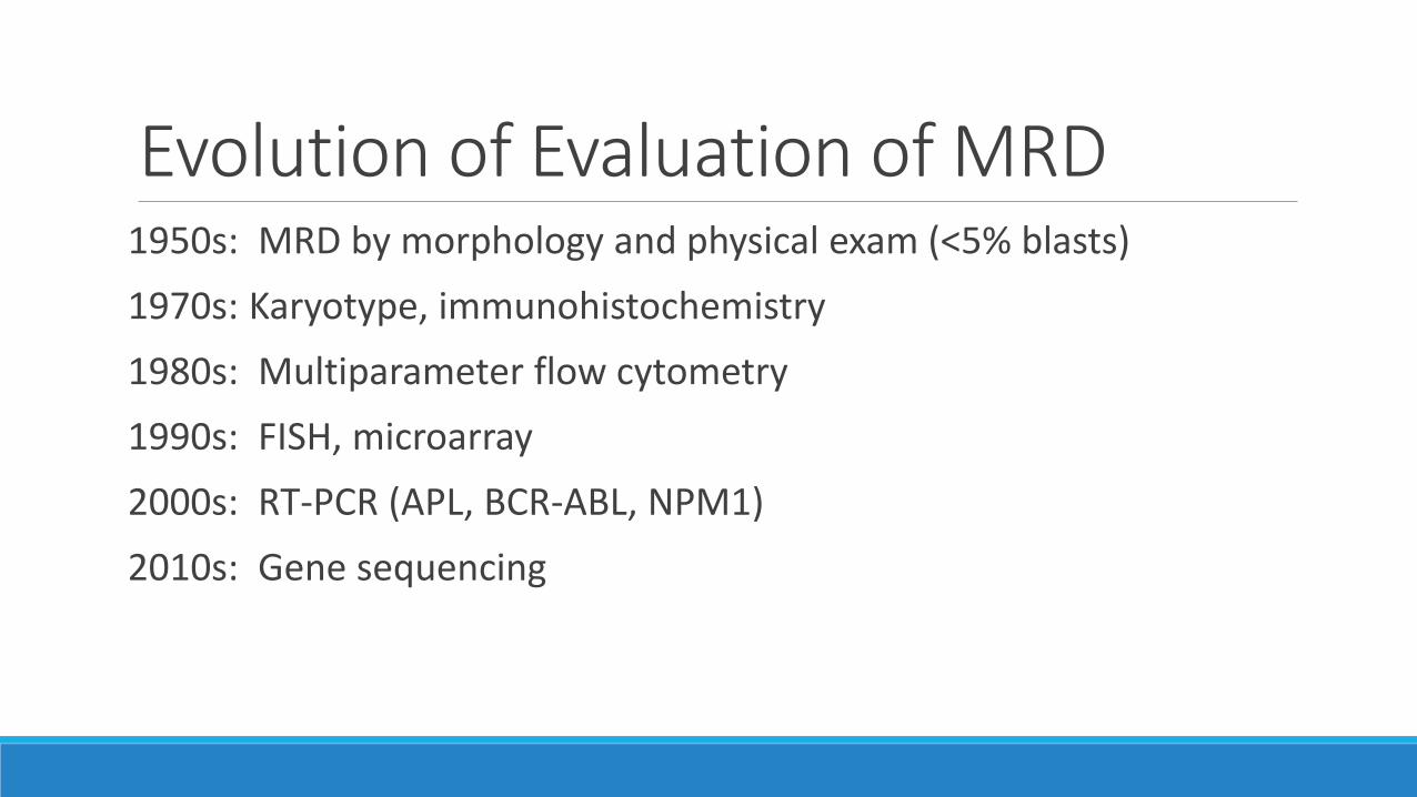
Evolution of Evaluation of MRD1950s: MRD by morphology and physical exam (<5% blasts)
1970s: Karyotype, immunohistochemistry
1980s: Multiparameter flow cytometry
1990s: FISH, microarray
2000s: RT-PCR (APL, BCR-ABL, NPM1)
2010s: Gene sequencing

Limited Number of Recurrent Mutations in MDS

Limited Number of Recurrent Mutations in AML

Recurrent Abnormalities in AML

Minimal Residual Disease in AMLs•Molecular risk stratification and evaluation for minimal residual disease relies on identification of abnormalities at diagnosis
•Repeat sequencing at 30 days after 7+3 induction to determine clearance of abnormalities◦ Positive MRD on day 14 after intensive induction chemotherapy is not
invariably associated with treatment failure
•Patients with mutation detection at variant allele frequency (VAF) greater than 2.5% at day 30 almost all died within 5 years

Minimal Residual Disease in AMLs•Doesn’t matter the mutation, detection at day 30 is a bad prognosticator◦ Positive MRD test identifies patients with
higher risk of relapse and shorter survival than similarly treated patients in morphological remission who test negative for MRD

WHO 2016 Updates-Myeloid•New Chapters
◦ Mast cells
◦ Myeloid/lymphoid neoplasms with eosinophilia and gene rearrangement
◦ Myeloid neoplasms with germline predisposition
•MDS Categorization◦ Elimination of “refractory anemia”
◦ SF3B1

Case Presentation•67 year old with macrocytic anemia, rule out lymphoma and myeloma◦ Iron studies, B12 level, and folate level are within normal limits
◦ Flow cytometry: No discrete blast population is identified by CD45 versus side scatter. There is no evidence of B-cell surface light chain restriction or aberrant T-cell immunophenotype. Plasma cells are identified by bright CD38 and CD138 expression and they show no evidence of cytoplasmic light chain restriction.
◦ Normal cytogenetics and MDS FISH panel


Ring sideroblastsOne in every 20 erythroid cells
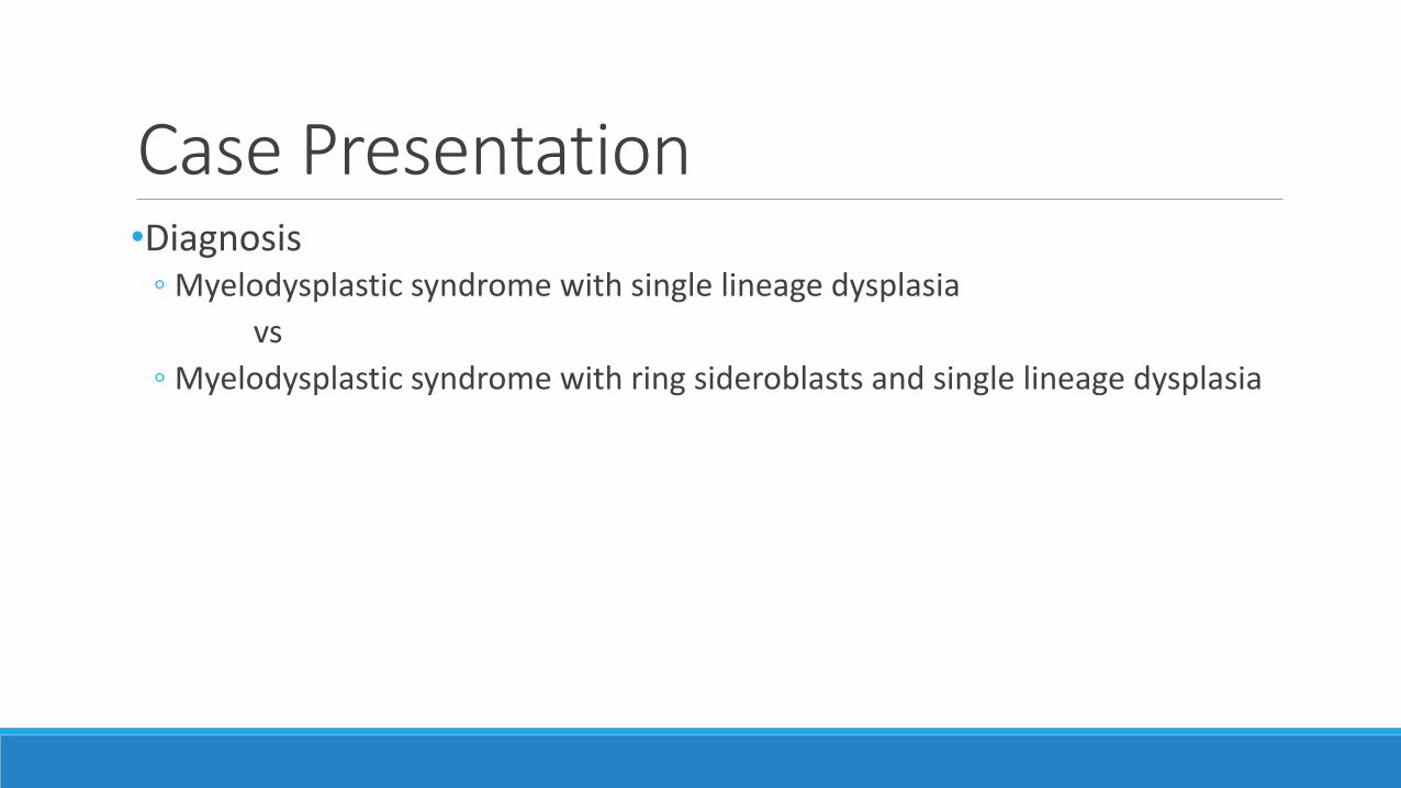
Case Presentation•Diagnosis
◦ Myelodysplastic syndrome with single lineage dysplasia
vs
◦ Myelodysplastic syndrome with ring sideroblasts and single lineage dysplasia

Lymphoid

WHO 2016 Updates-Lymphoid•High grade B-cell lymphoma with MYC and BCL2 and/or BCL6 rearrangements
•Mutations identified◦ Hairy cell leukemia
◦ Lymphoplasmacytic lymphoma

Clinical Relevance of Mutational Profiles in Lymphoid Neoplasms•Diagnostic criteria to refine entities
•Identification of patient subsets
•Prognostic and predictive significance
•Monitoring disease evolution
•Identification of actionable mutations

Early and Late Mutations in Follicular Lymphoma•Early driver mutations in chromatin regulator genes◦ CREBBP
◦ EZH2
◦ KMT2D (MLL2)
•Mutations gained at transformation◦ MYD88
◦ TNFAIP3

Somatic Mutations in LPLMYD88 CXCR4 BTK
95% of LPLs/Waldenstrom’sMyeloid Differentiation Factor 88
Activates BTKIncreases cell survival
25-40% of LPLs/WMC-X-C chemokine receptor 4
Often assoc. with MYD88More active disease
Less lymphadenopathyMore resistant to treatment
Bruton’s Tyrosine KinaseAdditional mutations prior to
progressionIbrutinib
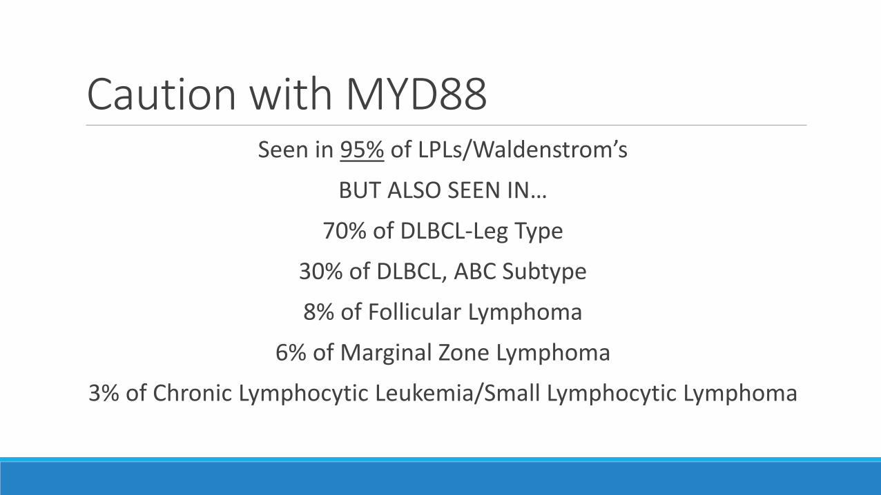
Caution with MYD88Seen in 95% of LPLs/Waldenstrom’s
BUT ALSO SEEN IN…
70% of DLBCL-Leg Type
30% of DLBCL, ABC Subtype
8% of Follicular Lymphoma
6% of Marginal Zone Lymphoma
3% of Chronic Lymphocytic Leukemia/Small Lymphocytic Lymphoma

Recurrent Mutated Pathways in Small B-Cell Lymphomas
Pathway U-CLL M-CLL MCL FL LPL MZL HCL
DNA Damage + +
SF3B1 + +/-
NOTCH1/2 + +/- + +/- +
Chromatin remodeling + +
BCR-Signaling + +/-
NFkB + + +/-
MYD88 +/- +/- +
MAPK +

What To Do?

CAP Guidelines for AML•Published in December 2016
•Panel of experts and advisors in hematology and hematopathology◦ EP: 7 pathologists, 1 hematologist, 1 hem/onc, 1 method consultant
◦ AP: 1 pt advocate, 1 cytogeneticist, 3 heme/onc, 1 med onc, 2 hemepath
•Recommendations derived from strength of evidence and feedback during a public comment period

CAP Guidelines for AMLDesignation Recommendation Rationale
Strongrecommendation
Recommend for, or against, a particularpractice. (Can include ‘‘must’’ or ‘‘should.’’)
Supported by convincing or adequate quality of evidence and clear benefit that outweighs harms.
Recommendation Recommend for, or against, a particularpractice. (Can include ‘‘should’’ or ‘‘may.’’)
Some limitations in quality of evidence, balance of benefits and harms, values, or costs, but panel concluded that there is sufficient evidence and/or benefit to inform a recommendation.
Expert consensusopinion
Recommend for, or against, a particularpractice. (Can include ‘‘should’’ or ‘‘may.’’)
Serious limitations in quality of evidence, balance of benefits and harms, values or costs, but panel consensus was that a statement was necessary.
No recommendation
No recommendation for, or against, a practice. Insufficient evidence or agreement of the balance of benefits and harms, values, or costs to provide arecommendation.

Guideline Designation
1 Clinical data/patient history provided by treating clinician Strong
2 PE and imaging data provided by treating clinician Recommendation
3 Pathologist review of CBC and diff Strong
4 Review of bone marrow aspirate and core biopsy Strong
5 Flow cytometry, cytogenetics, molecular studies Strong
6 Cytochemical stains (PAS, MPO, NSE) Consensus
7 Molecular/genetic studies on nucleic acid, cropreserved, FFPE tissue Recommendation
8 Pathologist review of CSF analysis/differential in ALLs Strong
9 Pathologist review of CSF analysis/differential in AMLs Consensus
10 Use of flow cytometry in analyzing CSF Recommendation
11 Tissue evaluation in patients with extramedullary disease Strong
12 Ensure thorough flow cytometry/cytogenetics/molecular for MRD Strong
13 Pediatric B-ALL: t(12;21), t(9;22), KMT2A (MLL), iAMP21, +4, +10 Strong
14 Adult B-ALL: t(9;22) Strong
15 Testing for PAX5, JAK1, JAK2, IKZF1, NOTCH1, FBXW7, CRLF2 Recommendation

Guideline Designation
16 FLT3 testing in AML Strong
17 KIT mutation analysis in adults with CBF-AML Strong
18 In suspected APL, rapid t(15;17) detection and testing for DIC Strong
19 AML patients should be tested for NMPM1, CEBPA, RUNX1 Strong
20 Testing for methylation, microRNA/gene expression in acute leukemias No recs
21 Mixed phenotype AMLs should have t(9;22) and KMT2A (MLL) testing Strong
22 All testing performed in labs with regulatory compliance Strong
23 Patients requiring immediate referral to institution with expertise, initial institution should, defer invasive procedures to avoid duplicateprocedures, associated patient discomfort, and additional costs
Strong
24 If patient is referred, provide all labs, slides, flow/CG data, pending tests Strong
25 Initial report should include laboratory, morphologic, IPT data, on which the diagnosis along with a list of any pending tests.
Strong
26 Ensure all tests are entered into the medical record Strong
27 Use the current WHO terminology Strong

MUSC Evaluation in New Leukemias•AMLs
◦ Peripheral Blood: morphology, flow cytometry
◦ Bone Marrow: morphology, flow cytometry, stat FISH t(15;17), karyotype, microarray, next gen sequencing panel
•ALLs◦ Peripheral Blood: morphology, flow cytometry
◦ Bone Marrow: morphology, flow cytometry, stat FISH t(9;22) and KMT2A, karyotype, additional FISH, microarray
◦ CSF: morphology, +/- flow cytometry
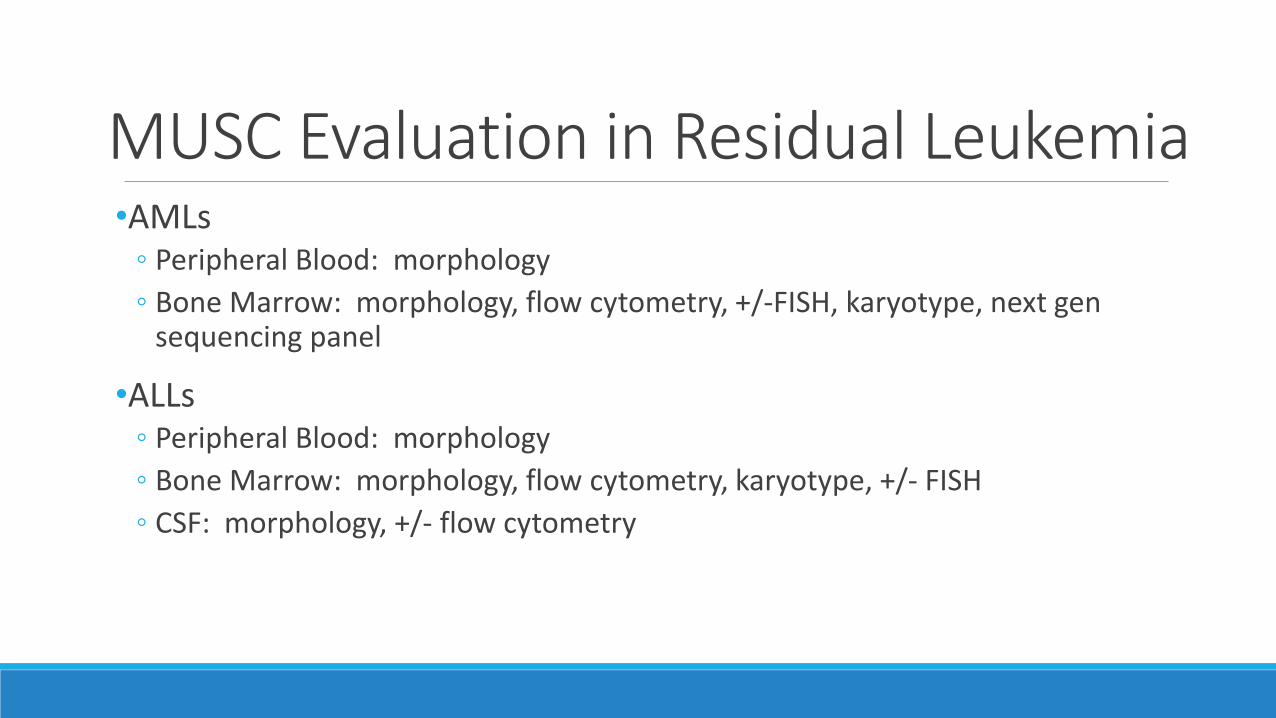
MUSC Evaluation in Residual Leukemia•AMLs
◦ Peripheral Blood: morphology
◦ Bone Marrow: morphology, flow cytometry, +/-FISH, karyotype, next gen sequencing panel
•ALLs◦ Peripheral Blood: morphology
◦ Bone Marrow: morphology, flow cytometry, karyotype, +/- FISH
◦ CSF: morphology, +/- flow cytometry

MUSC Evaluation of Lymphomas•Morphology and immunohistochemistry
•Flow cytometry
•Karyotype
•FISH
•Molecular testing: BRAF V600E, MYD88

Acknowledgements•Daynna Wolff, PhD
•Iya Znoyko, PhD
•W. Bailey Glen, PhD
•Tara Ellingham

Thank you!!!