mold and mycotoxins: effects on the neurological and ... and mycotoxins...in addition,...
TRANSCRIPT
JIT-ISJJ, ed.), pp. 94-98. Eastern.-•star. New York..r.c Sliwinski, M. (20OO). The
r. individuals with traumatic
Ashman. T. A., Breeze, }.,- impairment associated with:n of previous findings. Appl.
•-•:' :r:teliectual functioning after
"i edification and Assessment>v Vork.u v., sort. In "Foreosic Neuro-: Ziiiiinger, The Netherlands.-: D., Jarvis, B., Robbias, H..
jr.ed pulmonary disease from. s.-s/cofor. /, Occup. Environ.
ka:man, J. (2O01). The Word: malingered memory deficits.
Si. T. (19B8). How long does itv 22, 853-858.o.-;e. K., Dalberg, C., Gerber, D..: :iuld traumatic brain injury.
aencits: What is really wrong?145-183. Swets and Zeitlinger,
:'J «d Oxford University Press,
^iil. L. F., Tabaddor, K., High.:u :come following minor head
>ical Management of Mild TBI"a University Press, New YorL• in the aged. In "Handbook for
ed.). pp. 2Z&-237, American
-rc,;ogical diseases. In "Clinicalt (P.}. Snyder and P. D. Nuss-•Aociation. Washington, DC.ged building: An intervention
The Psychological Corporation,
h
iMold and Mycotoxins: Effects on the Neurological and
Immune Systems in Humans
ANDREW W. CAMPBELL,* JACK D. THRASHER,* MICHAEL R. GRAY,* ANDAiUSTO VOJDANI8
"Medical Center for Immune and Toxic Disorders, Spring, Texas
fSam-J Trust, Alto, New Mexico
^Progressive Health Care Group, Benson, Arizona^Immunosciances Laboratories, Beverly Hills, California
1. Introduction 3/bD. Water Damage aad Associated Molds 378
A. Mycobiota 378B. Mycotoxins Produced fay Toxigenic Molds 378C. Human Exposure 380
HI. Symptoms, Upper and Lower Respiratory Tract 380A. Symptoms 380B. Upper Respiratory Fungal Infections 3fl2C. Lower Respiratory Tract 383D. Proinflanimatory Cytokines and Biomarkers 385
IV, IgA, IgC, and igE Antibodies to Molds and Mycotoxins 385A. Salivary IgA Antibodies to Molds 385B. Serum IgA, IgM, IgG, and IgE Antibodies to Molds 386C. Cross-Reactivity of Antibodies to Molds 387D. Antibodies to Extracellular Polysaccharides (EPS) 390
V. Alterations in T and B Cells, Natural Kilior (NK) Cells, and OtherImmune Parameters in Humans Exposed to Toxigenic Molds 390A. Alterations in Percentage of T and B cells 390B. Mitogen Activity 391C. Autoantibodies 392D. Immune Complexes 392E. Concluding Remarks on Immunological Observations 393
VI. Neurological Abnormalities 393A. Neurocognitive Deficits and Central Nervous System Dysfunction 394B. Peripheral Motor and Sensory Neuropathy 396C. Neuronal Antibodies 39f;D. Demyelioation of Peripheral Nerves 397
VII. Conclusion 397References 399
!. Introduction
The potential harmful effects of exposure to molds in inhabitedbuildings were recognized in early Biblical times. In the Old Testa-ment {King James Version, Oxford 1888 Edition, Chapter XIV: Vorses
375
ADVANCES IN APPLIED M5CROBKJLOCY. VOLUME 55Ojprngh! 20&4. Ei*evier IDC.
A!! fights naiervsd,OOfc',-2H»4/!>4 $35.00
376 ANDREW W. CAMPBELL at a/.
34 thru 47} Leviticus put forth a detailed protocol for the remediationof contaminated structures, including the destruction of dwellings andpersonal belongings if remediation failed. Currently it is recognizedthat water intrusion into buildings leads to amplification of molds(Andersson et aL, 1997; Gravesen ef aL, 1999; Hodgson et aL, 1998;Jaakkola et aL, 2002; Johanning et aL, 1996; Nielsen, 2003; Peltola et aL,2001), which often requires remediation.
Fungal fragments occur in indoor air as biocontaminants (Burge, 1990;Gorney et aL, 2002). Potentially toxic and immunogenic byproducts offungi and molds include mycotoxins (Croft et aL, 1986; Johanning et aL,2002; Nielsen et aL, 1999; Nieminen et al., 2002; Tuomi et aL, 1998,2000); 1,3-alpha-D-gIucans (Andersson etal,, 1997), extracellular poly-sacharrides (EPS) (Duowes etal,, 1999; Notermans etal., 1988; Wouterset aL, 2000); exodigestive enzymes (EDS) (Monod et aL, 2002), andsolvents (Claeson et al., 2002). In addition, trichothecenes. ochratoxinA, sterigmatocystin, and other mycotoxins have been identified in venti-lation duct dust and in the air in buildings where occupants have experi-enced adverse health effects related to mold exposure (Croft et al., 1986;Engelhart et aL, 2003; Jarvis, 2002; Johanning et aL, 2002; Nieminenef al., 2002; Skaug et aL, 2000; Smoragiewicz et a!., 1993; Tuomi et aL,1998). The worst-case scenario appears to be repeated episodes ofwater damage that promote fungal growth and mycotoxin production,followed by drier conditions leading to release of spores and hyphalfragments (Nielsen, 2003).
Occupants of affected structures develop multiple organ symptomsand have adverse effects of the upper and lower respiratory system,central and peripheral nervous system, skin, gastrointestinal tract,kidneys and urinary tract, connective tissue, and the musculoskeletalsystem (Anyanwu et al., 2003a; Croft et al., 1986; Gunnbjornsdottiret al., 1998; Gray et al., 2003; Hodgson et aL, 1998; Jaakkola et aL,2002; Johanning et aL, 1996; Kilburn, 2002; Sailvilahti et aL, 2000).Human illness caused by fungi can result via one or all of the following:(1) mycotic infections (mycoses) (Anaissie et al., 2002; Eucker et al.,2001; Fraser, 1993; Grossi etal., 2000), (2) fungal rhino-sinusitis (Braunet aL, 2003; Ponikau et aL, 1999; Thrasher and Kingdom, 2003), (3) IgE-mediated sensitivity and asthma (Barnes ef al., 2002; Lander et al.,2001; Zureik et aL, 2002), (4) hypersensitivity pneumonitis and relatedinflammatory pulmonary diseases (Erkinjuntti-Pekkanen et al,, 1999;Ojanen, 1992; Patel et aL, 2001; Sumi ef al., 1994). (5) cytotoxicity(Desai et aL, 2002; Gareis, 1995; Jones et aL, 2002; Nagata et al., 2001;Poapolathep et aL, 2002), (6) immune suppression/modulation (Bereketal., 2001; BondyandPetska, 2000; JakabetoJf., 1994), (7) mitochondria!
toxic il\n *
Malauocity (A.format:Hsieh.etal., ',digesththromb1985;Ket aL, 2
The ichybotrbeen chinfant pase (sta.has alsrrelatedbeen destallaficin coneindicatiet aL, 2quandtimodel,;leakage2002 j". .-imonaryF in juvwith coidistend*observesMcCraeand pro*of mold
Reccepie moletion of p;coloniesenvironxtoring deroutine -
MUUJ AND MYCJUTUXINS '.I'/'/
for the remediationon of dwellings andtly it is recognizedilifi cation of molds;dgson et al., 1998;, 2Q03;Peltolaet al.,
inants (Surge, 1990;genie byproducts of86; Johanning et al.,Tuomi et al., 1998,extracellular poly-
-ra/.,1988;Woutersi et al., 2002), and^scenes, ochratoxin: identified in venti-upants have experi-:e (Croft era/., 1986;il, 2002; Nieminen1993; Tuomi eta].,
peated episodes ofrotoxin production,spores and hyphal
ie organ symptomsrespiratory system,strointestinal tract,he musculoskeletal>: Gunnbjornsdottir98; Jaakkola et al,,iiahti et al., 2000).all of the following:1002; Eucker et al.,no-sinusitis (Braundom, 2003), (3) IgE-002; Lander et a].,monitis and relatedianen et al., 1999;•4), (5) cytotoxicityXagata et al., 2001;modulation (Berek
;),(7)mitochcradrial
toxicity (Hoehler, et al., 1997; Niranjan et al, 1982; Pace, 1983, 1988;Sajan et al., 1997; Wei et al., 1984), (8) careinogeriicity (Dommguez-Malagon and Gaytan-Graham, 2001; Schwartz, 2002), (9) nephrotoxi-city (Anyanwu et al., 2003c; Pfohl-Leszkowicz et al., 2002), (10) theformation of nuclear and mitochondria! DNA adducts (Hsieh andHsieh, 1993; Petkova-Bochatrova et al., 1998; Pfhlohl-Leszkowiczet al., 1993). Finally, in the infectious state, molds secrete extracellulardigestive enzymes (EDE) that cause tissue destruction, angioiiivasion,thrombosis, infarction and other manifestations of mycosis (Ebina et al.,1985; Kordula et aJ., 2002; Kudo et al., 2002; Monod et al., 2002; Ribesef al, 2000; Vesper et al., 2000).
The pathological and inflammatory conditions associated with Sta-chybotrys chartarum in infants with pulmonary hemosiderosis havebeen characterized. S. chartarum isolated from the lungs of an affectedinfant produced a heinolysin (stachylysin), a siderophore, and a prote-ase (stachyrase) (Kordula et al., 2002; Vesper et al, 2000). Stachylysinhas also been demonstrated in the serum of adults ill from a building-related exposure (Von Emon et al., 2003). In rodents, its presence hasbeen demonstrated by an immunocytochemical method following in-stallation of S. chartarum spores into lungs. The hemolysin increasesin concentration from 24 to 72 hours following instillation of spores,indicating that production/release is a relatively slow process (Gregoryet al., 2003). In addition, strains of S. chartarum produce differentquantities of toxic trichothecenes 0arvis et al, 1998). In an earthwormmodel, stachyiysin increased the permeability of blood vessels, causingleakage through the vessel endothelium and walls (Vesper and Vesper,2002). Additionally, pathology may result from the interference of pul-monary surfactant synthesis by S. chartarum spores and isosatratoxin-F in juvenile mice. Ultrastructural changes in type II alveolar cells—with condensed mitochondria, increased cytoplasmic rarefaction, anddistended lamellar bodies with irregularly shaped lamellae—have beenobserved following exposure to S. chartarum (Mason eta/., 1998, 2001;McCrae et al., 2001; Rand et al, 2001). Thus, hemolysins, siderophores,and proteases also appear to have an important role in the pathogenesisof mold infections.
Recognizing the complexity of health problems associated with multi-ple mold exposure, we have previously reported a multi-center investiga-tion of patients with chronic health complaints from exposure to multiplecolonies of indoor fungi and molds. We utilized detailed health andenvironmental history-gathering questionnaires, environmental moni-toring data, physical examination, pulmonary function testing protocols,routine clinical chemistries, measurements of lymphocyte phonotypic

378 ANDREW W. CAMPBELL et al.
markers (on T, B, and NK ceils), antibodies to molds, mycotoxins,neuronal antigen antibodies, leukocyte apoptosis, neurocognitive test-ing, 16-channel quantitative EEGs (QEEG), nerve conduction studies(NCS), brainstem auditory evoked potentials (BAER), visual evokedresponses (VER), and other neurological testing. The following is a sum-mary of our findings on symptoms, pulmonary function, alterations inperipheral lymphocyte phenotypes, autoantibodies, and neurologicalabnormalities observed in patients by us and others. Currently we referto the illness of these individuals as a "mold mycotoxicosis" involvingthe immune system, the lungs, the central and peripheral nervous sys-tems, and generalized inflammatory and irritant responses to exposure tospores, hyphal fragments, mycotoxins, solvents, and other byproducts(e.g., EPS, EDS).
II. Water Damage and Associated Molds
A. MYCOBIOTA
Water intrusion into buildings can lead to an amplification of molds.Molds growing on building materials (e.g., wall board, particle board,plaster board, ceiling tiles, carpeting) are classifiable according to theirwater activity, aw (Nielsen, 2003) as follows: (1) primary colonizershave an aw of <0.8 with an optimal water requirement approaching 1for growth. The group includes Penicillium chrysogenum and Aspergil-Jus versicohr, followed by other species ofAspergiHus {niger, fumigatus,sydowii, ustus}, several Eurotium species, Penicillium species (brevi-cotnpactum, commune, corylophilum, pelicans], Paecilomyces variottiand Wallemia sebi. (2) Secondary colonizers requiring a minimum ofbetween 0.8 and 0.9 aw include species of Altemaria, Cladosporium,Phoma, and Ulocladium. (3) Tertiary colonizers (water-damage molds)that require 0.9 aw or greater include the most toxic molds: Chaetomiumgloblosum, Stachybotrys chartarum, Memnoniella echinata, and Tricho-derma species (viride, citrinoviride, harzianum and longibrachiaium).For a more detailed review, see Nielsen (2003).
Stachvtx-charta
Altft.-naritenuisf.
E. MYCOTOXINS PRODUCED BY TOXIGENIC Mouxs
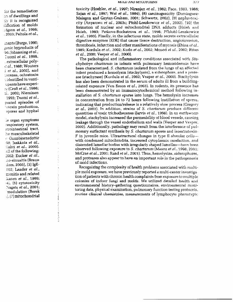
Fungi produce many metabolites, which are believed to play a cru-cial role in their natural habitats. In addition, many of the metaboliteshave been identified. Those that are toxic to animals and humans arecalled mycotoxins. Paradoxically, antibiotics isolated from molds aremycotoxins and are beneficial to humans. Table I lists the molds com-monly found in water-damaged buildings and the toxic metabolites
,i. mycotoxins,ocognitive test-auction studies
visual evoked-nvingisasum-
~, alterations inr.d neurological;rrendy we refer:csis" involving-rai nervous sys-•c s to exposure totner byproducts
5
^cation of molds.i, particle board,.: cording to their:mary colonizers..1 approaching 1-m and AspergU-niger, fumigatus,
zi species (brevi-cilomyces variotting a minimum of>z. Cladosporium,er-damage molds)"ids: Chaetomiumfnafo.and Tricho--'ongibrachiatum].
". ^>LDS
:ved to play a cru-of the metabolites
is and humans are»d from molds tire
.1 . 1 _3 _ — *..»
MOLD AND MYCDTOXINS 3 79
TABLE I
TOXIGENK: MOUJS IN WATER-DAMAGED BUTI.BINCS
Mold
Stachybotryschartarum
Altetnariaienuissima
Aspargillusflaws
Aspergillusfumigatus
Aspergillus niger
Aspergillusochraceous
Aspergillus ustutsAspergillas
versicolor
Penicilliumchn'sogenum
Chaetomiumglobosum
Memnoniellaechinata
Penicilliumbrencompactutn
Penicilliumexpansum
Peniciiiiumpolonicum
Trichodennaspades
Metabolites
Spirocyclic drimanes;satratoxins G, H and F;hydroxyroridin E, verrucarin J;frinhodermin; dolabelianos;atrones B and C;stachyotryaaiide;stachyotrylactams; stachylysin
Altornariols; tentoxin;tenuazonk; acids; altertoxin £
Aflatoxin Bl; kojic acid;aspBrgillic acid;3-nitropropionic acid;cyclopiazonic acid
Fumigaclavines; fumitoxius;fumilreoiorgens; gliotoxins;trvploquivalines; varrucuiogen
Ochratoxin A
Ochratoxin A, peniciiiic acid;xajithomegnin: viomelleio,vioxanthin
KotaninsSterigmatocvstin;
5 - met hoxy-starigmatocy sti n
Secaloriic acid D
Chaeiomins;chaetnglobosins A and C
Griseofulvin;dcchiorogriseofulvins;trichodermin; trichodermol
Mycophenolic acid;botryodipioidin.
Patulin; cifrinin; chaetoglobosin;Roquefortine C
Varrur.osidins:peniciiiic acid;nephrotoxic glyt:opeptides
Trichothecones; txichodermo!;trichodermin; gliotoxin: viridin
Health concern
Pulmonary hemosiderosis;Induces proinflammatorycytokines
Unknown
Carcinogenegis;aspergillosis
Tremors and CNS injurv;immune damage bygiiotoxin: aspergiiiosis;
Nephropathv
Nepbropatby
UnknownCarcinogenesi s :
aspergiiiosis
Unknown
Cyloxicity; inhibition ofcell division
Unknown
Toxic (mutagenir.}
Immune toxicitv.cvtntoxic; trfitnorgnnic
Tremors; crtotoxiritr:riephropathv
Toxicity associatedwith irichothecane?
toxic metabolites This table summarizes (lie taxigenic molds found and/or identified in water-damaged building,-..Tlie mvcotoxins isolated from the molds aad thair generaj toxic effects axe also summarised i bt.information in this table was obtained from tile review Nielsen (2U03).
380 ANDREW W. CAMPBELL et al.
(mycotoxins) that tliey produce with general statements on their loxic-ity. Readers are referred to the literature cited in this chapter and inNielsen (2003) for more detailed information.
C. HUMAN EXPOSURE
Humans can be exposed to mycotoxins and metabolites of molds inthe indoor environment via (1) ingestion (contaminated foods, dirt, anddust) (2) the skin (contaminated clothing and surfaces), and (3) inhala-tion. Inhalation is the primary mode of exposure in the inhalation ofspores [3 to 7 ^m), hyphal fragments, and partieulate matter down to0.05 fj.m. It has been shown that particles smaller than spores can beshed from colonies of molds (Gorney et al., 2002; Kildeso etal., 2000).Large quantities of particles < 0.03 /an can be released from colonies,creating a 300-fold higher concentration of fungal fragments as com-pared with the number of spores released (Gorney et al., 2002). There isno apparent correlation between the number of particles and the num-ber of spores. Factors that influence the release of spores and particu-lates include low humidity (stimulates release), ventilation, externalwind speeds, human activity, and pressure shocks (e.g., elevators,doors). Finally, because it is difficult to quantify the partieulate mattershed by colonies, very few meaningful correlations have been foundbetween spore concentrations and adverse health effects on humansfrom indoor exposure to toxigenic molds (Nielsen, 2003). Thus biomar-kers for molds and mycotoxins have been and need to be furtherdeveloped for exposure assessment.
One successful approach has been to use DNA adducts to determineexposure to aflatoxin Bl (Makarananda el al., 1998) and ochratoxin A(Pfhohl-Leszkowicz, 1993a,b). However, another effective approach hasbeen the development of immune assays to detect the presence of anti-bodies to mold-specific antigens and mycotoxins. Also, an appreciationof the adverse health effects can be obtained by utilizing neurophysio-logical, neuropsychological, and immunological diagnostic procedures(see below).
I I I . Symptoms, Upper and Lower Respiratory Tract
A. SYMPTOMS
Occupants of water-damaged buildings express multiple organ symp-toms. Table II summarizes observations made on 209 adults exposed athome and/or at the workplace. Complaints significantly different fromcontrols occurred as follows: (I) central nervous system (headache,
Symptc-r.
Exrassive Fati^
Headache
Nasal Symptom
Memory Prcb'ei
Spaciness
Sinus Discomfc
Coughing
Watery Kycs
Throat DiscoraiSlurred Speer;;
Lightheaded nes
joint Discoinfor
Dizziness
Weakness
Bloating
InsomniaWeak VoiceSpasms
Coordination p-
Visual Char,.a-?i
Rash
Numbness
Cold Intolerant
Heat Intolerant
Chest Tightr.es:-
Ches! Discomfc
Urine Frequerx
Excessive Thin;
Kinging Ears
Wheezing
Swallowing Pr;:
Flushing Skla
Bladder Contro
Rapid Fuise

-i on their toxic-chapter and in
lites of molds ind foods, dirt, and
i.i, and (3) inhala-die inhalation ofe matter down to'.an spores can be.desoetaA, 2000).;ed from colonies,fragments as com-jL, 2002). There is,cles and the num-
• sores and particu-.--ntilation, external.ks (e.g., elevators,
-,- particulate matter!» have been foundeffects on humans
20U3J. Thus biomar-need to be further
d ducts to determine?) and ochratoxin A:rective approach hasvhe presence of anti-
Aiso, an appreciationLiiizing neurophysio-;iagnostic procedures
ratory Tract
• multiple organ symp-209 adults exposed at
Scantlv different fromus system (headache,
MOLD AND MYCXJTOXINS
TABLE II
FREQUENCY OF SYMPTOMS
381
Symptom"
Excessive Fatigue
HeadacheNasal Symptoms
Memory ProblemsSpaciness
Sinus Discomfort
CoughingWatery Eyes
Throat DiscomfortSlurred SpeechLightheadednessJoint DiscomfortDizziness
Weakness
BloatingInsomniaWedt VoiceSpasmsCoordination ProblemsVisual Changes
Rash
NumbnessCold IntoleranceHeat IntoleranceChest TightnessChest Discomfort
Urine FrequencyExcessive Thirst
Ringing Ears
WheezingSwallowing Problems
Flushing SkinBladder ControlRapid Pulse
Mold Patients(N = } 209
5,8 ±1.9
5.2 ± 1,9
5.1 ± 2,2
5.1 ± Z.I4.8 ± 2.3
4.7 ± 2.2
4.6 ± 2.2
4.6 ± 2.1
4.5 ± 2,1
4.5 i- 2.3
4.4 ± 2.24.4 ± 2.34.3 ± 2.1
4.2 ± 2.3
4.2 dt 2.24.1 ± 2.2
4.1 ± 2.2
4 ± 2.2
4 ± 2.2
3.9 ± 2.3
3.9 ± 2.2
3.9 ± 2.2
3.9 ± 2.43.8 ± 2.4
3.8 ± 2,23.7 * 2.23.7 ± 2.3
3.6 ± 2.3
3.6 i 2.2
3.6 ± 2
3.2 ± 2
3.1 ± 2.13.1 ± 2
3 ± 2
Controls N = 28
4.3 ± 2.14.1 ± 2
4.1 ± 2
3.3 ± 1.63.2 ± 1.8
3.6 ± 1.8
3.2 i 1.6
3.4 1 1.7
3.4 ± 1.7
3.1 ± 2
3.2 ± 1.4
3,7 ± 2.1
3.1 ± 1.4
3 ± 1.7
3.2 ± 1.6
3.8 ± 2
2.8 ± 1.4
3.8 ir 2.1
2.9 ± 1.4
2.9 ir 1.4
2.9 ± 1.7
3.4 ± 1.7
3.1 ± 1.8
3.8 ± 2
2.6 ± 1.33 ± 1.3
3.8 ± 2.1
3.4 i 2
4.4 ± 2.4
2.6 ± 1.3
3 + 1.7
2.8 £ 1.82.8 =: 1.42.6 ± 0.9
F value
O.OOOi
0.005
0.02
0.0002
0,0007
0.01
0.001
0.004
0.008
0.002
0.006
NS
0.005
O.O08
0,02
NS
0.003
NS
0.01
0.02
0.02
NS
NSNS
0.006
NSNS
NS
NS
0.02
NS
NSNSNS
382 ANDREW W. CAMPBELL et a!.
TABLE!! (ConHnued}
Moid PatientsSymptom"
Palpitations
Bruising
Swelling Ankles
Hearing Changes
(N - )
2.8 T.
2,8 ±2.7 ±.
2,7 i
209
1.9
1.7
1.8
1.8
Controls N — 28
2.4 ± 0.8
2.4 i 0.9
2.6 ± 1.5
2.6 ± 1.5
P value
NS
NSNSNS
This table summarizes the frequency of symptoms of the 38 most frequently reported symptoms inthe patients vs the controls. To obtain these data, a total of 209 patients filled out questionnaire*.Critical t-test analysis was peribrm&d and p values are given for each symptom of patients vs rx>ntrols(NS = Nat Significant).
"Th« symptoms compared wore for femalei versus males. The females had significantly greaterfrequency for 21 of the 38 reported symptom* (data not shown; see Results section].
short-term memory loss, lightheadedness, dizziness, blurred vision,tinnitus, and cognitive function loss), (2) the upper respirator}1' tract (nasalcongestion and chronic sinusitis), (3) the lower respirator}' tract (cough,wheezing, chest tightness, exertional dyspnea, and irritation of thethroat), and (4) general ill feeling (excessive fatigue, weakness, jointaches and pains, and rashes) (Campbell et al, 2003; Gray et a/., 2003).In addition, others have shown similar increases in the incidence ofneurological and respiratory symptoms in individuals ill from moldexposure in water-damaged buildings (Hodgson et al., 1998; Johanninget al, 1996; Kilburn, 2002; Vojdani et al., 2003). Vojdani et al, (2003)reported that patients exposed to molds had significant increases inrecurrent flu-like illnesses, anxiety, and symptoms of severe allergies.It has become increasingly obvious that exposure to multiple toxigenicmolds in water-damaged buildings leads to an increased incidence ofmultiple organ symptoms in the affected individuals.
B. UPPER RESPIRATORY FUNGAL INFECTIONS
Symptoms of upper respiratory involvement include nasal conges-tion, sinusitis, sinus pain, and nasal bleeding (chronic rhinosinusitis}.Individuals with this condition do not respond to ordinary antibiotictherapy.
Several reports have appeared in the literature demonstrating that alarge proportion of individuals with chronic rhinosinusitis (CRS) haveinfections with molds and yeast. CRS is characterized by the presenceof eosinophilic mucin, fungal hyphae, Charcot-Leyden crystals, andthe presence or absence of polyposis (Ponikau et al., 1999; Taylor et al..
MOLD AND MYCOTOXINK 383
Is N =- 28 P value
4 = 0,8
.. - 0.9
.6 ~ 1.5
.- = 1.5
NSNSNSNS
reported symptoms in. filled out questionnaires.
-. iymptom of patients vs controls
rr.iaales had significantly greateri Results sacticm).
tziness, blurred vision,er respiratory tract (nasalrespiratory tract (cough,a. and irritation of thefatigue, weakness, joint2003; Gray et al, 2003).ises in the incidence ofiividuals ill from moldnet al., 1998; Johanning3). Vojdani ef al. (2003)significant increases in
toms of severe allergies.ire to multiple toxigenic; increased incidence ofiduals.
,FECTIONS
it include nasal eonges-(chronic rhinosinusitis).id to ordinary antibiotic
ure demonstrating that aainosinusitis (CRS) havecterized by the presence:ot-Leyden crystals, andet al., 1999; Taylor et al.,
2002). The incidence of fungal involvement in different case studies was82% to 100% (Braun et al., 2003; Dosa et al., 2001: Pordkau et al., 1999)and 100% (Taylor etaL, 2002). The fungal genera isolated and culturedfrom nasal secretions include such indoor contaminants as Aspergiilussp., Alternaria, Chaetomium, Cladosporium, Epicoccum, Penicillium,Phoma, Trichoderma, and others (Dosa etaL, 2002; Ponikau et al., 1999;Taylor et al., 2002). The isolation of fungi and the presence of eosiuo-phils and eosinophilic mucin rule out type I (IgE) hypersensitivity(allergy) and strongly point to the role of invasive fungi as the cause ofCRS (Braun et al., 2003; Ponikan et al.. 1999).
C. LOWER RESPIRATORY TRACT
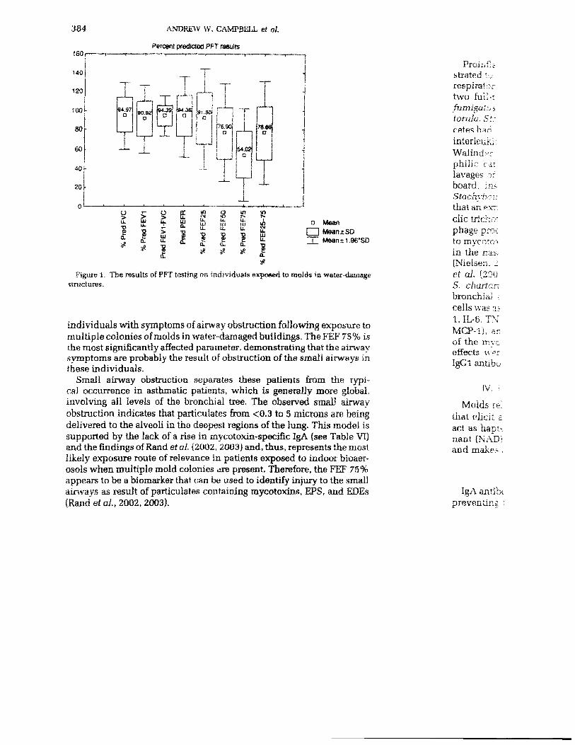
Molds can cause lung disease by different mechanisms: allergic asthma(Jaakkola et al., 2002; Zureik et al, 2002), infections (e.g., aspergillosis)(Eraser, 1993; Sumi etaL, 1994), and inflammation (e.g., hypersensitivitypneumonitis, and farmer's lung disease) (Fan, 2002; Ojanene. 1990,1992; Patel et al., 2001). Chest x-rays can be used to detect pathologicalchanges associated with infections (e.g., aspergillosis and granuloma-tous lesions}. Pulmonary function testing (PFT) is used to diagnoseairway restriction caused by allergies to molds as well as inflammatoryconditions (hypersensitivity pneumonitis and farmer's lung disease).PFT measures flow rates in the airways of the lungs. The forced vitalcapacity (FVC) is the maximum amount of air expelled during Forcedexpiration. The fraction of the vital capacity expired in one secon d is theFEVj. The importance of these measurements arises from the fact thatduring disease states, (e.g., asthma), the FVC may be normal while theFEVj is reduced because of increased airway resistance. However, thesetwo measurements do not discriminate between the airways of differentcaliber and therefore are not able to distinguish between the stat us of thelarge, medium, and small airways. Airborne particulate matter andspores (bioaerosols) from fungi range from 0.03 to 10 microns. "Respira-ble particles" range from 5 microns to 0.005 microns and are capableof reaching the small airways and alveoli of lungs. Therefore, PFTmeasurements used must also detect inflammatory or obstructivechanges within the small airways. The PFT measurements most suiisdfor small airway obstruction are FEF 75% and FEF 25-75%. Thesemeasure the flow rates at 75% and 25-75% of the exhalation and areindicative of air flow through the small airways. A reduction in thesePFT values is evidence of small airway obstruction. The results pre-sented in Fig. 1 show the mean and standard deviation of PFT values in.
384 ANDREW \V. CAMPBELL et al.
Percent predicted PFT results
140
120
100
80
60
40
T94.37
0
. 1
UUL
10.
T90.92
O
i
UJ
SF
T94.33
1
£
>(Jj
§
_
^
94.3^o 1
j
£Ul
1OL
Ti
•y*!i
j±
!f>
su.
a8«
"a
"
SASS
.
8 £u. it.Ill 1U
1
r •D
•
sJ> D Mean
U. U. £
a: a. -5if J
[HI MeantSD~j~ Mean ±1.96*80
Figure 1. The results of PFT testing on individuals exposed to molds in water-damagestructures.
individuals with symptoms of airway obstruction following exposure tomultiple colonies of molds in water-damaged buildings. The FEF 75% isthe most significantly affected parameter, demonstrating that the airwaysymptoms are probably the result of obstruction of the small airways inthese individuals.
Small airway obstruction separates these patients from the typi-cal occurrence in asthmatic patients, which is generally more global,involving all levels of the bronchial tree. The observed small airwayobstruction indicates that particulates from <0.3 to 5 microns are beingdelivered to the alveoli in the deepest regions of the lung. This model issupported by the lack of a rise in mycotoxin-specific IgA {see Table VT)and the findings of Rand et al. (2002, 2003} and, thus, represents the mostlikely exposure route of relevance in patients exposed to indoor bioaer-osols when multiple mold colonies are present. Therefore, the FEF 75%appears to be a biomarker that can be used to identify injury to the smallairways as result of particulates containing mycotoxins, EPS, and EDEs(Rand et al,, 2002, 2003).
Proinfhstrated '•,respiratortwo full-:fumigutuatorula, St~.
interleuki:Walind»rphilic ratlavages ofboard, ins-StachytKt:thatanexr.clic tricho'phage pro*:to mycoto>in the nas,(Nielsen. 2et al. (200S. chartanbronchial ;cells was as1, IL-6. TN.MCP-1). arof the mvceffects werIgGl antibo
IV. I
Molds K-that elicit aact as hapunant (NAD)and make>
IgA antibcpreventing *
MOLD AND MYCOTOXINS
D. PROINFLAMMATOKY CYTOKINES AND BIOMARKEKS
385
a MeanI""] MeantSDHI Mean±1.96*SD
aids in water-damage
3 wing exposure to;s.TheFEF75%isjig that the airwaye small airways in
ts from the typi-cally more global,rved small airwaymicrons are being
ung. This model isIgA {see Table VI)
represents the most;d to indoor bioaer-efore, the FEF 75%injury to the small
Proinflammatory cytokines and other biomarkers have been demon-strated to be elevated in the nasal lavage fluid of individuals with upperrespiratory symptoms in moldy buildings versus control subjects. Thirty-two full-time employees in a school building contaminated with A.fumigatus and A. versicolor, Eurotium, Exophiala, Phialophora, Ehodo-torula, Stachybotrys, Trichoderma, Ulocladium, Willenia, and actinomy-cetes had increased concentrations of alpha-tumor necrosis factor (TNF),interieukin 6 (IL-6), and nitric oxide (Hirvonen etal., 1999). Furthermore,Walinder etal. (2001) demonstrated increased concentrations of eosinophilic cationic protein, myeloperoxidase, and albumin in the nasallavages of occupants in buildings with mold infestation of the gypsumboard, insulation, wallpaper, and wood. Multiple genera, includingStachybotrys, were identified. Finally, Nielsen et al. (2001) have shownthat an extract of metabolites from Stachybotrys independent of macrocy-clic trichothecenes and atranones is capable of inducing in vitro macro-phage production of alpha-TNF and IL-4. This suggests that in additionto mycotoxins, other metabolites (e.g., spirocyclic drimanes) have a rolein the nasal inflammatory process seen in mold exposure individuals(Nielsen, 2003; Nielsen et al., 2001). Further support comes from Leinoet al. (2003), who have shown that exposure of mice to spores fromS. chartarum increases rnonocytes, neutrophils, and lymphocytes inbronchial alveolar lavage fluid (BAL). The infiltration of inflammatorycells was associated with the induction of proinflammatory cytokines (IL-1, IL-6, TNF-alpha), chemokines (CGL3/MLP-1, CCL4/MIP-1, and CCL2/MCP-1), and mRNA levels in the lungs. This effect was independentof the mycotoxin satratoxin produced by this mold. Furthermore, theeffects were observed with no significant increase in IgE, IgG2a, aridIgGl antibody levels after exposure to S. chartarum.
IV. IgA, IgG, and IgE Antibodies to Molds and Mycotoxins
Molds release antigenic determinants (e.g., EPS, EDS, and proteins)that elicit an antigen-antibody response. In addition, mycotoxins canact as haptens, binding to proteins, forming a new antigenic determi-nant (NAD). The immune system then recognizes the NAD as foreignand makes antibodies directed against the NAD.
A. SALIVARY IcA ANTIBODIES TO MOLDS
IgA antibodies are the first line of defense against foreign invasion bys and toxins to epithelial
386 ANDREW W. CAMPBELL et al
cells by complexing antigens (Cnallancombe, 1987), Recently Vojdaniet al. (2003) tested for the presence of saliva secretory IgA antibodiesagainst molds and mycotoxins in occupants with upper respiratorysymptoms of a water-damaged building. The patients had significantlyincreased salivary IgA antibodies to Altemaria, Aspergillus, Chaeto-mium, Cladosporium, Epicoccum, PeniciUium, Stachyhotrys, satratoxinH, and other trichothecenes. It is probable that these IgA antibodies playa role in late-phase type-1 and type-2 hypersensitivity as well as type-3delayed sensitivities to molds and their byproducts. For example, infarmer's lung disease, serum IgA antibodies against A. fumigatus andother molds are elevated and are correlated with the state of the disease(Knutsen et al., 1994; Ojanen, 1992; Ojanen et al,, 1990). In addition,serum IgA antibodies to this organism are associated with exacerbationsof bronchopuimonary aspergillosis along with elevated IgE, peripheraleosinophilia, and roentgenographic infiltrations (Apter et a/., 1989).
B. SERUM IcA, IcM, IcG, AND IcE ANTIBODIES TO MOLDS
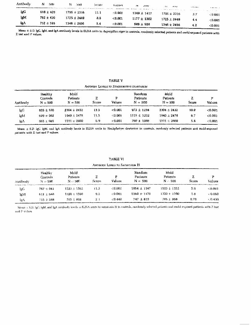
IgA, IgM, IgG, and IgE antibodies to 7 different molds {Altemaria,Aspergillus, Stachybotrys, Chaetomium. Cladosporium, Epicoccum,and Penicittium), satratoxin H, and other trichothecenes in 40 patientswith multiple organ symptoms were compared with 40 age- and sex-matched controls {Vojdani et al., 2003), The exposed individuals occu-pied a water-damaged building and were tested within days followingevacuation of the premises. Quantitative enzyme-linked immunosor-bent assay (ELISA) produced the following results: (!) IgG antibodies tothe molds and the two mycotoxins were significantly elevated in thepatients versus the controls. (2) Levels of serum IgA antibodies for eachmold and the mycotoxins were significantly elevated in the patients,with the exception of Epicoccum. The highest liters in descendingorder were found for Stachybotrys, PeniciHium, and Chaetomium.(3) IgM titers were significantly elevated in these patients versus thecontrols for Stachybotrys, Cladosporium, Altemaria, Aspergillus, sa-tratoxin H, and other trichothecenes. No difference in IgM titers wereobserved between patients and controls for Chaetomium, Epicoccum,and PeniciUium. (4) With respect to IgE antibodies, a significant in-crease in titers in these patients was found only for Aspergillus andsatratoxin H. It appears from these observations that randomly selectedcontrols without symptoms and apparent mold exposure have low titersof antibodies to a variety of mold and mycotoxins. However, mold-exposed symptomatic individuals have titers that are significantly-elevated over the control values.
In arudures. \ nige
fol lowi ito the !parent •referredresultsthe conthe blo<highestrespecttiters hwere csignificexposeigG mdata or(SDj wpatientpatientattend;et al. iIgG an'of!39potent
imininthe mi
mecritici,One i-IHaiseaboutpublicin
MOLD AND MYCXTTOXDMS 387
ntiy VojdaniA antibodies: respiratory-ignificantly.us, Chaeto-
;s, satratoxin"ibodies play•:ell as type-3example, in
izugatus andt the diseaseIn addition,
xacerbationsE. peripheral;iL 1989).
- (Altemaria,Epicoccum,: 40 patients
.-ige- and sex-v;duals occu-•;ys followingirnmunosor-
antibodies to-rated in thedies for each:he patients,: descendingZriaetomium..:s versus the•pergillus, sa-M liters were
Epicoccum,:gnificant in-pergillus andjmly selected-ave low titers•A'ever, mold-significantly
In another study, Vojdani et al (2003), utilizing ELISA assay proce-dures, tested for IgA, IgM, and IgG antibodies against S. chartamm.A. niger, P. notatum, satratoxin H, and other trichothecencs iu thefollowing three groups: healthy donors (N = 500); 500 patients referredto the laboratory for various diagnostic tests for illnesses without ap-parent exposure to molds (N = 500); and randomly selected patientsreferred for illness associated with exposure to molds (N — 500). Theresults of this study are summarized in Tables III through VI. Briefly,the concentration of IgA, IgM, and IgG antibody titers were lowest inthe blood donors, intermediate in the randomly selected patients, andhighest in the mold-exposed patients for each of the molds. Withrespect to satratoxin H and trichothecene antibodies, the antibodytiters had a different distribution. When the mold-exposed patientswere compared with the healthy controls, IgG and IgM titers weresignificantly elevated, while IgA titers were not. When the mold-exposed patients were compared with the random patients, only theIgG titers were significantly different. Moreover, on inspection of thedata on the random patients, it was noted that the standard deviation(SD) was large and overlapped with the mean value and SD of the moldpatients. It appears from these observations that the randomly selectedpatients may have been exposed to molds without recognition by theattending physician that such exposure might have occurred. Barneset al, (2002) reached similar conclusions. They demonstrated IgE andIgG antibodies to Stachybotrys chartamm in 9.4% and 42.2% of the seraof 139 blood donors. They concluded that sensitivity to S. chartaruni ispotentially much more widespread than previously appreciated. Thisfungus may affect the asthmatic and allergic population through bothimmunologic and toxic mechanisms. The significance of the fungus inthe milieu of allergenic fungi may need to be re-evaluated.
C. CROSS-REACTIVITY OF ANTIBODIES TO MOLDS
The use of antibodies to molds as a biomarker of exposure has beencriticized (Musmand, 2003). The critique is based on two publications.One is an abstract the full results of which have never been published(Halsey et al., 2001); therefore, it is impossible to determine anythingabout the methods used in this paper. The second is a position paperpublished on the Internet by the California Department of Public Servicesin which not a single experiment was conducted. Recently the question ofcross-reactivity between mold antigens (S. chartamm, A. niger, and P.notatum} was investigated by using affinity-purified rabbit sera {Vojdaniet al., 2004). The results of this study showed that non-immunized rabbits
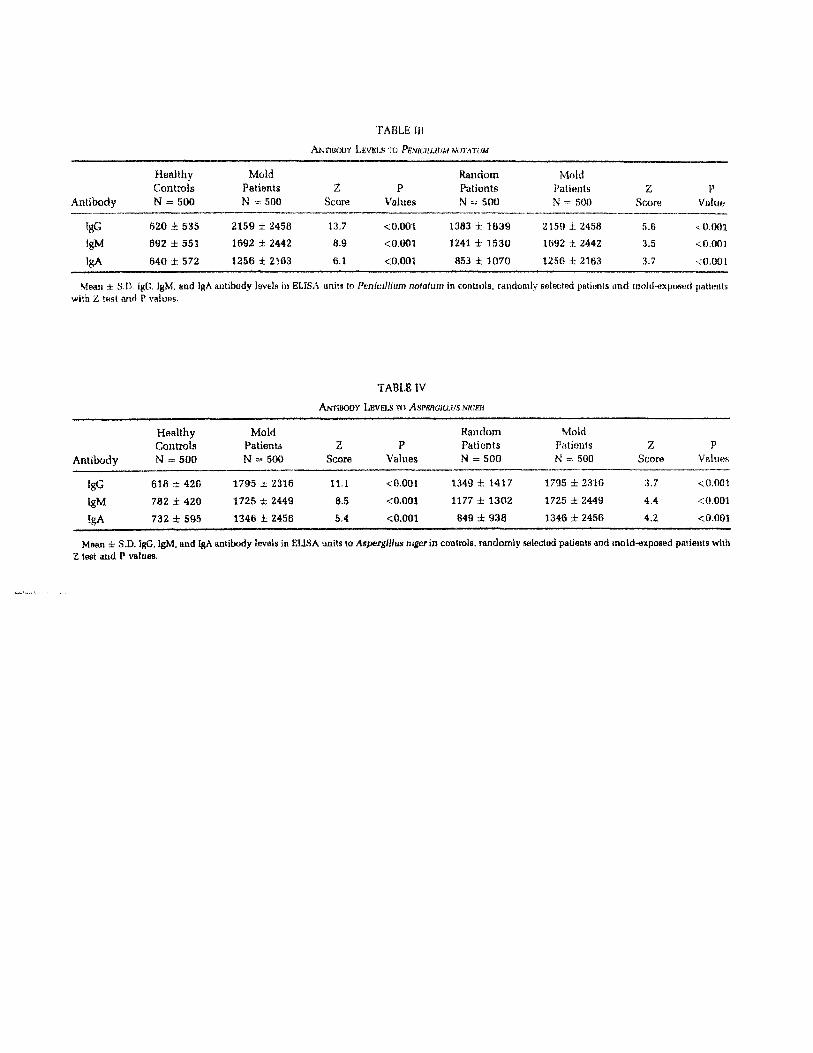
TABLE III
ANTIBODY LEVELS TO ftr.waujr/Af vw/rn/
Antibody
IgGIgM
IgA
HealthyControlsN = 500
620 ± 535
692 ± 551
640 ± 572
MoldPatientsN = 500
2159 ± 2458
1692 ± 2442
1256 ± 2193
ZScore
13,7
8,9
6.1
PValues
<0.001
<0.001
<0,001
RandomPatientsN ~ 500
1383 ± 1839
1241 ± 1530
853 £ 1070
MoldPatientsN = 500
2159 ± 2458
1692 ± 2442
1256 i 2163
ZScorn
5.63.53.7
PValue
<0.001
<0.001
<0.001
Mean ± S.D, IgD, IgM, and IgA antibody levels in ELISA units to Penicillium notatum in controls, randomiy seleetnd patients nnd rnolcl-sxposed pallfflHswith Z test and P values.
TABLE IV
ANTIBODY LEVELS TO ASPBRGIUMS mcen
HealthyControls
Antibody
IgGIgM
5gA
N = 500
618782732
-426
± 420
± 595
MoldPatientsN =
1795
1725
1346
= 500
± 2316
± 2449
±2456
ZScore
11.1
8.55.4
PValues
<0.001
< 0.001
<0.001
RandomPatientsN = 500
1349 ±1417
1177 ± 1302
849 ± 938
MoldPatientsN = 500
1795
1725
1346
± 2316
± 2449
± 2456
ZScore
3.7
4.4
4.2
PValues
<0.001
<0.001
<0.001
Msan i S.D. IgG, IgM, and IgA antibody levels in EUSA units to AspergiHus nigerin controls, randomly selected patients and moid-exposed patients withZ test and P values.
Anl i lMif ly
IgG
IgM
IgA
N SfW
618 ± 426
782 ± 420
732 ± 595
N ,'illlj
1795 ± 2316
1725 ± 2449
1346 ± 2456
.scorn
11,1
8.5
5,4
VUHMtS
<0,001
<0.001
<0.001
iv :iiiii
1349 ± 1417
1177 ± 1302
849 ± 938
1 •< ,HHt
1795 ± 2316
1725 ± 2449
1346 ± 2456
3.7
4,4
4.2
<0.001
<0.001
<0.001
Mean ± S.0. IgG, IgM, and IgA antibody lovels in ELISA units to Aspergillus niger in controls, randomly selected patients and raold-exposed pattonU with£j tost find P v8iuBs.
TABLE V
ANTIBODY LEVKLS TO STACHYBOTRYS aiAHTAHUM
Antibody
IgG
IgM
IgA
HealthyControlsN = 500
803 ± 530
S29 ± 602
665 ± 665
MoldPatientsN = 500
2304 ± 2432
1940 ± 2478
1511 ±2660
ZScore
13.5
11.5
6.9
PValues
<0.001
<0.001
<0.001
RandomPatientsN = 500
973 ± 1234
1115 ± 1212
760 ± 1086
MoldPatientsN = 500
2304 ± 2432
1940 ± 2478
1511 ± 2ft60
ZScore
10.9
6.7
5.8
PValues
<0.001
<0.001
<0.001
Mean ± S D. IgG, igM, and IgA antibody laveis in ELISA units to Stachybotrys chartarum in controls, randomly selncted patients and mold-exposedjiatients with Z t«st and !' values.
TABLE VI
ANTIBOOV LKVKI.S TO SATRATOXIN H
HealthvControls
Antibody
IgG
IgM
IgA
N =
767
f i l l
715
= 500
•i.-, 641
± 648
± 588
MoldPatientsN .
1523
1320
705
= 500
.i 1352
± 1590
:t 868
Z&;ore
11.3
9.2
2.1
PValues
<0.001
<0.001
<0.44!)
RandomPatientsN - 500
1054
1160
747
i 1147
± 1170
± 810
MoldPatientsN = 500
1523 -i 1352
1320 ± 1590
705 ± 868
ZScore
5.9
1.8
0.78
PValues
<0.001
< 0.060
<0.430
Moan i -S.D. IgCl. IgM, and IgAami I* V'ihins.
itxidv levnls in GL1SA units to satratuxiti H in controls, randomly stsSra'toii pat!«p.ts and inoid-cxpased [patients with Z lest
390 ANDREW W. CAMPBELL et al
develop IgG antibody titers to these molds that increase in concentrationwith age. The sera from these rabbits gave an impression of up to 52%cross-reaction with AspergiHus, Penicillium, and Stachybotrys. Whenusing affinity-purified antibodies in cross-inhibition studies, the antigen-ic cross-reaction between Stachybotrys and AspergiHus was between8.6% and 12.3%, and between Stachybotrys and Penicillium extracts; itshowed 9.3-9.6% antigenic similarities. Thus, for cross-reaction studiesbetween different molds, affinity-purified antibodies and a sensitive andquantitative assay with natural antigens should be used. When usingsuch an assay, it was concluded that cross-reactions between Stachybo-trys, AspergiHus, and Penicillium exist but are much less widespreadthan previously believed. Based on these observations, antibodies tomolds and mycotoxins as developed by this laboratory methodology arereliable biomarkers of mold and mycotoxin exposure.
D. ANTIBODIES TO EXTRACELLULAR POLYSACCHAKIOES (EPS)
EPS can cause type I and type in inflammatory processes. They havebeen shown to be present in mold-contaminated buildings and can beused as a marker of mold contamination and exposure (Duowes et a!.,1999; Wouters et- al., 2000}. Exposure to 1-3 beta-D-glucan caused air-way inflammation with symptoms of dry cough, phlegm, and hoarseness(Rylander, 1997; Rylander et al., 1998). IgG antibodies in immunizedrabbits against EPS from several mold genera have been reported(Notermans et al., 1987, 1988). The EPS antigens caused the productionof fairly specific antibodies, with some cross-reactivity as determined byan ELJSA. The EPS antigens produced by species of Penicillium, Asper-gillus, and Geotrichum lost their immunological activity with heating at100 °C at pH 1.8. The EPS antigens from Mucor recemosus, Rhizopusolgosporus, and C. cladosporoides were stable under the same conditions.It appears from these data that an ELJSA for antibodies to EPS released byvarious molds could be developed as an additional biomarker lor moldexposure.
V. Alterations in T and B Cells, Natural Killer (NK) Ceils, and OtherImmune Parameters in Humans Exposed to Toxigenic Molds
A. ALTERATIONS IN PERCENTAGE OF T AND B CELLS
Peripheral blood lymphocytes can be identified and quantified by-using fluorescent antibodies to cell surface antigens. Typical markersfor T cells axe designated as CD2, CD3, CD4, and CD8. B cells areidentified by CD19 or CD20. In addition, other markers can h« used to
MOLD AND MYCOTOXINS 391
identify activation of T and B ceils, (e.g., CD25, CD26, HLR-DR+,CD8CDl1b+). Patients chronically ill from exposure to toxigenic moldsin water-damaged office buildings, schools, and homes haxre alteredpercentages of lymphocyte markers in their peripheral blood whencompared with expected ranges (Gray et al., 2003). The patients hadincreased B cells (CD20) (75.6%). T cell activation markers increasedfor the following cell types: CD5CD25 (68.9%), CD3CD26 (91.2%),CD8HLR-DR+ (62%), and CD8CD38 (56.6%). Decreases were observedfor CDSCDlltM (15.6%) and natural killer (NK) cells {CD3CD16CD5B,38.5%). Moreover, Thrasher et al. (2004) found that individuals withan ongoing exposure to molds in a water-damaged building had re-lative increases over controls of the following: total lymphocyte count,T cells (CD2, CD3, CD4. CDS, and CTJ3CD16), B (CD19) cells, andNK cells (CD3CD16CD56).
B, MrrocEK ACTIVITY
T and B cells respond to specific and nonspecific antigens by under-going cell division (mitogenesis). Mitogenesis responses to nonspecificmitogens were as follows: phytohematogglutinin (PHA) was decreasedby 26.2% in mold-exposed subjects, while only 5.9% had decreasedresponse to Concanavalin A (ConA) (Gray et a!., 2003). PHA stimulatesT cells, while Con A causes T and B cells to divide.
Mitotic responses to ConA, PHA, PWM (pokeweed mitogen), andLPS (lipopolysaccharides) were examined in patients with an ongoingexposure to toxigenic molds. In general, mitogenesis to PHA and ConA was significantly elevated over controls, indicating increasedresponse of T cells to nonspecific antigens. In addition, rnitogenicresponse to B cell stimulators {ConA. PWM, and LPS) was also signifi-cantly elevated. Although mitogenesis was increased, the patientscould be subdivided into three distinct responses to each mitogen asfollows: suppression, elevation, and extremely elevated (Thrasheref al., 2004). Analysis of the NK cell (CD3CD16CD56) activity revealedthat 42.4% of these patients had decreased killing of target ceils.Furthermore, the CD4/CD8 (helper/suppressor) ratio was significantlyelevated.
These two studies (Gray et al, 2003; Thrasher et al, 2004) indicatedthat alterations in the percentages of T and B cells, mitogenesis, andNK cell activity ccurred in mold-exposed humans. The alterationsincluded an increase in activation markers, which may be a resultof antigenic stimulation. Furthermore, the changes in mitogenic re-sponse to both nonspecific and specific mitogens indicate immune

392 ANDREW W. CAMPBELL et al,
suppression occurred in some individuals, while others experiencedimmune stimulation. The decrease in NK ceils and their activity mayindicate that there was a decrease in immune surveillance, whichmay have importance with respect to cancer and/or infectious diseases.
C. AUTOANTIBODIES
Autoantibodies directed against self-antigens are known to occur ina variety of autoimmune diseases and degenerative neurologicdisorders. Antinuclear autoantibodies (ANA) are the ones most com-monly recognized and are usually associated with connective tissuedisease (e.g., lupus). However, other autoantibodies can be directedagainst a variety of self-antigens and can also be used as biomarkers oftoxic exposure (Thrasher et al, 2002; Vojdani et aL, 1992, 1993). Hu-mans exposed to toxigenic molds have abnormally elevated autoanti-bodies to the following: ANA, anti-smooth muscle, peripheral, andcentral nervous system myelin and eight different neural antigensincluding myelin basic protein, ganglioside Gl, sulfatide, tubulin,crystallin, filament, MOG, and MAG (Campbell et aL, 2003; Grayet al., 2003). Odds ratios for each autoantibody at 95% C.I. was signifi-cant, showing an increased risk for autoimmunity. Autoantibodies andautoimmune diseases are recognized as occurring from toxic exposures(Cooper et al., 2002; Griem et aL, 1998). For the significance regardingthe neural antigen autoantibodies, see Neurological Abnormalities.Section VI.
D. IMMUNE COMPLEXES
Immune complexes occur when antigen and antibodies combine andhave been implicated in numerous immunopathologic conditions, in-cluding systemic lupus erythematosus, rheumatoid arthritis, glomerulo-nephritis, and infectious induced inflammation (Abbas et aL, 1994).Deposition of immune complexes can occur from cell or tissue specificantibody-antigen reactions resulting in organ injury and/or immune com-plex diseases (Bigazzi et al., 1986). Thus it would appear from theseobservations on increased immune complexes that inflammation andautoimmune reactions may exist in mold-exposed patients. Circulatingimmune complexes containing IgG, IgM» and IgA antibodies can generatea variety of substances associated with muscle damage and the acutephase response that can activate the classic pathway of complement(Sorensen et a!., 2003). Autoantibodies are also known to activate thecomplement system.
MOLD AND MYCOTUXINS 393
E. CONCLUDING REMARKS ON IMMUNOLOGICAL OBSERVATIONS
The increase in B cells, activation markers, and helper/suppressorratio all indicate immune activation has occurred as demonstrated byGray et al. (2003) and Thrasher et al. (2004). Increased activation marker(CD45RO) has been reported for symptomatic children with exposure tomolds in contaminated homes (Dales et al., 1998). These observationsarc consistent with production of proinflammatory cytokines as dis-cussed above with antigenic stimulation. In addition, elevated immunecomplexes are further support for immune activation and antigenicstimulation. The presence of elevated immune complexes is compatiblewith increased production of antibodies to mold antigens as well as thepresence of ANA, anti-smooth muscle, and anti-neural antigen antibo-dies, The observations on immune alterations discussed above are alsoconsistent with the suggestion that mold exposure causes immune dys-regulation (Hirvonen et al., 1999; Wichman, et al, 2002). Recently areview by Anyanwu et al. (2003b) showed that natural killer cell activitywas adversely affected in patients with chronic exposure to indoormolds and may be implicated in causing neurological abnormalities.
VI. Neurological Abnormalities
Neurological abnormalities caused by mycotoxins from moldshave been described in the literature. The neurotoxic mycotoxinsinclude trichothecenes, citreoviridin, patulin, fumouisin, panitrem,verruculogen, rubratoxin, ergot alkaloids, and tremorgens.
Wilson et al. were the first to isolate a tremorgenic mycotoxin in \.The mycotoxin penitrem has been shown to induce tremors and con-vulsions in experimental animals (Hayes, 1980). Jorntner et al. (1986)and Morris et al. (1980) studied the neurological effects of the mycotox-ins penitrem A and verruculogen, which are known to cause a neuro-toxicity characterized by sustained tremors. Their findings support aprimary site of action of both of these mycotoxins as being presynaptic.Mycotoxins, being relatively nonpolar, pass through the blood-brainbarrier and thereby have access to synapses. The neurotoxic effects ofergot alkaloids are known to affect the postganglionic parasympatheticsynapses (Berde et al., 1978).
Wang et al (1998) in their study suggested that the primary site oftrichothecene action is the brain. Chapman (2003) reported how tricho-thecene mycotoxins from Stachybotrys cause neurological disorders hybeing neurotoxic. The clinical signs of trichothecene mycotoxicosisinclude eye pain, dyspnea, tachycardia, vomiting, mu.cl« tremor, ̂ d
394 ANDREW W. CAMPBELL et al.
weakness, lack of coordination, and confusion. Patients affected devel-op seizures, central nervous system dysfunction, hypotension, andmyelosuppression (Stahl et al., 1985). Studies have shown that expo-sure to molds can cause optic demyelinating neuritis and multifocalchoroiditis (Campbell et al., 2003; Rudich et al., 2003).
The ncphrotoxic and hepatotoxir. effects of mycotoxins have beenwell documented in several studies (Anyanwu et al., 2003c; Bhat et al.,1989; Etzel et al., 1998). The mycotoxin rubratoxin was studied byMoss (1971) and was shown to cause liver and kidney damageand lesions of the central nervous system. Ciegler and Bennett (1980)stated that trichothecene mycotoxins cause clinical conditions thatinclude skin irritations, vomiting, anorexia, diarrhea, hemorrhage,and convulsions.
Walsh et al. (1985) reviewed a large number of patients with necropsy-proven central nervous system aspergillosis and identified importantepidemiological, pathological, and clinical features. In their study, themost common central nervous system lesions were subcortical heraor-rhagic infarcts in the cerebral hemispheres or cerebellum, and they foundthat the most common entry of AspergiJlus into the central nervous systemwas the lower respirator)' tract, Aspergillosis of the central nervous sys-tem, lungs, and at least one other organ was found in almost 66% of thepatients. Beal era/. (1982), In their neuropathalogical review, discoveredthat the pathologic hallmark of neurologic aspergillosis cases was theinvasion of fungal hyphae into the blood vessel walls with subsequentnecrosis and thrombosis and spread into the surrounding tissues.
A. NEUROCOCNITIVE DEFICITS AND CENTRAL NERVOUS SYSTEM DYSFUNCTION
Pena (1970) noted subtle personality changes were observed as aninitial sign in cases of disseminated aspergillosis. Young et al. (1970)noted in their study of 13 patients with disseminated aspergillosis thatall had some degree of lethargy or fatigue. Malkin et al. (1998) in theirstudy at National Institute of Occupational Safety and Health reportedmultiple neurological symptoms in occupants of an office buildingcontaminated by several species of fungi, including Penicilliwn, As-pergillus, Altemaria, Candida, Cladosporium, Epicoccum, Fusarium,and Pullularia. Gordon et al. (1993) described a patient who after beingexposed to Aspergillus, Penicillium, and Rhizopus developed fatigue,headache, progressive somnolence, slowness of thinking, and severetremors. The patient had coarse fasciculations of the muscles of theface and tongue and was unable to stand unassisted. His EEC showed ageneral dysrythmia consistent with a toxic encephalopathy.
MOLD AND hWCOTOXINS 395
Baldo et al, (2002) studied the neuropsychological performance of10 patients exposed to molds (Stachybotrys aim, PeniciUium, andAspergillus). The patients had a variety of symptoms: fatigue, respira-tory problems, recurring bloody noses, nausea, frequent sore throats,and headaches, among others. The mold-exposed patients were im-paired on a number of cognitive measures, with the most consistentdeficits in visuospatial learning, visuospatial memory, verb, learning.and psychomotor speed. In addition, the mold-exposed patients hadevidence of Axis I and Axis II pathology. There was a significantcorrelation among patient's scores on the Beck Depression Inventory,with a number of neuropsychological tests falling within the impairedrange. The authors put forth a model by which to investigate the effectsof mold exposure on the central nervous system.
Crago et al. (2003) further demonstrated that measures of exposure \veruhighly predictive of neuropsychological test performance using two sub-tests from the Delis-Kaplan Executive Function System (D-KEFS) to mea-sure executive or higher-level cognitive functions. Significant predictivepower was observed for the D-KEFS Trail Making subtests of visualscanning, letter sequencing, number-letter sequencing, and motor speed;the D-KEFS Color-Word Inhibition/Switching subtest; the WAIS-JIIDigit Symbol Coding and Symbol Search subtests; arid the FVA-CPTfull-scale attention quotient and the visual and auditory attention quoti-ents. Crago el al. (2003) also reported that significant predictive powerwas found for estimates of degree of exposure and for the QEEG variabiesof mean frequency delta, relative power theta, relative power alpha,absolute power delta, absolute power theta, and absolute power alpha.In addition, the QEEG findings in confirmed moid-exposed patients in-dicated a restriction in the range of functioning (narrowed frequencybands) of the frontal lobes, that is, increased (accelerated) mean frequencyof the slower frequencies (delta range) and decreased (slowed) higherfrequencies (beta range), indicating a collapse toward the middle of thefrequency spectrum. These findings, coupled with observed increasedlevels of absolute and relative power theta and alpha waves in frontalsites, indicated hypoartivation of the frontal cortex consistent with insuf-ficient excitatory' input from the retieular activating system anatomicallyseated in the midbrain.
Finally, Kilburn (2002) reported on both objective neurological testsand neuropsychological evaluation of 20 mold-exposed patients. Ob-jective tests showed impaired balance, reaction time, color discrimina-tion, and visual fields in the mold-exposed patients. Neuropsychologicaltests showed impaired cognition, verbal recall, and trail making. Pulmo-nary function testing showed small airway obstruction was observed in 4
396 ANDREW W. CAMPBELL et aL
patients. Longer durations of exposure and aging appeared to increase thetotal abnormalities. He concluded "Moulds appear to ca\ise chemicalencephalopathy and these abnormalities."
Neurophysiological effects of mold exposure have been reported inchildren as compared with controls (Anyanwu et aL, 2003a). Brainstemauditory evoked response (BAER), electroencephalogram (EEC), visualevoked potential (VHP), and somatosensory evoked potential (SSEP) wereused to test neurological abnormalities. Three of 10 children had anabnormal EEC following mold exposure. The frontal-temporal theta waveactivity in the 10 patients seemed to indicate diffuse changes consistentwith metabolic encephalopathies. Also, 1 to 3 hertz delta activity wasasymmetric in the right hemisphere of 3 patients, BAER showed abnorm-alities in 9 patients with both 15' and 35" check sizes. A significant delayin waveform V occurred in the majority of patients, representing dysfunc-tional cognitive process and conductive hearing loss in both ears. VHPshowed clear abnormalities in 4 of the children with P100 amplitudes andlatencies decreased bilaterally. SSEP showed diffuse polyneuropathy intliree patients. The authors concluded that exposure to toxic molds canaffect neurological and behavioral status of children.
B. PERIPHERAL MOTOR AND SENSORY NEUROPATHY
Campbell et al. (2003) studied 119 patients with symptoms of neu.ro-toxicity with documented measured exposure to molds. These patientscomplained of fatigue, memory loss, cognitive function loss, headaches,tremors, numbness and tingling, blurred vision, tinnitus, and muscleweakness. Ninety-nine of these patients had abnormal clinical neuro-logical examinations, abnormal findings on neurophysiological testing,and elevated antibodies to neuronal antigens. Nerve conduction studies(NCVs) revealed three groups of abnormal patients (ABM) and onegroup of normal (NM): mixed sensory motor polyneuropathy (55ABN); motor neuropathy (17 ABN); sensory neuropathy (27 ABN); andsvmptoms without neuropnysiological abnormalities (20 NM, controls).
C. NEURONAL ANTIBODIES
Elevated autoantibodies by ELISA to several neuronal antigens werefound in patients with documented measured exposure to molds. Theliters of tie autoantibodies were significantly elevated over controls.These included IgA, IgG, and IgM antibodies to myelin basic protein,riiyeiin associated glycoprotein, oligodendrocyte glycoprotein, ganglin-side GM-1, chondroitin sulfate, crystalline, tubulin, and neurofilament.
MOLD AND MYCOTOXTNS 397
D, DEMYELJNATION OF PERIPHERAL NERVES
Campbell et ai. (2003) concluded their observations on changes innerve conduction velocities and the presence of neural antigen autoanti-bodies as follows: "The increased latency for motor and sensory nervesobserved in the 55 patients with mixed neuropathy is suggestive of ademyelinating process (Busby et al., 2003}." This was accompanied bya decrease in velocities for the median, ulnar, and peronoal nerves whilethe tibial nerve had a decrease in the amplitude. All three sensory nerves(median, ulnar, and superficial peroneal) exhibited increased latenciesand decreased amplitudes. Thus the polyneuropathy observed in thesepatients appeared to be a demyelinating process with decreased numberand size of fibers (decreased amplitude) and chronic involvement of thenerve (decreased velocities) (Busby et al., 2003; Steck et al., 1987).The motor neuropathies (17 patients) had decreases in latencies (peronealand tibial nerves), decreased amplitudes (median and peroneal nerves),and decreased velocities (median, ulnar, peroneal, and tibial nerves). Thisappeared to be a diffuse neuropathy and may involve some demyelination(Berger et al., 2003). Finally, the sensory neuropathies (27 patients) hadincreased latencies for all three nerves, with that of the superficial pero-neal being not significant. The increased latencies and the decreasedamplitude of the superficial peroneal suggested demyelination wasoccurring (Reindl et al, 1999; Willison and Yuki, 2002).
VII. Conclusion
Forgacs noted in 1962 that mold mycotoxicosis was called "theneglected disease." The manifestations and disorders in humanscaused by molds and mycotoxins continues to be overlooked or unno-ticed by many physicians. Each year studies continue to be publishedthroughout the world medical and scientific literature elucidating andexplaining the pathological processes and biomechanisms by whichexposure to molds and mycotoxins cause sickness in humans. We havedescribed in this chapter the most recent neuroimmune mechanisms ofdisease process in humans of molds and mycotoxins. The exactbiological and chemical actions through which these mechanisms un-fold is not completely understood. However, molds do produce meta-bolites (mycotoxins, solvents) and shed antigenic materials (spores,hyphae, extracellular polysaccharides, and enzymes), which are toxic(mycotoxins) and or cause immunologic responses (antigens). Scienceand medicine should continue its work in these areas and bring aboutthe much-needed change from "the neglected disease" to "the accepteddisease."
398 ANDREW W, CAMPBELL el al.
REFERENCES
Abbas, A. K,, Lichtman, A. H., and Pober, J. S. (1994). In Cellular and MolecularImmunology, 2nd ed., p. 393. WB Saunders and Company, Philadelphia.
Anaissie, E.}.. Straiten, S, L., Dignani, M. C., SummBrbell, R. C., Rex, J. H., Mcmson, T. F ,and Walsh, T. J. (2002). Pathogenic Aspergillus species recovered from a hospitalwater system: 3-year prospective study. Clin. Infect. Dis. 34, 780~7d9.
Andersson, M. A., Nikulin, M. Kooljalg, U., Anderson, M. C.. Rainey, K., Reuula, K'..Hiiitikka. E. L., and Salkinoja-Salonen, M. (1997). Bacteria, molds, and toxins inu-ater-damaged building materials. Appl. Knvimn. Mictobiol. 63, 387-393.
Apter. A. J.. Greenberger, P. A., Liolta, {. L., and Roberts, M. (1989). Fluctuations of sorurnIgA and its subclasses in allergic bronchopulmonary aspergillosis. /. Allergy Clin,Immunol. 84, 367-372.
Anyanwu, E. C., Campbell, A. W., and Vojdani, A. (20Q3a). Neurophysiological affects ofchronic indoor environmental exposure on children. Scientific World journal 3,281-290.
Anyanwu, E. C., Campbell, A. W.. Jones, ]., and Ehiri, J. (2003b). Tho neurologic*!significance of abnormal natural killer ceil activity in chronic toxlgenic mold expo-sures. Scientific World Journal 3, 1128-1137.
Anyanwu, E. C., Campbell, A. W,, Vojdani, A., and Ehiri, J. (2003c). Biochemical changesin the serum of patients with chronic toxigenic moid exposures: a risk factor formultiple renal dysfunctions. Scientific World Journal 3,1058-1064.
Baldo, (., Ahmad, L., and Ruff. R, (2002). Neuropsychological performance of patientsfollowing mold exposure. App. Neumpsych. 9, 193-202.
Barnes, C,, Buckley. S., Pacheco. F., and Portnoy, }. (2002). IgE-reactive proteins fromStachybotrys chartarum. Ann, Allergy Asthma Immunol. 90, 29-33.
Berger, T., Rubner, P.. Schautzer, p., E%$, R., Uitner, H,, Mayringer. 1., Dilitz, E,, Deisenham-mer, F., and Reindl, M. (2003). Antimyelin antibodies as a predictor of clinically definitemultiple sclerosis after a first demyelinating event. N. Engl. J. Med. 349,139-45.
Bhat. R.. Beedu. S.. Ramakrishna, Y., and Munshi, K. L. (1989). Outbreak of trichotheceneniycotoxicosis associated %vilb the consumption of moid damaged wheat products inKashmir Valley, India. Lancet 1, 35-37.
Bigazzi, P. E., Burek, C. L., and ROSUD. R. (1986). Antibodies to tissue-specific endocrine,gastrointestinal and neurological antigens. In Manual of Clinical Laboratory immu-nology (N. R. Rose, H. Friedman, and J. L. Fahey, fids.), p. 762. Amerie-.an Society ofMicrobiology, Washington D.C.
Beal, M. F., O'Carroll, C. P., Kleinman, G. M., and Grossman, R. 1. (1982). Aspergillosis ofthe nervous system. Neural, 32, 473—479.
Berde, fl., and Schild, H. (eds.) (1978). Ergot alkaloids and related compounds. Springer,New York.
Berek, L., Petri, i. B.. Mesterhazy, A., Teren, H., and Molnar, J. (2001). Effects of myco-toxins on human immune functions in vitro. Toxicol in Vitro. 15, 25-30,
Bondy, G. S.. and Petska, J. ). (2000). taummomodulation by fungal toxins. /. ToxicolEnviron. Health B Crit. Rev 3, 109-143.
Braun, H., Buzina, W., Freudenschuss, K.. Beham, A,, and Stammberger, H. (2003)."Eosinophilic fungal rhinosinusitis": A comnion disorder in Europe? Laryngoscope114. 264-269.
Barge, H, A. (1990). Bioaerosois: prevalence and health effects in the indoor environment./. Allergy Clin. Immunol. 86, 687-704.
MOLD AND MYCQTOXFNS M09
Busby, M., and Donaghy, M. (2003). Chronic dyaimmuae neuropathy. A subclassificationbased upon the clinical feature of 102 patients. /. Neurol. 250, 714-724.
California Department of Public Services Environmental Health Investigations Branch(2000). Misinterpretation of Stachybotrys serology. Available at: http://wivw.dhs.ca.gov/ehib/ehib2/ topics/SerologyfZ.htm.
(Campbell, A. W., Thrasher, {. D., Madison, R. A., Vojdani, A., and Gray, M. R. (20U3).Neural antigen autoantibodies and neurophysiology abnormalities in patientsexposed to moulds. Arch. Environ. Health 58 (In press).
Campbell, A. W., Anyanwu. E. C., and Vojdani, A. (2003). Combination of high-duseintravenous immunogiobiilins and itraconozole in treating chronic mycotic demyu-linating optic neuritis. Scientific World Journal 3, 640-646,
Ciegler, A., and Bennett, J. W. (1980). Mycotoxins and mycotoxicosis. Biascience 30,512-515.
Challancombe, S. J. (1987). The induction of secretory IgA responses. In "Food allergyand Intolerance" (J. Brostoff and S. J. Chaliancombe, eds.). WB Saunders, Eastutgn.East Sussex, England.
Chapman, J. (2003). Stachybotrys (chartarum=atra=altemans) and other problems causedby aliergenic fungi. Alter, and Asthm. Proc. 24, 1-7.
Claeson, A. S-, Levin, H., Biomquiat, G., and Sunesson, A. L. (2002). Voialile metabolitesfrom microorganisms grown on humid building materials and synthetic media./. Environ. Monit. 4, 667-672.
Cooper, G. S., Miller, F. W., and Germolec, D. R. (2002). Occupational exposures andautoimmune diseases. Inter. Immunopharmar.olagy 2, 303-313.
Crago, B. R., Gray, M. R., Nelson, L. A.. Davis, M., Arnold, L., and Thrasher, f. D. (2003).Neuropsychological and electrocortical effects of mixed mold exposure. ArchivesEnviron. Hith. 549-552.
Dales, R., Miller, U., White, J., Dulberg, C., and Lazarovits, A. 3. (1998). influence ofresidential fungal contamination on peripheral blood lymphocyte populations onchildren. Arch. Environ. Health S3, 190-195.
Desai, K., Sullards, M, C.. AHegood. J., Wang, E,, Schemlz, E. M., Hari, M., Humpf, H. U..Liolta, D. C., Peng. Q., and Merrill, A. H. (2002). Fumonisins and fumonisin analogsas inhibitors of ceramide synthase and inducers of apoptosis. Biochim. Biaphya. Acta1585, 188-192.
Dominguez-Malagon, H., and Gaj'tan-Graham, S. (2001). Hepatocelluiar carcinoma: anupdate. Ultrastruct. PathoL 25, 497~ai6.
Dosa, E., Coczi, L, Mojzes, L. Molnar, E. G., Varga, H. M.. and Nagy, E, (2002). Identifi-cation and incidence of fungal strains in chronic rhinosinusitis patients. Acta.Microbiol. Iwmunol. Hung, 49, 3337-3346.
Duowes. J., van der Sluis, B., Dockes, G., van Leusden, F.. Wijoarids, I,., van Strien, R.,Verhoff, A., and Brunerkreef, B. (1999). Fungal extracellular polysacchahdes inhouse dust as a marker for exposure to fungi: Relations with culturabie fungi, reportedhome dampness, and respiratory symptoms, /. Allergy Clin. Inununol. 103,494—500.
Ebina, K., Ichinowatari, S., and Yokota, K. (1985). Studies on toxin Aspergillus fumigatus.XXJI. Fashion of binding Asp-hemolysin to human erythrocytes and Asp-Hemolysi n-binding proteins of ervthrocyte membranes. Aficroiuo/- Immunol, 29, 91-101.
Engelhart, S., Loock, A., Skutlarck,D., Sagunski, H., Lommel. A,, Farver, H.. andExner, M.(2003). Occurrence of toxigenic Aspergillus versicolor isolates and steriginatocystin in carpet dust from damp indoor environments. Appl. Environ. Micrihinl. 68,3886-3890.
400 ANDREW W. CAMPBELL et al.
Erkinjuntti-Pekkanen. R.. Reiman. M,, Kakkarinen, J, L, Tukinnen, H. O., arid Terho. B O.(1999). IgG antibodies, chronic bronchitis, and pulmonary function values infarmer's lung patients and matched controls. Allergy. 54, 1181-1187.
Elzel. R. A., Balk, S. J., and Bearer, C. F. {1998}. Toxic effects of indoor molds, Pediair.101, 712-714.
Eucker, ]., Sezer, O.,Graf, B., and Possinger, K. (2001J. Mueonnycoses. Mycoses44,254- 260.Ezeamuzie, C. I., Al-Ali, S., Kahn, M., Kahn, Z., Dowaisan, A., Thomson, M. S., and
Georgi, J. (2000). IgE-mediated sensitizalion to mould allergens among patients withallergic respiratory diseases in a desert environment. Int. Arch. Allergy. Immunal.121, 300-307.
Fan. L. L. (2002). Hypersensitivity pneumonitis in children. Curr. Opin. Pediatr. 14,323-326.
Flannigan, B., McCabe, E. M., and McGarry, F. (1991). Allergenic and toxigenic micro-organisms in houses. /, Appl. Bact. Sym 70(suppl), 61-73.
Forgacs, J. (1962). Mycotoxicoses: The neglected diseases, Feedstuffs, pp. 34, 124.Fraser, R. S, (1993). Pulmonary aspergillosis: Pathologic and pathogenetic features
Pathoi. Anna. 38, 231-277.Gareis, M. (1995). Cytotoxicity testing of samples originating from problem buildings. In
"Proceedings of the International Conference: Fungi and Bacteria in Indoor Environ-ments: Health Effects, Detection and Remediation" (Eckart Johanning, S. Chin, andYang, eds.), pp. 139-144. Saratoga Springs, NY.
Gordon, K. E.. Masotti, R. E., and Waddell, W, R. (1993). Tremorgenie encephalopathy: Arole of mycotoxins in the production of CNS disease in humans. Con. /. Neural Sci.20, 237-239.
Gorney. R. L., Reponen, T., Willeke, K., Schmechel, B., Robina. E., Boissier. M.. andGrinshpuc, S. A. (2002). Fungal fragasents as indoor air contaminants. Appl. Envi-ron. Microbiol, 68, 3522-3551.
Gravesen, S., Nielsen, P. A., Iverson, R., and Nielsen, K. F. (1999). Microfungal contami-nation of damp buildings—examples of constructions and risk materials. Environ.Hlth. Perspect, 107(suppl. 3), 505-508.
Gray, M. R., Thrasher, J. D., Crago, R., Madison, R. A,, Campbell, A. W., and Vojdani. A.(2003). Mixed mold exposure: Immunological changes in humans with exposure inwater damaged buildings. Archives Environ. Hlth. 58, 410-420.
Gregory, L., Rand, T. G., Dearborn, D., Yike, A., and Vesper, S. (2003). Immunocytochem-ical localization of stachylysin in Slachybotrys chartarutn spores and spore-impact-ed mouse and rat lung tissue. Mycopathologia 156,109—117.
Griem, P., Wulferink, M., Stachs. B., Gonzalez. J. B.. and Gleichmann, E. (1998). Allergicand autoimmune reactions to xeaobiotics: How do they arise? Immanol. Today 134,133-141.
Grossi, P., Farina, C., Fiocchi, R.. and Balls Gasperina, D. (2000). Prevalence and outcomeof invasive fungal infections in 1,963 thoracic organ transplant recipients: a mul t i -center retrospective study. Italian study group of fungal infections in thoracic organtransplant recipients. Transplantation 70,112-116.
Gunnbjornsdortir,M. L,Norback, D., Plaschke,P.,Norman, E., Bjomson, E., and Janson, C.(2003). The relationship between indicators of building dampness and respiratoryhealth in young Swedish adults, liespir. Med. 97,301-308.
Halsey, J. F., Jensen, J. T., and Miller, J, D. (2001). taununological responses to Stachy-botrys chartamm anligen. /. Allergy Clin. Immunol. 1O7A1034.
Hayes, W. (1980), Mycotoxins: A review of biological effects and their role in humandiseases. Clin. Toxicology 17, 45-33.
MOLD AND MYCOTQXINS 401
Hirvouen, M.-R,, Ruotsalaiitcn, M. Roponnna, M., Hyvarinen, A., Husman, T,, KOSITIH,V, M., Komulainen, H., Savolaintin, K., find Nevalaineu, A. (1909). iSiitric oxide andprainfhunmatory cytokines in nasal lavagB Quid associated with symptoms andaxposuni to mold building microbes. Rasp, Grit. Care. Mod. 160, 1943-1946
Hodgson, M. JL Morey, P., Leung, W. Y,, Morrow, L., Miller, D., Jarvis, B., Kobbins. H.,Halsey, J. K., and Storey, E. (1998), Building-associated pulmonary disease fromexposure to Stachybotrys charttirum and Afp»rgillus vtirsicolor. /. Of.rup, Environ.Med. 40, 241-249.
Hsifih, L. L.. and Hsieh, T. T. (1993). Detection of aflatnxin Bl-ONA adduets in humanplacenta and cord blood. Cancer RMS. 53, 1278-1280,
Hoehler, D., Marquardt, R. R., Mclntosh, A. R., arid Hatch, G. M. (1997). Induction of fnwradicals in hepatocytes, mitochondria and microsornes of rats by ochratoxin A and i'.sanalogs. Biochim. Biophys. Acta 1357, 225-233.
Jaakkola, M., Nordman. H., Pilpari, R., Uitti, }., Laitinene, J,, Karejainen, A., Hahiola, P.,and Jaakkola, J. ]. (2002). Indoor dampness and molds and development of aduli-unset asthma: A population-based incident case-control study. Emir, tilth. Persp.110, 543-547.
jakab, G. J., Hrnieleski, R. R,, Hemenway, D. R., and Groopman, J. D, (1994). Respiratoryafiatoxicosis: suppression of pulmonary and systemic host defenses in rats and mice.Toxicol. Applied Pharm. 125, 198-205.
Jarvis, B. B. (2002), Chemistry and toxicology of molds isolated from water-damagedbuildings. In "Mycotoxins and Food Safety" 0. W. DeVries, M. W. Trucksess, andL. S. Jackson, eds.), pp. 43-52. Kluwer Academic/Plenum Publishers.
Jarvis, B. B., Soranson, W. G., Hintikka, E.-L, Nikulin, M., Zhou, Y., Jiang, j.. Wang- ?-..Hiakley, S.. Etzel, R. A., and Dearborn, D. (1998), Study of toxin production byisolates of Stachytfotrys chartarum and Memniella echinata isolated during a studyof pulmonary hotnosiderosis in infants, App. Environ. Micrabiol, 64, 3ti20-3G25.
Jones,C.. Ciacci-Zanella,}. R., Zhang, V., Hendeson, G., andDickman, M. (2002). Analysisof fumonisin Bl-induced apoptosi*. Environ. Health Persp. 109(suppl 2), 315-320
Jumtner, B. S., Erich. M., Katherman, A. E., Huckle, W. R., and Carterm. M. E. (1980).Effects of prolonged tremor due to pflwtrem A in mice. Drug. Chem. Toxicol. 9,101-116.
Johanning, E., Biagini. R., Hull, D.-L., Morey, P., Jarvis, B., and Landsbsrgis. P. (199G).Health and immunology study following exposure to toxigem'c rimgi (Stachyltoirvachartarum) in a water-damaged office environment. Int. Arch. Occup. Environ, tilth.68, 207-218.
Johanning, E., Gareis, M., Nielsen, K. F., Dietrich. R., and Martbauer, E. (2002). Airbornemycotoxins sampling and screening analysis. In {H. Levin, G. Bendy, and I. Cordel!,ed».], Indoor Air 2002, The 9th International Coni'arence on Indoor Air Quality andClimate, Monterey, CA. June 30-Juiy 5, 2002. Vol 5. pp. 1-6. The InternationalAcademy of Indoor Air Sciences, Santa Cruz.
Karlsson-Borga, A., jonssoii, P., arid Rolfsen, W. (1988). Specific IgE antibody to 16widespread mold gonera in patients with suspected mold allergy. Ann. Allergy 83,521-526.
Kildeso, J., Wurtz. V. M., Nielsen, K. F., Wilkins, C. K., Gravesen, S., Nielsen, P. A.,Thrane, U.. and Schneider, T. (2000). The release of fungal spores from waterdamaged building materials. Jn {O. Seppanen and J. Sateri, eds.), Proceedings ofHealthy Buiidings 2000, August 6-10, Espoo Finland. SY! Indoor Air InformationOy. Helsinki, pp. 131-318.
Kilbum, K. H. (2002). Inhalation of molds and mycotoxins. Bur. j. Oncai. 7, 197-202.
402 ANDREW W. CAMPBELL at al
Knutsen, A. P., Mueller, K. R., Hutcheson, P. S., and Slavin, R. G. (1994). Serum anti-AspergiUus fumigatus antibody iromunoblot and ELISA in cystic fibrosis vviih allergicbronchopulinonary aspergiiiosis. /. Clin. Immunol. 93, 926-931.
Kudo, Y,, Ootaisi, T., Kumangai, T., Fukuchi, Y., Ebina, K,, and Yokota, K. (2002),A novel oxidized low-density lipoproiein-binding protein. Asp-faetnoiysin recog-nizes lysophosphatydylcholine, BioL Pharm. Bull 25, 787-790.
Kordula. T., Banbula, A.. Macotnson. J.. and Ravis, J, (2002). Isolation and properties ofStachvrase A, a chymotrvpsin-iijke serine proteinase from Stachybotrys chartarum.Infection Invnun. 70, 419-421.
Kunip, V., Shen, H.-D., and Banerjec, B. (2000). Respiratory fungal allergy. Microbe* Inf.2, 1101-1110.
Lander, F., Meyer, H. W., and Norm, S. (2001). Serum IgE specific to moulds, measuredby basophil histarnine release, is associated with building-related symptoms indamp buildings. Inflamm. Res. 50, 227-231.
Leina, M., Makela, M., Reijula, K,, Haahtela, T., Rauhamaa, H., Tuomi. T., Hintikka. E. I...,and Alenius, H, (2004). Intranasal exposure to a damp building mould, Stachybatrys chartarum, induces lung inflarnmatioii in mice by satratoxin-independentmechanisms. Clin. Exper. Allergy 33.
Makarananda, K., Pengpan, U., Sriskulthong, M., Yoovathaworn, K..and Sriwatanakul, K,(1999). Monitoring of aflatoxin exposure by biomarkers. /. Toxicol. Set. 23(suppl 2),155-159.
Malkin, R., Martinez, K.. Marinkovich, V., Wilcox, T., Wall, D., and Biagitii, R, (19983.The relationship between symptoms and IgG and IgE antibodies in an office environ-ment. Env. Resear. 76, 85-93.
Mason, C. D., Rand. T. G., Oulion, M., MacDonaJd,). M.. and Scott, J. E. (1998), Effectsof Stachybotrys chartarum [atraj conidia and isolated toxin on lung surfactantproduction and homeostasis. Nat. Toxins 6, 27-33.
Mason, C. D., Raad, T. G., Oulton, M.. MacDonald,}. M., and Anthes, M. (2003). Effectsof Stachybotrys chartarum on surfactant convertase activity in juvenile mice.Toxicol. Appl. Pharm. 172, 21-28,
McCrae, K. C., Rand, T., Shaw, R, A.. Mason, C. D., Oulton, M. R, Hasting, C.. Cherlet. T..Thlivoris, J. A.. Mantscfa. H, H., MacDonald, }. M., and Scott, J. E. (2001).Analysis of pulmonary surfectanta by Fourier-transform infrared spectroscopy follow-ing exposure to Stachybotrys chartaniin latra) spores. Ghent Phys. Lipids 110,1-10,
Monod, M., Capoccia, S., Lechenne, B.. Zaugg, C., Holdom, M., and Jouddon, O. (2002).Secreted proteases from pathogenic fungi. Med, Microbiol. 282, 205-419.
Musmand, J. (2003). Dose Stacltybolrys actually cause adverse effects? Ana. AllergyAsthma Immunol. 90, 274-275.
Nagata, T., Suzuki, H., Ishigarni, N.. Dbinozuka, J,, Uetsuka, K.. Nakayarna, H., and Doi. K.(2001). Development of apopfosis and changes in lymphocyte subsets ia thymus,mesenteric lymph nodes and Payer's patches of mice orally inoculate with T-2 toxinExp. Toxicol. Path. 52, 3309-3315.
Nielsen, K. F. (2003). Mycoloxin production by indoor molds. Fungal (tonetics BioL 39,103-117.
Nielsen. K. F., Gravesen, S., and Neiisen, P. A. (1999), Production of mycotoxins onartificially and naturally infested building materials. Mycopathologia 145, 43—58
Nieminen, S, M.. Karki, R.. Auriola, S., Toivola, M., Laatsch, H., Laatikaineji, F.,>iyvarinen, A., and Von Wright, A. (2002). Isolation and identification of Asp&rgittusfumigatus mycotoxins on growth medium and some building materials. Microbiology68, 4871-487b.
MOLD AND MYCOTOXINS 403
Niranjan, B. K,, Bhat, N. K., and Avadhani, N. G. (1982). Preferential attack of mitochon-dria! DMA by aflatoxin Bl during hepaloeareinogenesis. Science 214(4528), 73-75.
Nielsen, K. F., Huttunan, K., Hyvarinen, A.. Andarsen, B., Jarvis, B. B., and Hirvnne. M. K.(2001). Mnlabolite profiles of Stachybotzys isolates from water-damaged buildingsand their induction of inflammatory mediators and cytoloxicity in macraphages,Mycopaiholgsa 154, 201-205.
Morris, P. J., Smith, C. C.. De Belieroche, ]., Bradford, H. F,, Mantle, V. G., Thomas, A. )..and Penny, R. H, (1980). Actions of tremorgenic fungal toxins on ueurotransmiiterrelease, /. Neumchem. 34, 33-42.
Nolermans, S., Dufrenne, J., Wijnands, L. M., and Engel. H, W. (1988). Human serumantibodies to extracellular polysaccharides (EPS) of moulds. /. Med. Vet. Myr.o't. 26,41-48.
Ojanen, T. (1992). Class specific antibodies in serodiagnosis of farmer's lung disease, flr/, Ind. Med. 49, 332-336.
Ojanen, T., Terho. B. O., Tukanineu, H., and Mantyyjarvi, R. A. 11990). Class specificantibodies during follow up of patients with fanner's lung disease. Eur Respir, ]. 3,257-280.
Pace, J. G. (1993). Effect of T-2 mycotoxin on rat liver mitochondria electron transportsystem. Toxicon. 21,675-680
Pace. J. G. (1998). T-2 mycotoxin inhibits mitochondria! protein synthesis, Toxicon. 26,77-85.
Patel. A. M., Ryu, J. H., and Reed, C. E, (2001). Hypersensitivity pneuinonitis: currentconcepts and future questions. /. Allergy din. Immune/. 108, 661-670.
Poltola, }., Andersson, M. A.. Haahtala, T., Mussalo-Rauhamaa, H., Rainey, F A .Kroppenstedt, R. M., Samson. R. A., and Salklnoja, M. S. (2001). Toxic-metabolite-producing bacteria and fungus in an indoor environment, Appl. Emir. Microbiol. 67,3269-3274.
Pena, C. E. (1970). Aspergillosis, In "The pathology and anatomy of mycoses: humanInfection with fungi" (R. E, Baker, ed.), pp. 762-831. Springer, Berlin.
Petkova-Bochatrova. T., Stoichev. I. I., Chernozemsky, I. N., Castegnaro, M.. and Pftiol-LesEkowicz, A. (1998). Fonnation of DNA adducts in tissue of mouse progeny throughtransplancental contamination and/or lactation after administration of a single dose ofochratoxin A to the pregnant mother. Environ. Mol. Mutagen. 32,155-162.
Pfhohl-Leszkowicz, A., Grosse, Y., Kane. A., Creppy. E, E.. and Dirheimer, G. (Ifl93a).Differential DNA adduct formation and disappearance in tkree mouse tissues aftertreatment with mycotoxin ochratoxin A. Mut. Res. 289, 265-273.
Pfhohl-Leskzkowicz, A., Grosse, Y., CJastegnaro, M., Nicolov, 1. G., Chernozernsky, 1. N . .Bartsch, H., Betheder, A. M., Creppy, E. E,, and Dirheimer, G. (I993b). OcbratoxinA-related DNA adducts in urinary tract tumors of Bulgarian subjects. lAftC Sci. Pub!.124, 141-148.
Pfohi-Leszkowicz, A., Petkova-Bocharova, T.. Ghemozemsky, I. N., and Castfignaro, M.(2002). Balkan endemic nephropathy and associated urinary tract tumours: A reviewon aetiolgical causes and the potential role of mycotoxins. Food Addit. Contani. tfl,282-302.
Poapolathep, A., Ohtsuks, R., Kiatipattonasakul, W., Ishigami, N., Nakayam, H.. and Doi. K.(2002). Nivalenol-induced apoptosis of thymus, spleen, and Peyer's patches of mice.Exp. Toxicol. Pathol. 53, 441-446.
Panikau, J. U., Sherris, D. A., Kern, E. G., Homburger, H. A., Frigas, E., Gaffcy. T. A. , andRoberts, G. D. (1999). The diagnosis and incidence of allergic fungal sinusitis. Mayo.Clin. Proc. 74, 877-884.
404 ANDREW W. HAMPBELL «t al.
Potter. P. C, Juritz,}., Little, F., McCaldin, M., and Dowdle, E. B. (1991), Clustering offungal allergen-specific IgE antibody responses in allergic subjects. Ann. Allergy 66,149-153.
Rand, L., Rand, T. G.. Dearborn, D., Yike, I., and Vesper, S. (2003). Inununocytochemicallocalis'.atiuti of stehyiysin in Stachybotrys ehartaruia spores and spore-impactedmouse and rate lung tissue. Mycopathologia 156.109-117.
Rand, T. G,, Mahoney, M., White, K.. and Ouiton, M. (2002). Microanatomical changes inalveolar typo II cells in juvenile mice intrathocaily exposed to Stachybotrys chartar-uin spores and toxin. Toxicol. Sci. 65, 239—245.
Reindl, M., Linington. C., Brehm, U.. Egg, R,. Dilitz, E., Deisenhammer, F., Powwe, VV..and Berger, T. (1999). Antibodies against the myelin oligodendrocyte glycoproteinand the ravelin basic protein in multiple sclerosis and other neurological diseases;A comparative study. Brain. 122, 2047-2056.
Ribes, J. A., Vanover-Sames, C. L., and Baker. D, J. (2000). Zygomycetes in human diseaseClin. MicrobioL Rev, 13, 23&-301.
Richard, J. L., Planner, R. D,, May,}., and Liska, S. L. (1999). The occurrence of ohratoxinA in dust collected from a problem household. Mycopathologia 146, 99-103,
Rudich, R., Santilli. J., and Rockwell, W. (2003). Indoor mold spore exposure: A possiblefactor in the etiology of multifoeai choroiditis. Am. J. OpthaSmol. 135, 402—404.
Ry lander, R. (1997). Airborne 3-beta-D-gJuean and airway disease in a day-care centerbefore and after renovation. Arch. Environ. Health 52, 281-285.
Rylander, R., Norrhall, M., Engdahl, U., Tunsater. A., and Hott, P. G. (1998). Airwaysinflammation, atopy, and (l-~3)-beta-D~giucan exposures in two schools. Am. J.Respir. Crit. Care Med. 158, 1685-1687.
Sajan, M. P., Satav, J. G., and Battaeharya, R. K. {1997). Effect of aflatoxin Bl in vitro on ratliver mitochondria! respiratory functions. Indian J. Exper, BioL 35, 1187-1190.
Salvilahti, R., Uitt, J., Laippaia, P., Husmari.T.. and Roto. P. (2000). Respiratory morbidityamong children following renovation of water-damaged school. Arch. Environ.Health 55, 305-410.
Schwartz, G. G. (2002). Does ochratoxin A cause tasticular cancer? Cancer Causes andControl 13, 91-100.
She)ton. B. F., Kirkland, K. H., Flanders, W. E.. and Morris, G. K. (2002). Profiles ofairborne fungi in buildings and outdoor environments in the United States. Appl.Environ. Microbioi. 68,1743-1753.
Skaug, M, A., Eduard, W., and Stormer, F. D. (2000). Ochratoxin A in airborne dust andfungal conidia. Mycopathologia 151, 93-95.
SmoragiewicE, W., Cossete, B., Boutrard, A., and Krzystyniak, K. (1993), Trichothocenernycotoxins in the dust of ventilation systems in office buildings. Inf. Arch. Occup.Environ. Health 65, 113-117.
Sorensen. B., Streib, J. E., Strand, M., Make, B., Giclas, P. C, Fleshner, M., and Jones,). F.(2003). Complement activation in a model of chronic fatigue syndrome. /. AllergyClin. Immunoi. 112, 397-403.
Stahl. C.}., Green, C. C., and Famum, J. B. (1985). The incident at Tuol Chrey: Pathologi-cal and toxicologic examinations of a casualty after chemical attacks. /. ForsnsScience 30, 317-337.
Steck, A. J., Murray, N., Dellagi, K., Brouet. f. C., and Seligmann. M. (1987). Peri-pheral neuropathy associated with monoclonal IgM autoaotibody. Ann. Afeur. 22,764-767.
Sumi, Y., Natura, H., Takeuchi, M., and Miyakawa, M. (1994). Granulomatous lesions inthe iung induced by inhalation of mold spores. Virchows Arch. 424, 661-668.
1 VII LJ iVl 1
Taylor, M. I., Ponikau, J. U., Shcrris, D. A,, Kern, E., Gaffey, T. A., Kephart, G., and Kita, H,(2002). Detection of fungal organisms In eoBmophilk: mucia using a fluorescoin-iabeled chitin-specific binding protein. Otolaiygol. Head Neck Surg. 127, 377-383.
Thrasher, J. D., Campbell, A. W. , Vojdani, A., Madison, R, A., and Gray, M. R. (2004). Immunealterations in humans chronically exposed to toxigenic molds. (In preparation].
Thrasher, J. D., Heuser, G., and Broughton, A. (2001). Autoimmunity and other immuno-logies] abnormalities in humans chronically exposed to Chlorpyrifos. Arch. Etivir.Health 57, 181-187.
Thrasher, R, D., and Kingdom, T. T. (2003). Fungal infections of the head and neck: Anupdate. Otoiarygol. Clin. North Am. 36, 577-594.
Tuorai, T., Johnson, T., Hemminki, K., Hintikka, E-L.. Lindroos. O., Kalso. S., KoufaiaKahkola, P., Mussalo-Rauhaniaa, H., and Haahtela, T. (2000). Mycotoxins in crudebuilding materials from water-damage buildings. Applied Environ. Microbiol. 66,1899-1904.
Tuomi, T., Saarinene, L., and Reijula, K. (1998). Detection of polar and inacrocyclictrichothacene mycotoxins from indoor environments. Analyst 123, 1835-1841.
Vesper, S. T., Dearborn, D. G., Elideinir, O., and Haugland, R. A. (2000). Quantification ofsiderophore and hemolysin from Stachybotrys chartarum strains, including a strainisolated from the lung of a child with pulmonary hamosidevosis. Appl. Environ,Microbiol. 66, 2678-2681.
Vesper, S. J., and Vesper, M. J. (2002). Stachylysin may be n cause of hemotrhagmg inhumans exposed to Stachybotrys chartarum. Infect, lirunun. 70, 2065-2069.
Vojdani, A., Campbell, A., Anyanwu, E., Kashanian, A., and Vojdani, E. (2002). Ana-bodies to neuron-specific antigens in children with autism: Possible cross-reactionwith encephalitogenic proteins from milk, Chlamydia pneumonias and Streptococ-cus A group. /. Neuroimmunology 128, 168-177.
Vojdani, A., Campbell, A.. Kashanian, A., and Vojdani, E. (2004). Antibodies againstmolds and mycotoxins following exposure to toxigenic fungi in a water-damagedbuilding. Arch. Environ. Health 58, 324-336.
Vojdani, A., Campbell, A., and Brautbar. N, (1993). Immune functional abnormalities inpatients with silicons breast implants. Toxicol. Indust. Health S, 415-^29.
Vojdani, A., Ghoneum, M., and Brautbar, N. (1992). Immune alteration associated withexposure to toxic chemicals. Toxicol. Indust. Health B, 239-254.
Vnjdani, A., Kashanian, A.. Vojdani. E., and Campbell, A. W. (2003). Saliva secretory IgAantibodies against molds and mycotoxins in patients exposed to toxigenic fungi.tamumophorm Jmmunotoxicol 25, 595-614.
Vojdani, A. , and Thrasher, J. D, (2004). Cross-reactivity between antigens of fungal extractsstudied by affinity-purified chromatography antibodies and ELJSA inhibition.[Manuscript in preparation).
Vojdani, A., Thrasher, J. D., Madison, R. A., Gray, M. R., Campbell A. W., and Heuser, G.(2003). Antibodies to molds and satratoxin in individuals exposed in water-damagedbuildings. Arch, of Environ. Health 58, 421-432.
Von Emon, J. M., Reed, A. W.. Yike. I, and Vesper, S. J. (2003). EUSA measurement ofstaehylysin™ in serum to quantify human exposures to the indoor mold Stachybotnvchartarum. j 'OEM 45, 582-591-
Walinder, R., Wieslander, G., Norback, D., Wesson, G., and Venge, P. (2001). Nasal lavag«biomarkers: Effects of water damage and inicrobial growth in ail office building.Arch. Environ. Health 56, 30-36.
Walsh, T. }., Heir, D. B., and Kaplan, L. R. (1985). Aspergillosis of the central nervoussystem: Cliaicopathological analysis of 17 patients. Ann. NeuroL 18, 574-582.
406 ANDREW W. CAMPBELL et al.
Wang, J., Fiztpatrick, D. W.. anei Wilson, J. R. (19985. Effects of the tricholhecene myco-toxin T-2 toxin on the neurotransmitters and metabolites in discrete areas of the ratbrain. Food Chem. Toxicol. 36, 947-953.
Wei, Y. H., Ding, W. H., and Wei, R. D. (1984). Biochemical effects of PR toxin on rat liverirutochondrial respiration and oxidative phosphoryiation. Arch. Biochtun. Biophys.230. 400-411.
Wichmann, G., Herbarth, O-, and Lehmann, 1. (2002), The mycotoxins citrintn, gliotoxiu. andpatulin affect interferon-gammafgreak symbolj rather than interl«ukin-4 production inhuman blood cells. Environ. Toxicol. 17,211-218.
Willison, H. J.. and Yuki, N. (2002). Peripheral neuropathies and anti-glycolipid antibodies.Brain 125, 2591-2625.
Wilson, B. J., and Wilson, C. H. (1964). Toxin from AspergilSus jlavus: production ou foodmaterial of a substance causing tremors in mice. Science 144, 177-178.
Wouters, 1. M., Douwes, f.. Dockets. C.. Thome, S., Brunekreef, B., and Heederik, D. j(2OOO). Increased levels of markers of microbial exposure in homes with indoorstorage of organic household waste. Appl. Environ. MicrobioL 66, S27-B31
Young, R. C., Bennett, J. E., Vogel, C. L., Carbone, P. P., and DC Vita, T. (1970). Aspergii-losis: The spectrum of disease in 98 patients. Medicine 49, 147-173 (Baltimore}.
Zureik, M., Neukirch, C, Leynaert, B., Liard, R., Boiisquet, {.. and Neukircb, F. (2002)Sensitization to airborne moulds and severity of asthma: Cross sectional study fromEuropean Community respiratory health survey. BMJ 325(7361). 411-414.