mol pharm 2013
TRANSCRIPT

Anti-VCAM‑1 and Anti-E-selectin SAINT-O-Somes for SelectiveDelivery of siRNA into Inflammation-Activated Primary EndothelialCellsPiotr S. Kowalski,† Lucas L. Lintermans,† Henriette W. M. Morselt,† Niek G. J. Leus,†
Marcel H. J. Ruiters,†,‡ Grietje Molema,† and Jan A. A. M. Kamps*,†
†Department of Pathology & Medical Biology, Medical Biology Section, Laboratory for Endothelial Biomedicine & Vascular DrugTargeting Research, University Medical Center Groningen, University of Groningen, Groningen, The Netherlands‡Synvolux Therapeutics, L.J. Zielstraweg 1, Groningen, The Netherlands
*S Supporting Information
ABSTRACT: Activated endothelial cells play a pivotal role in the pathology of inflammatory diseases and present a rationaltarget for therapeutic intervention by endothelial specific delivery of short interfering RNAs (siRNA). This study demonstratesthe potential of the recently developed new generation of liposomes based on cationic amphiphile SAINT-C18 (1-methyl-4-(cis-9-dioleyl)methyl-pyridinium-chloride) for functional and selective delivery of siRNA into inflamed primary endothelial cells. Tocreate specificity for inflamed endothelial cells, these so-called SAINT-O-Somes were harnessed with antibodies against vascularcell adhesion protein 1 (VCAM-1) or respectively E-selectin and tested in TNF-α activated primary endothelial cells from venousand aortic vascular beds. Both targeted SAINT-O-Somes carrying siRNA against the endothelial gene VE-cadherin specificallydownregulated its target mRNA and protein without exerting cellular toxicity. SAINT-O-Somes formulated with siRNA formedsmall particles (106 nm) with a 71% siRNA encapsulation efficiency. SAINT-O-Somes were stable in the presence of serum at 37°C, protected siRNA from degradation by serum RNases, and after i.v. injection displayed pharmacokinetic comparable toconventional long circulating liposomes. These anti-VCAM-1 and anti-E-selectin SAINT-O-Somes are thus a novel drug deliverysystem that can achieve specific and effective delivery of siRNA into inflamed primary endothelial cells and have physicochemicalfeatures that comply with in vivo application demands.
KEYWORDS: siRNA delivery, endothelium, adhesion molecules, Tumor Necrosis Factor, liposomes, SAINT
1. INTRODUCTION
Gene silencing by means of RNA interference (RNAi) is apowerful technique with a potential for pharmacologicalapplication. Short interfering RNAs (siRNAs) are extensivelyinvestigated as a new class of molecular drugs due to theirhighly specific interference with gene expression. However,unmodified or uncomplexed siRNAs (so-called “naked”siRNAs) are subjected to rapid clearance from the circulationby the liver and by renal filtration which limits their usefulnessas a therapeutic per se.1,2 Moreover, the physicochemicalproperties of siRNAs, including their high molecular weight(∼13−15 kDa), polyanionic nature (∼40 negatively chargedphosphate groups), and their susceptibility to degradation by
serum RNases, enforce application of chemical modifications orproper formulation into a delivery system to create potenttherapeutics.3,4 siRNA carriers have proven to be able ofknocking down targets in various diseases in vivo includinghypercholesterolemia,5 liver cirrhosis,6 and cancer.7 Moreover,first evidence of successful siRNA mediated gene silencing inhumans by systemically delivered siRNA nanoparticles has beenpublished recently.8
Received: February 27, 2013Revised: June 13, 2013Accepted: July 2, 2013Published: July 2, 2013
Article
pubs.acs.org/molecularpharmaceutics
© 2013 American Chemical Society 3033 dx.doi.org/10.1021/mp4001124 | Mol. Pharmaceutics 2013, 10, 3033−3044

Increased insight in the role of endothelial cells in thepathophysiology of cancer, inflammatory, and cardiovasculardiseases has drawn great interest in pharmacologicalinterventions toward the endothelial cells at diseased sites.9,10
Their prevalence throughout the body and accessibility forintravenously administered compounds make them attractivetargets for targeted drug delivery approaches. Interfering withmolecular processes in diseased endothelial cells by means ofsiRNA will advance to create novel endothelial cells directedtherapies. Santel and Aleku et al., for example, demonstratedthat in vivo silencing of protein kinase N3 (PKN3) in the tumorvasculature by siRNA containing lipoplexes significantlyinhibited growth of various types of tumors as well as formationof metastases.11,12
Despite successful attempts of siRNA delivery to tumorendothelium, targeted delivery to endothelial cells in inflamedsites is still ill-addressed. Upon pro-inflammatory stimulation,each organ displays a vascular bed specific pattern of celladhesion molecules as E-selectin or vascular cell adhesionprotein 1 (VCAM-1), providing challenging opportunities todeliver drugs or small RNAs to organ specific (micro)vascularendothelial subsets.13 E-selectin expression is restricted toendothelial cells and dramatically upregulated during inflam-mation.14,15 Also VCAM-1 is an attractive target as it is stronglyupregulated upon inflammatory stimuli and during angio-genesis.1,16 In previous studies, we successfully employedimmunoliposomes targeted to E-selectin for delivery of theanti-inflammatory drug dexamethasone and the cytotoxic drugdoxorubicine to inflamed endothelial cells.17−19
Limited processing of liposomes and subsequent release oftheir content displayed by primary endothelial cells remain aserious obstacle to achieve effective drug delivery.18 To addressthis, we recently developed a new generation of liposomescalled SAINT-O-Somes, based on formulation of conventionallong circulating liposomes and the cationic amphiphilic lipidSAINT-C18 (1-methyl-4-(cis-9-dioleyl)methyl-pyridinium-chloride). SAINT-O-Somes showed superior intracellularrelease of their content in endothelial cells compared toconventional liposomes. Moreover, this system displayed theability to efficiently encapsulate low molecular weightcompounds and siRNA.18 In the current study, we show thatsiRNA encapsulated in SAINT-O-Somes targeted to VCAM-1or E-selectin can be successfully delivered into inflamedprimary endothelial cells originating from distinct vascularbeds (venous and arterial). We investigated the potential ofSAINT-O-Somes to specifically deliver siRNA into activatedendothelial cells taking into account their physicochemicalfeatures (size, stability, protection of siRNA) and target cellconditions as appear in vivo. In addition, in vivo pharmacoki-netic (PK) behavior of siRNA SAINT-O-Somes was studied.Since little is known about the intracellular fate of siRNAdelivery systems internalized via E-selectin and VCAM-1, westudied the intracellular trafficking of both siRNA and SAINT-O-Somes in the activated endothelial cells. Based on our results,we concluded that SAINT-O-Somes fulfill important require-ments for successful targeted delivery of siRNA into inflamedendothelial cells and meet the requirements for in vivoapplication.
2. EXPERIMENTAL SECTION2.1. Materials. Lipids 1-palmitoyl-2-oleoyl-sn-glycero-3-
phosphocholine (POPC), 1,2-distearoyl-sn-glycero-3-phos-phoethanolamine-N-[methoxy(polyethylene glycol)-2000]-
maleimide (DSPE-PEG2000-Mal), and 2-distearoyl-sn-glycero-3-phosphoethanolamine-N-[methoxy(polyethylene glycol)-2000] (DSPE-PEG2000) were purchased from Avanti PolarLipids (Alabaster AL, USA). The cationic lipid 1-methyl-4-(cis-9-dioleyl)methyl-pyridinium-chloride (SAINT-C18) was ob-tained from Synvolux Therapeutics (Groningen, The Nether-lands). Cholesterol (Chol) and N-succinimidyl-S-acetylthioa-cetate (SATA) were obtained from Sigma (St. Louis MO,USA). Nucleic acid stain Hoechst 33342 (trihydrochloride),lipophilic tracers 1,1′-dioctadecyl-3,3,3′,3′-tetramethyl-indocar-bocyanine perchlorate (DiI), 1,1′-dioctadecyl-3,3,3′,3′-tetra-methyl-indodicarbocyanine perchlorate (DiD), and LysoTrack-er Green DND-26 probe were purchased from MolecularProbes (Leiden, The Netherlands). Lipofectamine 2000transfection reagent was purchased from Invitrogen (Breda,The Netherlands). All siRNAs were purchased from Qiagen(Venlo, The Netherlands).The H18/7-acb (mouse IgG2a antihuman E-selectin) and
E1/6-aa2 (mouse IgG1 antihuman VCAM-1 antibody)monoclonal antibody-producing hybridomas were kindlyprovided by Dr. M. Gimbrone from Harvard Medical School(Boston, MA, USA).
2.2. Preparation of siRNA Containing SAINT-O-Somes.SAINT-O-Somes were prepared as described previously withslight modifications.18 In brief, lipids from stock solutions ofPOPC, SAINT-C18, Chol, DSPE-PEG2000, and DSPE-PEG2000-Mal in chloroform−methanol (9:1) were mixed in a mol %ratio of 37:18:40:4:1. To fluorescently label the liposomalbilayer, DiI or DiD was added to the lipid mixture in a 0.25 mol% ratio of total lipid (TL). We prepared SAINT-O-Somescontaining VE-cadherin siRNA (Hu_CDH5_2 FlexiTubesiRNA, cat No. SI00028483) or control siRNA (AllStarsNegative Control, cat No. 1027281), with no homology to anyknown mammalian gene, with or without a fluorescent label.siRNA was dissolved according to the protocol of themanufacturer and mixed with dried lipids at a ratio of 1 nmolsiRNA per 1 μmol TL. After extrusion through polycarbonatefilters (50 nm pore size) nonencapsulated siRNA was removedby ion exchange chromatography on a DEAE Sepharose CL-6B(Sigma, The Netherlands) column using HN buffer (135 mMNaCl, 10 mM HEPES) pH 6.7 as an eluent. The concentrationof siRNA encapsulated in liposomes was measured using theQuant-iT Ribo-Green assay (Invitrogen, Breda, The Nether-lands) according to the protocol of the manufacturer. Theefficiency of siRNA encapsulation into liposomes was calculatedbased on the measurements of encapsulated siRNA in thepresence or absence of 1% (v/v) Triton X-100 (Sigma-Aldrich,Zwijndrecht, The Netherlands). The monoclonal anti-E-selectin and anti-VCAM-1 antibodies were thiolated bymeans of SATA and coupled to a maleimide group at thedistal end of the polyethylene glycol chain by sulfhydryl-maleimide coupling as described before for albumin.20 SAINT-O-Somes without an antibody were prepared from the samelipid mixture, but instead of being conjugated to the antibody,they were allowed to react with cysteine in a molar ratio twicethat of DSPE-PEG2000-Mal to block reactive maleimide groups.The SAINT-O-Somes were characterized by determining theprotein concentration using rat IgG as a standard andmeasuring the total lipid concentration by phosphorusassay.21 The particle size and zeta potential were analyzedusing a Nicomp model 380 ZLS submicrometer particleanalyzer. Particle size was measured using dynamic lightscattering (DLS) in the volume weighing mode (NICOMP
Molecular Pharmaceutics Article
dx.doi.org/10.1021/mp4001124 | Mol. Pharmaceutics 2013, 10, 3033−30443034
particle sizing systems, Santa Barbara, CA, USA). Thepolydispersity index (PDI) was calculated using a MalvernZetasizer Nano ZSP (Malvern Instruments ltd., Worcestershire,UK). The number of antibody molecules coupled per liposomewas calculated as described before.22 SAINT-O-Somes werestored at 4 °C under argon gas and were used within 4 weeks.2.3. Cryo-transmission Microscopy (Cryo-TEM). Cryo-
TEM analysis of SAINT-O-Somes was performed as describedpreviously.18
2.4. Cell Cultures. Human umbilical vein endothelial cells(HUVEC) were obtained from Lonza (Breda, The Nether-lands). Cells were cultured in EBM-2 medium supplementedwith EGM-2 MV Single Quot Kit Supplements & GrowthFactors (cat No. CC-3202, Lonza). In all experiments, cellsfrom passages 5 to 7 were used, and they were plated on cultureplates (Costar, Corning, NY) at a density of 1.8 × 10⁴ cells/cm2
one day before the experiment unless stated differently. Beforeseeding the cells, culture plates were incubated with EGM2MVmedium for 30 min.Human aortic endothelial cells (HAECs) were obtained from
Cascade Biologics Invitrogen cell culture (Life Technologies,Bleiswijk, The Netherlands). Cells were cultured in Medium200 (Life Technologies) supplemented with low serum growthcomponent (LSGS, containing fetal bovine serum, 2% v/v;hydrocortisone, 1 μg/mL; human epidermal growth factor, 10ng/mL; basic fibroblast growth factor, 3 ng/mL) and heparin10 μg/mL, streptomycin 100 μg/mL, and penicillin 100 IU/mL). In all experiments, cells from passage 5 to 10 were used,and they were plated on culture plates (Costar) at a density of 2× 10⁴ cells/cm2 one day before the experiment. All cell cultureswere maintained by the endothelial cell facility of UMCG.2.5. Influence of Serum on Particle Size Stability and
siRNA Integrity. To determine the influence of temperatureand serum on the size of siRNA SAINT-O-Somes, particleswere incubated for 2, 4, 6, and 24 h at 37 °C in the presence orabsence of 50% human serum. The size of liposomes wasmeasured as described above by dynamic light scattering.To investigate the integrity of encapsulated siRNA, SAINT-
O-Somes containing 160 ng of control siRNA were incubatedin the presence of 50% human serum for 0.5, 1, 4, 6, and 24 h at37 °C. An equal amount of naked control siRNA was incubatedwith human serum for the same period of time as SAINT-O-Somes. At the end of the incubation, 1% (v/v) Triton X-100and the gel loading dye (BioLabs, Leiden, The Netherlands)were added to the samples. Subsequently, samples were loadedon 2% agarose gel containing ethidium bromide and run for 15min at 110 V. Bands were visualized using the ChemiDoc XRSsystem (Biorad, Veenendaal, The Netherlands). The integrityof encapsulated siRNA after eight weeks of storage at 4 °Cunder argon gas was also analyzed by agarose gel electro-phoresis as described above.2.6. Investigation of SAINT-O-Somes Uptake by Flow
Cytometry and Fluorescence Microscopy. For flowcytometry experiments, HUVEC and HAEC were seeded in24-well plates. The 10 ng/mL TNF-α (BioSource Europe,Nivelles, Belgium) was added to the cells 2 h before addition of80 nmol TL/mL of SAINT-O-Somes containing AlexaFluor488siRNA. TNF-α remained present in the medium during furtherincubation with liposomes. To block E-selectin or respectivelyVCAM-1 protein, a 75-fold excess over the amount ofantibodies coupled to the liposomes of anti-E-selectin or anti-VCAM-1 monoclonal antibodies was added to the cells,together with the liposomes. At 4 or 24 h after addition of
SAINT-O-Somes, cells were washed twice with PBS anddetached from the surface using trypsin/EDTA (Sigma,Ayrshire, UK) after which they were immediately transferredto tubes containing 5% FBS (fetal bovine serum, ThermoScientific HyClone, Cramlington, UK) in PBS and kept on ice.Next, samples were centrifuged for 5 min at 500 g at 4 °C,followed by two washing steps with 3 mL of 5% FBS in PBSand resuspended in 0.2 mL PBS for flow cytometry analysis(Calibur, BD Biosciences, Franklin Lakes, NJ). When flowcytometry was performed the following day, cells were fixedwith 0.5% paraformaldehyde in PBS and stored at 4 °C.For fluorescence microscopy HUVEC were cultured on Lab-
Tek Chamber Slides (Nunc, Rochester, NY, USA). Beforeseeding the cells, chamber slides were incubated withEGM2MV medium for 30 min. Cells were activated withTNF-α and incubated with liposomes in a similar way asdescribed for the flow cytometry experiments. Targeted andnontargeted SAINT-O-Somes labeled with DiI and containingAlexaFluor488 labeled siRNA were used at 80 nmol TL/mL.Nuclei of the cells were stained using Hoechst 33342. At theend of the incubation, cells were washed twice with ice-coldserum-free culture medium, placed on ice, and subjected toimaging within 45 min. Fluorescence images of cells were takenwith a Leica DM/RXA fluorescence microscope (Wetzlar,Germany) using Quantimet HR600 image analysis software(Leica). Images were taken at excitation/emission wavelengthsof 550/570 nm for DiI, 490/520 nm for siRNA AlexaFluor488,and 350/461 nm for Hoechst 33342.
2.7. Investigation of Intracellular Processing of siRNAContaining SAINT-O-Somes by Confocal Microscopy.For confocal laser scanning microscopy (CLSM), HUVECwere cultured on sterile cover glasses (Menzel-Gla ser,Braunschweig, Germany) placed into the wells of a 12-wellplate. Before seeding the cells, the cover glasses were incubatedwith EGM2MV medium for 30 min. Prior to incubation withanti-E-selectin or anti-VCAM-1 SAINT-O-Somes, HUVECwere activated for 4 h with 10 ng/mL of TNF-α. Liposomescontaining membrane label (DiD) and/or fluorescent siRNAwere incubated with the cells at concentration of 80 nmol TL/mL for the indicated time periods. For staining of acidicorganelles, cells were incubated with LysoTracker at aconcentration of 75 nM for the last 20 min of the incubation.To stain late endosomal/lysosomal compartments cells wereplated at a density of 1.4 × 10⁴ cell/cm2 and transfected with 2μg/mL pLamp-1-RFP plasmid (obtained from Dr. Walther E.Mothes from Yale School of Medicine, New Haven, CT, USA)using Lipofectamine 2000, 48 h prior the experiment accordingto the manufacturer’s protocol. At the end of the incubation,cells were washed twice with ice-cold serum-free culturemedium, placed on ice, and subjected to imaging within 45 min.During this time, there was no change in cell morphology, andimages taken within this period were reproducible throughoutthe different experiments. For studying the internalization, cellswere fixed with 4% formaldehyde in PBS for 15 min at roomtemperature (RT), and subsequently slides were mounted withCiti-Fluor AF1 (Citifluor Ltd., London, UK) and kept at 4 °Cuntil analysis. Images were taken with a confocal laser scanningmicroscope (True Confocal Scanner SP2-AOBS; Leica,Heidelberg, Germany) equipped with argon (Ar) andhelium−neon lasers (HeNe) and coupled to a LeicaDM RXEmicroscope using an HCXPL APO CS 63 × 1.40 oil immersionobjective. Continuing scans were taken for fluorophore pairsAlexaFluor488/DiD and LysoTracker green/DiD, while sequen-
Molecular Pharmaceutics Article
dx.doi.org/10.1021/mp4001124 | Mol. Pharmaceutics 2013, 10, 3033−30443035

tial scans were obtained for fluorophore pairs AlexaFluor488/RFP to avoid bleed through. AlexaFluor488 was excited usingthe 488 nm Ar laser line, AlexaFluor546 and RPF were excitedusing the 543 nm HeNe laser line, and DiD was excited usingthe 633 nm HeNe laser line. All images were recorded in thelinear range, avoiding local saturation, and at an imageresolution of 1024 × 1024 pixels and with pinhole size of 1Airy unit. A series of xyz-scans with a 0.3 μm step size weretaken along the z-axis from top to bottom of the cells.Presented images show a single z-scan or maximum intensityprojection of all z-scans from a single series as indicated. Imageswere analyzed and processed using Leica Confocal SoftwareV2.61 and ImageJ V1.45s.2.8. Gene Expression Analysis by RT-qPCR. For gene
expression analysis, HUVEC and HAEC were seeded in 24-wellplates. After 2 h of activation with TNF-α (10 ng/mL), anti-E-selectin or anti-VCAM-1 SAINT-O-Somes containing VE-cadherin specific siRNA or control siRNA were added to thecells at 1 μM siRNA concentration and incubated for 48 h.TNF-α was present in the medium during the entire incubationperiod. After 48 h total RNA was isolated using the RNeasyMini Plus Kit (Qiagen, Venlo, The Netherlands) according tothe protocol of the manufacturer. The amount of RNA wasmeasured by a NanoDrop ND-1000 spectrophotometer(Wilmington, DE), and qualitative gel electrophoresis con-sistently showed intact RNA integrity. cDNA synthesis andquantitative (q) PCR, including data analysis, were performedas described previously.18 The real-time PCR primers forhuman VE-cadherin (Hs00174344_m1) and GAPDH(Hs99999905_m1) were purchased as Assay-on-Demandfrom Applied Biosystems (Nieuwekerk a/d IJssel, TheNetherlands). Gene expression levels were normalized to theexpression of the reference gene GAPDH and compared tocells treated only with TNF-α.2.9. Protein Expression Analysis by Western Blot and
ELISA. HUVEC and HAEC were seeded in 6-well plates, andafter 2 h of activation with TNF-α (10 ng/mL), anti-E-selectinor anti-VCAM-1 SAINT-O-Somes containing VE-cadherinspecific siRNA or control siRNA were added to the cells at 1μM siRNA concentration. After 48 h of incubation cells werelysed using RIPA buffer (Sigma-Aldrich, Zwijndrecht, TheNetherlands), and lysates were used for Western blot andELISA. Western blot analysis was performed as describedpreviously,23 with slight modifications. The membrane washorizontally cut through the 70 kDa prestained marker, and theupper part of the blot was incubated overnight at 4 °C withpolyclonal rabbit antihuman VE-cadherin antibody (cat. no.#2158, Cell Signaling Technology, Inc., Leiden, The Nether-lands). For loading control the lower part of the blot wasincubated for 1 h at RT with monoclonal mouse anti-humanGAPDH antibody (cat. no. mAbcam 9484, Abcam, Cambridge,UK). Antibody binding was visualized by horseradishperoxidase-conjugated anti-rabbit IgG (cat. no. 7074, CellSignaling Technology, Inc., Leiden, The Netherlands) and anti-mouse IgG H+L (cat No. 1010-05 Southern Biotech,Birmingham, AL, USA). Chemiluminescence (Thermo Scien-tific, Rockford, IL, USA) signals were quantified bydensitometric analysis using Quantity One software (Bio-Rad,Hercules, CA, USA). Protein expression from at least threeindependent experiments was quantified using DuoSet ICHuman Total VE-cadherin ELISA (R&D Systems, Abingdon,UK), according to the manufacturer’s protocol.
2.10. Investigation of Cellular Toxicity by MTS Assay.To investigate the influence of anti-E-selectin and anti-VCAM-1 siRNA SAINT-O-Somes on cell viability, HUVEC and HAECwere seeded in 96-well plates. Incubations with TNF-α andliposomes were performed as described for gene and proteinexpression analysis. After 48 h cells were washed twice withPBS followed by addition of 100 μL of fresh culture mediumand 20 μL of CellTiter 96 AQueous One Solution reagent(Promega, Leiden, The Netherlands). After 1.5 h, theabsorbance at 490 nm was recorded with a Varioskan FlashMultimode reader (Thermo Scientific, Breda, The Nether-lands). Absorbance of the activated endothelial cells withoutaddition of SAINT-O-Somes was considered 100%.
2.11. Pharmacokinetics of (siRNA) SAINT-O-Somes.Male C57bl/6OlaHsd mice (18−23 g) were purchased fromHarlan (Zeist, The Netherlands) and randomly divided intoexperimental groups. To induce an acute systemic inflammationmice were i.v. injected with 0.2 μg of recombinant mouse TNF-α (mrTNF-α; Gibco, Camarilo, CA, USA) in 0.9% NaCl, 2 hprior to injection of liposomes. Subsequently, animals receivedi.v. a single dose (10 μmol TL/kg, 3 mice/group) of [3H]cholesteryl hexadecyl ether (PerkinElmer, Shelton, CT, USA)labeled SAINT-O-Somes, empty or containing control siRNA.Blood was sampled at 10 min, 30 min, 1 h, 6 h, and 24 h. 3Hradioactivity was measured using a Packard Tri-Carb 2500 TRliquid scintillation analyzer (PerkinElmer Life and AnalyticalSciences, Waltham, MA). The total amount of radioactivity inthe serum, separated from the blood by centrifugation, wascalculated as described previously.20 Pharmacokinetic parame-ters were calculated according to population analysis using theinteractive two-stage Bayesian program.24 All animal experi-ments were performed according to national guidelines andupon approval of the local Animal Care and Use Committee ofGroningen University.
2.12. Statistical Analysis. Statistical analysis of the resultswas performed by a two-tailed unpaired Student’s t-test,assuming equal variances to compare two replicate means, orone-way ANOVA followed by Bonferroni posthoc analysis tocompare multiple replicate means. Differences were consideredsignificant when P < 0.05.
3. RESULTS3.1. Preparation and Characterization of SAINT-O-
Somes Loaded with siRNA. In the present study weformulated SAINT-O-Somes containing 18 mol % of SAINT-C18 and investigated their potential for effective siRNAdelivery into activated primary endothelial cells. The phys-icochemical properties of SAINT-O-Somes loaded with siRNAare given in Table 1. Using cryo-EM microscopy wedemonstrated that siRNA SAINT-O-Somes mainly consist of
Table 1. Physicochemical Properties of siRNA ContainingSAINT-O-Somesa
Formulation: POPC−SAINT−Chol−DSPE-PEG−DSPE-PEG-Mal,37:18:40:4:1
size [nm]polydispersity
index
zetapotential[mV]
Ab conjugatedmol/liposome
siRNAencapsulationefficiency (%)
106 ± 48 0.17 ± 0.03 3.8 ± 5 24 ± 5 71 ± 15
aData are presented as means of eight preparations ± SD. Ab, averageamount for anti-E-selectin or anti-VCAM-1 antibody conjugated toliposome.
Molecular Pharmaceutics Article
dx.doi.org/10.1021/mp4001124 | Mol. Pharmaceutics 2013, 10, 3033−30443036

liposomes. Additionally, disk-like micelles were observed in thepopulation (Figure 1 A), the formation of which could havebeen induced by the presence of DSPE-PEG2000 > 4 mol % TLin the liposome formulation as described by Johnsson andEdwards.25,26 However, based on volume-weighted Gaussiandistribution (data not shown) and the presented cryo-EMimages, disc-like micelles comprise a minor proportion (<10%)of siRNA SAINT-O-Somes preparation. The average liposomesize defined by DLS method was 106 nm with a correspondingpolydispersity index of 0.17 which according to multiplesources27,28 is considered acceptable for the sample to behomogeneous and monodisperse. siRNA SAINT-O-Somesshowed good size stability in the presence of 50% serum and37 °C (Figure 1B). The surface charge of targeted SAINT-O-Somes was neutral, and particles displayed a siRNAencapsulation efficiency of 71% ± 15 (Table 1). Based onthe Ribogreen measurements we estimated that additional 5−6% of siRNA was electrostatically attached to the surface ofliposomes. To investigate whether encapsulation into SAINT-O-Somes protects siRNA against RNases, we incubatedparticles with 50% human serum at 37 °C (Figure 1C).Nonencapsulated siRNA was entirely degraded in serum within4 h, whereas formulation into SAINT-O-Somes preserved theintegrity of siRNA for 24 h. Moreover, we demonstrated thatSAINT-O-Somes could be stored for at least 8 weeks at 4 °C inHN buffer without loss of siRNA integrity (Figure 1D) andwithout change of the particle size (data not shown).To demonstrate feasibility of SAINT-O-Somes for in vivo
administration, we investigated the pharmacokinetic behavior ofi.v. injected SAINT-O-Somes both empty and loaded withsiRNA. The plasma concentration of the particles showed two-phase clearance kinetics with a short initial half-life rangingfrom 9 to 12 min and a secondary half-life of >11 h (Figure S1and Table 2). siRNA SAINT-O-Somes showed a longercirculation time than empty SAINT-O-Somes, as indicated by
a longer secondary half-life and significantly lower plasmaclearance and steady state volume of distribution. Coupling ofantibody to siRNA SAINT-O-Somes resulted in significantlyprolonged secondary half-life from approximately 12 to 17 hand lower plasma clearance.
3.2. SAINT-O-Somes Targeted to VCAM-1 and E-selectin Specifically Deliver siRNA into TNF-α ActivatedPrimary Endothelial Cells. To achieve specific delivery ofsiRNA to activated endothelial cells we conjugated SAINT-O-Somes with antibodies directed against E-selectin or VCAM-1.Primary HUVEC and HAEC were used to investigatespecificity and efficacy of siRNA delivery. Cells were activatedwith TNF-α and incubated with targeted SAINT-O-Somescontaining fluorescently labeled siRNA. As compared to restingcells, anti-E-selectin and anti-VCAM-1 SAINT-O-Somesshowed a 35-fold increase in association (binding and uptake)of siRNA with activated HUVEC and a 25-fold increase withactivated HAEC (Figure 2A, B). HUVEC and HAEC exhibitedthe highest association with anti-VCAM-1 SAINT-O-Somesafter 24 h and comparable association between 4 and 24 h withanti-E-selectin SAINT-O-Somes, which corroborated the
Figure 1. Characterization of siRNA containing SAINT-O-Somes. (A) Cryo-EM images of siRNA containing SAINT-O-Somes; (B) size stability ofsiRNA SAINT-O-Somes in the (●) presence or (○) absence of 50% serum. Particles were incubated for 2, 4, 6, and 24 h at 37 °C in HN buffer pH7.4 or in 50% human serum. Data are presented as the diameter (nm) ± SD of three preparations; maintenance of siRNA integrity by encapsulationin SAINT-O-Somes was studied by comparison of equal amounts of nonencapsulated and encapsulated siRNA incubated in 50% human serum for0.5, 1, 4, 6, and 24 h at 37 °C. (C) Agarose gel electrophoresis of nonencapsulated and encapsulated siRNA. (D) Agarose gel electrophoresis ofsiRNA encapsulated in anti-E-selectin or respectively anti-VCAM-1 antibody conjugated SAINT-O-Somes that were stored for 8 weeks at 4 °C;intact naked VE-cadherin siRNA was used as a control.
Table 2. Pharmacokinetic Parameters of siRNA SAINT-O-Somesa
SAINT-O-Somes
parameter empty siRNA siRNA + Ab
CL, mL/h 2.87 ± 0.07 2.02 ± 0.11b 1.36 ± 0.04c
Vss, mL/g 32.65 ± 0.53 25.92 ± 1.39b 26.09 ± 0.55t1/2 (1), h 0.15 ± 0.01 0.21 ± 0.02 0.21 ± 0.02t1/2 (2), h 11.33 ± 0.06 11.72 ± 0.01b 17.36 ± 1.67c
aData are presented as mean values ± SD of 3 mice/group. CL, plasmaclearance; Vss, steady-state volume of distribution; t1/2 (1), initial half-life; t1/2 (2), secondary half-life.
bP < 0.05 empty vs siRNA. cP < 0.05siRNA vs siRNA + Ab (antibody).
Molecular Pharmaceutics Article
dx.doi.org/10.1021/mp4001124 | Mol. Pharmaceutics 2013, 10, 3033−30443037

protein expression kinetics of both adhesion molecules (datanot shown). In HUVEC, SAINT-O-Somes targeted to E-selectin or VCAM-1 showed comparable siRNA delivery,whereas in HAEC 50% more siRNA was delivered after 24 h byanti-VCAM-1 than by anti-E-selectin SAINT-O-Somes. Toprove that association of siRNA in activated endothelial cellswas mediated by E-selectin and VCAM-1, we coincubated cellswith an excess of antibodies against both target proteins. Thisled to a significant decrease in the siRNA association with theactivated cells (Figure 2A, B). Additionally, in the absence oftargeting antibody on the surface of SAINT-O-Somes noassociation of siRNA was observed either in resting or inactivated cells (Figure 2C and S2A, B). The uptake of targetedSAINT-O-Somes by activated endothelial cells was confirmedby fluorescence microscopy using DiI (red) labeled liposomescontaining fluorescent siRNA (green) (Figure 2C). The twolabels colocalized, indicating uptake of siRNA in conjunctionwith the carrier.3.3. Intracellular Trafficking of Anti-VCAM-1 and Anti-
E-selectin SAINT-O-Somes in Endothelial Cells. Uptakeand intracellular trafficking of the anti-E-selectin and anti-VCAM-1 SAINT-O-Somes loaded with labeled siRNA in TNF-
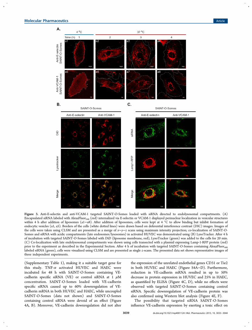
α activated HUVEC was investigated using CLSM. Alexa-Fluor546 siRNA (red) encapsulated in SAINT-O-Somes wasbinding to the surface of activated HUVEC at 4 °C (Figure 3Aa1, a5) and was internalized at 37 °C (Figure 3A a2−a4, a6−a8). siRNA SAINT-O-Somes endocytosed via E-selectin andVCAM-1 localized in acidic compartments (late endosomes/lysosomes) within 4 h after start of the incubation with the cells(Figure 3B, C), as demonstrated by colocalization withLysoTracker (Figure 3B) or lysosomally expressed Lamp-1-RFP (Figure 3C). By tracking simultaneously labeled SAINT-O-Somes and the encapsulated siRNA we demonstratedcolocalization of the two labels after 4 h and partial dissociationof the siRNA label from the lipid label after 24 h (Figure S3).
3.4. Anti-VCAM-1 and Anti-E-selectin SAINT-O-SomesCan Effectively Downregulate Target Genes in PrimaryEndothelial Cells. Using anti-E-selectin or anti-VCAM-1siRNA SAINT-O-Somes we aimed to knock down a gene thatis expressed in the endothelial cells and would pose a model forstudying the effects of the targeted system. VE-cadherin is anendothelial gene regulating formation of adherent celljunctions,29,30 the expression of which is restricted toendothelial cells and maintained under TNF-α stimulation
Figure 2. Selective delivery of siRNA to activated primary endothelial cells by targeted SAINT-O-Somes. Quiescent and TNF-α activated (A)HUVEC and (B) HAEC were incubated for 4 or 24 h with anti-E-selectin and anti-VCAM-1 SAINT-O-Somes containing AlexaFluor488 siRNA.Specificity of association to E-selectin respectively VCAM-1, was determined by coincubation with 75 times excess of anti-E-selectin or anti-VCAM-1monoclonal antibodies together with the liposomes. The association of siRNA with the cells was quantified by flow cytometric analysis. Data arepresented as mean florescence intensity (MFI) values ± SD of three independent experiments. * P < 0.05, # P < 0.05 - 4h vs 4h + Ab, $ P < 0.05 -24h vs 24h + Ab. (C) Fluorescence microscopy images of the uptake of targeted and nontargeted SAINT-O-Somes by cultured HUVEC. Theliposome membrane was labeled with DiI (red) and encapsulated siRNA was labeled with AlexaFluor488 (green), the nuclei of the cells were stainedusing Hoechst (blue). The presented data set shows representative images, presented as merged pictures, of tree independent experiments. Originalmagnification 100x.
Molecular Pharmaceutics Article
dx.doi.org/10.1021/mp4001124 | Mol. Pharmaceutics 2013, 10, 3033−30443038
(Supplementary Table 1), making it a suitable target gene forthis study. TNF-α activated HUVEC and HAEC wereincubated for 48 h with SAINT-O-Somes containing VE-cadherin specific siRNA (VE) or control siRNA at 1 μMconcentration. SAINT-O-Somes loaded with VE-cadherinspecific siRNA caused up to 60% downregulation of VE-cadherin mRNA in both HUVEC and HAEC, while uncoupledSAINT-O-Somes (data not shown) and SAINT-O-Somescontaining control siRNA were devoid of an effect (Figure4A, B). Moreover, VE-cadherin downregulation did not alter
the expression of the unrelated endothelial genes CD31 or Tie2in both HUVEC and HAEC (Figure S4A−D). Furthermore,reduction in VE-cadherin mRNA resulted in up to 50%decrease in protein expression in HUVEC and 25% in HAEC,as quantified by ELISA (Figure 4C, D), while no effects wereobserved with targeted SAINT-O-Somes containing controlsiRNA. Specific downregulation of VE-cadherin protein wasalso confirmed using Western blot analysis (Figure 4E, F).The possibility that targeted siRNA SAINT-O-Somes
influence VE-cadherin expression by exerting a toxic effect on
Figure 3. Anti-E-selectin and anti-VCAM-1 targeted SAINT-O-Somes loaded with siRNA directed to endolysosomal compartments. (A)Encapsulated siRNA labeled with AlexaFluor546 (red) internalized via E-selectin or VCAM-1 displayed perinuclear localization in vesicular structureswithin 4 h after addition of liposomes (a1−a8). After addition of liposomes, cells were kept at 4 °C to allow binding but inhibit formation ofendocytic vesicles (a1, a5). Borders of the cells (white dotted lines) were drawn based on deferential interference contrast (DIC) images. Images ofthe cells were taken using CLSM and are presented as a merge of x−y−z scans using maximum intensity projection; co-localization of SAINT-O-Somes and siRNA with acidic compartments (late endosomes/lysosomes) in activated HUVEC was demonstrated using (B) LysoTracker. After 4 hof incubation with targeted SAINT-O-Somes labeled with DiD (liposome membrane, red), LysoTracker (green) was added to the cells for 20 min.(C) Co-localization with late endolysosomal compartments was shown using cells transected with a plasmid expressing Lamp-1-RFP protein (red)prior to the experiment as described in the Experimental Section. After 4 h of incubation with targeted SAINT-O-Somes containing AlexaFluor488labeled siRNA (green), cells were visualized using CLSM and are presented as single z-scans. The presented data set shows representative images ofthree independent experiments.
Molecular Pharmaceutics Article
dx.doi.org/10.1021/mp4001124 | Mol. Pharmaceutics 2013, 10, 3033−30443039

the cells was excluded by their negligible effect on endothelialcell viability (Figure 5A, B). TNF-α activated HUVEC and
HAEC were incubated for 48 h with SAINT-O-Somescontaining VE-cadherin or control siRNA at 1 μM concen-
Figure 4. Effective downregulation of VE-cadherin in primary endothelial cells via targeted SAINT-O-Somes siRNA delivery. TNF-α activatedHUVEC and HAEC were incubated with targeted SAINT-O-Somes containing VE-cadherin (VE) or control siRNA (CS) at 1 μM concentration for48 h. After incubation, RNA or cell lysates were used for RT-qPCR (A,B), ELISA (C,D), and Western blot (E,F) as described in the ExperimentalSection. (A−B) Data are presented as relative expression ± SD, compared to cells treated only with TNF-α (Ctr), of a minimum of threeindependent experiments. *P < 0.05. (C−D) Data are presented as VE-cadherin protein expression ± SD of three independent experiments. (E−F)Images show a representative Western blot.
Figure 5. Targeted SAINT-O-Somes delivered siRNA do not affect cell viability. TNF-α activated HUVEC (A) and HAEC (B) were incubated withtargeted SAINT-O-Somes containing VE-cadherin or control siRNA at a 1 μM concentration for 48 h. After the incubation viability of cells wasinvestigated using CellTiter 96 AQueous One Solution reagent as described in the Experimental Section. The viability of activated endothelial cellswithout addition of SAINT-O-Somes was considered to be 100%. Data are presented as mean values ± SD of three independent experiments. *P <0.05.
Molecular Pharmaceutics Article
dx.doi.org/10.1021/mp4001124 | Mol. Pharmaceutics 2013, 10, 3033−30443040

tration. A small or no decrease in cell viability was observedeither in cells treated with SAINT-O-Somes containing VE-cadherin specific siRNA or in groups incubated with SAINT-O-Somes loaded with control siRNA.
4. DISCUSSIONInflamed endothelial cells play a significant role in thepathology of cancer and inflammatory diseases and presentan accessible target for systemically applied drug carriers.13 Assuch, development of siRNA delivery devices for pharmaco-logical intervention at the level of inflamed or angiogenicendothelium holds clinical potential. Development of a carrierwhich selectively delivers siRNA into endothelial cells at the siteof inflammation or angiogenesis can enhance pharmacologicalefficacy and limit possible side effects of the treatment. In thecurrent work we demonstrated that, by using SAINT-C18based liposomes (SAINT-O-Somes) surface-modified withantibodies specific for the inflammatory adhesion moleculesE-selectin or VCAM-1, we were able to selectively deliversiRNA into activated primary endothelial cells. We found thatE-selectin and VCAM-1 mediated internalization guidedSAINT-O-Somes to endolysosomal compartments and allowedthe release of the encapsulated siRNA. Targeted SAINT-O-Somes containing VE-cadherin specific siRNA showedsignificant downregulation of both the target gene mRNAand protein without exerting cellular toxicity. Furthermore, wedemonstrated that the physicochemical properties and in vivopharmacokinetic behavior of SAINT-O-Somes renders themsuitable for in vivo delivery of siRNA. From this study weconclude that anti-E-selectin and anti-VCAM-1 SAINT-O-Somes have the features that allows specific and effectivedelivery of siRNA to inflamed endothelial cells in vivo, whichjustifies further in vivo studies on the use of siRNA SAINT-O-Somes to interfere with inflammatory diseases.SAINT-O-Somes formulated with siRNA and 18 mol % of
SAINT-C18 in the liposomal bilayer form small and unilamellarparticles. Reported disc-like micelles are not expected tocontribute significantly to the physicochemical properties andfindings reported in this manuscript since they comprise <10%of siRNA SAINT-O-Some preparation, that with a polydisper-sity index of 0.17 is considered homogeneous and mono-disperse. The here-described SAINT-O-Somes are stable in thepresence of serum and show an siRNA encapsulation efficiencyof 71%, which is high compared to other liposomal deliveryplatforms incorporating cationic lipids.31,32 Good stability andencapsulation efficiency are crucial features of in vivo siRNAdelivery systems. For example, lipoplex formulations can reach>90% siRNA entrapment efficiency,33 but their limited stabilityunder physiological conditions restricts in vivo application.34
Our results indicated that the integrity of siRNA encapsulatedin SAINT-O-Somes is preserved under conditions thatresemble the in vivo situation (presence of 50% serum and 37°C) as well as upon long-term storage. We showed that i.v.injected SAINT-O-Somes display a two-phase PK with a longsecondary half-life >11 h, comparable to the PK behavior ofconventional long circulating liposomes, that have beensuccessfully used to target endothelial cells in vivo.17
Encapsulation of siRNA significantly influenced the PKparameters and resulted in delayed clearance from thecirculation. This could be explained by lower surface chargeof SAINT-O-Somes containing negatively charged siRNA.Coupling of antibody to siRNA SAINT-O-Somes prolongedthe circulation time from approximately 12 to 17 h. Combined,
our results justify use of siRNA SAINT-O-Somes in follow-upin vivo studies in the future.In the current study we focused on primary endothelial cells
as they more closely represent endothelial cells in vivo than celllines. Previous studies demonstrated that these cells are difficultto transfect23 and show limited processing capabilities,18 whichmakes them a challenging target for our newly designed siRNAcarrier. Recently, Whitehead et al.35 demonstrated that, incontrast to cell lines, primary cells were found to yield the mostpredictive correlations between in vitro and in vivo siRNAdelivery efficacy of lipid nanoparticles. Taking into account thewell-established heterogeneity of endothelial cells in vivo,36 weincluded primary cells from two distinct vascular beds in ourinvestigation (HUVECvenous, HAECarterial). BothVCAM-1 and E-selectin have been described as attractivetargets for endothelial specific drug delivery;13 however use ofVCAM-1 as a target for siRNA delivery to activated endothelialcells has not been demonstrated before. Here we comparedassociation of siRNA encapsulated into E-selectin or VCAM-1targeted SAINT-O-Somes with activated HUVEC and HAEC.We demonstrated that both anti-E-selectin and anti-VCAM-1SAINT-O-Somes allowed robust and specific siRNA deliveryinto activated cells and showed comparable downregulation ofVE-cadherin mRNA without exerting any cellular toxicity, asoften seen with siRNA delivery systems containing cationicamphiphiles.37 Up to 60% downregulation of VE-cadherinmRNA was reached in both cell types which was associated by50% decrease in protein expression in HUVEC and 25% inHAEC (Figure 4C−F). Sato et al.6 demonstrated that 60%downregulation of a target gene mRNA and protein by siRNA-bearing liposomes targeted to hepatic stellate cells was sufficientto almost completely resolve liver fibrosis and prolongedsurvival of cirrhotic rats. Thus, the demonstrated capacity oftargeted SAINT-O-Somes for gene silencing could be sufficientto interfere with the expression of disease-associated genes andrequires further investigation in animal models. Differences inthe efficacy of protein downregulation between HUVEC andHAEC in our study could not be attributed to differences inmRNA and protein expression levels (Table 2, SupportingInformation). Since HAEC and HUVEC exerted a similarextent of uptake of siRNA and siRNA delivered by the SAINT-O-Somes did not affect cell viability, possibly the ability of bothcells to process SAINT-O-Somes influencing siRNA release isresponsible for the observed differences. Moreover, the efficacyof the siRNA delivery can be affected by different molecularaspects that are part of the concept of heterogeneity ofendothelial cells.38 We demonstrated that anti-E-selectin andanti-VCAM-1 SAINT-O-Somes can effectively deliver siRNA toprimary endothelial cells originating from different vascularbeds, which provides an opportunity for effective targeting ofdisease-associated endothelial cell subsets in different vascularsegments.Target epitopes residing at the membrane of the target cells
should internalize upon ligand binding and allow intracellularrelease of the siRNA, which is a prerequisite for effective genedownregulation. It was previously shown that both E-selectinand VCAM-1 could mediate internalization of immunolipo-somes conjugated with antibodies targeting these proteins.15,39
Nevertheless little is known about the intracellular processingof siRNA delivered via this route. Therefore we studied the fateof anti-VCAM-1 and anti-E-selectin SAINT-O-Somes contain-ing siRNA after binding to the endothelial cell surface applyingconfocal microscopy. siRNA endocytosed via E-selectin and
Molecular Pharmaceutics Article
dx.doi.org/10.1021/mp4001124 | Mol. Pharmaceutics 2013, 10, 3033−30443041
VCAM-1 localized in endolysosomal compartments in theperinuclear region within 4 h after the start of incubation ofcells with SAINT-O-Somes. Our observations are in line withfindings describing transport of VCAM-1 and E-selectin ligandsmainly via clathrin-mediated endocytosis to lysosomes(summarized by Muro et al.40) although involvement ofother pathways, such as macropinocytosis or cell adhesionmolecule endocytosis cannot be excluded. Adrian et al.18
reported that at low pH (pH < 5.5) the SAINT-O-Some bilayeris destabilized, which enhances content release. Within theendolysosomal compartments the low pH may thus be able tofacilitate destabilization of the particles and enhance the releaseof siRNA. Furthermore, cationic lipids such as SAINT candestabilize the endosomal membrane by inducing “flipping” ofanionic lipids in the endosomal bilayer which facilitates releaseof the cargo into the cytoplasm.41 Under physiologicalconditions a polyethylene-glycol (PEG) coating limits inter-action of particles with the cell membrane by masking theirsurface charge.42 Destabilization of SAINT-O-Some bilayermay reduce the effect of PEG and allow SAINT to interact withthe endosomal membrane. Moreover, we showed that SAINT-O-Somes and encapsulated siRNA followed the same route ofinternalization and partially dissociated after 24 h, whichindicates limited release of siRNA from the carrier. Up to now,little is known about the relation between siRNA disassemblyfrom the carrier and the efficacy of gene silencing. The firsttools to study intracellular disassembly of siRNA nano-complexes have been developed only recently.43 Poorprocessing of liposomes by HUVEC, as reported by Adrianet al.,18 may as well contribute to limited release of siRNA.Based on the intracellular trafficking of SAINT-O-Somestargeted to VCAM-1 and E-selectin, the inclusion of pH-sensitive components in the formulation (e.g., pH-sensitivePEG) might improve the efficacy of siRNA release.32,44 Furtherstudies focusing on the exact siRNA release mechanisminvolving SAINT-O-Somes will shed more light and facilitatethe rational design of these SAINT-based carriers.We here report a liposomal formulation that allows selective
and functional siRNA delivery into inflammation activatedprimary endothelial cells, mediates significant downregulationof a target gene, and is suitable for in vivo application based ontheir physicochemical and pharmacokinetic features. Wedemonstrated that both VCAM-1 and E-selectin can serve asan efficient and specific entry route for siRNA delivery toinflamed endothelial cells. Liposomes based on cationicamphiphile SAINT-C18 (SAINT-O-Somes) are a suitablecarrier for siRNA and when harnessed with anti-VCAM-1 oranti-E-selectin antibodies allow effective delivery of siRNA toactivated primary endothelial cells from venous and aorticvascular beds. Anti-VCAM-1 and anti-E-selectin SAINT-O-Somes are thus a novel drug delivery system that provide anopportunity for siRNA delivery to disease-associated endothe-lial cell subsets in different vascular segments.
■ ASSOCIATED CONTENT*S Supporting InformationFigure S1 showing blood clearance of empty and siRNA loadedSAINT-O-Somes. Figure S2 showing the association ofnontargeted SAINT-O-Somes with endothelial cells. FigureS3 showing CLSM images of intracellular colocalization ofSAINT-O-Somes and siRNA in activated HUVEC. Figure S4showing expression of endothelial genes CD31 and Tie-2 inHUVEC and HAEC after transfection with anti-E-selectin and
anti-VCAM-1 SAINT-O-Somes containing VE-cadherin specif-ic or control siRNA. Supplementary Table 1 showing basalexpression of VE-cadherin, CD31, and Tie-2 in resting or TNF-α activated cells. This material is available free of charge via theInternet at http://pubs.acs.org.
■ AUTHOR INFORMATIONCorresponding Author*Department of Pathology and Medical Biology, MedicalBiology Section, University Medical Center Groningen,Hanzeplein 1, Internal Post Code EA11, 9713 GZ Groningen,The Netherlands. Tel.: +31503611293; fax: +31503619911. E-mail address: [email protected] (J.A.A.M.K.).
NotesThe authors declare no competing financial interest.
■ ACKNOWLEDGMENTSWe thank Henk Moorlag, Peter J. Zwiers, and Rianne M.Jongman for excellent technical assistance and Sabine Barnertfrom the Dept. of Pharmaceutical Technology and Biophar-macy (Albert-Ludwigs University, Freiburg) for her contribu-tion to the cryo-TEM experiments. Dr. Ur Rehman Zia and Dr.Jan Willem Kok from the Dept. of Membrane Cell Biology(UMCG, Groningen) are acknowledged for kindly providingpLamp-1-RFP plasmid and assistance with confocal micros-copy. We thank Dr. Ed Talman for providing high qualitySAINT-C18 and prof. Gerrit L. Scherphof for editing andproofreading the manuscript. Microscopic fluorescence imagingwas performed at the UMCG Microscopy & Imaging Center(UMIC), which is supported by The Netherlands Organizationfor Health Research and Developoment (ZonMW grant 40-00506-98-9021). This work was supported by EFRO (Euro-pean Fund for Regional Development) from the EuropeanUnion, project NTS 068 and 073 Drug Delivery and Targeting.The authors declare no competing financial interests. M.H.J.R.is CEO of Synvolux Therapeutics.
■ REFERENCES(1) Kim, I.; Moon, S. O.; Kim, S. H.; Kim, H. J.; Koh, Y. S.; Koh, G.Y. Vascular endothelial growth factor expression of intercellularadhesion molecule 1 (ICAM-1), vascular cell adhesion molecule 1(VCAM-1), and E-selectin through nuclear factor-kappa B activationin endothelial cells. J. Biol. Chem. 2001, 276 (10), 7614−20.(2) Juliano, R.; Bauman, J.; Kang, H.; Ming, X. Biological barriers totherapy with antisense and siRNA oligonucleotides. Mol. Pharmaceutics2009, 6 (3), 686−95.(3) Whitehead, K. A.; Langer, R.; Anderson, D. G. Knocking downbarriers: advances in siRNA delivery. Nat. Rev. Drug Discovery 2009, 8(2), 129−38.(4) Behlke, M. A. Chemical modification of siRNAs for in vivo use.Oligonucleotides 2008, 18 (4), 305−19.(5) Frank-Kamenetsky, M.; Grefhorst, A.; Anderson, N. N.; Racie, T.S.; Bramlage, B.; Akinc, A.; Butler, D.; Charisse, K.; Dorkin, R.; Fan,Y.; Gamba-Vitalo, C.; Hadwiger, P.; Jayaraman, M.; John, M.;Jayaprakash, K. N.; Maier, M.; Nechev, L.; Rajeev, K. G.; Read, T.;Rohl, I.; Soutschek, J.; Tan, P.; Wong, J.; Wang, G.; Zimmermann, T.;de Fougerolles, A.; Vornlocher, H. P.; Langer, R.; Anderson, D. G.;Manoharan, M.; Koteliansky, V.; Horton, J. D.; Fitzgerald, K.Therapeutic RNAi targeting PCSK9 acutely lowers plasma cholesterolin rodents and LDL cholesterol in nonhuman primates. Proc. Natl.Acad. Sci. U.S.A. 2008, 105 (33), 11915−20.(6) Sato, Y.; Murase, K.; Kato, J.; Kobune, M.; Sato, T.; Kawano, Y.;Takimoto, R.; Takada, K.; Miyanishi, K.; Matsunaga, T.; Takayama, T.;Niitsu, Y. Resolution of liver cirrhosis using vitamin A-coupled
Molecular Pharmaceutics Article
dx.doi.org/10.1021/mp4001124 | Mol. Pharmaceutics 2013, 10, 3033−30443042

liposomes to deliver siRNA against a collagen-specific chaperone. Nat.Biotechnol. 2008, 26 (4), 431−42.(7) Takeshita, F.; Minakuchi, Y.; Nagahara, S.; Honma, K.; Sasaki, H.;Hirai, K.; Teratani, T.; Namatame, N.; Yamamoto, Y.; Hanai, K.; Kato,T.; Sano, A.; Ochiya, T. Efficient delivery of small interfering RNA tobone-metastatic tumors by using atelocollagen in vivo. Proc. Natl. Acad.Sci. U.S.A. 2005, 102 (34), 12177−82.(8) Davis, M. E.; Zuckerman, J. E.; Choi, C. H.; Seligson, D.; Tolcher,A.; Alabi, C. A.; Yen, Y.; Heidel, J. D.; Ribas, A. Evidence of RNAi inhumans from systemically administered siRNA via targeted nano-particles. Nature 2010, 464 (7291), 1067−70.(9) Kamps, J. A. A. M.; Molema, G. Targeting Liposomes toEndothelial Cells in Inflammatory Diseases. In Interactions of Liposomeswith the Biological Milieu, Liposome Technology ed.; Gregoriadis, G.,Ed.; CRC Press: Boca Raton, FL, 2007; Vol. 3, pp 127−150.(10) Molema, G. Targeted drug delivery to the tumor neo-vasculature: concepts, advances, and challenges. In Angiogenesis: anintegrative approach from science to medicine; Folkman, J., Figg, W. D.,Eds.; Cambridge University Press: New York, 2008; Chapter 25, pp283−297.(11) Aleku, M.; Schulz, P.; Keil, O.; Santel, A.; Schaeper, U.;Dieckhoff, B.; Janke, O.; Endruschat, J.; Durieux, B.; Roder, N.; Loffler,K.; Lange, C.; Fechtner, M.; Mopert, K.; Fisch, G.; Dames, S.; Arnold,W.; Jochims, K.; Giese, K.; Wiedenmann, B.; Scholz, A.; Kaufmann, J.Atu027, a liposomal small interfering RNA formulation targetingprotein kinase N3, inhibits cancer progression. Cancer Res. 2008, 68(23), 9788−98.(12) Santel, A.; Aleku, M.; Roder, N.; Mopert, K.; Durieux, B.; Janke,O.; Keil, O.; Endruschat, J.; Dames, S.; Lange, C.; Eisermann, M.;Loffler, K.; Fechtner, M.; Fisch, G.; Vank, C.; Schaeper, U.; Giese, K.;Kaufmann, J. Atu027 prevents pulmonary metastasis in experimentaland spontaneous mouse metastasis models. Clin. Cancer Res. 2010, 16(22), 5469−80.(13) Kowalski, P. S.; Leus, N. G.; Scherphof, G. L.; Ruiters, M. H.;Kamps, J. A.; Molema, G. Targeted siRNA delivery to diseasedmicrovascular endothelial cells: cellular and molecular concepts.IUBMB Life 2011, 63 (8), 648−58.(14) van Meurs, M.; Kurniati, N. F.; Wulfert, F. M.; Asgeirsdottir, S.A.; de Graaf, I. A.; Satchell, S. C.; Mathieson, P. W.; Jongman, R. M.;Kumpers, P.; Zijlstra, J. G.; Heeringa, P.; Molema, G. Shock-inducedstress induces loss of microvascular endothelial Tie2 in the kidneywhich is not associated with reduced glomerular barrier function. Am.J. Physiol. Renal Physiol. 2009, 297 (2), F272−81.(15) Everts, M.; Koning, G. A.; Kok, R. J.; Asgeirsdottir, S. A.;Vestweber, D.; Meijer, D. K.; Storm, G.; Molema, G. In vitro cellularhandling and in vivo targeting of E-selectin-directed immunoconju-gates and immunoliposomes used for drug delivery to inflamedendothelium. Pharm. Res. 2003, 20 (1), 64−72.(16) van Meurs, M.; Wulfert, F. M.; Knol, A. J.; De Haes, A.;Houwertjes, M.; Aarts, L. P.; Molema, G. Early organ-specificendothelial activation during hemorrhagic shock and resuscitation.Shock 2008, 29 (2), 291−9.(17) Asgeirsdottir, S. A.; Zwiers, P. J.; Morselt, H. W.; Moorlag, H.E.; Bakker, H. I.; Heeringa, P.; Kok, J. W.; Kallenberg, C. G.; Molema,G.; Kamps, J. A. Inhibition of proinflammatory genes in anti-GBMglomerulonephritis by targeted dexamethasone-loaded AbEsel lip-osomes. Am. J. Physiol. Renal Physiol. 2008, 294 (3), F554−61.(18) Adrian, J. E.; Morselt, H. W.; Suss, R.; Barnert, S.; Kok, J. W.;Asgeirsdottir, S. A.; Ruiters, M. H.; Molema, G.; Kamps, J. A. TargetedSAINT-O-Somes for improved intracellular delivery of siRNA andcytotoxic drugs into endothelial cells. J. Controlled Release 2010, 144(3), 341−9.(19) Asgeirsdottir, S. A.; Kamps, J. A.; Bakker, H. I.; Zwiers, P. J.;Heeringa, P.; van der Weide, K.; van Goor, H.; Petersen, A. H.;Morselt, H.; Moorlag, H. E.; Steenbergen, E.; Kallenberg, C. G.;Molema, G. Site-specific inhibition of glomerulonephritis progressionby targeted delivery of dexamethasone to glomerular endothelium.Mol. Pharmacol. 2007, 72 (1), 121−31.
(20) Kamps, J. A.; Swart, P. J.; Morselt, H. W.; Pauwels, R.; DeBethune, M. P.; De Clercq, E.; Meijer, D. K.; Scherphof, G. L.Preparation and characterization of conjugates of (modified) humanserum albumin and liposomes: drug carriers with an intrinsic anti-HIVactivity. Biochim. Biophys. Acta 1996, 1278 (2), 183−90.(21) Bottcher, C. J. F.; Van Gent, C. M.; Pries, C. A rapid andsensitive sub-micro phosphorus determination. Anal. Chim. Acta 1961,24, 203.(22) Adrian, J. E.; Kamps, J. A.; Scherphof, G. L.; Meijer, D. K.; vanLoenen-Weemaes, A. M.; Reker-Smit, C.; Terpstra, P.; Poelstra, K. Anovel lipid-based drug carrier targeted to the non-parenchymal cells,including hepatic stellate cells, in the fibrotic livers of bile duct ligatedrats. Biochim. Biophys. Acta 2007, 1768 (6), 1430−9.(23) Asgeirsdottir, S. A.; Talman, E. G.; de Graaf, I. A.; Kamps, J. A.;Satchell, S. C.; Mathieson, P. W.; Ruiters, M. H.; Molema, G. Targetedtransfection increases siRNA uptake and gene silencing of primaryendothelial cells in vitroa quantitative study. J. Controlled Release2010, 141 (2), 241−51.(24) Proost, J. H.; Eleveld, D. J. Performance of an iterative two-stageBayesian technique for population pharmacokinetic analysis of richdata sets. Pharm. Res. 2006, 23 (12), 2748−59.(25) Johnsson, M.; Edwards, K. Liposomes, disks, and sphericalmicelles: aggregate structure in mixtures of gel phase phosphatidylcho-lines and poly(ethylene glycol)-phospholipids. Biophys. J. 2003, 85 (6),3839−47.(26) Johansson, E.; Lundquist, A.; Zuo, S.; Edwards, K. Nanosizedbilayer disks: attractive model membranes for drug partition studies.Biochim. Biophys. Acta 2007, 1768 (6), 1518−25.(27) Seguin, J.; Brulle, L.; Boyer, R.; Lu, Y. M.; Ramos Romano, M.;Touil, Y. S.; Scherman, D.; Bessodes, M.; Mignet, N.; Chabot, G. G.Liposomal encapsulation of the natural flavonoid fisetin improvesbioavailability and antitumor efficacy. Int. J. Pharmaceutics 2013, 444(1−2), 146−54.(28) Smith, G.; Raghunandan, R.; Wu, Y.; Liu, Y.; Massare, M.;Nathan, M.; Zhou, B.; Lu, H.; Boddapati, S.; Li, J.; Flyer, D.; Glenn, G.Respiratory syncytial virus fusion glycoprotein expressed in insect cellsform protein nanoparticles that induce protective immunity in cottonrats. PloS One 2012, 7 (11), e50852.(29) Dejana, E. Endothelial cell-cell junctions: happy together. Nat.Rev. Mol. Cell Biol. 2004, 5 (4), 261−70.(30) Crosby, C. V.; Fleming, P. A.; Argraves, W. S.; Corada, M.;Zanetta, L.; Dejana, E.; Drake, C. J. VE-cadherin is not required for theformation of nascent blood vessels but acts to prevent theirdisassembly. Blood 2005, 105 (7), 2771−6.(31) Adrian, J. E.; Wolf, A.; Steinbach, A.; Rossler, J.; Suss, R.Targeted delivery to neuroblastoma of novel siRNA-anti-GD2-liposomes prepared by dual asymmetric centrifugation and sterol-based post-insertion method. Pharm. Res. 2011, 28 (9), 2261−72.(32) Auguste, D. T.; Furman, K.; Wong, A.; Fuller, J.; Armes, S. P.;Deming, T. J.; Langer, R. Triggered release of siRNA frompoly(ethylene glycol)-protected, pH-dependent liposomes. J. Con-trolled Release 2008, 130 (3), 266−74.(33) Singhania, A.; Wu, S. Y.; McMillan, N. A. Effective Delivery ofPEGylated siRNA-Containing Lipoplexes to Extraperitoneal Tumoursfollowing Intraperitoneal Administration. J. Drug Delivery 2011, 2011,192562.(34) Zuhorn, I. S.; Visser, W. H.; Bakowsky, U.; Engberts, J. B.;Hoekstra, D. Interference of serum with lipoplex-cell interaction:modulation of intracellular processing. Biochim. Biophys. Acta 2002,1560 (1−2), 25−36.(35) Whitehead, K. A.; Matthews, J.; Chang, P. H.; Niroui, F.;Dorkin, J. R.; Severgnini, M.; Anderson, D. G. In vitro-in vivotranslation of lipid nanoparticles for hepatocellular siRNA delivery.ACS Nano 2012, 6 (8), 6922−9.(36) Molema, G. Heterogeneity in endothelial responsiveness tocytokines, molecular causes, and pharmacological consequences.Semin. Thromb. Hemost. 2010, 36 (3), 246−64.
Molecular Pharmaceutics Article
dx.doi.org/10.1021/mp4001124 | Mol. Pharmaceutics 2013, 10, 3033−30443043

(37) Kanasty, R. L.; Whitehead, K. A.; Vegas, A. J.; Anderson, D. G.Action and reaction: the biological response to siRNA and its deliveryvehicles. Mol. Ther. 2012, 20 (3), 513−24.(38) Langenkamp, E.; Molema, G. Microvascular endothelial cellheterogeneity: general concepts and pharmacological consequences foranti-angiogenic therapy of cancer. Cell Tissue Res. 2009, 335 (1), 205−22.(39) Voinea, M.; Manduteanu, I.; Dragomir, E.; Capraru, M.;Simionescu, M. Immunoliposomes directed toward VCAM-1 interactspecifically with activated endothelial cellsa potential tool forspecific drug delivery. Pharm. Res. 2005, 22 (11), 1906−17.(40) Muro, S. Challenges in design and characterization of ligand-targeted drug delivery systems. J. Controlled Release 2012, 164, 125−37.(41) Zelphati, O.; Szoka, F. C., Jr. Mechanism of oligonucleotiderelease from cationic liposomes. Proc. Natl. Acad. Sci. U.S.A. 1996, 93(21), 11493−8.(42) Morille, M.; Passirani, C.; Vonarbourg, A.; Clavreul, A.; Benoit,J. P. Progress in developing cationic vectors for non-viral systemic genetherapy against cancer. Biomaterials 2008, 29 (24−25), 3477−96.(43) Alabi, C. A.; Love, K. T.; Sahay, G.; Stutzman, T.; Young, W. T.;Langer, R.; Anderson, D. G. FRET-labeled siRNA probes for trackingassembly and disassembly of siRNA nanocomplexes. ACS Nano 2012,6 (7), 6133−41.(44) Simoes, S.; Moreira, J. N.; Fonseca, C.; Duzgunes, N.; de Lima,M. C. On the formulation of pH-sensitive liposomes with longcirculation times. Adv. Drug Delivery Rev. 2004, 56 (7), 947−65.
Molecular Pharmaceutics Article
dx.doi.org/10.1021/mp4001124 | Mol. Pharmaceutics 2013, 10, 3033−30443044