modulation of endothelial targeting by size of antibody–antioxidant enzyme conjugates
TRANSCRIPT

Journal of Controlled Release 149 (2011) 236–241
Contents lists available at ScienceDirect
Journal of Controlled Release
j ourna l homepage: www.e lsev ie r.com/ locate / jconre l
Modulation of endothelial targeting by size of antibody–antioxidantenzyme conjugates
Vladimir V. Shuvaev a, Samira Tliba a, Jeremy Pick a, Evguenia Arguiri b, Melpo Christofidou-Solomidou b,Steven M. Albelda b, Vladimir R. Muzykantov a,c,⁎a Institute for Translational Medicine and Therapeutics, University of Pennsylvania School of Medicine, Philadelphia, PA 19104, USAb Pulmonary Critical Care Division, Department of Medicine, University of Pennsylvania School of Medicine, Philadelphia, PA 19104, USAc Department of Pharmacology, University of Pennsylvania School of Medicine, Philadelphia, PA 19104, USA
⁎ Corresponding author. Institute for EnvironmePennsylvania Medical Center, 1 John Morgan BuildinWalk, Philadelphia, PA 19104-6068, USA. Tel.: +1 215 89
E-mail address: [email protected] (V.
0168-3659/$ – see front matter © 2010 Elsevier B.V. Adoi:10.1016/j.jconrel.2010.10.026
a b s t r a c t
a r t i c l e i n f oArticle history:Received 7 May 2010Accepted 20 October 2010Available online 31 October 2010
Keywords:Vascular immunotargetingEndotheliumAntioxidant deliveryLungs
Endothelial targeting of antioxidant enzymes attenuates acute vascular oxidative stress in animal studies.Superoxide dismutase (SOD) and catalase conjugated with antibodies to Platelet-Endothelial Cell AdhesionMolecule-1 (anti-PECAM/SOD and anti-PECAM/catalase) bind to endothelium, accumulate in the pulmonaryvasculature, and detoxify reactive oxygen species. In order to define the role of conjugate size in the efficacyand specificity of endothelial targeting, we synthesized anti-PECAM/enzyme conjugates of controlled size(40 nm–10,000 nm). Binding of anti-PECAM/enzymes to endothelial cells increased with conjugate size from300 nm to 2 μm (from 2.5 to 8.5% of bound fraction), and was specific, as conjugates did not bind to PECAM-negative cells. Pulmonary uptake of anti-PECAM/enzyme conjugates injected intravenously in mice alsoincreased from 4.5 to 16% of injected dose for particles from 200 to 800 nm. However, control conjugateslarger than 300 nm showed elevated non-specific pulmonary uptake, indicating that the targeting specificityof anti-PECAM/enzyme conjugates in vivo has a bell-shaped curve with amaximum close to 300-nm diameter.These results show that: i) the size of an antibody/enzyme conjugate modulates efficacy and specificity oftargeting, and ii) a size optimum should be defined in vivo to account for parameters that are difficult to modelin cell culture.
ntal Medicine, University ofg, 36th Street and Hamilton8 9823; fax: +1 215 898 0868.R. Muzykantov).
ll rights reserved.
© 2010 Elsevier B.V. All rights reserved.
1. Introduction
Targeted delivery of therapeutics to endothelial cells holdspromise to improve treatment of disease conditions involving thesevascular lining cells, including ischemia–reperfusion, inflammation,tumor growth, thrombosis and oxidative stress [1]. This can beachieved by conjugating drugs to antibodies that target specificendothelial surface molecules [2–7]. For example, conjugation ofdrugs and drug carriers with antibodies to Platelet-Endothelial CellAdhesion Molecule-1 (anti-PECAM) provides: i) specific binding ofconjugates to endothelial cells; ii) intracellular delivery of drugs; ii)preferential accumulation in highly vascularized organs after intra-venous injection (especially in the lungs, which is the main target foranti-PECAM conjugates); and, iv) enhanced local therapeutic effectsof drugs including genetic materials, as well as anti-thrombotic, anti-inflammatory and antioxidant enzymes [8–10].
In particular, endothelial targeting of the antioxidant enzymessuperoxide dismutase (SOD) and catalase is being developed as a
novel approach for effective containment of acute vascular oxidativestress [5,9,11–13]. Anti-PECAM/SOD and anti-PECAM/catalase con-jugates detoxify corresponding reactive oxygen species in endothelialcells [14,15]. After intravenous injection in animals, SOD and catalaseconjugated with antibodies to PECAM-1 and other endothelial targetsaccumulate in the pulmonary vasculature and protect against acutepulmonary oxidative stress, whereas native enzymes, or enzymesconjugated with control IgG, exert neither endothelial targeting norprotection [10,16,17]. This approach is especially attractive forpotential use in the alleviation of lung ischemia–reperfusion injury,such as that associated with transplantation injury [10,12,13,18].Therefore, understanding of mechanisms and further optimization ofendothelial targeting of anti-PECAM/SOD and anti-PECAM/catalaserepresent important goals.
One particularly intriguing, but still insufficiently understood,aspect is the role of the size of the conjugates in endothelial targeting.Most targeting studies have not measured the size of proteinconjugates and fusion constructs, which may vary from nanometers(small fusion proteins) to microns (supramolecular conjugates). Onereason for this lack of information is that the synthesis of proteinconjugates with controlled and stable size is very challenging. The fewstudies that have quantitatively measured size by dynamic lightscattering (DLS) employed conjugates within a narrow range of 250–

237V.V. Shuvaev et al. / Journal of Controlled Release 149 (2011) 236–241
350 nm in diameter [10,14]. Therefore, no systematic studies on therole of size of antibody/enzyme conjugates in endothelial targetingare available. To fill this gap in our knowledge, we designed a series ofprotein conjugates consisting of antioxidant enzymes coupled toeither anti-PECAM or control IgG that ranged in size from ~50 nm tomicrons and then studied the endothelial targeting of theseconjugates using isotope-labeled SOD or catalase in cell cultures andlab animals.
2. Materials and methods
2.1. Materials and cells
Cytochrome c, xanthine oxidase, xanthine (3,7-dihydro-purine-2,6-dione), dimethylformamide (DMFA) and fetal bovine serum werepurchased from Sigma (St. Louis, MO). Cu, Zn-superoxide dismutase(SOD) and catalase from bovine liver and streptavidin are fromCalbiochem (San Diego, CA). Radioisotope-containing sodium iodide(Na125I) was obtained from PerkinElmer (Wellesley, MA). Succinimi-dyl-6-[biotinamido]hexanoate (NHS-LC-biotin), 4-[N-maleimido-methyl]cyclohexane-1-carboxylate (SMCC), N-succinimidyl-S-acetylthioacetate (SATA), HABA (4′-hydroxyazobenzene-2-carboxylicacid) and Iodogen were from Pierce Biotechnology (Rockford, IL).Anti-PECAM monoclonal antibodies used were: (1) mAb 62, amonoclonal mouse IgG2α antibody directed against human Platelet-Endothelial Cell Adhesion Molecule-1 (PECAM) [9,19]; (2) mAb 390, arat monoclonal antibody reacting with murine PECAM [20], and (3)MEC-13.3, a rat IgG2α monoclonal antibody against murine PECAMfrom BD Biosciences (San Jose, CA). All cell culture mediumcomponents were from Life Technologies (Gaithersburg, MD) unlessotherwise noted. Rat IgG was from Jackson Immuno-ResearchLaboratory, Inc. (West Grove, PA). Human umbilical vein endothelialcells (HUVEC) were purchased from Clonetics (San Diego, CA).
2.2. Biotinylation of proteins and biotin measurement
Proteins were biotinylated with NHS-LC-biotin freshly dissolved inDMFA to a concentration of 0.1 M. SOD was biotinylated at 15:1 NHS-LC-biotin:protein molar excess. IgG were biotinylated at differentratio to modulate conjugates' size. After 2 h of incubation on ice, freebiotin was removed by dialysis against phosphate-buffered saline(PBS). Protein-bound fraction of biotin in b-SOD and b-IgG wasmeasured by HABA assay as we described earlier [21].
2.3. Measurements of SOD activity
Activity of SOD was measured using ferricytochrome c assay [22].Xanthine/xanthine oxidase systemwas used as a source of superoxideanion and cytochrome c as the indicating scavenger of the radicalcompeting with SOD. Working solution (0.6 ml) contained 50 mMphosphate buffer (pH 7.8), 0.1 EDTA, 50 μM xanthine, 20 μMcytochrome c and 10 μl of SOD-containing sample. Reaction wasinitiated by the addition of 10 μl of 0.2 U/ml xanthine oxidase and theabsorbance was monitored at 550 nm using Cary 50 spectrophotom-eter (Varian, Palo Alto, CA). One unit of SOD is defined as the amountof enzyme, which inhibits the rate of cytochrome c reduction by 50%at pH 7.8 and 25 °C. Each measurement was performed at least threetimes and results were expressed as mean±SD.
2.4. SOD and catalase radiolabeling
SOD and catalase were radiolabeled with Na125I using Iodogen asrecommended by the manufacturer. Iodogen was first dissolved inchloroform and a thin film was casted in glass tube by drying undernitrogen flow. After the iodination reaction was performed (15 min at25 °C), an excess of free label was removed by gel-filtration
chromatography using Bio-Spin 6 Chromatography columns (Bio-Rad Labs, Hercules, CA), while iodinated enzymes were transferred toPBS.
2.5. Preparation of immunoconjugates
Two modes of enzyme conjugation to antibody were used, usingstreptavidin–biotin system and chemical conjugation using SATA andSMCC cross-linkers. Biotinylated SOD was conjugated with biotiny-lated IgG (anti-PECAM or non-immune IgG) via streptavidin asdescribed elsewhere [9,21,23]. The molar ratio of enzyme to antibodywas kept 1:1. In the second technique, hetero-bifunctional cross-linker SMCC was used to introduce stable maleimide reactive groupinto enzyme molecule. The reaction was performed at 20–100 foldmolar excess of SMCC at room temperature during 1 h. In parallelsulfhydryl groups were introduced in the molecule of antibody orcontrol IgG through primary amine using SATA at 20-fold molarexcess of SATA at room temperature for 30 min. Sulfhydryl groupswere de-protected using hydroxylamine and antibody was conjugat-ed with SA at 2:1 IgG to SA molar ratio. At each step unreactedcomponents were removed using Spin Protein Columns (G-25Sephadex, Roche Applied Science, Indianapolis, IN). The effectivediameter of the obtained conjugates was measured by dynamic lightscattering apparatus 90Plus Particle Sizer (Brookhaven InstrumentsCorp., NY). Radiolabeled anti-PECAM conjugates were prepared byintroducing from 5 to 10 mol% of 125I-enzyme to unlabeled enzymeprior to its conjugation.
2.6. Cell culture
HUVEC were maintained in M199 medium (GIBCO, Grand Island,NY) with 15% fetal bovine serum supplemented with 100 μg/mlheparin (Sigma), 2 mM L-glutamine (GIBCO), 15 μg/ml endothelialcell growth supplement (Upstate, Lake Placid, NY), 100 U/mlpenicillin, and 100 μg/ml streptomycin (GIBCO). Cells were grownto confluency for higher PECAM expression level.
2.7. Binding of anti-PECAM conjugates to endothelial cells
HUVEC were plated in 24 well culture dishes and grown toconfluent culture. Radiolabeled conjugates were added to cells atsequential dilutions. Cells were incubated at 37 °C for 1 h, unboundlabel was washed out and cells were lysed with lysis buffer (1% TritonX-100, 1.0 M NaOH). Bound radioactivity was measured using aWallac 1470 Wizard™ gamma counter (Gaithersburg, MD). Boundmaterial was expressed as ng of enzyme per well. Each point wasperformed in quadruplicates and results were expressed as mean±SEM.
2.8. Pulmonary uptake and blood level of anti-PECAM conjugates in vivo
Animal experiments were performed according to the protocolapproved by the Institutional Animal Care and Use Committee(IACUC) of the University of Pennsylvania. Ten micrograms ofradiolabeled anti-PECAM/enzyme or IgG/enzyme was injected IV inC57BL/6J mice (The Jackson Laboratory, Bar Harbor, ME) via the tailvein. After 1 h, blood samples were withdrawn and animals weresacrificed. Internal organs were harvested, rinsed with saline, blotteddry, and weighed. Tissue radioactivity in organs and 100-μl samples ofblood was determined in a gamma counter. The level of 125I in theorgans was used to calculate parameters enzyme uptake (see [19] fordetailed explanations) expressed as mean±SD. Total dose circulatingin blood was calculated considering that mouse blood volume equalsnormally to 7.2% of animal weight. Immunospecificity index (ISI),parameter of targeting specificity, was calculated as %ID/g of targetedcarrier (anti-PECAM/enzyme) normalized per blood level and divided

238 V.V. Shuvaev et al. / Journal of Controlled Release 149 (2011) 236–241
by same parameter of control non-targeted counterpart (IgG/enzyme).
2.9. Statistical analysis
Statistical difference among groups was determined using one-way analysis of variance (ANOVA). When statistically significantdifferences were found (pb0.05) individual comparisons were madeusing the Student t-test upon normality confirmation and Holm–Sidakmethod (SigmaPlot 11.0).
3. Results
3.1. Synthesis of antibody-enzyme conjugates of controlled size
We conjugated SOD and catalase to anti-PECAM using twomethods and found that in both approaches the size of the resultantanti-PECAM/enzyme conjugates could be controlled by modulatingthe reaction conditions. In the first approach, we used a modularstreptavidin–biotin cross-linking technique: the enzymes and immu-noglobulins were biotinylated (4–7 biotin residues per proteinmolecule) and cross-linked by streptavidin (SA) [14]. DLS showedthat this allowed us to modulate the size of conjugates from ~50 nmto several microns by changing the ratio between SA and biotinylatedproteins (Fig. 1).
Using proteins with variable degree of biotinylation allows forfurther “tuning” of the conjugate size. For example, using anti-PECAMwith ~4 vs ~6 biotin residues per molecule provided SOD conjugateswith maximal size approximately 200 vs 400 nm (data not shown).Similarly, variations of streptavidin input and degree of biotinylationof catalase provided anti-PECAM/catalase conjugates of diverse size
400A
Eff
ecti
ve d
iam
eter
, nm
Eff
ecti
ve d
iam
eter
, nm
100
200
300
SA:(b-SOD+b-anti-PECAM), mol/mol0.0 0.5 1.0 1.5 2.0
0.0 0.5 1.0 1.5 2.0
0
600
800
1000B
SA:(Cat+αPECAM),mol/mol
0
Siz
e, μ
m
0
5
10
0
200
400
SA:(b-Catalase+b-anti-PECAM), mol/mol
21
Fig. 1. Modulation of streptavidin cross-linker input controls size of conjugates formedfrom biotinylated anti-PECAM and biotinylated enzymes. A. Anti-PECAM/SODconjugates. B. Anti-PECAM/catalase conjugates. Inset, using highly biotinylated catalaseforms larger particles. Size was measured using dynamic light scattering (DLS).Representative curves are shown.
ranging from ~30 nm to few microns (Fig. 1B). Similarly to otherprotein macrocomplexes such as formation of multimolecularimmune complexes, a molar excess of either component (biotinylatedproteins or streptavidin) serves as an inhibitor of the polymerizationreaction and yields smaller conjugates comparing with conjugatesformed at optimal molar ratio that in most situations tends to be closeto equimolar ratio (i.e. the equimolar ratio between biotin-bindingsites of streptavidin and number of biotin residues of biotinylatedproteins).
Conjugation using the bi-functional cross-linker SATA/SMCC at anoptimal molar ratio of modified proteins resulted in highly effectiveconjugation (Fig. 2A), which eliminated the need for separation ofimpurities. Similarly to streptavidin–biotin conjugation, this methodallowed control of size of protein conjugates (Fig. 2B). DLS showedthat conjugates of desired size could be produced by varying eitherthe time of reaction (Fig. 2B) or the extent of protein modification(Fig. 2B, inset). For example, catalase conjugation using SATA/SMCCcross-linker at a controlled extent of protein modification yielded aseries of anti-PECAM/catalase conjugates for in vivo studies witheffective mean diameters of 40, 80 and 300 nm and 2.0 μm (Fig. 1B).
3.2. Characterization of anti-PECAM conjugates in vitro and binding toendothelial cells
Both conjugation methods yielded SOD and catalase conjugatesretaining approximately 75%–80% of enzymatic activity, measured byassays of cytochrome c oxidation and H2O2 decay, respectively (notshown). Conjugates storedeither at−20 °C in50%glycerol or at−80 °C in7% sucrose retained their catalytic activity and size for a prolonged time.For example, Fig. 3A shows the stability of an anti-PECAM/SOD conjugatewith an initial size ~300 nmduring twoweeks of storage at−80 °C in 7%sucrose. Storage of sub-micron conjugates at these conditions for at leastup to two months did not change their properties (data not shown).
In initial studies of the effects of the conjugate size on endothelialtargeting, we tested the binding of anti-PECAM/125I-SOD conjugatesto endothelial cells in culture. The mass of the conjugate bound totarget cells achieved the highest level of 250 ng/well at a concentra-tion of 4.0 μg/ml of SOD for conjugates with an average diameter2.0 μm (Fig. 3B). Further, the amount of SOD delivered to cells wasdirectly proportional to the conjugate size: conjugates with meandiameter of 400 nm vs 2 μm achieved 4.4% vs 8.9% of added SOD ascalculated from the linear segment of the binding curves at 1 μg/ml
Fig. 2. Preparation of conjugates using SATA/SMCC linker. A. Non-reducing SDS-gelelectrophoresis of initial components IgG and catalase (Cat) and IgG/catalase conjugatepreparation (conj). Coomassie staining of 4–15% gradient gel. B. Effects of reaction timeon conjugate size. Catalase was modified at ratio catalase:SMCC 1:20 and IgG at IgG:SATA ratio 1:20. Conjugation reaction was performed for indicated time before sizemeasurements. Inset: Effects of extent of catalase modification on conjugate size.Catalase was modified as indicated, while IgG:SATA molar ratio was fixed at 1:20.Proteins were mixed and conjugation reaction was performed for 60 min at roomtemperature before size measurements.
4
5
300
400A
1
2
3
size
, nm
100
200
Time, days
0 2 4 6 8 10 12 14 16
SO
D a
ctiv
ity,
kU
/mg
0 0
150
200
250
300B
4
Bo
un
d S
OD
, ng
/wel
l
0
50
100
12
3
5
Added SOD, μg/well0 1 2 3 4
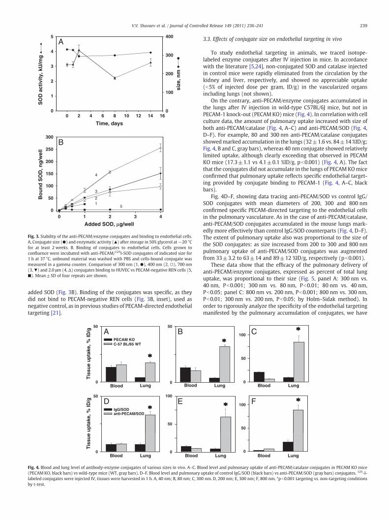
Fig. 3. Stability of the anti-PECAM/enzyme conjugates and binding to endothelial cells.A, Conjugate size (●) and enzymatic activity (▲) after storage in 50% glycerol at−20 °Cfor at least 2 weeks. B. Binding of conjugates to endothelial cells. Cells grown toconfluence were incubated with anti-PECAM/125I-SOD conjugates of indicated size for1 h at 37 °C, unbound material was washed with PBS and cells-bound conjugate wasmeasured in a gamma counter. Comparison of 300 nm (1, ●), 400 nm (2, ○), 700 nm(3,▼) and 2.0 μm (4, Δ) conjugates binding to HUVEC vs PECAM-negative REN cells (5,■). Mean±SD of four repeats are shown.
239V.V. Shuvaev et al. / Journal of Controlled Release 149 (2011) 236–241
added SOD (Fig. 3B). Binding of the conjugates was specific, as theydid not bind to PECAM-negative REN cells (Fig. 3B, inset), used asnegative control, as in previous studies of PECAM-directed endothelialtargeting [21].
50
PECAM KOC-57 BL/65 WT
50
A B
Blood Lung Blood
BloodBlood Lung
Tis
sue
up
take
, % ID
/gT
issu
e u
pta
ke, %
ID/g
0 0
50
IgG/SOD anti-PECAM/SOD
D
50
100
E
0 0
Fig. 4. Blood and lung level of antibody-enzyme conjugates of various sizes in vivo. A–C. Blo(PECAM KO, black bars) vs wild-type mice (WT, gray bars). D–F. Blood level and pulmonary ulabeled conjugates were injected IV, tissues were harvested in 1 h. A, 40 nm; B, 80 nm; C, 300by t-test.
3.3. Effects of conjugate size on endothelial targeting in vivo
To study endothelial targeting in animals, we traced isotope-labeled enzyme conjugates after IV injection in mice. In accordancewith the literature [5,24], non-conjugated SOD and catalase injectedin control mice were rapidly eliminated from the circulation by thekidney and liver, respectively, and showed no appreciable uptake(b5% of injected dose per gram, ID/g) in the vascularized organsincluding lungs (not shown).
On the contrary, anti-PECAM/enzyme conjugates accumulated inthe lungs after IV injection in wild-type C57BL/6J mice, but not inPECAM-1 knock-out (PECAMKO)mice (Fig. 4). In correlationwith cellculture data, the amount of pulmonary uptake increased with size ofboth anti-PECAM/catalase (Fig. 4, A–C) and anti-PECAM/SOD (Fig. 4,D–F). For example, 80 and 300 nm anti-PECAM/catalase conjugatesshowedmarked accumulation in the lungs (32±1.6 vs. 84±14 %ID/g;Fig. 4, B and C, gray bars), whereas 40 nm conjugate showed relativelylimited uptake, although clearly exceeding that observed in PECAMKO mice (17.3±1.1 vs 4.1±0.1 %ID/g, pb0.001) (Fig. 4, A). The factthat the conjugates did not accumulate in the lungs of PECAMKOmiceconfirmed that pulmonary uptake reflects specific endothelial target-ing provided by conjugate binding to PECAM-1 (Fig. 4, A–C, blackbars).
Fig. 4D–F, showing data tracing anti-PECAM/SOD vs control IgG/SOD conjugates with mean diameters of 200, 300 and 800 nmconfirmed specific PECAM-directed targeting to the endothelial cellsin the pulmonary vasculature. As in the case of anti-PECAM/catalase,anti-PECAM/SOD conjugates accumulated in the mouse lungs mark-edly more effectively than control IgG/SOD counterparts (Fig. 4, D–F).The extent of pulmonary uptake also was proportional to the size ofthe SOD conjugates: as size increased from 200 to 300 and 800 nmpulmonary uptake of anti-PECAM/SOD conjugates was augmentedfrom 33±3.2 to 63±14 and 89±12 %ID/g, respectively (pb0.001).
These data show that the efficacy of the pulmonary delivery ofanti-PECAM/enzyme conjugates, expressed as percent of total lunguptake, was proportional to their size (Fig. 5, panel A: 300 nm vs.40 nm, Pb0.001; 300 nm vs. 80 nm, Pb0.01; 80 nm vs. 40 nm,Pb0.05; panel C: 800 nm vs. 200 nm, Pb0.001; 800 nm vs. 300 nm,Pb0.01; 300 nm vs. 200 nm, Pb0.05; by Holm–Sidak method). Inorder to rigorously analyze the specificity of the endothelial targetingmanifested by the pulmonary accumulation of conjugates, we have
100 C
Lung Blood Lung
Blood LungLung
0
50
50
100 F
0
od level and pulmonary uptake of anti-PECAM/catalase conjugates in PECAM KO miceptake of control IgG/SOD (black bars) vs anti-PECAM/SOD (gray bars) conjugates. 125-I-nm. D, 200 nm; E, 300 nm; F, 800 nm. *pb0.001 targeting vs. non-targeting conditions

15
20C100
120A
0
5
10
0
20
40
60
80
Lu
ng
up
take
, % ID
Lu
ng
up
take
, % ID
15
20
8
10
200 300 800
200 300 800
Anti-PECAM/SOD conjugate, nm
DB
Anti-PECAM/catalase conjugate, nm
0
5
10
0
2
4
6
Imm
un
osp
ecif
icit
y in
dex
Imm
un
osp
ecif
icit
y in
dex
Anti-PECAM/SOD conjugate, nm
40 80 300
40 80 300
Anti-PECAM/catalase conjugate, nm
Fig. 5. Lung uptake (A and C) and immunospecificity index (ISI; B and D) of anti-PECAM/catalase (A and B) and anti-PECAM/SOD (C and D) conjugates of various size. Radiolabeledconjugates were injected i.v. and tissue was harvested 1 h post-injection. The immunospecificity index was calculated as ratio of targeted vs non-immune counterparts. Tissueuptake is shown as mean±SEM (n=3–5). *pb0.001 targeted conjugate vs. non-immune counterparts by t-test.
240 V.V. Shuvaev et al. / Journal of Controlled Release 149 (2011) 236–241
calculated the ratio of uptake of targeted vs. non-targeted formulationnormalized by their respective blood levels, i.e., the immunospecifi-city index (ISI, the most accurate measure of targeting specificity) [5].Pulmonary ISI of anti-PECAM/enzyme conjugates increased with sizeto 300 nm (Fig. 5, panels B and D). However, further larger conjugatesize (i.e., 800 nm) diminished the pulmonary ISI of anti-PECAM/SOD(Fig. 5D), because control IgG/SOD of this size showed enhancedpulmonary uptake. Therefore, anti-PECAM-enzyme conjugates withsize close to 300 nm diameter showed maximally specific endothelialtargeting in the vasculature vs. small conjugates (200 nm) and large(800 nm) conjugates. It is not known yet how sharp the optimal sizerange is. Based on in vivo studies with polymer particles [25,26] it mayextend within few hundred nanometers. In this case, it is plausiblethat conjugates with average diameter close to 400–500 nm mayexert superior specificity; yet this does not seem likely due todramatic reduction of specificity of 800 nm vs 300 nm conjugates.
4. Discussion
Carrier geometry plays a key role in drug delivery [27]. Forexample, the size and shape of polymer carriers markedly influencedtheir longevity in the circulation, their uptake by phagocytes andclearing tissues, their internalization by target cells and degradationrate, as well as drug loading and release [26,28,29]. In the context ofpulmonary endothelial targeting, recent studies showed that thegeometry (i.e., size and shape) of polymer carriers carrying anti-PECAM and other affinity moieties modulate the efficacy andspecificity of pulmonary accumulation of circulating carriers [25].
However, the role of size of protein conjugates has been much lessthoroughly addressed; in part, because it is more difficult to deviseprotein conjugates with controlled size than it is to manufacturecontrolled-sized polymer carriers [27]. Nevertheless, protein con-jugates and fusion constructs represent potentially useful classes oftargeted therapeutics andmany experimental agents in this class haveshown promising therapeutic and prophylactic effects in animalstudies.
This study describes the synthesis of differently sized antibody/enzyme conjugates (represented by SOD and catalase conjugatedwith IgG and PECAM antibodies) and testing their ability to target
endothelial cells. Conjugation of drug cargoes to antibodies andantibody fragments via streptavidin is widely employed in animalstudies [21]. This modular approach provides supramolecular con-jugates with unique targeting features including enhanced uptake bytarget cells [3,9]. From the translational perspective, however, it isimportant to note that an alternative conjugation technique (strepta-vidin is not used in patients in the USA) can also be adapted toproduce stable antibody-enzyme conjugates of desirable size within awide range of conjugate diameters (Fig. 2).
Targeting efficacy was directly proportional to the conjugate sizein both cell culture and in vivo (Figs. 3 and 4). This can be explained bythe increasing: i) drug load; and ii) number of antigen-binding sites ofthe conjugates due to enhanced number of antibody molecules perconjugate particle, which resulted in higher affinity of binding totarget. However, the elevated pulmonary uptake of large IgG/enzymeconjugates revealed that the specificity of in vivo targeting diminisheswhen the conjugate size begins to exceed 300 nm in average diameter(Fig. 5).
It is not clear why sub-micron sized IgG/enzyme conjugates exertelevated non-specific uptake in the pulmonary vasculature. They aresmaller than limiting size of the capillaries (i.e., ~800 nm vs ~5 μm),which argues against mechanical entrapment in the microvascula-ture. Studies in cell culture show no indication of enhanced non-specific cellular adhesion of larger conjugates (Fig. 3B). Perhaps, largeconjugates possessing multiple copies of IgG Fc-fragments moreextensively interact with Fc-receptor bearing cells in circulation(white blood cells and platelets), causing their agglomeration andretention in the capillaries. If this is the case, exceeding optimal sizemay cause adverse effects (see below); hence, understanding of themechanism of non-specific uptake is worth further inquiry.
Non-specific vascular uptake of sub-optimally large antibody/enzyme conjugates may have important implications. First, the Fc-fragment-mediated mechanism postulated above may predispose toinflammation and thrombosis. Second, conjugates non-specificallyretained in the vasculature are likely to reside in the lumen, withoutentering endothelial cells. Previous studies showed that for intra-endothelial delivery, anti-PECAM conjugatesmust interact specificallywith PECAM-1, causing endocytic signaling mediated by the PECAM-1cytosolic domain [30]. Most likely, antibody/enzyme conjugates non-

241V.V. Shuvaev et al. / Journal of Controlled Release 149 (2011) 236–241
specifically retained in a vessel will not induce endocytosis; hencetheir effects will be limited to the bloodstream. This location may besufficient and even preferential for some therapeutics (e.g., fibrino-lytics), but intracellular delivery is required for action of many otherdrugs [4].
In this study, we have focused on the ability of size to affectconjugate targeting to endothelium. Depending on the nature of thecargo, another important factor to consider is the ultimate fate of theconjugates, that is whether they stay on the cell surface (which wouldbe critical for an anti-thrombotic agent) or whether they areinternalized, and to which compartment (as might be critical forRNA, DNA, or antioxidants). In previous studies, we have shown thatoptimal size range for internalization of protein anti-PECAM con-jugates by endothelium is between ~100 and 500 nm, whereas largerconjugates are poorly internalized despite highly effective and specificbinding to endothelial cells [31]. Taken together with the data fromthis paper, our results suggest that a diameter close to 300 nm of anti-PECAM/enzyme conjugates is optimal for the specific intra-endothe-lial delivery.
Acknowledgements
This work was supported in part by the National Institutes ofHealth Grants RO1 HL073940 and HL087036 and Project 4 of PO1HL079063 (to VRM).
References
[1] B.S. Ding, T. Dziubla, V.V. Shuvaev, S. Muro, V.R. Muzykantov, Advanced drugdelivery systems that target the vascular endothelium, Mol. Interv. 6 (2) (2006)98–112.
[2] K.A. Massey, J.E. Schnitzer, Caveolae and Cancer. Recent Results Cancer Res 180(2010) 217-231.
[3] P. Valadon, B. Darsow, T.N. Buss, M. Czarny, N.M. Griffin, H.N. Nguyen, P. Oh, P.Borgstrom, A. Chrastina, J.E. Schnitzer, Designed auto-assembly of nanostrepta-bodies for rapid tissue-specific targeting in vivo. J Biol Chem 285 (1) (2010) 713–722.
[4] V.R. Muzykantov, Biomedical aspects of targeted delivery of drugs to pulmonaryendothelium, Expert Opin. Drug Deliv. 2 (5) (2005) 909–926.
[5] V.R. Muzykantov, E.N. Atochina, H. Ischiropoulos, S.M. Danilov, A.B. Fisher,Immunotargeting of antioxidant enzyme to the pulmonary endothelium, Proc.Natl Acad. Sci. USA 93 (11) (1996) 5213–5218.
[6] R. Pasqualini, D.M. McDonald, W. Arap, Vascular targeting and antigenpresentation, Nat. Immunol. 2 (7) (2001) 567–568.
[7] G.P. Robbins, R.L. Saunders, J.B. Haun, J. Rawson, M.J. Therien, D.A. Hammer,Tunable Leuko-polymersomes That Adhere Specifically to Inflammatory Markers.Langmuir 26 (17) (2010) 14089–14096.
[8] B.S. Ding, N. Hong, J.C. Murciano, K. Ganguly, C. Gottstein, M. Christofidou-Solomidou, S.M. Albelda, A.B. Fisher, D.B. Cines, V.R. Muzykantov, Prophylacticthrombolysis by thrombin-activated latent prourokinase targeted to PECAM-1 inthe pulmonary vasculature, Blood 111 (4) (2008) 1999–2006.
[9] V.R. Muzykantov, M. Christofidou-Solomidou, I. Balyasnikova, D.W. Harshaw, L.Schultz, A.B. Fisher, S.M. Albelda, Streptavidin facilitates internalization andpulmonary targeting of an anti-endothelial cell antibody (platelet-endothelial celladhesion molecule 1): a strategy for vascular immunotargeting of drugs, Proc.Natl Acad. Sci. USA 96 (5) (1999) 2379–2384.
[10] V.V. Shuvaev, M. Christofidou-Solomidou, F. Bhora, K. Laude, H. Cai, S. Dikalov, E.Arguiri, C.C. Solomides, S.M. Albelda, D.G. Harrison, V.R. Muzykantov, Targeteddetoxification of selected reactive oxygen species in the vascular endothelium,J. Pharmacol. Exp. Ther. 331 (2) (2009) 404–411.
[11] M. Christofidou-Solomidou, V.R. Muzykantov, Antioxidant strategies in respira-tory medicine, Treat. Respir. Med. 5 (1) (2006) 47–78.
[12] K. Nowak, C. Hanusch, K. Nicksch, R.P. Metzger, G. Beck, M.M. Gebhard, P.Hohenberger, S.M. Danilov, Pre-ischaemic conditioning of the pulmonaryendothelium by immunotargeting of catalase via angiotensin-converting-enzymeantibodies, Eur. J. Cardiothorac. Surg. (2009).
[13] K. Nowak, S. Weih, R. Metzger, R.F. Albrecht Ii, S. Post, P. Hohenberger, M.M.Gebhard, S.M. Danilov, Immunotargeting of catalase to lung endothelium via anti-ACE antibodies attenuates ischemia–reperfusion injury of the lung in vivo, Am. J.Physiol. Lung Cell. Mol. Physiol. (2007).
[14] V.V. Shuvaev, S. Tliba, M. Nakada, S.M. Albelda, V.R. Muzykantov, Platelet-endothelial cell adhesionmolecule-1-directed endothelial targeting of superoxidedismutase alleviates oxidative stress caused by either extracellular or intracellularsuperoxide, J. Pharmacol. Exp. Ther. 323 (2) (2007) 450–457.
[15] T.D. Sweitzer, A.P. Thomas, R. Wiewrodt, M.T. Nakada, F. Branco, V.R. Muzykantov,PECAM-directed immunotargeting of catalase: specific, rapid and transientprotection against hydrogen peroxide, Free Radic. Biol. Med. 34 (8) (2003)1035–1046.
[16] M. Christofidou-Solomidou, A. Scherpereel, R. Wiewrodt, K. Ng, T. Sweitzer, E.Arguiri, V. Shuvaev, C.C. Solomides, S.M. Albelda, V.R. Muzykantov, PECAM-directed delivery of catalase to endothelium protects against pulmonary vascularoxidative stress, Am. J. Physiol. Lung Cell. Mol. Physiol. 285 (2) (2003) L283–L292.
[17] V.R. Muzykantov, E.N. Atochina, A. Kuo, E.S. Barnathan, K. Notarfrancesco, H.Shuman, C. Dodia, A.B. Fisher, Endothelial cells internalizemonoclonal antibody toangiotensin-converting enzyme, Am. J. Physiol. 270 (5 Pt 1) (1996) L704–L713.
[18] B.D. Kozower, M. Christofidou-Solomidou, T.D. Sweitzer, S. Muro, D.G. Buerk, C.C.Solomides, S.M. Albelda, G.A. Patterson, V.R. Muzykantov, Immunotargeting ofcatalase to the pulmonary endothelium alleviates oxidative stress and reducesacute lung transplantation injury, Nat. Biotechnol. 21 (4) (2003) 392–398.
[19] A. Scherpereel, J.J. Rome, R. Wiewrodt, S.C. Watkins, D.W. Harshaw, S. Alder, M.Christofidou-Solomidou, E. Haut, J.C. Murciano, M. Nakada, S.M. Albelda, V.R.Muzykantov, Platelet-endothelial cell adhesion molecule-1-directed immunotar-geting to cardiopulmonary vasculature, J. Pharmacol. Exp. Ther. 300 (3) (2002)777–786.
[20] H.C. Yan, J.M. Pilewski, Q. Zhang, H.M. DeLisser, L. Romer, S.M. Albelda,Localization of multiple functional domains on human PECAM-1 (CD31) bymonoclonal antibody epitope mapping, Cell Adhes. Commun. 3 (1) (1995) 45–66.
[21] V.V. Shuvaev, T. Dziubla, R. Wiewrodt, V.R. Muzykantov, Streptavidin–biotincrosslinking of therapeutic enzymes with carrier antibodies: nanoconjugates forprotection against endothelial oxidative stress, Meth. Mol. Biol. 283 (2004) 3–20.
[22] J.M. McCord, I. Fridovich, Superoxide dismutase. An enzymic function forerythrocuprein (hemocuprein), J. Biol. Chem. 244 (22) (1969) 6049–6055.
[23] A. Scherpereel, R. Wiewrodt, M. Christofidou-Solomidou, R. Gervais, J.C. Murciano,S.M. Albelda, V.R. Muzykantov, Cell-selective intracellular delivery of a foreignenzyme to endothelium in vivo using vascular immunotargeting, FASEB J. 15 (2)(2001) 416–426.
[24] A. Bayati, O. Kallskog, B. Odlind, M.Wolgast, Plasma elimination kinetics and renalhandling of copper/zinc superoxide dismutase in the rat, Acta Physiol. Scand. 134(1) (1988) 65–74.
[25] S. Muro, C. Garnacho, J.A. Champion, J. Leferovich, C. Gajewski, E.H. Schuchman, S.Mitragotri, V.R. Muzykantov, Control of endothelial targeting and intracellulardelivery of therapeutic enzymes by modulating the size and shape of ICAM-1-targeted carriers, Mol. Ther. 16 (8) (2008) 1450–1458.
[26] D.A. Canelas, K.P. Herlihy, J.M. DeSimone, Top-down particle fabrication: controlof size and shape for diagnostic imaging and drug delivery, Wiley Interdiscip. Rev.Nanomed. Nanobiotechnol. 1 (4) (2009) 391–404.
[27] E.A. Simone, T.D. Dziubla, V.R. Muzykantov, Polymeric carriers: role of geometry indrug delivery, Expert Opin. Drug Deliv. 5 (12) (2008) 1283–1300.
[28] J.A. Champion, S. Mitragotri, Role of target geometry in phagocytosis, Proc. NatlAcad. Sci. USA 103 (13) (2006) 4930–4934.
[29] Y. Geng, P. Dalhaimer, S. Cai, R. Tsai, M. Tewari, T. Minko, D.E. Discher, Shapeeffects of filaments versus spherical particles in flow and drug delivery, Nat.Nanotechnol. 2 (4) (2007) 249–255.
[30] C. Garnacho, V. Shuvaev, A. Thomas, L. McKenna, J. Sun, M. Koval, S. Albelda, V.Muzykantov, S. Muro, RhoA activation and actin reorganization involved inendothelial CAM-mediated endocytosis of anti-PECAM carriers: critical role fortyrosine 686 in the cytoplasmic tail of PECAM-1, Blood 111 (6) (2008)3024–3033.
[31] R. Wiewrodt, A.P. Thomas, L. Cipelletti, M. Christofidou-Solomidou, D.A. Weitz, S.I.Feinstein, D. Schaffer, S.M. Albelda, M. Koval, V.R. Muzykantov, Size-dependentintracellular immunotargeting of therapeutic cargoes into endothelial cells, Blood99 (3) (2002) 912–922.