modificationofavolume … · a levitronix centrimag (levitronix llc, waltham, mass) (1st case) and...
TRANSCRIPT
International Scholarly Research NetworkISRN CardiologyVolume 2011, Article ID 831062, 6 pagesdoi:10.5402/2011/831062
Research Article
Modification of a Volume-Overload Heart Failure Model to TrackMyocardial Remodeling and Device-Related Reverse Remodeling
Egemen Tuzun,1, 2 Roger Bick,3 Cihan Kadipasaoglu,1 Jeffrey L. Conger,1 Brian J. Poindexter,3
Igor D. Gregoric,1 O. H. Frazier,1 Jeffrey A. Towbin,4 and Branislav Radovancevic1
1 Cardiovascular Surgical Research Laboratories, Texas Heart Institute, Houston, TX 77225, USA2 Texas A&M Institute for Preclinical Studies, 800 Raymond Stotzer Pkwy, Ste 2060, College Station, TX 77843-4478, USA3 Department of Pathology, University of Texas Health Science Center, Houston, TX 77030-1501, USA4 Department of Pediatric Cardiology, Baylor College of Medicine, Houston, TX 77030, USA
Correspondence should be addressed to Egemen Tuzun, [email protected]
Received 4 March 2011; Accepted 2 May 2011
Academic Editor: M. Guazzi
Copyright © 2011 Egemen Tuzun et al. This is an open access article distributed under the Creative Commons Attribution License,which permits unrestricted use, distribution, and reproduction in any medium, provided the original work is properly cited.
Purpose. To provide an ovine model of ventricular remodeling and reverse remodeling by creating congestive heart failure (CHF)and then treating it by implanting a left ventricular assist device (LVAD). Methods. We induced volume-overload heart failure in2 sheep; 20 weeks later, we implanted an LVAD and assessed recovery 11 weeks thereafter. We examined changes in histologicand hemodynamic data and levels of cellular markers of CHF. Results. After CHF induction, we found increases in LV end-diastolic pressure, LV systolic and diastolic dimensions, wall thickness, left atrial diameter, and atrial natriuretic protein (ANP)and endothelin-1 (ET-1) levels; β-adrenergic receptor (BAR) and dystrophin expression decreased markedly. Biopsies confirmedLV remodeling. After LVAD support, LV systolic and diastolic dimensions, wall thickness, and mass, and ANP and ET-1 levelsdecreased. Histopathologic and hemodynamic markers improved, and BAR and dystrophin expression normalized. Conclusions.We describe a successful sheep model for ventricular and reverse remodeling.
1. Introduction
Congestive heart failure (CHF) is characterized by an initialcardiac insult, followed by a compensatory response thatresults in ventricular remodeling. At the cellular level, CHFinvolves myocyte hypertrophy, cell death, and replacementfibrosis. At the subcellular level, the disease causes lossof myofilaments, alterations in cytoskeletal proteins, andadrenergic desensitization.
For the past 2 decades, the diagnosis of heart failurehas relied on noninvasive methods (physical examination,chest roentgenography, electrocardiography, and echocar-diography) and invasive tests [1]. Various biomarkers havealso been used to diagnose heart failure with marginalclinical utility. In patients with ischemic heart disease andmyocardial infarction, the following biomarkers have shownvarying levels of sensitivity in detecting heart failure: lacticdehydrogenase, creatine kinase myocardial band (CKMB),troponins T and I, C-reactive protein, norepinephrine,
endothelin-1 (ET-1), interleukin-6, tumor necrosis factor-α(TNF-α), atrial natriuretic protein (ANP), B-type natriureticpeptide, beta-adrenergic receptors (BARs), and angiotensinII [2–12]. Another cellular marker of heart failure isdystrophin, a protein that connects the extracellular matrixwith the sarcomere via actin. Dystrophin abnormalitieshave been associated with heart failure caused by geneticsusceptibility and viral infection [13]. Treatment of CHFwith left ventricular assist devices (LVADs) can reverse theventricular remodeling associated with heart failure, andthis reversal is correlated with normalization of cellularmarkers in myocytes [14, 15]. The present pilot study wasundertaken to provide an ovine experimental model forcreating CHF, then reversing this condition by implantingan LVAD. We assessed the changes in immunohistochemical,histopathologic, and hemodynamic parameters to verify theoccurrence and extent of the remodeling events in heartfailure development (remodeling) and subsequent LVAD-assisted recovery (reverse remodeling).
2 ISRN Cardiology
(a) (b)
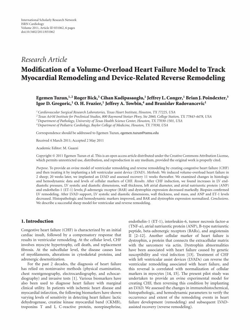
Figure 1: (a) Normal ventriculogram obtained before creation of mitral regurgitation (MR). (b) After chordal disruption, theventriculogram shows 2nd- to 3rd-degree MR.
(a)
†
(b)
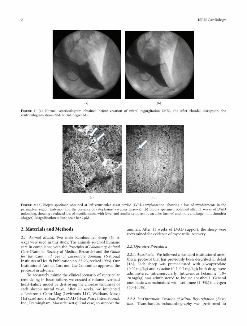
Figure 2: (a) Biopsy specimen obtained at left ventricular assist device (LVAD) implantation, showing a loss of myofilaments in theperinuclear region (asterisk) and the presence of cytoplasmic vacuoles (arrows). (b) Biopsy specimen obtained after 11 weeks of LVADunloading, showing a reduced loss of myofilaments, with fewer and smaller cytoplasmic vacuoles (arrow) and more and larger mitochondria(dagger). Magnification ×2500; scale bar 2 μM.
2. Materials and Methods
2.1. Animal Model. Two male Rambouillet sheep (54 ±4 kg) were used in this study. The animals received humanecare in compliance with the Principles of Laboratory AnimalCare (National Society of Medical Research) and the Guidefor the Care and Use of Laboratory Animals (NationalInstitutes of Health Publication no. 85-23, revised 1996). OurInstitutional Animal Care and Use Committee approved theprotocol in advance.
To accurately mimic the clinical scenario of ventricularremodeling in heart failure, we created a volume-overloadheart-failure model by destroying the chordae tendineae ofeach sheep’s mitral valve. After 20 weeks, we implanteda Levitronix CentriMag (Levitronix LLC, Waltham, Mass)(1st case) and a HeartWare LVAD (HeartWare International,Inc., Framingham, Massachusetts) (2nd case) to support the
animals. After 11 weeks of LVAD support, the sheep werereexamined for evidence of myocardial recovery.
2.2. Operative Procedures
2.2.1. Anesthesia. We followed a standard institutional anes-thesia protocol that has previously been described in detail[16]. Each sheep was premedicated with glycopyrrolate(0.02 mg/kg) and xylazine (0.2–0.7 mg/kg); both drugs wereadministered intramuscularly. Intravenous ketamine (10–20 mg/kg) was administered to induce anesthesia. Generalanesthesia was maintained with isoflurane (1–3%) in oxygen(40–100%).
2.2.2. 1st Operation: Creation of Mitral Regurgitation (Base-line). Transthoracic echocardiography was performed to
ISRN Cardiology 3
25000
20000
15000
10000
5000
0
FPD
Myocardial biopsy time
ANPBAR
ET-1DYS
A B C
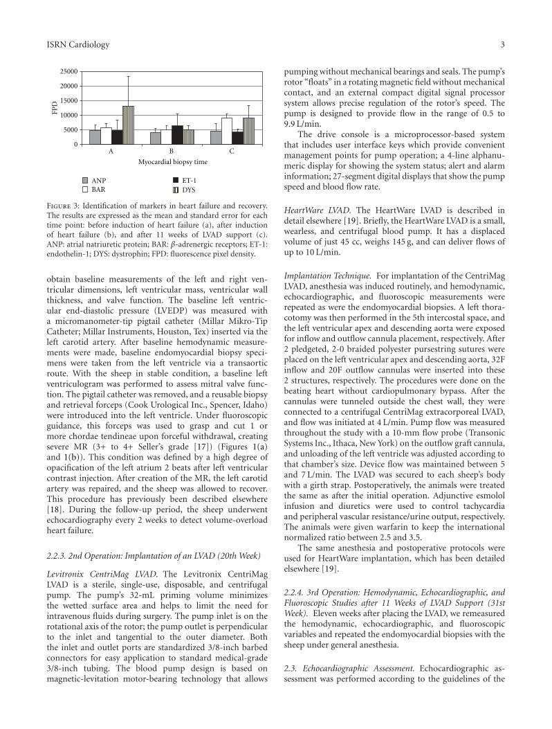
Figure 3: Identification of markers in heart failure and recovery.The results are expressed as the mean and standard error for eachtime point: before induction of heart failure (a), after inductionof heart failure (b), and after 11 weeks of LVAD support (c).ANP: atrial natriuretic protein; BAR: β-adrenergic receptors; ET-1:endothelin-1; DYS: dystrophin; FPD: fluorescence pixel density.
obtain baseline measurements of the left and right ven-tricular dimensions, left ventricular mass, ventricular wallthickness, and valve function. The baseline left ventric-ular end-diastolic pressure (LVEDP) was measured witha micromanometer-tip pigtail catheter (Millar Mikro-TipCatheter; Millar Instruments, Houston, Tex) inserted via theleft carotid artery. After baseline hemodynamic measure-ments were made, baseline endomyocardial biopsy speci-mens were taken from the left ventricle via a transaorticroute. With the sheep in stable condition, a baseline leftventriculogram was performed to assess mitral valve func-tion. The pigtail catheter was removed, and a reusable biopsyand retrieval forceps (Cook Urological Inc., Spencer, Idaho)were introduced into the left ventricle. Under fluoroscopicguidance, this forceps was used to grasp and cut 1 ormore chordae tendineae upon forceful withdrawal, creatingsevere MR (3+ to 4+ Seller’s grade [17]) (Figures 1(a)and 1(b)). This condition was defined by a high degree ofopacification of the left atrium 2 beats after left ventricularcontrast injection. After creation of the MR, the left carotidartery was repaired, and the sheep was allowed to recover.This procedure has previously been described elsewhere[18]. During the follow-up period, the sheep underwentechocardiography every 2 weeks to detect volume-overloadheart failure.
2.2.3. 2nd Operation: Implantation of an LVAD (20th Week)
Levitronix CentriMag LVAD. The Levitronix CentriMagLVAD is a sterile, single-use, disposable, and centrifugalpump. The pump’s 32-mL priming volume minimizesthe wetted surface area and helps to limit the need forintravenous fluids during surgery. The pump inlet is on therotational axis of the rotor; the pump outlet is perpendicularto the inlet and tangential to the outer diameter. Boththe inlet and outlet ports are standardized 3/8-inch barbedconnectors for easy application to standard medical-grade3/8-inch tubing. The blood pump design is based onmagnetic-levitation motor-bearing technology that allows
pumping without mechanical bearings and seals. The pump’srotor “floats” in a rotating magnetic field without mechanicalcontact, and an external compact digital signal processorsystem allows precise regulation of the rotor’s speed. Thepump is designed to provide flow in the range of 0.5 to9.9 L/min.
The drive console is a microprocessor-based systemthat includes user interface keys which provide convenientmanagement points for pump operation; a 4-line alphanu-meric display for showing the system status; alert and alarminformation; 27-segment digital displays that show the pumpspeed and blood flow rate.
HeartWare LVAD. The HeartWare LVAD is described indetail elsewhere [19]. Briefly, the HeartWare LVAD is a small,wearless, and centrifugal blood pump. It has a displacedvolume of just 45 cc, weighs 145 g, and can deliver flows ofup to 10 L/min.
Implantation Technique. For implantation of the CentriMagLVAD, anesthesia was induced routinely, and hemodynamic,echocardiographic, and fluoroscopic measurements wererepeated as were the endomyocardial biopsies. A left thora-cotomy was then performed in the 5th intercostal space, andthe left ventricular apex and descending aorta were exposedfor inflow and outflow cannula placement, respectively. After2 pledgeted, 2-0 braided polyester pursestring sutures wereplaced on the left ventricular apex and descending aorta, 32Finflow and 20F outflow cannulas were inserted into these2 structures, respectively. The procedures were done on thebeating heart without cardiopulmonary bypass. After thecannulas were tunneled outside the chest wall, they wereconnected to a centrifugal CentriMag extracorporeal LVAD,and flow was initiated at 4 L/min. Pump flow was measuredthroughout the study with a 10-mm flow probe (TransonicSystems Inc., Ithaca, New York) on the outflow graft cannula,and unloading of the left ventricle was adjusted according tothat chamber’s size. Device flow was maintained between 5and 7 L/min. The LVAD was secured to each sheep’s bodywith a girth strap. Postoperatively, the animals were treatedthe same as after the initial operation. Adjunctive esmololinfusion and diuretics were used to control tachycardiaand peripheral vascular resistance/urine output, respectively.The animals were given warfarin to keep the internationalnormalized ratio between 2.5 and 3.5.
The same anesthesia and postoperative protocols wereused for HeartWare implantation, which has been detailedelsewhere [19].
2.2.4. 3rd Operation: Hemodynamic, Echocardiographic, andFluoroscopic Studies after 11 Weeks of LVAD Support (31stWeek). Eleven weeks after placing the LVAD, we remeasuredthe hemodynamic, echocardiographic, and fluoroscopicvariables and repeated the endomyocardial biopsies with thesheep under general anesthesia.
2.3. Echocardiographic Assessment. Echocardiographic as-sessment was performed according to the guidelines of the
4 ISRN Cardiology
American Society of Echocardiography [20] by an echocar-diologist using a Sonos 2000 ultrasound system (Hewlett-Packard Co., Palo Alto, Calif) equipped with a 2.5-MHzphased-array transducer.
2.4. Histopathologic Evaluation. Endomyocardial biopsieswere performed before MR creation (baseline), during the2nd operation (week 20), and during the 3rd operation(week 31) and were evaluated with transmission electronmicroscopy. Macroscopic postexplant analyses of the pumpwere also done after termination of the study.
2.5. Immunohistochemical Staining. β-adrenergic receptor,ANP, and ET-1 levels were selected as markers of heart failureand were fluorescently labeled. Fluorescence was quantifiedas pixel densities of specific colors in split color images,using Corel Draw (Corel Corporation, Ottawa, ON, Canada)and Sigma Scan Pro software (Aspire Software International,Leesburg, Va).
2.6. Quantification of Dystrophin in Ventricular Biopsy Spec-imens. Expression of dystrophin was quantified by Westernblotting of whole tissue lysates prepared from biopsy tissues.Consequently, confocal immunofluorescence was used toobtain robust quantitative information about the number ofimmunoreactive signals at discrete sites within the cell.
3. Results
3.1. Induction (Remodeling Phase) and Treatment (ReverseRemodeling Phase) of Heart Failure. In both cases, com-parison of hemodynamic, fluoroscopic, echocardiographic,histopathologic, and immunohistochemical data recorded(1) during the 1st operation and (2) immediately beforepump implantation confirmed the effectiveness of the heartfailure induction protocol.
After the 2nd operation, both sheep recovered fromanesthesia without complications and were extubated withinthe 1st postoperative hour. Eleven weeks after LVAD implan-tation, they reached the scheduled endpoint without experi-encing anorexia, infection, or a neurologic disorder.
3.2. Device Examination and Animal Necropsy. The explant-ed pumps’ interior, inflow cannula, and outflow cannulawere completely free of thrombus. There was no evidence ofischemia or infarction in any of the peripheral end organs(brain, liver, spleen, and kidneys).
3.3. Hemodynamic Data. The mean LVEDP, which was 8 ±2 mmHg at baseline, increased to 16± 2 mmHg immediatelyafter chordal disruption and to 27± 4 mmHg during the 2ndoperation. After 11 weeks of LVAD support, the mean LVEDPmeasured 14 ± 2 mmHg.
3.4. Fluoroscopic Data. After the mitral chordae tendineaewere disrupted, left ventriculography confirmed the presenceof 2nd- to 3rd-degree MR (Figures 1(a) and 1(b)). During
the 2nd operation, repeat ventriculography showed 4th-degree MR and a dilated left atrium. After 11 weeks of LVADsupport, ventriculography revealed 4th-degree MR and adilated left atrium at a pump flow of 5 L/min in both sheep.
3.5. Echocardiographic Data. Table 1 shows the mean echo-car diographic results obtained during the 1st operation(after mitral chordal disruption), 2nd operation (LVADimplantation), and 3rd operation (after 11 weeks of LVADsupport). The echocardiographic data demonstrate thatvolume-overload heart failure was successfully created inboth animals, with a resulting increase in the left ventric-ular diastolic dimension (LVDD), left ventricular systolicdimension (LVSD), and left ventricular mass. After LVADsupport, the echocardiographic measurements improved,with a substantial decrease in the LVDD, LVSD, and leftventricular mass.
3.6. Histopathologic Data. Transmission electron microscopyof the myocardial biopsy specimens obtained during LVADplacement (2nd operation) showed a 58 ± 5% loss ofmyofilaments in the perinuclear region; the presence ofcytoplasmic vacuoles >1.5 μm in diameter decreased by33 ± 4% compared to baseline biopsy findings (Figure 2(a)).
After 11 weeks of LVAD unloading, the percentage ofcells with moderate to severe myofilament loss decreasedfrom 58 ± 5% (at LVAD implantation) to 18 ± 3%. Thepercentage of cells with cytoplasmic vacuoles >1.5 μm indiameter decreased from 33 ± 4% to 5 ± 2%. Cardiacmyocytes showed an increase in the number and size of theirmitochondria (Figure 2(b)).
3.7. Immunohistochemical Data. Compared to baseline val-ues, there was a marked increase in the fluorescence pixeldensity for ANP and ET-1 along with a reduction in BAR anddystrophin density (Figure 3), which is evidence of volume-overload heart failure.
After 11 weeks of LVAD support, biopsy samples showeda marked decrease in fluorescence pixel density for ANP andET-1, along with an increase in BAR and dystrophin density(Figure 3) which may be considered a sign of myocardialreverse remodeling.
4. Discussion
In our pilot study, the left ventricular diastolic and systolicdimensions increased with heart failure and decreased afterLVAD support, although baseline levels were not regainedafter 11 weeks of LVAD support. The cardiac index andleft ventricular mass were elevated during heart failure andreturned to near baseline levels after 11 weeks of LVADsupport. Together, these results indicated the successfuldevelopment of heart failure in sheep after chordal disrup-tion, followed by recovery after LVAD support, as determinedby physical measurements and hemodynamic data.
We used an adult sheep model, primarily because of theease with which heart failure can be induced and subsequent

ISRN Cardiology 5
Table 1: Mean echocardiographic measurements obtained in each individual sheep during the 1st operation (after mitral chordaldisruption), 2nd operation (left ventricular assist device (LVAD) implantation), and 3rd operation (after 11 weeks of LVAD support).
Time LVDD (mm) LVSD (mm) IVS (mm) PW (mm) RVDD (mm) LA (mm) EF (%) LV mass (g)
Sheep 1
1st operation (baseline) 32 22 7 7 19 32 55 201
2nd operation (wk 20) 59 44 11 9 26 48 44 570
3rd operation (wk 31) 41 26 11 10 29 48 56 210
Sheep 2
1st operation (baseline) 36 26 9 9 25 36 65 233
2nd operation (wk 20) 68 50 13 11 30 56 50 610
3rd operation (wk 31) 45 30 13 12 35 56 64 236
EF: ejection fraction; IVS: interventricular septal thickness; LA: left atrial dimension; LV: left ventricular; LVDD: left ventricular diastolic dimension; LVSD:left ventricular systolic dimension; PW: posterior wall thickness; RVDD: right ventricular diastolic dimension.
LVAD therapy can be applied in this model. Other funda-mental advantages of the sheep are the animal’s size (whichallows placement of a human-sized LVAD), minimal growthrate, ease of management, and hemodynamic similarities tohumans. Moreover, the ability to perform sequential biopsiesallowed us to monitor the progress of heart failure andto correlate the extent of remodeling and its reversal withANP, ET-1, BAR, and dystrophin levels. Because our volume-overload heart failure ovine model was not a previouslyestablished one, biopsies to detect heart failure creation(remodeling) and postimplant recovery timing (reverseremodeling) were scheduled and performed according to theguiding results of the echocardiographic follow-up data. Theechocardiographic findings of volume-overload heart failure(such as ejection fraction, left atrial diameter, left ventricularend-systolic and end-diastolic dimensions, ventricular massand thickness, etc.) and LVAD recovery correlated well withthe animal’s clinical picture and hemodynamics, so we alsoscheduled LVAD implantation and support times accordingto these echocardiographic results.
The changes in dystrophin expression in our pilotstudy were consistent with the development of heart failureand subsequent recovery after LVAD placement in humans[14]. Because dystrophin has been demonstrated to playa key role in linking the actin cytoskeletal networks andthe sarcolemmal dystrophin-associated protein complex,the lack of dystrophin is associated with altered structureand mechanical adherence of costameres to the underlyingcytoskeletal actin network, resulting in cardiomyopathy[15]. Therefore, the possible fragility (disruption) of thedystrophin-actin bonds may be important for tracking earlymyocardial structural changes in remodeling and reverseremodeling and for developing a more precise animal modelof heart failure to test the efficacy of short- or long-termLVAD support.
One of our study’s limitations was the lack of plasmaprofiles of dystrophin levels, which may be used as apredictive/clinical biomarker for left ventricular remodelingrelated to LVAD use. Such plasma profiles will be a mainfocus of our future studies involving a larger numberof animals. Moreover, the location of the outflow-graftanastomosis may be considered another study limitation, as
it may not be the anastomotic site currently preferred formost fully implantable LVAD outflow grafts.
5. Conclusion
In this pilot experimental study, we have demonstrated anexperimental animal model in which we created CHF in2 adult sheep and subsequently reversed this condition byimplanting 2 different centrifugal LVADs (1 device in eachanimal). The degree of remodeling was indicated by the dis-ruption of dystrophin expression, as well as hemodynamic,echocardiographic, histologic, molecular, and clinical signsof heart failure. The reversal of heart failure (myocardialrecovery), by means of mechanical circulatory assistance, wasindicated by the normalization of dystrophin expression andintegrity. We believe that this technique for creating andsubsequently treating CHF with LVAD therapy may be usefulin future studies investigating the diagnosis and preventionof CHF.
Conflict of Interests
None of the authors has any potential conflict of interest todeclare.
Disclosure
The work described herein was not supported by any grantsor other external funding and has not been presented at anymeetings.
References
[1] S. A. Hunt, D. W. Baker, M. H. Chin et al., “ACC/AHAguidelines for the evaluation and management of chronicheart failure in the adult: executive summary. A report of theAmerican College of Cardiology/American Heart Associationtask force on practice guidelines (committee to revise the1995 guidelines for the evaluation and management of heartfailure),” Circulation, vol. 104, no. 24, pp. 2996–3007, 2001.
[2] P. M. Ridker, C. H. Hennekens, J. E. Buring, and N. Rifai,“C-reactive protein and other markers of inflammation in the

6 ISRN Cardiology
prediction of cardiovascular disease in women,” New EnglandJournal of Medicine, vol. 342, no. 12, pp. 836–843, 2000.
[3] R. Sodian, M. Loebe, C. Schmitt et al., “Decreased plasmaconcentration of brain natriuretic peptide as a potential indi-cator of cardiac recovery in patients supported by mechanicalcirculatory assist systems,” Journal of the American College ofCardiology, vol. 38, no. 7, pp. 1942–1949, 2001.
[4] R. J. Raymond, G. J. Dehmer, T. C. Theoharides, and E.N. Deliargyris, “Elevated interleukin-6 levels in patients withasymptomatic left ventricular systolic dysfunction,” AmericanHeart Journal, vol. 141, no. 3, pp. 435–438, 2001.
[5] P. M. Ridker, N. Rifai, M. J. Stampfer, and C. H. Hennekens,“Plasma concentration of interleukin-6 and the risk of futuremyocardial infarction among apparently healthy men,” Circu-lation, vol. 101, no. 15, pp. 1767–1772, 2000.
[6] W. L. Roberts, R. Sedrick, L. Moulton, A. Spencer, and N. Rifai,“Evaluation of four automated high-sensitivity C-reactiveprotein methods: implications for clinical and epidemiologicalapplications,” Clinical Chemistry, vol. 46, no. 4, pp. 461–468,2000.
[7] J. Whicher, N. Rifai, and L. M. Biasucci, “Markers of the acutephase response in cardiovascular disease: an update,” ClinicalChemistry and Laboratory Medicine, vol. 39, no. 11, pp. 1054–1064, 2001.
[8] J. Niebauer, “Inflammatory mediators in heart failure,” Inter-national Journal of Cardiology, vol. 72, no. 3, pp. 209–213,2000.
[9] D. L. Brutsaert, “Cardiac endothelial-myocardial signaling:its role in cardiac growth, contractile performance, andrhythmicity,” Physiological Reviews, vol. 83, no. 1, pp. 59–115,2003.
[10] N. Hama, H. Itoh, G. Shirakami et al., “Rapid ventricularinduction of brain natriuretic peptide gene expression inexperimental acute myocardial infarction,” Circulation, vol.92, no. 6, pp. 1558–1564, 1995.
[11] A. Sugawara, K. Nakao, N. Mor II et al., “Synthesis of atrialnatriuretic polypeptide in human failing hearts. Evidence foraltered processing of atrial natriuretic polypeptide precursorand augmented synthesis of β-human ANP,” Journal of ClinicalInvestigation, vol. 81, no. 6, pp. 1962–1970, 1988.
[12] M. R. Bristow, “β-Adrenergic receptor blockade in chronicheart failure,” Circulation, vol. 101, no. 5, pp. 558–569, 2000.
[13] T. A. Manolio, K. L. Baughman, R. Rodeheffer et al., “Preva-lence and etiology of idiopathic dilated cardiomyopathy.Summary of a National Heart, Lung and Blood InstituteWorkshop,” American Journal of Cardiology, vol. 69, no. 17, pp.1458–1466, 1992.
[14] M. Vatta, S. J. Stetson, A. Perez-Verdia et al., “Molecularremodelling of dystrophin in patients with end-stage car-diomyopathies and reversal in patients on assistance-devicetherapy,” Lancet, vol. 359, no. 9310, pp. 936–941, 2002.
[15] M. Vatta, S. J. Stetson, S. Jimenez et al., “Molecular normal-ization of dystrophin in the failing left and right ventricle ofpatients treated with either pulsatile or continuous flow-typeventricular assist devices,” Journal of the American College ofCardiology, vol. 43, no. 5, pp. 811–817, 2004.
[16] O. H. Frazier, E. Tuzun, W. Cohn, D. Tamez, K. A. Kadi-pasaoglu, and M. Mallia, “Total heart replacement with dualcentrifugal ventricular assist devices,” ASAIO Journal, vol. 51,no. 3, pp. 224–229, 2005.
[17] R. D. Sellers, M. J. Levy, K. Amplatz, and C. W. Lillehei, “Leftretrograde cardioangiography in acquired cardiac disease.Technic, indications and interpretations in 700 cases,” TheAmerican Journal of Cardiology, vol. 14, no. 4, pp. 437–447,1964.
[18] E. Tuzun, J. Conger, I. D. Gregoric et al., “Evaluation of a newcardiac recovery system in a bovine model of volume overloadheart failure,” ASAIO Journal, vol. 50, no. 6, pp. 557–562, 2004.
[19] E. Tuzun, K. Roberts, W. E. Cohn et al., “In vivo evaluationof the HeartWare centrifugal ventricular assist device,” TexasHeart Institute Journal, vol. 34, no. 4, pp. 406–411, 2007.
[20] W. L. Henry, A. DeMaria, R. Gramiak et al., “Report ofthe American Society of Echocardiography Committee onnomenclature and standards in two-dimensional echocardio-graphy,” Circulation, vol. 62, no. 2, pp. 212–217, 1980.