models of heterogeneous dopamine signaling in an insect
TRANSCRIPT
RESEARCH ARTICLE
Models of heterogeneous dopamine signaling
in an insect learning and memory center
Linnie JiangID1,2, Ashok Litwin-KumarID
1*
1 Mortimer B. Zuckerman Mind Brain Behavior Institute, Department of Neuroscience, Columbia University,
New York, New York, United States of America, 2 Neurosciences Program, Stanford University, Stanford,
California, United States of America
Abstract
The Drosophila mushroom body exhibits dopamine dependent synaptic plasticity that
underlies the acquisition of associative memories. Recordings of dopamine neurons in this
system have identified signals related to external reinforcement such as reward and punish-
ment. However, other factors including locomotion, novelty, reward expectation, and internal
state have also recently been shown to modulate dopamine neurons. This heterogeneity is
at odds with typical modeling approaches in which these neurons are assumed to encode a
global, scalar error signal. How is dopamine dependent plasticity coordinated in the pres-
ence of such heterogeneity? We develop a modeling approach that infers a pattern of dopa-
mine activity sufficient to solve defined behavioral tasks, given architectural constraints
informed by knowledge of mushroom body circuitry. Model dopamine neurons exhibit
diverse tuning to task parameters while nonetheless producing coherent learned behaviors.
Notably, reward prediction error emerges as a mode of population activity distributed across
these neurons. Our results provide a mechanistic framework that accounts for the heteroge-
neity of dopamine activity during learning and behavior.
Author summary
Dopamine neurons across the animal kingdom are involved in the formation of associa-
tive memories. While numerous studies have recorded activity in these neurons related to
external and predicted rewards, the diversity of these neurons’ activity and their tuning to
non-reward-related quantities such as novelty, movement, and internal state have proved
challenging to account for in traditional modeling approaches. Using a well-characterized
model system for learning and memory, the mushroom body of Drosophila fruit flies,
Jiang and Litwin-Kumar provide an account of the diversity of signals across dopamine
neurons. They show that models optimized to solve tasks like those encountered by flies
exhibit heterogeneous activity across dopamine neurons, but nonetheless this activity is
sufficient for the system to solve the tasks. The models will be useful to generate testable
hypotheses about dopamine neuron activity across different experimental conditions.
PLOS Computational Biology | https://doi.org/10.1371/journal.pcbi.1009205 August 10, 2021 1 / 29
a1111111111
a1111111111
a1111111111
a1111111111
a1111111111
OPEN ACCESS
Citation: Jiang L, Litwin-Kumar A (2021) Models of
heterogeneous dopamine signaling in an insect
learning and memory center. PLoS Comput Biol
17(8): e1009205. https://doi.org/10.1371/journal.
pcbi.1009205
Editor: Abigail Morrison, Research Center Julich,
GERMANY
Received: June 9, 2020
Accepted: June 22, 2021
Published: August 10, 2021
Copyright: © 2021 Jiang, Litwin-Kumar. This is an
open access article distributed under the terms of
the Creative Commons Attribution License, which
permits unrestricted use, distribution, and
reproduction in any medium, provided the original
author and source are credited.
Data Availability Statement: Code implementing
the model is available at the following github
repository: https://github.com/alitwinkumar/jiang_
litwin-kumar_mb_rnn.
Funding: Research was supported by a Columbia
University Class of 1939 Summer Research
Fellowship (L. J.), the Columbia Science Research
Fellows Program (L. J.), the Burroughs-Wellcome
Foundation (A. L.-K.), NIH BRAIN Initiative award
R01EB029858 (A. L.-K.), the Simons Collaboration
on the Global Brain (A. L.-K.), the Gatsby Charitable
Foundation (L. J. and A. L.-K.), and NSF NeuroNex
Introduction
Dopamine release modulates synaptic plasticity and learning across vertebrate and inverte-
brate species [1, 2]. A standard view of dopamine activity, proposed on the basis of recordings
in the mammalian midbrain dopaminergic system, holds that dopamine neuron firing repre-
sents a “reward prediction error,” the difference between received and predicted reward [3].
This view is consistent with models of classical conditioning experiments and with reinforce-
ment learning algorithms that learn to choose the most rewarding sequence of actions [4]. A
frequent assumption in these models is that the scalar reward prediction signal is globally
broadcast to and gates the modification of synaptic connections involved in learning. How-
ever, studies in both vertebrates and invertebrates suggest that dopamine neuron activity is
modulated by other variables in addition to reward prediction error, and that this modulation
is heterogeneous across populations of dopamine neurons [5].
Early studies in arthropods identified roles for dopamine in a variety of functions [6–11]. In
Drosophila, both memory [12] and other functions including locomotion, arousal, sleep, and
mating have been associated with dopamine signaling [11]. Associative olfactory learning in
Drosophila requires a central brain area known as the mushroom body [13–15], and many
studies of dopamine neurons innervating this area have focused on activity related to reward
and punishment and its roles in the formation of appetitive and aversive memories [16–22]. In
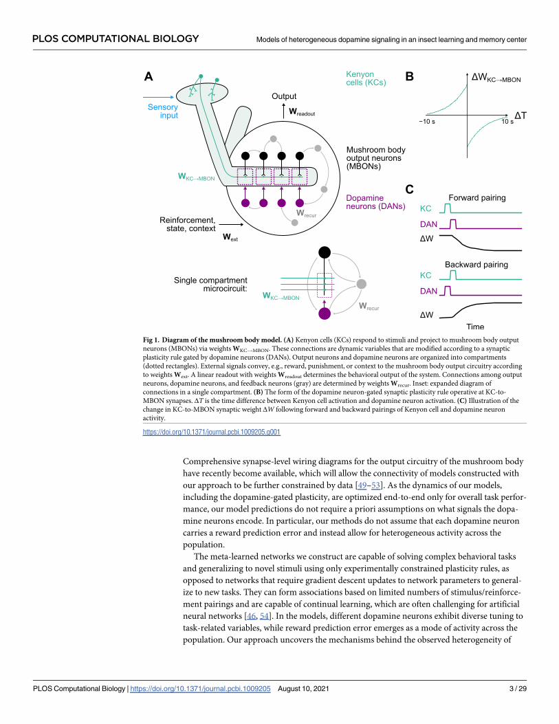
the mushroom body, Kenyon cells (KCs, green neurons in Fig 1A) conveying sensory informa-
tion, predominantly odor-related signals, send parallel fibers that contact the dendrites of
mushroom body output neurons (MBONs, black neurons in Fig 1A). The activation of specific
output neurons biases the organism toward particular actions [23, 24]. Output neuron den-
drites define discrete anatomical regions, known as “compartments,” each of which is inner-
vated by distinct classes of dopaminergic neurons (DANs, magenta neurons in Fig 1A). If the
Kenyon cells and dopamine neurons that project to a given output neuron are both active
within a particular time window, KC-to-MBON synapses are strengthened or weakened
depending on the relative timing of Kenyon cell and dopamine neuron activation [25–28]. The
resulting synaptic modifications permit flies to learn and update associations between stimuli
and reinforcement.
In addition to classical reward and punishment signals, recent studies have shown that vari-
ables including novelty [29], reward prediction [30–32], and locomotion-related signals [33]
are encoded by mushroom body dopamine neurons. In mammals, dopamine signals related to
movement, novelty and salience, and separate pathways for rewards and punishment have also
been identified in midbrain regions [5, 34–42]. These observations call for extensions of classic
models that assume dopamine neurons in associative learning centers are globally tuned to
reward prediction error [43]. How can dopamine signals gate appropriate synaptic plasticity
and learning if their responses are modulated by mixed sources of information?
To address this question, we develop a modeling approach in which networks that produce
dopamine signals suited to learning a particular set of behavioral tasks are constructed. Our
key methodological advance is to augment standard recurrent neural network models, which
employ fixed synaptic weights to solve tasks after optimization [44], with synapses that exhibit
fast dopamine-gated plasticity via an experimentally determined plasticity rule [28]. We
employ a “meta-learning” approach involving two phases [45–47]. First, we optimize the net-
work connections responsible for producing suitable learning signals in dopamine neurons.
Next, after these connections are fixed, we examine the network’s behavior on novel tasks in
which learning occurs only via biologically plausible dopamine-gated plasticity. Due to the
well-characterized anatomy of the mushroom body and knowledge of this plasticity rule, our
approach allows us to generate predictions about the activity of multiple neuron types [28, 48].
PLOS COMPUTATIONAL BIOLOGY Models of heterogeneous dopamine signaling in an insect learning and memory center
PLOS Computational Biology | https://doi.org/10.1371/journal.pcbi.1009205 August 10, 2021 2 / 29
Award DBI-1707398 (L. J. and A. L.-K.). The
funders had no role in study design, data collection
and analysis, decision to publish, or preparation of
the manuscript.
Competing interests: The authors have declared
that no competing interests exist.
Comprehensive synapse-level wiring diagrams for the output circuitry of the mushroom body
have recently become available, which will allow the connectivity of models constructed with
our approach to be further constrained by data [49–53]. As the dynamics of our models,
including the dopamine-gated plasticity, are optimized end-to-end only for overall task perfor-
mance, our model predictions do not require a priori assumptions on what signals the dopa-
mine neurons encode. In particular, our methods do not assume that each dopamine neuron
carries a reward prediction error and instead allow for heterogeneous activity across the
population.
The meta-learned networks we construct are capable of solving complex behavioral tasks
and generalizing to novel stimuli using only experimentally constrained plasticity rules, as
opposed to networks that require gradient descent updates to network parameters to general-
ize to new tasks. They can form associations based on limited numbers of stimulus/reinforce-
ment pairings and are capable of continual learning, which are often challenging for artificial
neural networks [46, 54]. In the models, different dopamine neurons exhibit diverse tuning to
task-related variables, while reward prediction error emerges as a mode of activity across the
population. Our approach uncovers the mechanisms behind the observed heterogeneity of
Fig 1. Diagram of the mushroom body model. (A) Kenyon cells (KCs) respond to stimuli and project to mushroom body output
neurons (MBONs) via weights WKC!MBON. These connections are dynamic variables that are modified according to a synaptic
plasticity rule gated by dopamine neurons (DANs). Output neurons and dopamine neurons are organized into compartments
(dotted rectangles). External signals convey, e.g., reward, punishment, or context to the mushroom body output circuitry according
to weights Wext. A linear readout with weights Wreadout determines the behavioral output of the system. Connections among output
neurons, dopamine neurons, and feedback neurons (gray) are determined by weights Wrecur. Inset: expanded diagram of
connections in a single compartment. (B) The form of the dopamine neuron-gated synaptic plasticity rule operative at KC-to-
MBON synapses. ΔT is the time difference between Kenyon cell activation and dopamine neuron activation. (C) Illustration of the
change in KC-to-MBON synaptic weight ΔW following forward and backward pairings of Kenyon cell and dopamine neuron
activity.
https://doi.org/10.1371/journal.pcbi.1009205.g001
PLOS COMPUTATIONAL BIOLOGY Models of heterogeneous dopamine signaling in an insect learning and memory center
PLOS Computational Biology | https://doi.org/10.1371/journal.pcbi.1009205 August 10, 2021 3 / 29

dopamine signals in the mushroom body and suggests that the “error” signals that support
associative learning may be more distributed than is often assumed.
Results
Modeling recurrent mushroom body output circuitry
The diversity of dopamine neuron activity challenges models of mushroom body learning that
assume these neurons convey global reward or punishment signals. Part of this discrepancy is
likely due to the intricate connectivity among output neurons, dopamine neurons, and other
neurons that form synapses with them [48, 52, 53]. We therefore modeled these neurons and
their connections, which we refer to collectively as the mushroom body “output circuitry,” as a
recurrent neural network (Fig 1A). This model network consists of 20 output neurons, 20
dopamine neurons, and 60 additional recurrent feedback neurons. Recurrent connections
within the network are defined by a matrix of synaptic weights Wrecur. Connections between
all of these 100 neurons are permitted, except that we assume connections from dopamine
neurons to output neurons are modulatory and follow a compartmentalized organization
(Fig 1A, inset). Synapses from 200 Kenyon cells onto output neurons provide the network
with sensory information and are represented by WKC!MBON. Separate pathways convey sig-
nals such as reward or punishment from other brain regions, via weights Wext.
The dynamics of the ith neuron in our model of the output circuitry are given by:
tdriðtÞdt¼ � riðtÞ þ
X
j
Wrecurij rjðtÞ þ bi þ IiðtÞ
" #
þ
; ð1Þ
where [�]+ represents positive rectification. The bias bi determines the excitability of neuron i,while Ii(t) represents its input from non-recurrent connections. If neuron i is an output neu-
ron, then its external input is given by IiðtÞ ¼P
kWKC!MBONik ðtÞrKCk ðtÞ, representing input from
Kenyon cells. If neuron i is a feedback neuron (FBN), then IiðtÞ ¼P
kWextik r
extk ðtÞ, representing
reinforcement, context, or state-dependent input from other brain regions. For dopamine neu-
rons, Ii(t) = 0, as all input to the dopamine neurons is relayed by feedback neurons, reflecting
our interpretation of the feedback neuron population as containing any pathway that conveys
information to the dopamine neurons. We do not constrain Wrecur, except that entries corre-
sponding to connections from dopamine neurons to output neurons are set to zero, based on
the assumption that these connections modulate plasticity of KC-to-MBON synapses rather
than output neuron firing directly (but see [50] and Discussion).
The objective of the network is to generate a desired pattern of activity in a readout that rep-
resents the behavioral bias produced by the mushroom body. The readout decodes this desired
output through a matrix of weights Wreadout. In our first set of experiments, this readout will
represent the one-dimensional valence (appetitive vs. aversive) of a stimulus decoded from the
output neurons (meaning that Wreadout is a 1 × NMBON matrix; later, we will consider more
sophisticated readouts):
vðtÞ ¼WreadoutrMBONðtÞ: ð2Þ
To achieve the task goal, trials are randomly generated and the following objective function,
which depends on the parameters of the network θ and represents the loss corresponding to
an individual trial consisting of T discretized timesteps {t1, t2, . . . tT}, is minimized through
PLOS COMPUTATIONAL BIOLOGY Models of heterogeneous dopamine signaling in an insect learning and memory center
PLOS Computational Biology | https://doi.org/10.1371/journal.pcbi.1009205 August 10, 2021 4 / 29

stochastic gradient descent:
Ly ¼1
T
XT
n¼1
ðvðtnÞ � v�ðtnÞÞ2þl
T
XT
n¼1
XNDAN
i¼1
½rDANi ðtnÞ � 0:1�2
þ: ð3Þ
The first term represents the difference between the decoded valence and a target valence v�
that is determined by the task being learned. The second term is a regularization term that
penalizes dopamine neuron activity that exceeds a baseline level of 0.1 (in normalized units of
firing rate and with λ = 0.1). This term was included to promote solutions that do not exhibit
high levels of non-task-related dopamine activity, but we verified with simulations that the reg-
ularization does not significantly affect overall network performance. Example loss curves over
the course of network optimization are shown in S1 Fig.
Implementation of dopamine-gated plasticity
Recurrent network modeling approaches typically optimize all parameters θ of the network in
order to produce a desired behavior. This approach assumes that, after optimization, connec-
tions are fixed to constant values during the execution of the behavior. However, connections
between Kenyon cells and output neurons are known to exhibit powerful and rapid dopa-
mine-gated synaptic plasticity. This plasticity is dependent on the relative timing of Kenyon
cell and dopamine neuron activation (notably, it does not appear to depend on the postsynap-
tic output neuron firing rate [26]) and can drive substantial changes in evoked output neuron
activity even after brief KC-DAN pairings [28]. We therefore augmented our networks with a
model of this plasticity by assuming that each element of WKC!MBON is a dynamic quantity
that tracks the variables wij (with a time constant of τW = 5 s; see below). These variables,
which determine the strength of the connection from the jth Kenyon cell to the ith output neu-
ron, obey the following update rule:
dwijðtÞdt
¼ �rDANi ðtÞrKCj ðtÞ � �rKCj ðtÞrDANi ðtÞ; ð4Þ
where rKCj and rDANi are the firing rates of the jth Kenyon cell and the dopamine neuron that
innervates the ith compartment, and �rKCj and �rDANi are synaptic eligibility traces constructed by
low-pass filtering rKCj and rDANi . The time constants of the low-pass filters used to generate the
eligibility traces determine the time window within which pairings of Kenyon cell and dopa-
mine neuron activity elicit appreciable changes of w. Each weight element of WKC!MBON is
initially set to its maximum value of 0.05 and subsequently updated according to
tWdWKC!MBON
ij ðtÞ
dt ¼ � WKC!MBONij ðtÞ þ wijðtÞ. The timescale of τW = 5 s accounts for the timescale
of the induction of LTD or LTP.
Odors are encoded by sparse activation of random subsets of Kenyon cells, which is accom-
plished in the model by setting 10% of the elements of rKC to 1 and the rest to 0. When Kenyon
cell and dopamine neuron firing rates are modeled as pulses separated by a time lag ΔT, the
dependence of the change in wij on ΔT takes the form of a biphasic timing-dependent function
(Fig 1B and 1C), consistent with a recent experimental characterization [28]. The seconds-
long timescale of this curve is compatible with the use of continuous firing rates rather than
discrete spike timing to model KC-to-MBON plasticity, as we have done in Eq 4.
Importantly, the weight update rule in Eq 4 is a smooth function of network firing rates,
allowing networks with this update rule to be constructed using gradient descent. Specifically,
we minimize the loss function Eq 3 under the assumption that the network follows the dynam-
ics defined by Eqs 1 and 4. The parameters to be optimized are θ = {Wrecur, Wext, Wreadout, b}
PLOS COMPUTATIONAL BIOLOGY Models of heterogeneous dopamine signaling in an insect learning and memory center
PLOS Computational Biology | https://doi.org/10.1371/journal.pcbi.1009205 August 10, 2021 5 / 29

(the connections describing the mushroom body output circuitry and the biases), while
WKC!MBON is treated as a dynamic quantity. We refer to the gradient descent modification of
θ as the “optimization” phase of constructing our networks. This optimization represents the
evolutionary and developmental processes that produce a network capable of efficiently learn-
ing new associations [55]. After this optimization is complete, the output circuitry is fixed, but
KC-to-MBON weights are subject to synaptic plasticity according to Eq 4. Our approach there-
fore separates synaptic weight changes that are the outcome of evolution and development
from those due to experience-dependent KC-to-MBON plasticity, which would be conflated if
all parameters were optimized with gradient descent (Fig 2). We show that, after optimization,
only the latter form of biologically plausible weight update is sufficient to solve the tasks we
consider and generalize to related but new tasks.
To begin, we assume that KC-to-MBON weights are set to their baseline values at the begin-
ning of each trial in which new associations are formed. Later, we will consider the case of con-
tinual learning of many associations.
Models of associative conditioning
We begin by considering models of classical conditioning, which involve the formation of
associations between a conditioned stimulus (CS) and unconditioned stimulus (US) such as
reward or punishment. A one-dimensional readout of the output neuron population is taken
to represent the stimulus valence (Eq 2), which measures whether the organism prefers
(valence > 0) or avoids (valence < 0) the CS. In the model, CS are encoded by the activation of
a random ensembles of Kenyon cells. Rewards and punishments are encoded by external
inputs to the network that provide input through Wext (see Methods).
To construct the model, we optimized the mushroom body output circuitry to produce an
estimate of the target valence in the readout during presentation of CS+ that have been paired
with US (first-order conditioning; Fig 3A and 3B, top). During presentations of novel CS-US
pairings after optimization, this valence is reported for CS+ but not unconditioned stimulus
(CS-) presentations. The activities of subsets of model output neurons are suppressed follow-
ing conditioning, indicating that the network learns to modify its responses for CS+ but not
CS- responses (Fig 3A and 3B, bottom). This form of classical conditioning requires an appro-
priate mapping from US pathways to dopamine neurons, but recurrent mushroom body out-
put circuitry is not required; networks without recurrence also produce the target valence
(Fig 3E; top). We therefore considered a more complex set of tasks. Networks were optimized
to perform first-order conditioning, to extinguish associations upon repeated presentation of a
CS+ without US, and also to perform second-order conditioning.
During extinction, the omission of a US following a previously conditioned CS+ reduces
the strength of the learned association (Fig 3C). In second-order conditioning, a CS (CS1) is
first paired with a reward or punishment (Fig 3D, left), and then a second CS (CS2) is paired
with CS1 (Fig 3D, center). Because CS2 now predicts CS1 which in turn predicts reward or
punishment, the learned valence of CS1 is transferred to CS2 (Fig 3D, right). In both extinction
and second-order conditioning, a previously learned association must be used to instruct
either the modification of an existing association (in the case of extinction) or the formation of
a new association (in the case of second-order conditioning). We hypothesized that recurrent
output circuitry would be required in these cases. Indeed, non-recurrent mushroom body net-
works are unable to solve these tasks, while recurrent networks are (Fig 3E, center, bottom).
Non-recurrent networks optimized for multiple tasks also exhibited errors on first-order con-
ditioning (0.0 and 0.42 error rate for recurrent and non-recurrent networks respectively,
p< 10−8, Mann-Whitney U-test), indicating a general failure to optimize. Recurrent networks
PLOS COMPUTATIONAL BIOLOGY Models of heterogeneous dopamine signaling in an insect learning and memory center
PLOS Computational Biology | https://doi.org/10.1371/journal.pcbi.1009205 August 10, 2021 6 / 29

generalized to related tasks that they were not optimized for, such as reversal learning (S2 Fig),
further supporting the conclusion that they implement generalizable learning strategies.
We also examined whether the addition of direct connections from Kenyon cells to dopa-
mine neurons influenced our results (S3 Fig). Such connections are present across mushroom
Fig 2. Schematic of meta-learning procedure. (A) Two phases of meta-learning and testing. Left: During the optimization phase, connections that
form the mushroom body output circuitry are updated with gradient descent (orange). Kenyon cell to output neuron weights evolve “online” (within
each trial) according to dopamine-dependent synaptic plasticity. Right: After optimization is complete, the network is tested on a new set of trials. In
this phase, connections that form the output circuitry are fixed. (B) Illustration of trials involving CS/US associations presented during training (left)
and testing (right). Each trial involves new CS/US identities and timing.
https://doi.org/10.1371/journal.pcbi.1009205.g002
PLOS COMPUTATIONAL BIOLOGY Models of heterogeneous dopamine signaling in an insect learning and memory center
PLOS Computational Biology | https://doi.org/10.1371/journal.pcbi.1009205 August 10, 2021 7 / 29

body compartments, but their functional properties are unclear [49, 56]. Our qualitative results
were unchanged when these connections were added, whether we assumed they were fixed or
subject to synaptic plasticity. Thus, indirect connections from Kenyon cells to dopamine neu-
rons through recurrent mushroom body circuitry are sufficient for the tasks we consider.
Comparison to networks without plasticity
Standard recurrent neural networks can maintain stimulus information over time through
persistent neural activity, without modification of synaptic weights. This raises the question of
whether the dopamine-gated plasticity we implemented is necessary to recall CS-US associa-
tions, or if recurrent mushroom body output circuitry alone is sufficient. We therefore com-
pared the networks described above to networks lacking this plasticity. For non-plastic
networks, connections from Kenyon cells to output neurons are optimized through gradient
descent (with no constraints on excitatory or inhibitory sign) and fixed after optimization.
These networks evolve similarly to plastic networks except that the dynamics are determined
only by Eq 1 and not by the dopamine-gated plasticity of Eq 4.
In non-plastic networks, information about CS-US associations must be stored in the per-
sistent activity of the mushroom body output circuitry as an “attractor” of neural activity [57],
as opposed to being encoded in the KC-to-MBON weights. Such activity must maintain both
the identity of the CS+ odor and which US it was paired with in order to recall the learned
Fig 3. Behavior of network during reward conditioning paradigms. (A) Behavior of output neurons (MBONs) during first-order conditioning. During training, a CS+
(blue) is presented, followed by a US (green). Top: The network is optimized so that a readout of the output neuron activity during the second CS+ presentation encodes
valence (gray curve). Black curve represents the target valence and overlaps with the readout. Bottom: Example responses of output neurons. (B) Same as A, but for CS-
presentation without US. (C) Same as A, but for extinction, in which a second presentation of the CS+ without the US partially extinguishes the association. (D) Same as
A, but for second-order conditioning, in which a second stimulus (CS2) is paired with a conditioned stimulus (CS1). (E) Error rate averaged across networks in different
paradigms. An error is defined as a difference between reported and target valence with magnitude greater than 0.2 during the test period. Networks optimized with
recurrent output circuitry (control, black) are compared to networks without recurrence (no recur., red), and networks prior to optimization (initialization, gray). Error
rates for each network realization are evaluated over 50 test trials and used to generate p-values with a Mann-Whitney U-test over 20 network realizations.
https://doi.org/10.1371/journal.pcbi.1009205.g003
PLOS COMPUTATIONAL BIOLOGY Models of heterogeneous dopamine signaling in an insect learning and memory center
PLOS Computational Biology | https://doi.org/10.1371/journal.pcbi.1009205 August 10, 2021 8 / 29

valence without generalizing that valence to a different odor. We hypothesized that it would be
challenging for networks to support a large number of such attractors and therefore investi-
gated the performance of non-plastic networks in simulated environments in which there are
a fixed number of odors. We optimized networks both to respond appropriately to CS+
(Fig 4A) and avoid responding to neutral CS (Fig 4B).
Non-plastic networks can form CS-US associations (Fig 4A). Compared to networks with
dopamine-gated plasticity (Fig 3A), output neurons exhibit stronger persistent activity follow-
ing a CS-US pairing. However, when the number of odors in the environment is large, non-
plastic networks exhibit a high degree of overgeneralization of learned associations to neutral
CS that have not been paired with US (Fig 4B). This likely reflects the non-plastic networks’
inability to distinguish between odor identities when many odors are present. When odor
identities cannot be distinguished, the best compromise is to assume that the learned CS
+ valence applies to both the CS+ and to neutral CS, and indeed when many odors are present
Fig 4. Comparison to networks without dopamine-gated plasticity. (A) Behavior during first-order conditioning,
similar to Fig 3A, but for a non-plastic network. Because of the need for non-plastic networks to maintain information
using persistent activity, performance degrades with longer delays between training and test phases. We therefore
chose this delay to be shorter than in Fig 3A. Results are shown for a network optimized with 10 odors. (B) Same as A,
but for a trial in which a CS-US pairing is followed by the presentation of a neutral CS. (C) Difference in response
(reported valence) for CS+ and neutral CS as a function of the number of odors. Each CS+ is associated with either a
positive or negative US. For comparison, the corresponding response difference for networks with dopamine-gated
plasticity is shown in blue. Error bars represent s.e.m. over 8 network realizations.
https://doi.org/10.1371/journal.pcbi.1009205.g004
PLOS COMPUTATIONAL BIOLOGY Models of heterogeneous dopamine signaling in an insect learning and memory center
PLOS Computational Biology | https://doi.org/10.1371/journal.pcbi.1009205 August 10, 2021 9 / 29

the difference in the reported valence for these two classes of stimuli decreases to zero
(Fig 4C). Networks with dopamine-gated plasticity do not suffer from this shortcoming, as
they can store and update the identities of arbitrary novel stimuli in KC-to-MBON weights
(Fig 4C, blue curve).
In total, the comparison between plastic and non-plastic networks demonstrates that the
addition of dopamine-gated plasticity at KC-to-MBON synapses improves capacity and
reduces overgeneralization. Furthermore, plastic networks need not rely solely on persistent
activity in order to store associations (compare Figs 3A and 4A), likely prolonging the time-
scale over which information can be stored without being disrupted by ongoing activity.
Distributed representations across dopamine neurons
We next examined the responses of dopamine neurons to neutral, unconditioned, and condi-
tioned stimuli in the networks we constructed, to examine the “error” signals responsible for
learning (Fig 5A). Dopamine neurons exhibited heterogeneity in their responses. We per-
formed hierarchical clustering to identify groups of dopamine neurons with similar response
properties (Fig 5B, gray; see Methods). This procedure identified two broad groups of
Fig 5. Population analysis of dopamine neuron (DAN) activity. (A) First-order conditioning trials with positive or negative valence US.
(B) Responses of model dopamine neurons from a single network. Neurons are sorted according to hierarchical clustering (illustrated with
gray dendrogram) of their responses. (C) Principal components analysis of dopamine neuron population activity. Left: Response to CS
before conditioning. Middle: Response to a positive (green) or negative (red) valence US. Right: Response to a previously conditioned CS.
https://doi.org/10.1371/journal.pcbi.1009205.g005
PLOS COMPUTATIONAL BIOLOGY Models of heterogeneous dopamine signaling in an insect learning and memory center
PLOS Computational Biology | https://doi.org/10.1371/journal.pcbi.1009205 August 10, 2021 10 / 29

dopamine neurons—one that responds to positive-valence US and another that responds to
negative-valence US—as well as more subtle features in the population response. Consistent
with the known logic of the mushroom body output circuitry [48] and learning involving
depression of KC-to-MBON synapses, compartments whose dopamine neurons signal positive
valence US tend to have output neurons whose activation signals negative valence (S4 Fig).
While some dopamine neurons increase their firing only for US, many also respond to rein-
forced CS. In some cases, this response includes a decrease in firing rate in response to the
omission of a predicted US that would otherwise cause an increase in rate, consistent with a
reward prediction error. In other cases, neurons respond only with increases in firing rate for
US of a particular valence, and for omitted US of the opposite valence, consistent with cross-
compartmental interactions supporting the prediction of valence [31]. The presence of both
reward prediction error-like responses and valence-specific omission responses suggests that
multiple mechanisms are employed by the network to perform tasks such as extinction and
second-order conditioning.
The examination of their responses demonstrates that dopamine neurons in our models are
diversely tuned. This tuning implies that KC-to-MBON synapses change in a heterogeneous
manner in response to CS and US presentations, but that these changes are sufficient to pro-
duce an appropriate behavioral response collectively. Consistent with this idea, principal com-
ponents analysis of dopamine neuron responses identified modes of activity with
interpretable, task-relevant dynamics. The first principal component (Fig 5C) reflects US
valence and predicted CS+ valence, while rapidly changing sign upon US omission, consistent
with a reward prediction error. It is notable that such a signal emerges as the dominant mode
of dopamine neuron activity, as our optimization procedure does not explicitly require the for-
mation of a reward prediction error. Subsequent principal components include components
that respond to CS and US of both valences (principal component 2) or are tuned primarily to
a single stimulus, such as a positive valence CS+ (principal component 4). When we con-
strained networks to have fewer compartments, error increased (S5 Fig) suggesting that diver-
sity in dopamine signaling improves performance, though we note that this trend does not
distinguish task difficulty and ease of optimization.
To further explore how dopamine neuron responses depend on the task being learned, we
extended the model to require encoding of novelty and familiarity, inspired by a recent study
that showed that the mushroom body is required for learning and expressing an alerting
behavior driven by novel CS [29]. We added a second readout that reports CS novelty, in addi-
tion to the readout of valence described previously. Networks optimized to report both vari-
ables exhibit enhanced CS responses and a large novelty-selective component in the
population response identified by principal components analysis (Fig 6), compared to net-
works that only report valence (Fig 5B). These results suggest that dopamine neurons collec-
tively respond to any variables relevant to the task for which the output circuitry is optimized,
which may include variables distinct from reward prediction. Furthermore, the distributed
nature of this representation implies that individual variables may be more readily decoded
from populations of dopamine neurons than from single neurons.
Continual learning of associations
In the previous sections, we modeled the dynamics of networks during individual trials con-
taining a limited number of associations. We next ask whether these networks are capable of
continual learning, in which long sequences of associations are formed, with recent associa-
tions potentially overwriting older ones. Such learning is often challenging, particularly when
synaptic weights have a bounded range, due to the tendency of weights to saturate at their
PLOS COMPUTATIONAL BIOLOGY Models of heterogeneous dopamine signaling in an insect learning and memory center
PLOS Computational Biology | https://doi.org/10.1371/journal.pcbi.1009205 August 10, 2021 11 / 29

minimum or maximum value after many associations are formed [58]. To combat this, a
homeostasic process that prevents such saturation is typically required. We therefore asked if
our optimized networks can implement such homeostasis.
In certain compartments of the mushroom body, it has been shown that the activation of
dopamine neurons in the absence of Kenyon cell activity leads to potentiation of KC-to-
MBON synapses [33]. This provides a mechanism for the erasure of memories formed follow-
ing synaptic depression. We hypothesized that this non-specific potentiation could implement
a form of homeostasis that prevents widespread synaptic depression after many associations
are formed. We therefore augmented our dopamine-gated synaptic plasticity rule (Fig 1C)
with such potentiation (Fig 7A). The new synaptic plasticity rule is given by:
dwijðtÞdt
¼ �rDANi ðtÞrKCj ðtÞ � �rKCj ðtÞrDANi ðtÞ þ b�rDANi ðtÞ; ð5Þ
where β represents the rate of non-specific potentiation (compare with Eq 4). We allowed β to
be optimized by gradient descent individually for each compartment but constrained it to be
nonnegative.
We modeled long sequences of associations in which CS+, CS-, and US are presented ran-
domly (Fig 7B) and the network is again optimized to produce a target valence (Eq 3). In opti-
mized networks, the KC-to-MBON weights are initialized at the beginning (t = 0) of trial n to
be equal to those at the end (t = T) of trial n − 1, WKC!MBONn ð0Þ ¼WKC!MBON
n� 1ðTÞ, rather than
Fig 6. Behavior of a network that encodes both valence and novelty. The network is similar to Fig 5 but a second readout that computes novelty is added. The novelty
readout is active for the first presentation of a given CS and zero otherwise. (A) The addition of novelty as a readout dimension introduces dopamine neuron responses
that are selective for novel CS. Compare with Fig 5B. (B) The first principal component (PC1) for the network in A is selective for CS novelty. Compare with Fig 5C.
https://doi.org/10.1371/journal.pcbi.1009205.g006
PLOS COMPUTATIONAL BIOLOGY Models of heterogeneous dopamine signaling in an insect learning and memory center
PLOS Computational Biology | https://doi.org/10.1371/journal.pcbi.1009205 August 10, 2021 12 / 29

being reset to their baseline values as done previously. We examined the distribution of KC-
to-MBON synaptic weights after such sequences of trials. Without non-specific potentiation,
most synaptic weights are clustered near 0 (Fig 7C, red). However, the addition of this potenti-
ation substantially changes the synaptic weight distribution, with many weights remaining
potentiated even after thousands of CS and US presentations (Fig 7C, black). We also exam-
ined performance and dopamine neuron responses in the two types of networks. Without
non-specific potentiation, dopamine neuron responses are weaker and the reported valence
less accurately tracks the target valence, compared to networks with such potentiation (Fig 7D
and 7E).
These results suggest that such homeostatic mechanisms, or other modifications to the syn-
aptic plasticity rule in Eq 4 that avoid weights clustering near 0, are important for performance
on continual learning tasks. However, we note that non-specific potentiation might also
shorten memory lifetime, for example in a situation where a CS-US pairing is followed by
unpaired US presentations. Investigating how this tradeoff is resolved across compartments is
an interesting topic for future study.
Associating stimuli with changes in internal state
In the previous sections, we focused on networks whose dopamine neurons exhibited transient
responses to the presentation of relevant external cues. Recent studies have found that dopa-
mine neurons also exhibit continuous fluctuations that track the state of the fly, even in the
Fig 7. Model behavior for long sequences of associations. (A) Illustration of non-specific potentiation following dopamine neuron
activity (compare with Fig 1C). (B) Example sequence of positive and negative associations between two odors CS+ and CS2+ and US.
Neutral gray odors (CS-) are also presented randomly. (C) Histogram of synaptic weights after a long sequence of CS and US
presentations for networks with (black) and without (red) non-specific potentiation. Weights are normalized to their maximum value.
The means of the distributions across 18 network realizations for each condition were significantly different (p< 2 � 10−7, Mann-
Whitney U-test). (D) Left: dopamine neuron responses for the sequence of CS and US presentations. Right: same as left, but for a
network without non-specific potentiation. (E) Error rate (defined as a difference between reported and target valence with magnitude
greater than 0.5 during a CS presentation; we used a higher threshold than Fig 3 due to the increased difficulty of the continual learning
task) for networks with (black) and without (red) non-specific potentiation. Error rates for each network realization are evaluated over
20 test trials and used to generate p-values with a Mann-Whitney U-test over 18 network realizations.
https://doi.org/10.1371/journal.pcbi.1009205.g007
PLOS COMPUTATIONAL BIOLOGY Models of heterogeneous dopamine signaling in an insect learning and memory center
PLOS Computational Biology | https://doi.org/10.1371/journal.pcbi.1009205 August 10, 2021 13 / 29

absence of overt external reinforcement. These fluctuations are correlated with transitions
between, for example, movement and quiescence [33], or hunger and satiation [59]. Under-
standing the functional role of this activity is a major challenge for models of dopamine-
dependent learning. We hypothesized that such activity could permit the association of stimuli
with a transition to an arbitrary internal state of the organism. This could allow downstream
networks to read out whether a stimulus has previously been experienced in conjuction with a
particular change in state, which might inform an appropriate behavioral response to that
stimulus.
To test this hypothesis, we constructed networks that, in addition to supporting associative
conditioning (as in Fig 3), also transitioned between a set of three discrete internal states, trig-
gered on input pulses that signal the identity of the next state (Fig 8A). This input represents
signals from other brain areas that drive state transitions. We optimized the output circuitry
to, in addition to encoding valence as before, continuously maintain a state representation,
quantified by the ability of a linear readout of dopamine neuron activity to decode the current
state (Fig 8B, top). Specifically, the loss function equaled
Ly ¼1
T
XT
n¼1
ðvðtnÞ � v�ðtnÞÞ2þ
1
T
XT
n¼1
jjsðtnÞ � s�ðtnÞjj2; ð6Þ
where s = Softmax(Wstater rDAN) is a 3-dimensional vector that represents the decoded proba-
bilities of being in each state and s� is a vector with one nonzero entry corresponding to the
actual current state. Here, Wstate is a 3 × NDAN matrix of weights that represents a linear read-
out of the state from DANs, while as before valence is read out from MBONs. Because we were
interested in networks that exhibited continuous fluctuations in dopamine neuron activity, we
did not impose an additional penalty on dopamine neuron firing rates as in Eq 3. Optimizing
networks with this loss function led to widespread state-dependent activity throughout the net-
work, including among dopamine neurons (Fig 8B, bottom). This activity coexists with activity
evoked by CS or US presentation.
We next examined output neuron responses to the presentation of stimuli that had previ-
ously preceded a transition to some state. If a transition to a given state reliably evokes a partic-
ular pattern of dopamine neuron activity, then KC-to-MBON synapses that are activated by
any stimulus preceding such a transition will experience a similar pattern of depression or
potentiation. We assessed this response similarity by computing the Pearson’s correlation
coefficient CorrðrMBONA ; rMBON
B Þ, where rMBONA is the average output neuron activity during the
presentation of stimulus A. Consistent with this prediction, the pattern of output neuron
responses evoked by a stimulus that predicts a transition to state S1 is more similar to the cor-
responding responses to other stimuli that predict the same state than any other state S2
(Fig 8C). The representations of state-transition-predictive stimuli are thus “imprinted” with
the identity of the predicted state. While these modifications could potentially interfere with
the ability of the system to support associative conditioning, these networks still exhibited high
performance on the tasks we previously considered (Fig 8D). Thus, state-dependent activity
and activity required for conditioning are multiplexed in the network. The presence of state-
dependent fluctuations could allow circuits downstream of the mushroom body to consistently
produce a desired behavior that depends on the internal state, instead of or in addition to the
external reinforcement, that is predicted by a stimulus. Our model thus provides a hypothesis
for the functional role of state-dependent dopamine neuron activity.
PLOS COMPUTATIONAL BIOLOGY Models of heterogeneous dopamine signaling in an insect learning and memory center
PLOS Computational Biology | https://doi.org/10.1371/journal.pcbi.1009205 August 10, 2021 14 / 29

Mixed encoding of reward and movement in models of navigation
We also examined models of dynamic, goal directed behaviors. An important function of
olfactory associations in Drosophila is to enable navigation to the sources of reward-predicting
odor cues, such as food odors [60]. We therefore modeled an agent that is first presented with
Fig 8. Behavior of a network whose activity transitions between a sequence of discrete states in addition to supporting
associative conditioning. (A) Brief pulse inputs to the network signal that a switch to a new state should occur. (B) Top: A linear
readout of dopamine neuron activity can be used to decode the network state. Bottom: dopamine neuron (DAN) activity exhibits
state-dependent fluctuations in addition to responding to CS and US. (C) Decoding of stimuli that predict state transitions. Heatmap
illustrates the correlation between output neuron population responses to the presentation of different stimuli that had previously
been presented prior to a state transition. Stimuli are ordered based on the state transitions that follow their first presentation. Blue
blocks indicate that stimuli that predict the same state transition evoke similar output neuron activity. (D) Performance of networks
on conditioning tasks. For each network realization, error rates are computed over 50 test trials and bars represent s.e.m. over 40
network realizations.
https://doi.org/10.1371/journal.pcbi.1009205.g008
PLOS COMPUTATIONAL BIOLOGY Models of heterogeneous dopamine signaling in an insect learning and memory center
PLOS Computational Biology | https://doi.org/10.1371/journal.pcbi.1009205 August 10, 2021 15 / 29

a CS+ followed by reward and then is placed in a two-dimensional environment and must nav-
igate to the rewarded odor (Fig 9A, top). The activity of the mushroom body output circuitry
controls the forward velocity u(t) and angular velocity ω(t) of the agent. The agent’s heading is
given by dydt ¼ oðtÞ, which, along with the forward velocity, determines the change in its loca-
tion dxdt ¼ uðtÞ cosyðtÞx1 þ sin yðtÞx2ð Þ (Fig 9A). We assumed that these movement variables
are not decoded directly from output neurons but from other feedback neurons which may
represent locomotion-related downstream regions (see Methods). The environment contains
Fig 9. Model behavior for a navigation task. (A) Top: Schematic of navigation task. After conditioning, the simulated organism
uses odor concentration input (blue) and information about wind direction w relative to its heading h. Bottom: Diagram of a
network that uses these signals to compute forward and angular velocity signals for navigation. Velocity signals are read out from
other neurons in the mushroom body output circuitry (gray), rather than output neurons. (B) Position of the simulated organism as
a function of time during navigation. Black: Simulation with intact dopamine-gated plasticity during navigation; Red: Simulation
with plasticity blocked. Arrowheads indicate direction of movement. In the top left plot, the starting location (gray circle) is
indicated. (C) Position error (mean-squared distance from rewarded odor source at the end of navigation) for control networks and
the same networks in which dopamine-gated plasticity is blocked during the navigation phase. For each network realization, error
rates are computed over 50 test trials and bars represent s.e.m. over 30 network realizations. Significance is computed with a
Wilcoxon signed-rank test. (D) Forward (top) and angular (bottom) velocity as a function of time during one example navigation
trial. (E) Left: Dopamine neuron activity during CS and US presentation in the conditioning phase of a trial. Right: Dopamine
neuron activity during the navigation phase of the trial (same trial as in D).
https://doi.org/10.1371/journal.pcbi.1009205.g009
PLOS COMPUTATIONAL BIOLOGY Models of heterogeneous dopamine signaling in an insect learning and memory center
PLOS Computational Biology | https://doi.org/10.1371/journal.pcbi.1009205 August 10, 2021 16 / 29

multiple odor sources that produce odor plumes that the the agent encounters as it moves. The
mushroom body output circuitry supports this behavior by integrating odor concentration
input from Kenyon cells and information from other brain areas about wind direction relative
to the agent’s orientation [61] (Fig 9A, bottom; see Methods for a description of how wind
input is encoded). Because x(t) is a differentiable function of network parameters, we can use
as a loss function the Euclidean distance between the agent’s location and the rewarded odor
source at the end of this navigation period:
Ly ¼ jjxðTÞ � x�jj2; ð7Þ
where x� is the location of the rewarded odor source and T is the time at which the navigation
period ends. Successfully executing this behavior requires storing the identity of the rewarded
odor, identifying the upwind direction for that odor, moving toward the odor source using
concentration information, and ignoring neutral odors.
The agent successfully navigates to the rewarded odor source (Fig 9B), and success requires
plasticity during conditioning that encodes the CS+/US pairing (S6 Fig). We wondered
whether dopamine-gated plasticity might also be operative during navigation, based on recent
findings that recorded ongoing dopamine neuron fluctuations correlated with movement [33].
We asked whether such plasticity during navigation affects the behavior of the model by exam-
ining the performance of networks in which it is blocked after optimization. Blocking plasticity
during navigation impairs performance (Fig 9C). In particular, networks lacking plasticity
often exhibit decreased forward velocity after entering a plume corresponding to a rewarded
odor (Fig 9B), suggesting that ongoing plasticity may reinforce salient odors as they are
encountered and promote odor-seeking, consistent with a recent report [62]. These results
make the prediction that increased levels of dopamine neuron activity and dopamine-gated
plasticity occur upon encounters of rewarded odor plumes.
We also examined the relationship between dopamine neuron activity and movement vari-
ables during navigation. The agent exhibits increased forward velocity and turning upon
encountering an odor, with greater increases for rewarded than for neutral odors (Fig 9D).
Model dopamine neurons exhibit activity during navigation that correlates with movement
(Fig 9E and S7 Fig). Many of the same dopamine neurons also exhibit reward-related activity,
as has been observed in neural recordings [33].
An important caveat to our results is that it is possible to construct networks in which plas-
ticity is active during the conditioning phase but gated off during the navigation phase of the
task from the beginning of optimization. In natural environments where learning and naviga-
tion are not clearly separated into distinct phases, such a gating mechanism may be difficult to
implement. However, in our setting, these networks exhibit similar performance to networks
in which plasticity is always active (S6 Fig). Thus, unconstrained optimization of networks
produces solutions in which ongoing plasticity during navigation is behaviorally relevant (Fig
9B and 9C), but our results cannot be taken to conclude that this plasticity is always required
to solve our task. More complex tasks that require moment-by-moment decisions to be made
throughout the navigation process may rely on such plasticity and are an exciting direction for
future study.
Discussion
We have developed models of the mushroom body that use a biologically plausible form of
dopamine-gated synaptic plasticity to solve a variety of learning tasks. By optimizing the mush-
room body output circuitry for task performance, these models generate patterns of dopamine
neuron activity sufficient to produce the desired behaviors. Model dopamine responses are
PLOS COMPUTATIONAL BIOLOGY Models of heterogeneous dopamine signaling in an insect learning and memory center
PLOS Computational Biology | https://doi.org/10.1371/journal.pcbi.1009205 August 10, 2021 17 / 29

distributed, tuned to multiple task-relevant variables, and exhibit rich temporal fluctuations.
This diversity is a result of optimizing our models only for task performance rather than
assuming that dopamine neurons uniformly represent a particular quantity of interest, such as
a global reward prediction error signal [3]. Our results predict that individual dopamine neu-
rons may exhibit diverse tuning while producing coherent activity at the population level.
They also provide the first unified modeling framework that can account for valence and
reward prediction (Fig 5), novelty (Fig 6), and movement-related (Fig 9) dopamine neuron
responses that have been recorded in experiments.
Relationship to other modeling approaches
To construct our mushroom body models, we took advantage of recent advances in recurrent
neural network optimization to augment standard network architectures with dopamine-
gated plasticity. Our approach can be viewed as a form of “meta-learning” [45–47], or “learn-
ing to learn,” in which a network learns through gradient descent to use a differentiable form
of synaptic plasticity (Eq 4) to solve a set of tasks. As we have shown, this meta-learning
approach allows us to construct networks that exhibit continual learning and can form associa-
tions based on single CS-US pairings (Fig 7). Recent studies have modeled networks with
other forms of differentiable plasticity, including Hebbian plasticity, [63–65] but have not
studied gated plasticity of the form of Eq 4. Another recent study examined networks with a
global neuromodulatory signal rather than the heterogeneous signals we focus on [66]. Meta-
learning approaches have also recently been applied to infer alternative learning algorithms to
backpropagation through time [67].
Another recent study used a meta-learning approach to model dopamine activity and activ-
ity in the prefrontal cortex of mammals [68]. Unlike our study, in which the “slow” optimiza-
tion is taken to represent evolutionary and developmental processes that determine the
mushroom body output circuitry, in this study the slow component of learning involved dopa-
mine-dependent optimization of recurrent connections in prefrontal cortex. This process
relied on gradient descent in a recurrent network of long short-term memory (LSTM) units,
leaving open the biological implementation of such a learning process. Like in actor-critic
models of the basal ganglia [69], dopamine was modeled as a global reward prediction error
signal.
In our case, detailed knowledge of the site and functional form of plasticity [28] allowed us
to build models that solved multiple tasks using only a biologically plausible synaptic plasticity
rule. This constraint allows us to predict patterns of dopamine neuron activity that are suffi-
cient for solving these tasks (Fig 5). Similar approaches may be effective for modeling other
brain areas in which the neurons responsible for conveying “error” signals can be identified,
such as the cerebellum or basal ganglia [2, 70].
Heterogeneity of dopamine signaling across species
Dopamine is responsible for a variety of functions in arthropods, including associative mem-
ory in honeybees [6], central pattern generation in the stomatogastric ganglion of lobsters [7],
escape behaviors [8] and salivation [9] in the cockroach, and flight production in moths [10].
While dopamine similarly plays many roles in Drosophila, including the regulation of locomo-
tion, arousal, sleep, and mating [11], until recently most studies of Drosophila mushroom body
dopamine neurons have focused on their roles in appetitive and aversive memory formation
[12, 13, 16, 18, 20–22]. In mammals, while numerous studies have similarly focused on reward
prediction error encoding in midbrain dopaminergic neurons [2], recent reports have also
described heterogeneity in dopamine signals reminiscent of the heterogeneity across
PLOS COMPUTATIONAL BIOLOGY Models of heterogeneous dopamine signaling in an insect learning and memory center
PLOS Computational Biology | https://doi.org/10.1371/journal.pcbi.1009205 August 10, 2021 18 / 29

dopamine neurons in the mushroom body [5, 43]. These include reports detailing distinct sub-
types of dopamine neurons that convey positive or negative valence signals or respond to
salient signals of multiple valences [39, 71], novelty responses [34–38, 40], responses to threat
[72], and modulation of dopamine neurons by movement [41, 42]. In many cases, these sub-
types are defined by their striatal projection targets, suggesting a compartmentalization of
function similar to that of the mushroom body [5]. However, the logic of this compartmentali-
zation is not yet clear.
Standard reinforcement learning models of the basal ganglia, such as actor-critic models,
assume that dopamine neurons are globally tuned to reward prediction error signals [69]. Pro-
posals have been made to account for heterogeneous dopamine responses, including that dif-
ferent regions produce prediction errors based on access to distinct state information [73], or
that dopamine neurons implement an algorithm for learning the statistics of transitions
between states using sensory prediction errors [74]. Our results are compatible with these the-
ories, but different in that our model does not assume a priori that all dopamine neurons
encode prediction errors. Instead, prediction error coding by particular modes of population
activity emerges in our model as a consequence of optimizing for task performance (Fig 5).
This heterogeneity emerged even though we penalized dopamine activity that exceeded a base-
line value (Eq 3). In networks in which this penalization is absent, such as networks whose
dopamine neurons encode arbitrary changes in internal state (Fig 8), an even higher level of
dopamine fluctuations is present in the optimized models.
Connecting mushroom body architecture and function
The identification of groups of dopamine neurons that respond to positive or negative valence
US [16, 24, 30, 75, 76], output neurons whose activity promotes approach or avoidance [26],
and dopamine-gated plasticity of KC-to-MBON synapses [27, 28, 77] has led to effective mod-
els of first-order appetitive and aversive conditioning in Drosophila. A minimal model of such
learning requires only two compartments of opposing valence and no recurrence among out-
put neurons or dopamine neurons. The presence of extensive recurrence [33, 48, 52, 78] and
dopamine neurons that are modulated by other variables [29, 31–33] suggests that the mush-
room body modulates learning and behavior along multiple axes.
The architecture of our model reflects the connectivity between Kenyon cells and output
neurons, compartmentalization among output neurons and dopamine neurons, and recur-
rence of the mushroom body output circuitry. These constraints match the key architectural
features of the mushroom body, but also reflect simplifications made in the absence of addi-
tional data. While the identities and functional properties of output neurons and dopamine
neurons have been mapped anatomically [48, 79], the feedback pathways have not, so the feed-
back neurons in our model (gray neurons in Fig 1A) represent any neurons that participate in
recurrent loops involving the mushroom body, which may involve paths through other brain
areas. For most of our analyses (but see S3 Fig), we also neglected direct projections from Ken-
yon cells to dopamine neurons [49, 56]. When they were added to the model, our qualitative
results were unchanged, although it is possible that future studies may uncover a specific role
for these connections. Our model could also be extended by including direct depolarizing or
hyperpolarizing effects of dopamine on output neurons, which has been observed experimen-
tally [50], or by introducing recurrence among Kenyon cells [49]. Additionally, explicitly
modeling the integration of projection neuron to Kenyon cell signaling could provide a more
realistic account of the representation of sensory stimuli [80].
Our model could also be extended by considering other forms of synaptic plasticity. There
is evidence that dopamine-gated synaptic plasticity rules (Fig 1B) are heterogeneous across
PLOS COMPUTATIONAL BIOLOGY Models of heterogeneous dopamine signaling in an insect learning and memory center
PLOS Computational Biology | https://doi.org/10.1371/journal.pcbi.1009205 August 10, 2021 19 / 29

compartments [26, 27], and non-dopamine-dependent plasticity could also lead to new behav-
ior [80]. While we have primarily focused on the formation of associations over short time-
scales because the detailed parameters of compartment-specific learning rules have not been
described, such heterogeneity will likely be particularly important in models of long-term
memory [21, 81–85].
Our model makes several predictions. It predicts that reward prediction error should
emerge as a dominant mode of population activity across dopamine neurons, even though
individual dopamine neurons may be diversely tuned (Fig 5). It predicts that compartments
that exhibit a large degree of non-specific potentiation may be particularly important for form-
ing short-term associations in complex environments with many background or distractor
odors (Fig 7). It also suggests the possibility of pairing an odor presentation with a change in
internal state and reading out this pairing from the pattern of output neuron activity upon a
subsequent presentation (Fig 8C). Our results also suggest that plasticity during navigation
may promote odor-seeking (Fig 9), an idea with experimental support [62]. For each of these
predictions, input to dopamine neurons from pathways other than those that convey purely
external reinforcement is required. Identifying the pathways that convey these signals is an
important direction. In the absence of an explicit correspondence between neurons in our
model and their biological counterparts, direct analysis of the connectivity in our optimized
networks is unlikely to be sufficient to do so. Future studies should build models that incorpo-
rate recently available mushroom body wiring diagrams to further constrain models [49, 50,
52, 53].
However, it is unlikely that purely anatomical information, even at the level of a synaptic
wiring diagram, will be sufficient to infer how the mushroom body functions [86]. We have
used anatomical information and parametrized synaptic plasticity rules along with hypotheses
about which behaviors the mushroom body supports to build “task-optimized” models, related
to approaches that have been applied to sensory systems [87]. The success of these approaches
for explaining neural data relies on the availability of complex tasks that challenge and con-
strain the computations performed by the models. Therefore, experiments that probe the axes
of fly behavior that the mushroom body supports, including behaviors that cannot be
described within the framework of classical conditioning, will be a crucial complement to con-
nectivity mapping efforts as models of this system are refined.
Methods
Time discretization
For computational efficiency and ease of training, we assume τ in Eq 1 is equal to 1 s and simu-
late the system with a timestep of Δt = 0.5 s, but our results do not depend strongly on these
parameters.
Optimization
Parameters are optimized using PyTorch (www.pytorch.org) with the RMSprop optimizer
[88] and a learning rate of 0.001. The loss to be minimized is described by Eqs 3 and 6 or Eq 7
for networks optimized for conditioning tasks, continuous state representations, or navigation
respectively. Optimization is performed over a set number of epochs, each of which consists of
a batch of B = 30 trials. The loss LtotyðmÞ for epoch m is the average of the individual losses over
PLOS COMPUTATIONAL BIOLOGY Models of heterogeneous dopamine signaling in an insect learning and memory center
PLOS Computational Biology | https://doi.org/10.1371/journal.pcbi.1009205 August 10, 2021 20 / 29

each trial in the batch:
LtotyðmÞ ¼
1
B
XB
b¼1
Lyðb;mÞ; ð8Þ
where Lyðb;mÞ represents the loss for bth trial drawn on epoch m.
All optimized weights are initialized as zero mean Gaussian variables. To initialize Wrecur,
weights from a neuron belonging to neuron type X (where X = MBON, DAN, or FBN) have 0
mean and variance equal to 1ffiffi2p
NX, where NX equals the number of neurons of type X. For Wread-
out, the variance is 1/NMBON while for Wext, the variance is 1. Bias parameters are initialized at
0.1. At the beginning of each trial, firing rates are reset to an initial state r0, with r0 = 0 for out-
put neurons and 0.1 for dopamine neurons or feedback neurons, to permit these neurons to
exhibit low levels of baseline activity.
Conditioning tasks
For conditioning tasks in which the predicted valence of a conditioned stimulus (CS) is
reported (such as first- and second-order conditioning and extinction), each CS is encoded by
setting 10% of the entries of rKC to 1 and the rest to 0. Unconditioned stimuli (US) are encoded
by rext which is equal to (1, 0)T when a positive-valence US is present, (0, 1)T when a negative-
valence US is present, and (0, 0)T otherwise. CS and US are presented for 2 s. Tasks are split
into 30 s intervals (for example conditioning and test intervals; see Fig 3). Stimulus presenta-
tion occurs randomly between 5 s and 15 s within these intervals. Firing rates are reset at the
beginning of each interval (e.g. r(t = 30 s) = r0), which prevents networks from using persistent
activity to maintain associations.
When optimizing networks in Fig 3, random extinction and second-order conditioning tri-
als were drawn. With probability 1/2, CS or US are randomly omitted (and the target valence
updated accordingly—e.g., if the US is omitted, the network should not report a nonzero
valence upon the second CS presentation; Fig 3B) in order to prevent the networks from over-
generalizing to CS that are not paired with reinforcement. Optimization progressed for 5000
epochs for networks trained to perform extinction and second-order conditioning. For net-
works trained only for first-order conditioning, (Fig 3E, top; Fig 4), only first-order condition-
ing trials were drawn, and optimization progressed for 2000 epochs.
Principal components of dopamine neuron activity (Fig 5) were estimated using 50 ran-
domly chosen trials of extinction and second-order conditioning in previously optimized net-
works. To order dopamine neurons based on their response similarity (Fig 5A), hierarchical
clustering was performed using the Euclidean distance between the vector of firing rates corre-
sponding to pairs of dopamine neurons during these trials.
For networks also trained to report stimulus novelty (Fig 6), an additional readout dimen-
sion n(t) that is active for the first presentation of a given CS and inactive otherwise is added.
The full network readout is then given by
RðtÞ ¼vðtÞ
nðtÞ
!
¼WreadoutrMBONðtÞ; ð9Þ
PLOS COMPUTATIONAL BIOLOGY Models of heterogeneous dopamine signaling in an insect learning and memory center
PLOS Computational Biology | https://doi.org/10.1371/journal.pcbi.1009205 August 10, 2021 21 / 29

and the loss equals
Ly ¼1
T
XT
n¼1
jjRðtnÞ � R�ðtnÞjj2þl
T
XT
n¼1
XNDAN
i¼1
½rDANi ðtnÞ � 0:1�2
þ: ð10Þ
Adding this second readout does not significantly impact the performance of the networks
for classical conditioning tasks.
Networks without dopamine-gated plasticity
For networks without dopamine-gated plasticity, KC-to-MBON synaptic weights were opti-
mized through gradient descent, similar to the weights that determine the output circuitry,
and then fixed. The time of CS+ presentation is 5 s, and the second CS presentation occurs at
15 s. Networks were optimized to perform first-order conditioning with positive and negative
valence US for a fixed set of stimuli numbering between 2 and 20. On half of the trials, a differ-
ent CS is presented instead of the second CS+ presentation (Fig 4B) and networks must not
respond to this CS.
Continual learning
To model continual learning (Fig 7), networks are augmented with non-specific potentiation
gated by dopamine neuron activity according to Eq 5. The potentiation parameter β is com-
partment-specific and updated through gradient descent. Each parameter is initialized at 0.01
and constrained to be positive.
Trials consist of 200 s intervals, during which two CS+ and two CS- odors are presented
randomly. For each CS, the number of presentations in this interval is chosen from a Poisson
distribution with a mean of 2 presentations. Unlike other networks, for these networks the val-
ues of WKC!MBON at the end of one trial are used as the initial condition for the next trial. To
prevent weights from saturating early in optimization, the weights at the beginning (t = 0) of
trial l are set equal to:
wlð0Þ ¼ ð1 � xÞw0 þ xwl� 1ðTÞ; ð11Þ
where w0 = 0.05 corresponds to the initial weight at the beginning of optimization, wl−1(T) are
the weights at the end (t = T) of trial l − 1, and x increases linearly from 0 to 1 during the first
2500 epochs of optimization. Hence, at the end of the optimization phase, wl(t) = wl−1(T). Net-
works were optimized for a total of 5000 epochs.
Networks that encode changes in state
For networks that encode changes in state (Fig 8), we modified our training protocol to
include an additional three-dimensional readout of dopamine neuron activity that encodes the
state (at each moment in time, the target is equal to 1 for the corresponding readout dimension
and 0 for the others; Eq 6). The external input rext is five-dimensional and signals both state
transitions using input pulses of length 2 s and the valence of US as before. The length of time
between pulses ΔTstate is a random variable distributed according to ΔTstate * 10 s � (1 + Exp
(1)). Networks were optimized for 500 epochs.
To test how state-dependent dopamine neuron dynamics affect stimulus encoding, a CS is
presented for 2 s, beginning 8 s prior to the second state change of a 300 s trial. Afterward, the
same CS is presented for 5 s. This was repeated for 50 CS, and the correlation coefficient
between output neuron responses during the second 5 s presentation was calculated (Fig 8C).
PLOS COMPUTATIONAL BIOLOGY Models of heterogeneous dopamine signaling in an insect learning and memory center
PLOS Computational Biology | https://doi.org/10.1371/journal.pcbi.1009205 August 10, 2021 22 / 29

Models of navigation
To model navigation toward a rewarded odor source (Fig 9), a CS+/US pairing is presented at
t = 2 s in a 20 s training interval with a US strength of rexti ¼ 0:1. This is followed by a 200 s
interval during which the model organism navigates in a two-dimensional environment.
During navigation, two odor sources are present, one CS+ and one neutral CS. The sources
are randomly placed at x1 = ±1 m and x2 chosen uniformly between 0 m and 2 m, with a mini-
mum spacing of 0.5 m. Associated with each odor source is a wind stream that produces an
odor plume that the model organism encounters as it navigates. These are assumed to be paral-
lel to the horizontal x1 axis and oriented so that the odor plume diffuses toward the origin,
with a vertical height of 0.5 m and centered on the x2 position of each odor source. For loca-
tions within these plumes and downwind of an odor source, the concentration of the odor is
given by:
cðDx1;Dx2Þ ¼1
1þ 0:5Dx1
exp � ðDx2Þ2=ð0:1Dx1Þ
� �; ð12Þ
where Δx1 and Δx2 are the horizontal and vertical displacements from the odor source in
meters. This equation expresses a Gaussian odor plume with a width that increases and magni-
tude that decreases with distance from the odor source.
During navigation, when the model organism encounters an odor plume, Kenyon cell
activity is assumed to be proportional to the pattern of activity evoked by an odor (as before, a
random pattern that activates 10% of Kenyon cells) scaled by c(Δx1, Δx2). The network further
receives 4-dimensional wind direction input via Wext (representing the magnitude in each of
the cardinal directions with respect to the model organism). Each input is given by [w � hi]+,
where w is a unit vector representing wind direction and hi for i = 1. . .4 is a unit vector point-
ing in the anterior, posterior, or lateral directions with respect to the model organism.
The organism is initially placed at the origin and at an angle distributed uniformly on the
range p
2ð1 � gÞ; p
2ð1þ gÞ
� �, with γ increasing linearly from 0 to 0.5 during the optimization.
The movement of the organism is given by two readouts of the feedback neurons. The first
determines the forward velocity u(t) = Softplus(Wu � r(t) + bu), and the second determines the
angular velocity ω(t) = Wω � r(t) + bω. The weights and bias parameters of these readouts are
included in the parameter vector θ that is optimized using gradient descent. For each trial, the
loss is determined by the Euclidean distance of the model organism from the rewarded odor
source at the end of the navigation interval (Eq 7). Networks were optimized for 500 epochs.
Networks that failed to converge (defined as an average position error of greater than 0.4 m)
were discarded.
Supporting information
S1 Fig. Loss function over the course of optimization. Loss is shown for five networks opti-
mized to perform first-order conditioning, second-order conditioning, and extinction (as in
Fig 3).
(EPS)
S2 Fig. Behavior of networks optimized to perform classical conditioning on a reversal
learning task. (A) Top: Schematic of reversal learning task. In the first phase, CS1 but not CS2
is paired with US, while during reversal the contingencies are reversed. Preference between
CS1 and CS2 is compared in the test phase. Bottom: Example MBON and DAN activity during
reversal learning. (B) The average difference in reported valence for CS2 vs. CS1. Positive or
negative values for positive or negative-valence US, respectively, indicate successful reversal
PLOS COMPUTATIONAL BIOLOGY Models of heterogeneous dopamine signaling in an insect learning and memory center
PLOS Computational Biology | https://doi.org/10.1371/journal.pcbi.1009205 August 10, 2021 23 / 29

learning. Bars indicate standard deviation across model networks.
(EPS)
S3 Fig. Behavior of networks with direct KC-to-DAN connections. Performance on extinc-
tion (top) and second-order conditioning (bottom) is shown for control networks (black), net-
works with optimized KC-to-DAN connections (blue), and networks with KC-to-DAN
connections that undergo plasticity according to Eq 4 (green). Corresponding networks that
lack recurrence are shown in red, cyan, and magenta, respectively. In all cases, the addition of
KC-to-DAN connections does not qualitatively change the results. Error rates for each net-
work realization are evaluated over 50 test trials and used to generate p-values with a Mann-
Whitney U-test over 12 network realizations. For networks in which KC-to-DAN connections
are optimized with gradient descent, we initialized the weights uniformly between 0 and 0.05
and constrained them to remain positive during optimization. The optimization procedure
reduced the magnitude of these weights significantly across networks in the control (0.025 to
0.021; p< 10−4) but not no recurrence condition (0.025 to 0.026; n.s.), suggesting that these
weights are not critical for performance.
(EPS)
S4 Fig. Relationship between US response and associated compartment readout. Results
are shown for a representative example optimized network as in Fig 3. Each point represents
one compartment. The horizontal axis denotes the corresponding entry of Wreadout and the
vertical axis denotes the difference between that compartment’s dopamine neuron’s response
to a positive and negative valence US. Consistent with learning involving depression of KC-to-
MBON synapses, compartments whose output neuron activity biases the readout toward nega-
tive valence (avoidance) have dopamine neurons that preferentially respond to positive valence
US.
(EPS)
S5 Fig. Performance of networks with fixed numbers of output neurons and dopamine
neurons, but different numbers of compartments. For a network with Nc compartments, the
dopamine neuron to output neuron connection matrix is block-diagonal with Nc blocks of
nonzero weights. Weights within blocks are equal to 1/Nc. Nc = 20 corresponds to the networks
used elsewhere in this manuscript. Error bars represent s.e.m. across 10 network realizations.
Error rates for each network realization are evaluated over 50 test trials and used to generate p-
values with a Mann-Whitney U-test over 10 network realizations. Comparisons are made to
networks with Nc = 20.
(EPS)
S6 Fig. Dependence of navigation performance on synaptic plasticity. (A) Performance of
control networks from Fig 9 (black), networks that were optimized with KC-to-MBON plastic-
ity only active during training and not navigation (gray), and networks lacking plasticity alto-
gether (blue). The gray condition corresponds to networks for which plasticity was blocked
during optimization and testing, rather than only during testing (red bar in Fig 9C). Error
rates for each network realization are evaluated over 50 test trials and used to generate p-values
with a Mann-Whitney U-test over 30 network realizations. (B) Similar to Fig 9B, but for a net-
work lacking KC-to-MBON synaptic plasticity altogether. The model organism is unable to
identify the rewarded odor and navigate toward it. Trajectories tend toward points located
between the two odor sources.
(EPS)
PLOS COMPUTATIONAL BIOLOGY Models of heterogeneous dopamine signaling in an insect learning and memory center
PLOS Computational Biology | https://doi.org/10.1371/journal.pcbi.1009205 August 10, 2021 24 / 29

S7 Fig. Example cross-correlation functions between dopamine neuron activity and veloc-
ity during navigation. Left: Expectation of d(t)u(t + τ), where d(t) is dopamine neuron activity
and u(t) is forward velocity. Each color represents a different dopamine neuron. Right: Same
as left, but for d(t)ω(t + τ), where ω(t) is angular velocity.
(EPS)
Acknowledgments
We wish to thank L. F. Abbott, Y. Aso, R. Axel, V. Ruta, M. Zlatic, and A. Cardona for insight-
ful discussions and comments on the manuscript. We are particularly grateful to L. F. Abbott
for discussions during the development of this study.
Author Contributions
Conceptualization: Linnie Jiang, Ashok Litwin-Kumar.
Formal analysis: Linnie Jiang, Ashok Litwin-Kumar.
Software: Linnie Jiang, Ashok Litwin-Kumar.
Writing – original draft: Linnie Jiang, Ashok Litwin-Kumar.
Writing – review & editing: Linnie Jiang, Ashok Litwin-Kumar.
References1. Perisse E, Burke C, Huetteroth W, Waddell S. Shocking revelations and saccharin sweetness in the
study of Drosophila olfactory memory. Current Biology. 2013; 23(17):R752–R763. https://doi.org/10.
1016/j.cub.2013.07.060
2. Watabe-Uchida M, Eshel N, Uchida N. Neural Circuitry of Reward Prediction Error. Annual Review of
Neuroscience. 2017; 40:373–394. https://doi.org/10.1146/annurev-neuro-072116-031109
3. Schultz W, Dayan P, Montague PR. A neural substrate of prediction and reward. Science. 1997; 275
(5306):1593–1599. https://doi.org/10.1126/science.275.5306.1593
4. Sutton RS, Barto AG. Reinforcement Learning: An Introduction. Cambridge, MA: MIT Press; 1998.
5. Watabe-Uchida M, Uchida N. Multiple Dopamine Systems: Weal and Woe of Dopamine. Cold Spring
Harbor Symposia on Quantitative Biology. 2019; p. 037648.
6. Bicker G, Menzel R. Chemical codes for the control of behaviour in arthropods. Nature. 1989; 337
(6202):33–39. https://doi.org/10.1038/337033a0
7. Marder E, Eisen JS. Electrically coupled pacemaker neurons respond differently to same physiological
inputs and neurotransmitters. Journal of Neurophysiology. 1984; 51(6):1362–1374. https://doi.org/10.
1152/jn.1984.51.6.1362
8. Casagrand JL, Ritzmann RE. Biogenic amines modulate synaptic transmission between identified giant
interneurons and thoracic interneurons in the escape system of the cockroach. Journal of Neurobiology.
1992; 23(6):644–655. https://doi.org/10.1002/neu.480230604
9. Evans AM, Green KL. Characterization of the dopamine receptor mediating the hyperpolarization of
cockroach salivary gland acinar cells in vitro. British Journal of Pharmacology. 1990; 101(1):103–108.
https://doi.org/10.1111/j.1476-5381.1990.tb12097.x
10. Claassen DE, Kammer AE. Effects of octopamine, dopamine, and serotonin on production of flight
motor output by thoracic ganglia of Manduca sexta. Journal of Neurobiology. 1986; 17(1):1–14. https://
doi.org/10.1002/neu.480170102
11. Yamamoto S, Seto ES. Dopamine Dynamics and Signaling in Drosophila: An Overview of Genes,
Drugs and Behavioral Paradigms. Experimental Animals. 2014; 63(2):107–119. https://doi.org/10.1538/
expanim.63.107
12. Han KA, Millar NS, Grotewiel MS, Davis RL. DAMB, a Novel Dopamine Receptor Expressed Specifi-
cally in Drosophila Mushroom Bodies. Neuron. 1996; 16(6):1127–1135. https://doi.org/10.1016/S0896-
6273(00)80139-7
13. de Belle J, Heisenberg M. Associative odor learning in Drosophila abolished by chemical ablation of
mushroom bodies. Science. 1994; 263(5147):692–695. https://doi.org/10.1126/science.8303280
PLOS COMPUTATIONAL BIOLOGY Models of heterogeneous dopamine signaling in an insect learning and memory center
PLOS Computational Biology | https://doi.org/10.1371/journal.pcbi.1009205 August 10, 2021 25 / 29

14. Dubnau J, Grady L, Kitamoto T, Tully T. Disruption of neurotransmission in Drosophila mushroom body
blocks retrieval but not acquisition of memory. Nature. 2001; 411(6836):476–480. https://doi.org/10.
1038/35078077
15. McGuire SE, Le PT, Davis RL. The Role of Drosophila Mushroom Body Signaling in Olfactory Memory.
Science. 2001; 293(5533):1330–1333. https://doi.org/10.1126/science.1062622
16. Schwaerzel M, Monastirioti M, Scholz H, Friggi-Grelin F, Birman S, Heisenberg M. Dopamine and octo-
pamine differentiate between aversive and appetitive olfactory memories in Drosophila. Journal of Neu-
roscience. 2003; 23(33):10495–10502. https://doi.org/10.1523/JNEUROSCI.23-33-10495.2003
17. Schroll C, Riemensperger T, Bucher D, Ehmer J, Voller T, Erbguth K, et al. Light-Induced Activation of
Distinct Modulatory Neurons Triggers Appetitive or Aversive Learning in Drosophila Larvae. Current
Biology. 2006; 16(17):1741–1747. https://doi.org/10.1016/j.cub.2006.07.023 PMID: 16950113
18. Kim YC, Lee HG, Han KA. D1 Dopamine Receptor dDA1 Is Required in the Mushroom Body Neurons
for Aversive and Appetitive Learning in Drosophila. Journal of Neuroscience. 2007; 27(29):7640–7647.
https://doi.org/10.1523/JNEUROSCI.1167-07.2007
19. Claridge-Chang A, Roorda RD, Vrontou E, Sjulson L, Li H, Hirsh J, et al. Writing Memories with Light-
Addressable Reinforcement Circuitry. Cell. 2009; 139(2):405–415. https://doi.org/10.1016/j.cell.2009.
08.034 PMID: 19837039
20. Aso Y, Siwanowicz I, Bracker L, Ito K, Kitamoto T, Tanimoto H. Specific Dopaminergic Neurons for the
Formation of Labile Aversive Memory. Current Biology. 2010; 20(16):1445–1451. https://doi.org/10.
1016/j.cub.2010.06.048
21. Aso Y, Herb A, Ogueta M, Siwanowicz I, Templier T, Friedrich AB, et al. Three Dopamine Pathways
Induce Aversive Odor Memories with Different Stability. PLOS Genetics. 2012; 8(7):e1002768. https://
doi.org/10.1371/journal.pgen.1002768 PMID: 22807684
22. Burke CJ, Huetteroth W, Owald D, Perisse E, Krashes MJ, Das G, et al. Layered reward signalling
through octopamine and dopamine in Drosophila. Nature. 2012; 492(7429):433–437. https://doi.org/10.
1038/nature11614 PMID: 23103875
23. Aso Y, Sitaraman D, Ichinose T, Kaun KR, Vogt K, Belliart-Guerin G, et al. Mushroom body output neu-
rons encode valence and guide memory-based action selection in Drosophila. eLife. 2014; 3:e04580.
https://doi.org/10.7554/eLife.04580 PMID: 25535794
24. Owald D, Felsenberg J, Talbot CB, Das G, Perisse E, Huetteroth W, et al. Activity of defined mushroom
body output neurons underlies learned olfactory behavior in Drosophila. Neuron. 2015; 86(2):417–427.
https://doi.org/10.1016/j.neuron.2015.03.025 PMID: 25864636
25. Tanimoto H, Heisenberg M, Gerber B. Event timing turns punishment to reward. Nature. 2004; 430
(7003):983–983. https://doi.org/10.1038/430983a
26. Hige T, Aso Y, Modi MN, Rubin GM, Turner GC. Heterosynaptic Plasticity Underlies Aversive Olfactory
Learning in Drosophila. Neuron. 2015; 88(5):985–998. https://doi.org/10.1016/j.neuron.2015.11.003
27. Aso Y, Rubin GM. Dopaminergic neurons write and update memories with cell-type-specific rules.
eLife. 2016; 5:e16135. https://doi.org/10.7554/eLife.16135
28. Handler A, Graham TGW, Cohn R, Morantte I, Siliciano AF, Zeng J, et al. Distinct Dopamine Receptor
Pathways Underlie the Temporal Sensitivity of Associative Learning. Cell. 2019; 178(1):60–75.e19.
https://doi.org/10.1016/j.cell.2019.05.040 PMID: 31230716
29. Hattori D, Aso Y, Swartz KJ, Rubin GM, Abbott LF, Axel R. Representations of Novelty and Familiarity
in a Mushroom Body Compartment. Cell. 2017; 169(5):956–969.e17. https://doi.org/10.1016/j.cell.
2017.04.028
30. Riemensperger T, Voller T, Stock P, Buchner E, Fiala A. Punishment prediction by dopaminergic neu-
rons in Drosophila. Current Biology. 2005; 15(21):1953–1960. https://doi.org/10.1016/j.cub.2005.09.042
31. Felsenberg J, Barnstedt O, Cognigni P, Lin S, Waddell S. Re-evaluation of learned information in Dro-
sophila. Nature. 2017; 544(7649):240–244. https://doi.org/10.1038/nature21716
32. Felsenberg J, Jacob PF, Walker T, Barnstedt O, Edmondson-Stait AJ, Pleijzier MW, et al. Integration of
Parallel Opposing Memories Underlies Memory Extinction. Cell. 2018; 175(3):709–722. https://doi.org/
10.1016/j.cell.2018.08.021 PMID: 30245010
33. Cohn R, Morantte I, Ruta V. Coordinated and compartmentalized neuromodulation shapes sensory pro-
cessing in Drosophila. Cell. 2015; 163(7):1742–1755. https://doi.org/10.1016/j.cell.2015.11.019
34. Steinfels GF, Heym J, Strecker RE, Jacobs BL. Behavioral correlates of dopaminergic unit activity in
freely moving cats. Brain Research. 1983; 258(2):217–228. https://doi.org/10.1016/0006-8993(83)
91145-9
35. Ljungberg T, Apicella P, Schultz W. Responses of monkey dopamine neurons during learning of behav-
ioral reactions. Journal of Neurophysiology. 1992; 67(1):145–163. https://doi.org/10.1152/jn.1992.67.1.
145
PLOS COMPUTATIONAL BIOLOGY Models of heterogeneous dopamine signaling in an insect learning and memory center
PLOS Computational Biology | https://doi.org/10.1371/journal.pcbi.1009205 August 10, 2021 26 / 29
36. Horvitz JC, Stewart T, Jacobs BL. Burst activity of ventral tegmental dopamine neurons is elicited by
sensory stimuli in the awake cat. Brain Research. 1997; 759(2):251–258. https://doi.org/10.1016/
S0006-8993(97)00265-5
37. Rebec GV, Christensen JRC, Guerra C, Bardo MT. Regional and temporal differences in real-time
dopamine efflux in the nucleus accumbens during free-choice novelty. Brain Research. 1997; 776
(1):61–67. https://doi.org/10.1016/S0006-8993(97)01004-4
38. Lak A, Stauffer WR, Schultz W. Dopamine neurons learn relative chosen value from probabilistic
rewards. eLife. 2016; 5:e18044. https://doi.org/10.7554/eLife.18044
39. Bromberg-Martin ES, Matsumoto M, Hikosaka O. Dopamine in Motivational Control: Rewarding, Aver-
sive, and Alerting. Neuron. 2010; 68(5):815–834. https://doi.org/10.1016/j.neuron.2010.11.022
40. Menegas W, Babayan BM, Uchida N, Watabe-Uchida M. Opposite initialization to novel cues in dopa-
mine signaling in ventral and posterior striatum in mice. eLife. 2017; 6:e21886. https://doi.org/10.7554/
eLife.21886
41. Howe MW, Dombeck DA. Rapid signalling in distinct dopaminergic axons during locomotion and
reward. Nature. 2016; 535(7613):505–510. https://doi.org/10.1038/nature18942
42. Engelhard B, Finkelstein J, Cox J, Fleming W, Jang HJ, Ornelas S, et al. Specialized coding of sensory,
motor and cognitive variables in VTA dopamine neurons. Nature. 2019; 570(7762):509. https://doi.org/
10.1038/s41586-019-1261-9 PMID: 31142844
43. Cox J, Witten IB. Striatal circuits for reward learning and decision-making. Nature Reviews Neurosci-
ence. 2019; 20(8):482–494. https://doi.org/10.1038/s41583-019-0189-2
44. Sussillo D, Abbott LF. Generating coherent patterns of activity from chaotic neural networks. Neuron.
2009; 63(4):544–557. https://doi.org/10.1016/j.neuron.2009.07.018
45. Duan Y, Schulman J, Chen X, Bartlett PL, Sutskever I, Abbeel P. RL2: Fast Reinforcement Learning via
Slow Reinforcement Learning. arXiv. 2016;1611.02779.
46. Finn C, Abbeel P, Levine S. Model-agnostic Meta-learning for Fast Adaptation of Deep Networks. In:
Proceedings of the 34th International Conference on Machine Learning. vol. 70; 2017. p. 1126–1135.
47. Wang JX, Kurth-Nelson Z, Tirumala D, Soyer H, Leibo JZ, Munos R, et al. Learning to reinforcement
learn. arXiv. 2016;1611.05763.
48. Aso Y, Hattori D, Yu Y, Johnston RM, Iyer NA, Ngo TT, et al. The neuronal architecture of the mush-
room body provides a logic for associative learning. eLife. 2014; 3:e04577. https://doi.org/10.7554/
eLife.04577 PMID: 25535793
49. Eichler K, Li F, Litwin-Kumar A, Park Y, Andrade I, Schneider-Mizell CM, et al. The complete connec-
tome of a learning and memory centre in an insect brain. Nature. 2017; 548(7666):175–182. https://doi.
org/10.1038/nature23455 PMID: 28796202
50. Takemura S, Aso Y, Hige T, Wong A, Lu Z, Xu CS, et al. A connectome of a learning and memory center
in the adult Drosophila brain. eLife. 2017; 6:e26975. https://doi.org/10.7554/eLife.26975 PMID:
28718765
51. Zheng Z, Lauritzen JS, Perlman E, Robinson CG, Nichols M, Milkie D, et al. A Complete Electron
Microscopy Volume of the Brain of Adult Drosophila melanogaster. Cell. 2018; 174(3):730–743. https://
doi.org/10.1016/j.cell.2018.06.019 PMID: 30033368
52. Eschbach C, Fushiki A, Winding M, Schneider-Mizell CM, Shao M, Arruda R, et al. Recurrent architec-
ture for adaptive regulation of learning in the insect brain. Nature Neuroscience. 2020; 23(4):544–555.
https://doi.org/10.1038/s41593-020-0607-9 PMID: 32203499
53. Li F, Lindsey JW, Marin EC, Otto N, Dreher M, Dempsey G, et al. The connectome of the adult Drosoph-
ila mushroom body provides insights into function. eLife. 2021; 10:e67510.
54. Kirkpatrick J, Pascanu R, Rabinowitz N, Veness J, Desjardins G, Rusu AA, et al. Overcoming cata-
strophic forgetting in neural networks. Proceedings of the National Academy of Sciences. 2017; 114
(13):3521–3526. https://doi.org/10.1073/pnas.1611835114 PMID: 28292907
55. Zador AM. A critique of pure learning and what artificial neural networks can learn from animal brains.
Nature Communications. 2019; 10(1):3770. https://doi.org/10.1038/s41467-019-11786-6
56. Cervantes-Sandoval I, Phan A, Chakraborty M, Davis RL. Reciprocal synapses between mushroom
body and dopamine neurons form a positive feedback loop required for learning. eLife. 2017; 6:e23789.
https://doi.org/10.7554/eLife.23789
57. Hopfield JJ. Neural networks and physical systems with emergent collective computational abilities.
Proceedings of the National Academy of Sciences. 1982; 79(8):2554–2558. https://doi.org/10.1073/
pnas.79.8.2554
58. Fusi S, Abbott LF. Limits on the memory storage capacity of bounded synapses. Nature Neuroscience.
2007; 10(4):485–493. https://doi.org/10.1038/nn1859
PLOS COMPUTATIONAL BIOLOGY Models of heterogeneous dopamine signaling in an insect learning and memory center
PLOS Computational Biology | https://doi.org/10.1371/journal.pcbi.1009205 August 10, 2021 27 / 29

59. Krashes MJ, DasGupta S, Vreede A, White B, Armstrong JD, Waddell S. A Neural Circuit Mechanism
Integrating Motivational State with Memory Expression in Drosophila. Cell. 2009; 139(2):416–427.
https://doi.org/10.1016/j.cell.2009.08.035
60. Gaudry Q, Nagel KI, Wilson RI. Smelling on the fly: sensory cues and strategies for olfactory navigation
in Drosophila. Current Opinion in Neurobiology. 2012; 22(2):216–222. https://doi.org/10.1016/j.conb.
2011.12.010
61. Suver MP, Matheson AMM, Sarkar S, Damiata M, Schoppik D, Nagel KI. Encoding of Wind Direction by
Central Neurons in Drosophila. Neuron. 2019; 102(4):828–842.e7. https://doi.org/10.1016/j.neuron.
2019.03.012
62. Sayin S, De Backer JF, Siju KP, Wosniack ME, Lewis LP, Frisch LM, et al. A Neural Circuit Arbitrates
between Persistence and Withdrawal in Hungry Drosophila. Neuron. 2019; 104(3):544–558. https://doi.
org/10.1016/j.neuron.2019.07.028 PMID: 31471123
63. Ba J, Hinton GE, Mnih V, Leibo JZ, Ionescu C. Using Fast Weights to Attend to the Recent Past. In:
Advances in Neural Information Processing Systems. vol. 29; 2016. p. 4331–4339.
64. Miconi T, Clune J, Stanley KO. Differentiable plasticity: training plastic neural networks with backpropa-
gation. In: Proceedings of Machine Learning Research. vol. 80; 2018. p. 3559–3568.
65. Orhan AE, Ma WJ. A diverse range of factors affect the nature of neural representations underlying
short-term memory. Nature Neuroscience. 2019; 22(2):275. https://doi.org/10.1038/s41593-018-0314-y
66. Miconi T, Rawal A, Clune J, Stanley KO. Backpropamine: training self-modifying neural networks with
differentiable neuromodulated plasticity. In: International Conference on Learning Representations.
2019.
67. Bellec G, Scherr F, Subramoney A, Hajek E, Salaj D, Legenstein R, et al. A solution to the learning
dilemma for recurrent networks of spiking neurons. Nature Communications 2019; 11(1):3625. https://
doi.org/10.1038/s41467-020-17236-y
68. Wang JX, Kurth-Nelson Z, Kumaran D, Tirumala D, Soyer H, Leibo JZ, et al. Prefrontal cortex as a
meta-reinforcement learning system. Nature Neuroscience. 2018; 21(6):860–868. https://doi.org/10.
1038/s41593-018-0147-8 PMID: 29760527
69. Barto AG. Adaptive critics and the basal ganglia. In: Models of information processing in the basal gan-
glia. Computational neuroscience. Cambridge, MA: The MIT Press; 1995. p. 215–232.
70. Ito M, Sakurai M, Tongroach P. Climbing fibre induced depression of both mossy fibre responsiveness
and glutamate sensitivity of cerebellar Purkinje cells. Journal of Physiology. 1982; 324:113–134. https://
doi.org/10.1113/jphysiol.1982.sp014103
71. Matsumoto M, Hikosaka O. Two types of dopamine neuron distinctly convey positive and negative moti-
vational signals. Nature. 2009; 459(7248):837–841. https://doi.org/10.1038/nature08028
72. Menegas W, Akiti K, Amo R, Uchida N, Watabe-Uchida M. Dopamine neurons projecting to the poste-
rior striatum reinforce avoidance of threatening stimuli. Nature Neuroscience. 2018; 21(10):1421–1430.
https://doi.org/10.1038/s41593-018-0222-1
73. Lau B, Monteiro T, Paton JJ. The many worlds hypothesis of dopamine prediction error: implications of
a parallel circuit architecture in the basal ganglia. Current Opinion in Neurobiology. 2017; 46:241–247.
https://doi.org/10.1016/j.conb.2017.08.015
74. Gardner MPH, Schoenbaum G, Gershman SJ. Rethinking dopamine as generalized prediction error.
Proceedings Biological Sciences. 2018; 285 (1891). https://doi.org/10.1098/rspb.2018.1645 PMID:
30464063
75. Mao Z, Davis RL. Eight different types of dopaminergic neurons innervate the Drosophila mushroom
body neuropil: anatomical and physiological heterogeneity. Frontiers in Neural Circuits. 2009; 3:5.
https://doi.org/10.3389/neuro.04.005.2009
76. Liu C, Placais PY, Yamagata N, Pfeiffer BD, Aso Y, Friedrich AB, et al. A subset of dopamine neurons
signals reward for odour memory in Drosophila. Nature. 2012; 488(7412):512–516. https://doi.org/10.
1038/nature11304 PMID: 22810589
77. Berry JA, Phan A, Davis RL. Dopamine Neurons Mediate Learning and Forgetting through Bidirectional
Modulation of a Memory Trace. Cell Reports. 2018; 25(3):651–662.e5. https://doi.org/10.1016/j.celrep.
2018.09.051
78. Ichinose T, Aso Y, Yamagata N, Abe A, Rubin GM, Tanimoto H. Reward signal in a recurrent circuit
drives appetitive long-term memory formation. eLife. 2015; 4:e10719. https://doi.org/10.7554/eLife.
10719
79. Tanaka NK, Tanimoto H, Ito K. Neuronal assemblies of the Drosophila mushroom body. Journal of
Comparative Neurology. 2008; 508(5):711–755. https://doi.org/10.1002/cne.21692
80. Kennedy A. Learning with naturalistic odor representations in a dynamic model of the Drosophila olfac-
tory system. bioRxiv. 2019; 783191.
PLOS COMPUTATIONAL BIOLOGY Models of heterogeneous dopamine signaling in an insect learning and memory center
PLOS Computational Biology | https://doi.org/10.1371/journal.pcbi.1009205 August 10, 2021 28 / 29

81. Tully T, Preat T, Boynton SC, Del Vecchio M. Genetic dissection of consolidated memory in Drosophila.
Cell. 1994; 79(1):35–47. https://doi.org/10.1016/0092-8674(94)90398-0
82. Trannoy S, Redt-Clouet C, Dura JM, Preat T. Parallel processing of appetitive short- and long-term
memories in Drosophila. Current Biology. 2011; 21(19):1647–1653. https://doi.org/10.1016/j.cub.2011.
08.032
83. Cervantes-Sandoval I, Martin-Peña A, Berry JA, Davis RL. System-like consolidation of olfactory mem-
ories in Drosophila. Journal of Neuroscience. 2013; 33(23):9846–9854. https://doi.org/10.1523/
JNEUROSCI.0451-13.2013
84. Pai TP, Chen CC, Lin HH, Chin AL, Lai JSY, Lee PT, et al. Drosophila ORB protein in two mushroom
body output neurons is necessary for long-term memory formation. Proceedings of the National Acad-
emy of Sciences. 2013; 110(19):7898–7903. https://doi.org/10.1073/pnas.1216336110 PMID:
23610406
85. Aso Y, Ray RP, Long X, Bushey D, Cichewicz K, Ngo TT, et al. Nitric oxide acts as a cotransmitter in a
subset of dopaminergic neurons to diversify memory dynamics. eLife. 2019; 8:e49257. https://doi.org/
10.7554/eLife.49257 PMID: 31724947
86. Bargmann CI, Marder E. From the connectome to brain function. Nature Methods. 2013; 10(6):483–
490. https://doi.org/10.1038/nmeth.2451
87. Yamins DLK, DiCarlo JJ. Using goal-driven deep learning models to understand sensory cortex. Nature
Neuroscience. 2016; 19(3):356–365. https://doi.org/10.1038/nn.4244
88. Tieleman T, Hinton G. Lecture 6.5—RMSProp: Divide the gradient by a running average of its recent
magnitude. COURSERA: Neural Networks for Machine Learning. 2012; 4(2).
PLOS COMPUTATIONAL BIOLOGY Models of heterogeneous dopamine signaling in an insect learning and memory center
PLOS Computational Biology | https://doi.org/10.1371/journal.pcbi.1009205 August 10, 2021 29 / 29