modelling the central nervous system: tissue engineering
TRANSCRIPT

Review Article
Modelling the central nervous system: tissueengineering of the cellular microenvironmentPaige A. Walczak1, Patricia Perez-Esteban1, David C. Bassett2 and Eric James Hill11College of Health and Life Sciences, School of Biosciences, Aston University, Birmingham, U.K.; 2Healthcare Technologies Institute, School of Chemical Engineering, Universityof Birmingham, Birmingham, U.K.
Correspondence: Eric James Hill ([email protected])
With the increasing prevalence of neurodegenerative diseases, improved models of thecentral nervous system (CNS) will improve our understanding of neurophysiology andpathogenesis, whilst enabling exploration of novel therapeutics. Studies of brain physi-ology have largely been carried out using in vivo models, ex vivo brain slices or primarycell culture from rodents. Whilst these models have provided great insight into complexinteractions between brain cell types, key differences remain between human and rodentbrains, such as degree of cortical complexity. Unfortunately, comparative models ofhuman brain tissue are lacking. The development of induced Pluripotent Stem Cells(iPSCs) has accelerated advancement within the field of in vitro tissue modelling.However, despite generating accurate cellular representations of cortical developmentand disease, two-dimensional (2D) iPSC-derived cultures lack an entire dimension ofenvironmental information on structure, migration, polarity, neuronal circuitry and spatio-temporal organisation of cells. As such, researchers look to tissue engineering in order todevelop advanced biomaterials and culture systems capable of providing necessary cuesfor guiding cell fates, to construct in vitro model systems with increased biological rele-vance. This review highlights experimental methods for engineering of in vitro culturesystems to recapitulate the complexity of the CNS with consideration given to previouslyunexploited biophysical cues within the cellular microenvironment.
IntroductionDisorders of the CNS bear significant economic and social burdens [1]. This is compounded by thefact that the development and approval processes for drugs that target the CNS take considerablylonger than non-CNS counterparts [2]. Models of the CNS provide invaluable support in the searchfor treatments by providing insight into pathogenesis and enabling convenient, early-stage testing ofnovel therapeutics [3]. Development of advanced in vitro CNS models will revolutionise therapeutictesting by supporting preclinical safety testing, with the potential for high-throughput drug screeningand creation of patient-specific models [4–6].In vivo studies and 2D tissue culture approaches equip scientists with insight into developmental
biology and treatment responses of the CNS. Animal models are a preclinical requirement whentesting therapeutics, as they are biologically relevant and are widely accepted by regulatory bodies forassessing safety and efficacy [7]. However, in isolation animal models often do not demonstrateenough biological relevance to humans to act as accurate predictors of therapeutic success [8–10]. Forexample, human astrocytes show increased complexity and diversity compared with rodent counter-parts [11,12]. In vitro models offer greater applicability to human tissues [5]; however, existing culturetechniques are predominantly carried out as 2D cultures and are incapable of recreating true three-dimensional (3D) tissue complexity. Such reductionist approaches are deemed unable to reliablypredict clinical outcomes in humans [3,13]. 3D culture techniques enable creation of in vitro modelsof the CNS with superior biological relevance, with an additional dimension of features that may
Version of Record published:15 September 2021
Received: 31 May 2021Revised: 16 August 2021Accepted: 27 August 2021
© 2021 The Author(s). This is an open access article published by Portland Press Limited on behalf of the Biochemical Society and the Royal Society of Biology and distributed under the Creative Commons
Attribution License 4.0 (CC BY).
1
Emerging Topics in Life Sciences (2021)https://doi.org/10.1042/ETLS20210245
Dow
nloaded from http://portlandpress.com
/emergtoplifesci/article-pdf/doi/10.1042/ETLS20210245/920649/etls-2021-0245.pdf by Aston U
niversity user on 16 September 2021
present novel phenotypic markers of disease [14]. Various methodologies enable modelling of the CNS in 3D,including use of aggregates, neurospheres and organoids [15,16]. Biomaterials such as hydrogels are employeddue to their tuneable and customisable nature enabling a range of biofabrication methods [17].To create a truly biomimetic model, we must first consider the complexity of CNS tissue in vivo. During
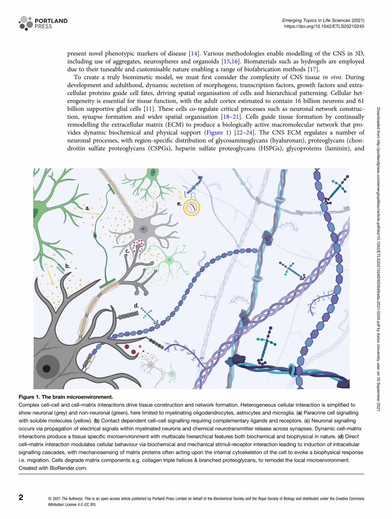
development and adulthood, dynamic secretion of morphogens, transcription factors, growth factors and extra-cellular proteins guide cell fates, driving spatial organisation of cells and hierarchical patterning. Cellular het-erogeneity is essential for tissue function, with the adult cortex estimated to contain 16 billion neurons and 61billion supportive glial cells [11]. These cells co-regulate critical processes such as neuronal network construc-tion, synapse formation and wider spatial organisation [18–21]. Cells guide tissue formation by continuallyremodelling the extracellular matrix (ECM) to produce a biologically active macromolecular network that pro-vides dynamic biochemical and physical support (Figure 1) [22–24]. The CNS ECM regulates a number ofneuronal processes, with region-specific distribution of glycosaminoglycans (hyaluronan), proteoglycans (chon-droitin sulfate proteoglycans (CSPGs), heparin sulfate proteoglycans (HSPGs), glycoproteins (laminin), and
Figure 1. The brain microenvironment.
Complex cell–cell and cell–matrix interactions drive tissue construction and network formation. Heterogeneous cellular interaction is simplified to
show neuronal (grey) and non-neuronal (green), here limited to myelinating oligodendrocytes, astrocytes and microglia. (a) Paracrine cell signalling
with soluble molecules (yellow). (b) Contact dependent cell–cell signalling requiring complementary ligands and receptors. (c) Neuronal signalling
occurs via propagation of electrical signals within myelinated neurons and chemical neurotransmitter release across synapses. Dynamic cell–matrix
interactions produce a tissue specific microenvironment with multiscale hierarchical features both biochemical and biophysical in nature. (d) Direct
cell–matrix interaction modulates cellular behaviour via biochemical and mechanical stimuli-receptor interaction leading to induction of intracellular
signalling cascades, with mechanosensing of matrix proteins often acting upon the internal cytoskeleton of the cell to evoke a biophysical response
i.e. migration. Cells degrade matrix components e.g. collagen triple helices & branched proteoglycans, to remodel the local microenvironment.
Created with BioRender.com.
© 2021 The Author(s). This is an open access article published by Portland Press Limited on behalf of the Biochemical Society and the Royal Society of Biology and distributed under the Creative Commons
Attribution License 4.0 (CC BY).
2
Emerging Topics in Life Sciences (2021)https://doi.org/10.1042/ETLS20210245
Dow
nloaded from http://portlandpress.com
/emergtoplifesci/article-pdf/doi/10.1042/ETLS20210245/920649/etls-2021-0245.pdf by Aston U
niversity user on 16 September 2021

fibrous proteins such collagen [23–25]. Notably, altered expression of matrix proteins such as CSPGs is asso-ciated with pathological states such as gliosis [26]. The ECM also contributes to mechanical properties such asstiffness, which is known to influence cellular behaviour [22,27]. Young’s elastic modulus of brain tissue is esti-mated to be ∼ 0.1 to 10 kilopascals (kPa), with precise measurement hindered by regional variation and a lackof standardised measurement parameters [28–30]. Cells detect mechanical cues through mechanosensing of thelocal microenvironment, causing changes in cell morphology and behaviour [31–33]. The concept of mechan-ical control of cell behaviour becomes increasingly relevant when we observe abnormal mechanical features (i.e.increased stiffness) in neurodevelopmental disorders, neurodegenerative disease and CNS injury [28,34,35].The dynamic reciprocity model dictates that continuous bidirectional cell–matrix interaction is imperative
for tissue development [26]; within the CNS, a myriad of biochemical and mechanical cues from the ECMinfluence individual cell fates and therefore tissue architecture, circuit formation and ultimately network func-tion (Figure 1). The dynamic and heterogeneous nature of living tissue is particularly pertinent when we con-sider the human CNS, where billions of cells are in communication to drive not only conscious behaviour, butalso micro-scale changes in tissue architecture and composition. Further investigation of such guidance cueswithin native tissue is necessary in order to translate findings into advanced tissue-engineered models withsuperior biological relevance.
In vitro modellingModels composed of primary cells from humans are limited due to lack of available tissue. Primary tissueobtained from animals can improve biological relevance by containing many of the elements found in vivo;however viability is short-term and models lack adequate species specificity for robust testing [3,5].Immortalised cell lines, such as human neuroblastoma SH-SY5Y, are cost-effective alternatives that provide pre-liminary insight into responses of single cells. However, such immortal lines are unsuitable for tissue modelling,often failing to display ‘normal’ cell behaviour when compared with in vivo equivalents and ex vivo primarycells. IPSC technology addresses many of these limitations, with pluripotent cells capable of generating a rangeof CNS cells of both healthy and diseased origin [36–38]. This is essential for creation of heterogeneous modelscomprised of both neurons and supportive cell types, with cellular maturity and network function dependenton neuron-glial interactions [19]. IPSC-derived Neural Progenitor Cells (NPCs) present an ideal source of cellsfor incorporation into model systems, however their capacity for differentiation is somewhat limited to neuralsubtypes. Alternatively, direct reprogramming of somatic cells can provide a source of induced neurons whilemaintaining features of ageing, where iPSC technology produces rejuvenated cells lacking such features [39].Genome editing enables creation of sophisticated humanised in vitro models, with the capability for persona-lised or disease-specific genotypes [40,41]. It is vital to select an appropriate cell source to suit the functionalneeds of the model and ensure outputs are translatable to in vivo conditions. However, identification and inclu-sion of every cellular component of the CNS is hampered by the complexity and limited availability of thispresently inimitable tissue. Ultimately researchers recognise the need for cellular heterogeneity within in vitromodels, requiring utilisation of pluripotent cell types and co-culture systems in order to create relevant models,with particular focus given to the necessity of vascular and immunomodulatory cells [18,19,40].
OrganoidsiPSC organoids are routinely used to recreate the biophysical spatial environment of the CNS, enabling physio-logically relevant culture conditions for both neuronal and glial cell types [42–44]. Cerebral organoids surpassexisting methods in terms of 3D cellular layering, structural folding, and network activity [14,42,43,45]. Thisapproach is used to model neurological diseases such as Alzheimers and glioblastoma [37,42,46,47]. Sadly,organoid protocols face issues with complexity and reproducibility. While homogenous cultures are less bio-logically relevant, increasing complexity and heterogeneity is associated with poor reproducibility [18,40,48,49].Methods to reduce variability rely on bioengineering of organoids and guiding cell fates via small molecules,genome editing, scaffolds, micropatterning, microfabrication techniques and organoid fusion [15,16,40,44,48,50].Homogeny of organoids is often lost over time with differentiation and uncontrolled morphogenesis.
Lancaster et al. [16], used small molecules to guide organoid culture and control neural cell fates [16,42,51].Introduction of vascular components though the addition of angiogenic factors such as VEGF and Wnt ligandshas been demonstrated [48,52]. Genetic engineering using CRISPR–Cas9 has enabled introduction of diseasephenotypes and induction of signalling centres for morphogenetic patterning, with the potential for
© 2021 The Author(s). This is an open access article published by Portland Press Limited on behalf of the Biochemical Society and the Royal Society of Biology and distributed under the Creative Commons
Attribution License 4.0 (CC BY).
3
Emerging Topics in Life Sciences (2021)https://doi.org/10.1042/ETLS20210245
Dow
nloaded from http://portlandpress.com
/emergtoplifesci/article-pdf/doi/10.1042/ETLS20210245/920649/etls-2021-0245.pdf by Aston U
niversity user on 16 September 2021
stimuli-responsive changes in expression [43,47,48,53]. Use of hydrogel scaffolds to guide organoid develop-ment and expansion is well established; however, there is a clear need for fine-tuning of characteristics to suitorganoid function e.g. softer hydrogels promote neurite outgrowth and maturation [54]. Fabrication of engi-neered microwells and micropatterning of culture vessels is an alternative method for guiding tissue morph-ology, with geometric confinement shown to influence cellular aggregation and organoid formation without theneed for hydrogel encapsulation [55,56]. Incorporation of organoids into bioreactors can improve distributionof nutrients and increase organoid size [57]. Furthermore, microscale bioreactors approach have enabled tightcontrol over organoid microenvironment and integrated analysis [48,52,58]. Alternatively Paşca et al., combinedcortical spheroids to form heterogeneous assembloid structures, thus improving model validity [15,59]. Thisapproach shows great promise for incorporation of vasculature within organoids, improving nutrient distribu-tion and promoting tissue maturity [15,49,60]. Unfortunately, the self-organising nature of organoids meansvascular components are often disorganised and incomplete, however recent advances in neurovascular modelsprovide great promise [61]. Organoids offer both cellular and structural complexity necessary for modellinghuman tissues in vitro; however, tissue engineering approaches including microfabrication and biomaterials arerequired to guide organoid morphology and architecture, with advancement enabling improved 3D culturewith reduced variability.
BiomaterialsPlastic 2D vessels for cell culture are several orders of magnitude stiffer than tissues in vivo[62], which hasbeen shown to induce cellular stress, with abnormal inflammatory morphologies of astrocytes and microgliawhen compared with 3D in vivo counterparts [63]. Unfortunately, development of biomaterials for CNS model-ling is hindered by the fact that soft tissues display unusual mechanics; the brain does not behave as a liquid orsolid, rather has viscoelastic properties commonly seen in highly hydrated tissues comprised of heterogeneouspolymer networks [64]. Structural and compositional heterogeneity, alongside hierarchical patterning, isresponsible for guiding cellular processes and dynamic non-linear mechanical behaviour of living tissue[32,65]. Hydrogels are an ideal biomaterial for CNS modelling due to their high water content and porousstructure enabling diffusion of metabolites, with the solid-phase polymer network providing relevant mechan-ical and spatial cues, tuneable mechanics and versatile chemical modification [17,34,65–67].
HydrogelsCommon polymers include both biologically active natural (collagen, hyaluronic acid), and biologically inertsynthetic polymers (polyethylene glycol, polyurethane) [17,65,67–70]. Chemical functionalisation permits tai-loring of cross-linking kinetics and material mechanics, but also enables improved bioactivity of synthetic poly-mers by promoting cell-material interactions [71]. Functionalisation can occur by direct binding or indirectconjugation of binding domains, often via ‘click chemistry’ [66,71–73]. Integration of cell adhesion proteins orsmall peptides (Laminin/IKVAV, fibronectin/RGD), have been shown to improve attachment and survival inneuronal tissue engineering [70,74–78]. Incorporation of ECM proteins and binding peptides provides bothstructural and functional support to cells, acting to sequester additional cell-secreted ECM components andthus better recreate the native cellular environment [75,79]. MatrigelTM [80] is a commercially available hydro-gel scaffold containing a variety of ECM proteins and small molecules to promote cell viability and functional-ity, however there is considerable batch variation [55]. Alternatively, decellularised tissue provides almost allthe components of the CNS ECM with added potential of retaining vascular structures, but again this approachis plagued with high variation [26,66]. Cellular processes such as migration or differentiation can also be con-trolled via inclusion of small molecules or growth factors, via conjugation or controlled release systems[17,67,73,81,82]. While natural polymers provide essential biological activity, synthetic polymers offer uniqueopportunities for hydrogel modification to address specific experimental questions, extending beyond biochem-ical functionalisation to promoting electrical conductivity and customisable cross-linking.
Tuneable mechanicsHydrogels are readily engineered for improved biocompatibility and bioactivity, however physical and mechan-ical properties are equally as important for recapitulating the natural tissue environment and guiding cell fates.The type of polymer, molecular weight and concentration, as well as the degree of cross-linking (influenced bymode of gelation, concentration of cross-linker, sequential rounds, etc.), influence bulk mechanical properties
© 2021 The Author(s). This is an open access article published by Portland Press Limited on behalf of the Biochemical Society and the Royal Society of Biology and distributed under the Creative Commons
Attribution License 4.0 (CC BY).
4
Emerging Topics in Life Sciences (2021)https://doi.org/10.1042/ETLS20210245
Dow
nloaded from http://portlandpress.com
/emergtoplifesci/article-pdf/doi/10.1042/ETLS20210245/920649/etls-2021-0245.pdf by Aston U
niversity user on 16 September 2021

such as stiffness and porosity. Homogenous tuning of scaffold stiffness is a means to promote desired pheno-types [35]; soft (∼1 kPa) hydrogel substrates promote neurogenesis, whereas differentiation of glial cells isfavoured on materials with an elastic modulus ∼1–10 kPa [22,27]. Modulation of scaffold stiffness is also sug-gested as a means to manipulate the secretome of encapsulated cells, further supporting the concept of mech-anical control of cell fates [83]. Generation of mechanogradients is suggested as an additional means toinfluence cell behaviour [84,85]. Xin et al. [85] employed microfluidics for reliable production of continuousmechanogradients, revealing critical stiffness thresholds for processes such as cell spreading.Hybrid hydrogels, comprised of blended polymers, enables even greater tailoring, i.e. altered mixing ratios to
modulate stiffness, stress-relaxation, or biofunctionality [54,72,86]. Moxon et al. [86] demonstrate that inclusionof collagen fibrils within alginate hydrogels acts dually to support neuronal culture by increasing stiffnesswhilst also improving bioactivity. Hybrid hydrogels have become increasingly attractive when considering thelimited biocompatibility of conductive polymers [68,87,88]. Inclusion of conductive elements is important asinhibition of electrical signalling by biomaterials can impede nervous tissue function [88]. Stress-relaxation,whereby internal stress force reduces over time as the material settles under constant strain, is an importantfeature of a polymer network, with relaxation shown to influence cell spreading independently of stiffness [31].Porosity is also an important consideration when guiding cellular migration and neurite outgrowth, althoughrandom interconnectedness does not guide neuronal network formation and the relevance of pore size andshape is still not fully understood [22,89,90]. Modulation of stiffness, porosity, stress relaxation and degradationis invaluable when guiding cell behaviour and tissue functionality [65,71]. It is vital to recognise that such fea-tures are interconnected and often reliant upon one another; Engler et al. sparked debate when first reportingstiffness mediated differentiation, overlooking interplay of other features such as porosity that effects proteintethering [29,30].Environmental cues within hydrogels for CNS modelling include not only the presence of biochemical mole-
cules (growth factors, proteins, small molecules) and bulk material properties (stiffness, conductivity, porosity),but also more complex heterogeneous aspects such as multicomponent structuring, dynamic or stimuli-respon-sive properties, and multiscale spatiotemporal topographical patterning to modulate cell behaviour. To sum-marise, exploiting biomaterial mechanics is a powerful tool for controlling cell fates and researchers must lookto understand interconnectedness of mechanical features, in order to capitalise on desirable synergistic effects.
Dynamic materialsDynamic heterogeneous hydrogels requires increased complexity and multiple components to enhance biophys-ical functionality of the material. Hybrid materials address limitations of homogenous hydrogel scaffolds,improving the range of applications [54,72]. Hybrid hydrogels extend beyond mixing of polymers and cross-linking approaches, to inclusion of multiple materials such as fibrillary, granular, crystalline and particulatecomponents. Granular features have shown to support infiltration of cells and vascularisation, a vital consider-ation when we consider the CNS to be a highly vascularised tissue [17]. Conductivity is another important con-sideration for CNS biomaterials. Inclusion of carbon components (crystals, nanotubes, wires, sheets, nanoclays)improves conductivity and promotes network functionality [68,71,87,101,102], and has been shown to regulatecellular differentiation and network stabilisation [26,45]. Inclusion of conductive components also enables inte-grated analysis of neuronal network activity [58]. Several studies have employed a combination of concurrentbiophysical features such as electrical and mechanical stimulation, to produce dynamic culture systems capableof promoting neural alignment and neurite extension [88,103]. Stimuli-responsive hydrogels respond to chem-ical or physical stimuli, including light, magnetic/electric fields, shear forces, temperature, pH, ions, chemicals,drugs, enzymes etc. [67]. Through modification of stimuli, it is possible to fine-tune mechanical propertiessuch as stiffness, swelling, gelation and degradation kinetics. The type of stimuli and response utilised is highlydependent upon the desired application; however, temperature and pH are highly investigated, as these stimulipossess the greatest biological relevance [67]. Dynamic ‘smart’ biomaterials also provide spatiotemporal controlover delivery of biochemical cues, with chemically and enzymatically degradable cross-links enabling a range ofapplications [70,72,81]. Alternatively, drug-releasing agents such as nanoparticles or gel droplets can beincluded within hydrogel formulations to function as controlled release systems [81]. Multiphase releasesystems are needed, to ensure sequential and spatiotemporal delivery of biochemical cues similar to naturallyoccurring tissues in vivo [82]. Ultimately, development of hydrogels with a combination of multiphase,dynamic and stimuli-responsive properties is the ideal approach to modelling complex living tissues in vitro [103].
© 2021 The Author(s). This is an open access article published by Portland Press Limited on behalf of the Biochemical Society and the Royal Society of Biology and distributed under the Creative Commons
Attribution License 4.0 (CC BY).
5
Emerging Topics in Life Sciences (2021)https://doi.org/10.1042/ETLS20210245
Dow
nloaded from http://portlandpress.com
/emergtoplifesci/article-pdf/doi/10.1042/ETLS20210245/920649/etls-2021-0245.pdf by Aston U
niversity user on 16 September 2021
BioprintingHydrogel biomaterials can also be equipped with novel shear-thinning behaviour by utilising small self-assembling peptides to form supramolecular gels or modifying processing conditions to form fluid gels [91].Use of supramolecular hydrogels is growing due to reversible cross-linking and potential for customisation.However complex processing and mechanical characterisation techniques has limited use within biologicalresearch [71,92]. Fluid gels display similar self-healing properties following displacement, lending both materi-als to injectable systems, such as bioprinting [93]. Bioprinting is a bottom-up approach that enables geometric-ally controlled assembly of complex 3D structures, with inclusion of functional elements producing scaffoldsthat encompass mechanically and biologically relevant cues [69,94]. Bioinks for neural bioprinting are continu-ally evolving, exploiting iPSC technology and an increased understanding of biochemical features of the CNS,to create specialised formulations capable of recapitulating a myriad of environmental cues [71,95,96]. Fluidgels show particular promise as bioinks, protecting cells from shear forces during the printing process thatwould otherwise reduce viability [69,93,97]. 3D bioprinting of hydrogels allows reliable production of spatiallydefined macrostructures, including vascular components, with the use of sacrificial inks suggested as a meansto create complex vascular networks [70,98,99]. Microfluidics present an alternative for creation of perfusableculture systems to mimic vasculature within 3D in vitro models of the CNS [52]. Bioprinting is also suggestedas a means to create structured constructs, an important macroscale feature for in vitro models of the CNSwhen we consider layering seen in the cortex in vivo [78]. Unfortunately, application of bioprinting is ham-pered by problematic low viscosity of bioinks required for mimicking mechanical properties of the CNS, neces-sitating a compromise on biocompatibility to ensure printability or the use of a secondary support phase[71,93,100]. Furthermore, printed constructs are often limited in size and resolution, with print nozzles oftenpossessing dimensions into the hundreds of microns. There is a clear need to develop bioprinting technology,by refining bioink formulations and exploring novel methods of printing, to achieve the high resolution neces-sary for recreating micron-scale cytoarchitecture found in the CNS.
Topographical patterningBiochemical and structural patterning of hydrogel scaffolds across multiple axes provides micro to nano scalecontrol over surface topography, guiding cellular processes and ultimately tissue formation [22,32]. Physicalpatterning of cell substrates is a powerful tool for influencing cell behaviour, with microscale variation insubstrate rigidity and size of patterns implicated in cell lineage commitment [56,104]. Patterning of soft bio-materials is troublesome due to incompatible mechanical properties, often requiring modified lithographicapproaches that are often limited in resolution. Light-based approaches, such as two-photon polymerisation,allow better resolution on the micro to nanoscale; however often require transparent materials [105–107].Photopatterning is also a well-established method of patterning biologically active growth factors, proteinsand peptides into hydrogel scaffolds [99,108]. Microfluidics enable creation of devices with microchannelsand segregated compartments [54,61]; as well as enabling tight control over microscale features via precisebiochemical patterning of organoids and hydrogel scaffolds [48,99]. Lithographic patterning and chemicalmodification has been used to support formation of segregated cell populations and microchannels within amicrofluidic device to study network connectivity [109], whilst fabrication techniques such as electrospinningof fibrous scaffolds or structural patterning of parallel grooves has shown to mimic in vivo ECM topographiesto induce axis alignment [32,68,110]. Traditional methods of incorporating biochemical or mechanical cuestake an ‘all or nothing’ approach, failing to account for interconnectedness of variables and synergy of cuessuch as microtopographies and biochemical gradients [111,112]. Researchers are beginning to recognise thepower of combining various approaches to create highly versatile and dynamic culture systems [54,61,108].Such advanced systems are capable of recreating some of the compositional and architectural complexity ofin vivo tissues, whilst also providing insight into the power of interconnected environmental cues on cellbehaviour.It is extremely difficult to achieve all of the architectural, compositional and biological features necessary for
valid and biologically relevant in vitro culture. A tissue engineering approach, utilising innovative techniquesfor structural patterning of hydrogels alongside bioconjugation and controlled delivery systems, holds the mostpromise for producing advanced biomaterials with the capability for dynamic spatiotemporal guidance of tissueformation [65,73,81,99]. However, it is vital to carefully consider the fabrication approach to find a balancebetween heterogeneity, functionality and practicality of the model.
© 2021 The Author(s). This is an open access article published by Portland Press Limited on behalf of the Biochemical Society and the Royal Society of Biology and distributed under the Creative Commons
Attribution License 4.0 (CC BY).
6
Emerging Topics in Life Sciences (2021)https://doi.org/10.1042/ETLS20210245
Dow
nloaded from http://portlandpress.com
/emergtoplifesci/article-pdf/doi/10.1042/ETLS20210245/920649/etls-2021-0245.pdf by Aston U
niversity user on 16 September 2021

Functional interrogationNeuronal models need to address fundamental experimental questions as accurately as possible, with specialconsideration given to the impact of materials on cell function, neuronal network architecture and ability tomonitor cellular growth and activity in real time, providing insight into tissue maturity and physiological rele-vance, yet this is lacking within CNS models [61]. Failure to account for interrogative methods during bioma-terial development may lead to interference with functional measurements [87]. Microfluidic devices haveproven to be particularly useful to enable the use of methods such as calcium imaging or measurement of elec-trical activity via Multi-Electrode Arrays [113,114]. However, the maturation of stem cell derived neuronal cul-tures and methods to enable functional interrogation in 3D may hamper attempts to fully utilise these modelsystems and will require significant optimisation.
Discussion and conclusionA multidisciplinary approach, integrating both biochemical and physical cues across multiple length scales isnecessary to mimic human CNS tissue. Many approaches fail to account for the dynamic interconnectednature of biological and physical cues, with insufficient consideration given to complex structure-function rela-tionships observed within the CNS in vivo. Advanced culture systems enable creation of superior in vitromodels by combining various tissue-engineering techniques to provide reliable spatiotemporal delivery of het-erogeneous, multiscale environmental cues. Unfortunately, limited understanding of how CNS cells constructand modulate elaborate microenvironments within the CNS in vivo has limited in vitro translation. Furtherchallenges relate to researchers often lacking the interdisciplinary knowledge, skillset and technology requiredfor creation and interrogation of models with suitable biochemical and mechanical features. Nevertheless, con-tinual advancement in the field of tissue engineering and increased multidisciplinary collaboration will acceler-ate the development of in vitro models of the CNS.
Summary• Engineering the mechanical properties of biomaterials is a lesser understood but powerful tool
for guiding cell fates.
• Inclusion of multiscale, heterogeneous environmental cues is necessary for recreating thehierarchical structure of living tissues in vitro.
• Hydrogels provide a versatile and tuneable alternative to traditional culture materials.
Competing InterestsThe authors declare that there are no competing interests associated with the manuscript.
Open AccessOpen access for this article was enabled by the participation of Aston University in an all-inclusive Read &Publish pilot with Portland Press and the Biochemical Society under a transformative agreement with JISC.
Author ContributionsE.J.H.: Conceptualization, resources, writing — original draft, writing — review and editing, supervision, fundingacquisition; D.C.B.: Writing — review and editing, supervision, funding acquisition; P.P.E.: Writing — review andediting, supervision; P.A.W.: Conceptualization, writing — original draft, writing — review and editing,visualization.
AcknowledgementsDr Hill acknowledges financial support from EPSRC-SFI Joint Centre for Doctoral Training in Engineered Tissuesfor Discovery, Industry and Medicine.
© 2021 The Author(s). This is an open access article published by Portland Press Limited on behalf of the Biochemical Society and the Royal Society of Biology and distributed under the Creative Commons
Attribution License 4.0 (CC BY).
7
Emerging Topics in Life Sciences (2021)https://doi.org/10.1042/ETLS20210245
Dow
nloaded from http://portlandpress.com
/emergtoplifesci/article-pdf/doi/10.1042/ETLS20210245/920649/etls-2021-0245.pdf by Aston U
niversity user on 16 September 2021
AbbreviationsCNS, central nervous system; CSPGs, chondroitin sulfate proteoglycans; ECM, extracellular matrix.
References1 Matthews, F.E., Stephan, B.C.M., Robinson, L., Jagger, C., Barnes, L.E., Arthur, A. et al. (2016) A two decade dementia incidence comparison from the
cognitive function and ageing studies I and II. Nat. Commun. 7, 11398 https://doi.org/10.1038/ncomms113982 Pankevich, D.E., Altevogt, B.M., Dunlop, J., Gage, F.H. and Hyman, S.E. (2014) Improving and accelerating drug development for nervous system
disorders. Neuron 84, 546–553 https://doi.org/10.1016/j.neuron.2014.10.0073 de Lange, E.C.M., van den Brink, W., Yamamoto, Y., de Witte, W.E.A. and Wong, Y.C. (2017) Novel CNS drug discovery and development approach:
model-based integration to predict neuro-pharmacokinetics and pharmacodynamics. Expert Opin. Drug Discov. 12, 1207–1218 https://doi.org/10.1080/17460441.2017.1380623
4 Medda, X., Mertens, L., Versweyveld, S., Diels, A., Barnham, L., Bretteville, A. et al. (2016) Development of a scalable, high-throughput-compatibleassay to detect tau aggregates using iPSC-derived cortical neurons maintained in a three-dimensional culture format. J. Biomol. Screen. 21, 804–815https://doi.org/10.1177/1087057116638029
5 Grainger, A.I., King, M.C., Nagel, D.A., Parri, H.R., Coleman, M.D. and Hill, E.J. (2018) In vitro models for seizure-liability testing using inducedpluripotent stem cells. Front. Neurosci. 12, 590 https://doi.org/10.3389/fnins.2018.00590
6 Clark, E.M., O’Donnell, J.C., Struzyna, L.A., Chen, H.I., Duda, J.E. and Cullen, D.K. (2020) Engineered microtissue as an anatomically inspired model ofParkinson’s disease. Curr. Opin. Biomed. Eng. 14, 75–83 https://doi.org/10.1016/j.cobme.2020.07.004
7 Communication from the commission on the European Citizen’s Initiative, ‘Stop Vivisection’ [press release]. Brussels 20158 Perry, C.J. and Lawrence, A.J. (2017) Hurdles in basic science translation. Front. Pharmacol. 8, 478 https://doi.org/10.3389/fphar.2017.004789 van Norman, G.A. (2019) Limitations of animal studies for predicting toxicity in clinical trials: is it time to rethink our current approach? JACC Basic
Transl. Sci. 4, 845–854 https://doi.org/10.1016/j.jacbts.2019.10.00810 Jang, K.-J., Otieno, M.A., Ronxhi, J., Lim, H.-K., Ewart, L., Kodella, K.R. et al. (2019) Reproducing human and cross-species drug toxicities using a
liver-Chip. Sci. Transl. Med. 11, eaax5516 https://doi.org/10.1126/scitranslmed.aax551611 Hodge, R.D., Bakken, T.E., Miller, J.A., Smith, K.A., Barkan, E.R., Graybuck, L.T. et al. (2019) Conserved cell types with divergent features in human
versus mouse cortex. Nature 573, 61–68 https://doi.org/10.1038/s41586-019-1506-712 Oberheim, N.A., Takano, T., Han, X., He, W., Lin, J.H.C., Wang, F. et al. (2009) Uniquely hominid features of adult human astrocytes. J. Neurosci. 29,
3276–3287 https://doi.org/10.1523/JNEUROSCI.4707-08.200913 Vagaska, B., Gillham, O. and Ferretti, P. (2020) Modelling human CNS injury with human neural stem cells in 2- and 3-dimensional cultures. Sci. Rep.
10, 6785 https://doi.org/10.1038/s41598-020-62906-y14 Fatehullah, A., Tan, S.H. and Barker, N. (2016) Organoids as an in vitro model of human development and disease. Nat. Cell Biol. 18, 246–254
https://doi.org/10.1038/ncb331215 Song, L., Yuan, X., Jones, Z., Griffin, K., Zhou, Y., Ma, T. et al. (2019) Assembly of human stem cell-derived cortical spheroids and vascular spheroids
to model 3-D brain-like tissues. Sci. Rep. 9, 5977 https://doi.org/10.1038/s41598-019-42439-916 Lancaster, M.A., Corsini, N.S., Wolfinger, S., Gustafson, E.H., Phillips, A.W., Burkard, T.R. et al. (2017) Guided self-organization and cortical plate
formation in human brain organoids. Nat. Biotechnol. 35, 659–666 https://doi.org/10.1038/nbt.390617 George, J., Hsu, C.-C., Nguyen, L.T.B., Ye, H. and Cui, Z. (2020) Neural tissue engineering with structured hydrogels in CNS models and therapies.
Biotechnol. Adv. 42, 107370 https://doi.org/10.1016/j.biotechadv.2019.03.00918 Fagerlund, I., Dougalis, A., Shakirzyanova, A., Gómez-Budia, M., Konttinen, H., Ohtonen, S. et al. (2020) Microglia orchestrate neuronal activity in brain
organoids. bioRxiv https://doi.org/10.1101/2020.12.08.41638819 Gilmour, A., Poole-Warren, L. and Green, R.A. (2019) An improved in vitro model of cortical tissue. Front. Neurosci. 13, 1349 https://doi.org/10.3389/
fnins.2019.0134920 Lee, J.-H., Kim, J.Y., Noh, S., Lee, H., Lee, S.Y., Mun, J.Y. et al. (2021) Astrocytes phagocytose adult hippocampal synapses for circuit homeostasis.
Nature 590, 612–617 https://doi.org/10.1038/s41586-020-03060-321 Molofsky, A.V., Kelley, K.W., Tsai, H.-H., Redmond, S.A., Chang, S.M., Madireddy, L. et al. (2014) Astrocyte-encoded positional cues maintain
sensorimotor circuit integrity. Nature 509, 189–194 https://doi.org/10.1038/nature1316122 Merryweather, D. and Roach, P. (2017) The need for advanced three-dimensional neural models and developing enabling technologies. MRS Commun.
7, 309–319 https://doi.org/10.1557/mrc.2017.5023 Miyata, S. and Kitagawa, H. (2017) Formation and remodeling of the brain extracellular matrix in neural plasticity: roles of chondroitin sulfate and
hyaluronan. Biochim. Biophys. Acta Gen. Subj. 1861, 2420–2434 https://doi.org/10.1016/j.bbagen.2017.06.01024 Rauti, R., Renous, N. and Maoz, B.M. (2020) Mimicking the brain extracellular matrix in vitro: a review of current methodologies and challenges. Israel
J. Chem. 60, 1141–1151 https://doi.org/10.1002/ijch.20190005225 Lam, D., Enright, H.A., Cadena, J., Peters, S.K.G., Sales, A.P., Osburn, J.J. et al. (2019) Tissue-specific extracellular matrix accelerates the formation of
neural networks and communities in a neuron-glia co-culture on a multi-electrode array. Sci. Rep. 9, 4159 https://doi.org/10.1038/s41598-019-40128-1
26 Sood, D., Cairns, D.M., Dabbi, J.M., Ramakrishnan, C., Deisseroth, K., Black, L.D. et al. (2019) Functional maturation of human neural stem cells in a3D bioengineered brain model enriched with fetal brain-derived matrix. Sci. Rep. 9, 17874 https://doi.org/10.1038/s41598-019-54248-1
27 Leipzig, N.D. and Shoichet, M.S. (2009) The effect of substrate stiffness on adult neural stem cell behavior. Biomaterials 30, 6867–6878 https://doi.org/10.1016/j.biomaterials.2009.09.002
28 Budday, S., Nay, R., de Rooij, R., Steinmann, P., Wyrobek, T., Ovaert, T.C. et al. (2015) Mechanical properties of gray and white matter brain tissue byindentation. J. Mech. Behav. Biomed. Mater. 46, 318–330 https://doi.org/10.1016/j.jmbbm.2015.02.024
29 Engler, A.J., Sen, S., Sweeney, H.L. and Discher, D.E. (2006) Matrix elasticity directs stem cell lineage specification. Cell 126, 677–689 https://doi.org/10.1016/j.cell.2006.06.044
© 2021 The Author(s). This is an open access article published by Portland Press Limited on behalf of the Biochemical Society and the Royal Society of Biology and distributed under the Creative Commons
Attribution License 4.0 (CC BY).
8
Emerging Topics in Life Sciences (2021)https://doi.org/10.1042/ETLS20210245
Dow
nloaded from http://portlandpress.com
/emergtoplifesci/article-pdf/doi/10.1042/ETLS20210245/920649/etls-2021-0245.pdf by Aston U
niversity user on 16 September 2021

30 Kim, H.N. and Choi, N. (2019) Consideration of the mechanical properties of hydrogels for brain tissue engineering and brain-on-a-chip. BioChip J. 13,8–19 https://doi.org/10.1007/s13206-018-3101-7
31 Chaudhuri, O., Gu, L., Klumpers, D., Darnell, M., Bencherif, S.A., Weaver, J.C. et al. (2016) Hydrogels with tunable stress relaxation regulate stem cellfate and activity. Nat. Mater. 15, 326–334 https://doi.org/10.1038/nmat4489
32 Coppari, S., Ramakrishna, S., Teodori, L. and Albertini, M.C. (2021) Cell signalling and biomaterials have a symbiotic relationship as demonstrated by abioinformatics study: the role of surface topography. Curr. Opin. Biomed. Eng. 17, 100246 https://doi.org/10.1016/j.cobme.2020.09.002
33 Hadden, W.J., Young, J.L., Holle, A.W., McFetridge, M.L., Kim, D.Y., Wijesinghe, P. et al. (2017) Stem cell migration and mechanotransduction on linearstiffness gradient hydrogels. Proc. Natl Acad. Sci. U.S.A. 114, 5647–5652 https://doi.org/10.1073/pnas.1618239114
34 Bartlett, R.D., Eleftheriadou, D., Evans, R., Choi, D. and Phillips, J.B. (2020) Mechanical properties of the spinal cord and brain: comparison withclinical-grade biomaterials for tissue engineering and regenerative medicine. Biomaterials 258, 120303 https://doi.org/10.1016/j.biomaterials.2020.120303
35 Baruffaldi, D., Palmara, G., Pirri, C. and Frascella, F. (2021) 3D cell culture: recent development in materials with tunable stiffness. ACS Appl. BioMater. 4, 2233–2250 https://doi.org/10.1021/acsabm.0c01472
36 Lee, K.M., Hawi, Z.H., Parkington, H.C., Parish, C.L., Kumar, P.V., Polo, J.M. et al. (2020) The application of human pluripotent stem cells to model theneuronal and glial components of neurodevelopmental disorders. Mol. Psychiatry 25, 368–378 https://doi.org/10.1038/s41380-019-0495-0
37 Nassor, F., Jarray, R., Biard, D.S.F., Maïza, A., Papy-Garcia, D., Pavoni, S. et al. (2020) Long term gene expression in human induced pluripotent stemcells and cerebral organoids to model a neurodegenerative disease. Front. Cell. Neurosci. 14, 14 https://doi.org/10.3389/fncel.2020.00014
38 Takahashi, K. and Yamanaka, S. (2006) Induction of pluripotent stem cells from mouse embryonic and adult fibroblast cultures by defined factors. Cell126, 663–676 https://doi.org/10.1016/j.cell.2006.07.024
39 Mertens, J., Reid, D., Lau, S., Kim, Y. and Gage, F.H. (2018) Aging in a dish: iPSC-derived and directly induced neurons for studying brain aging andage-related neurodegenerative diseases. Annu. Rev. Genet. 52, 271–293 https://doi.org/10.1146/annurev-genet-120417-031534
40 Kelava, I. and Lancaster Madeline, A. (2016) Stem cell models of human brain development. Cell Stem Cell 18, 736–748 https://doi.org/10.1016/j.stem.2016.05.022
41 Tan, H.Y., Cho, H. and Lee, L.P. (2021) Human mini-brain models. Nat. Biomed. Eng. 5, 11–25 https://doi.org/10.1038/s41551-020-00643-342 Lancaster, M.A., Renner, M., Martin, C.-A., Wenzel, D., Bicknell, L.S., Hurles, M.E. et al. (2013) Cerebral organoids model human brain development
and microcephaly. Nature 501, 373–379 https://doi.org/10.1038/nature1251743 Li, Y., Muffat, J., Omer, A., Bosch, I., Lancaster, M.A., Sur, M. et al. (2017) Induction of expansion and folding in human cerebral organoids. Cell Stem
Cell 20, 385–396.e3 https://doi.org/10.1016/j.stem.2016.11.01744 Velasco, S., Kedaigle, A.J., Simmons, S.K., Nash, A., Rocha, M., Quadrato, G. et al. (2019) Individual brain organoids reproducibly form cell diversity of
the human cerebral cortex. Nature 570, 523–527 https://doi.org/10.1038/s41586-019-1289-x45 Trujillo, C.A., Gao, R., Negraes, P.D., Gu, J., Buchanan, J., Preissl, S. et al. (2019) Complex oscillatory waves emerging from cortical organoids model
early human brain network development. Cell Stem Cell 25, 558–569.e7 https://doi.org/10.1016/j.stem.2019.08.00246 Gonzalez, C., Armijo, E., Bravo-Alegria, J., Becerra-Calixto, A., Mays, C.E. and Soto, C. (2018) Modeling amyloid beta and tau pathology in human
cerebral organoids. Mol. Psychiatry 23, 2363–2374 https://doi.org/10.1038/s41380-018-0229-847 Ogawa, J., Pao, G.M., Shokhirev, M.N. and Verma, I.M. (2018) Glioblastoma model using human cerebral organoids. Cell Rep. 23, 1220–1229
https://doi.org/10.1016/j.celrep.2018.03.10548 Fedorchak, N.J., Iyer, N. and Ashton, R.S. (2021) Bioengineering tissue morphogenesis and function in human neural organoids. Semin. Cell Dev. Biol.
111, 52–59 https://doi.org/10.1016/j.semcdb.2020.05.02549 Kim, J., Sullivan, G.J. and Park, I.-H. (2021) How well do brain organoids capture your brain? iScience 24, 102063 https://doi.org/10.1016/j.isci.2021.
10206350 Yoon, S.-J., Elahi, L.S., Pasca, A.M., Marton, R.M., Gordon, A., Revah, O. et al. (2019) Reliability of human cortical organoid generation. Nat. Methods
16, 75–78 https://doi.org/10.1038/s41592-018-0255-051 Lancaster, M.A. and Knoblich, J.A. (2014) Generation of cerebral organoids from human pluripotent stem cells. Nat. Protoc. 9, 2329–2340 https://doi.
org/10.1038/nprot.2014.15852 Linville, R.M., Arevalo, D., Maressa, J.C., Zhao, N. and Searson, P.C. (2020) Three-dimensional induced pluripotent stem-cell models of human brain
angiogenesis. Microvasc. Res. 132, 104042 https://doi.org/10.1016/j.mvr.2020.10404253 Mason, J.O. and Price, D.J. (2016) Building brains in a dish: prospects for growing cerebral organoids from stem cells. Neuroscience 334, 105–118
https://doi.org/10.1016/j.neuroscience.2016.07.04854 Liu, H., Wang, Y., Cui, K., Guo, Y., Zhang, X. and Qin, J. (2019) Advances in hydrogels in organoids and organs-on-a-chip. Adv. Mater. 31, e1902042
https://doi.org/10.1002/adma.20190204255 Chen, C., Rengarajan, V., Kjar, A. and Huang, Y. (2021) A matrigel-free method to generate matured human cerebral organoids using 3D-Printed
microwell arrays. Bioact. Mater. 6, 1130–1139 https://doi.org/10.1016/j.bioactmat.2020.10.00356 Knight, G.T., Lundin, B.F., Iyer, N., Ashton, L.M., Sethares, W.A., Willett, R.M. et al. (2018) Engineering induction of singular neural rosette emergence
within hPSC-derived tissues. eLife 7, e37549 https://doi.org/10.7554/eLife.3754957 Qian, X., Nguyen Ha, N., Song Mingxi, M., Hadiono, C., Ogden Sarah, C., Hammack, C. et al. (2016) Brain-region-specific organoids using
mini-bioreactors for modeling ZIKV exposure. Cell 165, 1238–1254 https://doi.org/10.1016/j.cell.2016.04.03258 Castiaux, A.D., Spence, D.M. and Martin, R.S. (2019) Review of 3D cell culture with analysis in microfluidic systems. Anal. Methods 11, 4220–4232
https://doi.org/10.1039/C9AY01328H59 Pasca, S.P. (2019) Assembling human brain organoids. Science 363, 126 https://doi.org/10.1126/science.aau572960 Pellegrini, L., Bonfio, C., Chadwick, J., Begum, F., Skehel, M. and Lancaster, M.A. (2020) Human CNS barrier-forming organoids with cerebrospinal
fluid production. Science (New York, NY) 369, eaaz5626 https://doi.org/10.1126/science.aaz562661 Fernandes, D.C., Reis, R.L. and Oliveira, J.M. (2021) Advances in 3D neural, vascular and neurovascular models for drug testing and regenerative
medicine. Drug Discov. Today 26, 754–768 https://doi.org/10.1016/j.drudis.2020.11.009
© 2021 The Author(s). This is an open access article published by Portland Press Limited on behalf of the Biochemical Society and the Royal Society of Biology and distributed under the Creative Commons
Attribution License 4.0 (CC BY).
9
Emerging Topics in Life Sciences (2021)https://doi.org/10.1042/ETLS20210245
Dow
nloaded from http://portlandpress.com
/emergtoplifesci/article-pdf/doi/10.1042/ETLS20210245/920649/etls-2021-0245.pdf by Aston U
niversity user on 16 September 2021

62 Sigma-Aldrich. CytoSoft® Elastic Modulus Plates: An Innovative Tool to Analyze the Effect of Matrix Stiffness/Rigidity on Regulating Cellular Behavior2021 [Available from: https://www.sigmaaldrich.com/technical-documents/articles/biology/cell-culture/cytosoft-elastic-modulus-plates.html
63 Watson, P.M.D., Kavanagh, E., Allenby, G. and Vassey, M. (2017) Bioengineered 3D glial cell culture systems and applications for neurodegenerationand neuroinflammation. SLAS Discov. 22, 583–601 https://doi.org/10.1177/2472555217691450
64 Libertiaux, V. and Pascon, F. (2009) Viscoelastic Modeling of Brain Tissue: A Fractional Calculus-Based Approach. In Mechanics of MicrostructuredSolids: Cellular Materials, Fibre Reinforced Solids and Soft Tissues (Ganghoffer, J.F. and Pastrone, F., eds), pp. 81–90, Springer Berlin Heidelberg,Berlin, Heidelberg
65 Eltom, A., Zhong, G. and Muhammad, A. (2019) Scaffold techniques and designs in tissue engineering functions and purposes: a review. Adv. Mater.Sci. Eng. 2019, 3429527 https://doi.org/10.1155/2019/3429527
66 Jensen, G., Morrill, C. and Huang, Y. (2018) 3D tissue engineering, an emerging technique for pharmaceutical research. Acta Pharm. Sin. B 8,756–766 https://doi.org/10.1016/j.apsb.2018.03.006
67 Mantha, S., Pillai, S., Khayambashi, P., Upadhyay, A., Zhang, Y., Tao, O. et al. (2019) Smart hydrogels in tissue engineering and regenerative medicine.Materials (Basel) 12, 3323 https://doi.org/10.3390/ma12203323
68 Boni, R., Ali, A., Shavandi, A. and Clarkson, A.N. (2018) Current and novel polymeric biomaterials for neural tissue engineering. J. Biomed. Sci. 25, 90https://doi.org/10.1186/s12929-018-0491-8
69 de la Vega, L., Lee, C., Sharma, R., Amereh, M. and Willerth, S.M. (2019) 3D bioprinting models of neural tissues: The current state of the field andfuture directions. Brain Res. Bull. 150, 240–249 https://doi.org/10.1016/j.brainresbull.2019.06.007
70 Fonseca, A.C., Melchels, F.P.W., Ferreira, M.J.S., Moxon, S.R., Potjewyd, G., Dargaville, T.R. et al. (2020) Emulating human tissues and organs: abioprinting perspective toward personalized medicine. Chem. Rev. 120, 11093–11139 https://doi.org/10.1021/acs.chemrev.0c00342
71 Chimene, D., Kaunas, R. and Gaharwar, A.K. (2020) Hydrogel bioink reinforcement for additive manufacturing: a focused review of emerging strategies.Adv. Mater. 32, 1902026 https://doi.org/10.1002/adma.201902026
72 Palmese, L.L., Thapa, R.K., Sullivan, M.O. and Kiick, K.L. (2019) Hybrid hydrogels for biomedical applications. Curr. Opin. Chem. Eng. 24, 143–157https://doi.org/10.1016/j.coche.2019.02.010
73 Willerth, S.M. (2017) Biomimetic strategies for replicating the neural stem cell niche. Curr. Opin. Chem. Eng. 15, 8–14 https://doi.org/10.1016/j.coche.2016.11.004
74 Barros, D., Conde-Sousa, E., Gonçalves, A.M., Han, W.M., García, A.J., Amaral, I.F. et al. (2019) Engineering hydrogels with affinity-bound laminin as3D neural stem cell culture systems. Biomater. Sci. 7, 5338–5349 https://doi.org/10.1039/C9BM00348G
75 Dobre, O., Azevedo Gonzalez Oliva, M., Ciccone, G., Trujillo, S., Rodrigo-Navarro, A., Venters, D. et al. (2021) A hydrogel platform that incorporateslaminin isoforms for efficient presentation of growth factors: neural growth and osteogenesis. Adv. Funct. Mater. 31, 2010225 https://doi.org/10.1002/adfm.202010225
76 Farrukh, A., Ortega, F., Fan, W., Marichal, N., Paez, J.I., Berninger, B. et al. (2017) Bifunctional hydrogels containing the laminin motif IKVAV promoteneurogenesis. Stem Cell Rep. 9, 1432–1440 https://doi.org/10.1016/j.stemcr.2017.09.002
77 Koivisto, J.T., Joki, T., Parraga, J.E., Pääkkönen, R., Ylä-Outinen, L., Salonen, L. et al. (2017) Bioamine-crosslinked gellan gum hydrogel for neuraltissue engineering. Biomed. Mater. 12, 025014 https://doi.org/10.1088/1748-605X/aa62b0
78 Lozano, R., Stevens, L., Thompson, B.C., Gilmore, K.J., Gorkin, R., Stewart, E.M. et al. (2015) 3D printing of layered brain-like structures using peptidemodified gellan gum substrates. Biomaterials 67, 264–273 https://doi.org/10.1016/j.biomaterials.2015.07.022
79 Tomaszewski, C.E., DiLillo, K.M., Baker, B.M., Arnold, K.B. and Shikanov, A. (2021) Sequestered cell-secreted extracellular matrix proteins improvemurine folliculogenesis and oocyte maturation for fertility preservation. Acta Biomater. Epub ahead of print https://doi.org/10.1016/j.actbio.2021.03.041
80 Corning. Matrigel Matrix 2020 [Available from: https://www.corning.com/worldwide/en/products/life-sciences/products/surfaces/matrigel-matrix.html#:∼:text=%20Use%20the%20online%20Corning%20Matrigel%20Lot%20Selection,to%20the%20previously%20requested%20lot%20number.%20More%20
81 He, W., Reaume, M., Hennenfent, M., Lee, B.P. and Rajachar, R. (2020) Biomimetic hydrogels with spatial- and temporal-controlled chemical cues fortissue engineering. Biomater. Sci. 8, 3248–3269 https://doi.org/10.1039/D0BM00263A
82 Wei, Z., Volkova, E., Blatchley, M.R. and Gerecht, S. (2019) Hydrogel vehicles for sequential delivery of protein drugs to promote vascular regeneration.Adv. Drug Deliv. Rev. 149–150, 95–106 https://doi.org/10.1016/j.addr.2019.08.005
83 Nasser, M. and Ghosh, G. (2018) Engineering Microenvironments to Regulate Mesenchymal Stem Cell Secretome. American Institute of ChemicalEngineers Annual Meeting; Wednesday, October 31,2018
84 Xia, T., Liu, W. and Yang, L. (2017) A review of gradient stiffness hydrogels used in tissue engineering and regenerative medicine. J. Biomed. Mater.Res. A 105, 1799–1812 https://doi.org/10.1002/jbm.a.36034
85 Xin, S., Dai, J., Gregory, C.A., Han, A. and Alge, D.L. (2019) Creating physicochemical gradients in modular microporous annealed particle hydrogels viaa microfluidic method. Adv. Funct. Mater. 30, 1907102 https://doi.org/10.1002/adfm.201907102
86 Moxon, S.R., Corbett, N.J., Fisher, K., Potjewyd, G., Domingos, M. and Hooper, N.M. (2019) Blended alginate/collagen hydrogels promote neurogenesisand neuronal maturation. Mater. Sci. Eng. C Mater. Biol. Appl. 104, 109904 https://doi.org/10.1016/j.msec.2019.109904
87 Lee, S., Ozlu, B., Eom, T., Martin, D.C. and Shim, B.S. (2020) Electrically conducting polymers for bio-interfacing electronics: From neural and cardiacinterfaces to bone and artificial tissue biomaterials. Biosens. Bioelectron. 170, 112620 https://doi.org/10.1016/j.bios.2020.112620
88 Magaz, A., Li, X., Gough, J.E. and Blaker, J.J. (2021) Graphene oxide and electroactive reduced graphene oxide-based composite fibrous scaffolds forengineering excitable nerve tissue. Mater. Sci. Eng. C Mater. Biol. Appl. 119, 111632 https://doi.org/10.1016/j.msec.2020.111632
89 George, J.H., Nagel, D., Waller, S., Hill, E., Parri, H.R., Coleman, M.D. et al. (2018) A closer look at neuron interaction with track-etched microporousmembranes. Sci. Rep. 8, 15552 https://doi.org/10.1038/s41598-018-33710-6
90 Broguiere, N., Husch, A., Palazzolo, G., Bradke, F., Madduri, S. and Zenobi-Wong, M. (2019) Macroporous hydrogels derived from aqueous dynamicphase separation. Biomaterials 200, 56–65 https://doi.org/10.1016/j.biomaterials.2019.01.047
91 Cooke, M.E., Jones, S.W., ter Horst, B., Moiemen, N., Snow, M., Chouhan, G. et al. (2018) Structuring of hydrogels across multiple length scales forbiomedical applications. Adv. Mater. 30, 1705013 https://doi.org/10.1002/adma.201705013
92 Chivers, P.R.A. and Smith, D.K. (2019) Shaping and structuring supramolecular gels. Nat. Rev. Mater. 4, 463–478 https://doi.org/10.1038/s41578-019-0111-6
© 2021 The Author(s). This is an open access article published by Portland Press Limited on behalf of the Biochemical Society and the Royal Society of Biology and distributed under the Creative Commons
Attribution License 4.0 (CC BY).
10
Emerging Topics in Life Sciences (2021)https://doi.org/10.1042/ETLS20210245
Dow
nloaded from http://portlandpress.com
/emergtoplifesci/article-pdf/doi/10.1042/ETLS20210245/920649/etls-2021-0245.pdf by Aston U
niversity user on 16 September 2021

93 Zandi, N., Sani, E.S., Mostafavi, E., Ibrahim, D.M., Saleh, B., Shokrgozar, M.A. et al. (2021) Nanoengineered shear-thinning and bioprintable hydrogelas a versatile platform for biomedical applications. Biomaterials 267, 120476 https://doi.org/10.1016/j.biomaterials.2020.120476
94 Ouyang, L., Armstrong, J.P.K., Salmeron-Sanchez, M. and Stevens, M.M. (2020) Assembling living building blocks to engineer complex tissues. Adv.Funct. Mater. 30, 1909009 https://doi.org/10.1002/adfm.201909009
95 Sokolovski, S.G., Crowe, J.A., Nagel, D., Hill, E.J., El-Tamer, A., Koroleva, A.V. et al. (2018) Printing brain in vitro at 3D scaffolds: materials andpatterns. 2018 International Conference Laser Optics (ICLO); 4-8 June 2018
96 Walus, K., Beyer, S. and Willerth, S.M. (2020) 3D bioprinting healthy and disease models of brain tissue using stem cells. Curr. Opin. Biomed. Eng. 14,25–33 https://doi.org/10.1016/j.cobme.2020.03.002
97 Chen, M.H., Wang, L.L., Chung, J.J., Kim, Y.-H., Atluri, P. and Burdick, J.A. (2017) Methods to assess shear-thinning hydrogels for application asinjectable biomaterials. ACS Biomater. Sci Eng. 3, 3146–3160 https://doi.org/10.1021/acsbiomaterials.7b00734
98 Eltaher, H.M., Abukunna, F.E., Ruiz-Cantu, L., Stone, Z., Yang, J. and Dixon, J.E. (2020) Human-scale tissues with patterned vascular networks byadditive manufacturing of sacrificial sugar-protein composites. Acta Biomater. 113, 339–349 https://doi.org/10.1016/j.actbio.2020.06.012
99 Primo, G.A. and Mata, A. (2021) 3D patterning within hydrogels for the recreation of functional biological environments. Adv. Funct. Mater. 31,2009574 https://doi.org/10.1002/adfm.202009574
100 Senior, J.J., Cooke, M.E., Grover, L.M. and Smith, A.M. (2019) Fabrication of complex hydrogel structures using suspended layer additive manufacturing(SLAM). Adv. Funct. Mater. 29, 1904845 https://doi.org/10.1002/adfm.201904845
101 Liu, J. (2018) Three-Dimensional Macroporous Nanoelectronics Scaffold Innervated Synthetic Tissue. In Biomimetics Through Nanoelectronics:Development of Three Dimensional Macroporous Nanoelectronics for Building Smart Materials, Cyborg Tissues and Injectable Biomedical Electronics(Liu, J., ed.), pp. 39–63, Springer International Publishing, Cham
102 Tiwari, S., Patil, R., Dubey, S.K. and Bahadur, P. (2020) Graphene nanosheets as reinforcement and cell-instructive material in soft tissue scaffolds.Adv. Colloid Interface Sci. 281, 102167 https://doi.org/10.1016/j.cis.2020.102167
103 Shahin-Shamsabadi, A. and Selvaganapathy, P.R. (2020) Tissue-in-a-Tube: three-dimensional in vitro tissue constructs with integrated multimodalenvironmental stimulation. Mater. Today Bio. 7, 100070 https://doi.org/10.1016/j.mtbio.2020.100070
104 Biggs, M.J.P., Fernandez, M., Thomas, D., Cooper, R., Palma, M., Liao, J. et al. (2017) The functional response of mesenchymal stem cells toelectron-beam patterned elastomeric surfaces presenting micrometer to nanoscale heterogeneous rigidity. Adv. Mater. 29, 10.1002/adma.201702119https://doi.org/10.1002/adma.201702119
105 Applegate, M.B., Coburn, J., Partlow, B.P., Moreau, J.E., Mondia, J.P., Marelli, B. et al. (2015) Laser-based three-dimensional multiscalemicropatterning of biocompatible hydrogels for customized tissue engineering scaffolds. Proc. Natl Acad. Sci. U.S.A. 112, 12052 https://doi.org/10.1073/pnas.1509405112
106 Liao, C., Wuethrich, A. and Trau, M. (2020) A material odyssey for 3D nano/microstructures: two photon polymerization based nanolithography inbioapplications. Appl. Mater. Today 19, 100635 https://doi.org/10.1016/j.apmt.2020.100635
107 You, S., Li, J., Zhu, W., Yu, C., Mei, D. and Chen, S. (2018) Nanoscale 3D printing of hydrogels for cellular tissue engineering. J. Mater. Chem. B 6,2187–2197 https://doi.org/10.1039/C8TB00301G
108 Yu, C., Miller, K.L., Schimelman, J., Wang, P., Zhu, W., Ma, X. et al. (2020) A sequential 3D bioprinting and orthogonal bioconjugation approach forprecision tissue engineering. Biomaterials 258, 120294 https://doi.org/10.1016/j.biomaterials.2020.120294
109 Kamudzandu, M., Köse-Dunn, M., Evans, M.G., Fricker, R.A. and Roach, P. (2019) A micro-fabricated in vitro complex neuronal circuit platform.Biomed. Phys. Eng. Express 5, 045016 https://doi.org/10.1088/2057-1976/ab2307
110 Roach, P., Parker, T., Gadegaard, N. and Alexander, M.R. (2013) A bio-inspired neural environment to control neurons comprising radial glia, substratechemistry and topography. Biomater. Sci. 1, 83–93 https://doi.org/10.1039/C2BM00060A
111 Kundu, A., Micholt, L., Friedrich, S., Rand, D.R., Bartic, C., Braeken, D. et al. (2013) Superimposed topographic and chemical cues synergistically guideneurite outgrowth. Lab Chip 13, 3070–3081 https://doi.org/10.1039/c3lc50174d
112 Mumford, T.R., Roth, L. and Bugaj, L.J. (2020) Reverse and forward engineering multicellular structures with optogenetics. Curr. Opin. Biomed. Eng.16, 61–71 https://doi.org/10.1016/j.cobme.2020.100250
113 Gladkov, A., Pigareva, Y., Kutyina, D., Kolpakov, V., Bukatin, A., Mukhina, I. et al. (2017) Design of cultured neuron networks in vitro with predefinedconnectivity using asymmetric microfluidic channels. Sci. Rep. 7, 15625 https://doi.org/10.1038/s41598-017-15506-2
114 Kane, K.I.W., Moreno, E.L., Hachi, S., Walter, M., Jarazo, J., Oliveira, M.A.P. et al. (2019) Automated microfluidic cell culture of stem cell deriveddopaminergic neurons. Sci. Rep. 9, 1796 https://doi.org/10.1038/s41598-018-34828-3
© 2021 The Author(s). This is an open access article published by Portland Press Limited on behalf of the Biochemical Society and the Royal Society of Biology and distributed under the Creative Commons
Attribution License 4.0 (CC BY).
11
Emerging Topics in Life Sciences (2021)https://doi.org/10.1042/ETLS20210245
Dow
nloaded from http://portlandpress.com
/emergtoplifesci/article-pdf/doi/10.1042/ETLS20210245/920649/etls-2021-0245.pdf by Aston U
niversity user on 16 September 2021