modeling cytoadhesion of plasmodium falciparum‐infected …biophys/pdf/febs2016lanzer.… · gesa...
TRANSCRIPT

Modeling cytoadhesion of Plasmodium falciparum-infected erythrocytes and leukocytes—common principlesand distinctive featuresGesa Helms1,*, Anil Kumar Dasanna2,3,*, Ulrich S. Schwarz2,3 and Michael Lanzer1
1 Department of Infectious Diseases, Heidelberg University, Germany
2 BioQuant, Heidelberg, Germany
3 Institute for Theoretical Physics, Heidelberg University, Germany
Correspondence
M. Lanzer, Department of Infectious
Diseases, Parasitology, Heidelberg
University, Im Neuenheimer Feld 324,
69120 Heidelberg, Germany
Fax: +49 6221 564643
Tel: +49 6221 567845
E-mail: [email protected]
and
U. Schwarz, Institute for Theoretical Physics,
Heidelberg University, Philosophenweg 19,
69120 Heidelberg, Germany
Fax: +49 6221 549331
Tel: +49 6221 549431
E-mail: [email protected]
*These authors contributed equally.
(Received 28 December 2015, revised 1
February 2016, accepted 7 February 2016)
doi:10.1002/1873-3468.12142
Edited by Wilhelm Just
Cytoadhesion of Plasmodium falciparum-infected erythrocytes to the
microvascular endothelial lining shares striking similarities to cytoadhesion of
leukocytes. In both cases, adhesins are presented in structures that raise them
above the cell surface. Another similarity is the enhancement of adhesion
under physical force (catch bonding). Here, we review recent advances in our
understanding of the molecular and biophysical mechanisms underlying
cytoadherence in both cellular systems. We describe how imaging, flow cham-
ber experiments, single-molecule measurements, and computational modeling
have been used to decipher the relevant processes. We conclude that although
the parasite seems to induce processes that resemble the cytoadherence of
leukocytes, the mechanics of erythrocytes is such that the resulting behavior
in shear flow is fundamentally different.
Keywords: catch bond; cytoadhesion; leukocyte; malaria; mesoscopic
model; modeling
A drop of blood (10 lL) contains an estimated 50 mil-
lion erythrocytes and approximately 5000 leukocytes,
among other cells. Both red and white blood cells are
easy to spot under the microscope because of their dif-
ference in color and shape. Erythrocytes are red and
are shaped in the form of a biconcave discoid, whereas
leukocytes are colorless and have a spherical appear-
ance. The distinct colors and shapes are a reflection of
their different biological functions. Erythrocytes are
part of the respiratory and circulatory system, carrying
oxygen in a complex with the red chromophore hemo-
globin from the lung to the periphery of the body.
Abbreviations
AFM, atomic force microscopic; CSA, chondroitin-4-sulfate; DPD, dissipative particle dynamics; FENE, finitely extensible nonlinear elastic;
GPCR, G-protein-coupled receptors; HEV, human endothelial venules; ICAM 1, intercellular adhesion molecule 1; KAHRP, knob-associated
histidine-rich protein; MPCD, multiparticle collision dynamics; PfEMP1, Plasmodium falciparum erythrocyte membrane protein 1; Pf, Plas-
modium falciparum; PSGL-1, P-selectin glycoprotein ligand-1; SEM, scanning electron microscopy; TSP, thrombosporin.
1FEBS Letters (2016) ª 2016 The Authors. FEBS Letters published by John Wiley & Sons Ltd on behalf of Federation of European Biochemical Societies.
This is an open access article under the terms of the Creative Commons Attribution-NonCommercial-NoDerivs License, which permits use and
distribution in any medium, provided the original work is properly cited, the use is non-commercial and no modifications or adaptations are made.

Leukocytes are cells of the immune system and are
involved in controlling infections and removing foreign
antigens.
Leukocytes usually move passively with the blood
flow. However, in response to an inflammatory stimu-
lus, they can roll along the microvascular endothelium
and, eventually, firmly adhere (Fig. 1) [1]. Leukocytes
then migrate over the endothelium until they extrava-
sate into the surrounding tissue, where they home in
on infections [2]. In comparison, erythrocytes do not
adhere to the endothelial lining under normal physio-
logical conditions. The exceptions are sickle cell ery-
throcytes that can adhere to postcapillary venule
endothelium, a condition that is associated with vaso-
occlusive pain [3]. Erythrocytes stay in circulation until
they are cleared by the reticuloendothelial system of
the spleen and liver after an average lifetime of
120 days.
The rheological properties of erythrocytes dramati-
cally change upon infection with the human malaria
parasite Plasmodium falciparum. Now erythrocytes
acquire adhesive properties mediated by parasite-
encoded adhesins that are presented on the host cell
surface in knob-like protrusions (Fig. 1) [4,5]. Strik-
ingly, the cytoadherence of parasitized erythrocytes
mimics the way leukocytes adhere (Table 1) [6].
Leukocytes are covered with hundreds of microvilli
that carry adhesins such as L-selectin on their tips [1].
These adhesins interact with receptors on the surface
of vascular endothelial cells, leading to cytoadhesive
events that phenotypically resemble the rolling and
firm adhesion displayed by P. falciparum-infected ery-
throcytes [1,6]. Thus, both infected erythrocytes and
leukocytes use localized and elevated adhesive domains
to adhere to vessel walls. Another similarity is the use
of tension-enhanced adhesion systems [7,8]. However,
in marked contrast to parasitized erythrocytes, cytoad-
herence of leukocytes is cooperative with the endothe-
lium and leads to extravasation rather than sustained
adhesion as is seen for P. falciparum-infected erythro-
cytes.
In this review, we discuss the similarities and differ-
ences between cytoadhesion in these two medically
important cellular systems. In Cytoadhesion: a molecu-
lar perspective, we will briefly introduce the molecular
players mediating cytoadhesion of P. falciparum-
infected erythrocytes and leukocytes to endothelial
cells. For a comprehensive review of the interacting
receptor–ligand pairs, we refer the interested reader to
specialized literature on this topic [1,5,9,10]. In Bio-
physics of adhesive bonds: slip or catch, we will dis-
cuss the phenomenon of tensile force-enhanced
cytoadhesion. Adhesive dynamics of white blood cells
and Adhesive dynamics of P. falciparum-infected ery-
throcytes will deal with the dynamic of cytoadhesion
and how mathematical modeling and simulation can
help us understand the mechano-physical and biophys-
ical principles underpinning cytoadhesion in flow. In
particular, we will explain the reason for placing adhe-
sins in structures elevated above the cell surface. We
close with a discussion of the similarities and differ-
ences between the two systems and an outlook on
open issues that should be addressed in the future.
Cytoadhesion: a molecularperspective
Pathology and cytoadhesion during a
P. falciparum infection
Malaria remains a leading cause of morbidity and
mortality in many developing countries. According to
Fig. 1. Schematic illustration of the
adhesive phenotypes displayed by
P. falciparum-infected erythrocytes and
leukocytes. Selected receptor and ligand
pairs are shown.
2 FEBS Letters (2016) ª 2016 The Authors. FEBS Letters published by John Wiley & Sons Ltd on behalf of Federation of European Biochemical Societies
Adhesion of leukocytes and infected erythrocytes G. Helms et al.

the world malaria report of 2014, there were 198 mil-
lion cases of malaria in 2013 and 584 000 deaths [11].
Of the five Plasmodium species that can cause malaria
in humans, P. falciparum is the most virulent and
deadly form, being responsible for most of the malar-
ia-associated deaths. P. falciparum is transmitted to
humans by the bite of an infected Anopheles mosquito.
Sporozoites released into the skin enter blood vessels
and are carried with the blood flow to the liver where
they invade hepatocytes. An infected hepatocyte can
give rise to more than 10 000 daughter cells, termed
merozoites, which are released into the blood stream
where they infect erythrocytes. After 48 h the infected
erythrocyte bursts, releasing typically 16 or 32 mero-
zoites that then again infect erythrocytes, thereby
establishing the intraerythrocytic life cycle. In extreme
cases, 40% of the approximately 2.6 9 1013 erythro-
cytes of an adult malaria patient can be infected by
parasites. The entire life cycle is completed when an
Anopheles mosquito takes up sexual stages during a
blood meal.
The asexual intraerythrocytic stages of P. falciparum
are responsible for much of the pathology associated
with malaria, whereas the liver stages are asymptomatic
[12]. The virulence of the asexual blood stages is largely
due to the fact that the parasite alters the rheological
and morphological properties of the infected erythro-
cyte. Uninfected erythrocytes move in the middle of the
blood vessel where the flow velocity is maximum, leav-
ing a rim around the vessel wall free of cells [13]. This
phenomenon can be explained by the Magnus effect.
As soon as a cell moves out of the central part of the
capillary the velocity gradient causes it to spin. The cell
will then feel a force perpendicular to the flow direction
and the rotational axis, which forces it back to the cen-
tral region [13]. P. falciparum-infected erythrocytes also
move with the blood flow but only for the first hours
post invasion. After 16–20 h post invasion, they
acquire adhesive properties and sequester in the deep
microvascular bed of inner organs, including lung and
brain [12]. By sequestering in the microvasculature,
parasitized erythrocytes avoid passage through the
spleen and, hence, elimination by splenic clearance
mechanisms that remove senescent and infected ery-
throcytes from circulation. Cytoadhesion is part of the
parasite’s strategy to colonize its host. However, it
causes severe disease [12]. The pathological sequelae
that develop in the affected blood vessels include
obstruction of microvascular blood flow, disturbed tis-
sue perfusion, hypoxia, and systemic microvascularitis
that can progress to life-threatening complications, as
presented by patients suffering from cerebral malaria
[12,14].
Cytoadhesion is mediated by a family of parasite-
encoded immuno-variant adhesins collectively called
Plasmodium falciparum erythrocyte membrane protein
1 (PfEMP1). PfEMP1 proteins can be classified into
different groups based on domain architecture and
functional specialization for interaction with particular
host receptors [10]. While the majority of the 60
Table 1. Comparison of cytoadhesion of leukocytes and P. falciparum-infected erythrocytes.
Leukocytes Pf-infected erythrocytes
Adhesive structures 100s of microvilli, each 300 nm high 10 000s of knobs, each 20 nm high
Mechanics of adhesive structure Viscoelastic, elongates under shear flow Compact and stiff
Cell size, shape, and mechanics Diameter 8–30 lm; spherical; flexible envelope Diameter 6.2–8.2 lm; varies between biconcave and
round, but not perfectly spherical; stiff envelope
Molecular system Mainly selectins, very specific ligands on HEV PfEMP-1, broad range of receptors specific to distinct
microvascular beds
On-set of adhesion Activated by chemokines, GPCR Developmentally controlled, commences 16 h post
invasion until end of replicative cycle
Interplay with endothelium Inside out signaling to recruit leukocytes Contact-dependent intracellular signaling; clustering and
activation of host adhesion receptors
Movement during cytoadhesion
under flow conditions
Rolling on HEV Sometimes rolling, but often irregular movement with
flipping, depending on life cycle
Biological purpose Preface to extravasation Evasion of splenic clearance
GPCR, G-protein-coupled receptors; HEV, human endothelial venules; Pf, Plasmodium falciparum.
3FEBS Letters (2016) ª 2016 The Authors. FEBS Letters published by John Wiley & Sons Ltd on behalf of Federation of European Biochemical Societies
G. Helms et al. Adhesion of leukocytes and infected erythrocytes
PfEMP1 variants encoded in the haploid P. falciparum
genome have unique binding properties, a few
PfEMP1 variants can interact with several host cell
receptors, for example, CD36 and ICAM-1 [10]. Col-
lectively, PfEMP1 proteins can interact with a broad
range of receptors, including CD36, intercellular adhe-
sion molecule 1 (ICAM 1), and P-selectin [4,5]. In
addition, a certain PfEMP1 variant can mediate
attachment of parasitized erythrocytes to chondroitin-
4-sulfate (CSA), a component of the proteoglycan
matrix present in the intervillous space and moderately
on the syncytiotrophoblast cell layer of the human pla-
centa [15–19]. Other parasite-encoded factors impli-
cated in cytoadhesive events include the RIFINS and
STEVOR immunovariant antigens that are thought to
mediate rosetting of infected erythrocytes with unin-
fected erythrocytes [20,21].
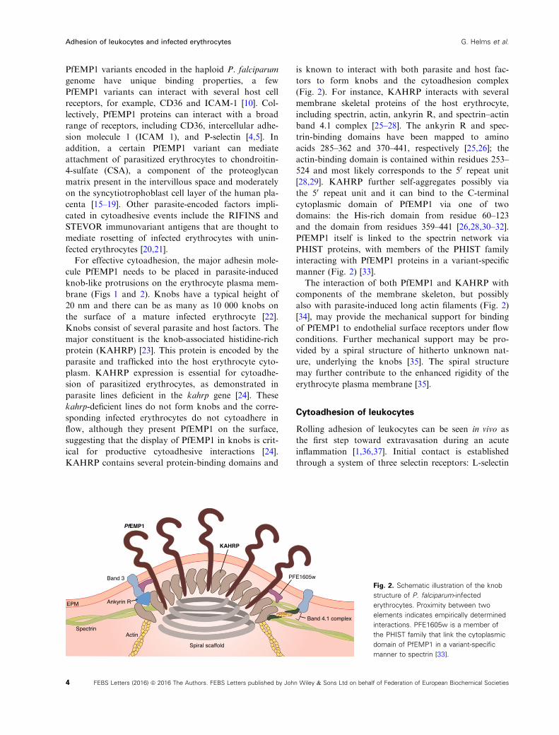
For effective cytoadhesion, the major adhesin mole-
cule PfEMP1 needs to be placed in parasite-induced
knob-like protrusions on the erythrocyte plasma mem-
brane (Figs 1 and 2). Knobs have a typical height of
20 nm and there can be as many as 10 000 knobs on
the surface of a mature infected erythrocyte [22].
Knobs consist of several parasite and host factors. The
major constituent is the knob-associated histidine-rich
protein (KAHRP) [23]. This protein is encoded by the
parasite and trafficked into the host erythrocyte cyto-
plasm. KAHRP expression is essential for cytoadhe-
sion of parasitized erythrocytes, as demonstrated in
parasite lines deficient in the kahrp gene [24]. These
kahrp-deficient lines do not form knobs and the corre-
sponding infected erythrocytes do not cytoadhere in
flow, although they present PfEMP1 on the surface,
suggesting that the display of PfEMP1 in knobs is crit-
ical for productive cytoadhesive interactions [24].
KAHRP contains several protein-binding domains and
is known to interact with both parasite and host fac-
tors to form knobs and the cytoadhesion complex
(Fig. 2). For instance, KAHRP interacts with several
membrane skeletal proteins of the host erythrocyte,
including spectrin, actin, ankyrin R, and spectrin–actinband 4.1 complex [25–28]. The ankyrin R and spec-
trin-binding domains have been mapped to amino
acids 285–362 and 370–441, respectively [25,26]; the
actin-binding domain is contained within residues 253–524 and most likely corresponds to the 50 repeat unit
[28,29]. KAHRP further self-aggregates possibly via
the 50 repeat unit and it can bind to the C-terminal
cytoplasmic domain of PfEMP1 via one of two
domains: the His-rich domain from residue 60–123and the domain from residues 359–441 [26,28,30–32].PfEMP1 itself is linked to the spectrin network via
PHIST proteins, with members of the PHIST family
interacting with PfEMP1 proteins in a variant-specific
manner (Fig. 2) [33].
The interaction of both PfEMP1 and KAHRP with
components of the membrane skeleton, but possibly
also with parasite-induced long actin filaments (Fig. 2)
[34], may provide the mechanical support for binding
of PfEMP1 to endothelial surface receptors under flow
conditions. Further mechanical support may be pro-
vided by a spiral structure of hitherto unknown nat-
ure, underlying the knobs [35]. The spiral structure
may further contribute to the enhanced rigidity of the
erythrocyte plasma membrane [35].
Cytoadhesion of leukocytes
Rolling adhesion of leukocytes can be seen in vivo as
the first step toward extravasation during an acute
inflammation [1,36,37]. Initial contact is established
through a system of three selectin receptors: L-selectin
Fig. 2. Schematic illustration of the knob
structure of P. falciparum-infected
erythrocytes. Proximity between two
elements indicates empirically determined
interactions. PFE1605w is a member of
the PHIST family that link the cytoplasmic
domain of PfEMP1 in a variant-specific
manner to spectrin [33].
4 FEBS Letters (2016) ª 2016 The Authors. FEBS Letters published by John Wiley & Sons Ltd on behalf of Federation of European Biochemical Societies
Adhesion of leukocytes and infected erythrocytes G. Helms et al.
is localized at the tips of the leukocyte microvilli and
binds to sulfated sialyl-Lewis X-like sugars (e.g.,
PNAd) in high endothelial venules of the endothelium.
P- and E-selectin are activated in inflamed endothe-
lium and bind to partners, such as P-selectin glycopro-
tein ligand-1 (PSGL-1), which are also localized at the
tips of the leukocyte microvilli (Fig. 1) [1,38]. Some
integrins, such as a4b7 and a4b1, also contribute to
rolling adhesion. One essential aspect of rolling is that
it exposes the leukocytes to chemoattractant signals,
such as the chemokine IL-8 that then activates G-pro-
tein-coupled receptors (GPCR) [36,37]. This, in turn,
leads to an inside-out activation of b2 or a4-integrinsthat establishes firm adhesion by binding to endothe-
lial immunoglobins. The most prominent integrins in
this context are LFA-1 (aLb2) or Mac-1 (aMb2) that
can bind to ICAM-1 or ICAM-2 presented on the sur-
face of endothelial cells (Fig. 1). There are hundreds
of microvilli on a leukocyte with a typical height of
300 nm. In marked contrast to the knobs on infected
erythrocytes, the microvilli of leukocytes can strongly
increase their length under force, due to visoelastic
flow in the membrane and the cytoskeleton [39]. Once
firmly attached, the leukocytes start to migrate along
the endothelium in order to induce the extravasation
process that usually proceeds in cooperativity with the
endothelial cells at cell–cell boundaries (paracellular
route). However, there is also a second mode of
extravasation, in which leukocytes simply cross
endothelial cells through their cell body (transcellular
route). Both processes require tight regulation of the
actin cytoskeleton [40]. While rolling adhesion can be
reconstituted easily in flow chambers, the study of
extravasation requires more complex coculture assays
or intravital microscopy.
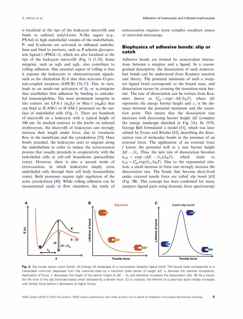
Biophysics of adhesive bonds: slip orcatch
Adhesive bonds are formed by noncovalent interac-
tions between a receptor and a ligand. In a coarse-
grained description, the dissociation of such noncova-
lent bonds can be understood from Kramers reaction
rate theory. The potential minimum of such a recep-
tor–ligand bond corresponds to the bound state, and
dissociation occurs by crossing the transition-state bar-
rier. The rate of dissociation can be written from Kra-
mers theory as k0off � expð�DE=kBTÞ, where DErepresents the energy barrier height and xc is the dis-
tance between the potential minimum and the transi-
tion point. This means that the dissociation rate
increases with decreasing barrier height DE (compare
the energy landscape sketched in Fig. 3A). In 1978,
George Bell formulated a model [41], which was later
refined by Evans and Ritchie [42], describing the disso-
ciation rate of molecular bonds in the presence of an
external force. The application of an external force
f lowers the potential well to a new barrier height
DE � fxc. Thus, the new rate of dissociation becomes
koff � exp(�(DE � fxc)/kBT), which leads to
koff ¼ k0off expðfxc=kBTÞ. Due to the exponential rela-
tion, a small increase in force can strongly increase the
dissociation rate. The bonds that become short-lived
under external tensile force are called slip bonds [43]
(Fig. 3B). This concept has been confirmed for many
receptor–ligand pairs using dynamic force spectroscopy
Slip bond Catch-slip bond
Lif
etim
e o
f b
on
d
Lif
etim
e o
f b
on
d
Tensile force Tensile force
A B
E (
x)
X
C
xc
ΔE - ƒxc
ΔE
E
E - ƒx
Fig. 3. Slip bonds versus catch bonds. (A) Energy (E) landscape of a noncovalent receptor–ligand bond. The bound state corresponds to a
metastable minimum separated from the unbound state by a transition state barrier of height DE. xc denotes the reactive compliance.
Application of force, f, decreases the height of the barrier height to DE � fxc and therefore increases the dissociation rate. (B) As a result,
the life time of the slip bond decreases when stressed by a tensile force. (C) In contrast, the lifetime of a catch-slip bond initially increases
with tensile force before it decreases at higher forces.
5FEBS Letters (2016) ª 2016 The Authors. FEBS Letters published by John Wiley & Sons Ltd on behalf of Federation of European Biochemical Societies
G. Helms et al. Adhesion of leukocytes and infected erythrocytes
[44]. In such experiments, the bonds are loaded by a
dynamic ramp of force (which is easier to control than
a constant force). From dynamic force spectroscopy
measurements, the most probable rupture forces were
shown to depend linearly on the logarithm of the load-
ing rate, as expected from the Kramers–Bell–Evansmodel and as initially validated for biotin–streptavidinand biotin–avidin bonds [44].
Interestingly, not all receptor–ligand pairs behave as
slip bonds. Some bonds are enhanced by tensile force
and are referred to as catch bonds [45,46]. The concept of
catch bonds was first introduced as a theoretical consid-
eration by Dembo et al. [43] and is explained by an
increase in the bond’s lifetime with increasing loading.
Given that every bond slips at very high forces, a pure
catch bond is not possible and one rather deals with a
catch-slip bond that is long-lived at intermediate forces
(Fig. 3C). Enhancement of adhesion in shear flow in the
case of bacteria [47,48] and in the case of leukocytes [49]
supports the possibility of catch bond behavior at the cel-
lular level. The first direct observation of catch-slip
bonds at the molecular level came from atomic force
microscopic (AFM) measurements in which Marshall
et al. [7] measured lifetimes of L-selectin/PSGL-1 bonds
under conditions of external loading. Off-rates of L-
selectin and PSGL-1 bonds were also estimated in flow
chamber experiments using L-selectin-coated micro-
spheres [50]. Off-rates were measured for two sizes of
microspheres in the presence and absence of ficoll, a
sugar that increases the viscosity of the medium and,
therefore, the hydrodynamic force. It was found that the
catch bond behavior of L-selectin and PSGL-1 bonds is
responsible for shear-enhanced adhesion. Catch bond
behavior is also observed in actomyosin bonds, with
maximum bond lifetime at around 6 pN below which the
bond lifetime decreases [51]. Other examples of catch
bond behavior include FimH-mediated adhesion of
E. coli bacteria [52] and the cell-matrix adhesion recep-
tor integrin a5b1 [53].Different biological systems might have evolved dif-
ferent molecular mechanisms for catch bonding. Also
different theoretical models have been suggested in this
context. For instance, the deformation model suggests
that application of an external force optimizes the rela-
tive position of the receptor and the ligand to one
another, which, in turn, is believed to increase the
binding strength [54]. Other models evoke allosteric
effects or sliding and rebinding of bonds when stressed
by a tensile force [55–59]. The two-pathway model
takes kinetic aspects into consideration, by proposing
that the molecular bond has two pathways for dissoci-
ation, one with a high-energy barrier and another one
with a low-energy barrier [60–63].
Adhesive dynamics of white bloodcells
Leukocyte rolling has been studied in vitro using flow
chamber experiments where rolling of leukocytes was
examined on culture dishes and artificial lipid bilayers
coated with appropriate ligands. It was found that the
dissociation rate of the P-selectin-glycoprotein ligand
bond increases with tensile force, indicative of a slip
bond, making the P-selectin–glycoprotein ligand inter-
action suitable for rolling adhesion [64,65]. In compar-
ison, L-selectin-mediated adhesion involves a shear
threshold, indicative of a catch bond, and only at high
tensile force does the bond behave like a slip bond
[50,66].
Catch bonds can be considered as hydrodynamic
sensors. Like sites of trauma, sites of inflammation are
characterized by vasodilation and increased shear flow.
The biological purpose of catch bonds, therefore,
seems to be to facilitate the adhesion of leukocytes
exactly where it is needed.
Modeling adhesion of white blood cells centers
around two key aspects: hydrodynamics and bond
kinetics, which together are referred to as adhesive
dynamics. The first to address this issue were Hammer
and Apte, who took a reductionist approach by
assuming that leukocytes are ideal spheres homoge-
nously covered with receptors and that the interacting
ligands are homogenously distributed on a planar sur-
face [67–69]. A more refined version of the model was
developed by Korn and Schwarz [70] who incorpo-
rated explicit positions for receptors and ligands. The
adhesion dynamics was simulated using a Langevin
equation:
otXðtÞ ¼ u1 þMfFD þ FSg þ kBTrMþ nðtÞwhere X(t) is a six-dimensional vector for both
position and orientation. Here, the generalized force,
F, includes both the force and the torque. The terms
FD and FS are direct and shear forces, respectively.
The direct forces include gravitational force and recep-
tor–ligand bond forces. The 6 9 6-dimensional mobil-
ity matrix M converts forces into velocities and u∞ is
the unperturbed flow velocity. The stochastic random
force, ξ(t), represents the thermal forces from the envi-
ronment, which are crucial to bring spatially resolved
receptors and ligands into binding range. The spurious
drift term with the spatial derivative ∇M ensures that
the stochastic dynamics satisfy the requirements of
equilibrium physics. In the case of a sphere in
unbounded flow, the mobility matrix becomes position
independent, eliminating the drift term, ∇M. The
ligand–receptor bond forms when the distance between
6 FEBS Letters (2016) ª 2016 The Authors. FEBS Letters published by John Wiley & Sons Ltd on behalf of Federation of European Biochemical Societies
Adhesion of leukocytes and infected erythrocytes G. Helms et al.

ligand and receptor is less than the capture radius,
r0, with a probability 1 � exp(�jonDt), where jon is
the on-rate. The dissociation of the bond is imple-
mented via the Bell equation and occurs with a proba-
bility of 1 � exp(�joffDt), where j0off is the unstressed
off-rate and joff ¼ j0offexpðF=FdÞ. Here, Fd is the
detachment force scale that can be estimated directly
from flow chamber experiments [71].
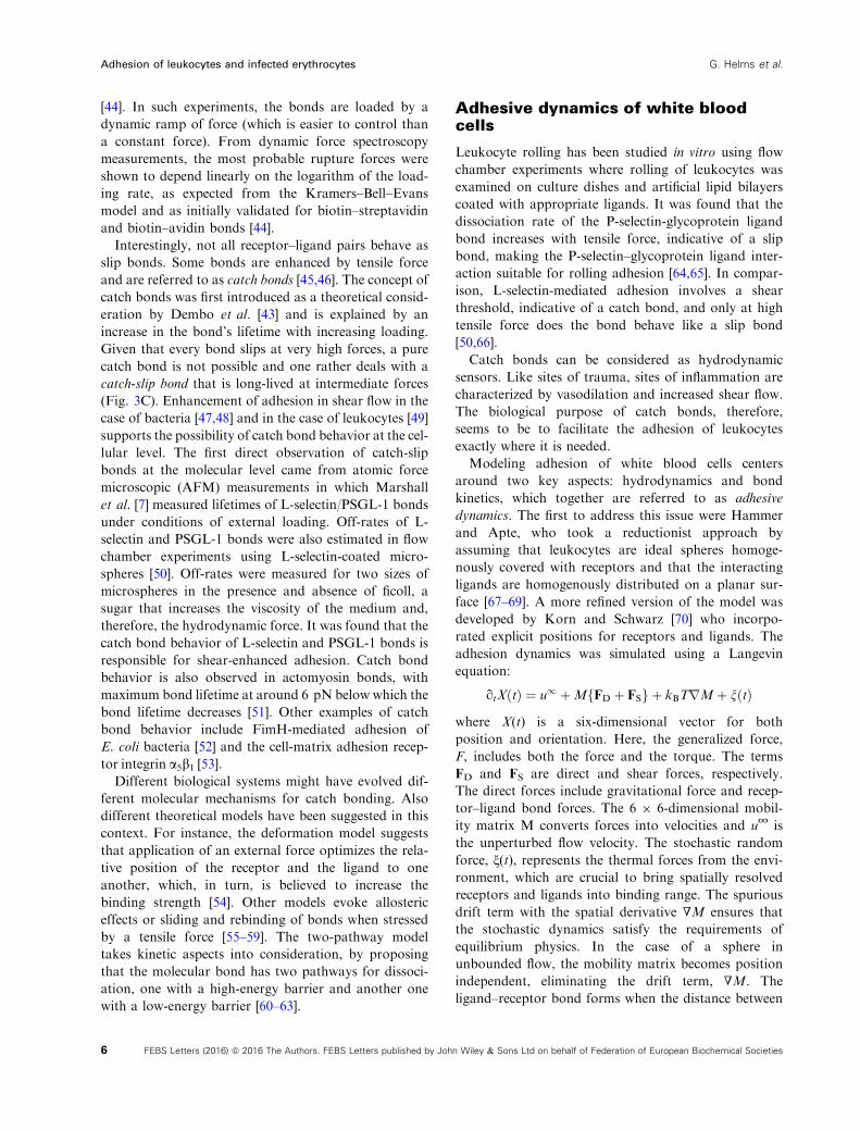
Adhesive dynamics is able to reproduce different
dynamical states, such as rolling adhesion, firm adhe-
sion, and transient adhesion. When systematically
varying the model parameters, the model allows phase
diagrams to be constructed, for example, as a function
of molecular association and dissociation rates [72]
(Fig. 4). The different phases correspond to different
characteristics of the individual cell trajectories. Dur-
ing free motion both translational and angular veloci-
ties change only in a smooth manner and no
interactions occur between the cell and the substrate.
Firm adhesion is achieved when the cell is firmly
attached to the substrate, thus translational and rota-
tional velocities are almost zero. During both transient
and rolling adhesion, the cell moves along the sub-
strate by constantly forming new bonds at the front
end and breaking the old bonds at the rear end. This
leads to strong variations in the velocities. Translation
and rotation are synchronized in the case of rolling
adhesion, while they are more variable and more
decoupled for transient adhesion. The on–off state dia-
gram depicted in Fig. 4 depends on many other
parameters, including the receptor–ligand kinetics, the
viscosity of the medium, and receptor and ligand den-
sity. For example, increases in viscosity leads to a
more stable rolling at the expense of firm adhesion,
leaving free motion and transient adhesion unchanged.
Particularly, the receptor and ligand density strongly
affects the cytoadherence efficiency [38,70]. Simulation
further revealed that the binding efficiency is signifi-
cantly increased if the receptors are elevated above the
cell surface [70]. The physical reason is that hydrody-
namic flow becomes slowed down toward the cell sur-
face. Protrusions are, therefore, exposed to larger
flows and can sample more environmental space than
the cell surface. This might explain why the adhesion
receptors are placed at the tip of microvilli in leuko-
cytes and why the adhesin PfEMP1 is presented in
knob-like protrusions on the surface of P. falciparum-
infected erythrocytes. However, leukocytes with their
microvilli of 300 nm in height seem to exploit this
principle to a larger extent than do infected erythro-
cytes where knobs are only a moderate 20 nm in
height. However, the large number of ten thousands of
knobs as opposed to hundreds of microvilli per cell
might compensate for the inferior height.
Adhesive dynamics has also been used to investigate
the importance of thermal fluctuations at both low and
high shear rates on adhesive behaviors [73]. Moreover,
adhesive dynamics can be applied to other systems, for
Firm adhesion
Transient adhesion
Rollingadhesion
Free motion
On rate
Off
rat
e
0
40
80
120
0
20
40
0
40
80
120
0
60
120
180
17.5 18 18.5 19 19.5 20
17.5 18 18.5 19 19.5 20
17.5 18 18.5 19 19.5 20
17.5 18 18.5 19 19.5 20
v(t)(t)
v(t)
,(t
) (µ
m·s
–1)
t(s)
t(s)
v(t)
,(t
) (µ
m·s
–1) v(t)
(t)
v(t)(t)
v(t)
,(t
) (µ
m·s
–1)
t(s)
v(t)
,(t
) (µ
m·s
–1)
v(t)(t)
t(s)
Fig. 4. Phase diagram of adhesive dynamics with representative trajectories. Computer simulations can be used to predict how adhesive
cells, such as leukocytes or infected red blood cells, move in hydrodynamic shear flow above a wall [69,70]. As a function of molecular
association rate (x-axis) and dissociation rate (y-axis), one typically finds the four different regimes as shown here. Free motion is
characterized by smooth changes in translational and angular velocities. At lower dissociation rates, transient adhesion occurs, with
repeated binding and unbinding leading to strongly variable trajectories. Rolling is characterized by synchronization of translation and rotation
and occurs for a high association rate and an intermediate dissociation rate, for example, as is seen for selectin-mediated adhesion of
leukocytes. Firm adhesion with low translational and rotational velocities occurs for low dissociation rates, which corresponds to adhesion
through activated integrins.
7FEBS Letters (2016) ª 2016 The Authors. FEBS Letters published by John Wiley & Sons Ltd on behalf of Federation of European Biochemical Societies
G. Helms et al. Adhesion of leukocytes and infected erythrocytes

example, to adhesion of tumor cells [74,75]. The model
was also used to study a two receptor system [76] and
catch-slip bond behavior [77], explaining the shear
threshold nature of leukocyte cytoadhesion [50].
Adhesive dynamics of P. falciparum-infected erythrocytes
Cytoadhesion in flow
Plasmodium falciparum-infected erythrocytes display
complex cytoadhesion phenotypes that include tum-
bling, rolling, and firm adhesion depending on the
receptor–ligand interaction (Fig. 1). They firmly attach
to CD36 and CSA, but roll over surfaces containing
ICAM 1 and P-selectin at physiological wall shear
stresses, as shown in flow chamber assays [78–81](Fig. 1). Both rolling and firm adhesion can be
observed in flow experiments using endothelial cells
that express multiple receptors, such as ICAM1 and
CD36 [80]. Li et al. [82] measured the interaction of
parasitized erythrocytes with surface-coated CD36 and
thrombosporin (TSP). They found that TSP initiates
the cell adhesion before they firmly adhere to CD36
[82].
In a recent development, Rieger et al. [8] have stud-
ied adhesion of P. falciparum-infected erythrocytes to
artificial membranes coated with CSA under variable
hydrodynamic conditions. They found that the number
of adhering cells first increases and then decreases as
the wall shear stress increases. Since this behavior is
reminiscent of stress-enhanced adhesion in other sys-
tems, Rieger et al. [8] proposed that the interaction
between PfEMP1 and CSA behaves like a catch bond
when stressed by a tensile force. Shear-enhanced adhe-
sion is also evident for rolling adhesion of parasitized
erythrocytes to ICAM-1 [78,80], suggesting that it
might be a feature common to different PfEMP1/re-
ceptor interactions. Shear force-enhanced bonds might
have evolved as sensors for those blood vessels that
provide optimal hydrodynamic and nutritional condi-
tions for parasite growth and propagation. Consistent
with this conclusion, Rieger et al. reported that the wall
shear stress of 0.2 � 0.1 Pa that affords optimal adhe-
sion of parasitized erythrocytes to CSA in in vitro flow
experiments corresponds to the hydrodynamic condi-
tions present in the intervillous space of the placenta
where CSA-binding parasite sequester in high numbers
during primigravidae [8,83]. In subsequent pregnancies,
mothers and their unborn children are protected from
placental malaria by acquired antibody-mediated
immune responses directed against parasite populations
expressing the CSA-binding PfEMP1 variant [83].
The receptor density not only modulates the effi-
ciency of cytoadherence, it is a defining factor of
whether productive cytoadhesive bonds are formed in
flow or not. In the case of CSA, P. falciparum-
infected erythrocytes adhered to CSA only when the
distance between neighboring CSA molecules is less
than 15 nm, corresponding to 4400 CSA molecules
per square micrometer [8]. This affords cytoadhesion
of P. falciparum-infected erythrocytes in the placental
intervillous space, where the CSA density is high,
and not elsewhere in the microvasculature where the
CSA content on endothelial cells is low—mainly con-
fined to single CSA units attached to thrombomod-
ulin [84].
A recent study has examined rolling of P. falci-
parum-infected erythrocytes over ICAM-1-coated sur-
faces as a function of the capillary size, using
microfluidic devices [85]. Unexpectedly, cytoadherence
was reduced in channels the size of an erythrocyte,
compared to wider vessels [85]. This finding might
challenge the long held view that the first capillaries to
clog in vivo during a P. falciparum infection are the
very fine microvessels.
Some studies have attempted to quantify the adhe-
sion force between parasitized erythrocytes and
endothelial cells or receptor-coated surfaces, by expos-
ing adherent parasitized erythrocytes to gradually
increasing wall shear stresses and determining the pres-
sure (s50) at which 50% of the cells detach [78]. For
cytoadherence to CSA, this value is approximately
1.3 Pa [8,81,86]. Interestingly, the wall shear stress at
which 50% of the cells detach from CSA-coated sur-
faces depends on the receptor density in a manner that
suggests cooperative binding [8]. Whether binding of
PfEMP1 variants to other host receptors is also coop-
erative remains to be seen.
By attaching infected erythrocyte to an AFM can-
tilever and then bringing them in contact with
endothelial cells, Davis et al. [87] found that the adhe-
sion force from the formation of single or multiple
bonds with CD36 is initially in the range of
167 � 208 pN. Once an initial contact is made, the
adhesion force increases by five to sixfold through a
signal pathway that leads to CD36 clustering and
actin recruitment and polymerization at the focal
point of cytoadhesion [87]. The newly formed actin
filaments are thought to reinforce the interacting
CD36 molecules, by anchoring them to the cytoskele-
ton such that they withstand the shear forces from
flow [88]. Other studies have pointed out that the
adhesion strength increases with parasite maturation
and decreases when the temperature is raised from
37 °C to 41 °C [89,90].
8 FEBS Letters (2016) ª 2016 The Authors. FEBS Letters published by John Wiley & Sons Ltd on behalf of Federation of European Biochemical Societies
Adhesion of leukocytes and infected erythrocytes G. Helms et al.
Modeling cytoadhesion of P. falciparum-infected
erythrocytes
Modeling adhesive dynamics of infected erythrocytes
requires prior knowledge of cell mechanics and the
corresponding physical parameters, such as the elastic
Young’s modulus. In addition, modeling requires
mesoscopic models of the shape of infected erythro-
cytes and of the fluid flow.
The shape of an infected erythrocyte changes dra-
matically as the parasite matures. For the first 20 h of
parasite development the host cell largely maintains its
biconcave discoid shape, although a slight deformation
is visible at the periphery of the host cell where the
parasite resides. This quiescent phase is followed by a
phase of volume increase brought about by the activa-
tion of parasite-encoded solute channels in the host
cell plasma membrane [91–93]. These new permeation
pathways lead to an influx of Na+, Cl�, and osmotic
water. As the host cell swells due to the Na+-driven
fluid gain, it changes its shape form a biconcave dis-
coid to a sphere [91,94]. For characterizing these tem-
poral changes in host cell volume, several microscopic
methods have been used [95,96]. For instance, 3D
models of surface-rendered views have been obtained
from consecutive serial stacks of confocal fluorescence
images [94], providing information regarding surface
area and volume. However, only structures larger than
200 nm can be resolved by conventional microscopy.
As the size of the knobs is in the range between 90
and 150 nm, with the distance between the knobs
varying between 130 and 220 nm [22], standard optical
microscopy is not suitable for analyzing these fine
structures. Atomic force microscopy (AFM) and scan-
ning electron microscopy (SEM) have recently pro-
vided detailed images and quantitative information
regarding knob morphology and size, showing that
there is a wide range of knob densities depending on
the parasite strain and the type of host erythrocyte
[8,22,97,98]. For instance, parasitized erythrocytes con-
taining the sickle cell hemoglobin S or hemoglobin C
(in which glutamic acid at position 6 in b-globin is
substituted by valine and lysine, respectively) display
abnormally enlarged and dispersed knobs [99,100].
These aberrations have been associated with the
reduced cytoadherence displayed by these infected
hemoglobinopathic erythrocytes [99,100]. Moreover,
aberrant knob formation and impaired cytoadhesion
might underpin the mechanism by which these hemo-
globinopathies protect carriers from severe malaria
[101,102].
As a result of the numerous parasite-induced alter-
ations, the elastic moduli of the host plasma mem-
brane changes during intraerythrocytic development.
For instance, the parasite reorganizes the actin of the
host cell, mining it from the junctional complexes of
the membrane skeleton and using it to generate long
actin filaments required for vesicular transport of
adhesins and other virulence factors from Maurer’s
clefts (unilamellar membrane profiles of parasite origin
in the cytoplasm of the host cell) to the erythrocyte
surface [34,103,104]. As a result of actin mining and
other parasite-induced modifications of the host cell’s
membrane skeleton, the original spectrin network is
broken down and reorganized in the form of spectrin
aggregates around the knobs, as shown by AFM imag-
ing [98]. As a consequence of actin and spectrin
remodeling and due to the generation of knobs, the
erythrocyte plasma membrane becomes stiffer and less
deformable [98]. Using an optical stretcher, Mauritz
et al. [105] detected that the infected erythrocytes have
a reduced ability to stretch as compared to uninfected
erythrocytes.
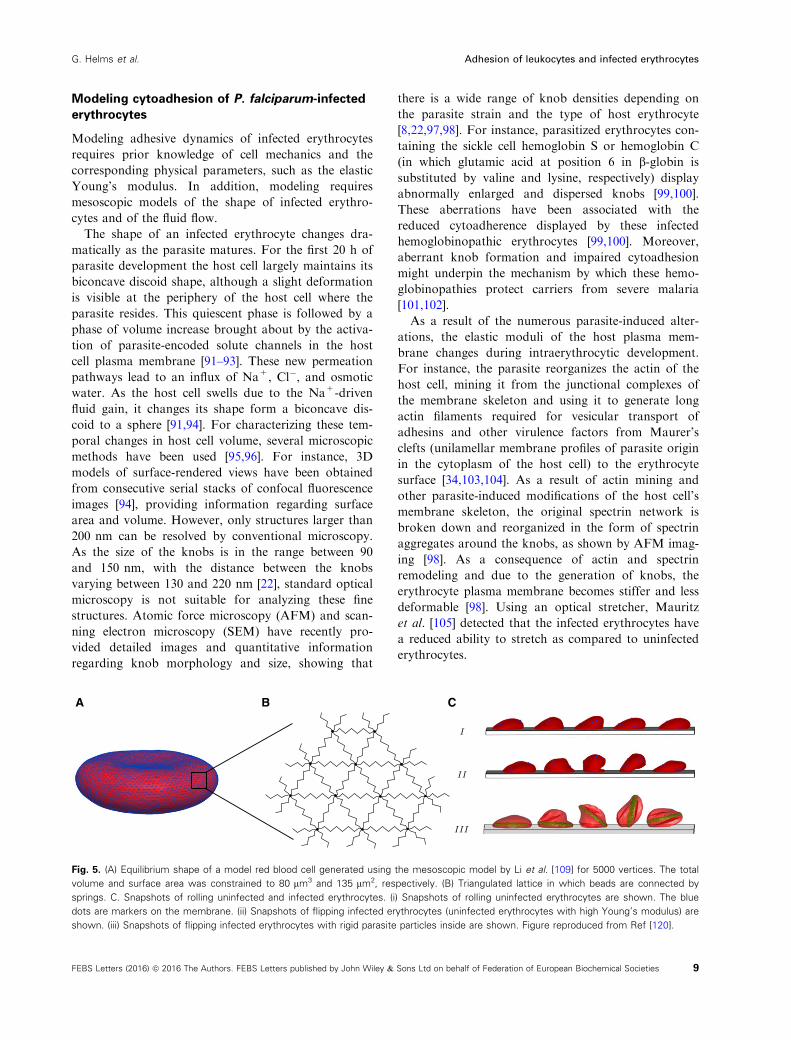
A B C
Fig. 5. (A) Equilibrium shape of a model red blood cell generated using the mesoscopic model by Li et al. [109] for 5000 vertices. The total
volume and surface area was constrained to 80 lm3 and 135 lm2, respectively. (B) Triangulated lattice in which beads are connected by
springs. C. Snapshots of rolling uninfected and infected erythrocytes. (i) Snapshots of rolling uninfected erythrocytes are shown. The blue
dots are markers on the membrane. (ii) Snapshots of flipping infected erythrocytes (uninfected erythrocytes with high Young’s modulus) are
shown. (iii) Snapshots of flipping infected erythrocytes with rigid parasite particles inside are shown. Figure reproduced from Ref [120].
9FEBS Letters (2016) ª 2016 The Authors. FEBS Letters published by John Wiley & Sons Ltd on behalf of Federation of European Biochemical Societies
G. Helms et al. Adhesion of leukocytes and infected erythrocytes

Mesoscopic models for red blood cell shape
Study of adhesive dynamics in flow requires meso-
scopic modeling of red blood cells. Mesoscopic or
coarse-grained models of any biological system con-
sider lower resolution subunits. One such class of
mesoscopic model is a bead-spring network model
where the 3D shape is represented by a triangulated
network of beads connected via springs. Using this
approach, the red blood cell shape has been modeled
at the spectrin level (Fig. 5A,B) [106–110]. The posi-
tions of the beads or vertices are given by xj. The
total surface area of an erythrocyte is the sum of
areas of individual triangles, Ai, and the total volume
is the sum of volumes contributed by each triangle,
n̂i � xið ÞAi=3. The total free energy of the system is,
FðfxjgÞ ¼ F in�plane þ F bend þ F area þ F volume
where F area conserves the total surface area of the cell
and F volume conserves the total volume of the cell.
Both F area and F volume are implemented as harmonic
potentials about A0 and V0 with rigidity factors kvolume
and karea, which set the strengths of the constraining
potentials for surface area and volume, respectively,
where A0 and V0 are total surface area and total vol-
ume of the cell, respectively.
The equilibrium length of the edge is ‘0. The total
bending energy is written as,
F bend ¼X
i
jb 1� cos hi � h0ð Þð Þ
where hi is the angle between the normals of adja-
cent triangles, jb is the bending constant, and h0 is
the spontaneous curvature angle. The bending con-
stant, jb, is related to the bending moduli of the
lipid membrane, jc, as jb ¼ 2ffiffiffi3
pjc [108,109]. The
last term in free energy F in�plane is the elastic energy
stored in the membrane. It is written as the sum of
the elastic energy stored in spectrin links and the
hydrostatic elastic energy stored in each triangle. It
is written as
F in�plane ¼Xlinks
i
Vsð‘iÞ þXtriangles
i
Cq
Aqi
For Vs(‘i), there exists many nonlinear spring
models such as the Worm-like Chain model (WLC)
and the Finitely Extensible Non-linear Elastic
(FENE) polymer model [111]. In the second term,
Cq is a constant that depends on spring parameters
[108,109] and q is an exponent that is chosen to be
1. The elastic moduli, such as Young’s modulus and
shear modulus, can be deduced in model parameters
using a linear analysis. Figure 5A shows the equilib-
rium shape of a model red blood cell for 5000 ver-
tices, a total volume constrained to V0 = 80 lm3,
and a total surface area of S0 = 135 lm2. The cur-
rently considered mesoscopic model contains a fixed
network, meaning that it does not consider the fluid
nature of lipid membranes. Noguchi and Gompper
took this into account by employing dynamically tri-
angulated lattices [112,113]. Recently, it has been
shown that the surface properties of erythrocytes are
also strongly determined by active elements (e.g., ion
pumps) in the membrane [114].
Modeling adhesion of parasitized erythrocyte
under flow
There are several coarse-grained or mesoscopic models
of fluid, such as Multi Particle Collision Dynamics
(MPCD) [115] and Dissipative Particle Dynamics
(DPD) [116]. MPCD and DPD are off-lattice particle-
based simulation techniques to model the fluid. Nogu-
chi and Gompper [113] used MPCD for simulating the
fluid along with a dynamically triangulated network
model for red blood cells to study the shape transi-
tions under capillary flow. The dynamics of solvent
particles was coupled to the dynamics of membrane
vertices, by including membrane vertices in solvent
exchange of momentum step [117,118]. They found
that model red blood cells transform from nonaxis
symmetric discocyte shapes to axis symmetric para-
chute shapes in capillary flow and that the speed of
this transition decreases with increasing elastic moduli,
such as the bending modulus and the shear modulus.
Fedosov et al. [108] simulated the mechanics and the
dynamics of model red blood cells in flow, using the
DPD method. The external solvent makes bounce-
back interactions with the membrane, separating the
internal and external solvent and preserving no-slip
boundary conditions at the membrane. The membrane
and solvent dynamics are linked via viscous force cou-
pling.
Fedosov et al. [119,120] extended the approach to
the adhesion of infected erythrocytes. Different
dynamical states were recovered, such as firm adhe-
sion, rolling adhesion, and flipping motion. Infected
erythrocytes were modeled by increasing the mem-
brane stiffness in relation to uninfected red blood
cells and by placing solid particles mimicking the
intracellular parasite inside the host cell cytosol. In
Fig. 5C, the different adhesive behaviors of unin-
fected and infected erythrocytes are illustrated. Model
uninfected erythrocytes change from rolling motion
10 FEBS Letters (2016) ª 2016 The Authors. FEBS Letters published by John Wiley & Sons Ltd on behalf of Federation of European Biochemical Societies
Adhesion of leukocytes and infected erythrocytes G. Helms et al.

to flipping behavior when the Young’s modulus is
increased three times. Irregular flipping dynamics is
observed when rigid particles are placed inside the
erythrocyte cytosol as models for the parasite. Thus,
the transition from rolling to flipping is due to
increased stiffness and the presence of an intracellular
rigid parasite.
Hosseini et al. modeled infected erythrocytes at the
schizont stage based on uninfected red blood cells with
an internal spherical rigid parasite to simulate stretch-
ing of the cell as observed using optical tweezers
[121,122]. They found that the deformability of the cell
under external force decreases owing to the increased
stiffness and the presence of the internal parasite.
Conclusions and perspectives
Leukocyte rolling adhesion and extravasation is a
widely investigated subject in biophysics, cell biology,
and immunology. In comparison, the biophysical inter-
actions of infected red blood cells with the endothe-
lium are much less explored in spite of the medical
relevance for the pathophysiology of malaria. Table 1
provides an overview of the main differences between
the two systems as discussed here. It is evident that,
despite the striking similarities that might have evolved
due to the physical limitations of cytoadhesion in flow,
there are also marked differences that certainly matter
for the biological function. Overall the leukocytes
seems to be better adapted to cell capture in shear
flow, as evidenced by the round shape, the mechanical
design of the microvilli and the molecular properties
of the different members of the selectin family.
Together, these elements lead to a cascade of adhesion
events that ensures efficient and specific cell capture.
In direct comparision, the adhesive knobs of the
infected erythrocyte, which are less elevated and stiffer
than the microvilli, seem to be less efficient in this
regard, possibly because the biological function does
not require high effectivity or specificity. On the other
hand, their large number and complex composition
and architecture suggest that the situation is more
complex, for example, involving interactions with the
endothelium and the immune system that we do not
yet fully understand.
While there has recently been substantial progress
in our understanding of the cytoadherence of P. falci-
parum-infected erythrocytes in flow, there are still
open questions and technical and methodological chal-
lenges to be met before this important pathological
behavior of parasitized erythrocytes can be appreci-
ated in its full complexity. From the comparison con-
ducted in this review, it is evident that the mechanics
of infected erythrocytes is crucial to a deeper insight
into interactions with the endothelium. However, the
mesoscopic shape model of parasitized erythrocytes
needs to be improved as current models do not yet
consider the effect of the knobs or temporal changes
in host erythrocyte shape during the course of intra-
cellular parasite development. In a recent develop-
ment, Zhang et al. [123] have approached this gap in
understanding, by calculating the shear modulus of
infected erythrocytes, using a coarse-grained model
where the lipid bilayer and the spectrin network were
modeled separately and connected with vertical link-
age. The knobs were simulated by circular areas with
increased membrane stiffness and more connections
with the spectrin network [123]. Such modeling
advances have to be pushed further in the future as
empirical data on the relevant parameters become
available.
There is further a gap in our knowledge about the
number of PfEMP1 molecules on the host cell surface
and how many PfEMP1 molecules are present per
knob. It is further not fully understood how they are
displayed in knobs and with which factors they inter-
act in these protrusions. Single-molecule counting in
combination with super-resolution microscopy might
help address these questions. First experiments using
various super-resolution microscopic techniques [in-
cluding stimulated emission microscopy (STED),
photo-activated localization microscopy (PALM),
stochastic optical reconstruction microscopy
(STORM), and structional illumination microscopy
(SIM)] are encouraging [124–126], promising novel
insights into host erythrocyte shape, knob formation,
and PfEMP1 assembly and presentation in the near
future. Other techniques that might help elucidate the
mechanical properties of the host cell membrane
include flicker microscopy, which has recently been
used to study the effect of endotoxins on uninfected
erythrocytes [127]. A central aspect here will be the
role of passive versus active contributions to the flick-
ering spectrum [114]. For example, it might well be
that the stiffening observed for infected erythrocytes
not only results from the remodeling of the spectrin
network but also from changes in the cellular meta-
bolism by the parasite. While we have first informa-
tion regarding the adhesion strength at the whole cell
level, the contribution of single-molecule bonds is
unclear. Here, single-molecule force measurements
using atomic force microscopy are anticipated to
resolve this issue, following the lead in other systems
[128]. Once such results are available, they can be
used for adhesive dynamics simulations for cell cap-
ture from the flow.
11FEBS Letters (2016) ª 2016 The Authors. FEBS Letters published by John Wiley & Sons Ltd on behalf of Federation of European Biochemical Societies
G. Helms et al. Adhesion of leukocytes and infected erythrocytes

Another pending question pertains to cytoadhesion
and flow dynamics of parasitized hemoglobinopathic
erythrocytes. As mentioned above carriers of the sickle
cell hemoglobins S and C are protected from severe
malaria (which explains why these hemoglobin poly-
morphisms are highly prevalent in malaria-endemic
areas). Carriers are not sterile to infection. In fact,
they can have parasitemias as high as those seen in
patients with normal hemoglobin. However, they are
less likely to develop cerebral malaria and to die from
severe complications [102]. These hemoglobinopathic
erythrocytes, and also fetal and a-thalassemic erythro-
cytes (which also protect from severe malaria), possess
fewer and abnormally large knobs on their surface
than wild-type erythrocytes, when infected with P. fal-
ciparum [99,100,102,129–132]. In addition, the amount
of PfEMP1 molecules presented is reduced and
aberrantly displayed [99,100,130]. How sickle cell
hemoglobins S and C affect knob formation and
cytoadherence is largely unclear as are the ramifica-
tions of the aberrant knob morphology and density
for the interaction of these parasitized hemoglobino-
pathic erythrocytes with the microvascular endothelial
lining in flow. Do they bind to endothelial cells with
sufficient strength to activate the endothelium? If the
adhesion strength is insufficient to trigger the life-
threatening downstream inflammatory events, as
recently proposed [130], then this might explain the
malaria-protective trait of hemoglobinopathic and fetal
erythrocytes.
Understanding cytoadhesion dynamics of leuko-
cytes has substantially benefited from computational
modeling and the application of biophysical methods
and approaches. In the case of parasitized erythro-
cytes, this research is still at its infancy. However,
progress is anticipated to accelerate given the ability
of the malaria community to implement novel tech-
niques and to learn from the experience gained in
other fields.
Acknowledgements
This work was in part supported by the Deutsche
Forschungsgemeinschaft under the Collaborative
Research Center SFB 1129. USS and ML are members
of the cluster of excellence CellNetworks. We thank
Britta Nyboer for the art work.
Author contributions
USS and ML outlined the manuscript. GH, AKD,
USS and ML wrote the article.
References
1 Simon SI and Goldsmith HL (2002) Leukocyte
adhesion dynamics in shear flow. Ann Biomed Eng 30,
315–332.2 Carman CV and Springer TA (2008) Trans-cellular
migration: cell-cell contacts get intimate. Curr Opin
Cell Biol 20, 533–540.3 Hebbel RP (1991) Beyond hemoglobin polymerization:
the red blood cell membrane and sickle disease
pathophysiology. Blood 77, 214–237.4 Tembo D and Montgomery J (2010) Var gene
expression and human Plasmodium pathogenesis.
Future Microbiol 5, 801–815.5 Sherman IW, Eda S and Winograd E (2003)
Cytoadherence and sequestration in Plasmodium
falciparum: defining the ties that bind. Microbes Infect
5, 897–909.6 Ho M, Hickey MJ, Murray AG, Andonegui G and
Kubes P (2000) Visualization of Plasmodium
falciparum-endothelium interactions in human
microvasculature: mimicry of leukocyte recruitment. J
Exp Med 192, 1205–1211.7 Marshall BT, Long M, Piper JW, Yago T, McEver RP
and Zhu C (2003) Direct observation of catch
bonds involving cell-adhesion molecules. Nature 423,
190–193.8 Rieger H, Yoshikawa HY, Quadt K, Nielsen MA,
Sanchez CP, Salanti A, Tanaka M and Lanzer M
(2015) Cytoadhesion of Plasmodium falciparum-infected
erythrocytes to chondroitin-4-sulfate is cooperative and
shear enhanced. Blood 125, 383–391.9 Smith JD (2014) The role of PfEMP1 adhesion
domain classification in Plasmodium falciparum
pathogenesis research. Mol Biochem Parasitol 195, 82–87.
10 Smith JD, Rowe JA, Higgins MK and Lavstsen T
(2013) Malaria’s deadly grip: cytoadhesion of
Plasmodium falciparum-infected erythrocytes. Cell
Microbiol 15, 1976–1983.11 World Health Organization (2014) World Malaria
Report 2014.
12 Miller LH, Baruch DI, Marsh K and Doumbo OK
(2002) The pathogenic basis of malaria. Nature 415,
673–679.13 Viallat A and Abkarian M (2014) Red blood cell: from
its mechanics to its motion in shear flow. Int J Lab
Hematol 36, 237–243.14 Storm J and Craig AG (2014) Pathogenesis of cerebral
malaria–inflammation and cytoadherence. Front Cell
Infect Microbiol 4, 100.
15 Viebig NK, Gamain B, Scheidig C, Lepolard C,
Przyborski J, Lanzer M, Gysin J and Scherf A (2005)
A single member of the Plasmodium falciparum var
multigene family determines cytoadhesion to the
12 FEBS Letters (2016) ª 2016 The Authors. FEBS Letters published by John Wiley & Sons Ltd on behalf of Federation of European Biochemical Societies
Adhesion of leukocytes and infected erythrocytes G. Helms et al.

placental receptor chondroitin sulphate A. EMBO Rep
6, 775–781.16 Goel S and Gowda DC (2011) How specific is
Plasmodium falciparum adherence to chondroitin 4-
sulfate? Trends Parasitol 27, 375–381.17 Fried M and Duffy PE (1996) Adherence of
Plasmodium falciparum to chondroitin sulfate A in the
human placenta. Science 272, 1502–1504.18 Clausen TM, Christoffersen S, Dahlback M, Langkilde
AE, Jensen KE, Resende M, Agerbaek MO, Andersen
D, Berisha B, Ditlev SB et al. (2012) Structural and
functional insight into how the Plasmodium falciparum
VAR2CSA protein mediates binding to chondroitin
sulfate A in placental malaria. J Biol Chem 287,
23332–23345.19 Srivastava A, Gangnard S, Round A, Dechavanne S,
Juillerat A, Raynal B, Faure G, Baron B, Ramboarina
S, Singh SK et al. (2010) Full-length extracellular
region of the var2CSA variant of PfEMP1 is required
for specific, high-affinity binding to CSA. Proc Natl
Acad Sci USA 107, 4884–4889.20 Niang M, Bei AK, Madnani KG, Pelly S, Dankwa S,
Kanjee U, Gunalan K, Amaladoss A, Yeo KP, Bob
NS et al. (2014) STEVOR is a Plasmodium falciparum
erythrocyte binding protein that mediates merozoite
invasion and rosetting. Cell Host Microbe 16, 81–93.21 Goel S, Palmkvist M, Moll K, Joannin N, Lara P,
Akhouri RR, Moradi N, Ojemalm K, Westman M,
Angeletti D et al. (2015) RIFINs are adhesins
implicated in severe Plasmodium falciparum malaria.
Nat Med 21, 314–317.22 Quadt KA, Barfod L, Andersen D, Bruun J, Gyan B,
Hassenkam T, Ofori MF and Hviid L (2012) The
density of knobs on Plasmodium falciparum-infected
erythrocytes depends on developmental age and varies
among isolates. PLoS One 7, e45658.
23 Pologe LG, Pavlovec A, Shio H and Ravetch JV
(1987) Primary structure and subcellular localization of
the knob-associated histidine-rich protein of
Plasmodium falciparum. Proc Natl Acad Sci USA 84,
7139–7143.24 Crabb BS, Cooke BM, Reeder JC, Waller RF,
Caruana SR, Davern KM, Wickham ME, Brown GV,
Coppel RL and Cowman AF (1997) Targeted gene
disruption shows that knobs enable malaria-infected
red cells to cytoadhere under physiological shear
stress. Cell 89, 287–296.25 Weng H, Guo X, Papoin J, Wang J, Coppel R,
Mohandas N and An X (2014) Interaction of
Plasmodium falciparum knob-associated histidine-rich
protein (KAHRP) with erythrocyte ankyrin R is
required for its attachment to the erythrocyte
membrane. Biochim Biophys Acta 1838, 185–192.26 Pei X, An X, Guo X, Tarnawski M, Coppel R and
Mohandas N (2005) Structural and functional
studies of interaction between Plasmodium
falciparum knob-associated histidine-rich protein
(KAHRP) and erythrocyte spectrin. J Biol Chem
280, 31166–31171.27 Oh SS, Voigt S, Fisher D, Yi SJ, LeRoy PJ,
Derick LH, Liu S and Chishti AH (2000)
Plasmodium falciparum erythrocyte membrane
protein 1 is anchored to the actin-spectrin junction
and knob-associated histidine-rich protein in the
erythrocyte skeleton. Mol Biochem Parasitol 108,
237–247.28 Kilejian A, Rashid MA, Aikawa M, Aji T and Yang
YF (1991) Selective association of a fragment of the
knob protein with spectrin, actin and the red cell
membrane. Mol Biochem Parasitol 44, 175–181.29 Rug M, Prescott SW, Fernandez KM, Cooke BM and
Cowman AF (2006) The role of KAHRP domains in
knob formation and cytoadherence of P. falciparum-
infected human erythrocytes. Blood 108, 370–378.30 Magowan C, Nunomura W, Waller KL, Yeung J,
Liang J, Van Dort H, Low PS, Coppel RL and
Mohandas N (2000) Plasmodium falciparum histidine-
rich protein 1 associates with the band 3 binding
domain of ankyrin in the infected red cell membrane.
Biochim Biophys Acta 1502, 461–470.31 Ganguly AK, Ranjan P, Kumar A and Bhavesh NS
(2015) Dynamic association of PfEMP1 and KAHRP
in knobs mediates cytoadherence during Plasmodium
invasion. Sci Rep 5, 8617.
32 Waller KL, Cooke BM, Nunomura W, Mohandas N
and Coppel RL (1999) Mapping the binding domains
involved in the interaction between the Plasmodium
falciparum knob-associated histidine-rich protein
(KAHRP) and the cytoadherence ligand P. falciparum
erythrocyte membrane protein 1 (PfEMP1). J Biol
Chem 274, 23808–23813.33 Oberli A, Slater LM, Cutts E, Brand F, Mundwiler-
Pachlatko E, Rusch S, Masik MF, Erat MC, Beck HP
and Vakonakis I (2014) A Plasmodium falciparum
PHIST protein binds the virulence factor PfEMP1 and
comigrates to knobs on the host cell surface. FASEB J
28, 4420–4433.34 Cyrklaff M, Sanchez CP, Kilian N, Bisseye C, Simpore
J, Frischknecht F and Lanzer M (2011) Hemoglobins
S and C interfere with actin remodeling in Plasmodium
falciparum-infected erythrocytes. Science 334, 1283–1286.
35 Watermeyer JM, Hale VL, Hackett F, Clare DK,
Cutts EE, Vakonakis I, Fleck RA, Blackman MJ and
Saibil HR (2016) A spiral scaffold underlies
cytoadherent knobs in Plasmodium falciparum-infected
erythrocytes. Blood, 127, 343–351.36 Springer TA (1994) Traffic signals for lymphocyte
recirculation and leukocyte emigration: the multistep
paradigm. Cell 76, 301–314.
13FEBS Letters (2016) ª 2016 The Authors. FEBS Letters published by John Wiley & Sons Ltd on behalf of Federation of European Biochemical Societies
G. Helms et al. Adhesion of leukocytes and infected erythrocytes
37 Luster AD, Alon R and von Andrian UH (2005)
Immune cell migration in inflammation: present and
future therapeutic targets. Nat Immunol 6, 1182–1190.38 Sundd P, Pospieszalska MK, Cheung LS,
Konstantopoulos K and Ley K (2011) Biomechanics
of leukocyte rolling. Biorheology 48, 1–35.39 Shao JY, Ting-Beall HP and Hochmuth RM (1998)
Static and dynamic lengths of neutrophil microvilli.
Proc Natl Acad Sci USA 95, 6797–6802.40 Heasman SJ and Ridley AJ (2010) Multiple roles for
RhoA during T cell transendothelial migration. Small
GTPases 1, 174–179.41 Bell GI (1978) Models for the specific adhesion of cells
to cells. Science 200, 618–627.42 Evans E and Ritchie K (1997) Dynamic strength
of molecular adhesion bonds. Biophys J 72, 1541–1555.
43 Dembo M, Torney DC, Saxman K and Hammer D
(1988) The reaction-limited kinetics of membrane-to-
surface adhesion and detachment. Proc R Soc Lond B
Biol Sci 234, 55–83.44 Merkel R, Nassoy P, Leung A, Ritchie K and Evans E
(1999) Energy landscapes of receptor-ligand bonds
explored with dynamic force spectroscopy. Nature 397,
50–53.45 Zhu C, Lou J and McEver RP (2005) Catch bonds:
physical models, structural bases, biological function
and rheological relevance. Biorheology 42, 443–462.46 Chakrabarti S, Hinczewski M and Thirumalai D
(2016) Phenomenological and microscopic theories for
catch bonds. arXiv:1601.02050 [q-bio.BM].
47 Thomas WE, Nilsson LM, Forero M, Sokurenko EV
and Vogel V (2004) Shear-dependent ‘stick-and-roll’
adhesion of type 1 fimbriated Escherichia coli. Mol
Microbiol 53, 1545–1557.48 Nilsson LM, Thomas WE, Trintchina E, Vogel V and
Sokurenko EV (2006) Catch bond-mediated adhesion
without a shear threshold: trimannose versus
monomannose interactions with the FimH adhesin of
Escherichia coli. J Biol Chem 281, 16656–16663.49 Finger EB, Puri KD, Alon R, Lawrence MB, von
Andrian UH and Springer TA (1996) Adhesion
through L-selectin requires a threshold hydrodynamic
shear. Nature 379, 266–269.50 Yago T, Wu J, Wey CD, Klopocki AG, Zhu C and
McEver RP (2004) Catch bonds govern adhesion
through L-selectin at threshold shear. J Cell Biol 166,
913–923.51 Guo B and Guilford WH (2006) Mechanics of
actomyosin bonds in different nucleotide states are
tuned to muscle contraction. Proc Natl Acad Sci USA
103, 9844–9849.52 Thomas WE, Trintchina E, Forero M, Vogel V and
Sokurenko EV (2002) Bacterial adhesion to target cells
enhanced by shear force. Cell 109, 913–923.
53 Kong F, Garcia AJ, Mould AP, Humphries MJ and
Zhu C (2009) Demonstration of catch bonds
between an integrin and its ligand. J Cell Biol 185,
1275–1284.54 Pereverzev YV and Prezhdo OV (2006) Force-induced
deformations and stability of biological bonds. Phys
Rev E Stat Nonlin Soft Matter Phys 73, 050902.
55 Lou J, Yago T, Klopocki AG, Mehta P, Chen W,
Zarnitsyna VI, Bovin NV, Zhu C and McEver RP
(2006) Flow-enhanced adhesion regulated by a selectin
interdomain hinge. J Cell Biol 174, 1107–1117.56 Lou J and Zhu C (2007) A structure-based sliding-
rebinding mechanism for catch bonds. Biophys J 92,
1471–1485.57 Yago T, Lou J, Wu T, Yang J, Miner JJ, Coburn L,
Lopez JA, Cruz MA, Dong JF, McIntire LV et al.
(2008) Platelet glycoprotein Ibalpha forms catch bonds
with human WT vWF but not with type 2B von
Willebrand disease vWF. J Clin Invest 118, 3195–3207.58 Thomas W, Forero M, Yakovenko O, Nilsson L,
Vicini P, Sokurenko E and Vogel V (2006) Catch-bond
model derived from allostery explains force-activated
bacterial adhesion. Biophys J 90, 753–764.59 Yakovenko O, Sharma S, Forero M, Tchesnokova V,
Aprikian P, Kidd B, Mach A, Vogel V, Sokurenko E
and Thomas WE (2008) FimH forms catch bonds that
are enhanced by mechanical force due to allosteric
regulation. J Biol Chem 283, 11596–11605.60 Pereverzev YV and Prezhdo OV (2007) Universal
laws in the force-induced unraveling of biological
bonds. Phys Rev E Stat Nonlin Soft Matter Phys 75,
011905.
61 Pereverzev YV, Prezhdo OV, Forero M, Sokurenko
EV and Thomas WE (2005) The two-pathway model
for the catch-slip transition in biological adhesion.
Biophys J 89, 1446–1454.62 Pereverzev YV, Prezhdo OV, Thomas WE and
Sokurenko EV (2005) Distinctive features of the
biological catch bond in the jump-ramp force regime
predicted by the two-pathway model. Phys Rev E Stat
Nonlin Soft Matter Phys 72, 010903.
63 Prezhdo OV and Pereverzev YV (2009) Theoretical
aspects of the biological catch bond. Acc Chem Res 42,
693–703.64 Lawrence MB and Springer TA (1991) Leukocytes roll
on a selectin at physiologic flow rates: distinction from
and prerequisite for adhesion through integrins. Cell
65, 859–873.65 Alon R, Hammer DA and Springer TA (1995)
Lifetime of the P-selectin-carbohydrate bond and its
response to tensile force in hydrodynamic flow. Nature
374, 539–542.66 Schwarz US and Alon R (2004) L-selectin-mediated
leukocyte tethering in shear flow is controlled by
multiple contacts and cytoskeletal anchorage
14 FEBS Letters (2016) ª 2016 The Authors. FEBS Letters published by John Wiley & Sons Ltd on behalf of Federation of European Biochemical Societies
Adhesion of leukocytes and infected erythrocytes G. Helms et al.

facilitating fast rebinding events. Proc Natl Acad Sci
USA 101, 6940–6945.67 Caputo KE and Hammer DA (2005) Effect of
microvillus deformability on leukocyte adhesion
explored using adhesive dynamics simulations. Biophys
J 89, 187–200.68 Chang KC, Tees DF and Hammer DA (2000) The
state diagram for cell adhesion under flow: leukocyte
rolling and firm adhesion. Proc Natl Acad Sci USA 97,
11262–11267.69 Hammer DA and Apte SM (1992) Simulation of cell
rolling and adhesion on surfaces in shear flow: general
results and analysis of selectin-mediated neutrophil
adhesion. Biophys J 63, 35–57.70 Korn C and Schwarz US (2006) Efficiency of initiating
cell adhesion in hydrodynamic flow. Phys Rev Lett 97,
138103.
71 Alon R, Chen S, Puri KD, Finger EB and Springer
TA (1997) The kinetics of L-selectin tethers and the
mechanics of selectin-mediated rolling. J Cell Biol 138,
1169–1180.72 Korn CB and Schwarz US (2008) Dynamic states of
cells adhering in shear flow: from slipping to rolling.
Phys Rev E Stat Nonlin Soft Matter Phys 77,
041904.
73 Ramesh KV, Thaokar R, Prakash JR and Prabhakar
R (2015) Significance of thermal fluctuations and
hydrodynamic interactions in receptor-ligand-mediated
adhesive dynamics of a spherical particle in wall-
bound shear flow. Phys Rev E Stat Nonlin Soft Matter
Phys 91, 022302.
74 Cheung LS-L, Zheng X, Wang L, Baygents JC,
Guzman R, Schroeder JA, Heimark RL and Zohar Y
(2011) Adhesion dynamics of circulating tumor cells
under shear flow in a bio-functionalized microchannel.
J Micromech Microeng 21, 054033.
75 Zheng X, Cheung LS, Schroeder JA, Jiang L and
Zohar Y (2011) Cell receptor and surface ligand
density effects on dynamic states of adhering
circulating tumor cells. Lab Chip 11, 3431–3439.76 Bhatia SK, King MR and Hammer DA (2003) The
state diagram for cell adhesion mediated by two
receptors. Biophys J 84, 2671–2690.77 Caputo KE, Lee D, King MR and Hammer DA
(2007) Adhesive dynamics simulations of the
shear threshold effect for leukocytes. Biophys J 92,
787–797.78 Nash GB, Cooke BM, Marsh K, Berendt A, Newbold
C and Stuart J (1992) Rheological analysis of the
adhesive interactions of red blood cells parasitized by
Plasmodium falciparum. Blood 79, 798–807.79 Cooke BM, Berendt AR, Craig AG, MacGregor J,
Newbold CI and Nash GB (1994) Rolling and
stationary cytoadhesion of red blood cells parasitized
by Plasmodium falciparum: separate roles for ICAM-1,
CD36 and thrombospondin. Br J Haematol 87, 162–170.
80 Yipp BG, Anand S, Schollaardt T, Patel KD,
Looareesuwan S and Ho M (2000) Synergism of
multiple adhesion molecules in mediating
cytoadherence of Plasmodium falciparum-infected
erythrocytes to microvascular endothelial cells under
flow. Blood 96, 2292–2298.81 Cooke BM, Rogerson SJ, Brown GV and Coppel RL
(1996) Adhesion of malaria-infected red blood cells to
chondroitin sulfate A under flow conditions. Blood 88,
4040–4044.82 Li A, Lim TS, Shi H, Yin J, Tan SJ, Li Z, Low BC,
Tan KS and Lim CT (2011) Molecular mechanistic
insights into the endothelial receptor mediated
cytoadherence of Plasmodium falciparum-infected
erythrocytes. PLoS One 6, e16929.
83 Duffy PE (2007) Plasmodium in the placenta:
parasites, parity, protection, prevention and possibly
preeclampsia. Parasitology 134, 1877–1881.84 Martin FA, Murphy RP and Cummins PM (2013)
Thrombomodulin and the vascular endothelium:
insights into functional, regulatory, and therapeutic
aspects. Am J Physiol Heart Circ Physiol 304, H1585–H1597.
85 Antia M, Herricks T and Rathod PK (2007)
Microfluidic modeling of cell-cell interactions in
malaria pathogenesis. PLoS Pathog 3, e99.
86 Avril M, Traore B, Costa FT, Lepolard C and Gysin J
(2004) Placenta cryosections for study of the adhesion
of Plasmodium falciparum-infected erythrocytes to
chondroitin sulfate A in flow conditions. Microbes
Infect 6, 249–255.87 Davis SP, Amrein M, Gillrie MR, Lee K, Muruve DA
and Ho M (2012) Plasmodium falciparum-induced
CD36 clustering rapidly strengthens cytoadherence via
p130CAS-mediated actin cytoskeletal rearrangement.
FASEB J 26, 1119–1130.88 Davis SP, Lee K, Gillrie MR, Roa L, Amrein M and
Ho M (2013) CD36 recruits alpha(5)beta(1) integrin to
promote cytoadherence of P. falciparum-infected
erythrocytes. PLoS Pathog 9, e1003590.
89 Xu X, Efremov AK, Li A, Lai L, Dao M, Lim CT
and Cao J (2013) Probing the cytoadherence of
malaria infected red blood cells under flow. PLoS One
8, e64763.
90 Carvalho PA, Diez-Silva M, Chen H, Dao M and
Suresh S (2013) Cytoadherence of erythrocytes invaded
by Plasmodium falciparum: quantitative contact-
probing of a human malaria receptor. Acta Biomater
9, 6349–6359.91 Mauritz JM, Esposito A, Ginsburg H, Kaminski CF,
Tiffert T and Lew VL (2009) The homeostasis of
Plasmodium falciparum-infected red blood cells. PLoS
Comput Biol 5, e1000339.
15FEBS Letters (2016) ª 2016 The Authors. FEBS Letters published by John Wiley & Sons Ltd on behalf of Federation of European Biochemical Societies
G. Helms et al. Adhesion of leukocytes and infected erythrocytes

92 Desai SA (2012) Ion and nutrient uptake by malaria
parasite-infected erythrocytes. Cell Microbiol 14, 1003–1009.
93 Lew VL, Tiffert T and Ginsburg H (2003) Excess
hemoglobin digestion and the osmotic stability of
Plasmodium falciparum-infected red blood cells. Blood
101, 4189–4194.94 Esposito A, Choimet JB, Skepper JN, Mauritz JM,
Lew VL, Kaminski CF and Tiffert T (2010)
Quantitative imaging of human red blood cells
infected with Plasmodium falciparum. Biophys J 99,
953–960.95 Cho S, Kim S, Kim Y and Park Y (2012) Optical
imaging techniques for the study of malaria. Trends
Biotechnol 30, 71–79.96 Mauritz JM, Esposito A, Tiffert T, Skepper JN,
Warley A, Yoon YZ, Cicuta P, Lew VL, Guck JR and
Kaminski CF (2010) Biophotonic techniques for the
study of malaria-infected red blood cells. Med Biol
Eng Comput 48, 1055–1063.97 Subramani R, Quadt K, Jeppesen AE, Hempel C,
Petersen JE, Hassenkam T, Hviid L and Barfod L
(2015) Plasmodium falciparum-infected erythrocyte
knob density is linked to the PfEMP1 variant
expressed. MBio 6, e01456–15.98 Shi H, Liu Z, Li A, Yin J, Chong AG, Tan KS,
Zhang Y and Lim CT (2013) Life cycle-dependent
cytoskeletal modifications in Plasmodium falciparum
infected erythrocytes. PLoS One 8, e61170.
99 Cholera R, Brittain NJ, Gillrie MR, Lopera-Mesa
TM, Diakite SA, Arie T, Krause MA, Guindo A,
Tubman A, Fujioka H et al. (2008) Impaired
cytoadherence of Plasmodium falciparum-infected
erythrocytes containing sickle hemoglobin. Proc Natl
Acad Sci USA 105, 991–996.100 Fairhurst RM, Baruch DI, Brittain NJ, Ostera GR,
Wallach JS, Hoang HL, Hayton K, Guindo A,
Makobongo MO, Schwartz OM et al. (2005)
Abnormal display of PfEMP-1 on erythrocytes
carrying haemoglobin C may protect against malaria.
Nature 435, 1117–1121.101 Taylor SM, Cerami C and Fairhurst RM (2013)
Hemoglobinopathies: slicing the Gordian knot of
Plasmodium falciparum malaria pathogenesis. PLoS
Pathog 9, e1003327.
102 Taylor SM, Parobek CM and Fairhurst RM (2012)
Haemoglobinopathies and the clinical epidemiology of
malaria: a systematic review and meta-analysis. Lancet
Infect Dis 12, 457–468.103 Cyrklaff M, Sanchez CP, Frischknecht F and Lanzer
M (2012) Host actin remodeling and protection from
malaria by hemoglobinopathies. Trends Parasitol 28,
479–485.104 Kilian N, Dittmer M, Cyrklaff M, Ouermi D, Bisseye
C, Simpore J, Frischknecht F, Sanchez CP and Lanzer
M (2013) Haemoglobin S and C affect the motion of
Maurer’s clefts inPlasmodium falciparum-infected
erythrocytes. Cell Microbiol 15, 1111–1126.105 Mauritz JM, Tiffert T, Seear R, Lautenschlager F,
Esposito A, Lew VL, Guck J and Kaminski CF (2010)
Detection of Plasmodium falciparum-infected red blood
cells by optical stretching. J Biomed Opt 15, 030517.
106 Boey SK, Boal DH and Discher DE (1998)
Simulations of the erythrocyte cytoskeleton at large
deformation. I. Microscopic models. Biophys J 75,
1573–1583.107 Discher DE, Boal DH and Boey SK (1998)
Simulations of the erythrocyte cytoskeleton at large
deformation. II. Micropipette aspiration. Biophys J 75,
1584–1597.108 Fedosov DA, Caswell B and Karniadakis GE (2010)
A multiscale red blood cell model with accurate
mechanics, rheology, and dynamics. Biophys J 98,
2215–2225.109 Li J, Dao M, Lim CT and Suresh S (2005) Spectrin-
level modeling of the cytoskeleton and optical tweezers
stretching of the erythrocyte. Biophys J 88, 3707–3719.110 Pivkin IV and Karniadakis GE (2008) Accurate
coarse-grained modeling of red blood cells. Phys Rev
Lett 101, 118105.
111 Marko JF and Siggia ED (1995) Stretching DNA.
Macromolecules 28, 8759–8770.112 Gompper G and Kroll DM (1997) Network models of
fluid, hexatic and polymerized membranes. J Phys
Condens Matter 9, 8795–8834.113 Noguchi H and Gompper G (2005) Shape transitions
of fluid vesicles and red blood cells in capillary flows.
Proc Natl Acad Sci USA 102, 14159–14164.114 Turlier H, Fedosov DA, Audoly B, Auth T, Gov NS,
Sykes C, Joanny J-F, Gompper G and Betz T (2016)
Equilibrium physics breakdown reveals the active
nature of red blood cell flickering. Nat Phys,
doi:10.1038/nphys3621.
115 Malevanets A and Kapral R (1999) Mesoscopic model
for solvent dynamics. J Chem Phys 110, 8605–8613.116 Hoogerbrugge PJ and Koelman JMVA (1992)
Simulating microscopic hydrodynamic phenomena
with dissipative particle dynamics. Europhys Lett 19,
155–190.117 Kikuchi N, Pooley CM, Ryder JF and Yeomans JM
(2003) Transport coefficients of a mesoscopic fluid
dynamics model. J Chem Phys 119, 6388–6395.118 Ripoll M, Mussawisade K, Winkler RG and Gompper
G (2004) Low-Reynolds-number hydrodynamics of
complex fluids by multi-particle-collision dynamics.
Europhys Lett 68, 106–112.119 Fedosov DA, Caswell B, Suresh S andKarniadakis GE
(2011) Quantifying the biophysical characteristics of
Plasmodium falciparum-parasitized red blood cells in
microcirculation. Proc Natl Acad Sci USA 108, 35–39.
16 FEBS Letters (2016) ª 2016 The Authors. FEBS Letters published by John Wiley & Sons Ltd on behalf of Federation of European Biochemical Societies
Adhesion of leukocytes and infected erythrocytes G. Helms et al.

120 Fedosov DA, Caswell B and Karniadakis GE (2011)
Wall shear stress-based model for adhesive dynamics
of red blood cells in malaria. Biophys J 100, 2084–2093.
121 Dao M, Lim CT and Suresh S (2003) Mechanics of
the human red blood cell deformed by optical
tweezers. J Mech Phys Solids 51, 2259–2280.122 Hosseini SM and Feng JJ (2012) How malaria
parasites reduce the deformability of infected red
blood cells. Biophys J 103, 1–10.123 Zhang Y, Huang C, Kim S, Golkaram M, Dixon
MW, Tilley L, Li J, Zhang S and Suresh S (2015)
Multiple stiffening effects of nanoscale knobs on
human red blood cells infected with Plasmodium
falciparum malaria parasite. Proc Natl Acad Sci USA
112, 6068–6073.124 Hans N, Relan U, Dubey N, Gaur D and Chauhan
VS (2015) Identification and localization of a Novel
Invasin of Plasmodium falciparum. Mol Biochem
Parasitol 202, 38–43.125 Hliscs M, Millet C, Dixon MW, Siden-Kiamos I,
McMillan P and Tilley L (2015) Organization and
function of an actin cytoskeleton in Plasmodium
falciparum gametocytes. Cell Microbiol 17, 207–225.126 Riglar DT, Richard D, Wilson DW, Boyle MJ,
Dekiwadia C, Turnbull L, Angrisano F, Marapana
DS, Rogers KL, Whitchurch CB et al. (2011) Super-
resolution dissection of coordinated events during
malaria parasite invasion of the human erythrocyte.
Cell Host Microbe 9, 9–20.127 Ito H, Kuss N, Rapp BE, Ichikawa M, Gutsmann T,
Brandenburg K, Poschl JM and Tanaka M (2015)
Quantification of the iInfluence of endotoxins on the
mechanics of adult and neonatal red blood cells. J
Phys Chem B 119, 7837–7845.128 Wijeratne SS, Martinez JR, Grindel BJ, Frey EW, Li
J, Wang L, Farach-Carson MC and Kiang CH (2016)
Single molecule force measurements of perlecan/
HSPG2: a key component of the osteocyte pericellular
matrix. Matrix Biol 50, 27–38.129 Amaratunga C, Lopera-Mesa TM, Brittain NJ,
Cholera R, Arie T, Fujioka H, Keefer JR and
Fairhurst RM (2011) A role for fetal hemoglobin
and maternal immune IgG in infant resistance to
Plasmodium falciparum malaria. PLoS One 6,
e14798.
130 Fairhurst RM, Bess CD and Krause MA (2012)
Abnormal PfEMP1/knob display on Plasmodium
falciparum-infected erythrocytes containing
hemoglobin variants: fresh insights into malaria
pathogenesis and protection. Microbes Infect 14, 851–862.
131 Fairhurst RM, Fujioka H, Hayton K, Collins KF and
Wellems TE (2003) Aberrant development of
Plasmodium falciparum in hemoglobin CC red cells:
implications for the malaria protective effect of the
homozygous state. Blood 101, 3309–3315.132 Krause MA, Diakite SA, Lopera-Mesa TM,
Amaratunga C, Arie T, Traore K, Doumbia S,
Konate D, Keefer JR, Diakite M et al. (2012)
alpha-Thalassemia Impairs the cytoadherence of
Plasmodium falciparum-infected erythrocytes. PLoS
One 7, e37214.
17FEBS Letters (2016) ª 2016 The Authors. FEBS Letters published by John Wiley & Sons Ltd on behalf of Federation of European Biochemical Societies
G. Helms et al. Adhesion of leukocytes and infected erythrocytes