mobilization of human cd34 cd133 stem cells in vivo by ... · in cd34+ cells was 25% (pb.0001)....
TRANSCRIPT

Cardiovascular Revascularization
Mobilization of human CD34+CD133+ and CD34+CD133� stem cells in
vivo by consumption of an extract from Aphanizomenon flos-aquae—
related to modulation of CXCR4 expression by an L-selectin ligand?B
Gitte S. Jensena,4, Aaron N. Hartb, Lue A.M. Zaskeb, Christian Drapeauc, Niraj Guptad,
David J. Schaeffere, J. Alex Cruickshankb
aHolger NIS, 601 13 Avenue NE, Calgary, Alberta, Canada T2E 1C7bNIS Labs, 1437 Esplanade, Klamath Falls, OR, USA
cStemTech Health Sciences Inc., 1011 Calle Amanecer, San Clemente, CA, USAdCancer Treatment Center, Merle West Medical Center, 2610 Uhrmann Rd, Klamath Falls, OR, USA
eDepartment of Veterinary Biosciences, College of Veterinary Medicine, University of Illinois at Urbana-Champaign, USA
Abstract Objective: The goal of this study was to evaluate effects on human stem cells in vitro and in vivo of
1553-8389/07/$ – see
doi:10.1016/j.carrev.2
Abbreviations: AFB This project wa
Aphanizomenon flos-a
4 Corresponding
E-mail address: g
The coauthor C. D
this paper. No other a
an extract from the edible cyanobacterium Aphanizomenon flos-aquae (AFA) enriched for a novel
ligand for human CD62L (L-selectin).
Experimental approach: Ligands for CD62L provide a mechanism for stem cell mobilization in
conjunction with down-regulation of the CXCR4 chemokine receptor for stromal derived factor 1.
Affinity immunoprecipitation was used to identify a novel ligand for CD62L from a water extract
from AFA. The effects of AFA water extract on CD62L binding and CXCR4 expression was tested
in vitro using human bone marrow CD34+ cells and the two progenitor cell lines, KG1a and K562. A
double-blind randomized crossover study involving 12 healthy subjects evaluated the effects of
consumption on stem cell mobilization in vivo.
Results: An AFA extract rich in the CD62L ligand reduced the fucoidan-mediated externalization of
the CXCR4 chemokine receptor on bone marrow CD34+ cells by 30% and the CD62L+ CD34+ cell
line KG1A by 50% but did not alter the CXCR4 expression levels on the CD34� cell line K562. A
transient, 18% increase in numbers of circulating CD34+ stem cells maximized 1 hour after
consumption (Pb .0003). When 3 noncompliant volunteers were removed from analysis, the increase
in CD34+ cells was 25% (Pb.0001).
Conclusion: AFAwater extract contains a novel ligand for CD62L. It modulates CXCR4 expression
on CD34+ bone marrow cells in vitro and triggers the mobilization of CD34+ CD133+ and CD34+
CD133� cells in vivo.
D 2007 Elsevier Inc. All rights reserved.
Keywords: L-selectin; Ligand; Human; Adult stem cell; CD34; CD133; KG1a; K562; Bone marrow; Mobilization; Blue-
green algae; Cyanobacteria; Aphanizomenon; In vivo; In vitro
Medicine 8 (2007) 189–202
front matter D 2007 Elsevier Inc. All rights reserved.
007.03.004
A, Aphanizomenon flos-aquae; PBMC, Peripheral blood mononuclear cells; PMN, Polymorph-nucleated cells.
s supported by the Merle-West Center for Medical Research, a nonprofit research organization, Desert Lake Technologies, a harvester of
quae, and NIS Labs, an independent natural products research laboratory.
author. Tel.: +1 403 277 4134.
[email protected] (G.S. Jensen).
rapeau is Chief Science Officer for StemTech Health Sciences, which distributes the product StemEnhance, used for the in vivo part of
uthors have any commercial interest in the subject matter.

G.S. Jensen et al. / Cardiovascular Revascularization Medicine 8 (2007) 189–202190
1. Introduction
Much recent research has focused on the role of
selectins and their ligands in mobilization of bone marrow
stem cells. L-selectin belongs to the selectin family of cell
adhesion molecules involved in cellular migration during
normal immunosurveillance and inflammatory conditions.
L-selectin is best known as a homing molecule for
recirculating lymphocytes to recognize high endothelial
venules during the process of extravasation [1–3] and
for leukocytes to recognize and home to inflamed tissues
[4–8]. However, L-selectin plays significant roles in other
physiological cell adhesion processes as well, including the
retention vs. release of bone marrow stem cells into the
blood circulation [9–11].
Of special importance are findings that engagement of
L-selectin by some ligands will modulate the expression of
the CXCR4 chemokine receptor [12]. The CXCR4 receptor
specifically recognizes the chemokine stromal derived
factor 1 (SDF-1), which acts as a potent chemoattractant
for stem cells and assists in retaining stem cells within the
bone marrow environment [13–16]. The chemoattractant
properties of SDF-1 on stem cells were shown in vitro [17]
as well as in vivo to be directly associated with recruitment
of stem cells into kidney [18] and liver [19]. The
mobilization of recruitment of stem cells is associated with
repair of the central nervous system [20,21], heart [22,23],
and other tissues [24]. Stem cell mobilization and homing
involve a series of G-protein-coupled receptors that can
interact with each other as well as with adhesion mole-
cules [25,26]. It is proposed that loss of responsiveness
towards CXCR4 may be one of several contributing
mechanisms that allow some bone marrow stem cells to
detach and leave the bone marrow as part of the mobi-
lization process [27,28].
The use of selectin ligands has been proposed as a
mechanism for stem cell mobilization [29]. Some
L-selectin ligands (LSLs), including fucoidan and sulfa-
tide, have a proven effect on stem cell mobilization
[29,30]. The mobilization appears to happen in selectin-
dependent and -independent mechanisms in tandem. As an
example, the sulfated polysaccharide fucoidan can act as
an LSL and up-regulate the chemokine receptor CXCR4,
a receptor for SDF-1. However, fucoidan also assists stem
cell detachment within the marrow by binding to
another adhesion receptor, CD11b, during stem cell mobi-
lization [30].
The objective of this study was to evaluate the effects
on human stem cells in vitro and in vivo of an extract from
Aphanizomenon flos-aquae (AFA), enriched for a novel
ligand for human L-selectin. We report here that a novel
compound from the blue-green algae AFA binds to the
ligand-binding area of human L-selectin. The effect of this
compound was tested in various in vitro assays as well as
on stem cell mobilization in humans.
2. Materials and methods
2.1. Buffers and media
For cell cultures, freshly isolated human marrow cells, as
well as the KG1a and K562 cell lines, were resuspended and
cultured in RPMI-1640 with 10% fetal calf serum (Gibco,
Grand Island, NY, USA), 1% penicillin and streptomycin,
and L-glutamine. For immunostaining, cells were washed,
resuspended, and stained in phosphate-buffered saline
(PBS) containing 0.02% azide and 1% fetal calf serum or
bovine serum albumin.
2.2. Cyanobacterial extracts
Dried powder of the freshwater blue-green algae AFA
was obtained from Desert Lake Technologies, Keno, OR,
USA. Dried powder of Spirulina platensis was obtained
from Healthforce Nutritionals, Escondido, CA, USA. One
gram of dried algal material was resuspended in 10 ml
PBS and incubated for one hour at 48C and protected
from light. The resulting slush was mixed by repeated
inversion of the vial and centrifuged at 400 g for 10 min.
The bright blue supernatant was decanted and sterile-
filtered using a 0.22-mm filter. This filtrate of AFA water
extract, AFA-W, was stored cold and dark and used
within the same day of preparation.
2.3. Monoclonal antibodies
The CD62L monoclonal antibody TQ1 (specific for the
ligand-binding area of the L-selectin molecule) linked to
phycoerythrin (PE) was purchased from Coulter (Hialeah,
FL, USA). CD45-PerCP, CD11b-PE, CD14-PE, and isotype
control antibodies were obtained from Becton–Dickinson
(San Jose, CA, USA). Monoclonal antibodies for CXCR4
(clone 12G5) and CCR9 were obtained from R&D Systems
(Minneapolis, MN, USA).
2.4. Capturing of ligand using Dynabeads and
chimera proteins
In order to identify the molecular weight of the L-
selectin binding compound, we used a cell-free method in
which Dynabeads (Dynal Biotech, Lake Success, NY,
USA) coated with protein G were incubated with an L-
selectin chimera protein (R&D Systems). The chimera
protein is a fusion of the extracellular domain of human L-
selectin with the Fc portion of human immunoglobulin G
(IgG), thereby facilitating binding to protein G. The
chimera protein was captured and subsequently covalently
linked to the protein G-coated Dynabeads using the
protocol recommended by the manufacturer. Beads were
incubated for 1 h in a freshly made 5.4-mg/ml solution of
dimethyl pimelimidate � 2HCl (Sigma Aldrich, St Louis,
MO, USA) in 0.2 M triethanolamine buffer (pH 8.0)

G.S. Jensen et al. / Cardiovascular Revascularization Medicine 8 (2007) 189–202 191
(Sigma Aldrich). The cross-linking was stopped by remov-
ing the beads from the cross-linking solution and resus-
pending them in 50 mM TRIS buffer (pH 7.5) (Sigma
Aldrich) for 15 min. Unbound chimera was eluted off the
beads by two washes in citrate/citric acid buffer (pH 2.8).
The beads were then washed several times in PBS (pH 7.4),
and added to a freshly made AFA water extract. Bound
material from the AFA water extract was eluted in one of
three ways: (1) boiling in Laemmli buffer containing beta-
mercaptoethanol, (2) pH 12.5, or (3) competition for the
LSL binding site using heparin. In parallel experiments,
beads coated with recombinant human L-selectin/IgG1
fusion protein were used to see whether a similar water
extract from another blue-green algae, S. platensis, con-
tained a similar selectin-binding compound.
2.5. Electrophoresis
Samples of elutant from the Dynabead affinity method
were prepared for gel electrophoresis by mixing 1:1 v/v in
Laemmli sample buffer (BioRad cat# 161-0737) with
mercaptoethanol. Sodium dodecyl sulfate (SDS) gel electro-
phoresis was performed on 4–15% gels (BioRad) in TRIS/
glycine/SDS buffer (Biorad cat# 161-0732) for 1 h at 120 V.
Electrophoresis for native protein was performed with SDS-
free reagents, using native sample buffer (BioRad cat# 161-
0738) for loading and TRIS/glycine buffer (BioRad cat#
161-0734) for electrophoresis.
2.6. Human subjects
Peripheral venous blood samples were obtained from
healthy human volunteers between 20 and 45 years of age
upon informed consent. Freshly drawn marrow was
obtained upon informed consent, approved by the Merle
West Medical Center Institutional Review Board (FWA
00002603). Blood and bone marrow samples were obtained
under aseptic conditions and processed immediately.
2.7. Immunostaining for L-selectin
Polymorph-nucleated cells (PMN) cells were purified by
gradient centrifugation, washed twice in PBS, and distrib-
uted into wells in a V-bottom 96-well microtiter plate at the
concentration of 105 cells per well. Serial dilutions of freshly
prepared AFA-W were added to the cells in the presence of
sodium azide to inhibit cytoskeletal movement and block
L-selectin shedding. Cells were incubated at room temper-
ature and in the dark for 20 min. Cells were washed twice
and resuspended in a volume of 50 Al PBS containing 1%
fetal calf serum and 0.05% azide. Staining was performed
with the TQ1-RD monoclonal antibody for 10 min; cells
were washed, resuspended in 50 Al buffer, and fixed in 1%
formalin. Samples were kept cold and dark until acquisition
by flow cytometry. Acquisition was performed within 24 h
of fixation.
2.8. Immunostaining for CXCR4 expression on different
types of progenitor cells
The binding of fucoidan to L-selectin results in external-
ization of premade CXCR4 onto the cell surface. This is
followed by internalization, creating a window of time for
responsiveness to chemotactic factors.We used this system to
examine whether AFA-W would compete with fucoidan for
binding to L-selectin on the leukocyte cell surface and to
assess whether it would block the externalization of CXCR4
triggered by fucoidan. To do so, freshly purified human bone
marrow peripheral blood mononuclear cells (PBMC), as well
as KG1a and K562 cells, were resuspended in RPMI at 106
cells per milliliter and distributed in a series of round-bottom
microwells. Fucoidan was added to one series of wells, AFA-
W to another series, and a mixture of fucoidan and AFA-W to
the third series of wells. At different time points (1, 10, 20, 30,
40, 60 min), PBS containing sodium azide was added to wells
in order to stop cytoskeletal movements and thereby stop the
recycling of CXCR4. This allowed us to stain for CXCR4
expressed at the cell surface at each time point. Cells were
washed in PBS containing sodium azide, stained with
CXCR4-PE and CD34-fluorescein isothiocyanate (FITC)
using the staining protocol described above, fixed in
formalin, and acquired by flow cytometry. Analysis was
performed by gating on the lymphocyte population using the
forward and side scatter properties, then gating on the CD34+
cells and analyzing the CD34+ bone marrow-derived stem
cells (BMSC) for their mean fluorescence intensity, which is
proportional to their CXCR4 expression.
2.9. Induction of CXCR4 expression on various types of
stem and progenitor-type cells
Using the same method as described above for BMSC,
we evaluated the effects of fucoidan and AFA-W on the two
cell lines KG1a and K562, both obtained from American
Type Culture Collection, Manassas, VA, USA. The KG1a
cell line is strongly positive for the stem cell marker CD34
and is phenotypically and functionally less mature than the
parent cell line KG-1. KG1a is characterized as a
promyeloblast cell line but does not spontaneously differ-
entiate into more mature myeloid cells. The K562 cell line is
also characterized as a highly undifferentiated, multipoten-
tial hematopoietic cell line but is negative for CD34 and
does spontaneously differentiate into progenitors for eryth-
rocytoid and myeloid cell types. Both cell lines were
maintained in log phase, washed in PBS, resuspended in
RPMI-1640, and used in the CXCR4 expression assay.
2.10. Study design for in vivo testing of consumption of an
LSL-rich fraction of AFA
Two consumables were tested: StemEnhance (StemTech
HealthSciences, San Clemente, CA, USA) and placebo.
StemEnhance is a proprietary blend of the cytoplasmic and

Fig. 1. SDS gel electrophoresis on eluted material after immunoprecipita-
tion of an LSL from AFA is shown in the center lane labeled bAFA.Q Thisligand was affinity-purified from AFA-W by paramagnetic Dynabeads
covalently linked with the fusion protein rHuL-selectin/IgG Fc chimera and
eluted from the beads by alkaline treatment at pH 12. The eluted material
was subjected to SDS gel electrophoresis under reducing conditions. The
negative control, as shown in the lane labeled bC,Q was prepared by
incubating the Dynabeads coated with the fusion protein rHuL-selectin/IgG
Fc chimera with PBS instead of AFA-extract. The molecular weight
standards included bovine serum albumin with a molecular weight at
66 kDa, as indicated by the arrow in the lane labeled bMW.Q Two bands areseen with apparent molecular weight of 57 and 54 kDa, respectively. The
data shown are representative of 12 independent experiments.
G.S. Jensen et al. / Cardiovascular Revascularization Medicine 8 (2007) 189–202192
cell wall-rich fractions of the whole plant biomass, enriched
approximately fivefold in content of the LSL compared to
the raw AFA biomass. One gram of StemEnhance or
placebo was given to volunteers with 4–6 oz water. The
appearance of the placebo was identical to that of the
StemEnhance and consisted of green-dyed, finely ground
potato flakes encapsulated in vegetable capsules. The
following exclusion criteria were used: under 20 or over
65 years of age, pregnancy, severe asthma and allergies
requiring daily medication, any known chronic illness or
previous/current venereal disease, frequent recreational drug
use, and impaired digestive function (including previous
major gastrointestinal surgery). Twelve volunteers were
scheduled on two study days one week apart. Testing was
always performed at the same time of the day (8–11 a.m.) to
minimize the effect of circadian fluctuations. Due to the
interference from stress with the release vs. homing of other
types of lymphocytes [31], effort was taken to minimize any
physical and mental stress during testing. In addition, on
each study day, volunteers were instructed to complete a
questionnaire aimed at determining any exceptional stress-
related circumstances that might affect the person on that
particular study day. Predetermined criteria for exclusion
from final analysis included significant lack of sleep and
severe anxiety. After completing the questionnaire, volun-
teers were instructed to remain quiescent for 3 h, comfort-
ably seated in a chair. After the first hour, the baseline blood
sample was drawn. Immediately after drawing the baseline
sample, a consumable was provided. Blood samples were
later drawn 30, 60 and 120 min after ingestion of the
consumable. At each time point, 5 ml of blood was drawn
into heparin, and 2 ml blood was drawn into EDTA. The
blood vials were placed on a rocking plate until use. The
blood drawn into EDTA was used for obtaining a complete
blood count (CBC) with differential, using a Coulter counter
(Micro Diff II, Beckman Coulter). All CBCs were
performed within an hour of drawing the sample. All CBCs
were performed in triplicate. The heparinized blood was
used for purification of the PBMC fraction by gradient
centrifugation and processed for immunostaining and flow
cytometry. The stem cell markers CD34-FITC (clone 8G12,
BD BioSciences, San Jose, CA, USA) and CD133-PE
(Miltenyi Biotech, Auburn, CA, USA) were used for two-
color immunofluorescence. Staining of all samples with
CD34-FITC/CD133-PE was performed in triplicate. IgG1-
FITC and IgG1-PE isotype controls (BD BioSciences) were
used in parallel samples. Separate, positive control samples
for each donor included CD45-FITC and CD14-PE. Stained
PBMC were fixed in 1% formalin and acquired by flow
cytometry immediately. Files of 200,000 events were
collected on each triplicate sample. The percent CD34+
CD133�, CD34+ CD133+, and the CD34� CD133+ subsets
were analyzed separately and were analyzed again after
multiplying with the lymphocyte cell counts, as obtained
from the average of the triplicate lymphocyte counts
obtained by the CBC differential count.
2.11. Statistical analysis
Flow cytometry data from the in vitro analysis of
phenotypical and functional changes were analyzed by
CellQuest Pro (BD BioSciences) and FlowJo (Tree Star,
Ashland, OR, USA). These data were exported to Microsoft
Excel for further analysis, including Student’s t test. Data
were considered significant at Pb.05.
In the human in vivo assay, for each volunteer, the time 0
(preingestion) level of CD34+ cells for a given treatment
(placebo or StemEnhance) was subtracted from the levels of
CD34+ cells in the samples collected at 30, 60 and 120 min
post ingestion. Thus, the data used for analysis are
normalized to each person’s baseline. These data are
repeated measures on a person, as well as repeated measures
for treatment and time. The two trial factors were volunteer
ID (number code) and analysis replicate. Normality of the
dependent data was determined by the Shapiro–Wilk test.
The data were analyzed using repeated measures analysis of
variance, followed by contrast tests to compare placebo and
StemEnhance at 30, 60 and 120 min. Significance was
declared at Pb.05. Analyses were carried out using Systat
11.01 (Systat, Richmond, CA, USA).
3. Results
3.1. AFA contains a ligand for human L-selectin (CD62L)
Using a cell-free affinity purification system in which
paramagnetic Dynabeads were coated with a human L-

Fig. 2. Competition for the LSL binding site between the monoclonal antibody TQ1 against the compound from AFA. The top histogram shows the
fluorescence intensity of TQ1 staining of PMN in the absence of competition for L-selectin binding (UT, untreated). The bottom histogram shows the reduction
in TQ1 binding in the presence of AFA-W extract. The mean fluorescence intensity (MFI) of TQ1 staining on the PMN is shown for each histogram. The data
are representative of five separate experiments.
G.S. Jensen et al. / Cardiovascular Revascularization Medicine 8 (2007) 189–202 193

Fig. 3. Treatment of BMSC and the cell lines KG1a and K562 with the LSL fucoidan (Fu) resulted in rapid externalization of the chemokine receptor CXCR4,
as measured by immunostaining and flow cytometry. Row A shows the baseline expression of CXCR4 on UT cells. Row B shows the level of CXCR4
expression after treatment with Fu. Row C shows the inhibition of CXCR4 expression on KG1a and CD34+ BMSC cells where both Fu and AFA-W were
added simultaneously. Row C also shows that the effect did not extend to the CD34� cell line K562. The last row D shows that AFA-W alone did not trigger
CXCR4 expression on the cell surface. The data shown are representative of testing involving three different bone marrow samples and three separate
experiments involving the two cell lines.
G.S. Jensen et al. / Cardiovascular Revascularization Medicine 8 (2007) 189–202194

G.S. Jensen et al. / Cardiovascular Revascularization Medicine 8 (2007) 189–202 195
selectin/IgG1 Fc fusion protein, we captured an L-selectin-
binding compound from the AFA water extract (AFA-W).
Under reducing conditions, this compound showed a
distinct double band. Two proteins had apparent molecular
weights at 57 and 54 kDa, respectively (Fig. 1). The native
protein is larger and estimated to be of an approximate
molecular weight of 160–180 kDa.
Comparing band density to SDS gel electrophoresis of a
standard curve of known amounts of bovine serum
albumin, we estimated that the ligand is present at 0.2 Ag/gof dried AFA biomass (data not shown). The molecular
Fig. 4. Time course evaluation of CXCR4 expression on K562 and KG1a cells w
treated with Fu, AFA-W, or a mixture of both Fu and AFA-W. Treatment with
expression on both cell types. Incubation of the cells with a mixture of Fu and AFA
but not K562, cells, indicating a competition between Fu and the AFA-derived L
weights of the two subunits of the AFA-LSL were present in
equal amounts, as estimated using scanned gels from a series
of experiments.
3.2. AFA-W specifically reduces TQ1 immunostaining of
L-selectin on human PMN cells
The incubation of PMNwith AFA-W resulted in reduction
of immunostaining with the TQ1 anti-human L-selectin
monoclonal antibody, which is known to be specific for the
ligand-binding area of L-selectin [32]. On PMN, an
as performed by immunostaining and flow cytometry after the cells were
Fu led to an immediate (5 min) and sustained (1 h) increase in CXCR4
-W resulted in a significant reduction of CXCR4 expression on the KG1A,
SL on KG1a cells.

G.S. Jensen et al. / Cardiovascular Revascularization Medicine 8 (2007) 189–202196
approximate 50-fold reduction in TQ1 staining was seen
when cells were preincubated with AFA-W (Fig. 2). The
AFA-W-mediated reduction of TQ1 staining was strongest
on lymphocytes and PMN but was also observed on
monocytes (data not shown). The expression of CD11b
was slightly up-regulated, while no significant changes were
observed for other adhesion markers (CD11a, CD18, CD29,
CD49d, CD49e and CD44; data not shown). Formalin-fixed
peripheral blood lymphocytes were incubated in the absence
or presence of serial dilutions of AFA-W. Staining of
lymphocytes with the TQ1 antibody showed a dose-depend-
ent reduction in TQ1 binding to L-selectin with increasing
concentrations of AFA-W. As the effect was seen also on the
formalin-fixed lymphocytes, the reduced staining could not
be due to shedding of L-selectin but was indeed a result of a
direct binding to the ligand-binding area.
3.3. AFA-W inhibits the fucoidan-induced CXCR4
expression on CD34+ cells from bone marrow and
on the KG1a CD34bright cell line but not on the CD34�
cell line K562
CXCR4 expression was evaluated on CD34+ cells from
human bone marrow. The CD34+ cells from bone marrow
Fig. 5. Histograms showing the percent CD34+ lymphocytes immediately bef
StemEnhance (S) or placebo (P). The data shown represent one of the 12 study p
responded to fucoidan by increasing the expression of
CXCR4 from intracellular stores (Fig. 3A, right column of
histograms). Fucoidan-induced expression of CXCR4
receptors was partially inhibited by AFA-W.
In addition to the CD34+ BMSC, the two primitive cell
lines KG1a and K562 were compared in terms of
responsiveness to L-selectin ligation by fucoidan and the
ability of AFA-W to inhibit this response. Both cell lines are
brightly positive for L-selectin, as evaluated by staining
with the TQ1 monoclonal antibody. KG1a is brightly
positive for CD34, whereas K562 is further differentiated;
it is negative for CD34 and positive for GlyA due to its
commitment to the erythromyeloid lineages. Both cell lines
contain intracellular reservoirs of the CXCR4 chemokine
receptor, as revealed by intracellular staining for CXCR4
(data not shown), but only the KG1a cell line responds to L-
selectin ligation by externalizing this receptor (Fig. 3A, left
and center columns). AFA-water extract is able to block the
fucoidan-mediated effect on CXCR4 expression on KG1a.
The time course for the fucoidan-induced CXCR4
expression on KG1a and K562 is shown in Fig. 4. The
inhibition of CXCR4 expression by AFA-W was effective
across the time course and was statistically significant
(P15 minb .02).
ore consumption (T0) and at 60 min after consumption (T60) of either
articipants.
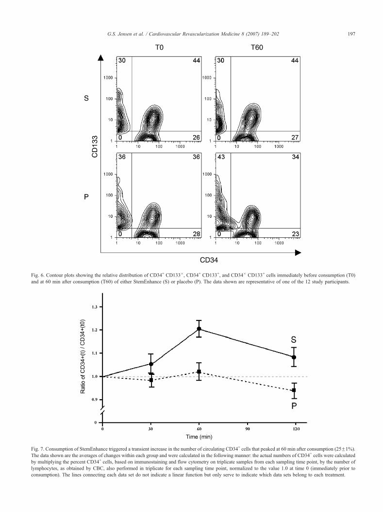
Fig. 6. Contour plots showing the relative distribution of CD34+ CD133�, CD34+ CD133+, and CD34� CD133+ cells immediately before consumption (T0)
and at 60 min after consumption (T60) of either StemEnhance (S) or placebo (P). The data shown are representative of one of the 12 study participants.
Fig. 7. Consumption of StemEnhance triggered a transient increase in the number of circulating CD34+ cells that peaked at 60 min after consumption (25F1%).
The data shown are the averages of changes within each group and were calculated in the following manner: the actual numbers of CD34+ cells were calculated
by multiplying the percent CD34+ cells, based on immunostaining and flow cytometry on triplicate samples from each sampling time point, by the number of
lymphocytes, as obtained by CBC, also performed in triplicate for each sampling time point, normalized to the value 1.0 at time 0 (immediately prior to
consumption). The lines connecting each data set do not indicate a linear function but only serve to indicate which data sets belong to each treatment.
G.S. Jensen et al. / Cardiovascular Revascularization Medicine 8 (2007) 189–202 197

G.S. Jensen et al. / Cardiovascular Revascularization Medicine 8 (2007) 189–202198
3.4. In vivo: consumption of an AFA extract rich in AFA-LSL
resulted in a transient increase of circulating CD34+ cells
The level of circulating CD34+ stem cells was compared
before and after ingestion of 1 g of the AFA-LSL-rich
extract StemEnhance or placebo. The staining included both
Fig. 8. Multiple testing on one individual on 16 different test days revealed a simila
the response varied between tests. The box plot shows the 25–75% spread of the re
CD34-FITC and CD133-PE on the PBMC, and analysis was
performed on CD34+ overall in parallel to CD34+ CD133�,
CD34+ CD133+ and CD34� CD133+. The change in
percent CD34+ lymphocytes from T0 to 60 min after
consumption (T60) of either StemEnhance or placebo are
shown for 1 out of the 12 study participants (Fig. 5).
r response to StemEnhance on different test days. However, the magnitude of
sults, the median (M), the average (A), and the lowest and highest response.

Table 1
Lack of effect of StemEnhance on the level of white blood cells and the lymphocyte subset
StemEnhance Placebo
Time (min) 0 30 60 120 0 30 60 120
WBC 100 104F1 104F1 109F5 100 107F3 104F3 111F4
Lymphocytes 100 107F2 108F2 118F5 100 109F3 109F3 124F5
WBC, white blood cells.
G.S. Jensen et al. / Cardiovascular Revascularization Medicine 8 (2007) 189–202 199
Only the analysis of CD34+ showed a significant
difference upon consumption of StemEnhance. The relative
distribution of CD34 vs. CD133 on the lymphocyte
population, gated to exclude any cell that did not express
either stem cell marker, is shown in Fig. 6. The proportion
of CD34+ CD133+ vs. CD34+ CD133� cells remained
constant, indicating that the mobilized progenitor cells
included cells of both phenotypes.
When including all volunteers, ingestion of StemEnhance
resulted in an 18F3% increase in the number of circulating
CD34+ cells, maximizing around 60 min after ingestion
(Pb.0003). This was in contrast to placebo, which resulted in
only minor fluctuations of the levels of CD34+ cells in the
blood circulation over 2 h. Questionnaires completed by the
volunteers on every experimental day revealed that three of
the volunteers met criteria for exclusion (e.g., significant lack
of sleep, severe anxiety) on at least one experimental day.
Exclusion of these volunteers in the analysis resulted in a
25F1% increase in the number of circulating stem cells at
60 min (Pb.0001) (Fig. 7). As expected a priori, effects
for people (ID), the interaction between people (ID), and
the interactions between people and treatment type
(StemEnhance or placebo) and people and time differed
significantly. There was no significant difference between
replicate analyses, nor the interactions between replicate
analyses and treatment type (StemEnhance or placebo) and
replicate analysis and time. That is, the analytical
variability was similar across people, treatment types,
and times. Also, overall analysis on normalized data for
each study participant showed significance.
In order to test the repeatability of the effect of
consumption of StemEnhance on the levels of CD34+ cells
in the peripheral blood, 16 separate experiments were
performed on one volunteer. The average increase in the
number of circulating stem cells was 53F16%, with a
median of 36% and a highest and lowest increase of 233%
and �4%, respectively (Fig. 8).
No statistically significant changes were observed when
comparing numbers of total leukocytes or lymphocytes
between the two treatments (StemEnhance vs. placebo)
(Table 1).
4. Discussion
Dietary strategies for supporting stem cell biology
represent an emerging field of nutritional medicine. The
understanding of the effect of nutrition on stem cells needs
to include stem cell viability, proliferation, mobilization,
and tissue-specific homing. It has been reported that
Spirulina consumption may increase erythropoiesis in a
mouse model [33]. In addition, antioxidant-rich blueberry
and green tea extracts have been reported to increase stem
cell life span and proliferation in vitro [34], which may
provide a better understanding of some aspects of
antioxidant therapy in aging.
This study involving human stem cell mobilization was
triggered by a few cases of empirical evidence that
consumption of an extract from AFA, enriched for the
LSL, resulted in unexpected extent of recovery after
traumatic injuries to the central nervous system. The
mobilization of stem cells is complex but involves two
key features: (1) interference with the adhesion of stem cells
to the bone marrow via L-selectin and (2) a reduction of the
chemotactic response to SDF-1 via the CXCR4 chemokine
receptor. We found that the cyanobacterium AFA contains a
novel compound that specifically binds to the ligand-
binding area of human L-selectin. It is composed of two
subunits with apparent molecular weight around 54–57 kDa
under reducing conditions. This compound differs from the
100,000 kDa polysaccharide from AFA previously
described [35]. This ligand for human L-selectin, precipi-
tated from AFA water extract (AFA-W), was able to
modulate the functional response on human lymphocytes
in vitro and interfered with the up-regulation of CXCR4
when bone marrow stem cells were exposed to another LSL,
fucoidan. In parallel to human bone marrow stem cells, the
primitive CD34bright KG1a cell line was responsive to L-
selectin-mediated up-regulation of CXCR4, possibly due to
its stage of differentiation being comparative to a subset of
bone marrow-resident stem cells. The ability of AFA-W to
down-regulate the expression of CXCR4 on BMSC and
KG1a, but not K562, suggests that this ligand could play a
role in stem cell mobilization from the bone marrow.
This may specifically have relevance for mobilization of
stem cells from marrow into the circulation, as it has
previously been shown that interference with the SDF-1/
CXCR4 axis is a primary mechanism of stem cell
mobilization from the bone marrow [11]. The LSL may
also have a direct effect on stem cell release, as LSLs have
been proposed as a therapeutic method for stem cell release
and increase of the number of circulating stem cells [29].
A double-blinded placebo-controlled crossover study
involving 12 healthy subjects showed that consumption of
an AFA extract enriched in this ligand (StemEnhance)
resulted in a small but significant increase in the number of

G.S. Jensen et al. / Cardiovascular Revascularization Medicine 8 (2007) 189–202200
circulating CD34+ stem cells, peaking at 1 h after
consumption. The effect was statistically significant
(Pb.0001). When tested on one individual on many
occasions, the increase in the number of circulating stem
cells after consumption of StemEnhance averaged 52F16%
and varied greatly from 96% to 333% of baseline value.
Interestingly, the average response in the one individual
tested repeatedly on 16 different study days, and the average
response to StemEnhance in the double-blind randomized
study involving 12 people was similar, with an increase in
CD34+ cells at 153% vs. 125%, respectively. The hypoth-
esis that StemEnhance transiently increases the levels of
circulating CD34+ cells is supported by significance for the
difference between the two treatments and the interactions
of this difference with person and time. This suggests a
significant consistency in the response, despite day-to-day
fluctuations, which may have contributed to an under-
estimation of the response to StemEnhance in the double-
blind study.
The increase in the number of circulating CD34+ cells
peaked within 1 h after consumption of StemEnhance. This
is in contrast with the response time seen with the known
mobilizer granulocyte colony-stimulating factor (G-CSF),
the response of which peaks after a few days of injection
[36,37]. It is believed that G-CSF triggers stem cell
mobilization by activating proteolytic activity in the
marrow, which degrades SDF-1, interfering with the SDF-
1/CXCR4 axis [11]. More comparable to StemEnhance is
the response to the CXCR4 antagonist AMD3100 that peaks
around 6 h after injection [38]. This supports the view that
the effect of StemEnhance on stem cell mobilization may be
caused by its LSL, down-regulating the expression of
CXCR4. The magnitude of the mobilization obtained with
StemEnhance (18–25%) is much smaller than what is seen
with G-CSF and AMD3100 (20- to 200-fold [36,37]).
Recent studies using G-CSF and AMD3100 have added
evidence for the potential role of stem cell mobilizers in the
mitigation of various diseases such as cardiomyopathies
[39,40], kidney failure [41], multiple sclerosis [42], stroke
[43,44], wound healing [45], as well as many other health
conditions [46]. Such compounds, however, can only be
used for short periods of time due to severe side effects [28].
However, such an extreme increase in the number of
circulating stem cells may not be required to achieve health
benefits. Tomoda and Aoki [47] quantified the level of
circulating stem cells in victims of acute myocardial
infarction and reported that individuals with more stem
cells showed greater recovery of ejection fraction 6 months
after the incident. Werner et al. [48] related the levels of
circulating stem cells with the risk of cardiovascular
incidents in 519 patients with coronary artery disease and
concluded that the level of circulating CD34+ endothelial
progenitor cells predicted the occurrence of cardiovascular
events and death from cardiovascular causes.
Two recent publications have further confirmed the
association between G-CSF-mediated stem cell mobilization
after acute myocardial infarction (AMI) and improved
cardiac repair. The degenerative remodeling of the heart
often seen over time after AMI was prevented by G-CSF
treatment, as long as percutaneous coronary intervention
(PCI) was performed early rather than late [49]. G-CSF
treatment provided a significant increase in the short-term
myocardial perfusion [50]. The conclusions presented from
these two trials are different from a Korean study in which
G-CSF-mediated mobilization alone had little effect on
increased cardiovascular output [51]. The Korean study
found greater effect when intracoronary injection with stem
cells was performed in conjunction with G-CSF treatment. It
is important to note the different timing of G-CSF treatment
in relationship to the PCI procedure when comparing these
two studies. In the German study protocol, the PCI was
performed first and then followed by G-CSF injections, and
the G-CSF injections were started within 90 min after
PCI. In the Korean study protocol, an initial 4-day course
of G-CSF injections were followed by the PCI, and no
further G-CSF treatment was given after the PCI procedure
was completed. The relative simplicity of G-CSF injections
as a singular treatment option makes this an attractive option
if proven to provide significant benefit to the patients. In
addition to further understanding the consequences from the
two different study protocols, it would also be interesting to
further evaluate whether geographical differences exist in the
underlying causes of cardiac function, including diet- and
stress-related factors, as well as additional differences in
medical management of cardiac disease prior to and during
myocardial infarct between studies.
At this point, the effect of a massive but transient
increase in the number of circulating stem cells, as induced
by G-CSF injection, has not been compared to the effect of a
mild but daily increase in the number of circulating stem
cells, such as what is seen after consumption of StemEn-
hance. The 25% increase in the number of circulating stem
cells occurring after ingestion of 1 g of StemEnhance may
have positive effects on various health conditions and, when
triggered daily for several weeks or months, might contrib-
ute to cardiac tissue regeneration. Studies should be
conducted to investigate the potential of such approach for
therapeutic purposes.
The observation that the mobilized progenitor cells
included cells of both the CD34+ CD133+ and CD34+
CD133� phenotypes may indicate that a broader range of
different types of progenitor cells are affected, instead of a
single type or developmental stage of stem cells. Circulat-
ing CD133+ cells are reported to include endothelial
progenitor cells, which play a role in endothelial repair.
The study by Engelmann et al. [50] showed that the
phenotypes of G-CSF-mobilized stem cells contributing to
this improvement included CD31+ CD34+ CD117+
CD133+ cells. In patients with coronary heart disease, a
reduced number of circulating CD133+ cells has been
proposed as an independent risk factor for erectile
dysfunction [52]. In particular, our data raises the question

G.S. Jensen et al. / Cardiovascular Revascularization Medicine 8 (2007) 189–202 201
as to whether daily consumption of StemEnhance may
counteract the reduced number of CD133+ cells in the
circulation of patients with cardiovascular disease linked to
endothelial dysfunction, including erectile dysfunction.
Empirical observations suggest that consumption of Ste-
mEnhance for longer periods of time might indeed bring
significant improvement in various health conditions,
including specific neurodegenerative diseases, chronic
obstructive pulmonary disease, kidney insufficiency, and
other degenerative problems. However, rigorous studies are
necessary to examine the effects of StemEnhance on
specific degenerative diseases.
In conclusion, our data presents the observation that
consumption of an herbal extract can significantly alter the
proportion of stem cells in the circulation. The novel LSL
isolated from AFA has complex biological activities in vitro
that could explain the increase in circulating stem cells
observed after consumption of the AFA extract StemEn-
hance in vivo. The extent to which this LSL may be
responsible for the in vivo effect on stem cell mobilization
and the effect of such mobilization on various health
conditions are currently subject for further study.
Acknowledgments
We are grateful to Kelly M. Patterson and Amber R.
Coaty for technical assistance.
References
[1] Gallatin WM, Weissman IL, Butcher EC. A cell-surface molecule
involved in organ-specific homing of lymphocytes. Nature
1983;304(5921):30–4.
[2] Tedder TF, Matsuyama T, Rothstein D, Schlossman SF, Morimoto C.
Human antigen-specific memory T cells express the homing receptor
(LAM-1) necessary for lymphocyte recirculation. Eur J Immunol
1990;20(6):1351–5.
[3] van Zante A, Rosen SD. Sulphated endothelial ligands for L-selectin
in lymphocyte homing and inflammation. Biochem Soc Trans
2003;31(2):313–7.
[4] Frenette PS, Wagner DD. Insights into selectin function from
knockout mice. Thromb Haemost 1997;78(1):60–4.
[5] Rainer TH, Ng MH, Lam NY, Chan TY, Cocks RA. Role of monocyte
L-selectin in the development of post-traumatic organ failure.
Resuscitation 2001;51(2):139–49.
[6] Rosen SD. Ligands for L-selectin: homing, inflammation, and beyond.
Annu Rev Immunol 2004;22:129–56.
[7] Khan AI, Kubes P. L-selectin: an emerging player in chemokine
function. Microcirculation 2003;10(3–4):351–8.
[8] Barkhausen T, Krettek C, van Griensven M. L-selectin: adhesion,
signalling and its importance in pathologic posttraumatic endotox-
emia and non-septic inflammation. Exp Toxicol Pathol 2005;57(1):
39–52.
[9] Frenette PS, Subbarao S, Mazo IB, von Andrian UH, Wagner DD.
Endothelial selectins and vascular cell adhesion molecule-1 promote
hematopoietic progenitor homing to bone marrow. Proc Natl Acad Sci
U S A 1998;95(24):14423–8.
[10] Hidalgo A, Weiss LA, Frenette PS. Functional selectin ligands
mediating human CD34(+) cell interactions with bone marrow
endothelium are enhanced postnatally. J Clin Invest 2002;110(4):
559–69.
[11] Petit I, Szyper-Kravitz M, Nagler A, Lahav M, Peled A, Habler L,
Ponomariov T, Taichman RS, Arenzana-Seisdedos F, Fujii N,
Sandbank J, Zipori D, Lapidot T. G-CSF induces stem cell
mobilization by decreasing bone marrow SDF-1 and up-regulating
CXCR4. Nat Immunol 2002;3(7):687–94.
[12] Ding Z, Issekutz TB, Downey GP, Waddell TK. L-selectin stimulation
enhances functional expression of surface CXCR4 in lymphocytes:
implications for cellular activation during adhesion and migration.
Blood 2003;101(11):4245–52.
[13] Eaves CJ. SDF-1 tells stem cells to mind their P’s and Z’s. J Clin
Invest 2005;115(1):27–9.
[14] Hidalgo A, Sanz-Rodriguez F, Rodriguez-Fernandez JL, Albella B,
Blaya C, Wright N, Cabanas C, Prosper F, Gutierrez-Ramos JC,
Teixido J. Chemokine stromal cell-derived factor-1alpha modulates
VLA-4 integrin-dependent adhesion to fibronectin and VCAM-1 on
bone marrow hematopoietic progenitor cells. Exp Hematol 2001;
29(3):345–55.
[15] Kollet O, Spiegel A, Peled A, Petit I, Byk T, Hershkoviz R, Guetta E,
Barkai G, Nagler A, Lapidot T. Rapid and efficient homing of human
CD34(+)CD38(�/low)CXCR4(+) stem and progenitor cells to the
bone marrow and spleen of NOD/SCID and NOD/SCID/B2m(null)
mice. Blood 2001;97(10):3283–91.
[16] Gazitt Y. Immunologic profiles of effector cells and peripheral blood
stem cells mobilized with different hematopoietic growth factors.
Stem Cells 2000;18(6):390–8.
[17] Son BR, Marquez-Curtis LA, Kucia M, Wysoczynski M, Turner AR,
Ratajczak J, Ratajczak MZ, Janowska-Wieczorek A. Migration of
bone marrow and cord blood mesenchymal stem cells in vitro is
regulated by stromal-derived factor-1-CXCR4 and hepatocyte growth
factor-c-met axes and involves matrix metalloproteinases. Stem Cells
2006;24(5):1254–64.
[18] Togel F, Isaac J, Hu Z, Weiss K, Westenfelder C. Renal SDF-1 signals
mobilization and homing of CXCR4-positive cells to the kidney after
ischemic injury. Kidney Int 2005;67(5):1772–84.
[19] Kollet O, Shivtiel S, Chen YQ, Suriawinata J, Thung SN, Dabeva MD,
Kahn J, Spiegel A, Dar A, Samira S, Goichberg P, Kalinkovich A,
Arenzana-Seisdedos F, Nagler A, Hardan I, Revel M, Shafritz DA,
Lapidot T. HGF, SDF-1, and MMP-9 are involved in stress-induced
human CD34+ stem cell recruitment to the liver. J Clin Invest
2003;112(2):160–9.
[20] Lazarini F, Tham TN, Casanova P, Arenzana-Seisdedos F, Dubois-
Dalcq M. Role of the alpha-chemokine stromal cell-derived factor
(SDF-1) in the developing and mature central nervous system. Glia
2003;42(2):139–48.
[21] Reiss K, Mentlein R, Sievers J, Hartmann D. Stromal cell-derived
factor 1 is secreted by meningeal cells and acts as chemotactic factor
on neuronal stem cells of the cerebellar external granular layer.
Neuroscience 2002;115(1):295–305.
[22] Abbott JD, Huang Y, Liu D, Hickey R, Krause DS, Giordano FJ.
Stromal cell-derived factor-1alpha plays a critical role in stem cell
recruitment to the heart after myocardial infarction but is not sufficient
to induce homing in the absence of injury. Circulation 2004;110(21):
3300–5.
[23] Tang YL, Qian K, Zhang YC, Shen L, Phillips MI. Mobilizing of
haematopoietic stem cells to ischemic myocardium by plasmid
mediated stromal-cell-derived factor-1alpha (SDF-1alpha) treatment.
Regul Pept 2005;125(1–3):1–8.
[24] Kucia M, Ratajczak J, Reca R, Janowska-Wieczorek A, Ratajczak
MZ. Tissue-specific muscle, neural and liver stem/progenitor cells
reside in the bone marrow, respond to an SDF-1 gradient and are
mobilized into peripheral blood during stress and tissue injury. Blood
Cells Mol Dis 2004;32(1):52–7.
[25] Mohle R, Boehmler AM, Denzlinger C, Kanz L. Nonpeptide
mediators in the hematopoietic microenvironment. Ann N Y Acad
Sci 2003;996:61–6.

G.S. Jensen et al. / Cardiovascular Revascularization Medicine 8 (2007) 189–202202
[26] Aiuti A, Webb IJ, Bleul C, Springer T, Gutierrez-Ramos JC. The
chemokine SDF-1 is a chemoattractant for human CD34+ hema-
topoietic progenitor cells and provides a new mechanism to explain
the mobilization of CD34+ progenitors to peripheral blood. J Exp Med
1997;185(1):111–20.
[27] Papayannopoulou T, Priestley GV, Bonig H, Nakamoto B. The role of
G-protein signaling in hematopoietic stem/progenitor cell mobiliza-
tion. Blood 2003;101(12):4739–47.
[28] Cottler-Fox MH, Lapidot T, Petit I, Kollet O, DiPersio JF, Link D,
Devine S. Stem cell mobilization. Hematology (Am Soc Hematol
Educ Program) 2003;419–37.
[29] Frenette PS, Weiss L. Sulfated glycans induce rapid hematopoietic
progenitor cell mobilization: evidence for selectin-dependent and
independent mechanisms. Blood 2000;96(7):2460–8.
[30] Sweeney EA, Priestley GV, Nakamoto B, Collins RG, Beaudet AL,
Papayannopoulou T. Mobilization of stem/progenitor cells by sulfated
polysaccharides does not require selectin presence. Proc Natl Acad Sci
U S A 2000;97(12):6544–9.
[31] Atanackovic D, Schnee B, Schuch G, Faltz C, Schulze J, Weber CS,
Schafhausen P, Bartels K, Bokemeyer C, Brunner-Weinzierl MC,
Deter HC. Acute psychological stress alerts the adaptive immune
response: stress-induced mobilization of effector T cells. J Neuro-
immunol 2006;176(1–2):141–52.
[32] Spertini O, Kansas GS, Reimann KA, Mackay CR, Tedder TF.
Function and evolutionary conservation of distinct epitopes on the
leukocyte adhesion molecule-1 (TQ-1 Leu-8) that regulate leukocyte
migration. J Immunol 1991;147(3):942–9.
[33] Zhang C-W. Effects of polysaccharide and phycocyanin from spirulina
on peripheral blood and hematopoietic system of bone marrow in
mice. Nanjing Univ China Pub in Proc of Second Asia Pacific Conf
on Algal Biotech Univ of Malaysia, April 1994 p. 58 [China].
[34] Bickford PC, Tan J, Shytle RD, Sanberg CD, El-Badri N, Sanberg PR.
Nutraceuticals synergistically promote proliferation of human stem
cells. Stem Cells Dev 2006;15(1):118–23.
[35] Pugh N, Ross SA, ElSohly HN, ElSohly MA, Pasco DS. Isolation of
three high molecular weight polysaccharide preparations with potent
immunostimulatory activity from Spirulina platensis, Aphanizome-
non flos-aquae and Chlorella pyrenoidosa. Planta Med 2001;67(8):
737–42.
[36] Bodine DM, Seidel NE, Orlic D. Bone marrow collected 14 days after
in vivo administration of granulocyte colony-stimulating factor and
stem cell factor to mice has 10-fold more repopulating ability than
untreated bone marrow. Blood 1996;88(1):89–97.
[37] Majolino I, Buscemi F, Scime R, Indovina A, Santoro A, Vasta S,
Pampinella M, Catania P, Fiandaca T, Caronia F, et al. Treatment of
normal donors with rhG-CSF 16 micrograms/kg for mobilization of
peripheral blood stem cells and their apheretic collection for
allogeneic transplantation. Haematologica 1995;80(3):219–26.
[38] Broxmeyer HE, Orschell CM, Clapp DW, Hangoc G, Cooper S, Plett
PA, Liles WC, Li X, Graham-Evans B, Campbell TB, Calandra G,
Bridger G, Dale DC, Srour EF. Rapid mobilization of murine and
human hematopoietic stem and progenitor cells with AMD3100, a
CXCR4 antagonist. J Exp Med 2005;201(8):1307–18.
[39] Kong D, Melo LG, Gnecchi M, Zhang L, Mostoslavsky G, Liew CC,
Pratt RE, Dzau VJ. Cytokine-induced mobilization of circulating
endothelial progenitor cells enhances repair of injured arteries.
Circulation 2004;110(14):2039–46.
[40] Orlic D, Kajstura J, Chimenti S, Limana F, Jakoniuk I, Quaini F,
Nadal-Ginard B, Bodine DM, Leri A, Anversa P. Mobilized bone
marrow cells repair the infracted heart, improving function and
survival. Proc Natl Acad Sci U S A 2001;98(18):10344–9.
[41] Iwasaki M, Adachi Y, Minamino K, Suzuki Y, Zhang Y, Okigaki M,
NakanoK, KoikeY,Wang J,MukaideH, Taketani S,Mori Y, Takahashi
H, Iwasaka T, Ikehara S. Mobilization of bone marrow cells by G-CSF
rescues mice from cisplatin-induced renal failure, andM-CSF enhances
the effects of G-CSF. J Am Soc Nephrol 2005;16(3):658–66.
[42] Saccardi R, Mancardi GL, Solari A, Bosi A, Bruzzi P, Di
Bartolomeo P, Donelli A, Filippi M, Guerrasio A, Gualandi F, La
NasaG,MurialdoA, Pagliai F, Papineschi F, Scappini B,MarmontAM.
Autologous HSCT for severe progressive multiple sclerosis in a
multicenter trial: impact on disease activity and quality of life. Blood
2005;105(6):2601–2607.
[43] Kawada H, Takizawa S, Takanashi T, Morita Y, Fujita J, Fukuda K,
Takagi S, Okano H, Ando K, Hotta T. Administration of hema-
topoietic cytokines in the subacute phase after cerebral infarction is
effective for functional recovery facilitating proliferation of intrinsic
neural stem/progenitor cells and transition of bone marrow-derived
neuronal cells. Circulation 2006;113(5):701–10.
[44] Shyu WC, Lin SZ, Yang HI, Tzeng YS, Pang CY, Yen PS, Li H.
Functional recovery of stroke rats induced by granulocyte colony-
stimulating factor-stimulated stem cells. Circulation 2004;110(13):
1847–54.
[45] Bozlar M, Aslan B, Kalaci A, Baktiroglu L, Yanat AN, Tasci A.
Effects of human granulocyte-colony stimulating factor on fracture
healing in rats. Saudi Med J 2005;26(8):1250–4.
[46] Kan I, Melamed E, Offen D. Integral therapeutic potential of bone
marrow mesenchymal stem cells. Curr Drug Targets 2005;6(1):31–41.
[47] Tomoda H, Aoki N. Bone marrow stimulation and left ventricular
function in acute myocardial infarction. Clin Cardiol 2003;26(10):
455–7.
[48] Werner N, Kosiol S, Schiegl T, Ahlers P, Walenta K, Link A, BohmM,
Nickenig G. Circulating endothelial progenitor cells and cardiovascular
outcomes. N Engl J Med 2005;353(10):999–1007.
[49] Ince H, Petzsch M, Kleine HD, Eckard H, Rehders T, Burska D,
Kische S, Freund M, Nienaber CA. Prevention of left ventricular
remodeling with granulocyte colony-stimulating factor after acute
myocardial infarction: final 1-year results of the Front-Integrated
Revascularization and Stem Cell Liberation in Evolving Acute
Myocardial Infarction by Granulocyte Colony-Stimulating Factor
(FIRSTLINE-AMI) trial. Circulation 2005;112(Suppl 9):I73–80.
[50] Engelmann MG, Theiss HD, Hennig-Theiss C, Huber A, Winter-
sperger BJ, Werle-Ruedinger AE, Schoenberg SO, Steinbeck G, Franz
WM. Autologous bone marrow stem cell mobilization induced by
granulocyte colony-stimulating factor after subacute ST-segment
elevation myocardial infarction undergoing late revascularization:
final results from the G-CSF-STEMI (Granulocyte Colony-Stimulat-
ing Factor ST-Segment Elevation Myocardial Infarction) trial. J Am
Coll Cardiol 2006;48(8):1712–21.
[51] Kang HJ, Kim HS, Koo BK, Kim YJ, Lee D, Sohn DW, Oh BH, Park
YB. Intracoronary infusion of the mobilized peripheral blood stem cell
by G-CSF is better than mobilization alone by G-CSF for improve-
ment of cardiac function and remodeling: 2-year follow-up results of
the Myocardial Regeneration and Angiogenesis in Myocardial
Infarction with G-CSF and Intra-Coronary Stem Cell Infusion
(MAGIC Cell) 1 trial. Am Heart J 2007;153(2):237.e1–8.
[52] Baumhakel M, Werner N, Bohm M, Nickenig G. Circulating
endothelial progenitor cells correlate with erectile function in patients
with coronary heart disease. Eur Heart J 2006;27(18):2184–8.