mmed dissertation december 2007 a study of perforated
TRANSCRIPT
MMED DISSERTATION
DECEMBER 2007
A Study of Perforated Acute Appendicitis at the University Teaching Hospital, Lusaka, Zambia
B Y
DR PETER S A M U E L PHIRI BSc (HB), M B C H B (UNZA)
A Dissertation submitted to the University of Zambia in a partial fiilfillment of the requirement for the degree of Master of Medicine in General Surgery.
(School of Medicine) THE UNIVERSITY OF ZAMBIA
LUSAKA

COPYRIGHT DECLARATION
I, PHIRI S A M U E L PETER, hereby declare that this dissertation represents my own work
and that it has never previously been published in part or in full for a diploma or degree
in any university.
Date: / Candidate signature....
I have read this dissertation and approve it for examination.
Date. .Q^.j^.^£>^.^.^ Supervisors sig nature../
Supervisor: Prof Kosher Odimba
Professor of surgery
Department of surgery
School of medicine
University of Zambia
i i

APPROVAL
This dissertation of DR PHIRI PETER SAMUEL is approved in partial
fulfillment of the requirements for the award of the Master of Medicine in
Surgery by the University of Zambia.
Date
3
4
ii i
EXAMINERS
Signature

ABBREVIATIONS
B M J : British Medical Journal
E. Afr. Med J: East African Medical Journal
UTH: University Teaching Hospital
USA: United States of America
SSI: Surgical Site Infection
iv

ETHICAL CONSIDERATION
I. Informed consent was obtained for the study.
II. Standard management and operative techniques were used, so ethical consideration
did not arise.
V

ACKNOWLEDGEMENT
I would like to thank my supervisor Prof B.F.K. Odimba consultant general surgeon and
coordinator of the Masters programme who guided me in the prepeiration, implementation
and write up of this dissertation.
Candidate signature
vi

SUMMARY
A prospective descriptive study of perforated appendicitis as seen at The University
Teaching hospital (UTH), Lusaka, Zambia, was carried out over a period of nine
months.(l^' March to 30'̂ November 2007). The aim was to establish the appendiceal
perforation rate, to describe some of the factors associated with perforated appendicitis as
well as to describe the major associated early complications.
The inclusion criteria were a confirmed intra operative diagnosis of perforated or non
perforated appendicitis. A l l Patients were recruited into the study until the sample size
was reached. The details of each patient were entered on an evaluation form designed for
the study. Each patient was followed up for four weeks.
A total of 71 appendicectomies were done. The appendiceal perforation rate was at 43.6
percent. 64.5 percent presented with generalized peritonitis necessitating laparotomy
through the midline. The male to female ratio of perforation was 2.5:1. The commonest
perforations were in the 30 to 40 year age group. The majority of those with perforation
presented between the third and fifth days after the onset of symptoms whereas the
majority of those with non-perforated acute appendicitis presented within the first forty
eight hours. The main factor attributed to perforation was pre- hospital delay by the
patient. 50 percent of those with perforation came Irom highly populated residential areas
and with poor socioeconomic background and subsequent poor access to quality health
care. 11 percent used traditional medicine prior to admission to UTH. There was no in-
hospital delay attributed to the surgeons or surgery.
Perforation was associated with high levels of morbidity with a 33.3 percent wound
infection and a further 22.2 percent requiring relaparotomies for intra abdominal
abscesses. The overall mortality rate was 1.4 percent.
The high rate of perforated appendicitis is due to pre-hospital patients' delay, therefore,
public education, specifically targeting those groups at risk, may provide a significant
solution to the problem.
vii

LIST OF TABLES AND FIGURES
Table 1 14
Figure 1 14
Table 2 15
Figure 2 15
Table 3 16
Figure 3 16
Table 4 17
Figure 4 17
Table 5 18
Figure 5 18
Table 6 19
Figure 6 19
Table 7 20
Figure 7 20
Table 8 21
Figure 8 21
Table 9 22
Figure 9 22
Table 10 23
Figure 10 23
Table 11 24
Figure 11 24
Table 12 25
Figure 12 25
Table 13 26
Figure 13 26
Table 14 27
Figure 14 27
Table 15 28
Figure 15 28
Table 16 ? 29
Figure 16 29
Table 17 30
Figure 17 30
Table 18 31
Figure 18 31
T A B L E O F C O N T E N T S
Certification i
Declaration ii
Approval i i i
Abbreviations iv
Ethical consideration v
Acknowledgement vi
Summary vii
List of tables and figures viii
Table of contents ix
Dedication x
Definition of terms • • • 1
Introduction 3
Objectives 4
Rationale 5
Literature review 6
Patients and method 9
Results 12
Discussion 32
Conclusion 37
Recommendation 38
Reference (Bibliography) 39
Appendix 44
ix

DEDICATION
This work is dedicated to my wife Mildred and my two sons Peter and Juma who were so
supportive during the study period.
X

DEFINITION OF TERMS
The University Teaching Hospital is a referral hospital in the capital city, Lusaka,
Zambia. It receives both referred and direct patients.
(I) Total operations:
Refers to all operations performed in the department of surgery.
(H) Abdominal surgery: Was defined as all the operations performed in the abdominal cavity.
(iii) Acute appendicitis:
Patients who presented for the first time with typical features of appendicitis and were
submitted for appendicectomy were all included under this.
(iv) Generalized peritonitis:
Patients who had appendicitis complicated by pus within the peritoneal cavity.
(v) Laparotomy:
Was defined as an opening of the peritoneal cavity.
(vi) Perforated appendix:
Was defined as an inflamed appendix comphcated by macroscopic perforation of its wall.
(vii) Wound infection:
Was defmed as inflammation and induration witii or without abscess formation.
I
(viii) Morbidity:
Was defined as postoperative complication occurring within a month from the time of
operation.
(ix) Mortality:
Was defined as death occurring within a month after the operation.
(x) Short term complication:
Was defined as a complication occurring within a period of four weeks from the time of
operation.
(xi) Prevalence
Number of cases present in a population in a particular time.
(xii) Incidence:
Number of new cases which occur in a population over a defined period of time.
(xiii) Rate:
Number of events in a period of time
Statistical significance is defined as a probability of less than 1 in 20 of an event being
the result of chance, i.e. P < 0.05.
Statistical analysis was confined to the use of basic tables accepting the converting level
of p < 0.05 as significant.
2

BmiODUCTION
Perforation of the vermiform appendix is the most severe comphcation of acute
appendicitis .This is associated with high levels of mortality and morbidity as compared
to appendicectomy for simple appendicitis, Ae reason being that perforation is associated
with the development of an appendicular abscess with subsequent septicaemia and shock
or with the development of generalised peritonitis.' For the purpose of this study,
perforation was defmed as an appendix with a visible hole at Laparotomy.^ The outcome
of non-perforated appendicitis is favourable, and because the morbidity and mortality
increases sharply with perforation, the priority in assessing patients with appendicitis
should be to perform a prompt appendicectomy but if this is delayed in any way,
perforation ensues. However, little is known on the fectors that lead to high levels of
perforation at the University Teaching Hospital, Lusaka, Zambia. This study addresses
the issue.
3

OBJECTIVES:
1. To determine the number of patients presenting with perforated appendicitis at
The University Teaching Hospital.
2. To determine the main factors associated with high levels of perforated
appendicitis at The University Teaching Hospital.
3. To compare the associated factors in those with perforated appendicitis to those
presenting with non-perforated appendicitis.
4. To describe and compare the short-term complications associated with surgery in
patients with perforated and non perforated appendicitis at The University
Teaching Hospital.
4

RATIONALE
Available data has demonstrated an appendiceal perforation rate of 32 % at the
University Teaching hospital with rates of 46 % and 49 % in the very young and very old
respectively. Authors do agree that perforation of the appendix is associated with high
levels of morbidity and mortality. A study of this nature, which gives an insight into the
contributing factors for such a high figure, gives useful information which in turn can be
used as a starting point in intervening in an effort to reduce these levels.
5

L r r E R A T U R E R E V I E W
The first appendicectomy for perforated appendix was preformed by Claudius Amyand in
1735 on an 11 year boy and the boy recovered well.^ The second one was performed in
1848 on a 30 year old woman who had just delivered and presented with generalized
peritonitis secondary to rupture of the appendix.'* A similar operation was done by
Lawson Tait on a 17 year old female patient in whom the pre-operative diagnosis of
perforated appendix was already made.'
An outstanding contribution was fi-om Fitrz Gerald who in 1886 awakened the medical
profession in America about the importance of the vermiform appendix undergoing
inflammation and coined the term appendicectomy. He advocated for early
appendicectomy.^
Controversy existed about the timing of operative intervention through out the first
quarter of the twentieth century, particularly on the patients first seen with advanced
disease. Mortality at that time was shockingly high varying from 5% to 50 %f
In 1900, Mcbumey, popularly known for the Mcbumeys' point, emphasized on the
importance of early diagnosis and prompt surgical intervaition. Charles McBumey and
other pioneering surgeons began to intervene early in acute appendicitis.''^ These
clinicians advocated prompt clinical diagnosis and surgical intervention. Their surgical
aim was to operate in a timely &shion before appendiceal perforation and peritonitis
developed.
During the next three decades, the mortality reduced to about 5% as a result of
dissemination of information to the public and physicians on signs of appendicitis. In the
following twenty five years, a combination of improved surgical technique, better pre and
post operative care, advances in anestfiesia, and the development of antimicrobials
reduced the mortality rate to less than one percent**''''
The patient usually presents with peri-umbihcal pain which after a few hours shifts to the
right iliac fossae. If no surgical intervention is offered, the initial pain which was aching
in character suddoily becomes more severe, spreading over the remainder of the
abdomen as diffuse peritonitis develops. The general condition of the patients deteriorates
rapidly, the pulse rate increases and the temperature rises. Within a few hours, there
6
might be signs of circulatory failure. The patient looks i l l , dehydrated and toxic."
Peritonitis occurs as a result of free migration of bacteria from the inflamed or
gangrenous appendix. Factors which promote this include extremes of age,
immunosupresion, diabetes mellitus, faecalith obstruction and previous abdominal
surgery. Current reports indicate that appendicectomy represents one percent of all
surgical operations in the West."
A few decades ago, intestinal obstmction was the leading cause of abdominal emergency
admission in many tropical countries. There has been a change in pattem and acute
appendicitis has become the major cause of emergency admission world wide.'^
Appendicitis is more common in the western world as compared to Africa. This has been
attributed to high levels of refined diet in the West. However, Katzaski " in 1979
attributed the low levels in Zambians due to dual blood supply to the appendix in
Africans. In the U S A the incidence of acute appendicitis is at 11 per 10 000 population,
whereas in South Africa it is at 0.95 per 10 000 population. In Bulavv^yo, Zimbabwe,
only 20 cases were recorded by Oliver in one year in a population of 2.5 million people in
1987.'^ In Zambia, Haque " in 1997 showed that the incidence was at 0.79 per 10 000
population and that this accounted for 0.80 % of all the operations at U T H and that it
comprised 11% of all abdominal operations at UTH. Appendicitis is more common in
males than in females and predominantly occurs in the young people." In Ethiopia;
kottiso'* in 1996 reported a male to female ratio of 1.6:1.
If not diagnosed and treated promptly, as shown above, acute appendicitis complicates
by perforation. Nanda'^ in 2004 concluded that morbidity caused by acute appendicitis
correlated directly with delay in treatment. This in turn can lead either to local or
generalized peritonitis. This may be due to a number of reasons such as problems with
access to care, feilure by patient to interpret symptoms as important as well misdiagnosis
by clinicians.
Madiba ^ in 1998 demonstrated a perforation rate of 43% in a South Africa population.
In the same study, he also demonstrated that tiiose who presented with right-sided
7

abdominal pain out-numbered the classical presentation of periumbilical pain.
Mutupheni^' within the same year had demonstrated a perforation rate of 25 % within
the same population indicating differences according to location. Levy ^ in 1997
attributed perforations in black South Africans to delay in presentation though Walker ^
in 1989 had attributed this delay to the seeking of traditional treatment before
presentation to hospital because throughout Africa, traditional healers are held in high
esteem. He reported 35% seeking traditional treatment before seeking modem treatment.
Out ^ in 1989 showed that patients with acute appendicitis presented late with a median
of five days from the onset of symptoms with a perforation rate of 20%. Wilmore, in
2001 in a Kenyan hospital attributed the perforation to pre hospital delay though he did
not demonstrate the exact reasons for such a delay. On the other hand, Ofoegbu ^ 17
attributed the perforations to time spent in private hospitals. In Zambia, Haque in 1997
demonstrated a perforation rate of 32%. In the same study, it was shown that fte
perforation rate in the very young and very old was at 46 % and 49 % respectively.
A perforated appendix is associated with high levels of mortality and morbidity. Lee
demonstrated 28% morbidity and 2.3% mortality in those patients above sbrty years
presenting witfi perforated appendix. Walters ^ had earlier on dranonstrated that 25 % of
all cases of peritonitis at U T H were due to perforated appendix.
Madiba demonstrated two percent mortality in patients with perforated appendix
whereas here at UTH, Mwangala ^ demonstrated a 35% wound infection rate in those
with generalized peritonitis and 25% mortality in this group. The incidence of perforated
appendix has been on the increase and authors do agree that this is associated with
serious morbidity. For now, little is known on factors contributing to this at UTH. The
study addresses the issue.
8

PATIENTS AND METHODS
This was a nine month prospective cross section descriptive study carried out at the
University Teaching Hospital, Lusaka, Zambia from March to November 2007.
For participation in the study, the following criterion was followed.
I. Consent by patient or guardian to participate in the study,
n. A confirmed intra-operative diagnosis of perforated and non- perforated
appendicitis.
Patients were recruited from the five general surgical units as well as from the
paediatric surgical unit.
A questionnaire was administered to every recruited patient. Patients were seen on the
day of recruitment (day 0) and were followed up until discharge. They were seen after
a week and then at one month. The information included the sex, age, socioeconomic
background, referring clinic, duration of illness, use of traditional medicine and
surgical complications encountered.
Patient selection.
A l l patients were recruited into the study until the sample size was reached. A total of
36 patients were recruited of whom half had perforated and the other half non-
perforated appendicitis. The sex, age and nature of the appendicitis in those not
formally recruited into the study was also taken and analyzed.
Pre operative care
Patients for appendicectomy were admitted via casualty to either the male or female
surgical wards upon making a diagnosis of acute abdomen. A detailed history was
taken and fiiU examination done at which a diagnosis of acute appendicitis or that of
peritonitis was confirmed. This was primarily a clinical diagnosis and very few
investigations were done. The patients were then assessed for fitness to undergo
9

general anesthesia. A l l patients were resuscitated before being taken for surgery
depending on the general condition especially in those with peritonitis.
The operations
All operations were preceded by standard skin preparation wilh savlon, iodine and
methylated spirit. The patients were then covered with sterile drapes. For those with
simple appendicitis, a right sided gridiron incision of about six to eight centimeters
was made. For those with generalized peritonitis, a midline incision was used. The
appendix was removed in a standard way which included the burying of the stamp
whenever possible, hi the case of peritonitis, lavage with copious amounts of warm
saline was done with some surgeons leaving a drain into the abdomen. Mass closure
with nylon was done.
Post operative care
Patient progress was monitored on the ward on a daily basis and complications
recorded until tiie patients were discharged. They were reviewed after a week and
then after a month although this was difficult as some never bothered to come back
for the second review.
Ethical Considerations
Permission to cany out a study involving human beings was sought from the research
ethics committee of the University of Zambia. This study methodology has been used
before in other studies and is well acceptable.
There was no manipulation of humans during the study. Operations were decided
upon and done by the respective surgical units and their team of surgeons.
Permission -was requested from relevant authorities i.e. fi-om the U T H Managing
director for data collection, from heads of concerned units and consent from the
patients. Patients were also be given transport money for reviews.
The study subjects were treated with dignity and respect. Confidentiality was
maintained i.e. Participants' names and their study were unlinked
10

Sample size
A total of thirty two patients were recruited in the study. This was calculated using
the following formulae;
N = pqzVd^
Where N is the sample size
P is the prevalence
Qis 100-p
Z is 1.96
Andd = 5
For this study, a prevalence rate of 1% was used. The calculated sample size was
multiplied by two to take care of the perforated and non perforated appendicitis.
11

RESULTS
During the study period, a total of seventy three appendicectomies were done of whom
two were interval appendicectomies and so were not included in the study. Of the
remaining seventy one, thirty one were perforated with either local or generalized
peritonitis. The age ranged from six months to sixty years. Of those with perforation, 68
% had generalized peritonitis. Further analysis was only done in those formally recruited
in the study. 18 of these had perforation and the other 18 had simple appendicitis.
Table 1 and figure 1 show the frequency of perforated appendix during the study period
of 43.6%. Table 2 and figure 2 show the frequency of perforation according to age. Most
of the perforations were in the age group between 30 and 40. Table 3 and figure 3 show
the frequency of perforation according to sex. Most of the perforations were in males.
Table 4 and figure 4 show the distribution of perforation according to residential area.
High rate of perforation was associated with people from highly populated areas. Table 5
and figure 5 show the frequency of perforation in relationship to duration of illness at
home before admission to UTH. None of the patients with perforation came within the
first twenty four hours as opposed to those with no perforation in whom over 60 % came
within the first forty eight hours (p < 0.001).
Table 6 and figure 6 show Ae frequency of perforation in relation to mode of admission
to UTH. The mode of admission did not have an influence over perforation. Table 7 and
figure 7 show the frequency of perforation according to time spent at the private clinic
before referral to UTH. This did not contribute significantly as there was no delay
attributed to private clinic consultation.
Table 8 and figure 8 show the frequency of perforation in relation to private clinic
diagnosis. Table 9 and figure 9 show the frequency of perforation in relation to pre
admission antibiotic administration. Table 10 and figure 10 show the frequency of
perforation in relation to use of traditional medicine.
12

11 percent of those with perforation used traditional medicine before admission to UTH.
Table 11 and figure 11 show the frequency of perforation in relation to amount eamed
per month.
The majority of those with perforation eamed less than five hundred thousand kwacha
per month. Table 12 and figure 12 show the frequency of perforation in relation to family
size. Perforation is associated with households of six or more people Table 13 and figure
13 show the frequency of perforation in relation to education level of patient or guardian.
Most of the patients with perforation were associated with education level of less than
grade seven.
Table 14 and figure 14 show the frequency of perforation in relation to period spent at
U T H before actual surgery. The majority of the people from the two groups were taken
to theatre for the actual surgery after four hours.
Table 15 and figure 15 show tiie complications associated with appendicectomy. 33.3%
in the perforated group had wound infection and 22 % had re-laparotomy done between
the fourtii and the tenth post operative day.
Table 16 and figure 16 show the frequency of admission to intensive care vmit. Table 17
and figure 17 show the duration of post operative hospital stay after surgery. The
majority of those without perforation were discharged between the third and fifth post
operative day as opposed to those with perforation who were discharged after the sixth
post operative day with 22.2 % going beyond the tenth day.
Table 18 and figure 18 show the mortality associated with appendicectomy. 3.2 % of the
perforated group died where as none of the patients with simple appendicitis died. The
charts following the tables are a graphical presentation of the results in the tables.
13

TABLES AND FIGURES
Table 1. Frequency of perforated appendix during the study period.
Total appendicectomies done 71 Number of perforated appendicitis 31 Percentage 43.6 %
Figure 1. Frequency of perforated appendix during the study period
55.40%
43.60%
0 perforated • none perforated
The perforation rate was at 43.6 %
14

Table 2. Frequency of perforation according to age.
Age Total number of appendicectomi es
Perforated Percentage of total perforations
Perforation rate for the age group
< 10 08 04 12.9 50 11-20 10 06 19.3 60 21-30 19 05 16.1 26.3 31-40 24 13 41.9 54.1 41-60 09 02 06.4 22.2 >60 01 01 03.2 100 Total 71 31
Figure 2 Frequency of perforation according to age.
> 60
I 31-40
|> 21-30
11-20
< 10
0 5 10 15 20 25 30 35 40 45 %
The highest perforation rate was between the 30 to 40 age group and those below 20 and above 60 had the highest chance to perforate.
15

Table 3.Frequency of perforation according to sex
Sex Perforated Percentage Male 22 70.9 Female 09 29.1
Figures. Frequency of perforation according to sex.
8 0 -j 7 0 1 1 6 0 5 0 4 0 3 0 I 2 0 10
0 J J 1———. 1 m a l e f e m a l e
The majority of those with perforation were male with a male to female perforation ratio of 2.5:1.
16

Table 4. Distribution of perforation according to residential area.
Residential area
Low density Medium density
High density
Peri-urbum Rural Residential area
No. % No. % No. % No. % No. %
Perforated appendix
02 11 02 11 09 50 02 11 03 16
Non-perforated
04 22 07 38 05 27 02 11 00 00
Figure 4. Distribution of perforation according to residential area.
low medium high periurban urban
i E3 perforated • non perforated
50% of those presenting with perforation were from highly populated residential areas.
17

Table 5. Frequency of perforation in relationship to duration of illness at home before admission to U T H
Duration at home before admission to
UTH
<24 hours 25-48 3-5 days 6-7 days >8 days Duration at home before admission to
UTH No. % No. % No. % No. % No. %
Perforated appendix
00 00 05 27 06 33 04 22 03 16
Non perforated
11 61 04 22 02 11 01 5.5 00 00
Figure 5. Frequency of perforation in relationship to duration of illness at home before admission to UTH
70
<24 25 to 48 3 to 5 6 to 7 >8 days hours hours days days
m perforated • non perforated
None of those with perforation came to U T H within the first 24 hours as compared to those without perforation in whom 60 % came within the first 24 hours.
18

Table 6. Frequency of perforation in relation to mode of admission to U T H
Item Direct admission to U T H
Referral from G V T clinic Referral from PVT clinic Item
No. % No. % No. %
Perforated appendix
2 11 12 66.6 4 22
Non-perforated
2 11 11 61.1 5 27.7
Figure 6. Frequency of perforation in relation to mode of admission to U T H
direct G V T cl inic P V T cl inic
n perforated • non perforated
There was no association between perforation and the mode of referral to U T H .
19

Table 7.Frequency of perforation according to time spent at the private clinic before referral
PVT clinic delay before Less than 24 hours More than 24 hours. referral to U T H No. % No. % Perforated 4 100 0 0 Non perforated 4 80 1 20
Figure 7.Frequency of perforation according to time spent at the private clinic before referral.
1 2 0 1 0 0
8 0 0̂ 6 0
4 0 2 0
O < 2 4 h o u r s > 2 4 h o u r s
m p e r f o r a t e d • n o n p e r f o r a t e d
Private clinics referred patients on the same day in whom acute appendicitis was suspected
20

Table 8. Frequency of perforation in relation to private clinic diagnosis
item Correct diagnosis Wrong diagnosis No. % No. %
Perforated 2 50 2 50 Non perforated 5 100 0 0
Figure 8. Frequency of perforation in relation to private clinic diagnosis.
1 2 0 1 0 0
8 0 6 0 AO 2 0
O w r o n g c o r r e c t
m p e r f o r a t e d • n o n p e r f o r a t e d
A l l those with perforation had the correct diagnosis made at the private clinics.

Table 9. Frequency of perforation in relation to preadmission antibiotic administration.
Antibiotic given before referral
Antibiotic No antibiotic Antibiotic given before referral No. % No. % Perforated 9 50 9 50 Non perforated 10 55 8 45
Figure 9. Frequency of perforation in relation to preadmission antibiotic administration.
no antibiotic
antibiotic given
h J
3
2 0 4 0 6 0
%
perforated • non perforated
50% of those with perforation had preadmission antibiotics given for a few days compared to 55% of those with simple appendicitis. There was no significant difference between the two groups in relation to pre-hospital antibiotic administration.
22

Table 10. Frequency of perforation in relation to use of traditional medicine.
Item Use of traditional medicine No use of traditional medicine No. % No. %
Perforated 2 11 16 89 Non perforated 0 0 18 100
Figure 10. Frequency of perforation in relation to use of traditional medicine
perforated non perforated
11% of those with perforation used traditional medicine at home for some days before coming to UTH.

Table 11. Frequency of perforation in relation to amount eamed per month.
Amount eamed in Kwacha
< 500 000 500 000 to 1 000 000
> 1 000 000 Amount eamed in Kwacha
No. % No. % No. %
Perforated appendix 14 77.7 1 5.5 3 16.6
Non perforated 6 33.3 5 27.7 7 38.8
Figure 11. Frequency of perforation in relation to amovmt eamed per month.
A B C
A < K500 000 B kSOO 000 - K1 000 000 C > K 1 000 000
77% of those with perforated appendicitis live on less than K500 000 per month.
24
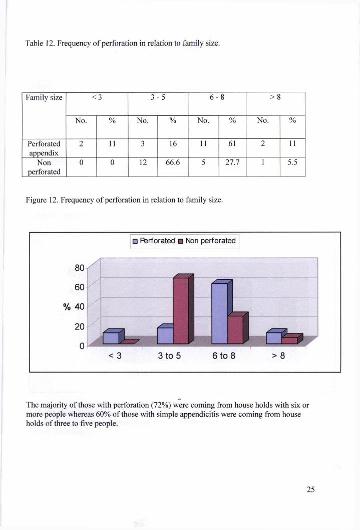
Table 12. Frequency of perforation in relation to family size.
Family size <3 3 -5 6 -8 >8 Family size
No. % No. % No. % No. %
Perforated appendix
2 11 3 16 11 61 2 11
Non perforated
0 0 12 66.6 5 27.7 1 5.5
Figure 12. Frequency of perforation in relation to family size.
The majority of those with perforation (72%) were coming from house holds with six or more people whereas 60% of those with simple appendicitis were coming from house holds of three to five people.
25

T A B L E 13. Frequency of perforation in relation to education level of patient or guardian.
Education level
< grade 4 Grade 5 - 7 Grade 8 - 9 Grade 10-12 Tertially education
Education level
No. % No. % No. % No. % No. %
Perforated appendix
5 27.7 4 22 2 11 4 22 3 16
Non perforated
0 0 0 0 4 22.2 8 44.4 6 33.3
Figure 13. . Frequency of perforation in relation to education level of patient or guardian
• Perforated • Non perforated
Half of those with perforation or their guardians for those less than 18 years of age had primary education alone whereas those without perforation went beyond the level of primary education.
26

Table 14. Frequency of perforation in relation to period spent at U T H before actual surgery
Period before surgery
< 1 hour 1 - 2 hours 2 - 4 hours > 4 hours Period before surgery
No. % No. % No. % No. %
Perforated appendix
0 0 1 5.5 4 22.2 13 72.2
Non perforated 0 0 0 0 4 22.2 14 77.7
Figure 14. Frequency of perforation in relation to period spent at U T H before actual surgery.
• Perforated • Non perforated
60
% 40
< 1hour 1 to 2 hrs 3 to 4 hrs > 4 hrs
The majority of the patients from the two groups (> 60%) were taken for the actual surgery more than four hours after being admitted to UTH.
27

Table 15. Complications associated with appendicectomy.
Complication Haemorrhage Surgical site infection
Re-Laparotomy for intra abdominal
abscess
Complication
No. % No. % No. %
Perforated appendix 0 0 6 33.3 4 22.2
Non perforated 0 0 1 5.5 1 5.5
Figure 15. Complications associated with appendicectomy
i Perforated i Non perforated
% 2 0 10
A B C
A = Haemorrhage B = S u g i c a i site infection C =Relaparotomy
There was a 33.3% wound infection rate in those with perforation as compared to 5.5 % in those with simple appendicitis. 22.2 % had re-Laparotomy for intra abdominal abscess formation as compared to 5.5% who had re-Laparotomy in the non perforated group.
28

Table 16. Frequency of admission to intensive care unit.
Admission to intensive care unit Yes No Admission to intensive care unit
No. % No. %
Perforated appendix 1 5.5 17 94.4
None perforated 0 0 18 100
Figure 16. Frequency of admission to intensive care unit.
Only 5.5% of the perforated group was admitted to the intensive care unit but none of those with simple appendicitis.
29

Table 17. Duration of post operative hospital stay after surgery.
Duration of admission
< 2 days 3 - 5 days 6 - 1 0 days > 10 days Duration of admission
No. % No. % No. % No. %
Perforated appendix
0 0 8 44.4 6 33.3 4 22.2
Non perforated 1 5.5 16 88.8 0 0 1 5.5
Figure 17. Duration of post operative hospital stay after surgery.
%
100
80
60
40
20
Perforated • Non perforated
1 ^
< 2 days 3-5 days 6-10 days >10 days
55.5% of those with perfpf^tion were discharged after the sixth day as opposed to 88-8 % of those with simple appendicitis who were discharged between the third and fifth postoperative day.
30

Table 18. Mortality associated with appendicectomy
Item Total number Mortality Percentage perforated 31 01 3.2 Non perforated 40 00 00 Total 71 01 1.4
Figure 18. Mortality associated with appendicectomy
4 M. J |
3
2 1 1 • 0
p e r f o r a t e d n o n p e r f o r a t e d
3.2 % of those with perforated appendicitis died as compared to non in those with simple appendicitis. The overall mortality rate is 1.4 %.
31

DISCUSSION
This study has clearly demonstrated an appendiceal perforation rate of 43.6%. This is
well above the 31.6% that was observed 10 years ago This increase can be attributed to
the increasing incidence of acute appendicitis as more people resort to a more refined
diet. This figure is of course similar to figures obtained in other Afi-ican countries.
Madiba in 1998 had shown a perforation rate of 43 percent in a study done in South
Afiica. This figure is of course higher than what has been reported in other studies.
Mutuphei, for example, demonstrated a perforation rate of 25 percent in the same year
in a different part of South Africa. Willmore ^' demonstrated a perforation rate of 22
percent in a Nigerian hospital. Rates as low as 12 percent have been reported in
Sweden.^" On the other hand, Von Titte demonstrated a 90 percent adult perforation
rate in a hospital in the USA. This demonstrates a wide range of variation from country to
country.
Table 2 shows that half of those less than ten years of age presented with perforation.
This is in line with what has been seen in many other studies as the diagnosis of acute
appendicitis in childrrai is usually difficuh and so the surgeon needs to have a high index
ofsuspicion.'^-^'*'^
Of those above the age of 40, only 30 percent presented with perforation. This is not in
line with what has been reported in other studies where the majority of those above 40
present with perforation. Just like in children, elderly patients are predisposed to
perforation due to low immunity."
Perforations were more common in the male than female patients with a ratio of 2.5. 1.
However, it was noted that a bigger percentage of the female patients with acute
appendicitis came with perforation. This could be attributed to the pain being linked to
other gynecological conditions such as pelvic inflammatory disease.
32

Half of all those patients with perforation were from highly populated residential areas
where as those with no perforation were from low or medium populated areas. The
former was also associated with households of more than six people. The possible
explanation for this could be that people from highly populated areas are associated with
poor socioeconomic background and so might have problems in accessing medical care.
This is in line with what has been demonstrated in other series where those with no
proper medical insurance were at a higher risk of presenting with perforation because of
issues of access to health. '̂'̂ ^" '̂ Hjortsberg in a study done here in Zambia in 2003
concluded that individuals were influenced by income, insurance and distance from a
health center on the decision to seek medical care for various conditions.
78 percent of those with perforation came after the third day of symptoms at home as
opposed to 60 percent of the non- perforated who presented within the first 24 hours (p <
0.001). This clearly demonstrates that pre-hospital delay is the main factor associated with
perforation. Bickell in 2006 showed that the risk of rapture increases by 5 % every
twelve hours after the thirty sixth hour from the onset of the symptoms. Omundesen
reported no complications in those who presented within a period of 24 hours. The
question that arises therefore is why the prehospital delay by the patients. From this
study, it can be concluded that the delay could be attributed to the low socioeconomic
status of the patients. Firstly, 77 percent of those with perforation live on less than 500
000 thousand kwacha per month yet have house-holds composed of six or more
members. Secondly, over half of the patients or guvdians of those who presented with
perforation never went beyond the seventh grade and so may luck the initiative to
interpret the symptoms of acute appendicitis as important.
There was no difference in the perforation rate between the two groups according to the
mode of admission to UTH. The perforation rate was the same in those who referred
themselves straight to the Emergence Department and those who were referred by the
government clinics. This does not agree with what was observed by Robert'** in a study
done in the U S A in which those who referred themselves straight to the Emergency
33

Department had a lesser chance of perforation as opposed to those who were referred
from other health sources or centers.
Consultation to private clinics or hospitals prior to admission to The University Teaching
Hospital did not contribute significantly to the prehospital delay. A l l the patients with
suspected acute appendicitis from the private institutions were referred to U T H on the
same day. This was not in line with what was observed by Ofoegbu in a Nigerian
hospital in which the prehospital delay was attributed to the consuhation of private clinics
before coming to public institutions.
11 percent of those with perforation used traditional medicine before seeking modem
medicine as opposed to none in the other group. This again is in line with what was
demonstrated by walker ^ in which prehospital delay was associated with the seeking of
traditional medicine before seeking orthodox medicine. A study done here at U T H
showed that 75 percent of all patients take traditional m^icine before presenting to
UTH.'*^ Despite this low figure, there is still need to educate our tradition healers on the
need for early referral when ever they are in doubt.
The administration of antibiotics before referral did not contribute significantly on
prevention of perforation as half of each group was given these before referral. This, also,
did not have a bearing on the postoperative complications. A l l patients were routinely put
on triple antibiotics after surgery. Peri-operative antibiotics in acute appendicitis play an
important role and this can never be over emphasized. Lack of antibiotic administration
by the referring clinics did not in any way contribute to an increase in perforation.
The other issue of concern is the in-hospital delay by the surgeon, that is, between the
time the diagnosis is made and the time the patient is taken to theatre for the actual
surgery. In this study, over 90 percent of patients from both groups were taken for
surgery after a period of over 4 hours on the ward. This could be due to the need to
resuscitate the patients before surgery. There was no difference between the two groups
in terms of in hospital delay; therefore it can not be associated vwth the high levels of
34

perforation observed in this study. The longest period of delay was observed on one
patient who was admitted wrongly to a medical ward and was treated as a case of peptic
ulcer disease. The surgeons saw the patient after three days but the poor state of the
patient could not allow immediate surgery. This was done ten days post admission to
U T H . Physicians should be encouraged to seek early surgical opinion whenever in doubt.
Our findings do not agree with what other authors have stated that perforation is a
surgeon dependant variable. To this effect, it can be concluded that the appendiceal
perforation rate of a particular institution can not be used as a way to assess the
effectiveness of a hospital in delivering its services as has been postulated by other
authorities, at least, not in a developing country like ours.
Perforation of the appendix is associated with high levels of morbidity and mortality.
64.5 percent of those with perforated appendicitis had generalized peritonitis requiring
access into the abdomen through the midline. Such an operation demands more time,
material and also increases the morbidity as compared to a small right sided incision used
for simple appendicitis.
One third of the patients with perforated appendicitis had surgical site infection as
compared to 5.5 percent of those with no perforation. This is in Une in what has been
seen in other studies worldwide in which perforation is associated with high levels of
complications. This is in agreement with what was observed by Von Titte in whom 60
% of those with perforated appendicitis presented with major complications. This
definitely has a bearing on the hospital post operative stay.
22.2 percent of those with perforation had re-laparotomies done for intra abdominal
abscesses between the fifth and tenth post operative day. One patient had two re
laparotomies for peritonitis; however, all these patients recovered well. The only thing of
note was that the post-optative hospital stay was prolonged in the group with
perforation. Most of these patients were discharged aAer the sixth post operative day with
22.2 percent going beyond the tenth day. 89 percent of those without perforation were
discharged between the second and fifth postoperative day. This clearly demonstrates that
35

patients with comphcated appendicitis are more prone to post operative complications.
This of course increases the morbidity and has a bearing on the hospital budget. On the
other hand, only one patient with simple acute appendicitis had a re- laparotomy on the
fifth post-operative day. He had a leak from the appendicular stump. This complication
was primarily attributed to the surgical technique.
From all the appendicectomies done during the study period, there was only one mortality
giving an overall mortality rate of less than two percent. This figure is in line with what
has been documented in other parts of the world. Despite the high appendiceal
perforation rate, the mortality rate is within acceptable levels.
This study had several important limitations. The diagnosis of acute appendicitis was
primarily based on what the operating surgeon reported. This was never confirmed by
histology. Some of them recruited as acute appendicitis may not, after all, have been
inflamed. The other thing is that the number of patients recruited was not very large.
There is still need to do a similar study with a large sample size.
36

CONCLUSION
Having looked at perforated appendicitis for a period of nine months; and having had
compared some of the associated factors in those with and without perforation, the
following conclusions can be drawn:
1. The perforation rate is 43.6 % which clearly shows that there is an increase from
the figures described a decade ago of 32 %.
2. Perforation is common in those betw^n 30 and 40 years.
3. Perforations are more common in the males than females with a perforation ratio
of2.5: 1.
4. The main factor associated with perforation of the appendix is pre hospital delay
by patient related factors.
5. Those coming from high density residential areas are prone to perforation as
compared to those coming from low or medium populated areas.
6. Perforation is more likely in those with house holds of six or more people and
also those living on less than five hundred thousand kwacha per household per
month.
7. Lack of education beyond the seventh grade predisposes people to rupture when
they have acute appendicitis.
8. In hospital delay by surg^n related factors was not the cause of perforated
appendicitis.
9. Form the aforementioned factors, it can be deduced that the pre hospital delay by
the patient is linked to poor access to quality health care.
10. The commonest complication associated with appendicectomy in those with
perforation is surgical site infection with a good percentage requiring
relaparotomy for intra abdominal abscesses.
11. Perforation increases the post operative hospital stay; this has a direct implication
on the hospital budget.
12. Despite an increase in the rate of appendiceal perforation, the overall mortality is
still less than two percent.
37

RECOMMENDATIONS
1. The high rate of perforated appendicitis with its subsequent sequelae of increased
morbidity and resource expenditure is the primary result of patient delay in seeking
medical attention and not the resuh of diagnostic dilemma or surgical delay, therefore,
public education, specifically targeting those groups at risk, may provide a significant
solution to the problem.
2. There is need to sensitize the community as well as the primary heahh workers on the
signs and symptoms of common surgical emergencies like acute appendicitis in order to
cut down on the pre- hospital delay.
3. A large study, whh special emphasis on factors causing pre- hospital delay in patients
with acute appendicitis, is strongly advised.
38

BIBLIOGRAPHY
1. Kelvin P, Appendix: Sabiston Textbook of surgery Sixteenth edition. 2001. WB.
Sandler company, London.
2. CD-9 CM-540-l(1987). International Classification of Diseases, Ninth Revision,
Clinical Modification (ICD-9-CM)
3. Creese P G (1953). The first appendicectomy.Surg cryecol obstet 97: 643.
4. Hannock H (1848). Disease of the appendix coeci cured by operation lancet 2:380
5. Tait L (1890). surgical treatment of typhylitis. Birmingham med Rev 27:26.
6. Fitze R H (1886). Perforating inflammation of the vermiform appendix, with
special reference to its early diagnosis and treatment. Trans Assoc Am Physicians
1886; 1:107-144.
7. McBurney C (1889). Experiences with early operative interference in cases of
diseases of the vermiform appendix. N Y Med J 1889; 50:676-684.
8. Murphy JB (1904). Two thousand operations for appendicitis, with deductions
from his personal experience. Am J Med Sci 1904; 128:187-211.
9. Ellis H (1991). Early operative treatment of acute appendicitis. Contemp Surg
38(1)35.
10. Anderson BR, Cleave F L , Anderson H K (2003) Antibiotics versus placebo for
prevention of postoperative infection after appendicectomy. Cochrane Database
Syst Rev. 2005 jul 20 ;(3) CD001439.
39

11. Ronan P (2004) The vermifonn Appendix: Love and Bailey. Principals of surgery
twenty fourth edition. 2004. Gouldy company, India.
12. Ajao O G (1981) Br J Surg May 68(5):345-7.Abdominal emergences in tropical
Africa.
13. Katzaski M , Gopai Rao, Brandy K (1979) Blood supply and position of the
appendix in Zambians. Med Y Zambia 1978 Apr-May: 13 (2) 32-4
14. Addis DG, Shaffer N , Tauxe R V (1990). The epidemiology of acute appendicitis
and appendicectomy in United States, American journal of epidemiology. 123 (5):
916-25.
15. FuUon J, Lazarus C (1995). Acute appendicitis among black south Africans. S Afr
J Surg. 1995 Dec; 33(4): 165-6.
16. Oliver M J (1987). Generalised peritonitis due to appendicitis. The proceedings of
the association of East Africa 10: 26-27.
17. Haque M E (1997). A study on the incidence of appendicectomy in the university
teaching hospital, Lusaka, Zambia. M.Med dissertation.
18. Kottiso B , Messele G (1996). Acute appendicitis in Ethiopia. E Afr. Med. J.
73(4); 251-252.
19. Nanda K (2004). Delay in surgery for acute appendicitis. A N Z journal of surgery
Vol 74 issue 9 p773- Sept 2004.
20. Madiba TE, Haflfeje AA Mbete DL,Chaitina H (1998). East Frica Med J. 1998
Fed ;75 (2); 81-4 Appendocectomy among African Population at king Edward IV
hospital, Durban South Africa.
40

21. Muthupheni M N , Morwamoche P (1998). The surgical pathology of the appendix
in South Africa. Central Africa jr med 1998 jcol (4): 9-11.
22. Levy RD, Degiannis E, Kantarovsky A Marbeti P M , Wello M , Hartitheo C
(1997). Audit of acute appendicitis in black South African population. S Afr J
surg 1997 Nov 35 (4): 19&- 202.
23. Walker AR, Walker BF, Mantsi B , Tsotetsi N G , Segal I (1989). Appendicitis in
Soweto: Traditional healers and the Hospital. JR soc Health 1989 Dec ; 109(6);
190-2.
24. Out A A (1989). Tropical surgical abdominal emergency; Acute appendicitis. Trop
GeogrMed 1989: 41 (2) 118-22.
25. Willmore WS, Hi l l A G (2001). Acute appendicitis in Kenya hospital. East Afr
Med J. 2001:78(7) 355-7.
26. Ofoegba C K , Odi T, Ogundipe O, Taimoz, Solagberu B A (2005). Epidemiology
of non-trauma deaths. West Afr J. med (2005) oct-dec: 24(4) 321-4.
27. Lee J F, Leon CK, Lan W Y (2000). Appendicitis in the elderly. A N T Surgery.
2000 Aug; 70(8):593-6.
28. Watters D (1988). Severe peritoneal sepsis. Baillieerels clinical tropical medicine
and communicable diseases, 3(2): 275-95.
29. Mwangala M M (1993) The acute abdomen in the university teaching Hospital: A
comparative study of surgery in H I V seropositive and seron^tive patients,
Lusaka, Zambia, M.Med dissertation.
30. Anderson RE (1992). Diagnostic accuracy and perforation rate in appendicitis
with age and sex. Eur. J Surg 1992 Jan 158; 1337-41.
41

31. Von Titte SN, McCabe CJ, Ottinger L W (1996). Delayed appendectomy for
appendicitis: causes and consequences. A m J Emerg Med 1996 Nov;14(7):620-2.
32. Nance ML,Adamson WT, Hendrick H L . (2000).Appendicitis in the young child: a
continuing diagnostic challenge. Pediatr Emerg Care. 2000 Jun; 16(3): 160-2.
33. Rajendra S, Feargal Q, Prem P (1995). Appendicitis in preschool children.
Pediatric Surgery International, vol 10, Nun 2-3, Feb, 1995.
34. Colvin JM, Bachur R, Kharbanada. (2007).The presentation of appendicitis in
preschool children, Pediatr Emeg Care, Dec; 23(12):849-55.
35. Susan L, Charles M ( 2000). Acute Appendicitis Risks of Complications: Age and
Medicaid Insurance PEDIATRICS Vol. 106 No. 1 July 2000, pp. 75-78.
36. O'Toole SJ, Karamanoukian HL,Allen JF, Caty M G ( 1996).Insurance-related
differences in the presentation of pediatric appendicitis,Pediatric Surg. 1996 Aur;
31(8): 1032-4.
37. Susan L B , Charles MH,John HT (2000). Acute Appendicitis Risks of
Complications: Age and Medicaid Insurance. PEDIATRICS Vol . 106 No. 1 July
2000, pp. 75-78.
38. Hjortsberg C (2003). Why do the sick not utilize heath care? The case of Zambia.
Heah Econ, 2003 Sep; 12(9):755-70.
39. Bickell N A Aufses A H , Rojas M . (2066). How time affects the risk of rapture.
Jam Coll Surg, 2006 Mar 20; 202(3):401-6.
40. Omundsen M , Dennett E (2006). Delay to appendicectomy and associated
morbidity: a retrospective review. A N Z J Surg 2006 Mar;76(3):153.
42

41. Robert G, Janet D, Dean K (1999). The risk of appendiceal rapture based on
hospital admission source. Academic emergence medicine vol. 6, number 6, 596-
601.
42. Horan T C. (1999). Guideline for prevention of surgical site infection: A Special
Report, http.premerinc.com.
43

DATA COLLECTION SHEET DATE:
APPENDIX I
PATIENT ID NO:
L S E X : M [ 1 F[l
2. AGE: < 5 [] 6-10 0 11-15 Q 16-20 [] 21-30 [] 31-40 [] 41-60 [ ] > 60 []
3. RESroENTIAL AREA: L O W [] M E D I U M [] H I G H [] PERIUBURN [] R U R A L []
4. DURATION OF ILLNESS BEFORE ADMISSION TO UTH
< 6H0URS [] 7-12 HOURS [] 13-24 HOURS [] 25-72 HOURS [] 3 -5 D A Y S [] 5-7 D A Y S [] > 7 D A Y S []
5. PREADMISSION CARE: DIRECT ADMISSION TO U T H Y [] N [] R E F E R A L FROM: PVT CLINIC Y [] N Q G V T CLINIC Y [] N 0
ANTIBIOTICS; Y [ ] N [ ] TRADITION MEDICINE Y[] N[]
6.S0CI0EC0N0MIC STATUS
AMOUNT EARNED PER MONTH: <K100 000[] KlOOOOO-500 000 [] 500 000-1 M I L L I O N [] > 1 M I L L I O N []
7. FAMILY SIZE <3n3-5[]5-8[]>8[]
8. DURATI0N BEFORE BEEING TAKEN TO THEATRE <1 HOUR [] 1-2 HOURS [] 2-4 HOURS [] > 4H0URS []
44

INTRAOPERATIVE FINDINGS:
PERFORATED [] NON-PERFORATED []
POSTOPERA TIVE PERIOD MICU: YES [] NO []
MOBIDITY 1. H A E M O R R H A G E : Y [] N O [] 2. SURGICAL SITE INFECTION Y [] N [] 3. W 0 U N D DEHISCENCE Y[] N[] 4. R E L A P A R O T O M Y ; Y [] N[] D A Y S [ ]
DURATION BEFORE DISCHARGE 1-2 [] 3-5 [] 6-10 [] >10 []
D E A T H Y a N []
PRIVATE CLINIC CONSULTATION; Y [] N [] IF YES HOW M A N Y D A Y S BEFORE R E F E R A L []
WORKING DL\GNOSIS:

APPENDIX II
Criteria for defining surgical site infection (SSIs) developed by CPC^s NNIS system Superficial incisional SSI Infection occurs within 30 days after the qjeration and infection involves only skin or subcutaneous tissue of the incision and at least one of the following: 1. Purulent drainage, with or withcHit laboratory confirmation, from the superficial incision. 2. Organisms isolated from an asq)tically obtained culture of fluid or tissue from the superficial incision. 3. At least one of the following signs or symptoms of infection: pain or tenderness, localized swelling, redness, or heat and superficial incision is deliberately qjened by surgeon, unless incision is culture-negative. 4. D i^os i s of superficial incisional SSI by the surgeon or attending physician.
Deep incisional SSI Infection occurs within 30 days after the operation if no implant is left in place or within one year if implant is in place and ftie infection appears to be related to the operation ami infection involves deqp soft tissues (e.g., fescial and muscle layers) of the incision and at least one of the following: 1. Purulent drainage from the deep incision but not from the organ/space component of the surgical site. 2. A deep incision spontaneously dehisces or is deliberately opened by a surgecMi when the patient has at least one of the following signs or symptoms: fever (>38° C), localized pain, or tenderness, unless site is culture-negative. 3. An abscess or other evidence of infection involving the deep incision is found on direct examination, during reoperation, or by hist(q>athologic or radiologic examination. 4. Diagnosis of a deep incisional SSI by a surgeon or attendii^ physician.
Organ/space SSI Infection occurs within 30 days after the operation if no implant is left in place or within one year if implant is in place and the infection appears to be related to the operation and infection involves any part of the anatomy (e.g., organs or spaces) other than the incision, which was opened or manipulated during an opeiaikm and at least one of the following: 1. Purulent drainage from a drain that is placed through a stab wcwmd into the organ/space. 2. Organisms isolated from an aseptically obtained culture of fluid or tissue in the organ/space. 3. An abscess or other evidence of infection involving the organ/space that is found on direct examination, during reoperation, or by histopathologic or radiologic examination. 4. Diagnosis of an organ/space SSI by a surgeon or attending physician.
46