mitotic and meiotic spindle dynamics comparison in fission
TRANSCRIPT

HAL Id: tel-03174872https://tel.archives-ouvertes.fr/tel-03174872
Submitted on 19 Mar 2021
HAL is a multi-disciplinary open accessarchive for the deposit and dissemination of sci-entific research documents, whether they are pub-lished or not. The documents may come fromteaching and research institutions in France orabroad, or from public or private research centers.
L’archive ouverte pluridisciplinaire HAL, estdestinée au dépôt et à la diffusion de documentsscientifiques de niveau recherche, publiés ou non,émanant des établissements d’enseignement et derecherche français ou étrangers, des laboratoirespublics ou privés.
Mitotic and meiotic spindle dynamics comparison infission yeast
Ana Loncar
To cite this version:Ana Loncar. Mitotic and meiotic spindle dynamics comparison in fission yeast. Cellular Biology.Université Paris sciences et lettres, 2020. English. �NNT : 2020UPSLT003�. �tel-03174872�

Préparée à l’Institut Curie
Mitotic and meiotic spindle dynamics comparison in
fission yeast
Comparaison de la dynamique du fuseau mitotique et
méiotique chez la levure à fission
Soutenue par
Ana LONČAR Le 11 septembre 2020
Ecole doctorale n° 577
Structure et dynamique des
systèmes vivants
Spécialité
Sciences de la vie et de la
santé
Composition du jury:
Hiroyuki OHKURA
Professeur
WTCCB – The university of Edinburgh Président
Iva TOLIĆ
Professeur
Institut Ruđer Bošković Rapporteur
Julien DUMONT
Directeur de recherche
Institut Jacques Monod –
CNRS - Université Paris Diderot Rapporteur
Sylvie TOURNIER
Directeur de recherche
CBI – CNRS Université de Toulouse Examinateur
Marie-Emilie TERRET
Directeur de recherche
Collège de France – INSERM Examinateur
Phong TRAN
Directeur de recherche
Institut Curie - CNRS Directeur de thèse

Acknowledgements 1
Mitotic and meiotic spindle dynamics comparison in fission yeast
Acknowledgements
Firstly, I thank Phong Tran, for giving me the opportunity to undertake this journey, and for being
a kind and considerate supervisor. He let me learn at my own pace, and allowed me to have my
own experiences to learn from. The autonomy and confidence I possess are the product of his
mentorship style.
Secondly, I would like to offer thanks my Thesis Committee members: Renata Basto, Daniele
Fachinetti, and Matthieu Piel, for insightful comments, stimulating discussions, and for helping me
navigate through my PhD. I would like to extend thanks to the thesis jury - Julien Dumont, Hiroyuki
Ohkura, Marie-Emilie Terret, Iva Tolić, and Sylvie Tournier - for graciously accepting to evaluate
my work.
Next, I thank the wonderful lab I was a part of: Anne Paoletti, an exceptional role model; Sergio
Rincon, fellow runner and a mentor; Frederique Carlier-Grynkorn, the Molière behind this thesis
Résumé; Federica Arbizzani, the deceptively quiet Lilliputian; Imène Bouhlel, the confident and
passionate student-matriarch; and Marion Arraou, who started on the same day as me, helped me
navigate the French bureaucracy on countless occasions, and is the biggest fan of my grandma’s
produce (and whose last name I still have to check for fear of missing a syllable).
The biggest thanks goes to Lara Katharina Krüger and Manuel Lera Ramirez, who have not only
shared office space with me, but also stoically endured my colorful personality. I cherish every
~garden break~ I had with Lara, and every hilarious meme expertly recognized and shared by
Manu. I could not have imagined better colleagues, or friends, to have by my side, whether in the
office/lab, or having drinks and trying out new restaurants. You have made difficult times bearable,
and lovely times more memorable. I do not deserve you, and I remain in your debt.
For inviting me to share their living space, and discovering the little world contained in Italy, I
thank my two roommates and friends Piergiuseppe and Jacopo. Thank you for teaching me which
type of pasta goes with which sauce, sharing focaccia and watching RuPaul’s Drag Race with me.
I leave our flat knowing which regions of Italy do not exist (sorry Fra <3), and knowing that Puglia
is the best region. (After seeing a typical Puglia pizza topping – French fries, I remain convinced
that pineapple is an acceptable pizza topping.)

Acknowledgements 2
Mitotic and meiotic spindle dynamics comparison in fission yeast
I thank Yunlong, my sister from another mister, for allowing me to be his friend. He, ironically,
sees much more in me (and others) than I could ever see myself. Know that I wish you a good
ephening or an aphternoon, at whatever time you may be reading this.
I thank Ralfs the magnificent, who was always up for a pique-nique, and that’s the characteristic
that pleases me most in people. He will also remain immortalized as my last human contact before
the Great Confinement of 2020, and the connoisseur of the struggle of stuffing oneself with weird
thin pizzas. I thank the beautiful Carlos, the amazing chef who does not know what a cheesecake
is, but who can teach anyone how to love themselves despite everything being against them. I thank
Sebastian, who has hosted our game nights, and with whom I shared the pleasure of experiencing
the worst falafel imaginable. I thank the fierce Benjamin, for serving it as is, and being
unapologetically himself. I thank Francesca, for enduring baking with me, and gracing me with her
bubbly company. I thank my running mate Linda, for all our heart to hearts and La Fourchette
explorations. I thank the ‘’always on time’’ Deep, for starting up the most interesting conversation
topics, and making sure everything my grandma prepared gets eaten.
I also thank the culinary enthusiast Liene, KFC super-fan Héctor, Iceland aficionado Joe, the 4th
floor of Institute Curie, and so many others that have spiced up my four-year stay in Paris.
I thank my French queens – Vlatka, the cuisine hacker, who was my welcome committee upon
arrival to Paris, and has come to be a cherished friend; Miriam, who proudly displayed the Croatian
flag in Paris during the finale of the World Cup; Daniela, the no-nonsense lawyer, whose signature
phrase ‘’emotions are not facts’’ should be taught in schools; and Petra, who I was lucky to be a
neighbour to, and kind-heartedly gossip about our boyfriends.
This work was aided by the Zagreb support system. I thank mamasita Tena for all our ‘’Krivi put’’
excursions, and the exams we prepared together. Thank you for introducing me to Jakov Kutnjak!
I thank Anja-Matea for asking me for a cigarette on our first day of university, and cooking French-
fries with me. I thank Bogy for being my number one Instagram follower, and teaching me about
big Sagg energy. I thank Marija for choosing to come to work to PrimeVigilance, and forming a
new circle of friends. I thank Martina for gossiping about our university colleagues every time I
would come to visit Croatia. I thank Eugen for all the lovely times spent at his flat praising the
Braun blender. I thank Karla for inspiring me to undertake this challenge and for giving me a friend
and support in the form of her sister Martina. I love you all!

Acknowledgements 3
Mitotic and meiotic spindle dynamics comparison in fission yeast
I especially want to say how grateful I am to my family – my mother Bosiljka, who is an amazing
queen in every conceivable aspect, and a true testament to strength of character and will power; to
my father Zlatko, for dealing the best way he could with what life put on his plate; to my brother
Martin, for watching all of the best cartoons with me, and hosting vivid interpretations of ‘’Ustaj
sine, majka zove’’. You all (and our favourite bunny Dražen Zečić) have supported me always and
forever, and I am eternally in your debt. (Except for that one time you went to that fancy restaurant
without me, I’ll keep that one in mind.)
I thank my dearest Ladyboys. I can barely find the words to express what their loyalty and
encouragement means to me. I thank Anja, for all our rollerblading sessions around the Jarun Lake.
She is the most resourceful woman I have ever met, and she motivates me to be more of a one as
well. I thank Dorica for being a creative treasure chest full of hidden gems which she shares with
me on Instagram. I thank Eva for growing up with me, and making me a part of her family.
Knowing her allowed me to know the world, and, importantly, Emil Geistlich. I thank Marina F
for guarding the secret of what was in that photo, and tolerating my Ville Valo inspired poetry
phase. I thank Marina Š, the reigning Yamb champion, for always staying until the very end –
whether it be a video-call or a random ~coffee~ in Hercegovina. I thank Natali, a typical Capricorn,
for all of our Bela wins, and for being one of the worst wolves the game has ever seen. I thank my
fellow key guardian Natalija, for her time with me in the Republic of Catalunya, and being cool
about getting lost in the world’s lamest labyrinth. I thank Nataša for being the most diligent
confinement officer, and, of course, for working on herself. I hope she will never rise to political
power.
Last, and possibly least, Nikola. Whatever words I use will not convey properly what I want to say.
You have lived this PhD as much as I have. Thank you (and Saucisse) for being by my side all this
time, supporting me. May we witness Keemstar and Jeremy Renner cancelled together.

Table of contents 4
Mitotic and meiotic spindle dynamics comparison in fission yeast
Table of contents
Acknowledgements .......................................................................................................................... 1
Table of contents .............................................................................................................................. 4
Figure Index ..................................................................................................................................... 6
Abbreviations ................................................................................................................................... 7
Introduction ...................................................................................................................................... 9
1. Cell division ....................................................................................................................... 10
1.1. Mitosis .......................................................................................................................... 10
1.2. Meiosis ......................................................................................................................... 12
2. Spindle components ........................................................................................................... 14
2.1. Microtubules (MTs) ..................................................................................................... 15
2.1.1. Kinetochore microtubules (KT-MTs) ................................................................... 17
2.1.2. Interpolar MTs (iMTs) .......................................................................................... 18
2.1.3. Astral MTs (aMTs) ............................................................................................... 19
2.2. Centrosome and spindle pole body (SPB) ................................................................... 20
2.3. Chromosomes and KTs ................................................................................................ 22
2.3.1. Chromokinesins & polar ejection force (PEF) ..................................................... 25
2.4. Motor proteins .............................................................................................................. 27
2.4.1. Dynein ................................................................................................................... 28
2.4.2. Kinesin-5 ............................................................................................................... 30
2.4.3. Kinesin-8 ............................................................................................................... 31
2.4.4. Kinesin-14 ............................................................................................................. 33
2.5. Non-motor microtubule associated proteins (MAPs) .................................................. 35
3. Non-centrosomal pathways of spindle assembly ............................................................... 36
3.1. Ran-GTP pathway. ....................................................................................................... 37
3.2. Chromosomal passenger complex (CPC) pathway ...................................................... 39
3.3. Acentriolar MTOCs (aMTOCs) ................................................................................... 40
3.4. Augmin pathway .......................................................................................................... 41
4. Fission yeast as model system for analysing mitotic and meiotic spindle dynamics ......... 42

Table of contents 5
Mitotic and meiotic spindle dynamics comparison in fission yeast
4.1. Phase I – Initial stages of spindle nucleation ............................................................... 44
4.2. Phase I – Establishment of a bipolar spindle ............................................................... 46
4.3. PhaseI/phaseII – Chromosome attachment to the spindle and congression ................. 49
4.4. Phase II – Spindle forces and force-balance maintenance in fission yeast .................. 51
4.5. Phase III – Final spindle elongation ............................................................................. 53
4.6. Comparison of mitotic and meiotic spindle dynamics in fission yeast ........................ 53
Aim of this work ............................................................................................................................. 55
Results ............................................................................................................................................ 56
Discussion .................................................................................................................................... 111
Résumé ......................................................................................................................................... 118
1. Introduction .......................................................................................................................... 119
1.1. Cell division ............................................................................................................... 119
1.1.1. La mitose .................................................................................................................... 119
1.1.2. La Méiose ............................................................................................................... 121
1.2. La levure fissipare S. pombe comme système modèle pour l’analyse de la dynamique
du fuseau mitotique et méiotique ............................................................................................. 123
1.2.1. Comparaison de la dynamique du fuseau mitotique et méiotique .......................... 124
1.2.2. Le double mutant de délétion kinésine-5 /kinésine-14 (cut7Δpkl1Δ) comme outil
permettant la comparaison des fuseaux mitotiques et méiotiques ....................................... 126
2. Resultats ............................................................................................................................... 127
2.1. La Dynamique du fuseau diffère en mitose et en méiose dans la levure fissipare .... 127
2.2. L’intégrité du fuseau est compromise spécifiquement en MI dans les zygotes du
double mutant cut7Δpkl1Δ.................................................................................................... 128
2.3. Le ratio Cut7-à-Pkl1 est plus élevé dans le fuseau MI qu’en mitose ......................... 129
2.4. La fonction de la kinésine 14 Klp2 exercée sur le fuseau est distincte en MI et en
mitose ................................................................................................................................... 130
2.5. La suppression de la dynamique des MTs restaurela bipolarité du fuseau de MI dans
les zygotes cut7Δpkl1Δ ......................................................................................................... 131
3. Discussion ............................................................................................................................ 133
References .................................................................................................................................... 137

Figure Index 6
Mitotic and meiotic spindle dynamics comparison in fission yeast
Figure Index
Figure 1.1. Vegetative cell cycle and mitosis. ................................................................................ 11
Figure 1.2. Meiosis consists of two divisions. ............................................................................... 14
Figure 2.1. MTs are characterized by dynamic instability. ............................................................ 16
Figure 2.2. Categories of spindle MTs. .......................................................................................... 17
Figure 2.3. Centrosome is the major MT organizing center in the cell. ......................................... 21
Figure 2.4. Architecture of an independent and paired chromosome. ........................................... 24
Figure 2.5. Chromokinesins exert forces on chromosomes to facilitate chromosome congression.
........................................................................................................................................................ 26
Figure 2.6. Dynein can induce centrosome separation. .................................................................. 29
Figure 2.7. Kinesin-5 is essential for spindle bipolarity. ................................................................ 30
Figure 2.8. Kinesin-8s regulate MT dynamics. .............................................................................. 32
Figure 2.9. Kinesin-14 is a minus-end directed motor capable of bundling MTs. ......................... 34
Figure 2.10. MAP65 members are cross-linker MAPs. ................................................................. 36
Figure 3.1. Chromosome associated pathways of spindle assembly. ............................................. 38
Figure 3.2. A model for bipolar spindle assembly in mouse oocytes. ........................................... 40
Figure 4.1. Fission yeast is a good tool to comparatively study mitotic and meiotic spindles. ..... 43
Figure 4.2. A simplified scheme showing spindle nucleation in fission yeast. .............................. 45
Figure 4.3. Cut7 establishes bipolarity, and Pkl1 organizes spindle poles. ................................... 47
Figure 4.4. Chromosome localization at spindle assembly onset and chromosome recapture. ..... 49
Figure 4.5. Simplified map of force producers in the fission yeast mitotic phase II spindle. ........ 52

Abbreviations 7
Mitotic and meiotic spindle dynamics comparison in fission yeast
Abbreviations
DNA - deoxyribonucleic acid
MT – microtubule
MTOC –MT organizing centre
KT - kinetochore
MAP – microtubule associated protein
SAC – spindle assembly checkpoint
MI – meiosis I
MII – meiosis II
GTP – guanosine triphosphate
KT-MT – kinetochore microtubule
iMT – interpolar microtubule
aMT – astral microtubule
PEF – polar ejection force
PCM – pericentriolar material
γ-TuSC – γ-tubulin small complex
γ-TuRC – γ-tubulin ring complex
SPB – spindle pole body
NEBD – nuclear envelope breakdown
γ-TuC – γ-tubulin complex
ncMTOC – non-centorsomal MTOC
APC – anaphase promoting complex
ATP – adenosine triphosphate
AAA – ATPase associated with diverse cellular activities

Abbreviations 8
Mitotic and meiotic spindle dynamics comparison in fission yeast
NE – nuclear envelope
GDP – guanosine diphosphate
aMTOC – acentriolar MTOC
CPC – chromosomal passenger complex
YE5S – yeast extract medium supplemented with Leu, Ura, Ade, His, and Lys
ME – malt extract
GFP – green fluorescent protein
mCherry – monomeric red fluorescent protein
IP – interphase
IK – interkinesis
DNA – deoxyribonucleic acid
MBC – methyl benzimidazole carbamate

Introduction 9
Mitotic and meiotic spindle dynamics comparison in fission yeast
Introduction

Introduction 10
Mitotic and meiotic spindle dynamics comparison in fission yeast
1. Cell division
The cell is a building block of all known organisms. During its life, the cell cycles through distinct
phases. A cell cycle consists of interphase, which encompasses G1, S and G2 phases, and cell
division (Figure 1.1A). Generally, in G1 cells grow and prepare for DNA (deoxyribonucleic acid)
replication that occurs in the S phase. In G2, cells continue to grow and prepare for cell division.
In eukaryotes, two types of cell division can be distinguished: mitosis and meiosis. The role of
mitosis is cell proliferation, while meiosis produces cells used for sexual reproduction. Both mitosis
and meiosis follow after a single round of DNA duplication – cells in mitosis divide once, but cells
in meiosis divide twice. The imperative task of cell division is to separate the duplicated genetic
material precisely into daughter cells.
1.1. Mitosis
Mitosis is a cell division type in which two identical daughter cells are produced from a single
parent cell, retaining the parental ploidy (Figure 1.1B). It is an essential process whose purpose is
proliferation – whether it be increasing the number of cells, e. g. growth and maintenance of a
multicellular organism, or reproduction, e. g. vegetative reproduction of plants. Therefore, it is
essential that chromosome segregation is error-free, as inaccuracies may lead to aneuploidy that
could result in catastrophic consequences such as cancer, developmental defects, or cell death
(Holland and Cleveland, 2009; Thompson, Bakhoum and Compton, 2010).
To achieve flawless chromosome separation necessary for ploidy maintenance, cells build a mitotic
spindle (Gatlin and Bloom, 2010; Heald and Khodjakov, 2015). The spindle is a macromolecular
apparatus that consists of microtubules (MTs), motor proteins and non-motor proteins (David J.
Sharp, Rogers and Scholey, 2000; Manning and Compton, 2008). A mitotic spindle is typically

Introduction 11
Mitotic and meiotic spindle dynamics comparison in fission yeast
formed in prophase, when centrosomes, the major MT organizing centres of the cell (MTOCs),
commence nucleating MTs (Rale, Kadzik and Petry, 2018). Growing MTs are attached via the
minus-end to the centrosome, and with the distal plus-end they explore the area around them in
search for other MTs or kinetochores (KTs), specialized protein structures located at the
centromeric region of DNA that serve as a platform for MT-mediated attachment of chromosome
to the spindle.
Figure 1.1. Vegetative cell cycle and mitosis. (A) In vegetative state, cells cycle between interphase
(phases G1, S, and G2) and mitosis (phase M). (B) A scheme depicting centrosomal mitotic spindle assembly
and elongation. The opposite centrosomes nucleate microtubules (MTs) that interdigitate and are crosslinked
by motors and MT associated proteins (MAPs). MTs from opposite poles attach the kinetochores (KTs) and
the chromosomes are positioned to the spindle equator. Once the spindle assembly checkpoint is silenced,
the cohesin between sister-chromatids is removed and the sister-chromatids are segregated to the opposite
spindle poles.
Spindle bipolarity is established when interdigitating MTs from the opposing centrosomes are
cross-linked via motors and MT associated proteins (MAPs) into antiparallel arrays and slid apart

Introduction 12
Mitotic and meiotic spindle dynamics comparison in fission yeast
(Tanenbaum and Medema, 2010). Chromosomes, which are attached to the spindle MTs via the
KTs, congress to the metaphase plate and align through a complex interplay of forces that maintain
a steady-state spindle length (Goshima et al., 2005; Dumont and Mitchison, 2009). Once all sister-
KTs are attached to the MTs emanating from the opposite spindle pole (the chromosomes are bi-
oriented), the spindle assembly checkpoint (SAC) is satisfied (Musacchio and Salmon, 2007). This
activates separase (Peters, 2006), which then cleaves cohesin, a protein complex that connects
sister-chromatids, thereby allowing the chromosomes to be pulled to the opposite spindle poles in
anaphase A.
Two mechanisms that govern chromosome movements can be discerned in anaphase A: Pac-Man
and poleward flux. In the Pac-Man mechanism, the MTs attached to the KTs (KT-fibre)
depolymerize at the plus-end, and shrink towards the spindle pole, simultaneously transporting the
chromosomes to the poles (Gorbsky, Sammak and Borisy, 1987). In poleward flux, the
chromosome is translocated toward the spindle pole by depolymerization of the KT-fiber near the
spindle pole (Mitchison, 1989). Expeditious spindle elongation follows in anaphase B that further
separates the genetic material away from the site of cytokinesis. Mitosis ends with two daughter
cells that have the same chromosome number as the mother cell.
1.2. Meiosis
Meiosis is a type of cell division that produces cell(s) with half of the starting genetic material.
These cells are used in sexual reproductions where they can fuse and produce a diploid zygote.
Meiosis consists of two consecutive divisions following one round of DNA duplication. Unlike
mitosis, the homologous chromosomes pair in prophase I of meiosis and undergo homologous
recombination (Page and Hawley, 2003). This process gives rise to new DNA combinations and
promotes genetic variability, and could be the purpose of sexual reproduction. Accurate

Introduction 13
Mitotic and meiotic spindle dynamics comparison in fission yeast
chromosome segregation in both meiotic divisions is imperative. In humans, chromosome
segregation in female meiosis is especially error-prone (Warburton, 1997; Holubcová et al., 2015;
Namgoong, Kim and Christenson, 2018), and such flaws result in congenital disorders or abortions.
As in mitosis, a MT based spindle is assembled to equally separate the genetic material into two
daughter cells (Figure 1.2A). Importantly, oocytes of many species do not have centrosomes
(Müller-Reichert et al., 2010; Dumont and Desai, 2012; Bennabi, Terret and Verlhac, 2016; Gruss,
2018), and rely on other pathways of MT nucleation (Chapter 3). One of the hallmarks of meiosis
is the reductional nature of the first meiotic division (MI). There are several mechanisms in place
that assure the halving of ploidy: 1) modified architecture of the sister-KTs and mono-orientation
of chromosomes (Yokobayashi and Watanabe, 2005; Hauf et al., 2007; Sakuno et al., 2011); 2)
specialized cohesin and hierarchical control of cohesin cleavage by separase (Watanabe and Nurse,
1999; Tomoya S. Kitajima et al., 2003; Kitajima, Kawashima and Watanabe, 2004; Miyazaki et
al., 2017). These mechanisms ensure mono-orientation of homologous chromosomes in a bivalent,
i.e. sister-KTs attach to the MTs emanating from the same spindle pole. When the SAC is satisfied,
separase cleaves the cohesin between the chromosome arms, but not in the centromeric region,
where cohesin is protected (Miyazaki et al., 2017). Spindle elongation in anaphase B of MI ensures
the homologous chromosomes from a bivalent are sufficiently separated, and MI ends with two
daughter cells that carry half of the chromosome number compared to the mother cell.
In some species, MI is followed by a brief period of rest called interkinesis, a so far cryptic form
of interphase, in which the two nuclei of daughter cells can be observed. Meiosis II (MII) is often
compared to mitosis, for the chromosome architecture is similarly organized (Figure 1.2B). An MII
spindle forms in each nucleus, and separates sister-chromatids into two new daughter cells. MII
ends with four daughter cells containing half of the starting amount of genetic material.

Introduction 14
Mitotic and meiotic spindle dynamics comparison in fission yeast
In female oocytes, cell division is very asymmetric in MI, and the products of MI are a mature egg,
with halved chromosome number, and a small first polar body that does not play a further role
(Sanders and Jones, 2018). The mature egg undergoes MII, and, in most vertebrates, it pauses at
metaphase of MII until it gets fertilized by a sperm. MII is also asymmetric, and upon fertilization
and resumption of division, an embryo and a second polar body are formed.
Figure 1.2. Meiosis consists of two divisions. (A) Two homologous chromosomes form a bivalent which
is held together by cohesin and chiasma, site of recombination. Meiosis I is a reductional division in which
the ploidy of the cells halves as recombinant homologous chromosomes are separated to the opposite spindle
poles. The spindle is shown to nucleate in a centrosome independent way, as the majority of oocytes
assemble a spindle without centrosomes. (B) In meiosis II, the spindle separates sister-chromatids to the
opposite spindle poles.
2. Spindle components
The key structure in cell division is the spindle. It is an intricate cellular machine composed of
hundreds of different proteins that fine-tune and regulate its function. Broadly speaking, spindle

Introduction 15
Mitotic and meiotic spindle dynamics comparison in fission yeast
components are shared between mitosis and meiosis, with some crucial differences that will be
explored in the following chapter.
2.1. Microtubules (MTs)
As MTs are the building blocks of the spindle, so is tubulin the building block of the MT (Figure
2.1A). There are two types of tubulin that make an MT: α-tubulin and β-tubulin (Bryan and Wilson,
1971). The α- and β-tubulin associate into dimers, which in turn assemble into a linear
protofilament. Typically, 13 protofilaments are linked in a parallel fashion to form a rigid, hollow
cylinder that is the MT, but other number of protofilaments have also been observed (Desai and
Mitchison, 1997; Chaaban and Brouhard, 2017).
The protofilament is polar in nature and the assembly of the protofilaments occurs on what is
termed the plus-end, where β-tubulin resides (Mitchison, 1993; Nogales, 1999). The α-tubulin is
found on the slow-polymerizing minus-end. Both tubulin units can bind one molecule of guanosine
triphosphate (GTP): non-hydrolysable on the α-tubulin and hydrolysable on the β-tubulin. During
the growth of the MT, GTP hydrolysis rate is slower than the rate of tubulin heterodimers
incorporation rate, and MT plus-ends are decorated with a GTP cap. When GTP hydrolysis is faster
than incorporation rate, the GTP cap is lost, which makes the MT unstable. This triggers a
catastrophe, an event where MTs switch from the period of growth to a period of shrinkage (Figure
2.1B, C) (Mitchison and Kirschner, 1984). During shrinkage, an MT may reverse back to growth,
an event termed rescue. This highly dynamic attribute of the MT where it shifts between periods
of growth and shrinkage is named dynamic instability (Mitchison and Kirschner, 1984). It was
proposed by Kirschner and Mitchison that the dynamic instability may facilitate KT capture during

Introduction 16
Mitotic and meiotic spindle dynamics comparison in fission yeast
the initial stages of spindle assembly, also known as the ‘‘search and capture’’ model (Kirschner
and Mitchison, 1986).
Figure 2.1. MTs are characterized by dynamic instability. (A) αβ-tubulin dimers associate into a
protofilament, and 13 protofilaments assemble into a cylindrical MT. Red dashed line indicates a ‘’seam’’,
a lattice discontinuity caused by lateral dimer association. Adapted from (Akhmanova and Steinmetz, 2008).
(B) Kymograph shows a MT (blue) growing from a seed (red), labelled with plus-end tracking protein
(green). The corresponding plot of MT length through time depicts MT dynamic instability. Phases of MT
growth and shrinkage, as well as the transitions between the two (catastrophe and rescue) are indicated on
the plot. (Zwetsloot, Tut and Straube, 2018) (C) Schematic view of MT dynamic instability. During MT
growth, a GTP-tubulin dimer rich region is present at the plus-end of the MT. During shrinkage, GTP-
tubulin dimers are disassociated from the plus-end, and the lateral bonds in the protofilament break. MT
polarity is indicated in red. Adapted from (Roostalu and Surrey, 2017).
The MT cytoskeleton has many essential roles, ranging from cellular trafficking to cell shape
maintenance. Notably, MTs assemble a spindle during cell division, and thereby orchestrate
chromosome segregation. Dynamic instability of MTs, i.e. growth and shrinkage of MTs in
Introduction 17
Mitotic and meiotic spindle dynamics comparison in fission yeast
spindles can produce pushing or pulling forces respectively (Dogterom et al., 2005). In early stages
of spindle assembly, MT polymerization can produce pushing forces that together with dynein and
kinesin-14 organize the bipolar spindle (Cytrynbaum, Scholey and Mogilner, 2003).
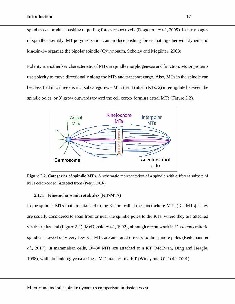
Polarity is another key characteristic of MTs in spindle morphogenesis and function. Motor proteins
use polarity to move directionally along the MTs and transport cargo. Also, MTs in the spindle can
be classified into three distinct subcategories – MTs that 1) attach KTs, 2) interdigitate between the
spindle poles, or 3) grow outwards toward the cell cortex forming astral MTs (Figure 2.2).
Figure 2.2. Categories of spindle MTs. A schematic representation of a spindle with different subsets of
MTs color-coded. Adapted from (Petry, 2016).
2.1.1. Kinetochore microtubules (KT-MTs)
In the spindle, MTs that are attached to the KT are called the kinetochore-MTs (KT-MTs). They
are usually considered to span from or near the spindle poles to the KTs, where they are attached
via their plus-end (Figure 2.2) (McDonald et al., 1992), although recent work in C. elegans mitotic
spindles showed only very few KT-MTs are anchored directly to the spindle poles (Redemann et
al., 2017). In mammalian cells, 10–30 MTs are attached to a KT (McEwen, Ding and Heagle,
1998), while in budding yeast a single MT attaches to a KT (Winey and O’Toole, 2001).

Introduction 18
Mitotic and meiotic spindle dynamics comparison in fission yeast
KT-MTs can be formed by the search and capture model (Kirschner and Mitchison, 1986), but the
stochastic character of this model is not consistent with the observed kinetics of K-fibre formation
in animal cells, as it would require hours to capture all of the KTs (Wollman et al., 2005). Studies
with Xenopus egg extracts have demonstrated that MTs can self-assemble around chromatin in the
absence of centrosomes via a Ran-GTP pathway (Heald et al., 1996, 1997; Gruss et al., 2001). The
same pathway of MT nucleation was also shown to operate in centrosome containing mammalian
cells and Drosophila S2 cell line, thus displaying the co-existence of these two pathways and their
simultaneous activity (Khodjakov et al., 2003; Maiato, Rieder and Khodjakov, 2004).
Besides silencing the SAC to prevent an early transition to anaphase (Musacchio and Salmon,
2007), the role of KT-MTs is to pull on the chromosomes at KTs, and maintain their position at the
cell equator, thus contributing to the net force of the spindle (Dogterom et al., 2005; Goshima et
al., 2005; Dumont and Mitchison, 2009; Pavin and Tolić, 2016). These pulling forces originate
from MT depolymerization, and can be modulated by motors and MAPs present at the KTs
(McIntosh, Grishchuk and West, 2002; Maddox et al., 2003; DeLuca et al., 2006).
2.1.2. Interpolar MTs (iMTs)
The set of MTs between spindle poles that is not attached to KTs is termed interpolar-MTs (iMTs)
(Figure 2.2). Minus-ends of iMTs can be found throughout meiotic spindles (Burbank et al., 2006;
Petry et al., 2013), and their nucleation was shown to occur throughout the mitotic spindles of
Drosophila S2 cell lines and human cell lines (Mahoney et al., 2006; Lawo et al., 2009; Uehara et
al., 2009).
It was proposed that iMTs ensure spindle bipolarity, and it was demonstrated in Xenopus that
bipolar meiotic spindles can assemble exclusively from non-KT-MTs (Heald et al., 1996). Some

Introduction 19
Mitotic and meiotic spindle dynamics comparison in fission yeast
iMTs are cross-linked in an antiparallel manner in the spindle midzone by MT cross-linker proteins
of the MAP65/Ase1/PRC1 family (Walczak and Shaw, 2010), and could provide structural stability
to the spindle.
In metaphase spindles, forces produced by antagonistic motor proteins maintain the spindle length
constant (Saunders and Hoyt, 1992; Goshima et al., 2005; Syrovaktina, Fu and Tran, 2013). Most
notably, kinesin-5 motors, that typically move toward the plus-end of the MT, separate the spindle
poles by cross-linking and sliding apart the antiparallel MTs (Hagan and Yanagida, 1990; Sharp et
al., 1999; Kapitein et al., 2005; Ferenz, Gable and Wadsworth, 2010). Kinesin-5 produced outward
pushing force is opposed by motors that move toward the minus-end of the MTs, such as kinesin-
14 motors or dynein, and that produce pulling forces (Chalovich and Eisenberg, 2013; Olmsted et
al., 2014). Hence, iMTs are a region of the spindle where motors operate and contribute pushing
or pulling forces in the spindle.
2.1.3. Astral MTs (aMTs)
A subgroup of MTs that are anchored via their minus-end at the spindle pole, and grow with their
plus end toward the cortex is called astral MTs (aMTs) (Figure 2.2). They can be found in
centrosomal mitotic and meiotic spindles (Miyazaki, Kato and Nemoto, 2005; Burdyniuk et al.,
2018), while their occurrence varies in different types of acentrosomal meiotic spindles. When
activated in vitro, acentrosomal spindles of Drosophila oocytes contained aMTs, but not in laid
eggs (Wilson and Borisy, 1998; Endow and Hallen, 2011), and aMTs are not reported in Xenopus
oocytes (Burbank et al., 2006). In other acentrosomal MI spindles, like those of mouse oocytes and
C. elegans, aMTs have been observed (Schuh and Ellenberg, 2007; Vargas et al., 2019). However,
these observations were not subsequently reported, and are currently considered to be the result of

Introduction 20
Mitotic and meiotic spindle dynamics comparison in fission yeast
poor experimental procedures, and not a reflection of reality. Importantly, human oocyte spindles
lack aMTs (Holubcová et al., 2015), but further research on healthy oocytes is necessary.
Studies showed aMTs are capable of producing pulling or pushing forces through dynamic
instability, that can also be promoted by action of motor proteins and MAPs, and the principal
function of aMTs appears to be spindle positioning (Pearson and Bloom, 2004). Additionally, aMTs
are involved in the production of so called polar ejection force (PEF), that is suggested to contribute
to chromosome congression and oscillation by pushing the arms of chromosomes away from the
spindle poles (Rieder et al., 1986). Drug induced destabilization/depolymerization of aMTs caused
chromosomes to no longer be repelled from the pole (Ault et al., 1991). Conversely,
stabilization/polymerization of aMTs resulted in an increase of PEFs, as chromosomes were pushed
further away from the pole than in non-treated cells. Moreover, in C. elegans, dynein-mediated
astral pulling forces prompt anaphase B spindle elongation (Hara and Kimura, 2009).
2.2. Centrosome and spindle pole body (SPB)
In the majority of dividing mitotic cells, MT nucleation is assigned to the centrosomes. The
centrosome is extensively studied, and much of its structure and proteome is mapped and described
(Andersen et al., 2003; Müller et al., 2010; Lawo et al., 2012; Ito and Bettencourt-Dias, 2018).
The centrosome is composed of two orthogonally oriented centrioles, one mother and one daughter
centriole, and of pericentriolar material (PCM), that nucleates and stabilizes the MTs, and anchors
MT minus-ends to the centrosomes (Figure 2.3A) (Wu and Akhmanova, 2017). Phosphorylation
of PCM components by PLK1 and Aurora A is pivotal to promote PCM assembly (Lee and Rhee,
2011; Joukov, Walter and De Nicolo, 2014; Conduit, Wainman and Raff, 2015). Two molecules
of γ-tubulin, a member of the tubulin superfamily indispensable for MT nucleation, GCP2 and

Introduction 21
Mitotic and meiotic spindle dynamics comparison in fission yeast
GCP3, form a γ-tubulin small complex (γ-TuSC) (Figure 2.3B). PCM proteins, like CDK5RAP2
and Pericentrin, promote the assembly of multiple γ-TuSCs into an active γ-tubulin ring complex
(γ-TuRC), which acts as a template for MT nucleation (Figure 2.3B) (Zimmerman et al., 2004;
Fong et al., 2008).
Figure 2.3. Centrosome is the major MT organizing center in the cell. (A) A schematic representation
of the centrosome. The orthogonally positioned centrioles are surrounded by the protein-packed
pericentriolar material (PCM). The centriole is a hollow cylinder made of nine MT triplets, which are
organized in a cartwheel-like formation. Adapted from (Rale, Kadzik and Petry, 2018) (B) Schematic view
of the γ-tubulin small complex (γ-TuSC). γ-TuSCs oligomerize into a more complex structure named the γ-
tubulin ring complex (γ-TuRC). Adapted from (Tovey and Conduit, 2018; Liu et al., 2020) (C) Schematic
view of the duplicated fission yeast spindle pole body (SPB) connected by a bridge (upper) and an
immunoelectron microscopy image of a duplicated SPB. The SPBs are embedded in the nuclear envelope
(NE) through a process named polar fenestration. Scale bar, 100 nm. Adapted from (Cavanaugh and
Jaspersen, 2017), (Paoletti et al., 2003)..
The centrosomes of fission yeast, the spindle pole bodies (SPBs), are ellipsoid, laminar structures
that do not contain centrioles (Figure 2.3C) (Cavanaugh and Jaspersen, 2017). As mitosis is closed
in fission yeast, i.e. no nuclear envelope breakdown (NEBD) occurs, the cytoplasmic interphase
SPBs have to be incorporated into the nuclear membrane to nucleate the spindle (Ding, McDonald
and McIntosh, 1993). As in mammalian cells, γ-TuC complex serves as a template for MT
nucleation in S. pombe and is composed of (human/fission yeast): γ-tubulin/Gtb1 and five GCP
proteins (GCP2/Alp4, GCP3/Alp6, GCP4/Gfh1, GCP5/Mod21 and GCP6/Alp16). Fission yeast

Introduction 22
Mitotic and meiotic spindle dynamics comparison in fission yeast
also contains Pericentrin, Pcp1, that recruits both γ-TuRC and PLK1/Plo1 to the nuclear side of the
SPB and is vital for mitotic entry and spindle formation (Fong, Sato and Toda, 2010). Unlike
centrosomes in most systems, in fission yeast meiosis SPBs are retained and function as spindle
MT nucleators.
Acentrosomal spindles, like the ones in most female oocyte meiosis, lack centrosomes, and
therefore rely on non-centrosomal MTOCs (ncMTOC) to nucleate MTs. Research so far has
showed that ncMTOCs during spindle assembly can be formed around chromatin, KTs or along
pre-existing MTs (Heald et al., 1996; Maiato, Rieder and Khodjakov, 2004; Petry et al., 2013).
Unlike the centrosome, little is known of the ncMTOC structure, but it is presumed they would
have to include MT minus-end proteins and adapter-proteins that could attach MTs to specific
locations (Sanchez and Feldman, 2015). Interestingly, it was shown that laser ablation of
centrosomes in mammalian cells does not abolish spindle assembly (Khodjakov et al., 2000),
demonstrating that ncMTOCs are operating during centrosomal spindle assembly as well.
2.3. Chromosomes and KTs
While regarded as passive elements during cell division in the past (Mazia, 1961), with the
observation of Ran-GTP-dependent pathway of MT nucleation during spindle assembly, as well as
the force contribution of the chromosomal region to force-balance in the metaphase spindle
(Civelekoglu-Scholey and Scholey, 2010), chromosomes are now recognized as important
contributors to spindle assembly and function.
In mitosis and MII, the chromosomes are individual entities, and consist of two sister-chromatids
connected by the cohesin complex (Figure 2.4A). Mitotic cohesin complex is made up of four
subunits (in human/fission yeast): Smc1α/Psm1, Smc3/Psm3, Rad21 and SA1/2/Psc3 (Tomonaga

Introduction 23
Mitotic and meiotic spindle dynamics comparison in fission yeast
et al., 2000; Mehta, Rizvi and Ghosh, 2012). Smc1 and Smc3 make a V-shaped dimer, which is
enclosed by Rad21 associated with SA1/2 (Figure 2.4B). This ring structure encompasses sister-
chromatids and keeps them together from S-phase until anaphase.
Each sister-chromatid contains a KT, where spindle MTs are attached during mitosis. To achieve
bi-orientation, sister-KTs on mitotic chromosomes are positioned back-to-back (Figure 2.4A), and
thus arranged to face the opposite spindle poles, which facilitates bipolar attachment (Östergren,
1951; Nicklas, 1997; Sakuno, Tada and Watanabe, 2009; Zaytsev and Grishchuk, 2015). In
prometaphase, Aurora-B kinase concentrates at centromeres, and is likely activated by the lack of
tension between the sister-chromatids. If Aurora B is close enough to the KTs (insufficient tension),
it will phosphorylate its substrates at the KT, and destabilize the attachment of MTs to KTs, thereby
promoting correct bi-orientation (Liu et al., 2009). KT attachment and bi-orientation of
chromosomes distances sister-KTs, and causes tension in the centromeric area that inactivates SAC
(Musacchio and Salmon, 2007). Anaphase promoting complex (APC) is activated by Cdc20, and
the ensuing cascade activates separase, that cleaves the cohesin between the sister-chromatids, and
triggers their disjunction.
In MI, the homologous chromosomes are synapsed into a bivalent through the action of the
synaptonemal complex, and consist of four sister-chromatids connected by cohesin and chiasmata
– regions where crossing-over occurred and where the homologous chromosomes are physically
linked (Figure 2.4A) (Miyazaki & Orr-Weaver, 1994; Page & Hawley, 2003). The meiotic cohesin
ring is modified, such that the Rad21 subunit is exchanged with the meiosis specific Rec8 subunit
(Figure 2.4B). Additionally, in mammals Smc1β replaces Smc1α (Revenkova et al., 2004), and
STAG3 replaces SA1/SA2 in meiosis (Prieto et al., 2001). In fission yeast, cohesin complexes are

Introduction 24
Mitotic and meiotic spindle dynamics comparison in fission yeast
organized in distinct domains – chromosome arm cohesin, where Rec11 is in complex with Rec8,
and centromeric cohesin, where Psc3 is in complex with Rec8 (Tomoya S Kitajima et al., 2003).
Figure 2.4. Architecture of an independent and paired chromosome. (A) An illustration of chromosomal
architecture in mitosis and meiosis. In mitosis, the chromosome is a singular entity where sister-chromatids
are connected by the mitotic cohesin complex. The KTs are oriented back-to-back, which facilitates bi-
orientation of the chromosome, and separation of sister-chromatids to the opposite spindle poles upon the
destruction of cohesin complex. In meiosis I (MI), the homologous chromosomes are paired into a bivalent,
which facilitates recombination. The yellow asterisks on the illustration indicate sites where recombination
occurred, and where the chromosomes are intertwined. Meiosis-specific cohesin and MEIKIN/Moa1
‘’clamp’’ the KTs on sister chromatids together, and bring them into a side-by-side arrangement. This
facilitates the mono-orientation of each homologous chromosome in a bivalent. Adapted from (Sakuno,
Tada and Watanabe, 2009). (B) Schematic representation of the cohesin complex in mitosis (regular font)
and meiosis (bold font) (Mehta et al., 2013).
Because of this meiosis-specific cohesin configuration, cohesin can be removed from the
chromosome in a step-wise manner (Miyazaki et al., 2017). First, once all the bivalents are bi-
oriented, cohesin is removed from the chromosome arms, but not from the centromeric region
(Figure 1.2A). This allows halving of ploidy, as the homologous chromosomes are separated from
the bivalent configuration to the opposite spindle poles in MI. In MII, the remaining centromeric
cohesin is removed (Figure 1.2B), and sister-chromatids are separated in anaphase II. Studies in
fission yeast have identified an evolutionary conserved protein Sgo1 as the key molecule protecting

Introduction 25
Mitotic and meiotic spindle dynamics comparison in fission yeast
the cohesin in the centromeric region from destruction (Kitajima, Kawashima and Watanabe, 2004;
Watanabe et al., 2005; Kitajima et al., 2006). Sgo1 is recruited through activity of Bub1
(Kawashima et al., 2010), and in turn recruits PP2A that ensures dephosphorylation of centromeric
cohesin during MI, thereby preventing its destruction (Riedel et al., 2006; Miyazaki et al., 2017).
KT architecture in MI is also altered to ensure mono-orientation of bivalents – unlike the
mitotic/MII back-to-back arrangement, the KTs in MI adopt a side-by-side arrangement (Figure
2.4A) (Östergren, 1951). The central molecule in establishment of cohesin-mediated monopolar
attachment of KTs is the mammalian MEIKIN/fission yeast Moa1 (Yokobayashi and Watanabe,
2005; Kim et al., 2015), that localizes in the centromeric region up to anaphase I onset. This
molecule is also implicated in the MI cohesin protection through recruitment of Plo1/PLK1 (Kim
et al., 2015; Miyazaki et al., 2017), whose activity at the KTs ensures Bub1 accumulation
(Miyazaki et al., 2017).
2.3.1. Chromokinesins & polar ejection force (PEF)
At chromosomes, PEF is generated through the activity of chromokinesins, kinesin-like motor
proteins that associate with chromatin during cell division. Chromokinesins identified so far belong
to the kinesin-4 and kinesin-10 families, as well as kinesin-12 family (Vanneste, Ferreira and
Vernos, 2011). They are low- or non-processive plus-end directed motors (Yajima et al., 2003;
Bringmann et al., 2004) which are thought to advance chromosome congression by actively
pushing the chromosome arms toward the spindle equator (Figure 2.5A). (Maiato et al., 2017).
The first characterized chromokinesin was from the kinesin-4 family and was isolated from
embryonic chick retina (Wang and Adler, 1995), and homologous proteins in Xenopus, Drosophila,
mouse and human cells are identified (Sekine et al., 1994; Vernos et al., 1995; Williams et al.,
1995; Ha et al., 2000). Depletion of both human and Drosophila kinesin-4 chromokinesin
Introduction 26
Mitotic and meiotic spindle dynamics comparison in fission yeast
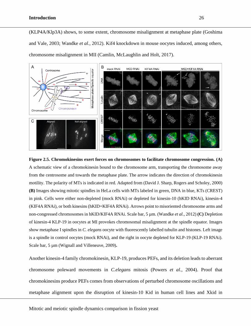
(KLP4A/Klp3A) shows, to some extent, chromosome misalignment at metaphase plate (Goshima
and Vale, 2003; Wandke et al., 2012). Kif4 knockdown in mouse oocytes induced, among others,
chromosome misalignment in MII (Camlin, McLaughlin and Holt, 2017).
Figure 2.5. Chromokinesins exert forces on chromosomes to facilitate chromosome congression. (A)
A schematic view of a chromokinesin bound to the chromosome arm, transporting the chromosome away
from the centrosome and towards the metaphase plate. The arrow indicates the direction of chromokinesin
motility. The polarity of MTs is indicated in red. Adapted from (David J. Sharp, Rogers and Scholey, 2000)
(B) Images showing mitotic spindles in HeLa cells with MTs labeled in green, DNA in blue, KTs (CREST)
in pink. Cells were either non-depleted (mock RNAi) or depleted for kinesin-10 (hKID RNAi), kinesin-4
(KIF4A RNAi), or both kinesins (hKID+KIF4A RNAi). Arrows point to misoriented chromosome arms and
non-congressed chromosomes in hKID/KIF4A RNAi. Scale bar, 5 µm. (Wandke et al., 2012) (C) Depletion
of kinesin-4 KLP-19 in oocytes at MI provokes chromosomal misalignment at the spindle equator. Images
show metaphase I spindles in C. elegans oocyte with fluorescently labelled tubulin and histones. Left image
is a spindle in control oocytes (mock RNAi), and the right in oocyte depleted for KLP-19 (KLP-19 RNAi).
Scale bar, 5 µm (Wignall and Villeneuve, 2009).
Another kinesin-4 family chromokinesin, KLP-19, produces PEFs, and its deletion leads to aberrant
chromosome poleward movements in C.elegans mitosis (Powers et al., 2004). Proof that
chromokinesins produce PEFs comes from observations of perturbed chromosome oscillations and
metaphase alignment upon the disruption of kinesin-10 Kid in human cell lines and Xkid in

Introduction 27
Mitotic and meiotic spindle dynamics comparison in fission yeast
Xenopus egg extracts (Antonio et al., 2000; Funabiki and Murray, 2000; Levesque and Compton,
2001). In human cells, kinesin-10 contributed the majority of PEFs while kinesin-4 stabilized MTs
and reduced MT dynamics (Figure 2.5B) (Brouhard and Hunt, 2005; Wandke et al., 2012).
Similarly to the effect observed in mitosis, absence of PEFs due to KLP-19 knockdown leads to
chromosome alignment defects in oocyte MI (Figure 2.5C) (Wignall and Villeneuve, 2009),
although a subsequent study did not confirm this role (Dumont, Oegema and Desai, 2010). In
female Drosophila meiosis, orphan kinesin Nod was proposed to produce PEFs required to align
chromosomes IV, which seldom recombine, but not the other chromosomes in meiosis or in mitosis
(Zhang et al., 1990; Theurkauf and Hawley, 1992). Alternatively, it was also proposed that Nod
could act through its cross-linking activity as a break, as a chromosome tension regulator, or as an
intermediate in chromosomal cohesion (Matthies, Baskin and Hawley, 2001).
In mitosis, computational analysis in the Drosophila embryo spindle estimated PEFs to be
approximately 1.1 pN per MT (Marshall et al., 2001). This value corresponds to both MT
polymerization driven mechanism of PEF generation (Chapter 2.1.3. Astral MTs) and motor-
generated PEF production. Research on the scope of PEFs produced per MT on a bivalent has not
been performed.
2.4. Motor proteins
Dyneins and kinesins are motor proteins that bind and hydrolyse adenosine triphosphate (ATP) and
translate that energy into movement along the MT lattice. MT-dependent motors are involved in a
myriad of cellular functions, ranging from organelle transport to cell division and motility. Motor
proteins exploit the polar nature of the MT in order to move along the lattice in a directed fashion.
In general, the majority of kinesins move towards the plus-end of the MT, while kinesin-14s and

Introduction 28
Mitotic and meiotic spindle dynamics comparison in fission yeast
dyneins move toward the minus-end. It is well established that motors can produce force through
binding and sliding of MTs or through modifying MT dynamics, and therefore play an integral role
in the spindle force network. In the following sections, I will focus on data available on motor
proteins that have functional homologues in fission yeast, and which play a role in fission yeast
cell division processes during prophase and metaphase.
2.4.1. Dynein
Dyneins can be classified in two distinct subgroups: the axonemal and cytoplasmic dynein. The
axonemal dynein is involved in ciliary and flagellar movements, and is not involved in spindle
function. The cytoplasmic dynein can be further divided into dynein-1 and dynein-2, of which the
latter is concerned with intra-flagellar transport. In this chapter, I will focus on cytoplasmic dynein-
1 and its roles in the spindle force production.
Dynein-1 is an approximately 1.4 MDa complex composed of multiple polypeptide chains, and
associates with numerous additional proteins and protein complexes to accomplish its cellular
activities (Cianfrocco et al., 2015). The stem is required for dimerization and for binding of other
dynein accessory subunits such as intermediate chains, light intermediate chains, and light chains
(Figure 2.6A). Two dynein heavy chains carry 6 AAA+-ATPase modules that make the motor-
domain (Neuwald et al., 1999). The motor and the tail domain are joined by a linker, an element
required for dynein motility (Burgess et al., 2003; Reck-Peterson et al., 2006). The MT binding
site is located on the N-terminal domain of dynein, the stalk, that extends from the motor domain
(Gee, Heuser and Vallee, 1997; Koonce, 1997). Moreover, the tail domain also mediates
interactions with cargo adaptor and dynein regulator proteins, which decide the function of dynein
in the cell.

Introduction 29
Mitotic and meiotic spindle dynamics comparison in fission yeast
Figure 2.6. Dynein can induce centrosome separation. (A) A scheme of dynein-1 composition. Modified
from (Raaijmakers and Medema, 2014) (B) An illustration of NE-bound dynein promoting centrosome
separation. The arrows indicate the direction of kinesin-5 (pink) and dynein (orange) motility. MT polarity
is indicated in red. Adapted from (Raaijmakers et al., 2012).
As a result of interacting with a variety of adaptor proteins, dynein is found throughout the cell,
from the nuclear envelope (NE) and cell cortex to MTs and KTs (Raaijmakers & Medema, 2014).
In most systems, kinesin-5 is essential for spindle assembly and bipolarity establishment through
centrosome separation. Some studies in mitotic and MII spindles have shown that dynein inhibition
permits bipolar spindle assembly in kinesin-5 depleted systems, and that dynein might counteract
kinesin-5 outward pushing forces (Mitchison et al., 2005; Tanenbaum et al., 2008; Chalovich and
Eisenberg, 2013). Additionally, NE associated dynein was found to promote centrosome separation
in C. elegans and Drosophila systems (Gönczy et al., 1999; Robinson et al., 1999). More recent
studies, performed in a human cell line in which spindle assembly is independent of kinesin-5
activity, have further demonstrated the role of dynein-dependent pulling forces exerted on
centrosomes in their separation (Figure 2.6B) (Raaijmakers et al., 2012). Dynein mediated pulling

Introduction 30
Mitotic and meiotic spindle dynamics comparison in fission yeast
was also proposed to move chromosomes towards the spindle pole in prometaphase (Sharp et al.,
2000; Yang et al., 2007).
2.4.2. Kinesin-5
Kinesin-5s are generally MT plus-end directed homotetramers, although there is experimental
proof of kinesin-5 minus-end directed movement as well (Yajima et al., 2003; Gerson-gurwitz et
al., 2011; Roostalu et al., 2011; Winters et al., 2019). Each monomer consists of the N-terminal
head domain containing the motor, a coiled-coil stalk, and a C-terminal tail domain (Cole et al.,
1994; Kashina et al., 1996). Such a structure allows kinesin-5 to crosslink and slide apart
antiparallel MTs in the initial stages of spindle assembly, thereby producing outward pushing forces
necessary to achieve spindle bipolarity (Figure 2.7A) (Kashina et al., 1996; Kapitein et al., 2005).
Kinesin-5 localizes to the centrosomes and spindle MTs, which is consistent with its ability to bind
both parallel and antiparallel MT bundles (Kapitein et al., 2005; van den Wildenberg et al., 2008).
Figure 2.7. Kinesin-5 is essential for spindle bipolarity. (A) A simple model of kinesin-5 dependent
spindle bipolarity establishment. In the initial stages of spindle assembly, kinesin-5 cross-link antiparallel
MTs. Cross-linked anti-parallel MTs are subsequently pushed apart, thereby also distancing the spindle
poles. (B) Chemical inhibition of kinesin-5 activity with monastrol in Xenopus egg extracts perturbs bipolar
spindle assembly. Left image shows a normal bipolar spindle (NBS) and the right image shows a monoastral
(monopolar) spindle (MA). Tubulin is shown in red and chromatin in blue. Scale bar, 5 µm (Kapoor et al.,
2000).

Introduction 31
Mitotic and meiotic spindle dynamics comparison in fission yeast
Kinesin-5s are essential in many systems, as their inactivity leads to formation of monopolar
spindles and centrosome separation failure (Figure 2.7B) (Kashina, Rogers and Scholey, 1997;
Kapoor et al., 2000). Among the first kinesin-5s discovered were the one in fungus A. nidulans
(Enos and Morris, 1990) and fission yeast (Hagan and Yanagida, 1990). In the following years
kinesin-5s were identified in Xenopus, Drosophila, and human (Sawin et al., 1992; Heck et al.,
1993; Blangy et al., 1995). Interestingly, kinesin-5 BMK-1 mutants of C. elegans, or CaKip1
deficient C. albicans, do not show monopolar spindles, suggesting that in these organisms kinesin-
5 is not essential for the establishment of spindle bipolarity (Bishop, Han and Schumacker, 2005;
Shoukat, Frazer and Allingham, 2019).
Kinesin-5 plays a role in meiosis as well, implied by immunostaining of Eg5 on meiotic spindles
in Xenopus (Houliston et al., 1994). Similarly to mitosis, inhibition of Eg5 in mouse oocytes leads
to an increase in frequency of monopolar spindles in MI, while also interfering with chromosome
alignment at the metaphase plate and causing multipolar spindle assembly (Mailhes, Mastromatteo
and Fuseler, 2004). Treatment of a human oocyte arrested in MII with monastrol, kinesin-5
inhibitor, switched the MII spindle from bipolar to monopolar state, (Duncan, Hornick and
Woodruff, 2012), further indicating that kinesin-5 operates in meiotic spindles. Differently,
knockdown of kinesin-5 in Drosophila oocytes did not result in monopolar, but in asymmetric
spindles, e.g. one half-spindle showed a higher tubulin intensity (Radford, Go and McKim, 2017).
The mechanism behind this result is not identified, but it is interesting to speculate that kinesin-5
may regulate MT nucleation as it was shown in fission yeast (Olmsted et al., 2014).
2.4.3. Kinesin-8
Kinesin-8s are MT plus-end directed dimers implicated in MT dynamics regulation (Figure 2.8A)
(Shrestha et al., 2019). The motor domain is located at the N-terminal part of the protein, connected
Introduction 32
Mitotic and meiotic spindle dynamics comparison in fission yeast
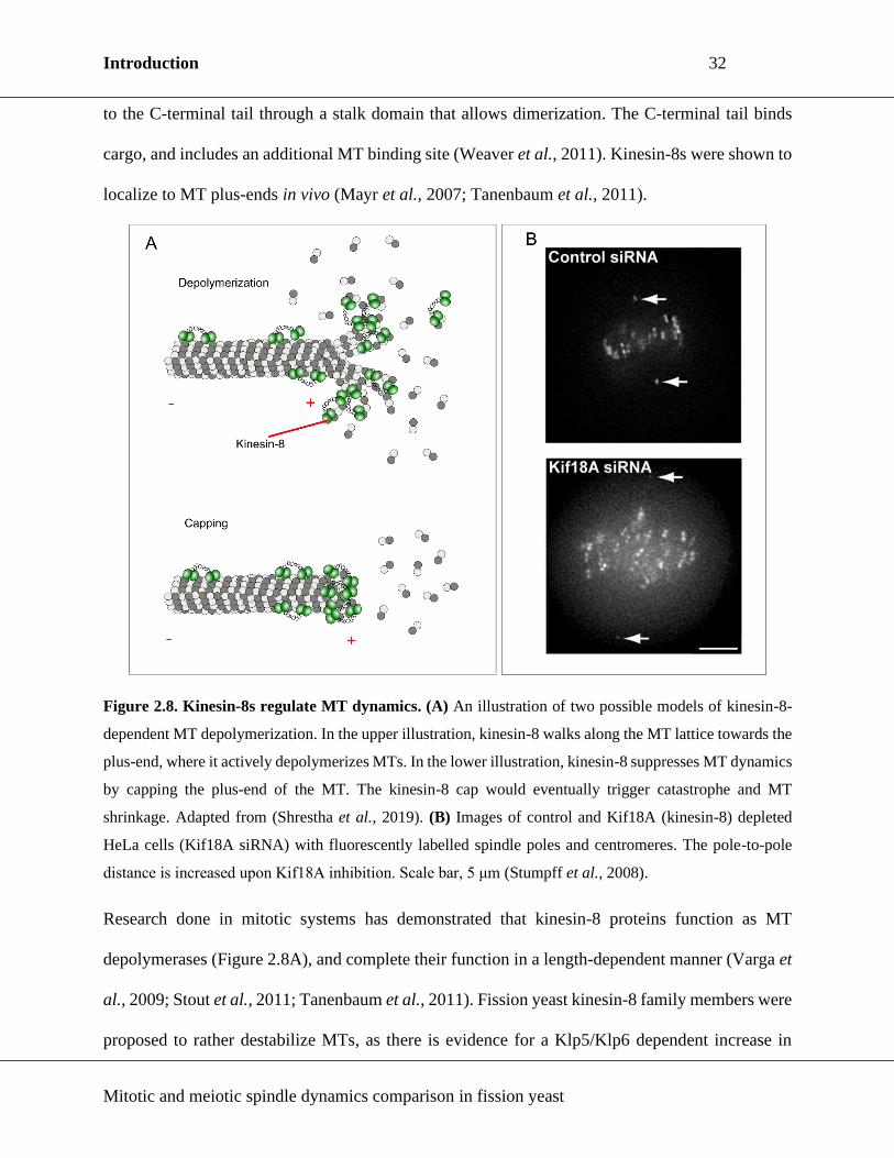
to the C-terminal tail through a stalk domain that allows dimerization. The C-terminal tail binds
cargo, and includes an additional MT binding site (Weaver et al., 2011). Kinesin-8s were shown to
localize to MT plus-ends in vivo (Mayr et al., 2007; Tanenbaum et al., 2011).
Figure 2.8. Kinesin-8s regulate MT dynamics. (A) An illustration of two possible models of kinesin-8-
dependent MT depolymerization. In the upper illustration, kinesin-8 walks along the MT lattice towards the
plus-end, where it actively depolymerizes MTs. In the lower illustration, kinesin-8 suppresses MT dynamics
by capping the plus-end of the MT. The kinesin-8 cap would eventually trigger catastrophe and MT
shrinkage. Adapted from (Shrestha et al., 2019). (B) Images of control and Kif18A (kinesin-8) depleted
HeLa cells (Kif18A siRNA) with fluorescently labelled spindle poles and centromeres. The pole-to-pole
distance is increased upon Kif18A inhibition. Scale bar, 5 μm (Stumpff et al., 2008).
Research done in mitotic systems has demonstrated that kinesin-8 proteins function as MT
depolymerases (Figure 2.8A), and complete their function in a length-dependent manner (Varga et
al., 2009; Stout et al., 2011; Tanenbaum et al., 2011). Fission yeast kinesin-8 family members were
proposed to rather destabilize MTs, as there is evidence for a Klp5/Klp6 dependent increase in

Introduction 33
Mitotic and meiotic spindle dynamics comparison in fission yeast
catastrophe frequency as a fuinction of MT length (Tischer, Brunner and Dogterom, 2009), and
they did not show depolymerization activity in vitro (Erent, Drummond and Cross, 2012).
Additionally, there is evidence that budding yeast kinesin-8 can cross-link and slide MTs (Su et al.,
2013; Rizk et al., 2014). Likely due to its roles in MT dynamics regulation, inactivation of kinesin-
8s disrupts chromosome congression in several systems (Figure 2.8B) (West, Malmstrom and
McIntosh, 2002; Stumpff et al., 2008; Savoian and Glover, 2010). Moreover, spindle length is
typically affected upon kinesin-8 disruption, so that the spindles in mutants are longer (West,
Malmstrom and McIntosh, 2002; Mayr et al., 2007; Stumpff et al., 2008; Weaver et al., 2011). The
observed phenotypes point to kinesin-8s as players in spindle length maintenance.
Kinesin-8 function is explored to a modest extent in meiotic systems. Studies performed in
Drosophila spermatocytes recapitulate roles for kinesin-8 in meiotic spindle length control and
chromosome congression (Gandhi et al., 2004; Savoian et al., 2004). In Xenopus, kinesin-8 was
shown to accumulate in oocytes as they progress through meiosis, and its depletion results in longer
MII spindles with unfocused spindle poles (Möckel et al., 2017). Further, fission yeast kinesin-8
mutants fail to progress through typical meiosis, evident by aberrant spore production (West et al.,
2001). No studies were done on kinesin-8 functions in acentrosomal MI spindles, but considering
the available data, it is plausible to assume kinesin-8 could be an important player in MI spindle
assembly/function, and further studies may be beneficial.
2.4.4. Kinesin-14
Kinesin-14s are dimeric MT minus-end directed motors (McDonald, Stewart and Goldstein, 1990).
Like other kinesins, they consist of a globular head with a motor domain, a central coiled-coil stalk
region, and a tail domain, with the distinction of the motor domain being C-terminal and the tail
Introduction 34
Mitotic and meiotic spindle dynamics comparison in fission yeast
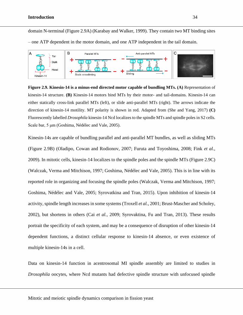
domain N-terminal (Figure 2.9A) (Karabay and Walker, 1999). They contain two MT binding sites
– one ATP dependent in the motor domain, and one ATP independent in the tail domain.
Figure 2.9. Kinesin-14 is a minus-end directed motor capable of bundling MTs. (A) Representation of
kinesin-14 structure. (B) Kinesin-14 motors bind MTs by their motor- and tail-domains. Kinesin-14 can
either statically cross-link parallel MTs (left), or slide anti-parallel MTs (right). The arrows indicate the
direction of kinesin-14 motility. MT polarity is shown in red. Adapted from (She and Yang, 2017) (C)
Fluorescently labelled Drosophila kinesin-14 Ncd localizes to the spindle MTs and spindle poles in S2 cells.
Scale bar, 5 μm (Goshima, Nédélec and Vale, 2005).
Kinesin-14s are capable of bundling parallel and anti-parallel MT bundles, as well as sliding MTs
(Figure 2.9B) (Oladipo, Cowan and Rodionov, 2007; Furuta and Toyoshima, 2008; Fink et al.,
2009). In mitotic cells, kinesin-14 localizes to the spindle poles and the spindle MTs (Figure 2.9C)
(Walczak, Verma and Mitchison, 1997; Goshima, Nédélec and Vale, 2005). This is in line with its
reported role in organizing and focusing the spindle poles (Walczak, Verma and Mitchison, 1997;
Goshima, Nédélec and Vale, 2005; Syrovatkina and Tran, 2015). Upon inhibition of kinesin-14
activity, spindle length increases in some systems (Troxell et al., 2001; Brust-Mascher and Scholey,
2002), but shortens in others (Cai et al., 2009; Syrovaktina, Fu and Tran, 2013). These results
portrait the specificity of each system, and may be a consequence of disruption of other kinesin-14
dependent functions, a distinct cellular response to kinesin-14 absence, or even existence of
multiple kinesin-14s in a cell.
Data on kinesin-14 function in acentrosomal MI spindle assembly are limited to studies in
Drosophila oocytes, where Ncd mutants had defective spindle structure with unfocused spindle

Introduction 35
Mitotic and meiotic spindle dynamics comparison in fission yeast
poles (Hatsumi and Endow, 1992). Further studies expanded on this findings, showing that MI
spindles in which kinesin-14 activity is compromised form multipolar MI spindles, fail to elongate
to wild-type (wt) MI spindle length, and either fail to achieve bipolarity, or are unable to maintain
it (Matthies et al., 1996; Endow and Komma, 1997). A model of acentrosomal MI spindle assembly
relying on Ncd was also proposed (Sköld, Komma and Endow, 2005). A compelling study in mouse
oocytes has shown that increasing the amount of kinesin-14 in MI results in a mitotic-like spindle
assembly and erroneous chromosome segregation (Bennabi et al., 2018). Available data indicates
kinesin-14 may play a crucial role in MI spindle assembly in a dose-dependent way, while not
being essential in mitotic spindle assembly.
2.5. Non-motor microtubule associated proteins (MAPs)
Spindle function and structure does not depend solely on motor proteins and MTs. The spindle and
its components are coordinated by proteins that control cell-cycle progression, regulate motor
function or promote MT nucleation (Manning and Compton, 2008). Among many regulatory and
accessory proteins associated with the spindle and its components, non-motor MAPs are tasked
with providing structural integrity to the spindle by crosslinking parallel or anti-parallel MT
bundles. In this chapter, I will discuss non-motor proteins in the context of spindle structure and
architecture.
Cross-linker proteins of the MAP65 family are arguably the most prominent spindle structure
maintaining proteins besides kinesin-5. Mammalian member of the MAP65 family, PRC1, is a
homodimer with four distinct domains: a dimerization domain, a rod domain, a spectrin domain,
and a C-terminal domain (Figure 2.10A) (Subramanian et al., 2010; Kellogg et al., 2016). MAP65
members are crucial elements of the spindle midzone, for they crosslink iMTs, and support the

Introduction 36
Mitotic and meiotic spindle dynamics comparison in fission yeast
spindle structure (Figure 2.10B) (Mollinari et al., 2002; Kurasawa et al., 2004; Loïodice et al.,
2005). Mitotic spindles in cells deficient for MAP65 frequently break upon anaphase onset,
producing two unorganized half-spindles.
Figure 2.10. MAP65 members are cross-linker MAPs. (A) An illustration of domain structure of full-
length PRC1. Numbers indicated below represent amino-acid residues. Adapted from (Kellogg et al., 2016).
(B) MAP65 members cross-link anti-parallel MTs at the spindle midzone. MT polarity is shown in red.
Depletion of SPD-1 in C. elegans female meiosis showed no defect in MT bundle formation,
arguing for a secondary role of SPD-1 in MT bundling (Verbrugghe and White, 2004; Mullen and
Wignall, 2017). Considering the spindle localization of MAP65 members during prometaphase in
mammalian and fission yeast cells (Mollinari et al., 2002; Fu et al., 2010; Polak et al., 2017), there
is a possibility that MAP65 might be an essential player in meiotic spindle integrity maintenance
in systems other than C. elegans. A recent study published by the Kitajima group has found that
Prc1 accumulates at the kinetochore during MI, and is required for correct chromosome
segregation. Additionally, such accumulation was not detected in human oocytes, where
chromosome segregation is especially error-prone (Yoshida et al., 2020).
3. Non-centrosomal pathways of spindle assembly
As mentioned earlier (Chapter 1.2. and Chapter 2.2.), many female oocytes lack centrosomes, yet
manage to nucleate MTs and assemble a spindle. Moreover, mitotic cells in which centrosomes are
ablated, or mutants that lack centrosomes, still assemble a mitotic spindle. Regardless of
Introduction 37
Mitotic and meiotic spindle dynamics comparison in fission yeast
differences, it appears that MT nucleation is mediated by γ-tubulin in all pathways. In the following
chapter, I will address the most prominent centrosome-independent spindle assembly pathways.
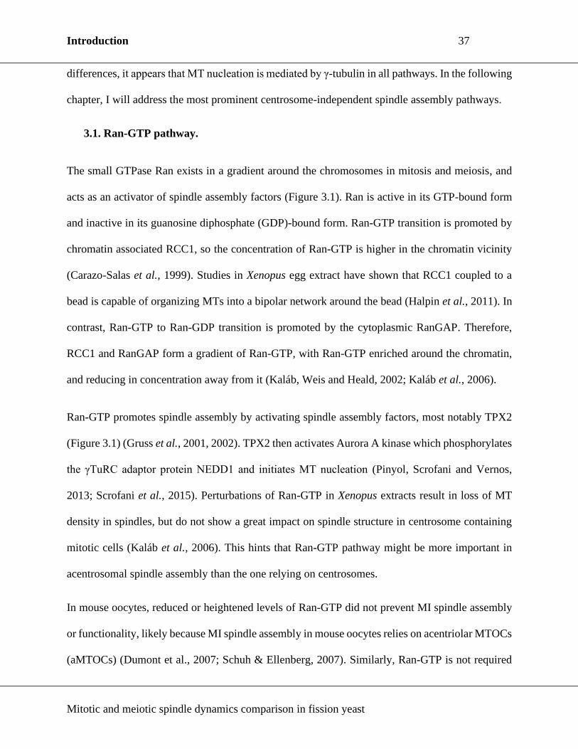
3.1. Ran-GTP pathway.
The small GTPase Ran exists in a gradient around the chromosomes in mitosis and meiosis, and
acts as an activator of spindle assembly factors (Figure 3.1). Ran is active in its GTP-bound form
and inactive in its guanosine diphosphate (GDP)-bound form. Ran-GTP transition is promoted by
chromatin associated RCC1, so the concentration of Ran-GTP is higher in the chromatin vicinity
(Carazo-Salas et al., 1999). Studies in Xenopus egg extract have shown that RCC1 coupled to a
bead is capable of organizing MTs into a bipolar network around the bead (Halpin et al., 2011). In
contrast, Ran-GTP to Ran-GDP transition is promoted by the cytoplasmic RanGAP. Therefore,
RCC1 and RanGAP form a gradient of Ran-GTP, with Ran-GTP enriched around the chromatin,
and reducing in concentration away from it (Kaláb, Weis and Heald, 2002; Kaláb et al., 2006).
Ran-GTP promotes spindle assembly by activating spindle assembly factors, most notably TPX2
(Figure 3.1) (Gruss et al., 2001, 2002). TPX2 then activates Aurora A kinase which phosphorylates
the γTuRC adaptor protein NEDD1 and initiates MT nucleation (Pinyol, Scrofani and Vernos,
2013; Scrofani et al., 2015). Perturbations of Ran-GTP in Xenopus extracts result in loss of MT
density in spindles, but do not show a great impact on spindle structure in centrosome containing
mitotic cells (Kaláb et al., 2006). This hints that Ran-GTP pathway might be more important in
acentrosomal spindle assembly than the one relying on centrosomes.
In mouse oocytes, reduced or heightened levels of Ran-GTP did not prevent MI spindle assembly
or functionality, likely because MI spindle assembly in mouse oocytes relies on acentriolar MTOCs
(aMTOCs) (Dumont et al., 2007; Schuh & Ellenberg, 2007). Similarly, Ran-GTP is not required

Introduction 38
Mitotic and meiotic spindle dynamics comparison in fission yeast
for MI spindle assembly in Xenopus or Drosophila oocytes (Cesario and McKim, 2011). These
data point to the existence of another essential spindle assembly mechanism, while Ran-GTP
mostly contributes to the speed and efficiency of MI spindle assembly in oocytes. At the same time,
MII spindle defects were much more pronounced upon Ran-GTP inactivation, and accompanied
by chromosome segregation defects, suggesting a critical role of Ran-GTP pathway in MII spindle
function.
Figure 3.1. Chromosome associated pathways of spindle assembly. On the left side of the chromosome,
the chromosomal passenger complex (CPC) dependent pathway of spindle assembly is illustrated. CPC is
at the KT, where one of its components, Aurora B, phosphorylates and thereby inactivates MT-destabilizing
factors. This results in an environment being formed around the KT that promotes MT stabilization. On the
right side of the chromosome, the Ran-GTP pathway of spindle assembly is illustrated. Chromatin bound
Rcc1 locally enriches the area around chromatin with Ran-GTP. In turn, Ran-GTP activates spindle
assembly factors, like TPX2, by releasing them from importins. Adapted from (Gruss, 2018) and (Meunier
and Vernos, 2016).
Blocking Ran-GTP dependent spindle assembly pathway delayed MT nucleation and hindered MI
spindle assembly in human oocytes, suggesting its necessity for successful meiosis (Holubcová et

Introduction 39
Mitotic and meiotic spindle dynamics comparison in fission yeast
al., 2015). However, oocytes used in this study did not progress through meiosis upon hormonal
cues, and may therefore behave differently than healthy human oocytes.
3.2. Chromosomal passenger complex (CPC) pathway
A second chromatin associated pathway of spindle assembly, which can operate separately of the
Ran-GTP pathway, is called the CPC pathway (Maresca et al., 2009). The CPC consists of
(human/fission yeast) INCENP/Pic1, Aurora B/Ark1, Survivin/Bir1/Cut17 and Borealin/Nlb1
(Carmena et al., 2012; van der Waal et al., 2012). The mechanism of CPC-dependent assembly
pathway is well explored in Xenopus, and involves Aurora B-dependent inhibition of MT-
destabilizing agents, which in turn creates an environment where MT assembly is promoted (Figure
3.1) (Andrews et al., 2004; Lan et al., 2004; Sampath et al., 2004; Gadea and Ruderman, 2006;
Kelly et al., 2007).
In Drosophila oocytes, CPC pathway is essential for acentrosomal spindle assembly, as its
suppression prevents MI spindle assembly (Colombié et al., 2008; Radford, Jang and McKim,
2012). In C. elegans, CPC was found to contribute to chromosome alignment in meiosis, the release
of cohesion, and the proper assembly of the meiotic (Kaitna et al., 2002; Rogers et al., 2002;
Wignall and Villeneuve, 2009; Dumont, Oegema and Desai, 2010). As Ran-GTP, inactivation of
the CPC-pathway through INCENP knockdown, or chemically by inactivating Aurora B/C, did not
abolish MI spindle assembly in mouse oocytes, but it did provoke chromosome misalignment at
meiotic metaphase (Shuda et al., 2009; Sharif et al., 2010).
In other studies utilizing mouse oocytes, MI spindles were aberrant and disorganized in Aurora B
and C mutants (Yang et al., 2010; Balboula and Schindler, 2014). Discrepancies in obtained results
may reflect the robustness of the methods or the targets used for the inhibition of the CPC-pathway.

Introduction 40
Mitotic and meiotic spindle dynamics comparison in fission yeast
Alternatively, they could be a consequence of redundancy, as multiple Auroras co-exist in mice.
Indeed, Aurora A can take over Aurora B/Aurora C roles in mouse oocytes when both are
inactivated (Nguyen et al., 2018). In any case, further studies in model organisms and human
oocytes are mandatory.
3.3. Acentriolar MTOCs (aMTOCs)
aMTOCs were discovered in mouse oocytes (Calarco, Donahue and Szollosi, 1972), and are of
significant importance in MI spindle assembly (Namgoong, Kim and Christenson, 2018). Their
structure and function is still under investigation, but it has been established that they associate
with canonical PCM proteins such as γ-tubulin, pericentrin and NEDD1 (Gueth-Hallonet et al.,
1993; Carabatsos et al., 2000; Ma, Baumann and Viveiros, 2010).
Figure 3.2. A model for bipolar spindle assembly in mouse oocytes. First, PLK1 triggers a
decondensation of MTOC structure. Secondly, BicD2 recruits dynein to the NE, which stretches the MTOCs
into ribbons and fragments them. Thirdly, kinesin-5 KIF11 further fragments the MTOCs so that they can
be arranged between the spindle poles (Clift and Schuh, 2015).
aMTOC-dependent spindle assembly is well characterized in mouse oocytes where three crucial
steps were observed (Figure 3.2) (Clift and Schuh, 2015). Mouse oocytes contain multiple aMTOCs

Introduction 41
Mitotic and meiotic spindle dynamics comparison in fission yeast
before NEBD (Clift and Schuh, 2015), which are first decondensed by PLK1. Next, dynein and
MTs stretch aMTOCs and split them into pieces along the NE (Łuksza et al., 2013). After NEBD,
kinesin-5 further fragments aMTOCs into smaller foci. It appears that aMTOC fragmentation is
required to achieve MI spindle bipolarity, as spindle assembly is defective when PLK1
decondensation of aMTOCs is inhibited (Clift and Schuh, 2015). Following NEBD, a MT ball is
formed through Ran-GTP pathway, and then organized into a stable central array (Brunet et al.,
2008; Clift and Schuh, 2015), along which aMTOCs are sorted to the spindle poles (Breuer et al.,
2010).
It remains to be explored whether such a mechanism of spindle assembly functions in human
oocytes. Studies conducted so far did not report aMTOCs in human atretic oocytes (Holubcová et
al., 2015). Whether they are present in healthy human oocytes is unknown.
3.4. Augmin pathway
MTs can be nucleated within the mitotic spindles from pre-existing MTs (Mahoney et al., 2006),
and the complex responsible for it is the Augmin complex (Goshima et al., 2008). Augmin is not
required for the initial spindle assembly, but rather to enrich spindles with MTs. Augmin promotes
MT nucleation from pre-existing MTs by recruiting γ-tubulin in several mitotic systems, including
human cell lines (Goshima et al., 2007; Uehara et al., 2009), and was also shown to recruit γ-TuRC
to MTs in vitro (Song et al., 2018).
Augmin deficiency lowers MT density within the spindle, compromises spindle bipolarity, and
results in chromosome misalignment (Lawo et al., 2009; Uehara and Goshima, 2010). In Xenopus
MII arrested extracts, both Augmin and Ran-GTP target TPX2 to initiate nucleation of branching

Introduction 42
Mitotic and meiotic spindle dynamics comparison in fission yeast
MTs (Petry et al., 2013). Because of that finding, and the reported co-immunoprecipitation of
Augmin with TPX2 (Petry et al., 2013), it was suggested that the two pathways are linked.
In Drosophila oocytes mutated for Augmin, chromosomes often misalign, but initial spindle
assembly is not perturbed (Meireles et al., 2009). Instead, Augmin is required for MT assembly
near the acentrosomal MI spindle poles (Colombié et al., 2013). Augmin pathway is functional in
male meiotic spindle assembly, but Augmin localizes to the KTs instead on the spindle MTs
(Savoian and Glover, 2014). The contribution of Augmin pathway obviously differs from system
to system, so it would be most beneficial to expand studies on it in MI spindles of other species,
like Xenopus, and ultimately, in human oocytes.
4. Fission yeast as model system for analysing mitotic and meiotic spindle
dynamics
Fission yeast is a single cell eukaryotic rod-like organism that typically reaches 12-14 µm in length
at mitotic division (Figure 4.1A). Because of the simplicity of its MT cytoskeleton, the evolutionary
conservancy of major players in spindle assembly and function, and the fact that, as in higher
eukaryotes, multiple MTs attach to a KT (Ding, McDonald and McIntosh, 1993), it makes for an
exceptional model organism to study the spindle in mitosis and meiosis.
When in nutrient-rich conditions, fission yeast mostly lives as a haploid, and undergoes mitosis in
order to proliferate. Like mammalian cells, it shows the typical progression of cell cycle stages:
G1, S, G2 and M (Figure 4.1A) (Mitchison and Creanor, 1971; Nurse, Thuriaux and Nasmyth,
1976). Under starvation conditions, most notably nitrogen starvation, fission yeast exits the
vegetative cell cycle and enters the meiosis pathway (Figure 4.1B) (Harigaya and Yamamoto,
2007). Two different mating types can be distinguished in fission yeast cells: h+ and h-. If the
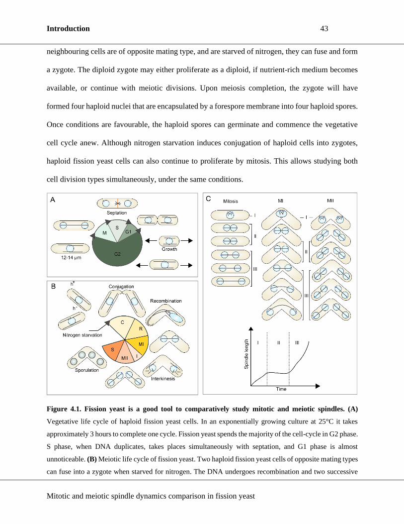
Introduction 43
Mitotic and meiotic spindle dynamics comparison in fission yeast
neighbouring cells are of opposite mating type, and are starved of nitrogen, they can fuse and form
a zygote. The diploid zygote may either proliferate as a diploid, if nutrient-rich medium becomes
available, or continue with meiotic divisions. Upon meiosis completion, the zygote will have
formed four haploid nuclei that are encapsulated by a forespore membrane into four haploid spores.
Once conditions are favourable, the haploid spores can germinate and commence the vegetative
cell cycle anew. Although nitrogen starvation induces conjugation of haploid cells into zygotes,
haploid fission yeast cells can also continue to proliferate by mitosis. This allows studying both
cell division types simultaneously, under the same conditions.
Figure 4.1. Fission yeast is a good tool to comparatively study mitotic and meiotic spindles. (A)
Vegetative life cycle of haploid fission yeast cells. In an exponentially growing culture at 25°C it takes
approximately 3 hours to complete one cycle. Fission yeast spends the majority of the cell-cycle in G2 phase.
S phase, when DNA duplicates, takes places simultaneously with septation, and G1 phase is almost
unnoticeable. (B) Meiotic life cycle of fission yeast. Two haploid fission yeast cells of opposite mating types
can fuse into a zygote when starved for nitrogen. The DNA undergoes recombination and two successive
Introduction 44
Mitotic and meiotic spindle dynamics comparison in fission yeast
meiotic divisions. Meiosis ends with four haploid nuclei encapsulated into four haploid spores. (C) Spindle
dynamics commonly show three phases of elongation. Upper illustration shows fission yeast cells in mitosis
and meiosis, with phases of spindle elongation indicated on the right side (mitosis) and in between the cells
(meiosis). The lower part depicts a spindle dynamics plot that shows spindle length through time. Dotted
lines indicate transition between three phases of spindle elongation.
Spindle assembly and elongation are stereotypical in fission yeast, in both mitosis and meiosis, and
consist of three distinct stages (Figure 4.1C) (Nabeshima, Nakagawa, Straight, Murray, et al.,
1998). Phase I of spindle assembly starts in prophase, and refers to the initial stages of spindle
formation, e.g. bipolarity establishment. Phase II is marked by steady-state spindle length
maintenance, accompanied by chromosome congression to the metaphase plate. At the end of phase
II, sister-chromatids are separated to the opposite poles. Finally, in phase III, the spindle elongates
rapidly and further separates the chromosomes.
4.1. Phase I – Initial stages of spindle nucleation
MT nucleation in fission yeast mitotic spindle assembly is mediated by the SPB, and dependent on
γ-tubulin (Masuda and Shibata, 1996; Fong, Sato and Toda, 2010). Mitosis is closed in fission
yeast, so to nucleate spindle MTs on the nuclear side, SPBs first have to be inserted into the NE in
the process known as fenestration (Jaspersen and Ghosh, 2012; Cavanaugh and Jaspersen, 2017).
Once inserted, SPBs can nucleate MTs through γ-TuC activity.
The γ-TuC in fission yeast is comparable in size and structure to the γ-TuRC of metazoans, and all
of the homologous subunits have been identified in fission yeast (Vardy and Toda, 2000; Fujita et
al., 2002; Venkatram et al., 2004; Anders, Lourenço and Sawin, 2006). γ-TuC components γ-
tubulin/Gtb1, GCP2/Alp4 and GCP3/Alp6 are essential, and mutants defective for these proteins

Introduction 45
Mitotic and meiotic spindle dynamics comparison in fission yeast
assembled aberrant monopolar spindles (Horio et al., 1991; Vardy and Toda, 2000). Interaction of
MOZART1/Mzt1 with GCP3/Alp6 allows γ-TuC to be targeted to the SPBs (Figure 4.2) (Fong,
Sato and Toda, 2010; Dhani et al., 2013; Masuda et al., 2013). Upon mitotic entry, CDK/Cdc2
phosphorylation of TACC/Alp7 triggers transport of the TACC-TOG/Alp7-Alp14 complex to the
nucleus, where it interacts with γ-TuC to initiate MT nucleation and spindle assembly (Sato et al.,
2004; Okada et al., 2014; Tang et al., 2014; Flor-Parra, Iglesias-Romero and Chang, 2018;
Yukawa, Kawakami, et al., 2019).
Figure 4.2. A simplified scheme showing spindle nucleation in fission yeast. Mzt1 forms a complex with
γ-tubulin, Alp4, and Alp6 (that comprise the γ-TuC), and it is necessary for γ-TuC attachment to the SPBs.
At mitosis, Alp7 in Alp7-Alp14 complex is phosphorylayed by Cdc2, which allows the interaction of Alp7-
Alp14 complex with the importin Cut15. The complex is translocated to the nucleus where it is recruited to
the SPB, among others, by Pcp1.

Introduction 46
Mitotic and meiotic spindle dynamics comparison in fission yeast
Ran-dependent spindle assembly pathway was also observed in fission yeast, albeit somewhat
differently than described in the chapter 3.1. Firstly, as mitosis is closed in fission yeast, there is
most likely no Ran-GTP gradient, and secondly, nucleation seems to be promoted at SPBs, and
remains unproven around chromatin in mitosis (Okada & Sato, 2015). Fission yeast mutants for
Ran-GTP/Sim1 and RCC1/Pim1 showed aberrant monopolar or star-shaped spindles, indicating
impaired spindle assembly (Fleig et al., 2000; Hirose et al., 2006; Salus et al., 2002). TACC/Alp7
was shown to be the primary target of RCC1/Pim1 (Sato & Toda, 2007). Upon RCC1 inactivation,
TACC/Alp7, and consequently TOG/Alp14, localization to the nucleus was prevented, and
therefore nuclear mitotic spindle assembly was perturbed.
In meiosis of fission yeast, spindle assembles from the SPBs as well, although some of its
components are modified from mitosis to meiosis (Ohta, Sato and Yamamoto, 2012). As research
on MT nucleation in meiosis of fission yeast is lacking, presumably, a similar mechanism of γ-
tubulin SPB-dependent MT nucleation operates in fission yeast meiosis as in mitosis. In support of
this hypothesis, Gtb1 mutants were shown to assemble monopolar spindles in meiosis,
accompanied by frequent chromosome mis-segregation (Tange et al., 2004). Interestingly, a recent
study has shown that a spindle can assemble in meiosis without SPB insertion into the NE (Pineda-
Santaella and Fernández-Álvarez, 2019). Taken with the fact that Alp7/TACC, a target of
RCC1/Pim1, localizes to the KTs at MI but not at mitotic entry (Kakui et al., 2013), it is plausible
to presume chromatin-dependent MT nucleation pathway may exist in MI of fission yeast.
4.2. Phase I – Establishment of a bipolar spindle
MT minus-ends are anchored at the SPBs (Toya et al., 2007; Yukawa, Ikebe and Toda, 2015),
while their plus-ends grow and explore the area around the SPBs. The growing MTs are bundled

Introduction 47
Mitotic and meiotic spindle dynamics comparison in fission yeast
together by fission yeast kinesin-5 Cut7, and it was proposed this is done by Cut7 rotating growing
MTs and organizing them into antiparallel fashion (Hagan and Yanagida, 1990, 1992a; Winters et
al., 2019). Cut7 exerts forces on the antiparallel-bundle and slides the MTs apart, thereby pushing
away the SPBs and establishing bipolarity. Cut7 is an essential protein, and mutants deficient for
Cut7 assemble monopolar mitotic spindles (Figure 4.3A) (Hagan and Yanagida, 1990, 1992a).
Cut7 localizes to the spindle poles and to the spindle MTs in the initial stages of spindle assembly
in both mitosis and meiosis.
Figure 4.3. Cut7 establishes bipolarity, and Pkl1 organizes spindle poles. (A) Cut7 is essential for
spindle bipolarity establishment in fission yeast. Cut7 mutants build a monopolar spindle, which ultimately
leads to cell death. (B) An illustration of Pkl1 function at the spindle poles. Pkl1 localizes to the SPB, where
it crosslinks parallel MTs. In cells lacking Pkl1, the spindle poles are unfocused. Cut7-dependent outward
pushing forces can slide minus-end MTs outward from the unfocused poles, thus forming a long MT
protrusion. The protrusion can push the chromosome to the cell division site, resulting in chromosome cut
and aneuploidy. Adapted from (Syrovatkina and Tran, 2015).
Another player in the initial stages of spindle assembly in fission yeast is a minus-end directed
motor protein, kinesin-14 Pkl1, which localizes to the SPBs and faintly to the spindle MTs at the
initial stages of mitotic spindle assembly (Pidoux, LeDizet and Cande, 1996; Syrovatkina and Tran,
2015; Yukawa, Ikebe and Toda, 2015). Pkl1 forms a complex together with Msd1 and Wdr8, which
is important in anchoring the spindle MTs to the SPBs (Toya et al., 2007; Yukawa, Ikebe and Toda,
2015). Additionally, Pkl1 probably crosslinks parallel MT bundles to provide structural stability at

Introduction 48
Mitotic and meiotic spindle dynamics comparison in fission yeast
the spindle poles in view of outward force exerted by Cut7. In fission yeast cells lacking Pkl1
activity, spindle poles are unfocused, which is evident in Cut7-mediated MT protrusion formation,
which may cause aneuploidy (Figure 4.3B) (Syrovatkina and Tran, 2015). After the report that
immunostaining failed to show Pkl1 localization on spindle MTs in meiosis (Pidoux, LeDizet and
Cande, 1996), its localization in meiosis was not further explored. However, it was reported that
zygotes deleted for Pkl1 produce the typical four spores upon meiosis completion less frequently
(Troxell et al., 2001), arguing for a role of Pkl1 in meiosis.
Two other significant players in spindle bipolarity establishment are a MAP65 family cross-linker
PRC1/Ase1 and CLASP/Peg1, a MT binding protein. Upon mitosis onset, Ase1 localizes around
the SPBs, and once bipolarity is established, it localizes to the SPBs and to the spindle midzone,
although its localization at the midzone is reduced compared to late mitosis (Loïodice et al., 2005;
Yamashita et al., 2005; Fu et al., 2010). Ase1 contributes to early spindle stability by recruiting
Peg1, and is required for Cut7-independent spindle assembly (Bratman and Chang, 2007; Rincon
et al., 2017; Yukawa et al., 2017). The role of Ase1 in the recruitment of Peg1 to early spindle
midzone is somewhat controversial, as one study found that Peg1 localization to early spindle
midzone was not dependent on Ase1 (Ebina, Ji and Sato, 2019), while another found that Peg1
localization required Ase1 (Rincon et al., 2017). In any case, strains lacking Ase1 activity show
defects in spindle integrity, but they are more pronounced in phases II and III than in phase I
(Loïodice et al., 2005; Yamashita et al., 2005; Syrovaktina, Fu and Tran, 2013). Fission yeast Peg1
localizes to the mitotic spindle midzone, and mutants were shown to generate aberrant phase I
mitotic spindles (Grallert et al., 2006; Bratman and Chang, 2007). Interestingly, localization and
function of these two molecules was not explored in fission yeast meiosis, except for a report that
meiotic nuclear oscillations are perturbed in Ase1 deficient zygotes (Yamashita et al., 2005).
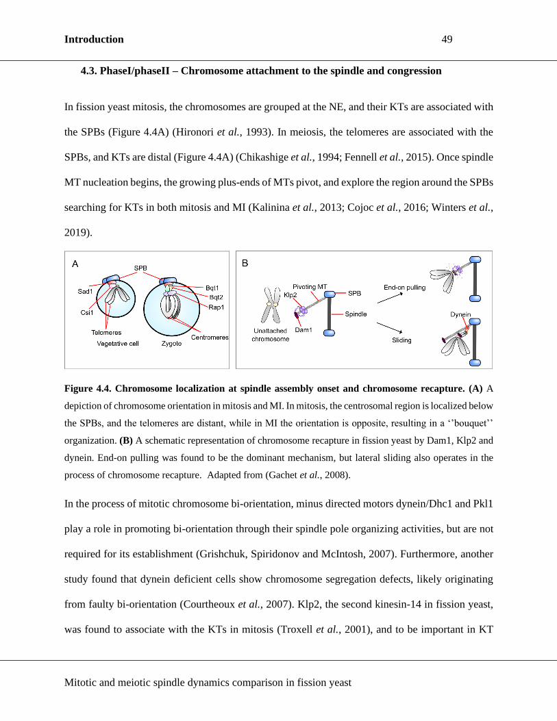
Introduction 49
Mitotic and meiotic spindle dynamics comparison in fission yeast
4.3. PhaseI/phaseII – Chromosome attachment to the spindle and congression
In fission yeast mitosis, the chromosomes are grouped at the NE, and their KTs are associated with
the SPBs (Figure 4.4A) (Hironori et al., 1993). In meiosis, the telomeres are associated with the
SPBs, and KTs are distal (Figure 4.4A) (Chikashige et al., 1994; Fennell et al., 2015). Once spindle
MT nucleation begins, the growing plus-ends of MTs pivot, and explore the region around the SPBs
searching for KTs in both mitosis and MI (Kalinina et al., 2013; Cojoc et al., 2016; Winters et al.,
2019).
Figure 4.4. Chromosome localization at spindle assembly onset and chromosome recapture. (A) A
depiction of chromosome orientation in mitosis and MI. In mitosis, the centrosomal region is localized below
the SPBs, and the telomeres are distant, while in MI the orientation is opposite, resulting in a ‘’bouquet’’
organization. (B) A schematic representation of chromosome recapture in fission yeast by Dam1, Klp2 and
dynein. End-on pulling was found to be the dominant mechanism, but lateral sliding also operates in the
process of chromosome recapture. Adapted from (Gachet et al., 2008).
In the process of mitotic chromosome bi-orientation, minus directed motors dynein/Dhc1 and Pkl1
play a role in promoting bi-orientation through their spindle pole organizing activities, but are not
required for its establishment (Grishchuk, Spiridonov and McIntosh, 2007). Furthermore, another
study found that dynein deficient cells show chromosome segregation defects, likely originating
from faulty bi-orientation (Courtheoux et al., 2007). Klp2, the second kinesin-14 in fission yeast,
was found to associate with the KTs in mitosis (Troxell et al., 2001), and to be important in KT

Introduction 50
Mitotic and meiotic spindle dynamics comparison in fission yeast
recapture and chromosome translocation to the SPBs through MT sliding and depolymerization
together with Dam1 (Figure 4.4B) (Grishchuk and McIntosh, 2006; Franco, Meadows and Millar,
2007; Gachet et al., 2008). That congression and bi-orientation of the chromosomes are not
impeded in the absence of one or more fission yeast minus-end directed proteins, demonstrates this
process is likely motor independent, and probably relies on MT dynamics (Grishchuk and
McIntosh, 2006; Grishchuk, Spiridonov and McIntosh, 2007). Even though each homologous
chromosome in a bivalent is attached to a single, opposite pole, the bivalents in MI are actually bi-
oriented. However, the role of motors in this process in meiosis was not explored in fission yeast.
Chromokinesins have not been identified in fission yeast (Wood et al., 2002), and astral MTs are
absent until end of phase II/onset of phase III, when they are assembled on the cytoplasmic side of
the nucleus (Hagan and Hyams, 1988). Therefore, PEFs probably do not operate in fission yeast
spindles to drive chromosome congression. A hetero-complex of kinesin-8, Klp5/Klp6, was
identified as a key complex in mitotic chromosome congression and bi-orientation, together with
Dam1 (Garcia, Koonrugsa and Toda, 2002; West, Malmstrom and McIntosh, 2002; Sanchez-Perez
et al., 2005; Grissom et al., 2009; Buttrick et al., 2012; Mary et al., 2015; Klemm et al., 2018).
Klp5/Klp6 localizes to KTs from phase I until the onset of phase III, when it localizes to the spindle
midzone. It governs chromosome congression by controlling pulling forces in a length-dependent
manner, i.e. accumulating at the plus-ends of long MTs, and promoting catastrophe (Figure 2.8A).
Function and localization of Klp5/Klp6 in meiosis was not reported. It was reported that Klp5 and
Klp6 appear to be essential for successful meiosis outcome, as zygotes defective for one or both
molecules failed to produce the typical four spores (West et al., 2001). As Klp5/Klp6 is not essential
for vegetative growth and mitosis, it would be beneficial to explore what is the cause of their
essentiality in meiosis.

Introduction 51
Mitotic and meiotic spindle dynamics comparison in fission yeast
4.4. Phase II – Spindle forces and force-balance maintenance in fission yeast
In phase II, the spindle maintains a constant length, while the KTs move back and forth along the
spindle. The constant length of phase II spindle was observed in many other organisms and cell
types, and was proposed to be the result of the balance of forces produced by spindle motors and
MAPs (Saunders and Hoyt, 1992; Dumont and Mitchison, 2009; Goshima and Scholey, 2010;
Syrovaktina, Fu and Tran, 2013).
In the fission yeast mitotic phase II spindle, kinesin-5 is recognized as a motor that produces
outward pushing force. Its inactivation results in phase II spindle shortening and collapse
(Syrovaktina, Fu and Tran, 2013). Regarding other motors and MAPs in fission yeast, spindle II
length changes have been observed upon Pkl1 and Ase1 inactivation (shorter spindles), and
Klp5/Klp6, Klp2, and Dam1 inactivation (longer spindles) (Troxell et al., 2001; Garcia, Koonrugsa
and Toda, 2002; Syrovaktina, Fu and Tran, 2013). Dam1 is a MAP conserved only in fungi, and is
one of the subunits of the DASH complex that is associated with the KTs during mitosis (Liu et
al., 2005). It was suggested that its role in inward pulling force production in phase II mitotic
spindles stems from its ability to convert MT depolymerization into chromosome movement
(Gachet et al., 2008; Grissom et al., 2009).

Introduction 52
Mitotic and meiotic spindle dynamics comparison in fission yeast
Figure 4.5. Simplified map of force producers in the fission yeast mitotic phase II spindle. In phase II,
the spindle length is maintained constant. The activity of indicated motors and MAPs was experimentally
shown to influence phase II spindle length in vivo. Additionally, simultaneous deletion of force generators
producing opposite forces has been shown to restore phase II spindle length.
Interestingly, Pkl1 overexpression also results in shorter spindles, arguing for its role in production
of inward pulling forces (Rincon et al., 2017). Finding that Pkl1 inactivation results in shorter
spindles could be the consequence of its role in spindle pole focusing and spindle assembly.
Moreover, simultaneous deletion of Cut7 and Pkl1, or simultaneous overexpression, restores
spindle bipolarity, thereby demonstrating these two proteins produce opposite forces and act
antagonistically to maintain constant mitotic phase II spindle length (Olmsted et al., 2014; Rincon
et al., 2017; Yukawa et al., 2017). Similar antagonistic relationship was also observed for Klp5-
Ase1 and Dam1-Ase1 (Syrovaktina, Fu and Tran, 2013).
KT-MT attachment and chromosome cohesion also influence mitotic phase II spindle length.
Mutants for KT proteins Mis12, Mis6 and Mis4 all show longer metaphase spindles (Goshima,
Saitoh and Yanagida, 1999). Chromosomes likely produce inward forces through tension
production upon attachment of KTs to MTs. Constant metaphase spindle length and force-balance

Introduction 53
Mitotic and meiotic spindle dynamics comparison in fission yeast
maintenance are important in promoting correct KT-MT attachment and chromosome segregation
(Choi and McCollum, 2012; Syrovaktina, Fu and Tran, 2013). Although necessary for precise
chromosome segregation, force-balance, or the key molecules producing force in phase II meiotic
spindles, have not been explored in fission yeast meiosis.
4.5. Phase III – Final spindle elongation
At the end of phase II, the genetic material is separated to the opposite sides of the cell as KT-MTs
depolymerize (Nabeshima, Nakagawa, Straight, Murray, et al., 1998; Ward et al., 2014). In phase
III, MTs resume growth and the spindle elongates rapidly.
The majority of anti-parallel MT sliding in phase III is carried out by the homo-tetrameric kinesin-
6 Klp9, while Ase1 cross-links the anti-parallel MTs at the spindle midzone, and maintains the
integrity of the spindle (Tolić-Nørrelykke et al., 2004; Courtheoux et al., 2009; Fu et al., 2010;
Krüger et al., 2019; Yukawa, Okazaki, et al., 2019). Cut7 was also shown to play a role in MT
sliding, as its inactivity results in lower phase III elongation velocity (Rincon et al., 2017; Krüger
et al., 2019; Yukawa, Okazaki, et al., 2019). Additionally, minus-end directed Klp2 is important
for final spindle elongation, as its persistence at the phase III spindle leads to lower spindle
elongation velocity (Mana-Capelli et al., 2012; Krüger et al., 2019). Unfortunately, our
understanding of meiosis spindle elongation is lacking, as no studies were performed to date.
4.6. Comparison of mitotic and meiotic spindle dynamics in fission yeast
Mitosis and meiosis have both been extensively studied, and many studies have investigated the
roles of motors, MAPs and MT dynamics in the spindle. Despite the considerable amount of
research, no studies to date have compared spindle dynamics simultaneously in mitosis and meiosis
in the same system. This is most likely because such a task remains technically impossible in many

Introduction 54
Mitotic and meiotic spindle dynamics comparison in fission yeast
systems, or there are too many factors that would influence data interpretation. Additionally, such
studies in humans are extremely complicated, because oocytes are regarded as precious material.
Taking into account the fundamental nature of mitotic and meiotic processes, a comparison of
mitotic and meiotic spindle dynamics would be central in exploring the differences and peculiarities
in each cell division type respectively.
Fission yeast has emerged as a good model system for such a study. Its MT cytoskeleton is simple
and well characterized, and the key molecular players in spindle assembly are conserved from
fission yeast to human. Importantly, fission yeast can undergo mitosis and meiosis in the same
environment, and under the same conditions, making comparative study of mitotic and meiotic
spindle dynamics feasible.

Aim of this work 55
Mitotic and meiotic spindle dynamics comparison in fission yeast
Aim of this work
Force-balance maintenance in phase II spindle, and components producing forces in the spindle are
very well explored in fission yeast mitosis. Additionally, spindle dynamics in wt and mutant
vegetative cells have been measured and studied. However, the corresponding data for meiotic
spindles is lacking. Moreover, no study was conducted to compare mitotic and meiotic spindle
dynamics in the same organism, and to reveal how and why cells modify spindles from mitosis to
meiosis.
The antagonistic relationship of Cut7 and Pkl1 is extensively investigated in fission yeast mitotic
spindles. That Pkl1 can bypass Cut7 essentiality is explained by its ability to generate forces in the
spindle that are opposite to Cut7-dependent forces (Rincon et al., 2017; Yukawa et al., 2017).
Additionally, it was proposed that Pkl1 may inhibit MT nucleation, and that Cut7 counters this
function (Olmsted et al., 2014). More recent studies have further suggested that another fission
yeast kinesin-14, Klp2 may collaborate with Pkl1 in antagonizing Cut7 (Yukawa et al., 2018).
We discovered that while cut7Δpkl1Δ mutants complete mitosis, they fail to successfully complete
meiosis, evident by their failure to produce typical four spores. This finding raised the possibility
of the spindle not being functional in meiosis of cut7Δpkl1Δ, although it is functional in mitosis,
and provided us with a tool to study differences in mitotic and meiotic spindles in fission yeast.
Taking into consideration that function of motors and MAPs has not been thoroughly explored in
fission yeast meiosis, I have set out to investigate why cut7Δpkl1Δ mutants fail to complete meiosis,
and what is the difference between mitotic and meiotic spindle dynamics in fission yeast.

Results 56
Mitotic and meiotic spindle dynamics comparison in fission yeast
Results

Results 57
Mitotic and meiotic spindle dynamics comparison in fission yeast
The results obtained in this study are presented in the form of an article entitled: Kinesin-14s and
microtubule dynamics define fission yeast mitotic and meiotic spindle assembly and elongation.
This article is published in the Journal of Cell Science.

Results 58
Mitotic and meiotic spindle dynamics comparison in fission yeast
Kinesin-14s and microtubule dynamics define fission yeast mitotic and meiotic spindle
assembly and elongation.
Ana Loncar1, Sergio A. Rincon2, Manuel Lera Ramirez1, Anne Paoletti1 & Phong T. Tran1,3
1 Institute Curie, PSL Research University, CNRS, UMR 144, F-75005 Paris, France
2 Instituto de Biología Funcional y Genómica/Departamento de Microbiología y Genética, Consejo
Superior de Investigaciones Científicas (CSIC)/Universidad de Salamanca, Salamanca, 37007
Spain
3 University of Pennsylvania, Department of Cell and Developmental Biology, Philadelphia, PA
19104, USA
* Correspondence: [email protected], [email protected]
Key words: Spindle, mitosis, meiosis, microtubule, kinesin, fission yeast
Running title: Mitotic and meiotic spindle analysis.
Summary
Chromosome segregation requires the microtubule-based spindle in both mitosis and meiosis. We
report that kinesin-14s and microtubule dynamics function differently between mitosis and meiosis.

Results 59
Mitotic and meiotic spindle dynamics comparison in fission yeast
Abstract
To segregate the chromosomes faithfully during cell division, cells assemble a spindle that captures
the kinetochores and pulls them towards opposite poles. Proper spindle function requires correct
interplay between microtubule motors and non-motor proteins. Defects in spindle assembly or
changes in spindle dynamics are associated with diseases like cancer or developmental disorders.
Here we compared mitotic and meiotic spindles in fission yeast. We show that even though mitotic
and meiotic spindles undergo the typical three phases of spindle elongation, they have distinct
features. We found that the relative concentration of kinesin-14 Pkl1 is decreased in meiosis I
compared to mitosis, while the concentration of kinesin-5 Cut7 remains constant. We identified the
second kinesin-14 Klp2 and microtubule dynamics as factors necessary for proper meiotic spindle
assembly. This work defines differences between mitotic and meiotic spindles in fission yeast, and
provides prospect for future comparative studies.

Results 60
Mitotic and meiotic spindle dynamics comparison in fission yeast
Introduction
Cell division is an essential feature of all living organisms. Mitosis allows cell renewal and
amplification by producing two daughter cells of the same ploidy as the mother cell. Meiosis
promotes genetic mixing within species by producing four gametes of halved ploidy (Page and
Hawley, 2003). In both cases, faithful chromosome segregation is crucial for a successful outcome
of cell division. To accomplish this, cells build a spindle – a complex molecular machine that is
comprised of microtubules (MTs), motors, non-motor MT-associated proteins (MAPs) and other
regulatory proteins.
As it is well recognized that irregularities in spindle dynamics can result in chromosome
segregation errors, potentially leading to cancer, congenital diseases and cell death (Holland and
Cleveland, 2009; Thompson, Bakhoum and Compton, 2013), knowing the similarities and/or
differences in spindle dynamics between mitosis and meiosis in the same organism may help our
understanding of these processes (Ohkura, 2015). Fission yeast is a simple, genetically tractable
organism with many of its genes conserved in humans. Importantly, fission yeast can complete
mitosis and meiosis in the same environment. This allows robust simultaneous study of both
processes and provides an opportunity to understand the intrinsic differences in their spindle
dynamics.
In fission yeast, the initial assembly of the mitotic spindle occurs in phase I, during
prophase/prometaphase, when nucleation of MTs from the spindle pole body (SPB, equivalent of
centrosome) starts inside the nucleus (Fig. 1A). MTs are anchored to the SPBs via their minus-
ends, which are bundled and focused by the kinesin-14 Pkl1 (Grishchuk, Spiridonov and McIntosh,
2007; Syrovatkina and Tran, 2015; Yukawa, Ikebe and Toda, 2015). Plus-ends of MTs originating

Results 61
Mitotic and meiotic spindle dynamics comparison in fission yeast
from the opposite SPBs grow and interact through motors and MAPs. Spindle bipolarity is achieved
when interdigitating MTs are cross-linked and organized into antiparallel arrays by the kinesin-5
Cut7 (Hagan and Yanagida, 1992b; Sharp et al., 1999; Kapitein et al., 2005), inducing the
separation of spindle poles. Spindle bipolarity then ensures the segregation of genetic material to
the opposite sides of the cell. The bipolar spindle grows in length until metaphase/anaphase A
(phase II), when a constant spindle length is reached and maintained by a “force-balance”
mechanism (Saunders and Hoyt, 1992; Goshima et al., 2005; Syrovaktina, Fu and Tran, 2013). The
forces involved in maintaining the spindle length during chromosome congression originate from
the spindle midzone (kinesin-5), the spindle poles (kinesin-14, dynein), the kinetochores (kinesin-
8) and from the cohesion between the sister chromatids (cohesin) (Saunders and Hoyt, 1992; Pavin
and Tolić, 2016). When all kinetochores are captured by MTs and the chromosomes are bi-oriented
(sister-kinetochores are attached to the opposite poles), the spindle assembly checkpoint (SAC) is
satisfied (Musacchio and Salmon, 2007). This activates an endopeptidase separase which abolishes
the cohesion between the sister-chromatids and triggers chromatid disjunction (Uhlmann,
Lottspeich and Nasmyth, 1999; Yanagida, 2000). In anaphase B (phase III), the spindle elongates,
thus segregating the chromatids into two daughter cells which keep the same ploidy as the mother
cell.
Haploid fission yeast cells can undergo mitosis, or, in nutrient-poor conditions, cells of opposite
mating type can fuse and form a zygote (Fig. 1B). Zygote undergoes two meiotic divisions and
produces four quiescent haploid spores. Meiotic spindle assembly is preceded by the so-called
nuclear horsetail movement – long range oscillations led by the SPB which promote meiotic
recombination (Chikashige et al., 1994; Yamamoto et al., 1999; Miki et al., 2002). Differently than
in mitosis, the homologous chromosomes are linked through the reciprocal recombination

Results 62
Mitotic and meiotic spindle dynamics comparison in fission yeast
(chiasmata) and form bivalents during the first meiotic division (MI). Due to specific meiotic
cohesion and an altered kinetochore architecture (Watanabe and Nurse, 1999; Yokobayashi and
Watanabe, 2005; Hauf et al., 2007; Sakuno, Tada and Watanabe, 2009), the homologous
chromosomes in the bivalent mono-orient (sister-kinetochores attach to the same pole). This, along
with step-wise removal of cohesion by separase (Kitajima, Kawashima and Watanabe, 2004;
Miyazaki et al., 2017), results in segregation of homologues into daughter cells and halved ploidy
in the two nuclei produced by the end of MI. In the second meiotic division (MII), the kinetochores
have a mitotic-like architecture, resulting in the segregation of sister-chromatids into daughter cells,
similarly to mitosis. The four haploid nuclei produced by the end of MII are finally encapsulated
into spores by growth of the forespore membrane around them (Nakamura-Kubo et al., 2003).
In most organisms, including fission yeast, kinesin-5 deficiency leads to mitotic monopolar
spindles and failure to segregate the chromosomes (Hagan and Yanagida, 1990; Kapoor et al.,
2000; Goshima et al., 2005; Olmsted et al., 2014). It was recently reported that force-balance and
spindle bipolarity are restored in fission yeast cells defective simultaneously for kinesin-5 Cut7 and
kinesin-14 Pkl1 (Olmsted et al., 2014; Rincon et al., 2017; Yukawa et al., 2018). In this work, we
show that, in contrast to what happens in mitosis, fission yeast zygotes deficient for Cut7 and Pkl1
fail to assemble a functional MI spindle, and instead persist in a monopolar state. While the total
amounts of all spindle components tested in this work scale up from mitosis to MI, the total amount
of kinesin-14 Pkl1 remains constant. The second fission yeast kinesin-14, Klp2, additionally
antagonizes Cut7 in MI and its deletion rescues the Cut7-independent bipolar spindle assembly.
Finally, we report that MTs are more dynamic in zygotes than in vegetative cells. This suggests
altered MT dynamics regulation between mitosis and meiosis, which also impacts the ability to
assemble a functional spindle in the absence of Cut7 and Pkl1. Our data point out precise
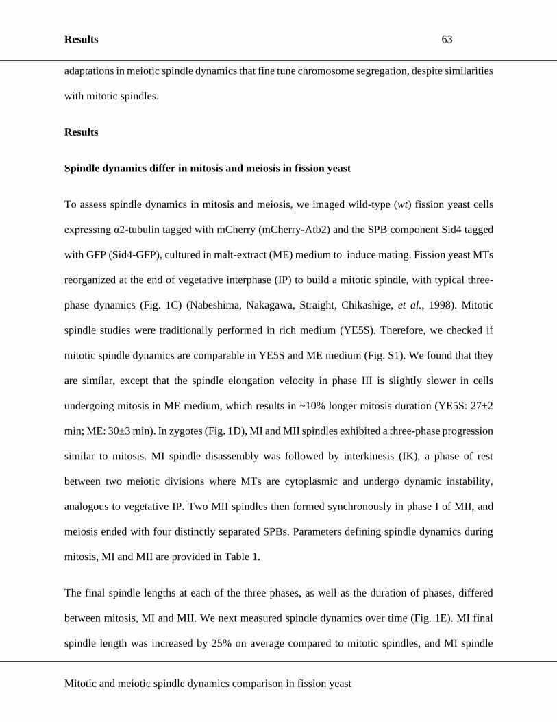
Results 63
Mitotic and meiotic spindle dynamics comparison in fission yeast
adaptations in meiotic spindle dynamics that fine tune chromosome segregation, despite similarities
with mitotic spindles.
Results
Spindle dynamics differ in mitosis and meiosis in fission yeast
To assess spindle dynamics in mitosis and meiosis, we imaged wild-type (wt) fission yeast cells
expressing α2-tubulin tagged with mCherry (mCherry-Atb2) and the SPB component Sid4 tagged
with GFP (Sid4-GFP), cultured in malt-extract (ME) medium to induce mating. Fission yeast MTs
reorganized at the end of vegetative interphase (IP) to build a mitotic spindle, with typical three-
phase dynamics (Fig. 1C) (Nabeshima, Nakagawa, Straight, Chikashige, et al., 1998). Mitotic
spindle studies were traditionally performed in rich medium (YE5S). Therefore, we checked if
mitotic spindle dynamics are comparable in YE5S and ME medium (Fig. S1). We found that they
are similar, except that the spindle elongation velocity in phase III is slightly slower in cells
undergoing mitosis in ME medium, which results in ~10% longer mitosis duration (YE5S: 27±2
min; ME: 30±3 min). In zygotes (Fig. 1D), MI and MII spindles exhibited a three-phase progression
similar to mitosis. MI spindle disassembly was followed by interkinesis (IK), a phase of rest
between two meiotic divisions where MTs are cytoplasmic and undergo dynamic instability,
analogous to vegetative IP. Two MII spindles then formed synchronously in phase I of MII, and
meiosis ended with four distinctly separated SPBs. Parameters defining spindle dynamics during
mitosis, MI and MII are provided in Table 1.
The final spindle lengths at each of the three phases, as well as the duration of phases, differed
between mitosis, MI and MII. We next measured spindle dynamics over time (Fig. 1E). MI final
spindle length was increased by 25% on average compared to mitotic spindles, and MI spindle

Results 64
Mitotic and meiotic spindle dynamics comparison in fission yeast
elongation velocity in phase III was faster than in mitosis. In contrast to vegetative mitotic cells,
zygotes spent the largest portion of spindle elongation time in phase II of MI and MII. Finally,
zygotes spent a similar proportion of time in phase I of MI and MII, but the velocity of spindle
assembly in zygotes was lower than in mitotic cells. These differences in spindle dynamics implied
qualitative and/or quantitative changes in spindle components, such as motors and MAPs, or
changes in MT dynamics between mitosis and meiosis.
Chromosome segregation is severely compromised in cut7Δpkl1Δ zygotes
Kinesin-14 Pkl1 and kinesin-5 Cut7 are antagonistic motors involved in mitotic spindle assembly.
While Cut7 deletion leads to monopolar spindles and is non-viable (Hagan and Yanagida, 1990),
the simultaneous deletion of Cut7 and Pkl1 resulted in the formation of a short bipolar spindle in
mitosis, capable of segregating the chromosomes (Syrovatkina and Tran, 2015; Rincon et al., 2017;
Yukawa et al., 2018). However, when these mutants were mated, their zygotes failed to produce
the typical four spores (20±1% zygotes with four spores; Fig. 2A, Fig. S2A), consistent with a
recent report (Shirasugi and Sato, 2019). This was suggestive of a problem in meiotic chromosome
segregation. We imaged wt and cut7Δpkl1Δ cells expressing GFP-Atb2 and Hht1-mCherry (histone
H3) to visualize chromosome segregation. While the mitotic spindle in cut7Δpkl1Δ cells was
capable of segregating the DNA into two separate pools of equal size (Fig. 2B), meiotic
chromosome segregation was perturbed, and three distinct mutant phenotypes in cut7Δpkl1Δ
meiosis could be detected (Fig. 2C). The first subset of zygotes managed to segregate the DNA
mass in MI, but one of the MII spindles failed to complete DNA separation, resulting in three DNA
masses upon MII completion (22% of zygotes). In the second subset of zygotes, the spindle
appeared to elongate, as if the zygotes entered phase III of MI, but without segregating the DNA
mass. A spindle assembled again, this time separating the DNA into two separate masses (45% of

Results 65
Mitotic and meiotic spindle dynamics comparison in fission yeast
zygotes). Finally, in the third subset, the spindle completely failed to separate the DNA mass,
resulting in one DNA mass at the end of meiosis (9% of zygotes). These observed errors in
chromosome segregation could explain the abnormal number of spores at the end of meiotic
divisions.
Spindle integrity is compromised specifically in MI of the cut7Δpkl1Δ zygotes
To study spindle dynamics in cut7Δpkl1Δ meiosis, we imaged cut7Δpkl1Δ zygotes expressing
mCherry-Atb2 and Sid4-GFP. Contrary to mitosis, where all of the cut7Δpkl1Δ cells assembled a
bipolar spindle, cut7Δpkl1Δ zygotes failed to do so in meiosis (Fig. S2B-D): 74±2% of cut7Δpkl1Δ
zygotes failed to establish a bipolar spindle and separate the SPBs during MI, consistent with a
recent report (Shirasugi and Sato, 2019). In addition, among the zygotes that had assembled bipolar
spindles in MI, another 8±2% failed to assemble a spindle in MII. Unsuccessful SPB separation in
MII could result from errors in the preceding MI, or from the intrinsic properties of cut7Δpkl1Δ
MII spindles.
In wt zygotes, there are two SPBs at the end of MI, which duplicate, and MII ends with four SPBs
(Fig. 1D). In the cut7Δpkl1Δ zygotes which have initially failed to assemble a spindle, we have
observed a second spindle assembly onset accompanied by an increase in SPB number (Fig. S2C,
far right panel). An increase in SPB number after MI spindle failure could indicate that the SPBs
in cut7Δpkl1Δ zygotes were duplicated a second time, and that cut7Δpkl1Δ zygotes entered MII
despite chromosome segregation failure in MI. We confirmed this result by imaging wt and
cut7Δpkl1Δ zygotes expressing Cdc13-GFP (cyclin-B) and mCherry-Atb2. In wt zygotes, Cdc13-
GFP localization was visible in the nucleus, on the spindle, and the SPBs (Izawa et al., 2005; Fu et
al., 2010). Upon phase III onset in MI, Cdc13-GFP signal was partially degraded (Fig. S2E). The

Results 66
Mitotic and meiotic spindle dynamics comparison in fission yeast
pool of Cdc13-GFP was replenished upon MII phase I onset, and was degraded again upon phase
III onset of MII. Similar dynamics of Cdc13-GFP were visible in cut7Δpkl1Δ zygotes that formed
monopolar spindles, except that Cdc13-GFP degradation relative to spindle assembly onset was
delayed 16 minutes on average in cut7Δpkl1Δ MI (Fig. S2F). This suggests that the spindle
assembly checkpoint (SAC) is active, but leaky, in cut7Δpkl1Δ zygotes, ultimately allowing
progression through the meiotic stages regardless of spindle or chromosome segregation defects.
We then compared wt and cut7Δpkl1Δ spindle dynamics. In mitosis, pre-phase III spindles were
shorter and phase III onset was delayed in the cut7Δpkl1Δ mutant compared to wt (Fig. 2D), which
is in agreement with previous studies (Rincon et al., 2017; Yukawa et al., 2018). Similar results
were observed for the MII spindle dynamics (Fig. 2E). In contrast, most MI spindles remained
monopolar. The 25% of MI cut7Δpkl1Δ spindles that managed to achieve bipolarity and separate
the SPBs, did not show the usual three-phase progression of spindle elongation (Fig. 2F), as phase
I and II of spindle elongation could not be distinguished. Combined, our data indicate that spindle
assembly and elongation are specifically perturbed during MI of cut7Δpkl1Δ zygotes.
We next analyzed the polarity of MT protrusions that formed in MI monopolar spindles by
checking the localization of the MT-plus end tracking protein Mal3-GFP (EB1) (Busch and
Brunner 2004). The analysis revealed that cut7Δpkl1Δ zygotes formed monopolar protrusions with
distal MT-plus ends (Fig. S2G). This phenotype is similar to the phenotype of mitotic cells lacking
Cut7 (Hagan and Yanagida, 1990).
We conclude that Pkl1 deletion cannot fully compensate for the absence of Cut7 in MI. This
suggests that Cut7 and Pkl1 are differently regulated between mitosis and MI.
The ratio of Cut7-to-Pkl1 is higher in MI than mitosis spindles

Results 67
Mitotic and meiotic spindle dynamics comparison in fission yeast
To assess if Cut7 and Pkl1 are differently regulated in mitosis and MI, we imaged Cut7 or Pkl1
tagged with GFP together with the MTs tagged with mCherry in wt mitosis and MI. We first
checked the impact of the GFP tag on motor functionality. Cut7 deficiency is lethal in fission yeast
(Hagan and Yanagida, 1992b), resulting in cell death when Cut7 is inactive or inhibited. Cells
expressing Cut7-GFP were viable. Of 45 observed zygotes with Cut7-GFP, 11% showed a
collapsed MI spindle, and only 1 vegetative cell of more than 100 observed showed a monopolar
spindle in mitosis. Tagging Cut7 with resulted in less zygotes producing the typical 4 spores (Fig.
S3A; 91% compared to the 97% for the strain with no GFP tag), and no difference in growth was
observed in serial dilution plate assay (Fig. S3B). Mitotic and MI spindle dynamics of the strain
with the tagged Cut7 are comparable to the strain with no GFP tag (Fig. S3C,D). The results
indicate that the GFP tagged version of Cut7 is mostly functional.
Pkl1 deficiency was reported to lead to protrusions in phases II and III in 85% of the mitotic cells
grown in YE5S medium (Syrovatkina and Tran, 2015). We have observed no protrusions in mitotic
cells with 3XGFP tagged Pkl1 grown in YE5S. In ME medium, we have observed 13% of mitotic
cells with protrusions in phase III and all of the zygotes showed protrusions in phase III of MI.
Despite protrusions in phase III of MI, labelling Pkl1 with 3XGFP had no significant effect on
spore production (Fig. S3A; 94% compared to the 97% for the strain with no GFP tag) or any
consequence on the growth in serial dilution plate assay (Fig. S3B). Mitotic spindle dynamics of
the strain with the Pkl1-3XGFP were similar to the dynamics of the strain with no GFP tag (Fig.
S3E), while MI spindle dynamics showed a longer phase II duration and a shorter phase II spindle
in MI (Fig. S3F). The results indicate that the 3XGFP tagged version of Pkl1 is mostly functional.
Despite slight changes regarding spindle dynamics, as well as observed protrusions and lower
percentage of zygotes with 4 spores, suggesting a partial but not complete loss of functionality, no

Results 68
Mitotic and meiotic spindle dynamics comparison in fission yeast
changes have been observed in phase I. Therefore, we conclude that Cut7-GFP and Pkl1-3XGFP
can be used to investigate the localization and recruitment of Cut7 and Pkl1 during phase I in both
mitosis and MI.
Similar to previous observation (Hagan and Yanagida, 1992b), Cut7-GFP localized to the spindle
and the spindle poles from spindle assembly to spindle disassembly in both mitosis and MI (Fig.
3A). Similarly, the localization pattern of Pkl1-3XGFP appeared unchanged between MI and
mitosis. Pkl1-3XGFP localized to the spindle poles shortly after spindle assembly started, and
remained associated with the SPBs until spindle disassembly. It was also detected on the spindle
in phase I to phase II transition (Fig. 3B).
We then measured Cut7-GFP and Pkl1-3XGFP signal distribution along the spindles during phase
I to phase II transition by linescan analysis, 1 minute before the spindle reached a constant length
that marks the beginning of phase II.
Both Pkl1-3XGFP and Cut7-GFP displayed maximum intensity peaks at the start and at the end of
the linescan, which corresponds to the spindle poles. Cut7-GFP maximum intensity was
considerably higher in MI (Fig. 3C), while Pkl1-3XGFP maximum intensity was similar in mitosis
and MI (Fig. 3D). Moreover, total intensity of Cut7-GFP, calculated by integrating the intensity
profiles from the linescan, was elevated in MI compared to mitosis, while total intensity of Pkl1-
3XGFP remained similar in both types of spindles (Fig. S3G, H). Because total MT intensity is
different in mitotic and MI spindles (Fig. S3I), we normalized the intensity values obtained for
Cut7-GFP and Pkl1-3XGFP to the MT intensity values. In this way we found that the relative
concentration of Cut7 remained unchanged (Fig. 3E), while the relative concentration of Pkl1-
3XGFP per MT was lower in MI than in mitotic spindles (Fig. 3F). This may explain why pkl1

Results 69
Mitotic and meiotic spindle dynamics comparison in fission yeast
deletion cannot compensate for cut7 deletion in MI, and suggests that an additional factor might
counteract Cut7 activity during phase II of MI to maintain force-balance.
Kinesin-14 Klp2 function is distinctive in MI and mitosis spindles
To identify additional spindle components that could counteract Cut7-dependent forces in meiosis
(Hagan and Yanagida, 1990; Syrovaktina, Fu and Tran, 2013), we screened other motors and MAPs
reported to produce forces in the spindle (Syrovaktina, Fu and Tran, 2013). We reasoned that if a
protein antagonizes Cut7, its deletion in the cut7Δpkl1Δ background should restore bipolar spindle
assembly in MI. This in turn would result in proper chromosome segregation and increase the
percentage of zygotes producing the typical four spores. We have systematically deleted candidate
motors and MAPs in the cut7Δpkl1Δ background, mated the triple mutants, and scored for spore
number (Fig. 4A). Of all the proteins tested, only the deletion of kinesin-14 Klp2 resulted in zygotes
that produced the typical four spores more frequently (44±2% of cut7Δpkl1Δklp2Δ zygotes
produced four spores compared to 20±1% in the cut7Δpkl1Δ background). Next, we turned to live
cell imaging of cut7Δpkl1Δklp2 zygotes to study how klp2 deletion conferred an increase in the
number of zygotes producing four spores.
Live cell imaging of cut7Δpkl1Δklp2Δ zygotes revealed that they succeeded to build a bipolar
spindle in MI that was capable of separating the SPBs (Fig. 4B). While only 26±2% of cut7Δpkl1Δ
zygotes managed to establish spindle bipolarity and separate SPBs, 49±2% of cut7Δpkl1Δklp2Δ
zygotes assembled bipolar spindles in MI (Fig. S4A). We next compared mitotic and MI spindle
dynamics in the triple mutant. MI spindle dynamics comparison revealed that not only was spindle
bipolarity reestablished, but the usual three-phase spindle elongation dynamics were partially

Results 70
Mitotic and meiotic spindle dynamics comparison in fission yeast
rescued (Fig. 4C). This differed strongly from cut7Δpkl1Δ zygotes in which the three-phase spindle
dynamics was not observed (Fig. 2F and 4C).
It was recently reported that klp2 deletion partially rescues cut7Δpkl1Δ mitotic spindle dynamics,
indicating that Klp2 produces inward forces alongside Pkl1, thereby counteracting the Cut7-
dependent forces (Yukawa et al., 2018). Our mitotic spindle analyses showed that klp2 deletion
partially rescued the time needed to reach phase III, but it did not rescue phase III spindle length in
cut7Δpkl1Δ cells (Fig. S4B-E). This result was unlike the one noted in cut7Δpkl1Δklp2Δ MI
spindles, where the pre-phase III spindle length was partially restored (Fig. 4C). Together, these
results suggest that kinesin-14 Klp2 counteracts Cut7-dependent outward pushing forces and
represents a new player in the force-balance maintenance in phase II of MI spindles.
Klp2 is a minus-end directed motor that can crosslink and slide MTs, affecting spindle length
(Troxell et al., 2001; Carazo-Salas, Antony and Nurse, 2005; Janson et al., 2007; Syrovaktina, Fu
and Tran, 2013). We tagged Klp2 with GFP to study its localization in mitotic and MI spindles.
Tagging Klp2 had no effect on serial dilution assay results when compared to the non-tagged strain
(Fig. S4F) and had a non-significant decrease in the percentage of zygotes producing 4 spores (Fig.
S4G; 94% compared to the 97% for the strain with no GFP tag). The velocity of spindle elongation
in phase III of mitotic spindle dynamics was faster and phase II duration was increased in the strain
with tagged Klp2 (Fig. S4H). Tagging Klp2 resulted in a longer phase II spindle in MI (Fig S.4I)
and 2 of the 17 observed zygotes required a longer time to reach spindle bipolarity. The results
indicate that Klp2-GFP is mostly functional.
Imaging of Klp2-GFP showed that it localized along the spindle, starting from spindle assembly
until early phase III in both mitosis and MI (Mana-Capelli et al., 2012; Yukawa et al., 2018) (Fig.

Results 71
Mitotic and meiotic spindle dynamics comparison in fission yeast
4D). Linescan analysis of Klp2-GFP showed higher maximum intensity peaks in MI, indicating an
increase in the total amount of Klp2-GFP in MI compared to mitosis (Fig. 4E). However, the
normalized concentration of Klp2-GFP relative to spindle MTs remained unchanged (Fig. 4F). We
also noticed that Klp2-GFP localized more intensely at the spindle poles in MI compared to mitosis.
This is likely not an artifact of mitosis and MI spindle length difference, as mitotic Pkl13XGFP
signal was clearly observed as two distinct peaks in linescan analysis (Fig. 3D), regardless of the
shorter length of the mitotic spindle.
Suppressing MT dynamics restored MI spindle bipolarity in cut7Δpkl1Δ zygotes
Considering that klp2 deletion rescued spindle bipolarity only partially in the cut7Δpkl1Δ zygotes,
we searched for additional factors that may participate in force-balance in MI phase II spindles.
We considered perhaps MT dynamics may be differently regulated between mitosis and meiosis.
Because current imaging limitations prevented us from measuring individual MT dynamics within
the spindles, where there are many crosslinked MTs, we instead measured individual MT bundle
dynamics before mitosis onset (IP) and in the pause period between MI and MII (IK). We
compared the dynamics of individual MT bundles that grew straight and originated from the SPBs.
Parameters defining MT dynamics during IP and IK are provided in Table 2.
Kymographs and plots of MT bundle dynamics in a vegetative cell and a zygote are shown in Fig.
5A. MT bundles begun their growth at the SPB and grew continuously until they abruptly
underwent catastrophe, switching to rapid shortening, and shrank back to the SPB. We found MT
bundles shrank slower and grew faster in IK, but reached shorter maximum length than bundles in
IP. Additionally, IK MT bundles underwent catastrophe two times more frequently than IP MT
bundles. In general, meiotic IK MTs are more dynamic than mitotic IP MTs. Although not

Results 72
Mitotic and meiotic spindle dynamics comparison in fission yeast
measured within the spindles, these results on IP and IK MT bundles suggest that MT dynamics
are differently regulated between mitotic cells and meiotic zygotes. We therefore tested if a
modification in MT dynamics could influence MI spindle assembly and spore formation in
cut7Δpkl1Δ zygotes.
We perturbed MT dynamics by utilizing low doses of methyl benzimidazole carbamate (MBC), a
drug that inhibits MT polymerization and suppresses MT dynamics (Yenjerla et al., 2009; Vela-
Corcía et al., 2018). Mating wt zygotes on ME plates with 5 µg ml-1 MBC resulted in a slightly
lower frequency of zygotes with four spores (Fig. 5B). In contrast, cut7Δpkl1Δ zygotes produced
four spores more frequently with the addition of MBC (44±1% with the addition of MBC compared
to 20±1% when no MBC was added). Live-cell imaging showed that the addition of 5 µg ml-1 MBC
allowed bipolar spindle assembly in MI in 80±4% of cut7Δpkl1Δ zygotes compared to 26±2% of
cut7Δpkl1Δ zygotes when no MBC was added (Fig. 5C and Fig. S5A).
We also noticed that in wt zygotes treated with MBC, the MI phase II spindle frequently exhibited
transient breakage (Fig. 5C, asterisk), a phenotype that was never observed in the cut7Δpkl1Δ
zygotes. Accordingly, we saw an abrupt decrease of SPB distance in phase II of MI upon spindle
breakage (Fig. 5D). However, spindle integrity was subsequently re-established and MI phase III
could proceed. Nevertheless, this spindle instability due to the MBC treatment could account for
the slightly lower number of wt zygotes producing four spores.
Cut7Δpkl1Δ spindle dynamics with the addition of MBC showed a delayed onset of MI phase III
as well as a shorter MI phase I/phase II spindle, similar to what was observed for cut7Δpkl1Δ
mitotic progression in the absence of MBC (Fig. 2D). Unlike cut7Δpkl1Δklp2Δ (Fig. 4C),
cut7Δpkl1Δ MI spindles in the presence of 5 µg ml-1 MBC did not exhibit a three-phase progression

Results 73
Mitotic and meiotic spindle dynamics comparison in fission yeast
of spindle elongation (Fig. 5D). This suggests that the mechanism of MBC-dependent rescue is
distinct from the one that relies on klp2 deletion.
Finally, addition of low doses of MBC did not seem to impact mitotic progression (Fig. S5B). The
relationship between wt and cut7Δpkl1Δ spindle dynamics with the addition of 5 µg ml-1 MBC
appeared similar to their relationship without MBC addition (Fig. 2D). We conclude that in phase
II of MI, MT dynamics counteract Cut7-dependent outward pushing forces together with Klp2 to
achieve proper force-balance and allow the formation of functional bipolar spindles.
Discussion
In this work, we have analyzed the differences between mitotic and meiotic spindle dynamics in
fission yeast. We found that the total amount of MTs, as well as the total amount of molecular
motors studied (except kinesin-14 Pkl1), were higher in MI than in mitosis. In fission yeast, spindle
length scales with cell size (Cortés et al., 2018; Krüger et al., 2019). A simple scaling mechanism
could therefore explain the increase in the final MI spindle lengths in all phases, as well as the
increased velocity of MI phase III (Table 1; Krüger et al., 2019). Because zygotes are diploid and
larger (Table 1), they presumably have more MTs, motors and MAPs to organize a spindle. The
average 25% increase in MI final spindle length compared to mitotic spindle could be readily
explained by the increased cell volume and increased availability of spindle components.
We further found that in MI, unlike in mitosis, the largest proportion of time was spent in phase II
(Table 1). In MI, the homologous chromosomes in the bivalent are held together by cohesin and
chiasmata (Watanabe and Nurse, 1999; Molnar et al., 2003; Tomoya S Kitajima et al., 2003). The
temporal requirement to complete the resolution of chiasmata could explain why phase II of MI is
prolonged compared to mitosis.

Results 74
Mitotic and meiotic spindle dynamics comparison in fission yeast
Finally, zygotes spent a similar time in phase I of the two meiotic divisions (Table 1), but the
velocity of spindle elongation in phase I of MI was lower than in mitosis, indicating that it may not
scale up with cell size, unlike in phase III. This lower phase I velocity might result from a
modification in MT dynamics (Table 2), an hypothesis that needs further testing, as we could not
to date access MT dynamics within the spindle for technical reasons.
It was reported that failure to form a meiotic bouquet leads to aberrant spindles (Tomita and Cooper,
2007). However, imaging of the telomere component Taz1-GFP and the SPB component Sid4-
mCherry did not reveal any defect in bouquet formation (Fig. S5C). We have also considered that
the kinetochores may not be attached to the spindle MTs in cut7Δpkl1Δ MI. Because of the bouquet
rearrangement, the kinetochores are far from the SPBs during MI, making it relatively easy to detect
kinetochore attachment to MTs. Imaging the kinetochore component Mis12 tagged with GFP and
the SPB component Sid4 tagged with mCherry did not reveal a defect in kinetochore attachment
(Fig. S5D).
Further, MI spindles have a specific organization of chromosomes in bivalents, with cohesin
between the homologous chromosomes, the existence of chiasmata, and changes in kinetochore
architecture (Molnar et al., 2003; Yokobayashi and Watanabe, 2005; Watanabe and Nurse, 1999;
Kitajima et al., 2004). We examined if this organization could influence spindle assembly in the
cut7Δpkl1Δ background, but the individual deletion of Rec8 (meiosis specific cohesin), Moa1
(meiotic kinetochore protein), Rec11 (meiotic sister-chromatid cohesion) or Rec12 (meiotic
recombination endonuclease) did not rescue the cut7Δpkl1Δ MI spindle phenotype nor the number
of zygotes producing the typical four spores (Fig. S5E). This indicates that the difference observed
between mitotic and MI spindles does not simply stem from the organization of chromosomes.
However, a study published by Shirasugi and Sato during the revision of this paper has

Results 75
Mitotic and meiotic spindle dynamics comparison in fission yeast
demonstrated that a double deletion of Rec12 and Moa1 partially rescued spindle bipolarity in
cut7Δpkl1Δ background (Shirasugi and Sato, 2019).
Unlike the other spindle components tested in this work, the concentration of kinesin-14 Pkl1
relative to spindle MTs is lower in MI than in mitosis (Fig. 3). The regulation behind this decrease
in relative concentration remains unknown. As we have measured the concentrations of motors in
mitosis and MI by analyzing the intensity of GFP attached to the motor, it is possible that it does
not reflect the wild-type state, especially in light of the fact that Cut7-GFP, Pkl1-3XGFP, and Klp2-
GFP were not fully functional (Fig. 3S and 4S). Nevertheless, by measuring the intensities in the
same cell but in different types of cell division, we argue that the differences we observe are indeed
intrinsic to the type of cell division, and are not artifacts from the fluorescent tagging. It was
recently published that mild overexpression of kinesin-14 HSET in MI accelerated spindle
bipolarization during phase I, and resulted in more focused spindle poles, essentially switching
from MI to a more mitotic-like spindle (Bennabi et al., 2018). This in turn resulted in aberrant
chromosome alignment. It is tempting to think that a downregulation of kinesin-14 concentration
from mitosis to MI could be evolutionary conserved.
We identified Klp2 as an additional Cut7 antagonist in MI. It was demonstrated that in mitosis Klp2
tethering to SPBs can, to some degree, replace the role of Pkl1 in spindle anchorage and force
generation, indicating that in such a situation, Klp2 can effectively oppose Cut7 outward forces
(Yukawa et al., 2018). Since Klp2-GFP localized more to the spindle poles in MI than in mitosis
(Fig. 4E), we propose that Klp2 function may differ in MI compared to mitosis, and that it plays an
important role in spindle bipolarity establishment by counteracting Cut7 outward pushing forces to
compensate for the increase in the Cut7-to-Pkl1 ratio. How Klp2 regulation is tuned and which
factors influence its functions remain open questions.
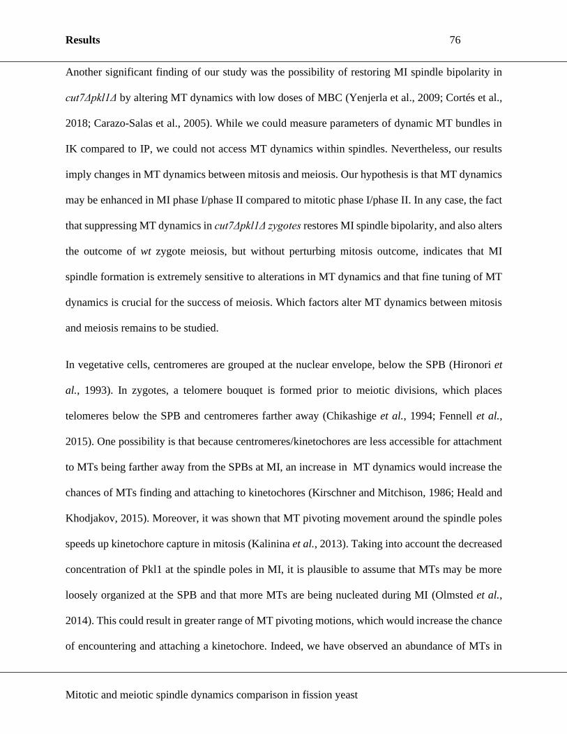
Results 76
Mitotic and meiotic spindle dynamics comparison in fission yeast
Another significant finding of our study was the possibility of restoring MI spindle bipolarity in
cut7Δpkl1Δ by altering MT dynamics with low doses of MBC (Yenjerla et al., 2009; Cortés et al.,
2018; Carazo-Salas et al., 2005). While we could measure parameters of dynamic MT bundles in
IK compared to IP, we could not access MT dynamics within spindles. Nevertheless, our results
imply changes in MT dynamics between mitosis and meiosis. Our hypothesis is that MT dynamics
may be enhanced in MI phase I/phase II compared to mitotic phase I/phase II. In any case, the fact
that suppressing MT dynamics in cut7Δpkl1Δ zygotes restores MI spindle bipolarity, and also alters
the outcome of wt zygote meiosis, but without perturbing mitosis outcome, indicates that MI
spindle formation is extremely sensitive to alterations in MT dynamics and that fine tuning of MT
dynamics is crucial for the success of meiosis. Which factors alter MT dynamics between mitosis
and meiosis remains to be studied.
In vegetative cells, centromeres are grouped at the nuclear envelope, below the SPB (Hironori et
al., 1993). In zygotes, a telomere bouquet is formed prior to meiotic divisions, which places
telomeres below the SPB and centromeres farther away (Chikashige et al., 1994; Fennell et al.,
2015). One possibility is that because centromeres/kinetochores are less accessible for attachment
to MTs being farther away from the SPBs at MI, an increase in MT dynamics would increase the
chances of MTs finding and attaching to kinetochores (Kirschner and Mitchison, 1986; Heald and
Khodjakov, 2015). Moreover, it was shown that MT pivoting movement around the spindle poles
speeds up kinetochore capture in mitosis (Kalinina et al., 2013). Taking into account the decreased
concentration of Pkl1 at the spindle poles in MI, it is plausible to assume that MTs may be more
loosely organized at the SPB and that more MTs are being nucleated during MI (Olmsted et al.,
2014). This could result in greater range of MT pivoting motions, which would increase the chance
of encountering and attaching a kinetochore. Indeed, we have observed an abundance of MTs in

Results 77
Mitotic and meiotic spindle dynamics comparison in fission yeast
the initial stages of spindle assembly in MI compared to mitosis where such structures were rarely
detected (Fig. S5F). It is possible these MTs pivot, but this hypothesis remains unconfirmed as
there are several MTs per time frame in MI, which makes tracking their position from frame to
frame challenging. The fact that suppressing MT dynamics by MBC rescued spindle bipolarity in
cut7Δpkl1Δ zygotes, suggests that MT dynamics also contribute to the force-balance establishment
within meiotic spindles, possibly by limiting the outward pushing forces generated by MT growth.
Our results open new questions on how motors, MAPs and MT dynamics are modified between
mitosis and meiosis. Given the high degree of evolutionary conservation of the molecular players
involved in spindle assembly, it will be most interesting to expand this study further in other
organisms to develop our global understanding of mitosis and meiosis.
Acknowledgements
We would like to thank Lara K. Krüger for her critical reading of the manuscript and valuable input.
We acknowledge the Cell and Tissue Imaging (PICT-IBiSA), Institute Curie, member of the French
National Research Infrastructure France-BioImaging (ANR10-INBS-04). Ana Loncar is supported
by a PhD fellowship from the European Union ITN-Divide Network (Marie Skłodowska-Curie
grant agreement No 675737s). This work is supported by grants from INCa, Fondation ARC, and
La Ligue National Contre le Cancer. The Tran lab is a member of the Labex CelTisPhyBio, part of
IdEx PSL.
Author contributions

Results 78
Mitotic and meiotic spindle dynamics comparison in fission yeast
A.L. and P.T.T. conceived and designed the study upon initial work by S.A.R. A.L. has performed
the experiments, analyzed the data, and wrote the paper. M.L.R. wrote the semi-automated Matlab
program for spindle dynamics measurements. S.A.R., A.P., and P.T.T. edited the manuscript.
Declaration of interest
The authors declare no competing interests.

Results 79
Mitotic and meiotic spindle dynamics comparison in fission yeast
List of Symbols and Abbreviations used
MT – microtubule
MAP – microtubule associated protein
SPB – spindle pole body
MI – meiosis I
MII – meiosis II
GFP – green fluorescent protein
mCherry – monomeric red fluorescent protein
ME – malt extract
IP - interphase
YE5S – yeast extract medium supplemented with Leu, Ura, Ade, His, and Lys
IK – interkinesis
DNA – deoxyribonucleic acid
SAC – spindle assembly checkpoint
MBC – methyl benzimidazole carbamate

Results 80
Mitotic and meiotic spindle dynamics comparison in fission yeast
References
Bähler, J., Wu, J.-Q., Longtine, M. S., Shah, N. G., Mckenzie III, A., Steever, A. B., Wach,
A., Philippsen, P. and Pringle, J. R. (1998). Heterologous modules for efficient and
versatile PCR-based gene targeting inSchizosaccharomyces pombe. Yeast 14, 943–951.
Bennabi, I., Quéguiner, I., Kolano, A., Boudier, T., Mailly, P., Verlhac, M. and Terret, M.
(2018). Shifting meiotic to mitotic spindle assembly in oocytes disrupts chromosome
alignment. EMBO Rep. 19, 368–381.
Carazo-Salas, R. E. (2005). The Kinesin Klp2 Mediates Polarization of Interphase Microtubules
in Fission Yeast. Science (80-. ). 309, 297–300.
Chikashige, Y., Ding, D., Funabiki, H., Haraguchi, T., Mashiko, S., Yanagida, M. and
Hiraoka, Y. (1994). Telomere-led premeiotic chromosome movement in fission yeast.
Science (80-. ). 264, 270–273.
Fennell, A., Fernández-álvarez, A., Tomita, K. and Cooper, J. P. (2015). Telomeres and
centromeres have interchangeable roles in promoting meiotic spindle formation. J. Cell Biol.
208, 415–428.
Fu, C., Ward, J. J., Loiodice, I., Velve-casquillas, G., Nedelec, F. J. and Tran, P. T. (2010).
Phospho-regulated interaction between kinesin-6 klp9p and microtubule bundler ase1p
promotes spindle elongation. 17, 257–267.
Funabiki, H., Hagan, I., Uzawa, S. and Yanagida, M. (1993). Cell cycle-dependent specific
positioning and clustering of centromeres and telomeres in fission yeast. J. Cell Biol. 121,
961–976.
G. Cortés, J. C., Ramos, M., Konomi, M., Barragán, I., Moreno, M. B., Alcaide-Gavilán,
M., Moreno, S., Osumi, M., Pérez, P. and Ribas, J. C. (2018). Specific detection of
fission yeast primary septum reveals septum and cleavage furrow ingression during early
anaphase independent of mitosis completion. PLOS Genet. 14, e1007388.
Goshima, G., Wollman, R., Stuurman, N., Scholey, J. M. and Vale, R. D. (2005). Length
control of the metaphase spindle. Curr. Biol. 15, 1979–1988.
Grishchuk, E. L., Spiridonov, I. S. and McIntosh, J. R. (2007). Mitotic Chromosome
Biorientation in Fission Yeast Is Enhanced by Dynein and a Minus-end–directed, Kinesin-
like Protein. Mol. Biol. Cell 18, 2216–2225.
Hagan, I. and Yanagida, M. (1990). Novel potential mitotic motor protein encoded by the
fission yeast cut7+ gene. Nature 347, 563.
Hagan, I. and Yanagida, M. (1992). Kinesin-related cut 7 protein associates with mitotic and
meiotic spindles in fission yeast. Nature 356, 74–76.
Hauf, S., Biswas, A., Langegger, M., Kawashima, S. A., Tsukahara, T. and Watanabe, Y.
(2007). Aurora controls sister kinetochore mono-orientation and homolog bi-orientation in
meiosis-I. EMBO J. 26, 4475–4486.
Heald, R. and Khodjakov, A. (2015). Thirty years of search and capture: The complex

Results 81
Mitotic and meiotic spindle dynamics comparison in fission yeast
simplicity of mitotic spindle assembly. J. Cell Biol. 211, 1103–1111.
Holland, A. J. and Cleveland, D. W. (2009). Boveri revisited: Chromosomal instability,
aneuploidy and tumorigenesis. Nat. Rev. Mol. Cell Biol. 10, 478–487.
Izawa, D., Goto, M., Yamashita, A. and Yamano, H. (2005). Fission yeast Mes1p ensures the
onset of meiosis II by blocking degradation of cyclin Cdc13p. 883, 879–883.
Janson, M. E., Loughlin, R., Loı, I., Fu, C., Brunner, D. and Ne, J. (2007). Crosslinkers and
Motors Organize Dynamic Microtubules to Form Stable Bipolar Arrays in Fission Yeast.
Cell 128, 357–368.
Kalinina, I., Nandi, A., Delivani, P., Chacón, M. R., Klemm, A. H., Ramunno-Johnson, D.,
Krull, A., Lindner, B., Pavin, N. and Tolić-Nørrelykke, I. M. (2013). Pivoting of
microtubules around the spindle pole accelerates kinetochore capture. Nat. Cell Biol. 15, 82–
87.
Kapitein, L. C., Peterman, E. J. G., Kwok, B. H., Kim, J. H., Kapoor, T. M. and Schmidt, C.
F. (2005). The bipolar mitotic kinesin Eg5 moves on both microtubules that it crosslinks.
Nature 435, 114–118.
Kapoor, T. M., Mayer, T. U., Coughlin, M. L. and Mitchison, T. J. (2000). Probing Spindle
Assembly Mechanisms with Monastrol, a Small Molecule Inhibitor of the Mitotic Kinesin,
Eg5. J. Cell Biol. 150, 975–988.
Keeney, J. B. and Boeke, J. D. (1994). Efficient targeted integration at leu1-32 and ura4-294 in
Schizosaccharomyces pombe. Genetics 136, 849–56.
Kirschner, M. and Mitchison, T. (1986). Beyond self-assembly: From microtubules to
morphogenesis. Cell 45, 329–342.
Kitajima, T. S. (2003). Distinct Cohesin Complexes Organize Meiotic Chromosome Domains.
Science (80-. ). 300, 1152–1155.
Kitajima, T. S., Kawashima, S. A. and Watanabe, Y. (2004). The conserved kinetochore
protein shugoshin protects centromeric cohesion during meiosis. Nature 427, 510–517.
Krüger, L. K., Sanchez, J.-L., Paoletti, A. and Tran, P. T. (2019). Kinesin-6 regulates cell-
size-dependent spindle elongation velocity to keep mitosis duration constant in fission yeast.
Elife 8, 1–22.
Mana-Capelli, S., McLean, J. R., Chen, C.-T., Gould, K. L. and McCollum, D. (2012). The
kinesin-14 Klp2 is negatively regulated by the SIN for proper spindle elongation and
telophase nuclear positioning. Mol. Biol. Cell 23, 4592–4600.
Miki, F., Okazaki, K., Shimanuki, M., Yamamoto, A., Hiraoka, Y. and Niwa, O. (2002). The
14-kDa Dynein Light Chain-Family Protein Dlc1 Is Required for Regular Oscillatory
Nuclear Movement and Efficient Recombination during Meiotic Prophase in Fission Yeast.
Mol. Biol. Cell 13, 930–946.
Miyazaki, S., Kim, J., Sakuno, T. and Watanabe, Y. (2017). Hierarchical Regulation of
Centromeric Cohesion Protection by Meikin and Shugoshin during Meiosis I. Cold Spring
Harb. Symp. Quant. Biol. 82, 259–266.

Results 82
Mitotic and meiotic spindle dynamics comparison in fission yeast
Molnar, M., Doll, E., Yamamoto, A., Hiraoka, Y. and Kohli, J. (2003). Linear element
formation and their role in meiotic sister chromatid cohesion and chromosome pairing. J.
Cell Sci. 116, 1719–1731.
Musacchio, A. and Salmon, E. D. (2007). The spindle-assembly checkpoint in space and time.
Nat. Rev. Mol. Cell Biol. 8, 379–393.
Nabeshima, K., Nakagawa, T., Straight, A. F., Murray, A., Chikashige, Y., Yamashita, Y.
M., Hiraoka, Y. and Yanagida, M. (1998). Dynamics of Centromeres during Metaphase–
Anaphase Transition in Fission Yeast: Dis1 Is Implicated in Force Balance in Metaphase
Bipolar Spindle. Mol. Biol. Cell 9, 3211–3225.
Nakamura-Kubo, M., Nakamura, T., Hirata, A. and Shimoda, C. (2003). The Fission Yeast
spo14 + Gene Encoding a Functional Homologue of Budding Yeast Sec12 Is Required for
the Development of Forespore Membranes. Mol. Biol. Cell 14, 1109–1124.
Ohkura, H. (2015). Meiosis: An overview of key differences from mitosis. Cold Spring Harb.
Perspect. Biol. 7, 1–15.
Olmsted, Z. T., Colliver, A. G., Riehlman, T. D. and Paluh, J. L. (2014). Kinesin-14 and
kinesin-5 antagonistically regulate microtubule nucleation by γ-TuRC in yeast and human
cells. Nat. Commun. 5, 5339.
Page, S. L. and Hawley, S. R. (2003). Chromosome Choreography: The Meiotic Ballet. Science
(80-. ). 301, 785–789.
Pavin, N. and Tolić, I. M. (2016). Self-Organization and Forces in the Mitotic Spindle. Annu.
Rev. Biophys. 45, 279–298.
Rincon, S. A., Lamson, A., Blackwell, R., Syrovatkina, V., Fraisier, V., Paoletti, A.,
Betterton, M. D. and Tran, P. T. (2017). Kinesin-5-independent mitotic spindle assembly
requires the antiparallel microtubule crosslinker Ase1 in fission yeast. Nat. Commun. 8, 1–
12.
Sakuno, T., Tada, K. and Watanabe, Y. (2009). Kinetochore geometry defined by cohesion
within the centromere. Nature 458, 852–858.
Saunders, W. S. and Hoyt, M. A. (1992). Kinesin-related proteins required for structural
integrity of the mitotic spindle. Cell 70, 451–458.
Sharp, D. J., McDonald, K. L., Brown, H. M., Matthies, H. J., Walczak, C., Vale, R. D.,
Mitchison, T. J. and Scholey, J. M. (1999). The bipolar kinesin, KLP61F, cross-links
microtubules within interpolar microtubule bundles of Drosophila embryonic mitotic
spindles. J. Cell Biol. 144, 125–138.
Shirasugi, Y. and Sato, M. (2019). Kinetochore-mediated outward force promotes spindle pole
separation in fission yeast. Mol. Biol. Cell 30, 2802–2813.
Syrovatkina, V. and Tran, P. T. (2015). Loss of kinesin-14 results in aneuploidy via kinesin-5-
dependent microtubule protrusions leading to chromosome cut. Nat. Commun. 6, 7322.
Syrovatkina, V., Fu, C. and Tran, P. T. (2013). Antagonistic Spindle Motors and MAPs
Regulate Metaphase Spindle Length and Chromosome Segregation. Curr. Biol. 23, 2423–

Results 83
Mitotic and meiotic spindle dynamics comparison in fission yeast
2429.
Thompson, S. L., Bakhoum, S. F. and Compton, D. A. (2013). Mechanisms of Chromosomal
Instability. Curr. Biol. 20, 1–23.
Tomita, K. and Cooper, J. P. (2007). The Telomere Bouquet Controls the Meiotic Spindle. Cell
130, 113–126.
Troxell, C. L., Sweezy, M. A., West, R. R., Reed, K. D., Carson, B. D., Pidoux, A. L., Cande,
W. Z. and McIntosh, J. R. (2001). pkl1(+)and klp2(+): Two Kinesins of the Kar3
Subfamily in Fission Yeast Perform Different Functions in Both Mitosis and Meiosis. Mol.
Biol. Cell 12, 3476–3488.
Uhlmann, F., Lottspeich, F. and Nasmyth, K. (1999). Sister-chromatid separation at anaphase
onset is promoted by cleavage of the cohesin subunit Scc1. Nature 400, 37–42.
Vela-Corcía, D., Romero, D., de Vicente, A. and Pérez-García, A. (2018). Analysis of β-
tubulin-carbendazim interaction reveals that binding site for MBC fungicides does not
include residues involved in fungicide resistance. Sci. Rep. 8, 7161.
Watanabe, Y. and Nurse, P. (1999). Cohesin Rec8 is required for reductional chromosome
segregation at meiosis. Nature 400, 461–464.
Yamamoto, A., West, R. R., McIntosh, J. R. and Hiraoka, Y. (1999). A Cytoplasmic Dynein
Heavy Chain Is Required for Oscillatory Nuclear Movement of Meiotic Prophase and
Efficient Meiotic Recombination in Fission Yeast. J. Cell Biol. 145, 1233–1250.
Yanagida, M. (2000). Cell cycle mechanisms of sister chromatid separation; Roles of
Cut1/separin and Cut2/securin. Genes to Cells 5, 1–8.
Yenjerla, M., Cox, C., Wilson, L. and Jordan, M. A. (2009). Carbendazim Inhibits Cancer Cell
Proliferation by Suppressing Microtubule Dynamics. J. Pharmacol. Exp. Ther. 328, 390–
398.
Yokobayashi, S. and Watanabe, Y. (2005). The kinetochore protein Moa1 enables cohesion-
mediated monopolar attachment at meiosis I. Cell 123, 803–817.
Yukawa, M., Ikebe, C. and Toda, T. (2015). The Msd1-Wdr8-Pkl1 complex anchors
microtubule minus ends to fission yeast spindle pole bodies. J. Cell Biol. 209, 549–562.
Yukawa, M., Yamada, Y., Yamauchi, T. and Toda, T. (2018). Two spatially distinct kinesin-14
proteins, Pkl1 and Klp2, generate collaborative inward forces against kinesin-5 Cut7 in S. pombe.
J. Cell Sci. 131, jcs210740.

Results 84
Mitotic and meiotic spindle dynamics comparison in fission yeast
Table 1. Spindle length dynamics in mitosis, MI and MII.
Spindle characteristics Mitosis (n=60) MI
(n=53)
MII
(n=99)
Phase I Final spindle length (μm) 1.26 ± 0.05 2.94 ± 0.11 1.64 ± 0.04
Duration (min) 4.27 ± 0.18 9.60 ± 0.45 7.17 ± 0.26
Relative duration of phase I
(%) 17 ± 1 24 ± 1 21 ± 1
Velocity (μm min-1) 0.36 ± 0.02 0.30 ± 0.02 0.27 ± 0.01
Phase II Final spindle length (μm) 2.70 ± 0.06 5.09 ± 0.08 2.28 ± 0.05
Duration (min) 8.52 ± 0.23 19.85 ± 0.60 21.23 ± 0.44
Relative duration of phase II
(%) 33 ± 1 49 ± 1 64 ± 1
Phase III Final spindle length (μm) 10.97 ± 0.13 14.23 ± 0.18 4.94 ± 0.15
Duration (min) 12.62 ± 0.27 10.72 ± 0.27 4.89 ± 0.17
Relative duration of phase
III (%) 50 ± 1 27 ± 1 15 ± 1
Velocity (μm min-1) 0.70 ± 0.02 0.99 ± 0.03 0.75 ± 0.03
Total duration (min) 25.40 ± 0.33 40.17 ± 0.79 33.34 ± 0.49
Cell length (μm) 13.27 ± 0.13 18.13 ± 0.26
Meiotic IK duration (min) NA 33.64 ± 1.03
Values in table are given as mean±s.e.m.
Table 2. Individual MT bundle dynamic parameters during IP and IK.
IP (n≥80) IK (n≥105) p-value
Growth velocity (µm min-1) 2.12 ± 0.05 2.53 ± 0.09 <0.001
Shrinkage velocity (µm min-1) 6.28 ± 0.25 3.26 ± 0.13 <0.0001
Catastrophe frequency (min-1) 0.54 ± 0.02 1.07 ± 0.04 <0.0001
Maximum length (µm) 4.47 ± 0.12 2.63 ± 0.07 <0.0001
p-value was calculated by the Mann-Whitney test. Values in table are given as mean±s.e.m.

Results 85
Mitotic and meiotic spindle dynamics comparison in fission yeast
Figure Legends
Fig. 1. Fission yeast mitotic and meiotic spindle have distinct dynamics. (A) Model of spindle
assembly and elongation in mitosis, MI and MII. (B) DIC image of a wt haploid cell post mitosis
(cell indicated by an asterisk on the septum) and wt diploid zygote post MI and MII (zygote
indicated by asterisks on 4 round spores) in the same field of view. (C) Time-lapse images of wt
cell expressing mCherry-Atb2 (α-tubulin) and Sid4-GFP (SPB component) from IP (30 min prior
to spindle formation) to spindle breakdown. Phases of mitotic spindle assembly and elongation are
indicated on the left side; I = phase I, II = phase II, III = phase III. Each frame represents a 5 min
interval. Scale bars, 5 µm. (D) Time-lapse images of wt zygotes expressing mCherry-Atb2 and
Sid4-GFP from spindle assembly in MI to spindle disassembly in MII. Phases of meiotic spindle
assembly and elongation are indicated on the left side. Each frame represents a 5 min interval. Scale
bar, 5 µm. (E) Comparative plot of wt spindle length dynamics in mitosis (n=20), MI (n=19) and
MII (n=34). Bold curves represent the average spindle.
Fig. 2. The cut7Δpkl1Δ spindle fails to segregate chromosomes during meiosis. (A) Bar graph
comparison of the number of wt (n=749) and cut7Δpkl1Δ (n=669) zygotes producing the typical
four spores. p-value was calculated by χ² test. (B) Time-lapse images of wt and cut7Δpkl1Δ mitotic
cells expressing GFP-Atb2 and Hht1-mCherry (histone H3) from spindle assembly to spindle
breakdown. Each frame represents a 5 min interval. Scale bar, 5 µm. (C) Time-lapse images of wt
and cut7Δpkl1Δ zygotes expressing GFP-Atb2 and Hht1-mCherry from spindle assembly in MI to
spindle breakdown in MII. Percentages below the three cut7Δpkl1Δ phenotypes represent their
frequency (n=54). Each frame represents a 10 min interval. Scale bar, 5 µm. (D) Comparative plot
of wt (n=19) and cut7Δpkl1Δ (n=20) mitotic spindle length dynamics. Dotted grey lines indicate a
shift between phases for wt spindles. (E) Comparative plot of wt (n=41) and cut7Δpkl1Δ (n=35)

Results 86
Mitotic and meiotic spindle dynamics comparison in fission yeast
MII spindle length dynamics. Dotted grey lines indicate a shift between phases for wt spindles. (F)
Comparative plot wt (n=17) and cut7Δpkl1Δ (n=20) MI spindle length dynamics. Dotted grey lines
indicate a shift between phases for wt spindles.
Fig. 3. Kinesin-14 Pkl1 relative concentration is lower in MI than in mitosis. (A) Time-lapse
images of cells expressing mCherry-Atb2 and kinesin-5 Cut7-GFP. Asterisks indicate the spindle
at phase I/phase II transition, 1 minute before the spindle reaches a constant length in phase II.
Scale bar, 5 µm. (B) Time-lapse images of cells expressing mCherry-Atb2 and kinesin-14 Pkl1-
3XGFP. Asterisks indicate the spindle at phase I/phase II transition, 1 minute before the spindle
reaches a constant length in phase II. Scale bar, 5 µm. (C) Linescan analysis of Cut7-GFP signal
distribution on the spindle at phase I/phase II transition in mitosis (n=60) and MI (n=60). (D)
Linescan analysis of Pkl1-3XGFP signal distribution on the spindle at phase I/phase II transition
in mitosis (n=57) and MI (n=58). (E) Box-and-whisker plot comparison of relative concentration
of Cut7-GFP per spindle MTs at phase I/phase II transition in mitosis (n=62) and MI (n=60).
n.s.=not significant. (F) Box-and-whisker plot comparison of relative concentration of Pkl1-
3XGFP per spindle MTs at phase I/phase II transition in mitosis (n=57) and MI (n=58). p-values
were calculated by Mann-Whitney test.
Fig. 4. Kinesin-14 Klp2 deletion partially rescues bipolar spindle defects of cut7Δpkl1Δ
zygotes during MI. (A) Bar graph comparison of the number of cut7Δpkl1Δ (n=669),
cut7Δpkl1Δklp2Δ (n=650), cut7Δpkl1Δklp6Δ (n=483), cut7Δpkl1Δdhc1Δ (n=694),
cut7Δpkl1Δdam1Δ (n=581), and cut7Δpkl1Δrec8Δ (n=522) zygotes producing the typical four
spores. The red bar represents the cut7Δpkl1Δ zygotes and the green bar represents
cut7Δpkl1Δklp2Δ zygotes. Black bars represent triple mutants that did not show a rescue in the
number of zygotes producing four spores. p-value was calculated by χ² test. (B) Time-lapse images

Results 87
Mitotic and meiotic spindle dynamics comparison in fission yeast
of cut7Δpkl1Δ and cut7Δpkl1Δklp2Δ zygotes expressing mCherry-Atb2 and Sid4-GFP from spindle
assembly to breakdown in MI. Each frame represents a 5 min interval. Scale bar, 5 µm. (C)
Comparative plot of cut7Δpkl1Δ (n=20) and cut7Δpkl1Δklp2Δ (n=20) MI spindle length dynamics.
Horizontal dotted lines indicate the final length of pre-phase III spindle for cut7Δpkl1Δ (red) and
cut7Δpkl1Δklp2Δ (green). The box-and-whisker graph on the right shows the comparison of the
final length of pre-phase III spindles in cut7Δpkl1Δ (n=24) and cut7Δpkl1Δklp2Δ (n=29). p-value
was calculated by Mann-Whitney test. (D) Time-lapse images of cells expressing mCherry-Atb2
and Klp2-GFP. Asterisks indicate the spindle at phase I/phase II transition, 1 minute before the
spindle reaches a constant length in phase II. Scale bar, 5 µm. (E) Linescan analysis of Klp2-GFP
signal distribution on the spindle at phase I/phase II transition in mitosis (n=58) and MI (n=59). (F)
Box-and-whisker plot comparison of relative concentration of Klp2-GFP per spindle MTs at phase
I/phase II transition in mitosis (n=58) and MI (n=59). n.s.=not significant.
Fig. 5. Inhibition of MT polymerization restores MI spindle bipolarity in cut7Δpkl1Δ zygotes.
(A) Representative zygote and vegetative cell expressing mCherry-Atb2 and Sid4-GFP. Scale bar,
5 µm. Kymographs are taken from boxed regions containing the SPB and the MT bundle in zygotes
in IK and in vegetative cells in IP. Kymographs on the right shows representative IK- and IP-MT
bundle dynamics over time. Scale bar, 2 µm. Comparative plot shows differences between IP- and
IK-MT bundle dynamics. (B) Comparison of the number of zygotes producing the typical 4 spores
with the addition of 5 µg ml-1 MBC (wt n=572, cut7Δpkl1Δ n=1162) and without the addition of
MBC (wt n=749, cut7Δpkl1Δ n=669). p-value was calculated by χ² test. (C) Time-lapse images of
wt and cut7Δpkl1Δ zygotes expressing mCherry-Atb2 and Sid4-GFP from spindle assembly to
breakdown in MI. Yellow asterisk indicates a collapsed metaphase spindle. Scale bar, 5 µm. (D)

Results 88
Mitotic and meiotic spindle dynamics comparison in fission yeast
Comparative plot of wt (n=20) and cut7Δpkl1Δ (n=20) MI spindle dynamics with the addition of 5
µg ml-1 MBC.

Results 89
Mitotic and meiotic spindle dynamics comparison in fission yeast
Figure 1. Loncar et al., J Cell Sci, 2020

Results 90
Mitotic and meiotic spindle dynamics comparison in fission yeast
Figure 2. Loncar et al., J Cell Sci, 2020

Results 91
Mitotic and meiotic spindle dynamics comparison in fission yeast
Figure 3. Loncar et al., J Cell Sci, 2020

Results 92
Mitotic and meiotic spindle dynamics comparison in fission yeast
Figure 4. Loncar et al., J Cell Sci, 2020

Results 93
Mitotic and meiotic spindle dynamics comparison in fission yeast
Figure 5. Loncar et al., J Cell Sci, 2020

Results 94
Mitotic and meiotic spindle dynamics comparison in fission yeast
Inventory of Supplemental information
Supplemental figure legends
Supplemental figures
Materials and methods
Supplemental table 1. List of strains used in this study

Results 95
Mitotic and meiotic spindle dynamics comparison in fission yeast
Fig. S1. Mitotic dynamics are comparable in YE5S and ME media. Comparative plot of wt
spindle dynamics in YE5S (n=20) and ME (n=20). Bold curves represent a mean spindle.
Fig. S2. Cut7Δpkl1Δ zygotes fail to establish a bipolar spindle in MI, but still enter MII. (A)
DIC image of cut7Δpkl1Δ zygotes. Yellow lines indicate zygotes with abnormal spores (asterisks).
(B) Time-lapse images of wt and cut7Δpkl1Δ vegetative cells expressing mCherry-Atb2 and Sid4-
GFP from spindle assembly to spindle breakdown. Scale bar, 5 µm. (C) Time-lapse images of wt
and cut7Δpkl1Δ zygotes expressing mCherry-Atb2 and Sid4-GFP from spindle assembly in MI to
spindle breakdown in MII. Percentages below the two cut7Δpkl1Δ phenotypes represent their
frequency (n=113). Scale bar, 5 µm. (D) Bar graph comparison of SPB separation in mitosis (wt
n=201, cut7Δpkl1Δ n=219), MI (wt n=102, cut7Δpkl1Δ n=113), and MII (wt n=157, cut7Δpkl1Δ
n=127). (E) Time-lapse images of wt and cut7Δpkl1Δ zygotes expressing mCherry-Atb2 and
Cdc13-GFP (cyclin-B). Scale bar, 5 µm. (F) Box-and-whisker plot comparison of Cdc13-GFP
degradation time after spindle assembly onset in MI of wt (n=37) and cut7Δpkl1Δ (n=22) zygotes.
(G) Representative image of a cut7Δpkl1Δ zygote in phase I/phase II of MI expressing mCherry-
Atb2 and Mal3-GFP. Scale bar, 5 µm. For insets, scale bar is 2 µm.
Fig. S3. Total amount of Pkl1 remains constant from mitosis to MI. (A) Bar-graph comparison
of the number of zygotes producing the typical four spores in strains without GFP (n=494), in
strains with Cut7-GFP (n=613), and in strains with Pkl1-3XGFP (n=490). p-values were calculated
by χ² test. (B) Serial dilution (fourfold) assay showing growth of strains without GFP, and strains
with Cut7-GFP or Pkl1-3XGFP. Plates were incubated 48 or 96 hours at the indicated temperatures.
(C) Comparative plot of mitotic spindle length dynamics in the strain without GFP (n=18) and in
the strain with Cut7-GFP (n=19). The green vertical dotted line represents a time point in phase
I/phase II transition when the intensity of Cut7-GFP was measured. (D) Comparative plot of MI

Results 96
Mitotic and meiotic spindle dynamics comparison in fission yeast
spindle length dynamics in the strain without GFP (n=17) and in the strain with Cut7-GFP (n=20).
The green vertical dotted line represents a time point in phase I/phase II transition when the
intensity of Cut7-GFP was measured. (E) Comparative plot of mitotic spindle length dynamics in
the strain without GFP (n=15) and in the strain with Pkl1-3XGFP (n=18). The green vertical dotted
line represents a time point in phase I/phase II transition when the intensity of Pkl1-3XGFP was
measured. (F) Comparative plot of MI spindle length dynamics in strains without GFP (n=24) and
in strains with Pkl1-3XGFP (n=20). The green vertical dotted line represents a time point in phase
I/phase II transition when the intensity of Pkl1-3XGFP was measured. (G) Box-and-whisker plot
comparison of Cut7-GFP total intensity in mitosis (n=62) and MI (n=60). (H) Box-and-whisker
plot comparison of Pkl1-3XGFP total intensity in mitosis (n=57) and MI (n=58). (I) Box-and-
whisker plot comparison of MT total intensity per spindle length in mitosis (n=57) and MI (n=58).
p-values were calculated by Mann-Whitney test.
Fig. S4. Klp2 deletion rescues metaphase duration, but not final metaphase spindle length in
cut7Δpkl1Δ mitosis. (A) Bar graph comparison of SPB separation in cut7Δpkl1Δ (n=113) and
cut7Δpkl1Δklp2Δ (n=83) MI. (B) Time-lapse images of cut7Δpkl1Δ and cut7Δpkl1Δklp2Δ mitotic
cells expressing mCherry-Atb2 and Sid4-GFP from spindle assembly to spindle disassembly. Scale
bar, 5 µm. (C) Comparative plot of cut7Δpkl1Δ (n=20) and cut7Δpkl1Δklp2Δ (n=17) mitotic spindle
length dynamics. (D) Box-and-whisker plot comparison of mitotic phase I/phase II duration in
cut7Δpkl1Δ (n=46) and cut7Δpkl1Δklp2Δ (n=46). (E) Box plot comparison of pre-phase III mitotic
spindle length of cut7Δpkl1Δ (n=46) and cut7Δpkl1Δklp2Δ (n=46). p-values were calculated by
Mann-Whitney test. (F) Serial dilution (fourfold) assay showing growth of strain without GFP, and
strain with Klp2-GFP. Plates were incubated 48 or 96 hours at the indicated temperatures. (G) Bar-
graph comparison of the number of zygotes producing the typical four spores in strains without

Results 97
Mitotic and meiotic spindle dynamics comparison in fission yeast
GFP (n=494) and with Klp2-GFP (n=445). p-values were calculated by χ² test. (H) Comparative
plot of mitotic spindle length dynamics in the strain without GFP (n=26) and in the strain with
Klp2-GFP (n=16). The green vertical dotted line represents an average time point in phase I/phase
II transition when the intensity of Klp2-GFP was measured. (F) Comparative plot of MI spindle
length dynamics in the strain without GFP (n=15) and in strains with Klp2-GFP (n=17). The green
vertical dotted line represents a time point in phase I/phase II transition when the intensity of Klp2-
GFP was measured.
Fig. S5. Chromosome organization and architecture are not the cause of failed cut7Δpkl1Δ
meiosis. (A) Bar graph comparison of SPB separation in cut7Δpkl1Δ zygotes with (n=110) and
without addition of 5 µg ml-1 MBC (n=124). (B) Comparative plot of wt (n=19) and cut7Δpkl1Δ
(n=20) mitotic spindle dynamics with the addition of 5 µg ml-1 MBC. (C) Kymographs of wt and
cut7Δpkl1Δ zygotes expressing Sid4-mCherry and a telomere component Taz1-GFP. Yellow
asterisks indicate a time point in which telomeres start to dissociate from the SPB. Scale bar, 2 µm.
(D) Kymographs of wt and cut7Δpkl1Δ zygotes expressing Sid4-mCherry and a kinetochore
component Mis12-GFP. Yellow asterisks indicate a time point in which all the kinetochores are
pulled to the SPB. Scale bar, 2 µm. (E) Bar graph comparison of the number of cut7Δpkl1Δ
(n=122), cut7Δpkl1Δrec11Δ (n=168), cut7Δpkl1Δrec12Δ (n=235), cut7Δpkl1Δsgo1Δ (n=264), and
cut7Δpkl1Δmoa1Δ zygotes (n=321) producing the typical four spores. (F) MTs in mitotic and MI
from spindle assembly onset in phase I to phase II onset. Yellow arrows in vegetative cell indicate
pivoting MTs. Yellow arrows in the zygote indicate a possible pivoting MT. Time indicated on the
images is in seconds. Scale bar, 5 µm. For insets, scale bar is 2 µm.
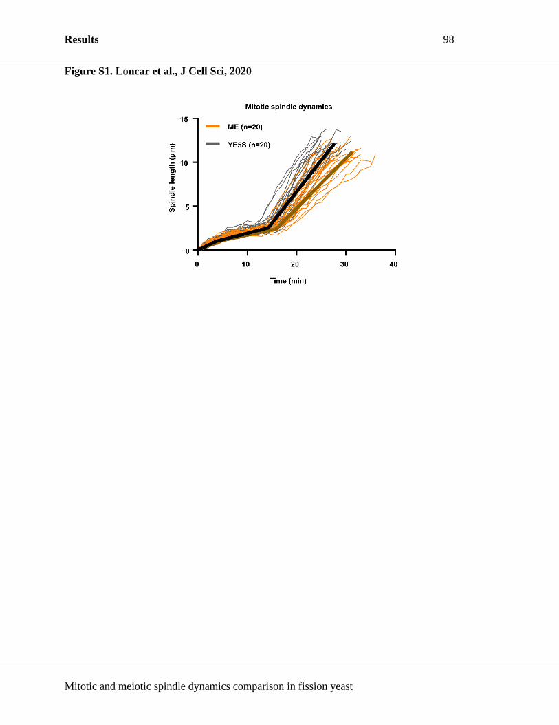
Results 98
Mitotic and meiotic spindle dynamics comparison in fission yeast
Figure S1. Loncar et al., J Cell Sci, 2020

Results 99
Mitotic and meiotic spindle dynamics comparison in fission yeast
Figure S2. Loncar et al., J Cell Sci, 2020

Results 100
Mitotic and meiotic spindle dynamics comparison in fission yeast
Figure S3. Loncar et al., J Cell Sci, 2020

Results 101
Mitotic and meiotic spindle dynamics comparison in fission yeast
Figure S4. Loncar et al., J Cell Sci, 2020

Results 102
Mitotic and meiotic spindle dynamics comparison in fission yeast
Figure S5. Loncar et al., J Cell Sci, 2020

Results 103
Mitotic and meiotic spindle dynamics comparison in fission yeast
Materials and methods
Production of S. pombe mutant strains
All strains used in this work are isogenic to wild-type 972 and were obtained from genetic crosses,
selected by random spore germination and replicated on plates with appropriate drugs or
supplements. All strains are listed in the Supplementary File 1. Gene deletion and tagging was
performed in the endogenous locus, except for Sid4-GFP and Mis12-GFP which were integrated
in the leu1-32 locus, as described previously (Keeney and Boeke, 1994; Bähler et al., 1998).
Transformations were performed by using the lithium-DTT method. 25 mL of exponentially
growing cells (OD600 0.5-0.8) were harvested by centrifugation and washed with 10 mM Tris HCl
pH 7.4. They were centrifuged again and re-suspended in 100 mM lithium acetate with 10 mM
DTT. Suspension was incubated on an orbital wheel at room temperature for 40 minutes. 100 µL
of suspension was mixed with 80 µL of 100 mM lithium acetate, 10 µL of single stranded DNA
from salmon testes (D9156-5ML, Sigma) and 1 µg of the purified DNA. After 10 minutes of
incubation on an orbital wheel, 300 µL of PEG 4000, dissolved in 100 mM lithium acetate was
added. After incubation of 10 minutes on the wheel, 15 µL of DMSO was added. Mixture was heat
shocked at 42°C for 20 minutes. Cells were washed with water and plated onto YE5S plates. After
two days of incubation at 25°C, the plates were replicated on plates containing the corresponding
selective media.
Fission yeast culture
All S. pombe strains were maintained at 25˚C and cultured in standard media. Generally, two days
before imaging, cells were refreshed on a YE5S plate. The following day, cells of opposite mating
types were mixed on an ME plate, or an ME plate supplemented with 5 µg ml-1 MBC, to induce
conjugation. The plate with mating cells was incubated at 25˚C for 16-20 hours before imaging.
For spore quantification, the mixture was incubated on an ME plate, or an ME plate supplemented
with 5 µg ml-1 MBC, at 25˚C for two days and subsequently imaged.
Serial dilutions were performed by growing tested strains on a YE5S plate at 25˚C for 24 hours,
and then resuspending some of the patch in water so the OD600 was 1.5. This suspension was serially
fourfold diluted, 5 µl of the prepared dilutions was inoculated on YE5S plates and then incubated
at 25˚C or 30 ˚C for 48 or 96 hours.

Results 104
Mitotic and meiotic spindle dynamics comparison in fission yeast
Live microscopy
For live-cell imaging, cells were put on ME agarose pads, containing 4% agarose (Tran et al.,
2004). In experiments where MT dynamics were perturbed, MBC was added to the patch at a final
concentration of 5 µg ml-1 MBC. Imaging was performed at 25˚C. Images were acquired on an
inverted Spinning Disk Confocal (Roper/Nikon), equipped with Plan Apochromat 100×/1.4 NA
objective lens (Nikon), a PIFOC (perfect image focus) objective stepper, and a charge-coupled
device camera (EMCCD 512x512 QuantEM; Photometrics).
For spindle dynamics, stacks of 7 planes spaced by 1 µm were acquired for each channel with 200
msec exposure for 561 nm wave length and 100 msec exposure for 491 nm wavelength, binning
one, and an electronic gain of 3 for both wavelengths. For each time-lapse movie, each stack was
taken every minute for a duration of 180–240 min.
For intensity measurements, stacks of 11 planes spaced by 0.5 µm were acquired for each channel
with 100 msec exposure, binning one, and electronic gain of 1 or 3. For each time-lapse movie,
each stack was taken every minute for a duration of 90 min.
For MT bundle dynamics, stacks of 7 planes spaced by 1 µm were acquired for each channel with
100 msec exposure for 561 nm wave length and 100 msec exposure for 491 nm wavelength, binning
one, and an electronic gain of 3 for both wavelengths. For each time-lapse movie, each stack was
taken every 5 seconds for a duration of 30 min.
To quantify spores, bright-field images of zygotes were taken at random, and the spores inside the
zygotes counted.
Image analysis
Analyses were performed by Metamorph 7.8 and ImageJ 1.52i. Maximum projections of each stack
were performed for analysis of spindle dynamics and for presentation, unless stated otherwise, and
sum projections for intensity measurements.
Spindle dynamics were determined by measuring the length of the mCherry-Atb2 signal between
Sid4-GFP signals over time, or, when 5 µg ml-1 MBC was added, by measuring the distance
between the SPBs over time, by a semi-automated Matlab program.
Results 105
Mitotic and meiotic spindle dynamics comparison in fission yeast
Intensity measurements were performed by linescan analysis, reading out the average intensity per
pixel, subtracting the background and adding all positive values to obtain a total amount of
intensity.
MT bundle dynamics were approximated by tracing the shape of mCherry-Atb2 signal on a
kymograph, and calculating the slopes of the gathered curves to obtain the velocity of MT bundle
growth and shrinkage. The origins and the endings of the curves were approximated by extending
the curves to the edges of the SPB signal. The movements of the SPB were accounted and corrected
for in our measurements. The catastrophe frequency was approximated as a reciprocal value of
time the MT bundle spent in elongation.
Image analysis: Feature detection
Microscopy images were analyzed with a semi-automated Matlab program that can find features
in microscopy images. After the computer finds the features, they are presented to the user, and the
user can manually correct them. In this work, the program was used to detect SPBs and spindles in
maximal projections of confocal microscopy stacks. Features are represented as the coordinates of
two points (SPBs) or a polygonal chain (spindle). The scripts for the analysis are integrated in a
Graphical User Interface, and the complete version of the program is available upon request. We
provide the Matlab scripts for feature extraction, and describe the pipeline of the analysis:
Cell segmentation: Initially, the user segments a cell manually in the relevant frames of the movie.
Only the selected frames, and the area inside the segmented region of interest is considered for
feature detection for a particular cell. Features are detected for each of the selected frames.
Spindle detection (1): Short spindles (less than 4 μm) are found by applying a 2 pixel gaussian blur
to the image, and subsequently thresholding applying Otsu’s method as provided in Matlab’s
multithresh (https://mathworks.com/help/images/ref/multithresh.htmL). Serial thresholding steps
are applied until the area above the threshold contains less than 10% of the pixels of the region
defined by the user as the cell. After that, regions above the threshold are merged using a convex
hull algorithm as provided in Matlab’s bwconvhull
(https://mathworks.com/help/images/ref/bwconvhull.htmL) and the coordinates of the pixels above
the threshold are fit to a first degree polynomial by regression. The resulting line is uniformly
sampled such that points are distributed along its length at 1 pixel distance, and constitutes the

Results 106
Mitotic and meiotic spindle dynamics comparison in fission yeast
spindle feature. Once a spindle is found to be longer than 4 μm, spindles are detected using the
method explained below.
Spindle detection (2): This method relies on the fact that long spindles are often aligned with the
major axis of the cell. Therefore, the distance with respect to the major axis of the cell is very
similar in neighboring pixels that belong to the spindle. A maximal projection of the image is made
along the major axis of the region defined by the user as the cell; for each projected pixel, the
position along the major axis ([X]), the intensity ([I]), and distance with respect to the major axis
([Y]) are stored in 1 dimensional vectors, noted in square brackets. To keep only the pixels
belonging to the spindle, subsequent discarding steps are applied: A moving standard deviation
filter of window size 3 pixels is applied to [Y], and positions with a standard deviation value above
3 pixels, are discarded. Next, groups (“group” denotes a series of consecutive non-discarded pixels)
containing less than 4 pixels are discarded. Pixels for which the intensity in [I] is smaller than 20%
of the maximum pixel in the cell are discarded. For the remaining groups, if the distance along the
major axis [X] between the closest points of 2 consecutive groups divided by the difference in
distance from the major axis [Y] between those two points is bigger than 1.5, the group that is
furthest from the center of the cell is discarded. The curve defined by [Y] is smoothened applying
a moving median filter of window size 10, resulting in [Y2]. Then, ([X,Y2]) coordinates are fit to
a first order polynomial if the cumulative distance along the curve [X,Y2] is smaller than 8 μm,
and to a third order polynomial otherwise. The resulting curve is sampled as described previously,
and constitutes the spindle feature.
SPB detection: The 10 most intense pixels inside a cell are considered for the analysis. If 2 or more
distinct regions are present in the image, the coordinates of the centroids of the two biggest regions
are returned. If there is only one region (often when Spindle Pole Bodies are too close), the pixel
with the weakest signal is removed from the analysis recursively until two distinct regions form,
and the coordinates of the centroids of the two regions are returned. If this method does not succeed,
the coordinates of the two most intense pixels in the cell are returned.
Image analysis: Measurements
Spindle length is defined from the N coordinates of the spindle polygonal chain [𝑥1, 𝑥2, … , 𝑥𝑁] and
[𝑦1, 𝑦2, … , 𝑦𝑁], as the sum of Eucledian distances between consecutive points:

Results 107
Mitotic and meiotic spindle dynamics comparison in fission yeast
∑ √(𝑥𝑖+1 − 𝑥𝑖)2 + (𝑦𝑖+1 − 𝑦𝑖)2
𝑁−1
𝑖=1
SPB separation is defined as the Eucledian distance between the two points defining the spindle
pole bodies.
Quantification and statistical tests
All graphs and box plots were generated using GraphPad 8.0 (Prism). All bar graph and line graph
data are showed as mean values with error bars representing s.e.m. Data in box and whisker graphs
are plotted by the Tukey method; the black line shows the median value. Analysis of statistical
significance was performed by using Mann-Whitney or χ² test. Obtained p-values are shown within
plots and data sets were defined as significantly different if p<0.05. n values, representing the
number of cells, are shown inside the plots. All shown experiments were performed at least 3 times.

Results 108
Mitotic and meiotic spindle dynamics comparison in fission yeast
Supplementary table 1. List of strains used in this study.
Identifier Genotype Source
AP240 h- ade6-M210 ura4-D18 leu1-32 Lab collection
AP241 h+ ade6-M210 ura4-D18 leu1-32 Lab collection
TP2872 h- cut7::Nat pkl1::ura4+ ade6-M210 ura4-D18 leu1-32 Lab collection
TP2873 h+ cut7::Nat pkl1::ura4+ ade6-M210 ura4-D18 leu1-32 Lab collection
TP2927 h- mCherry-Atb2:Hph leu1:Sid4-GFP leu1-32 ura4-D18 Lab collection
TP2944 h+ mCherry-Atb2:Hph leu1:Sid4-GFP leu1-32 ura4-D18 Lab collection
TP3793 h- GFP-Atb2:Hph Hht1-mCherry:Kan ade6-M210 ura4-D18 leu1-32 This study
TP3794 h+ GFP-Atb2:Hph Hht1-mCherry:Kan ade6-M210 ura4-D18 leu1-32 This study
TP3653 h- Hht1-mCherry:Kan GFP-Atb2:Hph cut7::Nat pkl1::ura4+ ade6-
M210 ura4-D18 leu1-32
This study
TP3654 h+ Hht1-mCherry:Kan GFP-Atb2:Hph cut7::Nat pkl1::ura4+ ade6-
M210 ura4-D18 leu1-32
This study
TP2491 h- cut7::Nat pkl1::ura4+ mCherry-Atb2:Hph Sid4-GFP:Kan leu1-32
ura4-D18
Lab collection
TP2492 h+ cut7::Nat pkl1::ura4+ mCherry-Atb2:Hph Sid4-GFP:Kan leu1-32
ura4-D18
Lab collection
TP3453 h+ Cdc13-GFP:Kan mCherry-Atb2:Hph ade6-M210 ura4-D18 leu1-
32
Lab collection
TP3454 h- Cdc13-GFP:Kan mCherry-Atb2:Hph ade6-M210 ura4-D18 leu1-
32
Lab collection
TP4090 h- Cdc13-GFP:Kan cut7::Nat pkl1::ura4+ mCherry-Atb2:Hph ade6-
M210 ura4-D18 leu1-32
This study
TP4091 h+ Cdc13-GFP:Kan cut7::Nat pkl1::ura4+ mCherry-Atb2:Hph ade6-
M210 ura4-D18 leu1-32
This study
TP4224 h- cut7::Nat pkl1::ura4+ Mal3-linker-GFP:Kan mCherry-Atb2::Hph
ura4-D18 leu1-32
This study
TP4225 h+ cut7::Nat pkl1::ura4+ Mal3-linker-GFP:Kan mCherry-Atb2::Hph
ura4-D18 leu1-32
This study
TP116 h- mCherry-Atb2::Hph ura4-D18 leu1-32 Lab collection
TP319 h+ mCherry-Atb2::Hph ura4-D18 leu1-32 Lab collection
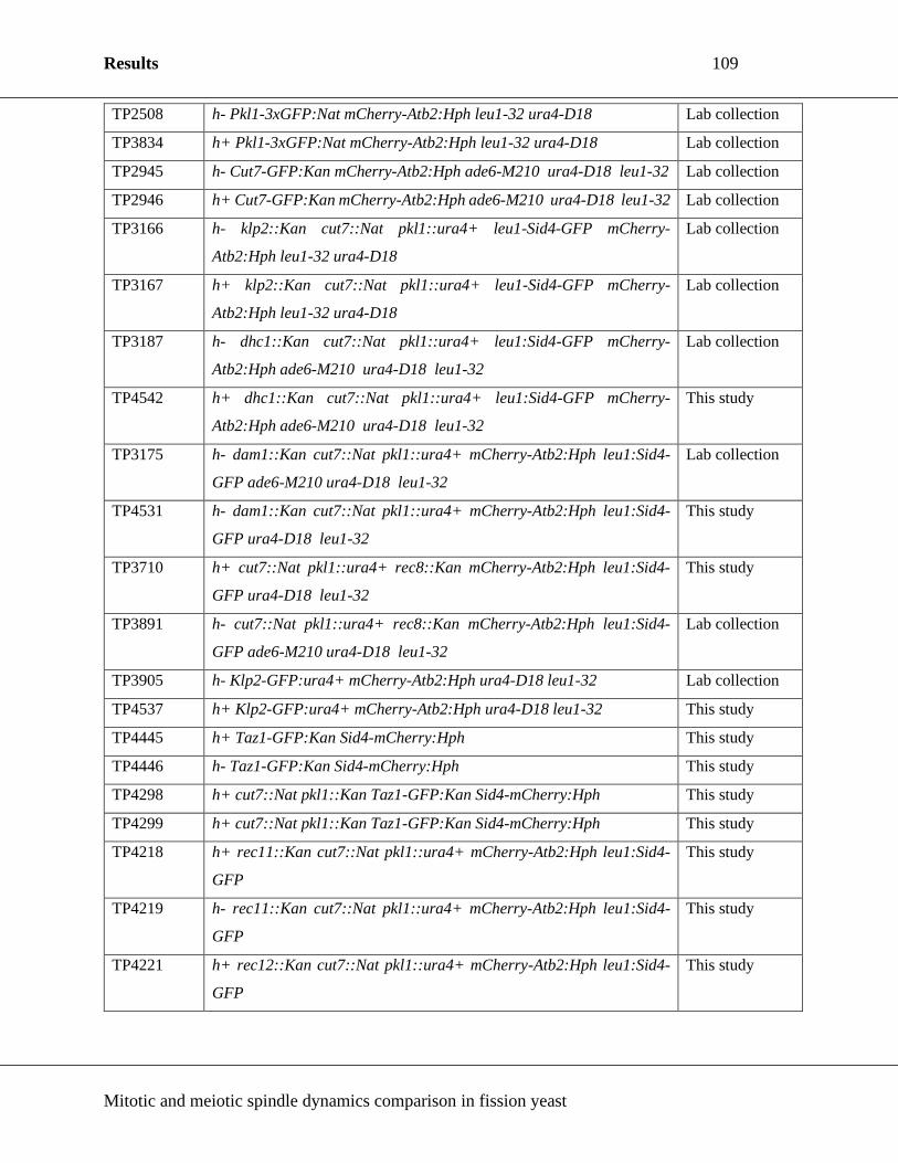
Results 109
Mitotic and meiotic spindle dynamics comparison in fission yeast
TP2508 h- Pkl1-3xGFP:Nat mCherry-Atb2:Hph leu1-32 ura4-D18 Lab collection
TP3834 h+ Pkl1-3xGFP:Nat mCherry-Atb2:Hph leu1-32 ura4-D18 Lab collection
TP2945 h- Cut7-GFP:Kan mCherry-Atb2:Hph ade6-M210 ura4-D18 leu1-32 Lab collection
TP2946 h+ Cut7-GFP:Kan mCherry-Atb2:Hph ade6-M210 ura4-D18 leu1-32 Lab collection
TP3166 h- klp2::Kan cut7::Nat pkl1::ura4+ leu1-Sid4-GFP mCherry-
Atb2:Hph leu1-32 ura4-D18
Lab collection
TP3167 h+ klp2::Kan cut7::Nat pkl1::ura4+ leu1-Sid4-GFP mCherry-
Atb2:Hph leu1-32 ura4-D18
Lab collection
TP3187 h- dhc1::Kan cut7::Nat pkl1::ura4+ leu1:Sid4-GFP mCherry-
Atb2:Hph ade6-M210 ura4-D18 leu1-32
Lab collection
TP4542 h+ dhc1::Kan cut7::Nat pkl1::ura4+ leu1:Sid4-GFP mCherry-
Atb2:Hph ade6-M210 ura4-D18 leu1-32
This study
TP3175 h- dam1::Kan cut7::Nat pkl1::ura4+ mCherry-Atb2:Hph leu1:Sid4-
GFP ade6-M210 ura4-D18 leu1-32
Lab collection
TP4531 h- dam1::Kan cut7::Nat pkl1::ura4+ mCherry-Atb2:Hph leu1:Sid4-
GFP ura4-D18 leu1-32
This study
TP3710 h+ cut7::Nat pkl1::ura4+ rec8::Kan mCherry-Atb2:Hph leu1:Sid4-
GFP ura4-D18 leu1-32
This study
TP3891 h- cut7::Nat pkl1::ura4+ rec8::Kan mCherry-Atb2:Hph leu1:Sid4-
GFP ade6-M210 ura4-D18 leu1-32
Lab collection
TP3905 h- Klp2-GFP:ura4+ mCherry-Atb2:Hph ura4-D18 leu1-32 Lab collection
TP4537 h+ Klp2-GFP:ura4+ mCherry-Atb2:Hph ura4-D18 leu1-32 This study
TP4445 h+ Taz1-GFP:Kan Sid4-mCherry:Hph This study
TP4446 h- Taz1-GFP:Kan Sid4-mCherry:Hph This study
TP4298 h+ cut7::Nat pkl1::Kan Taz1-GFP:Kan Sid4-mCherry:Hph This study
TP4299 h+ cut7::Nat pkl1::Kan Taz1-GFP:Kan Sid4-mCherry:Hph This study
TP4218 h+ rec11::Kan cut7::Nat pkl1::ura4+ mCherry-Atb2:Hph leu1:Sid4-
GFP
This study
TP4219 h- rec11::Kan cut7::Nat pkl1::ura4+ mCherry-Atb2:Hph leu1:Sid4-
GFP
This study
TP4221 h+ rec12::Kan cut7::Nat pkl1::ura4+ mCherry-Atb2:Hph leu1:Sid4-
GFP
This study

Results 110
Mitotic and meiotic spindle dynamics comparison in fission yeast
TP4222 h- rec12::Kan cut7::Nat pkl1::ura4+ mCherry-Atb2:Hph leu1:Sid4-
GFP
This study
TP4229 h- sgo1::Kan cut7::Nat pkl1::ura4+ leu1:Sid4-GFP mCherry-
Atb2:Hph
This study
TP4230 h+ sgo1::Kan cut7::Nat pkl1::ura4+ leu1:Sid4-GFP mCherry-
Atb2:Hph
This study
TP4539 h+ moa1::Kan cut7::Nat pkl1::ura4+ mCherry-Atb2:Hph leu1:Sid4-
GFP
This study
TP4538 h- moa1::Kan cut7::Nat pkl1::ura4+ mCherry-Atb2:Hph leu1:Sid4-
GFP
This study
TP1511 h+ Mis12-GFP:leu2+ Sid4-mCherry:Hph ura4-D18 leu1-32 Lab collection
TP2943 h- Sid4-mCherry:Hph Mis12-GFP:leu2+ ura4-D18 leu1-32 Lab collection
TP3223 h- cut7::Nat pkl1::ura4+ Sid4-mCherry:Hph Mis12-GFP:leu2+ ade6-
ura4-D18 leu1-32
Lab collection
TP3062 h+ cut7::Nat pkl1::ura4+ Sid4-mCherry:Hph Mis12-GFP:leu2+
ade6- ura4-D18 leu1-32
Lab collection

Discussion 111
Mitotic and meiotic spindle dynamics comparison in fission yeast
Discussion

Discussion 112
Mitotic and meiotic spindle dynamics comparison in fission yeast
Spindle dynamics are distinguishing mitotic and meiotic spindles
In this work, spindle dynamics were explored and compared simultaneously in mitosis and meiosis,
in the same organism, for the first time. This work has shown mitotic, MI and MII spindles exhibit
distinct spindle dynamics patterns (Figure 1E in Results section), and that the characterized
attributes of spindles are intrinsically different for each spindle type (Table 1 in Results section).
Moreover, MT bundle dynamics measured in the time interval between two mitotic divisions and
between two meiotic divisions showed that MT bundles are more dynamic in zygotes than in
vegetative cells, as they undergo catastrophe more often (Table 2 in Results section). These data
clearly show that mitotic and meiotic spindles are differently regulated, although they perform the
same function, i.e. chromosome segregation, and that MT dynamics may be modified to fine tune
spindle function in different cell-division types.
Zygotes are approximately 27% larger than vegetative cells, and assemble an MI spindle that is
longer in all three spindle elongation phases. The phenomenon of spindle length scaling with cell
size was observed in fission yeast previously (Krüger et al., 2019), but also in other organisms
(Wühr et al., 2008; Hara and Kimura, 2009; Yang et al., 2016), and it can be simply explained by
a limited components model, i.e. limiting cytoplasmic components (Good et al., 2013).
In mitosis of fission yeast, the spindle spends roughly half of its life in phase III, which is when
spindle elongates and further separates sister chromatids, unlike MI and MII spindles, which spend
nearly half or 2/3 of their life in phase II, respectively. As mitosis occurs in smaller cells, which
additionally septate, i.e. produce a wall at the cell equator to separate daughter cells into two
separate entities at the end of mitosis (Wolfe and Gould, 2005; Sipiczki, 2007), the principal role
of the mitotic spindle may be to remove the sister-chromatids as far away from the site of
cytokinesis to prevent the chromosome cut phenotype. In fission yeast meiosis there is no

Discussion 113
Mitotic and meiotic spindle dynamics comparison in fission yeast
cytokinesis to cause cut chromosomes, so there may be no necessity for the spindle to remain in
phase III. In line with that, MII final spindle length is 45% shorter than mitotic spindle length, and
only 15% of spindle life is spent in phase III, yet the DNA is successfully separated.
In MI, homologous chromosomes are associated into a bivalent, and physically linked at chiasmata
(Molnar et al., 2003; Hirose et al., 2011). Possibly the MI spindle spends the majority of its life in
phase II to resolve the chiasmata intermediates before homologous chromosomes are pulled to the
opposite spindle poles at the end of phase II. That eggs are arrested at metaphase I or metaphase II,
which coincides with phase II of meiosis spindle elongation in fission yeast, has been observed in
many systems, including humans (Sagata, 1996; Tunquist and Maller, 2003; Sanders and Jones,
2018). Possibly, these phenomena are somehow correlated and may prove an interesting direction
for future research.
Klp2 co-operates with Pkl1 to antagonize Cut7 in MI spindle
An important observation in this study was the failure of cut7Δpkl1Δ mutant to successfully
complete meiosis, although mitosis outcome is successful (Olmsted et al., 2014; Rincon et al.,
2017; Yukawa et al., 2017; Shirasugi and Sato, 2019). Our study revealed that spindle assembly
and elongation are specifically perturbed in MI, but not mitosis or MII. This confirms the
hypothesis that meiotic and mitotic spindles are intrinsically different.
We confirmed that Cut7 and Pkl1 localize similarly in mitosis and MI spindle, but their relative
amounts are changed, such that the ratio of Cut7 to Pkl1 is ~2 times higher in MI (Figure 3 in the
Results section). Consequently, there are more Cut7 molecules to produce outward pushing force
in MI spindle compared to the mitotic spindle. This has led us to speculate, that another molecule
may antagonize Cut7. We have found that Klp2 partially counteracts Cut7-dependent forces in MI

Discussion 114
Mitotic and meiotic spindle dynamics comparison in fission yeast
and restores pre-phase III spindle length (Figure 4C in the Results section). Moreover, Klp2
localized more to the spindle poles in MI spindles, contrary to its dispersed localization along the
spindle in mitosis (Figure 4D and E in in the Results section). It was shown previously that, in
mitotic spindles, tethering Klp2 to the spindle poles in Pkl1 deficient cells allows it to produce
inward forces to counteract Cut7-dependent forces (Yukawa et al., 2018). We conclude that
although Klp2 concentration is comparable in mitotic and MI spindles (Figure 4F in the Results
section), its MI-specific localization to the spindle poles permits it to antagonize Cut7. The
mechanism behind this regulation remains an open question. Considering that other organisms like
mice and humans possess multiple kinesin-14s, it would be interesting to see if such kinesin-14
specialization in MI is evolutionary conserved.
MT dynamics in zygotes and vegetative cells may fine tune spindle function
In this study, we have observed that MI and MII spindles took longer time to assemble and establish
a steady-state length, and have accomplished it at comparable velocities (Table 1 in the Results
section). This has prompted us to investigate if spindle dynamics may differ from mitosis to
meiosis. As technical limitations prevented us from studying MT dynamics inside the
prometaphase spindles because their size precludes us from monitoring MT dynamics, we have
opted to test MT bundle dynamics in between two mitotic divisions and the meiotic divisions.
We found MT bundles in zygotes grew faster and underwent catastrophe more often than in
vegetative cells (Table 2 in the Results section). Under the assumption that MTs are more dynamic
in MI, we have inhibited MT dynamics in cut7Δpkl1Δ zygotes by treatment with the MT drug MBC
(Vela-Corcía et al., 2018). Indeed, approximately 80% of treated cut7Δpkl1Δ zygotes assembled
bipolar spindles compared to 26% of non-treated cut7Δpkl1Δ zygotes (Figure S5A in the Results
section). Moreover, mitotic spindle dynamics were not sensitive to treatment with MBC, unlike MI

Discussion 115
Mitotic and meiotic spindle dynamics comparison in fission yeast
spindle dynamics, in both wt and cut7Δpkl1Δ zygotes (Figure 5D and FigureS5B in the Results
section).
In zygotes, KTs are located further away from the SPBs, while in mitosis they are located
underneath the SPBs (Figure 4.4A in the Introduction section) (Hironori et al., 1993). This
configuration may favour more dynamic MTs, as more frequent catastrophe would increase the
chance of MT growing in the direction of the KT, and subsequent MT-KT attachment (Mitchison
and Kirschner, 1984; Heald and Khodjakov, 2015).
In line with this, lower concentration of kinesin-14 Pkl1 in MI may further aid successful KT-MT
attachment. Pkl1 was shown to negatively influence MT nucleation in fission yeast, and Cut7 was
shown to inhibit this effect (Olmsted et al., 2014). It may be that lower Pkl1 concentration in MI
spindles favours more frequent MT nucleation. Moreover, as Pkl1 cross-links parallel MTs and
organizes the spindle poles (Syrovatkina and Tran, 2015; Yukawa, Ikebe and Toda, 2015), its
scarcity in in MI spindles could lead to MTs that are able to more freely explore the area around
them (Figure S5F in the Results section) (Kalinina et al., 2013; Cojoc et al., 2016; Winters et al.,
2019). Further investigations on role of Pkl1 and Klp2 in MI spindles are needed, and measuring
MT dynamics inside spindles is a necessary next step to confirm current findings.
Chromosome architecture is not a decisive factor in successful MI spindle assembly in
cut7Δpkl1Δ zygotes
Arguably the most prominent difference between mitosis and MI is the chromosomal organization
(Figure 2.4A in the Introduction section) (Ohkura, 2015). It was shown prior that cohesin influences
the length of the phase II spindle (Goshima, Saitoh and Yanagida, 1999; Goshima et al., 2005), and

Discussion 116
Mitotic and meiotic spindle dynamics comparison in fission yeast
proper tension between sister-chromatids is a feature used by the cell to monitor and amend proper
MT-KT attachments (Musacchio and Salmon, 2007; Liu et al., 2009).
We could not detect the influence of MI-specific chromosomal organization on spindle assembly
in the cut7Δpkl1Δ background, but a recent study that studied homothallic strains in meiosis, has
demonstrated that a double deletion of Rec12 and Moa1 partially rescued spindle bipolarity in
cut7Δpkl1Δ background (Shirasugi and Sato, 2019). However, a quadruple mutant still showed
only a modest, ~50% success in SPB separation, indicating that chromosomal architecture is not a
pivotal factor in successful bipolarity establishment in cut7Δpkl1Δ MI.
Future perspectives for mitotic and meiotic spindle comparison
The work presented in this dissertation is but a beginning in comparative spindle studies. While
Cut7, Pkl1, and Klp2 localization and intensities were investigated and compared, other motors and
MAPs remain to be studied. Moreover, while localization and amounts of these three molecules
are described, the regulation behind their respective peculiarities in MI compared to mitosis
remains to be examined.
MII spindles remain somewhat ambiguous. While MI and mitotic spindle analyses are relatively
straightforward, MII spindles are strongly impacted by the successfulness of preceding MI division.
In order to probe MII spindles, new methodologies concerning protein inhibition and over-
expression must be developed to specifically perturb a function of a molecule in MII but not in MI.
Further, it is possible to study the role of many essential genes in mitosis because thermo-sensitive
alleles of these genes exist, and as such, these proteins can be inactivated at 36°C. Such an approach
is somewhat feasible in meiotic spindle studies, under the condition that conjugation and
recombination are allowed to occur at permissive temperature (meiotic life cycle triggering is

Discussion 117
Mitotic and meiotic spindle dynamics comparison in fission yeast
severely inhibited at temperatures above 31°C). Generally speaking, methods to manipulate protein
expression in meiosis are lacking, but are essential to perform meiotic spindle studies. Future work
will surely have to be oriented towards new technology development, especially control of protein
levels in meiosis.
This study has identified that MT dynamics and kinesin-14s are differently regulated in mitotic and
MI spindles to coordinate chromosome segregation. In studies to follow, a more in depth
characterization of the spindle must be achieved, that will ultimately allow us to manipulate the
spindle in an efficient and targeted manner for therapeutic purposes.
Résumé 118
Mitotic and meiotic spindle dynamics comparison in fission yeast
Résumé

Introduction 119
Mitotic and meiotic spindle dynamics comparison in fission yeast
1. Introduction
1.1. Cell division
La cellule est une entité constituée de plusieurs organites communs aux organismes connus. Au
cours de sa vie, la cellule effectue plusieurs cycles successifs composés de plusieurs phases. Chaque
cycle cellulaire comprend l’interphase - comprenant les phases G1, S et G2 - suivie de la division
cellulaire appelée mitose. Généralement, en phase G1 la cellule croît et se prépare à la réplication
de son ADN (acide désoxyribonucléique) génomique qui aura lieu en phase S. Au cours de la phase
G2, la cellule continue sa croissance et se prépare à la division cellulaire. Chez les eucaryotes, on
distingue deux types de division cellulaire : la mitose et la méiose. La mitose permet la prolifération
cellulaire qui consiste à générer deux cellules filles identiques tandis que la méiose permet de
produire des cellules destinées à la reproduction sexuée. La mitose et la méiose se succèdent après
un unique cycle de duplication de l’ADN – les cellules de la mitose se divisent une fois alors que
lors de la méiose les cellules se divisent deux fois. La tâche impérative de la division cellulaire est
de séparer précisément le matériel génomique dupliqué en deux cellules filles identiques.
1.1.1. La mitose
La mitose est un type de division cellulaire dans laquelle deux cellules filles identiques sont
générées à partir d’une cellule parent unique, conservant la ploïdie parentale. C'est un processus
essentiel dont le but est la prolifération cellulaire - qu'il s'agisse d'augmenter le nombre de cellules,
pour permettre par exemple la croissance et le maintien d'un organisme multicellulaire, sa
reproduction, comme par exemple dans le cas de la reproduction végétative des plantes. Par
conséquent, il est essentiel que la ségrégation des chromosomes ne comporte aucune erreur car les
inexactitudes peuvent conduire à une aneuploïdie qui pourrait entrainer des conséquences

Introduction 120
Mitotic and meiotic spindle dynamics comparison in fission yeast
catastrophiques telles que le cancer, des défauts au cours du développement ou la mort cellulaire
(Holland and Cleveland, 2009; Thompson, Bakhoum and Compton, 2010).
Pour obtenir une séparation parfaite des chromosomes nécessaire au maintien de la ploïdie, les
cellules forment un fuseau mitotique (Gatlin and Bloom, 2010; Heald and Khodjakov, 2015). Le
fuseau mitotique est une structure macromoléculaire composée de microtubules (MTs), de
protéines motrices et de protéines non motrices (David J. Sharp, Rogers and Scholey, 2000;
Manning and Compton, 2008). Le fuseau mitotique est formé lors de la prophase lorsque les
centrosomes, centres organisateurs des microtubules (MTOC) majeurs, procèdent à la nucléation
des MTs (Rale, Kadzik and Petry, 2018). Les MTs en croissance sont attachées au centrosome par
leur extrémité “moins” et leur extrémité “plus” distale leur permet d’explorer l’espace qui les
entourent à la recherche d’autres MTs ou kinétochores (KTs), structures protéiques spécialisées
situées dans la région centromérique de l'ADN qui servent de plate-forme pour la fixation du
chromosome au fuseau médiée par les MTs.
La bipolarité du fuseau est établie lorsque l'interdigitation des MT des centrosomes opposés, sont
réticulés via des moteurs et des protéines associées aux MT (MAPs) en faisceaux antiparallèles qui
glissent dans deux directions opposés (Tanenbaum and Medema, 2010). Les chromosomes, qui
sont attachés aux MTs du fuseau via les KT, congressent vers sur la plaque métaphasique et
s'alignent par une interaction complexe de forces qui maintiennent une longueur de fuseau à l'état
d’équilibre (Goshima et al., 2005; Dumont and Mitchison, 2009). Une fois que tous les KT sœurs
sont attachés aux MTs émanant du pôle opposé du fuseau (les chromosomes sont bi-orientés), le
point de contrôle de l'assemblage du fuseau (‘spindle assembly checkpoint’ - SAC) est satisfait
(Musacchio and Salmon, 2007). Cela active alors la séparase (Peters, 2006), qui clive ensuite la

Introduction 121
Mitotic and meiotic spindle dynamics comparison in fission yeast
cohésine, un complexe protéique qui relie les chromatides sœurs, permettant ainsi aux
chromosomes d'être tirés vers les pôles opposés du fuseau en anaphase A.
On distingue deux mécanismes gouvernant les mouvements des chromosomes au cours de l’
anaphase A: les flux nommés Pac-Man et “poleward”, “vers le pôle”. Dans le flux Pac-Man, les
MTs attachés aux KTs (fibre- KT) dépolymérisent à l’extrémité plus et rétrécissent vers le pôle du
fuseau, transportant les chromosomes aux deux pôles de façon simultanée (Gorbsky, Sammak and
Borisy, 1987). Dans le flux ‘poleward’, le chromosome est translocalisé vers le pôle du fuseau par
dépolymérisation de la fibre-KT près du pole du fuseau. (Mitchison, 1989).
Puis, un allongement rapide du fuseau a lieu en anaphase B qui permet la séparation du matériel
génétique du site de la cytokinèse. La mitose se termine par l’obtention deux cellules filles qui ont
le même nombre de chromosomes que la cellule mère.
1.1.2. La Méiose
La Méiose est un type de division cellulaire produisant des cellules porteuses de la moitié du
matériel génétique de la cellule initiale. Ces cellules sont utilisées dans la reproduction sexuée
lorsqu’elles fusionnent et produisent un zygote diploïde. La méiose est composée de deux divisions
consécutives après un cycle de duplication de l’ADN. Contrairement à la mitose, les chromosomes
homologues s'apparient en prophase I de méiose et subissent une recombinaison homologue (Page
and Hawley, 2003). Ce processus donne naissance à de nouvelles combinaisons d'ADN et favorise
la variabilité génétique, qui pourrait être le but de la reproduction sexuée. Une ségrégation
chromosomique précise dans les deux divisions méiotiques est requise. Chez l'homme, la
ségrégation chromosomique dans la méiose féminine est particulièrement sujette aux erreurs
(Warburton, 1997; Holubcová et al., 2015; Namgoong, Kim and Christenson, 2018), et de tels
défauts entraînent des troubles congénitaux ou des avortements.

Introduction 122
Mitotic and meiotic spindle dynamics comparison in fission yeast
De façon similaire à la mitose, un fuseau composé de MTs est assemblé pour séparer de façon
identique le matériel génétique en deux cellules filles. Il est important de noter que les ovocytes de
nombreuses espèces n'ont pas de centrosomes (Müller-Reichert et al., 2010; Dumont and Desai,
2012; Bennabi, Terret and Verlhac, 2016; Gruss, 2018) et dépendent d'autres voies de nucléation
des MTs (chapitre 3). L'une des caractéristiques de la méiose est la nature réductrice de la première
division méiotique (MI). Plusieurs mécanismes permettent d’assurer la réduction de moitié de la
ploïdie: 1) architecture modifiée des KT sœurs et mono-orientation (Yokobayashi and Watanabe,
2005; Hauf et al., 2007; Sakuno et al., 2011); 2) Cohésine spécialisée et contrôle hiérarchique du
clivage de la cohésine par la séparase (Watanabe and Nurse, 1999; Tomoya S. Kitajima et al., 2003;
Kitajima, Kawashima and Watanabe, 2004; Miyazaki et al., 2017). Ces mécanismes garantissent
que les chromosomes homologues se mono-orientent, c'est-à-dire les KT sœurs, s'attachent aux
MTs émanant du même pôle du fuseau. Lorsque le point de contrôle de l’assemblage du fuseau est
satisfait, la séparase clive la cohésine entre les bras chromosomiques, mais pas dans la région
centromérique, où la cohésine est protégée (Miyazaki et al., 2017). L'allongement du fuseau au
cours de l'anaphase B de la méiose I (MI) garantit que les chromosomes homologues bivalents sont
suffisamment séparés, et le MI se termine par deux cellules filles qui portent la moitié du nombre
de chromosomes par rapport à la cellule mère.
Chez certaines espèces, la méiose I est suivie d'une brève période de repos appelée interkinésie,
une forme cryptique d'interphase, dans laquelle les deux noyaux de cellules filles peuvent être
observés. La méiose II (MII) est souvent comparée à la mitose, car l'architecture chromosomique
est organisée de manière similaire: un fuseau se forme dans chaque noyau et sépare les chromatides
sœurs en deux nouvelles cellules filles. Quatre cellules filles contenant la moitié de la quantité
initiale de matériel génétique sont ainsi formées en fin de MII.
Introduction 123
Mitotic and meiotic spindle dynamics comparison in fission yeast
Dans les ovocytes femelles, la division cellulaire est très asymétrique en MI, et les produits de la
méiose I sont contenus dans un œuf mature, contenant un nombre de chromosomes réduit de moitié,
et un petit premier corps polaire qui ne joue plus de rôle (Sanders and Jones, 2018). L'ovule mature
entre en méiose II, et s'arrête en métaphase jusqu'à ce qu'il soit fécondé par un sperme. La méiose
II est également asymétrique et, lors de la fécondation et de la reprise de la division, un embryon
et un deuxième corps polaire sont formés.
1.2. La levure fissipare S. pombe comme système modèle pour l’analyse de
la dynamique du fuseau mitotique et méiotique
La levure fissipare Schizosaccharomyces pombe (S. pombe) est un organisme eucaryote
unicellulaire en forme de bâtonnet qui atteint généralement une longueur de 12 à 14 µm à la division
mitotique. En raison de la simplicité de l’organisation de son cytosquelette de MTs, de la
conservation évolutive des principaux acteurs de l'assemblage et de la fonction du fuseau, et du fait
que, comme chez les eucaryotes supérieurs, plusieurs MT se fixent à un KT (Ding, McDonald and
McIntosh, 1993), cet organisme constitue un modèle exceptionnel d’intérêt pour l’étude du fuseau
mitotique et méiotique.
Lorsqu'elle se trouve dans des conditions riches en nutriments, la levure fissipare S. pombe vit
principalement comme un haploïde et entre une mitose pour proliférer. Comme les cellules de
mammifères, cet organisme suit la progression typique des étapes du cycle cellulaire: G1, S, G2 et
M (Mitchison and Creanor, 1971; Nurse, Thuriaux and Nasmyth, 1976). Dans des conditions de
carence , notamment en cas de déficit en azote, la levure de fission quitte le cycle cellulaire végétatif
et entre dans la voie de la méiose (Harigaya and Yamamoto, 2007). On distingue deux types sexuels
dans les cellules de levure S. pombe : h + et h-. Si les cellules voisines sont de type sexuel opposé

Introduction 124
Mitotic and meiotic spindle dynamics comparison in fission yeast
et sont privées d'azote, elles peuvent fusionner et former un zygote. Le zygote diploïde peut
proliférer en tant que diploïde, si un milieu riche en nutriments devient disponible, ou continuer
avec des divisions méiotiques. À la fin de la méiose, le zygote aura formé quatre noyaux haploïdes
encapsulés par une membrane générant quatre spores haploïdes. Une fois que les conditions sont
favorables, les spores haploïdes peuvent germer et recommencer le cycle cellulaire végétatif. Bien
que la privation d'azote induise la conjugaison des cellules haploïdes en zygotes, les cellules
haploïdes peuvent néanmoins continuer à proliférer par mitose. Cela permet d'étudier les deux types
de division cellulaire simultanément, dans les mêmes conditions.
L'assemblage et l'allongement du fuseau sont stéréotypés dans la levure fissipare S. pombe, tant
dans la mitose que dans la méiose, et se composent de trois étapes distinctes (Nabeshima,
Nakagawa, Straight, Murray, et al., 1998). La phase I de l'assemblage du fuseau en prophase se
réfère aux étapes initiales de la formation du fuseau, comme par exemple l’établissement de
bipolarité. La phase II est marquée par un maintien de la longueur du fuseau à l'état d'équilibre,
accompagné d'une congression chromosomique vers la plaque métaphasique. A la fin de la phase
II, les chromatides sœurs sont séparées aux pôles opposés. Enfin, en phase III, le fuseau s'allonge
rapidement et permet la séparation des chromosomes.
1.2.1. Comparaison de la dynamique du fuseau mitotique et méiotique
La mitose et la méiose ont fait l'objet de recherches approfondies et de nombreuses études ont
explore le rôle des moteurs moléculaires, des MAPs et de la dynamique des MT du fuseau. Malgré
de nombreux travaux de recherche, aucune étude à ce jour n'a comparé la dynamique du fuseau
dans la mitose et la méiose de façon simultanée dans le même système modèle. Très probablement
en raison du fait qu'une telle tâche reste techniquement impossible dans de nombreux systèmes, ou

Introduction 125
Mitotic and meiotic spindle dynamics comparison in fission yeast
bien qu'il existe trop de facteurs qui pourraient rendre difficile l'interprétation des données. De plus,
de telles études chez l'homme sont extrêmement compliquées, car les ovocytes sont considérés
comme une ressource biologique précieuse. Compte tenu de la nature fondamentale des processus
mitotiques et méiotiques, une comparaison de la dynamique du fuseau mitotique et méiotique serait
essentielle pour explorer respectivement les différences et les particularités de chaque type de
division cellulaire.
La levure de fission est devenue un bon système modèle pour une telle étude. Son cytosquelette de
MTs est simple et bien caractérisé, et les principaux acteurs moléculaires de l'assemblage du fuseau
sont conservés de la levure fissipare à l'homme. Il est important de noter que la levure S. pombe
peut subir une mitose et une méiose dans le même environnement et dans les mêmes conditions, ce
qui rend possible une étude comparative de la dynamique du fuseau mitotique et méiotique de façon
simultanée.
Dans des expériences conçues pour comparer la dynamique du fuseau mitotique et méiotique, des
cellules de types sexuels opposés contenant des composants du fuseau marqué par des molécules
fluorescentes ont d'abord été cultivées dans un milieu riche (YE5S) pendant une nuit, puis
mélangées sur un milieu pauvre en azote (ME) de façon à induire la conjugaison. Après 16-20
heures, les cellules ont été remises en suspension dans 10 µL de ME liquide et transférées sur des
coussins semi-solides composés d'agarose et de milieu malt-extrait (ME) et imagées pendant
plusieurs heures. En suivant ce protocole, un champ de cellules contenant un mélange de zygotes
et de cellules végétatives a pu être identifié et imagé avec le microscope confocal à disque rotatif
(spinning disk).

Introduction 126
Mitotic and meiotic spindle dynamics comparison in fission yeast
1.2.2. Le double mutant de délétion kinésine-5 /kinésine-14 (cut7Δpkl1Δ) comme outil
permettant la comparaison des fuseaux mitotiques et méiotiques
Chez S. pombe, lors de la phase I de l'assemblage du fuseau, les MTs en croissance sont regroupés
en faisceau antiparallèle par la kinesine-5 /Cut7 (Hagan and Yanagida, 1990, 1992a). Le moteur
homotétramérique Cut7 dirigé à l'extrémité plus des MTs exerce des forces sur le faisceau
antiparallèle et fait glisser les MTs vers l’extérieur, repoussant ainsi les ‘spindle pole body’ (SPB,
équivalent du centrosome chez S. pombe) permettant d’établir la bipolarité. Cut7 est une protéine
essentielle et il a été montré que les mutants Cut7 thermosensibles assemblent des fuseaux
mitotiques monopolaires (Hagan and Yanagida, 1990, 1992a).
Un autre acteur dans les étapes initiales de l'assemblage du fuseau chez S. pombe est une protéine
motrice dirigée vers l'extrémité “moins” des MTs, la kinésine 14 Pkl1, localisée au SPB et
faiblement aux MTs du fuseau aux étapes initiales de l'assemblage du fuseau mitotique (Pidoux,
LeDizet and Cande, 1996; Syrovatkina and Tran, 2015; Yukawa, Ikebe and Toda, 2015). Pkl1
forme un complexe avec les protéines Msd1 et Wdr8, important pour l’ancrage du fuseau
microtubulaire aux SPBs (Toya et al., 2007; Yukawa, Ikebe and Toda, 2015). De plus, Pkl1 réticule
probablement des faisceaux de MTs parallèles pour fournir une stabilité structurelle aux pôles du
fuseau en raison de la force exercée par Cut7 vers l'extérieur. Dans les cellules de S. pombe
dépourvues d'activité de Pkl1, les pôles du fuseau ne sont pas focalisés, ce qui est évident dans la
formation de protrusion MT médiée par Cut7, pouvant induire une aneuploïdie (Syrovatkina and
Tran, 2015).
Fait intéressant, la double délétion de Pkl1 et de Cut7 permet à la cellule mutante de former un
fuseau bipolaire mitotique capable de séparer les chromosomes; de plus, la relation antagoniste de

Resultats 127
Mitotic and meiotic spindle dynamics comparison in fission yeast
Cut7 et Pkl1 est largement étudiée dans les fuseaux mitotiques de S. pombe (Rincon et al., 2017;
Yukawa et al., 2017). Cependant, les zygotes cut7Δpkl1Δ sont incapables de poursuivre
normalement la méiose. En effet, à la fin de la méiose, les zygotes cut7Δpkl1Δ ne produisent pas
quatre spores régulières. Ce résultat suggère que les chromosomes de ce double mutant ne peuvent
pas être séparés et que la mécanique du fuseau peut être compromise. Ainsi, dans ce travail, il a été
étudié pourquoi la méiose échoue alors que la mitose se déroule normalement chez le mutant
cut7Δpkl1Δ de S. pombe, qui a été utilisé comme outil pour étudier les différences dans les fuseaux
mitotiques et méiotiques.
2. Resultats
2.1. La Dynamique du fuseau diffère en mitose et en méiose dans la levure fissipare
Afin d’évaluer la dynamique du fuseau dans la mitose et la méiose, nous avons imagé des cellules
de levure fissipare S. pombe de type sauvage (wt - wild type) exprimant l’alpha-2 (α2) tubuline
marquée avec mCherry (mCherry-Atb2) et le composant SPB Sid4 marqué avec GFP α2-tubuline
(Sid4-GFP), cultivé dans du malt- extrait (ME) afin d’induire la conjugaison. Pour évaluer la
dynamique du fuseau, nous avons mesuré la longueur du signal entre les deux SPB à des intervalles
de temps d'une minute. Il a été constaté que les fuseaux MI et MII présentaient une progression en
trois phases similaires aux fuseaux mitotiques.
La longueur finale du fuseau MI a été augmentée de 25% en moyenne par rapport aux fuseaux
mitotiques, et la vitesse d'élongation du fuseau MI en phase III était plus rapide qu'en mitose.
Contrairement aux cellules mitotiques végétatives, les zygotes ont passé la plus grande partie du
temps d'élongation du fuseau en phase II de MI et MII. Enfin, les zygotes ont passé une proportion
similaire de temps dans la phase I de MI et MII, mais la vitesse d'assemblage du fuseau chez les

Resultats 128
Mitotic and meiotic spindle dynamics comparison in fission yeast
zygotes était plus faible que dans les cellules mitotiques. Ces différences dans la dynamique du
fuseau impliquaient des changements qualitatifs et/ou quantitatifs dans les composants du fuseau,
tels que les moteurs moléculaires et les MAPs, ou des changements dans la dynamique MT entre
mitose et méiose.
2.2. L’intégrité du fuseau est compromise spécifiquement en MI dans les zygotes du
double mutant cut7Δpkl1Δ
Pour étudier la dynamique du fuseau dans la méiose du double mutant cut7Δpkl1Δ, nous avons
imagé des zygotes cut7Δpkl1Δ exprimant mCherry-Atb2 et Sid4-GFP. Contrairement à la mitose,
où toutes les cellules cut7Δpkl1Δ ont assemblé un fuseau bipolaire, les zygotes cut7Δpkl1Δ ont
échoué en méiose: 74 ± 2% des zygotes cut7Δpkl1Δ n'ont pas réussi à établir un fuseau bipolaire
et à séparer les SPB en MI, comme cela a été observé dans un rapport récent (Shirasugi and Sato,
2019). De plus, parmi les zygotes qui avaient assemblé des fuseaux bipolaires en MI, 8 ± 2%
supplémentaires n'ont pas réussi à assembler de fuseau en MII. Une séparation des SPB
infructueuse en MII, pourrait résulter d'erreurs dans la MI précédente ou des propriétés intrinsèques
des fuseaux cut7Δpkl1Δ de MII.
Nous avons ensuite comparé la dynamique des fuseaux de cellules cut7Δpkl1Δ et wt. En mitose,
les fuseaux pré-phasiques III étaient plus courts et l’entrée en phase III était retardé dans le doubles
mutant cut7Δpkl1Δ comparativement aux cellules wt, ce qui corrobore avec les études précédentes
(Rincon et al., 2017; Yukawa et al., 2018). Des résultats similaires ont été observés pour la
dynamique du fuseau MII. En revanche, la plupart des fuseaux MI sont restés monopolaires.
25%des fuseaux MI cut7Δpkl1Δ qui ont réussi à atteindre la bipolarité et à séparer les SPB n'ont
pas montré la progression triphasée habituelle de l'allongement du fuseau, car les phases I et II (de

Resultats 129
Mitotic and meiotic spindle dynamics comparison in fission yeast
l'allongement du fuseau) n'ont pas pu être distinguées. Combinées, nos données indiquent que
l'assemblage et l'allongement du fuseau sont spécifiquement perturbés pendant l'IM des zygotes
cut7Δpkl1Δ. Nous concluons que la suppression de Pkl1 ne peut pas compenser entièrement
l'absence de Cut7 en MI. Cela suggère que Cut7 et Pkl1 sont régulés différemment en mitose et en
MI.
2.3. Le ratio Cut7-à-Pkl1 est plus élevé dans le fuseau MI qu’en mitose
Afin d’évaluer si Cut7 et Pkl1 sont régulés différemment en mitose et en MI, nous avons imagé
Cut7 ou Pkl1 marqués avec la GFP et les MT avec mCherry-Atb2 en mitose et en MI dans les
cellules wt. Nous avons observé que Cut7-GFP était localisé au fuseau et aux pôles du fuseau depuis
l'assemblage jusqu’à sa rupture en mitose et en MI. De même, le profil de localisation de Pkl1-
3XGFP est apparu inchangé entre la MI et la mitose. Pkl1-3XGFP est localisé aux pôles du fuseau
peu de temps après le début de son assemblage, et reste associé aux SPB jusqu'à sa rupture. Pkl1-
3XGFP a également été détecté sur le fuseau lors de la transition entre la phase I et la phase II.
Nous avons ensuite mesuré la distribution du signal Cut7-GFP et Pkl1-3XGFP au fuseau pendant
la transition de la phase I à la phase II, 1 minute avant que le fuseau n'atteigne une longueur
constante qui marque le début de la phase II. L'intensité maximale de Cut7-GFP était plus élevée
dans en MI, tandis que l'intensité maximale de Pkl1-3XGFP était similaire en mitose et en MI. De
plus, l'intensité totale de Cut7-GFP, calculée en intégrant les profils d'intensité du ‘linescan’, était
élevée en MI comparativement à la mitose, tandis que l'intensité totale de Pkl1-3XGFP est restée
similaire dans les deux types de fuseau.
Parce que l'intensité totale des MTs est différente dans fuseaux mitotiques et en MI, nous avons
normalisé les valeurs d'intensité obtenues pour Cut7-GFP et Pkl1-3XGFP aux valeurs d'intensité

Resultats 130
Mitotic and meiotic spindle dynamics comparison in fission yeast
des MTs. De cette façon, nous avons constaté que la concentration relative de Cut7 restait
inchangée, tandis que la concentration relative de Pkl1-3XGFP par MT était plus faible en MI que
celle observée en mitose. Cela peut expliquer pourquoi la suppression de Pkl1 ne peut pas
compenser la suppression de Cut7 en MI, et suggère qu'un facteur supplémentaire pourrait
contrecarrer l'activité de Cut7 pendant la phase II de MI pour maintenir l'équilibre des forces.
2.4. La fonction de la kinésine 14 Klp2 exercée sur le fuseau est distincte en MI et en
mitose
Afin d’identifier des de nouveaux composants du fuseau qui pourraient contrecarrer les forces
dépendantes de Cut7 en méiose, nous avons systématiquement délété les moteurs et les MAPs
candidat-e-s dans le fond génétique cut7Δpkl1Δ, croisé les triples mutants et évalué le nombre de
spores. De toutes les protéines testées, seule la suppression de la kinésine-14 Klp2 a induit la
formation de zygotes qui ont produit quatre spores typiques de façon plus fréquente (44 ± 2% des
zygotes cut7Δpkl1Δklp2Δ ont produit quatre spores versus 20 ± 1% dans le fond génétique/les
doubles mutants de délétion cut7Δpkl1Δ).
L'imagerie des cellules vivantes a révélé que si seulement 26 ± 2% des zygotes cut7Δpkl1Δ ont
réussi à établir la bipolarité du fuseau et à séparer les SPB, 49 ± 2% des zygotes cut7Δpkl1Δklp2Δ
ont assemblé des fuseaux bipolaires en MI. La comparaison de la dynamique du fuseau de MI a
révélé que non seulement la bipolarité du fuseau a été rétablie, mais la dynamique d'élongation du
fuseau habituellement tri-phasique a été partiellement complémentée chez les zygotes
cut7Δpkl1Δklp2Δ. Cela différait fortement des zygotes cut7Δpkl1Δ dans lesquels la dynamique du
fuseau tri-phasique n'était pas observée. Nos analyses du fuseau mitotique ont montré que la
suppression de Klp2 complémentait partiellement le temps nécessaire pour atteindre la phase III,

Resultats 131
Mitotic and meiotic spindle dynamics comparison in fission yeast
mais cela n'a pas permis de restaurer la longueur du fuseau de phase III dans les cellules
cut7Δpkl1Δ. Ce résultat était différent de celui noté dans les fuseaux de MI du triple mutant
cut7Δpkl1Δklp2Δ, où la longueur du fuseau de pré-phase III a été partiellement restaurée.
L’ensemble de ces résultats suggèrent que la kinésine-14 Klp2 contrecarre les forces de poussée
vers l'extérieur dépendant de Cut7 et représente un nouvel acteur dans le maintien de l'équilibre des
forces du fuseau de MI au cours de la phase II.
Nous avons marqué Klp2 avec la GFP pour étudier sa localisation dans les fuseaux mitotiques et
de MI. L'imagerie de Klp2-GFP a montré sa localisation le long du fuseau, dès son assemblage
jusqu'au début de la phase III dans la mitose comme en MI (Mana-Capelli et al., 2012; Yukawa et
al., 2018). L'analyse ‘linescan’ de Klp2-GFP a montré des pics d'intensité maximale plus élevés en
MI, indiquant une augmentation de la quantité totale de Klp2-GFP en MI par rapport à celle
observée en mitose. Cependant, la concentration normalisée de Klp2-GFP par rapport aux MTs du
fuseau est restée inchangée. Nous avons également remarqué que Klp2-GFP se localisait plus
intensément aux pôles du fuseau en MI par rapport à la mitose.
2.5. La suppression de la dynamique des MTs restaurela bipolarité du fuseau de MI
dans les zygotes cut7Δpkl1Δ
Considérant que la délétion de klp2 n'a restauré que partiellement la bipolarité du fuseau dans les
zygotes cut7Δpkl1Δ, nous avons recherché des facteurs supplémentaires pouvant participer à
l'équilibre des forces des fuseaux de phase II de MI. Nous soupçonnions que la dynamique des MTs
pouvait être régulée de façon différente entre la mitose et la méiose. Étant donné que les limitations
d'imagerie actuelles ne nous permettent pas de mesurer la dynamique des MTs individuels du
fuseau, où il existe de nombreux MT réticulés, nous avons plutôt mesuré la dynamique des

Resultats 132
Mitotic and meiotic spindle dynamics comparison in fission yeast
faisceaux individuels de MTs avant le début de la mitose et pendant la période de pause entre MI
et MII.
Nous avons constaté que les faisceaux MT rétrécissaient plus lentement et croissaient plus vite chez
les zygotes, mais atteignaient une longueur maximale plus courte que les faisceaux dans les cellules
végétatives. De plus, les faisceaux de MTs ont subi une “catastrophe” deux fois plus fréquente chez
les zygotes que dans les cellules végétatives. En général, les faisceaux de MTs méiotiques sont plus
dynamiques que les faisceaux de MTs mitotiques. Bien que non mesurés dans les fuseaux, ces
résultats suggèrent que la dynamique des MTs est régulée différemment dans les cellules mitotiques
et les zygotes. Nous avons donc testé si une modification de la dynamique des MTs pouvait
influencer l'assemblage du fuseau de MI et la formation de spores chez les zygotes cut7Δpkl1Δ.
Nous avons perturbé la dynamique des MTs en utilisant de faibles doses de carbamate de méthyl
benzimidazole (MBC), une molécule qui inhibe la polymérisation des MTs et réprime leur
dynamique (Yenjerla et al., 2009; Vela-Corcía et al., 2018). L'accouplement de zygotes sur des
boîtes de ME avec 5 µg /ml de MBC a entraîné une fréquence légèrement inférieure de zygotes
contenant quatre spores. En revanche, les zygotes cut7Δpkl1Δ ont produit quatre spores plus
fréquemment avec l'ajout de MBC (44 ± 1% avec l'ajout de MBC par rapport à 20 ± 1% en absence
de MBC). L'imagerie des cellules vivantes a montré que l'ajout de 5 µg /ml MBC permettait un
assemblage du fuseau bipolaire en MI dans 80 ± 4% des zygotes cut7Δpkl1Δ, contre 26 ± 2% des
zygotes cut7Δpkl1Δ en absence de MBC.
La dynamique du fuseau Cut7Δpkl1Δ après ajout de MBC a montré un début retardé de la phase
III de la MI ainsi qu’un fuseau MI phase I / phase II plus court, similaire à ce qui a été observé lors
de la progression mitotique chez le double mutant cut7Δpkl1Δ en l'absence de MBC. Contrairement

Discussion 133
Mitotic and meiotic spindle dynamics comparison in fission yeast
aux fuseaux cut7Δpkl1Δklp2Δ, les fuseaux cut7Δpkl1Δ MI en présence de 5 µg /ml de MBC n'ont
pas présenté de progression en trois phases de l'allongement du fuseau. Cela suggère que le
mécanisme de restauration dépendant de MBC est distinct de celui sur lequel repose/dont dépend
la déplétion de Klp2.
Enfin, l'ajout de faibles doses de MBC ne semble pas avoir d'impact sur la progression mitotique.
Nous concluons que dans la phase II de la MI, la dynamique des MTs contrecarre les forces de
poussée vers l'extérieur dépendantes de Cut7 avec Klp2 pour atteindre un bon équilibre des forces
et permettre la formation de fuseaux bipolaires fonctionnelles.
3. Discussion
Dans ce travail, nous avons analysé les différences entre la dynamique du fuseau mitotique et
méiotique dans la levure de fission. Nous avons constaté que la quantité totale de MTs, ainsi que
la quantité totale de moteurs moléculaires étudiés (à l'exception de la kinésine-14 Pkl1), étaient
plus élevées en MI que dans la mitose. Dans la levure de fission, la longueur du fuseau varie en
fonction de la taille des cellules (Cortés et al., 2018; Krüger et al., 2019). Un mécanisme de mise à
l'échelle simple pourrait donc expliquer l'augmentation des longueurs finales de fuseau dans toutes
les phases de MI, ainsi que l'augmentation de la vitesse de la phase III de MI (Krüger et al., 2019).
Parce que les zygotes sont diploïdes et plus grands, ils contiennent probablement davantage de
MTs, de moteurs et de MAP pour organiser un fuseau. L'augmentation moyenne de 25% de la
longueur finale du fuseau de MI par rapport au fuseau mitotique pourrait être facilement expliquée
par l'augmentation du volume cellulaire et la disponibilité accrue des composants du fuseau.
Nous avons en outre constaté que dans la MI, contrairement à la mitose, la plus grande partie du
temps était consacrée à la phase II (tableau 1). En MI, les chromosomes homologues bivalents sont

Discussion 134
Mitotic and meiotic spindle dynamics comparison in fission yeast
maintenus ensemble par la cohésine et les chiasmes (Watanabe and Nurse, 1999; Molnar et al.,
2003; Tomoya S Kitajima et al., 2003). La nécessité temporelle de terminer la résolution des
chiasmas pourrait expliquer pourquoi la phase II de la MI est prolongée par rapport à la mitose.
Les fuseaux de MI ont une organisation spécifique des chromosomes bivalents: la cohésine se situe
entre les chromosomes homologues, l'existence de chiasmes ainsi que des changements dans
l'architecture des kinétochores (Watanabe and Nurse, 1999; Molnar et al., 2003; Kitajima,
Kawashima and Watanabe, 2004; Yokobayashi and Watanabe, 2005). Nous avons examiné si cette
organisation pouvait influencer l'assemblage du fuseau dans les cellules cut7Δpkl1Δ, mais la
suppression individuelle de Rec8 (cohésine spécifique de la méiose), Moa1 (protéine du
kinétochore méiotique), Rec11 (cohésion chromatide sœur méiotique) ou Rec12 (endonucléase de
recombinaison méiotique) n'a pas restaurer le phénotype du fuseau MI cut7Δpkl1Δ ainsi que le
nombre de zygotes produisant les quatre spores typiques. Cela indique que la différence observée
entre les fuseaux mitotiques et ceux de MI ne vient pas simplement de l'organisation des
chromosomes. Cependant, une étude publiée par Shirasugi et Sato lors de la révision de cet article
a démontré qu'une double suppression de Rec12 et Moa1 a partiellement complémenté la bipolarité
du fuseau observe dans les cellules/double mutant cut7Δpkl1Δ (Shirasugi and Sato, 2019).
Contrairement aux autres composants du fuseau testés dans ce travail, la concentration de kinésine-
14 Pkl1 par rapport aux MTs du fuseau est plus faible en MI qu’en mitose. La régulation de la
diminution de cette concentration relative reste inconnue. Il a été récemment publié qu'une légère
surexpression de la kinésine-14 HSET au cours de la MI accélérait la bipolarisation du fuseau
pendant la phase I, et entraînait des pôles de fuseau plus focalisés, passant essentiellement de la MI
à un fuseau similaire à celui de la mitose (Bennabi et al., 2018). Ceci a engendré un alignement

Discussion 135
Mitotic and meiotic spindle dynamics comparison in fission yeast
chromosomique aberrant. Il est tentant de penser qu'une régulation de la baisse de la concentration
en kinésine-14 pourrait être conservée évolutivement de la mitose à la méiose I.
Nous avons identifié Klp2 comme un antagoniste supplémentaire de Cut7 en MI. Il a été démontré
qu’au cours de la mitose, l’attachement de Klp2 aux SPBs peut, dans une certaine mesure,
remplacer le rôle de Pkl1 dans l'ancrage du fuseau et la génération de force, indiquant que dans une
telle situation, Klp2 peut efficacement s'opposer aux forces extérieures Cut7 (Yukawa et al., 2018).
Étant donné que Klp2-GFP est d’avantage localisé aux pôles du fuseau en MI qu’en mitose, nous
proposons que la fonction Klp2 puisse différer en MI par rapport à la mitose, et qu'elle joue un rôle
important dans l'établissement de la bipolarité du fuseau en contrecarrant les forces de poussée vers
l'extérieur exercées par Cut7 pour compenser la augmentation du rapport Cut7-à-Pkl1. La manière
dont Klp2 est régulée et les facteurs qui influencent ses fonctions restent des questions ouvertes.
Une autre découverte importante de notre étude était la possibilité de restaurer la bipolarité du
fuseau de MI des cellules cut7Δpkl1Δ en modifiant la dynamique des MTs avec de faibles doses
de MBC (Yenjerla et al., 2009; Cortés et al., 2018; Carazo-Salas et al., 2005). Notre hypothèse est
que la dynamique des MTs peut être améliorée en MI phase I / phase II par rapport à la phase
mitotique I / phase II. Dans tous les cas, le fait que la suppression de la dynamique MT dans les
zygotes cut7Δpkl1Δ restaure la bipolarité du fuseau en MI, et modifie également le résultat de la
méiose wt-zygote, mais sans perturber le résultat de la mitose, indique que la formation du fuseau
MI est extrêmement sensible aux altérations de la dynamique des MTs et que cette régulation fine
de la dynamique des MTs est crucial pour le succès de la méiose. Les facteurs qui modifient la
dynamique des MTs entre mitose et méiose restent à étudier.

Discussion 136
Mitotic and meiotic spindle dynamics comparison in fission yeast
Dans les cellules végétatives, les centromères sont regroupés dans l'enveloppe nucléaire, en dessous
du SPB (Hironori et al., 1993). Chez les zygotes, un bouquet de télomères se forme avant les
divisions méiotiques, ce qui place les télomères sous le SPB et les centromères plus loin
(Chikashige et al., 1994; Fennell et al., 2015). Une possibilité est que, parce que les centromères /
kinétochores sont moins accessibles pour l'attachement aux MTs plus éloignés des SPB en MI, une
augmentation de la dynamique des MTs augmenterait la probabilité que les MTs trouvent et
s'attachent aux kinétochores (Kirschner and Mitchison, 1986; Heald and Khodjakov, 2015). De
plus, il a été démontré que le mouvement de pivotement des MTs autour des pôles du fuseau
accélère la capture des kinétochores dans la mitose (Kalinina et al., 2013). Compte tenu de la
diminution de la concentration de Pkl1 aux pôles du fuseau en MI, il est plausible de supposer que
les MTs peuvent être organisés de manière plus lâche au SPB et que plus de MT sont nucléées
pendant la MI (Olmsted et al., 2014). Cela pourrait entraîner une plus grande plage de mouvements
de pivotement des MTs, ce qui augmenterait les chances de rencontrer et d'attacher un kinétochore.
Ces résultats ouvrent de nouvelles questions sur la façon dont les moteurs, les MAPs et la
dynamique des MTs sont modifiés entre mitose et méiose. Étant donné le degré élevé de
conservation évolutive des acteurs moléculaires impliqués dans l'assemblage du fuseau, il sera très
intéressant d'étendre cette étude à d'autres organismes afin de développer notre compréhension
globale de la mitose et de la méiose.

References 137
Mitotic and meiotic spindle dynamics comparison in fission yeast
References

References 138
Mitotic and meiotic spindle dynamics comparison in fission yeast
Akhmanova, A. and Steinmetz, M. O. (2008) ‘Tracking the ends: A dynamic protein network controls the
fate of microtubule tips’, Nature Reviews Molecular Cell Biology, 9(4), pp. 309–322. doi:
10.1038/nrm2369.
Anders, A., Lourenço, P. C. C. and Sawin, K. E. (2006) ‘Noncore Components of the Fission Yeast γ-
Tubulin Complex’, Molecular Biology of the Cell. Edited by T. Davis, 17(12), pp. 5075–5093. doi:
10.1091/mbc.e05-11-1009.
Andersen, J. S. et al. (2003) ‘Proteomic characterization of the human centrosome by protein correlation
profiling’, Nature, 426(6966), pp. 570–574. doi: 10.1038/nature02166.
Andrews, P. D. et al. (2004) ‘Aurora B regulates MCAK at the mitotic centromere’, Developmental Cell,
6(2), pp. 253–268. doi: 10.1016/S1534-5807(04)00025-5.
Antonio, C. et al. (2000) ‘Xkid, a chromokinesin required for chromosome alignment on the metaphase
plate’, Cell, 102(4), pp. 425–435. doi: 10.1016/S0092-8674(00)00048-9.
Ault, J. G. et al. (1991) ‘Studies on the ejection properties of asters: Astral microtubule turnover
influences the oscillatory behavior and positioning of mono-oriented chromosomes’, Journal of Cell
Science, 99(4), pp. 701–710.
Bähler, J. et al. (1998) ‘Heterologous modules for efficient and versatile PCR‐based gene targeting in
Schizosaccharomyces pombe’, Yeast, 14 (10)(February), pp. 943–951.
Balboula, A. Z. and Schindler, K. (2014) ‘Selective Disruption of Aurora C Kinase Reveals Distinct
Functions from Aurora B Kinase during Meiosis in Mouse Oocytes’, PLoS Genetics, 10(2). doi:
10.1371/journal.pgen.1004194.
Bennabi, I. et al. (2018) ‘Shifting meiotic to mitotic spindle assembly in oocytes disrupts chromosome
alignment’, EMBO reports, 19(2), pp. 368–381. doi: 10.15252/embr.201745225.
Bennabi, I., Terret, M.-E. and Verlhac, M.-H. (2016) ‘Meiotic spindle assembly and chromosome
segregation in oocytes’, Journal of Cell Biology, 215(5), pp. 611–619. doi: 10.1083/jcb.201607062.
Bishop, J. D., Han, Z. and Schumacker, J. M. (2005) ‘The Caenorhabditis elegans Aurora B kinase AIR-2
phosphorylates and is required for the localization of a BimC kinesin to meiotic and mitotic spindles’,
Molecular Biology of the Cell, 16(2), pp. 742–756. doi: 10.1091/mbc.E04-08-0682.
Blangy, A. et al. (1995) ‘Phosphorylation by p34cdc2 regulates spindle association of human Eg5, a
kinesin-related motor essential for bipolar spindle formation in vivo’, Cell, 83(7), pp. 1159–1169. doi:
10.1016/0092-8674(95)90142-6.
Bratman, S. V. and Chang, F. (2007) ‘Stabilization of Overlapping Microtubules by Fission Yeast
CLASP’, Developmental Cell, 13(6), pp. 812–827. doi: 10.1016/j.devcel.2007.10.015.
Breuer, M. et al. (2010) ‘HURP permits MTOC sorting for robust meiotic spindle bipolarity, similar to
extra centrosome clustering in cancer cells’, Journal of Cell Biology, 191(7), pp. 1251–1260. doi:
10.1083/jcb.201005065.
Bringmann, H. et al. (2004) ‘A Kinesin-like Motor Inhibits Microtubule Dynamic Instability’, Science,
303(5663), pp. 1519–1522. doi: 10.1126/science.1094838.
Brouhard, G. J. and Hunt, A. J. (2005) ‘Microtubule movements on the arms of mitotic chromosomes:
Polar ejection forces quantified in vitro’, Proceedings of the National Academy of Sciences of the United
States of America, 102(39), pp. 13903–13908. doi: 10.1073/pnas.0506017102.
Brunet, S. et al. (2008) ‘Meiotic regulation of TPX2 protein levels governs cell cycle progression in

References 139
Mitotic and meiotic spindle dynamics comparison in fission yeast
mouse oocytes’, PLoS ONE, 3(10). doi: 10.1371/journal.pone.0003338.
Brust-Mascher, I. and Scholey, J. M. (2002) ‘Microtubule Flux and Sliding in Mitotic Spindles of
Drosophila Embryos’, Molecular Biology of the Cell. Edited by J. R. McIntosh, 13(11), pp. 3967–3975.
doi: 10.1091/mbc.02-05-0069.
Bryan, J. and Wilson, L. (1971) ‘Are cytoplasmic microtubules heteropolymers?’, Proceedings of the
National Academy of Sciences of the United States of America, 68(8), pp. 1762–1766. doi:
10.1073/pnas.68.8.1762.
Burbank, K. S. et al. (2006) ‘A new method reveals microtubule minus ends throughout the meiotic
spindle’, Journal of Cell Biology, 175(3), pp. 369–375. doi: 10.1083/jcb.200511112.
Burdyniuk, M. et al. (2018) ‘F-Actin nucleated on chromosomes coordinates their capture by microtubules
in oocyte meiosis’, Journal of Cell Biology, 217(8), pp. 2661–2674. doi: 10.1083/jcb.201802080.
Burgess, S. A. et al. (2003) ‘Dynein structure and power stroke’, Nature, 421(6924), pp. 715–718. doi:
10.1038/nature01377.
Buttrick, G. J. et al. (2012) ‘Plo1 phosphorylates Dam1 to promote chromosome bi-orientation in fission
yeast’, Journal of Cell Science, 125(7), pp. 1645–1651. doi: 10.1242/jcs.096826.
Cai, S. et al. (2009) ‘Kinesin-14 Family Proteins HSET/XCTK2 Control Spindle Length by Cross-Linking
and Sliding Microtubules’, Molecular Biology of the Cell. Edited by K. S. Bloom, 20(5), pp. 1348–1359.
doi: 10.1091/mbc.e08-09-0971.
Calarco, P. G., Donahue, R. P. and Szollosi, D. (1972) ‘Germinal vesicle breakdown in the mouse
oocyte.’, Journal of Cell Science, 10(2), pp. 369–385.
Camlin, N. J., McLaughlin, E. A. and Holt, J. E. (2017) ‘Kif4 is essential for mouse oocyte meiosis’, PLoS
ONE, 12(1), pp. 1–17. doi: 10.1371/journal.pone.0170650.
Carabatsos, M. J. et al. (2000) ‘Sorting and reorganization of centrosomes during oocyte maturation in the
mouse’, Microscopy Research and Technique, 49(5), pp. 435–444. doi: 10.1002/(SICI)1097-
0029(20000601)49:5<435::AID-JEMT5>3.0.CO;2-H.
Carazo-Salas, R. E. et al. (1999) ‘Generation of GTP-bound ran by RCC1 is required for chromatin-
induced mitotic spindle formation’, Nature, 400(6740), pp. 178–181. doi: 10.1038/22133.
Carazo-Salas, R. E., Antony, C. and Nurse, P. (2005) ‘The Kinesin Klp2 Mediates Polarization of
Interphase Microtubules in Fission Yeast’, Science, 309(5372), pp. 297–300.
Carmena, M. et al. (2012) ‘The chromosomal passenger complex (CPC): from easy rider to the godfather
of mitosis’, Nature Reviews Molecular Cell Biology, 13(12), pp. 789–803. doi: 10.1038/nrm3474.
Cavanaugh, A. M. and Jaspersen, S. L. (2017) ‘Big Lessons from Little Yeast: Budding and Fission Yeast
Centrosome Structure, Duplication, and Function’, Annual Review of Genetics, 51(1), pp. 361–383. doi:
10.1146/annurev-genet-120116-024733.
Cesario, J. and McKim, K. S. (2011) ‘RanGTP is required for meiotic spindle organization and the
initiation of embryonic development in Drosophila’, Journal of Cell Science, 124(22), pp. 3797–3810. doi:
10.1242/jcs.084855.
Chaaban, S. and Brouhard, G. J. (2017) ‘A microtubule bestiary: Structural diversity in tubulin polymers’,
Molecular Biology of the Cell, 28(22), pp. 2924–2931. doi: 10.1091/mbc.E16-05-0271.
Chalovich, J. M. and Eisenberg, E. (2013) ‘NIH Public Access’, Magn Reson Imaging, 31(3), pp. 477–
References 140
Mitotic and meiotic spindle dynamics comparison in fission yeast
479. doi: 10.1016/j.immuni.2010.12.017.Two-stage.
Chikashige, Y. et al. (1994) ‘Telomere-Led Premeiotic Chromosome Movement in Fission Yeast’,
Science, 264, pp. 270–273.
Choi, S. H. and McCollum, D. (2012) ‘A Role for Metaphase Spindle Elongation Forces in Correction of
Merotelic Kinetochore Attachments’, Current Biology, 22(3), pp. 225–230. doi:
10.1016/j.cub.2011.12.022.
Cianfrocco, M. A. et al. (2015) ‘Mechanism and Regulation of Cytoplasmic Dynein’, Annual Review of
Cell and Developmental Biology, 31(1), pp. 83–108. doi: 10.1146/annurev-cellbio-100814-125438.
Civelekoglu-Scholey, G. and Scholey, J. M. (2010) ‘Mitotic force generators and chromosome
segregation’, Cellular and Molecular Life Sciences, 67(13), pp. 2231–2250. doi: 10.1007/s00018-010-
0326-6.
Clift, D. and Schuh, M. (2015) ‘A three-step MTOC fragmentation mechanism facilitates bipolar spindle
assembly in mouse oocytes’, Nature Communications. Nature Publishing Group, 6. doi:
10.1038/ncomms8217.
Cojoc, G. et al. (2016) ‘Paired arrangement of kinetochores together with microtubule pivoting and
dynamics drive kinetochore capture in meiosis i’, Scientific Reports. Nature Publishing Group, 6(May),
pp. 1–11. doi: 10.1038/srep25736.
Cole, D. G. et al. (1994) ‘A “slow” homotetrameric kinesin-related motor protein purified from
Drosophila embryos’, Journal of Biological Chemistry, 269(37), pp. 22913–22916.
Colombié, N. et al. (2008) ‘Dual roles of incenp crucial to the assembly of the acentrosomal metaphase
spindle in female meiosis’, Development, 135(19), pp. 3239–3246. doi: 10.1242/dev.022624.
Colombié, N. et al. (2013) ‘Meiosis-Specific Stable Binding of Augmin to Acentrosomal Spindle Poles
Promotes Biased Microtubule Assembly in Oocytes’, PLoS Genetics, 9(6). doi:
10.1371/journal.pgen.1003562.
Conduit, P. T., Wainman, A. and Raff, J. W. (2015) ‘Centrosome function and assembly in animal cells’,
Nature Reviews Molecular Cell Biology. Nature Publishing Group, 16(10), pp. 611–624. doi:
10.1038/nrm4062.
Cortés, J. et al. (2018) ‘Specific detection of fission yeast primary septum reveals septum and cleavage
furrow ingression during early anaphase independent of mitosis completion’, PLoS Genetics, 14(5), p.
e1007388.
Courtheoux, T. et al. (2007) ‘Dynein participates in chromosome segregation in fission yeast’, Biology of
the Cell, 99(11), pp. 627–637. doi: 10.1042/bc20070047.
Courtheoux, T. et al. (2009) ‘Ase1/Prc1-dependent spindle elongation corrects merotely during anaphase
in fission yeast’, Journal of Cell Biology, 187(3), pp. 399–412. doi: 10.1083/jcb.200902093.
Cytrynbaum, E. N., Scholey, J. M. and Mogilner, A. (2003) ‘A force balance model of early spindle pole
separation in Drosophila embryos’, Biophysical Journal, 84(2 I), pp. 757–769. doi: 10.1016/S0006-
3495(03)74895-4.
DeLuca, J. G. et al. (2006) ‘Kinetochore Microtubule Dynamics and Attachment Stability Are Regulated
by Hec1’, Cell, 127(5), pp. 969–982. doi: 10.1016/j.cell.2006.09.047.
Desai, A. and Mitchison, T. J. (1997) ‘Microtubule polymerization dynamic’, Annual Review of Cell and
Developmental Biology, 13(1), pp. 83–117. doi: 10.1146/annurev.cellbio.13.1.83.

References 141
Mitotic and meiotic spindle dynamics comparison in fission yeast
Dhani, D. K. et al. (2013) ‘Mzt1/Tam4, a fission yeast MOZART1 homologue, is an essential component
of the γ-tubulin complex and directly interacts with GCP3Alp6’, Molecular Biology of the Cell, 24(21),
pp. 3337–3349. doi: 10.1091/mbc.E13-05-0253.
Ding, R., McDonald, K. L. and McIntosh, J. R. (1993) ‘Three-dimensional reconstruction and analysis of
mitotic spindles from the yeast, Schizosaccharomyces pombe’, Journal of Cell Biology, 120(1), pp. 141–
152. doi: 10.1083/jcb.120.1.141.
Dogterom, M. et al. (2005) ‘Force generation by dynamic microtubules’, Current Opinion in Cell Biology,
17(1), pp. 67–74. doi: 10.1016/j.ceb.2004.12.011.
Dumont, J. et al. (2007) ‘A centriole- and RanGTP-independent spindle assembly pathway in meiosis I of
vertebrate oocytes’, Journal of Cell Biology, 176(3), pp. 295–305. doi: 10.1083/jcb.200605199.
Dumont, J. and Desai, A. (2012) ‘Acentrosomal spindle assembly and chromosome segregation during
oocyte meiosis’, Trends in Cell Biology. Elsevier Ltd, 22(5), pp. 241–249. doi: 10.1016/j.tcb.2012.02.007.
Dumont, J., Oegema, K. and Desai, A. (2010) ‘A kinetochore-independent mechanism drives anaphase
chromosome separation during acentrosomal meiosis’, Nature Cell Biology, 12(9), pp. 894–901. doi:
10.1038/ncb2093.
Dumont, S. and Mitchison, T. J. (2009) ‘Force and Length in the Mitotic Spindle’, Current Biology,
19(17), pp. R749–R761. doi: 10.1016/j.cub.2009.07.028.
Duncan, F. E., Hornick, J. E. and Woodruff, T. K. (2012) ‘Bipolar-to-monopolar spindle collapse in
human eggs’, Molecular Reproduction and Development, 79(9), pp. 587–587. doi: 10.1002/mrd.22069.
Ebina, H., Ji, L. and Sato, M. (2019) ‘CLASP promotes microtubule bundling in metaphase spindle
independently of Ase1/PRC1 in fission yeast’, Biology Open, 8(10), pp. 1–10. doi: 10.1242/bio.045716.
Endow, S. A. and Hallen, M. A. (2011) ‘Anastral spindle assembly and γ-tubulin in Drosophila oocytes’,
BMC Cell Biology, 12, pp. 1–11. doi: 10.1186/1471-2121-12-1.
Endow, S. A. and Komma, D. J. (1997) ‘Spindle dynamics during meiosis in Drosophila oocytes’, Journal
of Cell Biology, 137(6), pp. 1321–1336. doi: 10.1083/jcb.137.6.1321.
Enos, A. P. and Morris, N. R. (1990) ‘Mutation of a gene that encodes a kinesin-like protein blocks
nuclear division in A. nidulans’, Cell, 60(6), pp. 1019–1027. doi: 10.1016/0092-8674(90)90350-N.
Erent, M., Drummond, D. R. and Cross, R. A. (2012) ‘S. pombe kinesins-8 promote both nucleation and
catastrophe of microtubules’, PLoS ONE, 7(2). doi: 10.1371/journal.pone.0030738.
Fennell, A. et al. (2015) ‘Telomeres and centromeres have interchangeable roles in promoting meiotic
spindle formation’, Journal of Cell Biology, 208(4), pp. 415–428. doi: 10.1083/jcb.201409058.
Ferenz, N. P., Gable, A. and Wadsworth, P. (2010) ‘Mitotic functions of kinesin-5’, Seminars in Cell &
Developmental Biology, 21(3), pp. 255–259. doi: 10.1016/j.semcdb.2010.01.019.
Fink, G. et al. (2009) ‘The mitotic kinesin-14 Ncd drives directional microtubule-microtubule sliding’,
Nature Cell Biology, 11(6), pp. 717–723. doi: 10.1038/ncb1877.
Flor-Parra, I., Iglesias-Romero, A. B. and Chang, F. (2018) ‘The XMAP215 Ortholog Alp14 Promotes
Microtubule Nucleation in Fission Yeast’, Current Biology, 28(11), pp. 1681-1691.e4. doi:
10.1016/j.cub.2018.04.008.
Fong, C. S., Sato, M. and Toda, T. (2010) ‘Fission yeast Pcp1 links polo kinase-mediated mitotic entry to
γ-tubulin-dependent spindle formation’, EMBO Journal. Nature Publishing Group, 29(1), pp. 120–130.

References 142
Mitotic and meiotic spindle dynamics comparison in fission yeast
doi: 10.1038/emboj.2009.331.
Fong, K.-W. et al. (2008) ‘CDK5RAP2 Is a Pericentriolar Protein That Functions in Centrosomal
Attachment of the γ-Tubulin Ring Complex’, Molecular Biology of the Cell. Edited by S. Doxsey, 19(1),
pp. 115–125. doi: 10.1091/mbc.e07-04-0371.
Franco, A., Meadows, J. C. and Millar, J. B. A. (2007) ‘The Dam1/DASH complex is required for the
retrieval of unclustered kinetochores in fission yeast’, Journal of Cell Science, 120(19), pp. 3345–3351.
doi: 10.1242/jcs.013698.
Fu, C. et al. (2010) ‘Phospho-regulated interaction between kinesin-6 klp9p and microtubule bundler
ase1p promotes spindle elongation’, Developmental Cell, 17(2), pp. 257–267. doi:
10.1016/j.devcel.2009.06.012.
Fujita, A. et al. (2002) ‘A Fourth Component of the Fission Yeast γ-Tubulin Complex, Alp16, Is Required
for Cytoplasmic Microtubule Integrity and Becomes Indispensable When γ-Tubulin Function Is
Compromised’, Molecular Biology of the Cell. Edited by T. Stearns, 13(7), pp. 2360–2373. doi:
10.1091/mbc.02-01-0603.
Funabiki, H. and Murray, A. W. (2000) ‘The Xenopus chromokinesin Xkid is essential for metaphase
chromosome alignment and must be degraded to allow anaphase chromosome movement’, Cell, 102(4),
pp. 411–424. doi: 10.1016/S0092-8674(00)00047-7.
Furuta, K. and Toyoshima, Y. Y. (2008) ‘Minus-End-Directed Motor Ncd Exhibits Processive Movement
that Is Enhanced by Microtubule Bundling In Vitro’, Current Biology, 18(2), pp. 152–157. doi:
10.1016/j.cub.2007.12.056.
Gachet, Y. et al. (2008) ‘Sister Kinetochore Recapture in Fission Yeast Occurs by Two Distinct
Mechanisms, Both Requiring Dam1 and Klp2’, Molecular Biology of the Cell. Edited by D. Drubin, 19(4),
pp. 1646–1662. doi: 10.1091/mbc.e07-09-0910.
Gadea, B. B. and Ruderman, J. V. (2006) ‘Aurora B is required for mitotic chromatin-induced
phosphorylation of Op18/Stathmin’, Proceedings of the National Academy of Sciences of the United States
of America, 103(12), pp. 4493–4498. doi: 10.1073/pnas.0600702103.
Gandhi, R. et al. (2004) ‘The Drosophila Kinesin-like Protein KLP67A Is Essential for Mitotic and Male
Meiotic Spindle Assembly’, Molecular Biology of the Cell, 15(1), pp. 121–131. doi: 10.1091/mbc.e03-05-
0342.
Garcia, M. A., Koonrugsa, N. and Toda, T. (2002) ‘Two kinesin-like Kin I family proteins in fission yeast
regulate the establishment of metaphase and the onset of anaphase A’, Current Biology, 12(8), pp. 610–
621. doi: 10.1016/S0960-9822(02)00761-3.
Gatlin, J. C. and Bloom, K. (2010) ‘Microtubule motors in eukaryotic spindle assembly and maintenance’,
Seminars in Cell & Developmental Biology, 21(3), pp. 248–254. doi: 10.1016/j.semcdb.2010.01.015.
Gee, M. A., Heuser, J. E. and Vallee, R. B. (1997) ‘An extended microtubule-binding structure within the
dynein motor domain’, Nature, 390(6660), pp. 636–639. doi: 10.1038/37663.
Gerson-gurwitz, A. et al. (2011) ‘Directionality of individual kinesin-5 Cin8 motors is modulated by loop
8 , ionic strength and microtubule geometry’, The EMBO Journal, 30(24), pp. 4942–4954. doi:
10.1038/emboj.2011.403.
Gönczy, P. et al. (1999) ‘Cytoplasmic dynein is required for distinct aspects of MTOC positioning,
including centrosome separation, in the one cell stage Caenorhabditis elegans embryo’, Journal of Cell
Biology, 147(1), pp. 135–150. doi: 10.1083/jcb.147.1.135.

References 143
Mitotic and meiotic spindle dynamics comparison in fission yeast
Good, M. C. et al. (2013) ‘Cytoplasmic Volume Modulates Spindle Size During Embryogenesis’, Science,
342(6160), pp. 856–860. doi: 10.1126/science.1243147.
Gorbsky, G. J., Sammak, P. J. and Borisy, G. G. (1987) ‘Chromosomes move poleward in anaphase along
stationary microtubules that coordinately diassemble from their kinetochore ends’, Journal of Cell
Biology, 104(1), pp. 9–18. doi: 10.1083/jcb.104.1.9.
Goshima, G. et al. (2005) ‘Length control of the metaphase spindle’, Current Biology, 15(22), pp. 1979–
1988. doi: 10.1016/j.cub.2005.09.054.
Goshima, G. et al. (2007) ‘Genes Required for Mitotic Spindle Assembly in Drosophila S2 Cells’,
Science, 316(5823), pp. 417–421. doi: 10.1126/science.1141314.
Goshima, G. et al. (2008) ‘Augmin: A protein complex required for centrosome-independent microtubule
generation within the spindle’, Journal of Cell Biology, 181(3), pp. 421–429. doi: 10.1083/jcb.200711053.
Goshima, G., Nédélec, F. and Vale, R. D. (2005) ‘Mechanisms for focusing mitotic spindle poles by
minus end-directed motor proteins’, Journal of Cell Biology, 171(2), pp. 229–240. doi:
10.1083/jcb.200505107.
Goshima, G., Saitoh, S. and Yanagida, M. (1999) ‘Proper metaphase spindle length is determined by
centromere proteins Mis12 and Mis6 required for faithful chromosome segregation’, Genes and
Development, 13(13), pp. 1664–1677. doi: 10.1101/gad.13.13.1664.
Goshima, G. and Scholey, J. M. (2010) ‘Control of Mitotic Spindle Length’, Annual Review of Cell and
Developmental Biology, 26(1), pp. 21–57. doi: 10.1146/annurev-cellbio-100109-104006.
Goshima, G. and Vale, R. D. (2003) ‘The roles of microtubule-based motor proteins in mitosis:
Comprehensive RNAi analysis in the Drosophila S2 cell line’, Journal of Cell Biology, 162(6), pp. 1003–
1016. doi: 10.1083/jcb.200303022.
Grallert, A. et al. (2006) ‘S. pombe CLASP needs dynein, not EB1 or CLIP170, to induce microtubule
instability and slows polymerization rates at cell tips in a dynein-dependent manner’, Genes and
Development, 20(17), pp. 2421–2436. doi: 10.1101/gad.381306.
Grishchuk, E. L. and McIntosh, J. R. (2006) ‘Microtubule depolymerization can drive poleward
chromosome motion in fission yeast’, EMBO Journal, 25(20), pp. 4888–4896. doi:
10.1038/sj.emboj.7601353.
Grishchuk, E. L., Spiridonov, I. S. and McIntosh, J. R. (2007) ‘Mitotic Chromosome Biorientation in
Fission Yeast Is Enhanced by Dynein and a Minus-end–directed, Kinesin-like Protein’, Molecular biology
of the cell, 18(June), pp. 2216–2225. doi: 10.1091/mbc.E06.
Grissom, P. M. et al. (2009) ‘Kinesin-8 from Fission Yeast: A Heterodimeric, Plus-End–directed Motor
that Can Couple Microtubule Depolymerization to Cargo Movement’, Molecular Biology of the Cell.
Edited by K. S. Bloom, 20(3), pp. 963–972. doi: 10.1091/mbc.e08-09-0979.
Gruss, O. (2018) ‘Animal Female Meiosis: The Challenges of Eliminating Centrosomes’, Cells, 7(7), p.
73. doi: 10.3390/cells7070073.
Gruss, O. J. et al. (2001) ‘Ran induces spindle assembly by reversing the inhibitory effect of importin α on
TPX2 activity’, Cell, 104(1), pp. 83–93. doi: 10.1016/S0092-8674(01)00193-3.
Gruss, O. J. et al. (2002) ‘Chromosome-induced microtubule assembly mediated by TPX2 is required for
spindle formation in HeLa cells’, Nature Cell Biology, 4(11), pp. 871–879. doi: 10.1038/ncb870.
Gueth-Hallonet, C. et al. (1993) ‘γ-Tubulin is present in acentriolar MTOCs during early mouse

References 144
Mitotic and meiotic spindle dynamics comparison in fission yeast
development’, Journal of Cell Science, 105(1), pp. 157–166.
Ha, M. J. et al. (2000) ‘Assignment of the kinesin family member 4 genes (KIF4A and KIF4B) to human
chromosome bands Xq13.1 and 5q33.1 by in situ hybridization’, Cytogenetics and Cell Genetics, 88(1–2),
pp. 41–42. doi: 10.1159/000015482.
Hagan, I. M. and Hyams, J. S. (1988) ‘The use of cell division cycle mutants to investigate the control of
microtubule distribution in the fission yeast Schizosaccharomyces pombe.’, Journal of cell science, 89 ( Pt
3), pp. 343–357.
Hagan, I. and Yanagida, M. (1990) ‘Novel potential mitotic motor protein encoded by the fission yeast
cut7+ gene’, Nature. Nature Publishing Group, 347(18), pp. 563–566. doi: 10.1016/0021-9797(80)90501-
9.
Hagan, I. and Yanagida, M. (1992a) ‘Kinesin-related cut 7 protein associates with mitotic and meiotic
spindles in fission yeast’, Nature, 356(6364), pp. 74–76. doi: 10.1038/356074a0.
Hagan, I. and Yanagida, M. (1992b) ‘Kinesin-related cut7 protein associates with mitotic and meiotic
spindles in fission yeast’, Nature, 356(December 1991), pp. 1991–1993. doi: 10.1038/356074a0.
Halpin, D. et al. (2011) ‘Mitotic spindle assembly around RCC1-coated beads in xenopus egg extracts’,
PLoS Biology, 9(12). doi: 10.1371/journal.pbio.1001225.
Hara, Y. and Kimura, A. (2009) ‘Cell-Size-Dependent Spindle Elongation in the Caenorhabditis elegans
Early Embryo’, Current Biology. Elsevier Ltd, 19(18), pp. 1549–1554. doi: 10.1016/j.cub.2009.07.050.
Harigaya, Y. and Yamamoto, M. (2007) ‘Molecular mechanisms underlying the mitosis-meiosis decision’,
Chromosome Research, 15(5), pp. 523–537. doi: 10.1007/s10577-007-1151-0.
Hatsumi, M. and Endow, S. A. (1992) ‘Mutants of the microtubule motor protein, nonclaret disjunctional,
affect spindle structure and chromosome movement in meiosis and mitosis’, Journal of Cell Science,
101(3), pp. 547–559.
Hauf, S. et al. (2007) ‘Aurora controls sister kinetochore mono-orientation and homolog bi-orientation in
meiosis-I’, EMBO Journal, 26(21), pp. 4475–4486. doi: 10.1038/sj.emboj.7601880.
Heald, R. et al. (1996) ‘Self-organization of microtubules into bipolar spindles around artificial
chromosomes in Xenopus egg extracts’, Nature, pp. 420–425. doi: 10.1038/382420a0.
Heald, R. et al. (1997) ‘Spindle Assembly in Xenopus Egg Extracts: Respective Roles of Centrosomes and
Microtubule Self-Organization’, Journal of Cell Biology, 138(3), pp. 615–628. doi:
10.1083/jcb.138.3.615.
Heald, R. and Khodjakov, A. (2015) ‘Thirty years of search and capture: The complex simplicity of
mitotic spindle assembly’, Journal of Cell Biology, 211(6), pp. 1103–1111. doi: 10.1083/jcb.201510015.
Heck, M. M. et al. (1993) ‘The kinesin-like protein KLP61F is essential for mitosis in Drosophila.’, The
Journal of Cell Biology, 123(3), pp. 665–679.
Hironori, F. et al. (1993) ‘Cell Cycle-dependent Specific Positioning and Clustering of Centromeres and
Telomeres in Fission Yeast’, Journal of Cell Biology, 121(5), pp. 961–976.
Hirose, Y. et al. (2011) ‘Chiasmata promote monopolar attachment of sister chromatids and their co-
segregation toward the proper pole during meiosis I’, PLoS Genetics, 7(3). doi:
10.1371/journal.pgen.1001329.
Holland, A. J. and Cleveland, D. W. (2009) ‘Boveri revisited: Chromosomal instability, aneuploidy and

References 145
Mitotic and meiotic spindle dynamics comparison in fission yeast
tumorigenesis’, Nat Rev Mol Cell Biol, 10(7), pp. 478–487. doi: 10.1038/nrm2718.Boveri.
Holubcová, Z. et al. (2015) ‘Error-prone chromosome-mediated spindle assembly favors chromosome
segregation defects in human oocytes’, Obstetrical and Gynecological Survey, 70(9), pp. 572–573. doi:
10.1097/OGX.0000000000000240.
Horio, T. et al. (1991) ‘The fission yeast γ-tubulin is essential for mitosis and is localized at microtubule
organizing centres’, Journal of Cell Science, 99(4), pp. 693–700.
Houliston, E. et al. (1994) ‘The kinesin-related protein eg5 associates with both interphase and spindle
microtubules during xenopus early development’, Developmental Biology, pp. 147–159. doi:
10.1006/dbio.1994.1187.
Ito, D. and Bettencourt-Dias, M. (2018) ‘Centrosome Remodelling in Evolution’, Cells, 7(7), p. 71. doi:
10.3390/cells7070071.
Izawa, D. et al. (2005) ‘Fission yeast Mes1p ensures the onset of meiosis II by blocking degradation of
cyclin Cdc13p’, Nature, 883(2003), pp. 879–883.
Janson, M. E. et al. (2007) ‘Crosslinkers and Motors Organize Dynamic Microtubules to Form Stable
Bipolar Arrays in Fission Yeast’, Cell, 128, pp. 357–368. doi: 10.1016/j.cell.2006.12.030.
Jaspersen, S. L. and Ghosh, S. (2012) ‘Nuclear envelope insertion of spindle pole bodies and nuclear pore
complexes’, Nucleus (United States), 3(3), pp. 226–236. doi: 10.4161/nucl.20148.
Joukov, V., Walter, J. C. and De Nicolo, A. (2014) ‘The Cep192-Organized Aurora A-Plk1 Cascade Is
Essential for Centrosome Cycle and Bipolar Spindle Assembly’, Molecular Cell. Elsevier Inc., 55(4), pp.
578–591. doi: 10.1016/j.molcel.2014.06.016.
Kaitna, S. et al. (2002) ‘The aurora B kinase AIR-2 regulates kinetochores during mitosis and is required
for separation of homologous chromosomes during meiosis’, Current Biology, 12(10), pp. 798–812. doi:
10.1016/S0960-9822(02)00820-5.
Kakui, Y. et al. (2013) ‘Microtubules and Alp7-Alp14 (TACC-TOG) reposition chromosomes before
meiotic segregation’, Nature Cell Biology, 15(7), pp. 786–796. doi: 10.1038/ncb2782.
Kaláb, P. et al. (2006) ‘Analysis of a RanGTP-regulated gradient in mitotic somatic cells’, Nature,
440(7084), pp. 697–701. doi: 10.1038/nature04589.
Kaláb, P., Weis, K. and Heald, R. (2002) ‘Visualization of a Ran-GTP gradient in interphase and mitotic
Xenopus egg extracts’, Science, 295(5564), pp. 2452–2456. doi: 10.1126/science.1068798.
Kalinina, I. et al. (2013) ‘Pivoting of microtubules around the spindle pole accelerates kinetochore
capture’, Nature Cell Biology, 15(1), pp. 82–87. doi: 10.1038/ncb2640.
Kapitein, L. C. et al. (2005) ‘The bipolar mitotic kinesin Eg5 moves on both microtubules that it
crosslinks’, Nature, 435(7038), pp. 114–118. doi: 10.1083/jcb.150.5.975.
Kapoor, T. M. et al. (2000) ‘Probing Spindle Assembly Mechanisms with Monastrol , a Small Molecule
Inhibitor of the Mitotic Kinesin , Eg5 7’, Journal of Cell Biology, 150(5), pp. 975–988.
Karabay, A. and Walker, R. A. (1999) ‘Identification of microtubule binding sites in the Ncd tail domain’,
Biochemistry, 38(6), pp. 1838–1849. doi: 10.1021/bi981850i.
Kashina, A. S. et al. (1996) ‘A bipolar kinesin’, Nature, 379(6562), pp. 270–272. doi: 10.1038/379270a0.
Kashina, A. S., Rogers, G. C. and Scholey, J. M. (1997) ‘The bimC family of kinesins: Essential bipolar
mitotic motors driving centrosome separation’, Biochimica et Biophysica Acta - Molecular Cell Research,

References 146
Mitotic and meiotic spindle dynamics comparison in fission yeast
1357(3), pp. 257–271. doi: 10.1016/S0167-4889(97)00037-2.
Kawashima, S. A. et al. (2010) ‘Phosphorylation of H2A by Bub1 prevents chromosomal instability
through localizing shugoshin’, Science, 327(5962), pp. 172–177. doi: 10.1126/science.1180189.
Keeney, J. B. and Boeke, J. D. (1994) ‘Efficient Targeted Integration at leu1-32 and ura4-294 in
Schizosaccharomyces pombe’, Genetics, 136(March), pp. 849–856.
Kellogg, E. H. et al. (2016) ‘Near-atomic cryo-EM structure of PRC1 bound to the microtubule’,
Proceedings of the National Academy of Sciences of the United States of America, 113(34), pp. 9430–
9439. doi: 10.1073/pnas.1609903113.
Kelly, A. E. et al. (2007) ‘Chromosomal Enrichment and Activation of the Aurora B Pathway Are
Coupled to Spatially Regulate Spindle Assembly’, Developmental Cell, 12(1), pp. 31–43. doi:
10.1016/j.devcel.2006.11.001.
Khodjakov, A. et al. (2003) ‘Minus-end capture of preformed kinetochore fibers contributes to spindle
morphogenesis’, Journal of Cell Biology, 160(5), pp. 671–683. doi: 10.1083/jcb.200208143.
Kim, J. et al. (2015) ‘Meikin is a conserved regulator of meiosis-I-specific kinetochore function’, Nature,
517(7535), pp. 466–471. doi: 10.1038/nature14097.
Kirschner, M. and Mitchison, T. (1986) ‘Beyond Self-Assembly : From Microtubules to Morphogenesis’,
Cell, 45, pp. 329–342.
Kitajima, Tomoya S et al. (2003) ‘Distinct Cohesin Complexes Organize Meiotic Chromosome Domains’,
Science, 300(May), pp. 1152–1155.
Kitajima, Tomoya S. et al. (2003) ‘Rec8 cleavage by separase is required for meiotic nuclear divisions in
fission yeast’, EMBO Journal, 22(20), pp. 5643–5653. doi: 10.1093/emboj/cdg527.
Kitajima, T. S. et al. (2006) ‘Shugoshin collaborates with protein phosphatase 2A to protect cohesin’,
Nature, 441(1), pp. 46–52. doi: 10.1038/nature04663.
Kitajima, T. S., Kawashima, S. A. and Watanabe, Y. (2004) ‘The conserved kinetochore protein shugoshin
protects centromeric cohesion during meiosis’, Nature, 427(6974), pp. 510–517. doi:
10.1038/nature02312.
Klemm, A. H. et al. (2018) ‘Metaphase kinetochore movements are regulated by kinesin-8 motors and
microtubule dynamic instability’, Molecular Biology of the Cell, 29(11), pp. 1332–1345. doi:
10.1091/mbc.E17-11-0667.
Koonce, M. P. (1997) ‘Identification of a microtubule-binding domain in a cytoplasmic dynein heavy
chain’, Journal of Biological Chemistry, 272(32), pp. 19714–19718. doi: 10.1074/jbc.272.32.19714.
Krüger, L. K. et al. (2019) ‘Kinesin-6 regulates cell-size-dependent spindle elongation velocity to keep
mitosis duration constant in fission yeast’, eLIFE, 8, p. e42182. doi:
https://doi.org/10.7554/eLife.42182.001.
Kurasawa, Y. et al. (2004) ‘Essential roles of KIF4 and its binding partner PRC1 in organized central
spindle midzone formation’, EMBO Journal, 23(16), pp. 3237–3248. doi: 10.1091/mbc.e04-10-0899.
Lan, W. et al. (2004) ‘Aurora B Phosphorylates Centromeric MCAK and Regulates Its Localization and
Microtubule Depolymerization Activity’, Current Biology, 14(4), pp. 273–286. doi:
10.1016/j.cub.2004.01.055.
Lawo, S. et al. (2009) ‘HAUS, the 8-Subunit Human Augmin Complex, Regulates Centrosome and

References 147
Mitotic and meiotic spindle dynamics comparison in fission yeast
Spindle Integrity’, Current Biology, 19(10), pp. 816–826. doi: 10.1016/j.cub.2009.04.033.
Lawo, S. et al. (2012) ‘Subdiffraction imaging of centrosomes reveals higher-order organizational features
of pericentriolar material’, Nature Cell Biology. Nature Publishing Group, 14(11), pp. 1148–1158. doi:
10.1038/ncb2591.
Lee, K. and Rhee, K. (2011) ‘PLK1 phosphorylation of pericentrin initiates centrosome maturation at the
onset of mitosis’, Journal of Cell Biology, 195(7), pp. 1093–1101. doi: 10.1083/jcb.201106093.
Levesque, A. A. and Compton, D. A. (2001) ‘The chromokinesin Kid is necessary for chromosome arm
orientation and oscillation, but not congression, on mitotic spindles’, Journal of Cell Biology, 154(6), pp.
1135–1146. doi: 10.1083/jcb.200106093.
Liu, D. et al. (2009) ‘Sensing Chromosome Bi-Orientation by Spatial Separation of Aurora B Kinase from
Kinetochore Substrates’, Science, 323(5919), pp. 1350–1353. doi: 10.1126/science.1167000.
Liu, P. et al. (2020) ‘Insights into the assembly and activation of the microtubule nucleator γ-TuRC’,
Nature. Springer US, 578(7795), pp. 467–471. doi: 10.1038/s41586-019-1896-6.
Liu, X. et al. (2005) ‘Molecular analysis of kinetochore architecture in fission yeast’, EMBO Journal,
24(16), pp. 2919–2930. doi: 10.1038/sj.emboj.7600762.
Loïodice, I. et al. (2005) ‘Ase1p Organizes Antiparallel Microtubule Arrays during Interphase and Mitosis
in Fission Yeast’, Molecular Biology of the Cell, 16(4), pp. 1756–1768. doi: 10.1091/mbc.e04-10-0899.
Łuksza, M. et al. (2013) ‘Rebuilding MTOCs upon centriole loss during mouse oogenesis’,
Developmental Biology, 382(1), pp. 48–56. doi: 10.1016/j.ydbio.2013.07.029.
Ma, W., Baumann, C. and Viveiros, M. M. (2010) ‘NEDD1 is crucial for meiotic spindle stability and
accurate chromosome segregation in mammalian oocytes’, Developmental Biology. Elsevier Inc., 339(2),
pp. 439–450. doi: 10.1016/j.ydbio.2010.01.009.
Maddox, P. et al. (2003) ‘Direct observation of microtubule dynamics at kinetochores in Xenopus extract
spindles: Implications for spindle mechanics’, Journal of Cell Biology, 162(3), pp. 377–382. doi:
10.1083/jcb.200301088.
Mahoney, N. M. et al. (2006) ‘Making microtubules and mitotic spindles in cells without functional
centrosomes’, Current Biology, 16(6), pp. 564–569. doi: 10.1016/j.cub.2006.01.053.
Maiato, H. et al. (2017) ‘Mechanisms of chromosome congression during mitosis’, Biology, 6(1), pp. 1–
56. doi: 10.3390/biology6010013.
Maiato, H., Rieder, C. L. and Khodjakov, A. (2004) ‘Kinetochore-driven formation of kinetochore fibers
contributes to spindle assembly during animal mitosis’, Journal of Cell Biology, 167(5), pp. 831–840. doi:
10.1083/jcb.200407090.
Mailhes, J. B., Mastromatteo, C. and Fuseler, J. W. (2004) ‘Transient exposure to the Eg5 kinesin inhibitor
monastrol leads to syntelic orientation of chromosomes and aneuploidy in mouse oocytes’, Mutation
Research - Genetic Toxicology and Environmental Mutagenesis, 559(1–2), pp. 153–167. doi:
10.1016/j.mrgentox.2004.01.001.
Mana-Capelli, S. et al. (2012) ‘The kinesin-14 Klp2 is negatively regulated by the SIN for proper spindle
elongation and telophase nuclear positioning’, Molecular Biology of the Cell, 23(23), pp. 4592–4600. doi:
10.1091/mbc.E12-07-0532.
Manning, A. L. and Compton, D. A. (2008) ‘Structural and regulatory roles of nonmotor spindle proteins’,
Current Opinion in Cell Biology, 20(1), pp. 101–106. doi: 10.1016/j.ceb.2007.11.004.

References 148
Mitotic and meiotic spindle dynamics comparison in fission yeast
Maresca, T. J. et al. (2009) ‘Spindle Assembly in the Absence of a RanGTP Gradient Requires Localized
CPC Activity’, Current Biology. Elsevier Ltd, 19(14), pp. 1210–1215. doi: 10.1016/j.cub.2009.05.061.
Marshall, W. F. et al. (2001) ‘Chromosome elasticity and mitotic polar ejection force measured in living
Drosophila embryos by four-dimensional microscopy-based motion analysis’, Current Biology, 11(8), pp.
569–578. doi: 10.1016/S0960-9822(01)00180-4.
Mary, H. et al. (2015) ‘Fission yeast kinesin-8 controls chromosome congression independently of
oscillations’, Journal of Cell Science, 128(20), pp. 3720–3730. doi: 10.1242/jcs.160465.
Masuda, H. et al. (2013) ‘Fission yeast MOZART1/Mzt1 is an essential γ-tubulin complex component
required for complex recruitment to the microtubule organizing center, but not its assembly’, Molecular
Biology of the Cell, 24(18), pp. 2894–2906. doi: 10.1091/mbc.E13-05-0235.
Masuda, H. and Shibata, T. (1996) ‘Role of γ-tubulin in mitosis-specific microtubule nucleation from the
Schizosaccharomyces pombe spindle pole body’, Journal of Cell Science, 109(1), pp. 165–177.
Matthies, H. J. G. et al. (1996) ‘Anastral meiotic spindle morphogenesis: Role of the non-claret
disjunctional kinesin-like protein’, Journal of Cell Biology, 134(2), pp. 455–464. doi:
10.1083/jcb.134.2.455.
Matthies, H. J. G., Baskin, R. J. and Hawley, R. S. (2001) ‘Orphan kinesin NOD lacks motile properties
but does possess a microtubule-stimulated ATPase activity’, Molecular Biology of the Cell, 12(12), pp.
4000–4012. doi: 10.1091/mbc.12.12.4000.
Mayr, M. I. et al. (2007) ‘The Human Kinesin Kif18A Is a Motile Microtubule Depolymerase Essential
for Chromosome Congression’, Current Biology, 17(6), pp. 488–498. doi: 10.1016/j.cub.2007.02.036.
Mazia, D. (1961) ‘Mitosis and the Physiology of Cell Division’, in Brachet, J. and Mirsky, A. E. (eds) The
cell: Biochemistry, physiology, morphology. Elsevier Inc., pp. 77–412. doi: 10.1016/B978-0-12-123303-
7.50008-9.
McDonald, H. B., Stewart, R. J. and Goldstein, L. S. B. (1990) ‘The kinesin-like ncd protein of Drosophila
is a minus end-directed microtubule motor’, Cell, 63(6), pp. 1159–1165. doi: 10.1016/0092-
8674(90)90412-8.
McDonald, K. L. et al. (1992) ‘Kinetochore microtubules in PTK cells’, Journal of Cell Biology, 118(2),
pp. 369–383. doi: 10.1083/jcb.118.2.369.
McEwen, B. F., Ding, Y. and Heagle, A. B. (1998) ‘Relevance of kinetochore size and microtubule-
binding capacity for stable chromosome attachment during mitosis in PtK1 cells’, Chromosome Research,
6(2), pp. 123–132. doi: 10.1023/A:1009239013215.
McIntosh, J. R., Grishchuk, E. L. and West, R. R. (2002) ‘Chromosome-Microtubule Interactions During
Mitosis’, Annual Review of Cell and Developmental Biology, 18(1), pp. 193–219. doi:
10.1146/annurev.cellbio.18.032002.132412.
Mehta, G. D. et al. (2013) ‘Cohesin: Functions beyond sister chromatid cohesion’, FEBS Letters.
Federation of European Biochemical Societies, 587(15), pp. 2299–2312. doi:
10.1016/j.febslet.2013.06.035.
Mehta, G. D., Rizvi, S. M. A. and Ghosh, S. K. (2012) ‘Cohesin: A guardian of genome integrity’,
Biochimica et Biophysica Acta - Molecular Cell Research. Elsevier B.V., 1823(8), pp. 1324–1342. doi:
10.1016/j.bbamcr.2012.05.027.
Meireles, A. M. et al. (2009) ‘Wac: A new augmin subunit required for chromosome alignment but not for

References 149
Mitotic and meiotic spindle dynamics comparison in fission yeast
acentrosomal microtubule assembly in female meiosis’, Journal of Cell Biology, 184(6), pp. 777–784. doi:
10.1083/jcb.200811102.
Meunier, S. and Vernos, I. (2016) ‘Acentrosomal Microtubule Assembly in Mitosis: The Where, When,
and How’, Trends in Cell Biology. Elsevier Ltd, 26(2), pp. 80–87. doi: 10.1016/j.tcb.2015.09.001.
Miki, F. et al. (2002) ‘The 14-kDa Dynein Light Chain-Family Protein Dlc1 Is Required for Regular
Oscillatory Nuclear Movement and Efficient Recombination during Meiotic Prophase in Fission Yeast’,
Molecular Biology of the Cell biolo, 13(March), pp. 930–946. doi: 10.1091/mbc.01.
Mitchison, J. M. and Creanor, J. (1971) ‘Further measurements of DNA synthesis and enzyme potential
during cell cycle of fission yeast Schizosaccharomyces pombe’, Experimental Cell Research, 69(1), pp.
244–247. doi: 10.1016/0014-4827(71)90337-5.
Mitchison, T. J. (1989) ‘Polewards microtubule flux in the mitotic spindle: Evidence from photoactivation
of fluorescence’, Journal of Cell Biology, 109(2), pp. 637–652. doi: 10.1083/jcb.109.2.637.
Mitchison, T. J. (1993) ‘Localization of an exchangeable GTP binding site at the plus end of
microtubules’, Science, 261(5124), pp. 1044–1047. doi: 10.1126/science.8102497.
Mitchison, T. J. et al. (2005) ‘Roles of Polymerization Dynamics, Opposed Motors, and a Tensile Element
in Governing the Length of Xenopus Extract Meiotic Spindles’, Molecular Biology of the Cell, 16(6), pp.
3064–3076. doi: 10.1091/mbc.e05-02-0174.
Mitchison, T. and Kirschner, M. (1984) ‘Dynamic instability of microtubule growth’, Nature, 312(5991),
pp. 237–242. doi: 10.1038/312237a0.
Miyazaki, A., Kato, K. H. and Nemoto, S. I. (2005) ‘Role of microtubules and centrosomes in the
eccentric relocation of the germinal vesicle upon meiosis reinitiation in sea-cucumber oocytes’,
Developmental Biology, 280(1), pp. 237–247. doi: 10.1016/j.ydbio.2005.01.026.
Miyazaki, S. et al. (2017) ‘Hierarchical Regulation of Centromeric Cohesion Protection by Meikin and
Shugoshin during Meiosis I’, Cold Spring Harbor Symposia on Quantitative Biology, 82, pp. 259–266.
doi: 10.1101/sqb.2017.82.033811.
Miyazaki, W. Y. and Orr-Weaver, T. L. (1994) ‘Sister-Chromatid Cohesion in Mitosis and Meiosis’,
Annual Review of Genetics, 28(1), pp. 167–187. doi: 10.1146/annurev.ge.28.120194.001123.
Möckel, M. M. et al. (2017) ‘Xenopus laevis Kif18A is a highly processive kinesin required for meiotic
spindle integrity’, Biology Open, 6(4), pp. 463–470. doi: 10.1242/bio.023952.
Mollinari, C. et al. (2002) ‘PRC1 is a microtubule binding and bundling protein essential to maintain the
mitotic spindle midzone’, Journal of Cell Biology, 157(7), pp. 1175–1186. doi: 10.1083/jcb.200111052.
Molnar, M. et al. (2003) ‘Linear element formation and their role in meiotic sister chromatid cohesion and
chromosome pairing.’, Journal of cell science, 116(Pt 9), pp. 1719–1731. doi: 10.1242/jcs.00387.
Mullen, T. J. and Wignall, S. M. (2017) Interplay between microtubule bundling and sorting factors
ensures acentriolar spindle stability during C. elegans oocyte meiosis, PLoS Genetics. doi:
10.1371/journal.pgen.1006986.
Müller-Reichert, T. et al. (2010) ‘The elegans of spindle assembly’, Cellular and Molecular Life Sciences,
67(13), pp. 2195–2213. doi: 10.1007/s00018-010-0324-8.
Müller, H. et al. (2010) ‘Proteomic and functional analysis of the mitotic Drosophila centrosome’, EMBO
Journal, 29(19), pp. 3344–3357. doi: 10.1038/emboj.2010.210.

References 150
Mitotic and meiotic spindle dynamics comparison in fission yeast
Musacchio, A. and Salmon, E. D. (2007) ‘The spindle-assembly checkpoint in space and time’, Molecular
Cell Biology, 8(May), pp. 379–393. doi: 10.1038/nrm2163.
Nabeshima, K., Nakagawa, T., Straight, A. F., Murray, A., et al. (1998) ‘Dynamics of Centromeres during
Metaphase–Anaphase Transition in Fission Yeast: Dis1 Is Implicated in Force Balance in Metaphase
Bipolar Spindle’, Molecular Biology of the Cell. Edited by J. R. McIntosh, 9(11), pp. 3211–3225. doi:
10.1091/mbc.9.11.3211.
Nabeshima, K., Nakagawa, T., Straight, A. F., Chikashige, Y., et al. (1998) ‘Dynamics of Centromeres
during Metaphase – Anaphase Transition in Fission Yeast : Dis1 Is Implicated in Force Balance in
Metaphase Bipolar Spindle’, Molecular Biology of the Cell, 9(November), pp. 3211–3225.
Nakamura-kubo, M. et al. (2003) ‘The Fission Yeast spo14 ؉ Gene Encoding a Functional Homologue of
Budding Yeast Sec12 Is Required for the Development of Forespore Membranes’, Molecular Biology of
the Cell, 14(March), pp. 1109–1124. doi: 10.1091/mbc.E02.
Namgoong, S., Kim, N. H. and Christenson, L. K. (2018) ‘Meiotic spindle formation in mammalian
oocytes: Implications for human infertility’, Biology of Reproduction, 98(2), pp. 153–161. doi:
10.1093/biolre/iox145.
Neuwald, A. F. et al. (1999) ‘AAA+: A class of chaperone-like ATPases associated with the assembly,
operation, and disassembly of protein complexes’, Genome Research, 9(1), pp. 27–43. doi:
10.1101/gr.9.1.27.
Nguyen, A. L. et al. (2018) ‘Genetic Interactions between the Aurora Kinases Reveal New Requirements
for AURKB and AURKC during Oocyte Meiosis Report Genetic Interactions between the Aurora Kinases
Reveal New Requirements for AURKB and AURKC during Oocyte Meiosis’, Current Biology. Elsevier
Ltd., 28(21), pp. 3458-3468.e5. doi: 10.1016/j.cub.2018.08.052.
Nicklas, R. B. (1997) ‘How Cells Get the Right Chromosomes’, Science, 275(5300), pp. 632–637. doi:
10.1126/science.275.5300.632.
Nogales, E. (1999) ‘A structural view of microtubule dynamics’, Cellular and Molecular Life Sciences,
56(1–2), pp. 133–142. doi: 10.1007/s000180050012.
Nurse, P., Thuriaux, P. and Nasmyth, K. (1976) ‘Genetic control of the cell division cycle in the fission
yeast Schizosaccharomyces pombe’, MGG Molecular & General Genetics, 146(2), pp. 167–178. doi:
10.1007/BF00268085.
Ohkura, H. (2015) ‘Meiosis: An overview of key differences from mitosis’, Cold Spring Harbor
Perspectives in Biology, 7(5), p. a015859. doi: 10.1101/cshperspect.a015859.
Ohta, M., Sato, M. and Yamamoto, M. (2012) ‘Spindle pole body components are reorganized during
fission yeast meiosis’, Molecular Biology of the Cell, 23(10), pp. 1799–1811. doi: 10.1091/mbc.E11-11-
0951.
Okada, N. et al. (2014) ‘CDK-dependent phosphorylation of Alp7-Alp14 (TACC-TOG) promotes its
nuclear accumulation and spindle microtubule assembly’, Molecular Biology of the Cell, 25(13), pp.
1969–1982. doi: 10.1091/mbc.E13-11-0679.
Oladipo, A., Cowan, A. and Rodionov, V. (2007) ‘Microtubule Motor Ncd Induces Sliding of
Microtubules In Vivo’, Molecular Biology of the Cell. Edited by T. Salmon, 18(9), pp. 3601–3606. doi:
10.1091/mbc.e06-12-1085.
Olmsted, Z. T. et al. (2014) ‘Kinesin-14 and kinesin-5 antagonistically regulate microtubule nucleation by
g-TuRC in yeast and human cells’, Nature Communications. Nature Publishing Group, 5(May), p. 5339.

References 151
Mitotic and meiotic spindle dynamics comparison in fission yeast
doi: 10.1038/ncomms6339.
Östergren, G. (1951) ‘The mechanism of co-orientation in bivalents and multivalents. The theory of
orientation by pulling.’, Hereditas, 37(1–2), pp. 85–156. doi: 10.1111/j.1601-5223.1951.tb02891.x.
Page, S. L. and Hawley, S. R. (2003) ‘Chromosome Choreography: The Meiotic Ballet’, Science,
301(5634), pp. 785–789. doi: 10.1126/science.1086605.
Paoletti, A. et al. (2003) ‘Fission Yeast cdc31p Is a Component of the Half-bridge and Controls SPB
Duplication’, Molecular Biology of the Cell, 14(7), pp. 2793–2808. doi: 10.1091/mbc.e02-10-0661.
Pavin, N. and Tolić, I. M. (2016) ‘Self-Organization and Forces in the Mitotic Spindle’, Annual Review of
Biophysics, 45(1), pp. 279–298. doi: 10.1146/annurev-biophys-062215-010934.
Pearson, C. G. and Bloom, K. (2004) ‘Dynamic microtubules lead the way for spindle positioning’, Nature
Reviews Molecular Cell Biology, 5(6), pp. 481–492. doi: 10.1038/nrm1402.
Peters, J. M. (2006) ‘The anaphase promoting complex/cyclosome: A machine designed to destroy’,
Nature Reviews Molecular Cell Biology, 7(9), pp. 644–656. doi: 10.1038/nrm1988.
Petry, S. et al. (2013) ‘Branching microtubule nucleation in xenopus egg extracts mediated by augmin and
TPX2’, Cell. Elsevier Inc., 152(4), pp. 768–777. doi: 10.1016/j.cell.2012.12.044.
Petry, S. (2016) ‘Mechanisms of Mitotic Spindle Assembly’, Annual Review of Biochemistry, 85(1), pp.
659–683. doi: 10.1146/annurev-biochem-060815-014528.
Pidoux, a L., LeDizet, M. and Cande, W. Z. (1996) ‘Fission yeast pkl1 is a kinesin-related protein
involved in mitotic spindle function.’, Molecular biology of the cell, 7(10), pp. 1639–1655. doi:
10.1091/mbc.7.10.1639.
Pineda-Santaella, A. and Fernández-Álvarez, A. (2019) ‘Spindle assembly without spindle pole body
insertion into the nuclear envelope in fission yeast meiosis’, Chromosoma. Chromosoma, 128(3), pp. 267–
277. doi: 10.1007/s00412-019-00710-y.
Pinyol, R., Scrofani, J. and Vernos, I. (2013) ‘The role of NEDD1 phosphorylation by aurora a in
chromosomal microtubule nucleation and spindle function’, Current Biology. Elsevier Ltd, 23(2), pp. 143–
149. doi: 10.1016/j.cub.2012.11.046.
Polak, B. et al. (2017) ‘ PRC 1‐labeled microtubule bundles and kinetochore pairs show one‐to‐one
association in metaphase ’, EMBO reports, 18(2), pp. 217–230. doi: 10.15252/embr.201642650.
Powers, J. et al. (2004) ‘Loss of KLP-19 polar ejection force causes misorientation and missegregation of
holocentric chromosomes’, Journal of Cell Biology, 166(7), pp. 991–1001. doi: 10.1083/jcb.200403036.
Prieto, I. et al. (2001) ‘Mammalian STAG3 is a cohesin specific to sister chromatid arms in meiosis I’,
Nature Cell Biology, 3(8), pp. 761–766. doi: 10.1038/35087082.
Raaijmakers, J. A. et al. (2012) ‘Nuclear envelope-associated dynein drives prophase centrosome
separation and enables Eg5-independent bipolar spindle formation’, EMBO Journal, 31(21), pp. 4179–
4190. doi: 10.1038/emboj.2012.272.
Raaijmakers, J. A. and Medema, R. H. (2014) ‘Function and regulation of dynein in mitotic chromosome
segregation’, Chromosoma, 123(5), pp. 407–422. doi: 10.1007/s00412-014-0468-7.
Radford, S. J., Go, A. M. M. and McKim, K. S. (2017) ‘Cooperation between kinesin motors promotes
spindle symmetry and chromosome organization in oocytes’, Genetics, 205(2), pp. 517–527. doi:
10.1534/genetics.116.194647.

References 152
Mitotic and meiotic spindle dynamics comparison in fission yeast
Radford, S. J., Jang, J. K. and McKim, K. S. (2012) ‘The chromosomal passenger complex is required for
meiotic acentrosomal spindle assembly and chromosome biorientation’, Genetics, 192(2), pp. 417–429.
doi: 10.1534/genetics.112.143495.
Rale, M. J., Kadzik, R. S. and Petry, S. (2018) ‘Phase Transitioning the Centrosome into a Microtubule
Nucleator’, Biochemistry, 57(1), pp. 30–37. doi: 10.1021/acs.biochem.7b01064.
Reck-Peterson, S. L. et al. (2006) ‘Single-Molecule Analysis of Dynein Processivity and Stepping
Behavior’, Cell, 126(2), pp. 335–348. doi: 10.1016/j.cell.2006.05.046.
Redemann, S. et al. (2017) ‘C. elegans chromosomes connect to centrosomes by anchoring into the
spindle network’, Nature Communications, 8(May), pp. 1–13. doi: 10.1038/ncomms15288.
Revenkova, E. et al. (2004) ‘Cohesin SMC1β is required for meiotic chromosome dynamics, sister
chromatid cohesion and DNA recombination’, Nature Cell Biology, 6(6), pp. 555–562. doi:
10.1038/ncb1135.
Riedel, C. G. et al. (2006) ‘Protein phosphatase 2A protects centromeric sister chromatid cohesion during
meiosis I’, Nature, 441(1), pp. 53–61. doi: 10.1038/nature04664.
Rieder, C. L. et al. (1986) ‘Oscillatory movements of monooriented chromosomes and their position
relative to the spindle pole result from the ejection properties of the aster and half-spindle.’, The Journal of
Cell Biology, 103(2), pp. 581–591. doi: 10.1083/jcb.103.2.581.
Rincon, S. A. et al. (2017) ‘Kinesin-5-independent mitotic spindle assembly requires the antiparallel
microtubule crosslinker Ase1 in fission yeast’, Nature Communications. Nature Publishing Group,
8(May), p. 15286. doi: 10.1038/ncomms15286.
Rizk, R. S. et al. (2014) ‘The kinesin-8 Kip3 scales anaphase spindle length by suppression of midzone
microtubule polymerization’, Journal of Cell Biology, 204(6), pp. 965–975. doi: 10.1083/jcb.201312039.
Robinson, J. T. et al. (1999) ‘Cytoplasmic dynein is required for the nuclear attachment and migration of
centrosomes during mitosis in Drosophila’, Journal of Cell Biology, 146(3), pp. 597–608. doi:
10.1083/jcb.146.3.597.
Rogers, E. et al. (2002) ‘The aurora kinase AIR-2 functions in the release of chromosome cohesion in
Caenorhabditis elegans meiosis’, Journal of Cell Biology, 157(2), pp. 219–229. doi:
10.1083/jcb.200110045.
Roostalu, J. et al. (2011) ‘Supporting Online Material for Directional Switching of the Kinesin Cin8
Through Motor Coupling’. doi: 10.1126/science.1199945.
Roostalu, J. and Surrey, T. (2017) ‘Microtubule nucleation: Beyond the template’, Nature Reviews
Molecular Cell Biology. Nature Publishing Group, 18(11), pp. 702–710. doi: 10.1038/nrm.2017.75.
Sagata, N. (1996) ‘Meiotic metaphase arrest in animal oocytes: Its mechanisms and biological
significance’, Trends in Cell Biology, 6(1), pp. 22–28. doi: 10.1016/0962-8924(96)81034-8.
Sakuno, T. et al. (2011) ‘Repositioning of aurora b promoted by chiasmata ensures sister chromatid mono-
orientation in meiosis i’, Developmental Cell. Elsevier Inc., 21(3), pp. 534–545. doi:
10.1016/j.devcel.2011.08.012.
Sakuno, T., Tada, K. and Watanabe, Y. (2009) ‘Kinetochore geometry defined by cohesion within the
centromere’, Nature. Nature Publishing Group, 458(7240), pp. 852–858. doi: 10.1038/nature07876.
Sampath, S. C. et al. (2004) ‘The chromosomal passenger complex is required for chromatin-induced
microtubule stabilization and spindle assembly’, Cell, 118(2), pp. 187–202. doi:

References 153
Mitotic and meiotic spindle dynamics comparison in fission yeast
10.1016/j.cell.2004.06.026.
Sanchez-Perez, I. et al. (2005) ‘The DASH complex and Klp5/Klp6 kinesin coordinate bipolar
chromosome attachment in fission yeast’, EMBO Journal, 24(16), pp. 2931–2943. doi:
10.1038/sj.emboj.7600761.
Sanchez, A. D. and Feldman, J. L. (2015) ‘Microtubule-organizing centers: from the centrosome to non-
centrosomal sites’, Curr Opin Cell Biol, 40(4), pp. 1291–1296. doi:
10.1097/CCM.0b013e31823da96d.Hydrogen.
Sanders, J. R. and Jones, K. T. (2018) ‘Regulation of the meiotic divisions of mammalian oocytes and
eggs’, Biochemical Society Transactions, 0, pp. 1–10. doi: 10.1042/BST20170493.
Sato, M. et al. (2004) ‘Interdependency of Fission Yeast Alp14/TOG and Coiled Coil Protein Alp7 in
Microtubule Localization and Bipolar Spindle Formation’, Molecular Biology of the Cell, 15(4), pp. 1609–
1622. doi: 10.1091/mbc.e03-11-0837.
Saunders, W. S. and Hoyt, A. M. (1992) ‘Kinesin-Related Proteins Required for Structural Integrity of the
Mitotic Spindle’, Cell, 70, pp. 451–458.
Savoian, M. S. et al. (2004) ‘Drosophila Klp67A is required for proper chromosome congression and
segregation during meiosis I’, Journal of Cell Science, 117(16), pp. 3669–3677. doi: 10.1242/jcs.01213.
Savoian, M. S. and Glover, D. M. (2010) ‘Drosophila Klp67A binds prophase kinetochores to
subsequently regulate congression and spindle length’, Journal of Cell Science, 123(5), pp. 767–776. doi:
10.1242/jcs.055905.
Savoian, M. S. and Glover, D. M. (2014) ‘Differing requirements for augmin in male meiotic and mitotic
spindle formation in drosophila’, Open Biology, 4(MAY). doi: 10.1098/rsob.140047.
Sawin, K. E. et al. (1992) ‘Mitotic spindle organization by a plus-end-directed microtubule motor’,
Nature, 359(6395), pp. 540–543. doi: 10.1038/359540a0.
Schuh, M. and Ellenberg, J. (2007) ‘Self-Organization of MTOCs Replaces Centrosome Function during
Acentrosomal Spindle Assembly in Live Mouse Oocytes’, Cell, 130(3), pp. 484–498. doi:
10.1016/j.cell.2007.06.025.
Scrofani, J. et al. (2015) ‘Microtubule nucleation in mitosis by a RanGTP-dependent protein complex’,
Current Biology. Elsevier Ltd, 25(2), pp. 131–140. doi: 10.1016/j.cub.2014.11.025.
Sekine, Y. et al. (1994) ‘A novel microtubule-based motor protein (KIF4) for organelle transports, whose
expression is regulated developmentally’, Journal of Cell Biology, 127(1), pp. 187–201. doi:
10.1083/jcb.127.1.187.
Sharif, B. et al. (2010) ‘The chromosome passenger complex is required for fidelity of chromosome
transmission and cytokinesis in meiosis of mouse oocytes’, Journal of Cell Science, 123(24), pp. 4292–
4300. doi: 10.1242/jcs.067447.
Sharp, D. J. et al. (1999) ‘The Bipolar Kinesin, KLP61F, Cross-links Microtubules within Interpolar
Microtubule Bundles of’, Journal of Cell Biology, 144(1), pp. 125–138.
Sharp, D. J., Rogers, G. C. and Scholey, J. M. (2000) ‘Cytoplasmic dyein is required for poleward
chromosome movement during mitosis in Drosophila embryos’, Nature Cell Biology, 2(12), pp. 922–930.
doi: 10.1038/35046574.
Sharp, David J., Rogers, G. C. and Scholey, J. M. (2000) ‘Microtubule motors in mitosis’, Nature,
407(6800), pp. 41–47. doi: 10.1038/35024000.

References 154
Mitotic and meiotic spindle dynamics comparison in fission yeast
She, Z.-Y. and Yang, W.-X. (2017) ‘Molecular mechanisms of kinesin-14 motors in spindle assembly and
chromosome segregation’, Journal of Cell Science, 130(13), pp. 2097–2110. doi: 10.1242/jcs.200261.
Shirasugi, Y. and Sato, M. (2019) ‘Kinetochore-mediated outward force promotes spindle pole separation
in fission yeast’, Molecular Biology of the Cell. Edited by K. S. Bloom, 30(22), pp. 2802–2813. doi:
10.1091/mbc.E19-07-0366.
Shoukat, I., Frazer, C. and Allingham, J. S. (2019) ‘Kinesin-5 Is Dispensable for Bipolar Spindle
Formation and Elongation in Candida albicans , but Simultaneous Loss of Kinesin-14 Activity Is Lethal’,
mSphere. Edited by A. P. Mitchell, 4(6), pp. 1–17. doi: 10.1128/mSphere.00610-19.
Shrestha, S. et al. (2019) ‘Emerging insights into the function of kinesin-8 proteins in microtubule length
regulation’, Biomolecules, 9(1), pp. 1–18. doi: 10.3390/biom9010001.
Shuda, K. et al. (2009) ‘Aurora kinase B modulates chromosome alignment in mouse oocytes’, Molecular
Reproduction and Development, 76(11), pp. 1094–1105. doi: 10.1002/mrd.21075.
Sipiczki, M. (2007) ‘Splitting of the fission yeast septum’, FEMS Yeast Research, 7(6), pp. 761–770. doi:
10.1111/j.1567-1364.2007.00266.x.
Sköld, H. N., Komma, D. J. and Endow, S. A. (2005) ‘Assembly pathway of the anastral Drosophila
oocyte meiosis I spindle’, Journal of Cell Science, 118(8), pp. 1745–1755. doi: 10.1242/jcs.02304.
Song, J. G. et al. (2018) ‘Mechanism of how augmin directly targets the γ-tubulin ring complex to
microtubules’, Journal of Cell Biology, 217(7), pp. 2417–2428. doi: 10.1083/jcb.201711090.
Stout, J. R. et al. (2011) ‘Kif18B interacts with EB1 and controls astral microtubule length during
mitosis’, Molecular Biology of the Cell, 22(17), pp. 3070–3080. doi: 10.1091/mbc.E11-04-0363.
Stumpff, J. et al. (2008) ‘The Kinesin-8 Motor Kif18A Suppresses Kinetochore Movements to Control
Mitotic Chromosome Alignment’, Developmental Cell, 14(2), pp. 252–262. doi:
10.1016/j.devcel.2007.11.014.
Su, X. et al. (2013) ‘Microtubule-sliding activity of a kinesin-8 promotes spindle assembly and spindle-
length control’, Nature Cell Biology, 15(8), pp. 948–957. doi: 10.1038/ncb2801.
Subramanian, R. et al. (2010) ‘Insights into Antiparallel Microtubule Crosslinking by PRC1, a Conserved
Nonmotor Microtubule Binding Protein’, Cell, 142(3), pp. 433–443. doi: 10.1016/j.cell.2010.07.012.
Syrovaktina, V., Fu, C. and Tran, P. T. (2013) ‘Antagonistic spindle motors and MAPs regulate metaphase
spindle length and chromosome segregation’, Current Biology, 23(23), pp. 2423–2429. doi:
10.1016/j.chemosphere.2012.12.037.Reactivity.
Syrovatkina, V. and Tran, P. T. (2015) ‘Loss of kinesin-14 results in aneuploidy via kinesin-5-dependent
microtubule protrusions leading to chromosome cut’, Nature Communications, 6(1), p. 7322. doi:
10.1038/ncomms8322.
Tanenbaum, M. E. et al. (2008) ‘Dynein, Lis1 and CLIP-170 counteract Eg5-dependent centrosome
separation during bipolar spindle assembly’, EMBO Journal, 27(24), pp. 3235–3245. doi:
10.1038/emboj.2008.242.
Tanenbaum, M. E. et al. (2011) ‘A complex of Kif18b and MCAK promotes microtubule
depolymerization and is negatively regulated by aurora kinases’, Current Biology, 21(16), pp. 1356–1365.
doi: 10.1016/j.cub.2011.07.017.
Tanenbaum, M. E. and Medema, R. H. (2010) ‘Mechanisms of Centrosome Separation and Bipolar
Spindle Assembly’, Developmental Cell, 19(6), pp. 797–806. doi: 10.1016/j.devcel.2010.11.011.

References 155
Mitotic and meiotic spindle dynamics comparison in fission yeast
Tang, N. H. et al. (2014) ‘Targeting Alp7/TACC to the spindle pole body is essential for mitotic spindle
assembly in fission yeast’, FEBS Letters. Federation of European Biochemical Societies, 588(17), pp.
2814–2821. doi: 10.1016/j.febslet.2014.06.027.
Tange, Y. et al. (2004) ‘Functional dissection of the γ-tubulin complex by suppressor analysis of gtb1 and
alp4 mutations in Schizosaccharomyces pombe’, Genetics, 167(3), pp. 1095–1107. doi:
10.1534/genetics.104.027946.
Theurkauf, W. E. and Hawley, R. S. (1992) ‘Meiotic spindle assembly in Drosophila females: Behavior of
nonexchange chromosomes and the effects of mutations in the nod kinesin-like protein’, Journal of Cell
Biology, 116(5), pp. 1167–1180. doi: 10.1083/jcb.116.5.1167.
Thompson, S. L., Bakhoum, S. F. and Compton, D. A. (2010) ‘Mechanisms of Chromosomal Instability’,
Current Biology. Elsevier Ltd, 20(6), pp. R285–R295. doi: 10.1016/j.cub.2010.01.034.
Thompson, S. L., Bakhoum, S. F. and Compton, D. A. (2013) ‘Mechanisms of Chromosomal Instability’,
Current Biology, 20(6), pp. 285–295. doi: 10.1016/j.cub.2010.01.034.Mechanisms.
Tischer, C., Brunner, D. and Dogterom, M. (2009) ‘Force- and kinesin-8-dependent effects in the spatial
regulation of fission yeast microtubule dynamics’, Molecular Systems Biology, (250), pp. 1–10. doi:
10.1038/msb.2009.5.
Tolić-Nørrelykke, I. M. et al. (2004) ‘Positioning and Elongation of the Fission Yeast Spindle by
Microtubule-Based Pushing’, Current Biology, 14(13), pp. 1181–1186. doi: 10.1016/j.cub.2004.06.029.
Tomita, K. and Cooper, J. P. (2007) ‘The Telomere Bouquet Controls the Meiotic Spindle’, Cell, 130, pp.
113–126. doi: 10.1016/j.cell.2007.05.024.
Tomonaga, T. et al. (2000) ‘Characterization of fission yeast cohesin: Essential anaphase proteolysis of
Rad21 phosphorylated in the S phase’, Genes and Development, 14(21), pp. 2757–2770. doi:
10.1101/gad.832000.
Tovey, C. A. and Conduit, P. T. (2018) ‘Microtubule nucleation by γ-tubulin complexes and beyond’,
Essays in Biochemistry, 62(6), pp. 765–780. doi: 10.1042/EBC20180028.
Toya, M. et al. (2007) ‘γ-Tubulin complex-mediated anchoring of spindle microtubules to spindle-pole
bodies requires Msd1 in fission yeast’, Nature Cell Biology, 9(6), pp. 646–653. doi: 10.1038/ncb1593.
Troxell, C. L. et al. (2001) ‘pkl1(+)and klp2(+): Two Kinesins of the Kar3 Subfamily in Fission Yeast
Perform Different Functions in Both Mitosis and Meiosis’, Molecular Biology of the Cell. Edited by T.
Stearns. The American Society for Cell Biology, 12(11), pp. 3476–3488. Available at:
http://www.ncbi.nlm.nih.gov/pmc/articles/PMC60269/.
Tunquist, B. J. and Maller, J. L. (2003) ‘Under arrest: Cytostatic factor (CSF)-mediated metaphase arrest
in vertebrate eggs’, Genes and Development, 17(6), pp. 683–710. doi: 10.1101/gad.1071303.
Uehara, R. et al. (2009) ‘The augmin complex plays a critical role in spindle microtubule generation for
mitotic progression and cytokinesis in human cells’, Proceedings of the National Academy of Sciences of
the United States of America, 106(17), pp. 6998–7003. doi: 10.1073/pnas.0901587106.
Uehara, R. and Goshima, G. (2010) ‘Functional central spindle assembly requires de novo microtubule
generation in the interchromosomal region during anaphase’, Journal of Cell Biology, 191(2), pp. 259–
267. doi: 10.1083/jcb.201004150.
Uhlmann, F., Lottspeich, F. and Nasmyth, K. (1999) ‘Sister-chromatid separation at anaphase onset is
promoted by cleavage of the cohesin subunit Scc1’, Nature, 400, pp. 37–42.

References 156
Mitotic and meiotic spindle dynamics comparison in fission yeast
Vanneste, D., Ferreira, V. and Vernos, I. (2011) ‘Chromokinesins: Localization-dependent functions and
regulation during cell division’, Biochemical Society Transactions, 39(5), pp. 1154–1160. doi:
10.1042/BST0391154.
Vardy, L. and Toda, T. (2000) ‘The fission yeast g-tubulin complex is required in G1 phase and is a
component of the spindle assembly checkpoint’, The EMBO Journal, 19(22), pp. 6098–6111. doi:
10.1093/emboj/19.22.6098.
Varga, V. et al. (2009) ‘Kinesin-8 Motors Act Cooperatively to Mediate Length-Dependent Microtubule
Depolymerization’, Cell. Elsevier Ltd, 138(6), pp. 1174–1183. doi: 10.1016/j.cell.2009.07.032.
Vargas, E. et al. (2019) ‘Spherical spindle shape promotes perpendicular cortical orientation by preventing
isometric cortical pulling on both spindle poles during C. Elegans female meiosis’, Development
(Cambridge), 146(20), pp. 1–11. doi: 10.1242/dev.178863.
Vela-Corcía, D. et al. (2018) ‘Analysis of β-tubulin-carbendazim interaction reveals that binding site for
MBC fungicides does not include residues involved in fungicide resistance’, Scientific Reports, 8(1), p.
7161. doi: 10.1038/s41598-018-25336-5.
Venkatram, S. et al. (2004) ‘Identification and Characterization of Two Novel Proteins Affecting Fission
Yeast γ-tubulin Complex Function’, Molecular Biology of the Cell, 15(May), pp. 2287–2301. doi:
10.1091/mbc.E03.
Verbrugghe, K. J. C. and White, J. G. (2004) ‘SPD-1 Is Required for the Formation of the Spindle
Midzone but Is Not Essential for the Completion of Cytokinesis in C. elegans Embryos’, Current Biology,
14(19), pp. 1755–1760. doi: 10.1016/j.cub.2004.09.055.
Vernos, I. et al. (1995) ‘Xklp1, a chromosomal Xenopus kinesin-like protein essential for spindle
organization and chromosome positioning’, Cell, 81(1), pp. 117–127. doi: 10.1016/0092-8674(95)90376-
3.
van der Waal, M. S. et al. (2012) ‘Cell division control by the Chromosomal Passenger Complex’,
Experimental Cell Research. Elsevier Inc., 318(12), pp. 1407–1420. doi: 10.1016/j.yexcr.2012.03.015.
Walczak, C. E. and Shaw, S. L. (2010) ‘A MAP for bundling microtubules’, Cell, 142(3), pp. 364–367.
doi: 10.1016/j.cell.2010.07.023.
Walczak, C. E., Verma, S. and Mitchison, T. J. (1997) ‘XCTK2: A Kinesin-related Protein That Promotes
Mitotic Spindle Assembly in’, Cell, 136(4), pp. 859–870.
Wandke, C. et al. (2012) ‘Human chromokinesins promote chromosome congression and spindle
microtubule dynamics during mitosis’, Journal of Cell Biology, 198(5), pp. 847–863. doi:
10.1083/jcb.201110060.
Wang, S. Z. and Adler, R. (1995) ‘Chromokinesin: A DNA-binding, kinesin-like nuclear protein’, Journal
of Cell Biology, 128(5), pp. 761–768. doi: 10.1083/jcb.128.5.761.
Warburton, D. (1997) ‘Human female meiosis: New insights into an error-prone process’, American
Journal of Human Genetics, 61(1), pp. 1–4. doi: 10.1086/513911.
Ward, J. J. et al. (2014) ‘Mechanical design principles of a mitotic spindle’, eLife, 3, p. e03398. doi:
10.7554/eLife.03398.
Watanabe, Y. et al. (2005) ‘Shugoshin protects cohesin complexes at centromeres’, Philosophical
Transactions of the Royal Society B: Biological Sciences, 360(1455), pp. 515–521. doi:
10.1098/rstb.2004.1607.

References 157
Mitotic and meiotic spindle dynamics comparison in fission yeast
Watanabe, Y. and Nurse, P. (1999) ‘Cohesin Rec8 is required for reductional chromosome segregation at
meiosis’, Nature, 400(6743), pp. 461–464. doi: 10.1038/22774.
Weaver, L. N. et al. (2011) ‘Kif18A Uses a Microtubule Binding Site in the Tail for Plus-End Localization
and Spindle Length Regulation’, Current Biology, 21(17), pp. 1500–1506. doi: 10.1016/j.cub.2011.08.005.
West, R. R. et al. (2001) ‘Two related kinesins, klp5+ and klp6+, foster microtubule disassembly and are
required for meiosis in fission yeast’, Molecular Biology of the Cell, 12(12), pp. 3919–3932. doi:
10.1091/mbc.12.12.3919.
West, R. R., Malmstrom, T. and McIntosh, J. R. (2002) ‘Kinesins klp5+ and klp6+ are required for normal
chromosome movement in mitosis’, Journal of Cell Science, 115(5), pp. 931–940.
Wignall, S. M. and Villeneuve, A. M. (2009) ‘Lateral microtubule bundles promote chromosome
alignment during acentrosomal oocyte meiosis’, Nature Cell Biology. Nature Publishing Group, 11(7), pp.
839–844. doi: 10.1038/ncb1891.
van den Wildenberg, S. M. J. L. et al. (2008) ‘The Homotetrameric Kinesin-5 KLP61F Preferentially
Crosslinks Microtubules into Antiparallel Orientations’, Current Biology. Elsevier Ltd, 18(23), pp. 1860–
1864. doi: 10.1016/j.cub.2008.10.026.
Williams, B. C. et al. (1995) ‘The Drosophila kinesin-like protein KLP3A is a midbody component
required for central spindle assembly and initiation of cytokinesis’, Journal of Cell Biology, 129(3), pp.
709–723. doi: 10.1083/jcb.129.3.709.
Wilson, P. G. and Borisy, G. G. (1998) ‘Maternally expressed γTub37CD in drosophila is differentially
required for female meiosis and embryonic mitosis’, Developmental Biology, 199(2), pp. 273–290. doi:
10.1006/dbio.1998.8900.
Winey, M. and O’Toole, E. T. (2001) ‘The spindle cycle in budding yeast’, Nature Cell Biology, 3(1), pp.
23–27. doi: 10.1038/35050663.
Winters, L. et al. (2019) ‘Pivoting of microtubules driven by minus-end-directed motors leads to spindle
assembly’, BMC Biology, 17(1), pp. 1–18. doi: 10.1186/s12915-019-0656-2.
Wolfe, B. A. and Gould, K. L. (2005) ‘Split decisions: Coordinating cytokinesis in yeast’, Trends in Cell
Biology, 15(1), pp. 10–18. doi: 10.1016/j.tcb.2004.11.006.
Wollman, R. et al. (2005) ‘Efficient chromosome capture requires a bias in the “search-and-capture”
process during mitotic-spindle assembly’, Current Biology, 15(9), pp. 828–832. doi:
10.1016/j.cub.2005.03.019.
Wood, V. et al. (2002) ‘The genome sequence of Schizosaccharomyces pombe’, Nature, 415(6874), pp.
871–880. doi: 10.1038/nature724.
Wu, J. and Akhmanova, A. (2017) ‘Microtubule-Organizing Centers’, Annual Review of Cell and
Developmental Biology, 33(1), pp. 51–75. doi: 10.1146/annurev-cellbio-100616-060615.
Wühr, M. et al. (2008) ‘Evidence for an Upper Limit to Mitotic Spindle Length’, Current Biology, 18(16),
pp. 1256–1261. doi: 10.1016/j.cub.2008.07.092.
Yajima, J. et al. (2003) ‘The human chromokinesin Kid is a plus end-directed microtubule-based motor’,
EMBO Journal, 22(5), pp. 1067–1074. doi: 10.1093/emboj/cdg102.
Yamamoto, A. et al. (1999) ‘A Cytoplasmic Dynein Heavy Chain Is Required for Oscillatory Nuclear
Movement of Meiotic Prophase and Efficient Meiotic Recombination in Fission Yeast’, Journal of Cell
Biology, 145(6), pp. 1233–1249.

References 158
Mitotic and meiotic spindle dynamics comparison in fission yeast
Yamashita, A. et al. (2005) ‘The Roles of Fission Yeast Ase1 in Mitotic Cell Division, Meiotic Nuclear
Oscillation, and Cytokinesis Checkpoint Signaling’, Molecular Biology of the Cell, 16(3), pp. 1378–1395.
doi: 10.1091/mbc.e04-10-0859.
Yanagida, M. (2000) ‘Cell cycle mechanisms of sister chromatid separation ; Roles of Cut1 / separin and
Cut2 / securin’, Genes to Cells, 5, pp. 1–8.
Yang, C. F. et al. (2016) ‘Kinesin-5 Contributes to Spindle-length Scaling in the Evolution of Cancer
toward Metastasis’, Scientific Reports. Nature Publishing Group, 6(October), pp. 1–9. doi:
10.1038/srep35767.
Yang, K.-T. et al. (2010) ‘Aurora-C Kinase Deficiency Causes Cytokinesis Failure in Meiosis I and
Production of Large Polyploid Oocytes in Mice’, Molecular Biology of the Cell. Edited by S. Doxsey,
21(14), pp. 2371–2383. doi: 10.1091/mbc.e10-02-0170.
Yang, Z. et al. (2007) ‘Kinetochore Dynein Is Required for Chromosome Motion and Congression
Independent of the Spindle Checkpoint’, Current Biology, 17(11), pp. 973–980. doi:
10.1016/j.cub.2007.04.056.
Yenjerla, M. et al. (2009) ‘Carbendazim Inhibits Cancer Cell Proliferation by Suppressing Microtubule
Dynamics’, journal of pharmacology and experimental therapeutics, 328(2), pp. 390–398. doi:
10.1124/jpet.108.143537.Tubulin.
Yokobayashi, S. and Watanabe, Y. (2005) ‘The kinetochore protein Moa1 enables cohesion-mediated
monopolar attachment at meiosis I’, Cell, 123(5), pp. 803–817. doi: 10.1016/j.cell.2005.09.013.
Yoshida, S. et al. (2020) ‘Prc1-rich kinetochores are required for error-free acentrosomal spindle
bipolarization during meiosis I in mouse oocytes’, Nature Communications. Springer US, 11, p. 11:2652.
doi: 10.1038/s41467-020-16488-y.
Yukawa, M. et al. (2017) ‘A microtubule polymerase cooperates with the kinesin-6 motor and a
microtubule cross-linker to promote bipolar spindle assembly in the absence of kinesin-5 and kinesin-14 in
fission yeast’, Molecular Biology of the Cell, 28(25), pp. 3647–3659. doi: 10.1091/mbc.E17-08-0497.
Yukawa, M. et al. (2018) ‘Two spatially distinct kinesin-14 proteins, Pkl1 and Klp2, generate
collaborative inward forces against kinesin-5 Cut7 in S. pombe’, Journal of Cell Science, 131(1), p.
jcs210740. Available at: http://jcs.biologists.org/content/131/1/jcs210740.abstract.
Yukawa, M., Okazaki, M., et al. (2019) ‘Kinesin-6 Klp9 plays motor-dependent and -independent roles in
collaboration with Kinesin-5 Cut7 and the microtubule crosslinker Ase1 in fission yeast’, Scientific
Reports, 9(1), pp. 1–15. doi: 10.1038/s41598-019-43774-7.
Yukawa, M., Kawakami, T., et al. (2019) ‘Two XMAP215/TOG microtubule polymerases, Alp14 and
dis1, play non-exchangeable, distinct roles in microtubule organisation in fission yeast’, International
Journal of Molecular Sciences, 20(20). doi: 10.3390/ijms20205108.
Yukawa, M., Ikebe, C. and Toda, T. (2015) ‘The Msd1-Wdr8-Pkl1 complex anchors microtubule minus
ends to fission yeast spindle pole bodies’, Journal of Cell Biology, 209(4), pp. 549–562. doi:
10.1083/jcb.201412111.
Zaytsev, A. V. and Grishchuk, E. L. (2015) ‘Basic mechanism for biorientation of mitotic chromosomes is
provided by the kinetochore geometry and indiscriminate turnover of kinetochore microtubules’,
Molecular Biology of the Cell, 26(22), pp. 3985–3998. doi: 10.1091/mbc.E15-06-0384.
Zhang, P. et al. (1990) ‘A kinesin-like protein required for distributive chromosome segregation in
Drosophila’, Cell, 62(6), pp. 1053–1062. doi: 10.1016/0092-8674(90)90383-P.

References 159
Mitotic and meiotic spindle dynamics comparison in fission yeast
Zimmerman, W. C. et al. (2004) ‘Mitosis-specific Anchoring of γ Tubulin Complexes by Pericentrin
Controls Spindle Organization and Mitotic Entry’, Molecular Biology of the Cell, 15(8), pp. 3642–3657.
doi: 10.1091/mbc.e03-11-0796.
Zwetsloot, A. J., Tut, G. and Straube, A. (2018) ‘Measuring microtubule dynamics’, Essays in
Biochemistry, 62(6), pp. 725–735. doi: 10.1042/EBC20180035.
Akhmanova, A. and Steinmetz, M. O. (2008) ‘Tracking the ends: A dynamic protein network controls the
fate of microtubule tips’, Nature Reviews Molecular Cell Biology, 9(4), pp. 309–322. doi:
10.1038/nrm2369.
Anders, A., Lourenço, P. C. C. and Sawin, K. E. (2006) ‘Noncore Components of the Fission Yeast γ-
Tubulin Complex’, Molecular Biology of the Cell. Edited by T. Davis, 17(12), pp. 5075–5093. doi:
10.1091/mbc.e05-11-1009.
Andersen, J. S. et al. (2003) ‘Proteomic characterization of the human centrosome by protein correlation
profiling’, Nature, 426(6966), pp. 570–574. doi: 10.1038/nature02166.
Andrews, P. D. et al. (2004) ‘Aurora B regulates MCAK at the mitotic centromere’, Developmental Cell,
6(2), pp. 253–268. doi: 10.1016/S1534-5807(04)00025-5.
Antonio, C. et al. (2000) ‘Xkid, a chromokinesin required for chromosome alignment on the metaphase
plate’, Cell, 102(4), pp. 425–435. doi: 10.1016/S0092-8674(00)00048-9.
Ault, J. G. et al. (1991) ‘Studies on the ejection properties of asters: Astral microtubule turnover
influences the oscillatory behavior and positioning of mono-oriented chromosomes’, Journal of Cell
Science, 99(4), pp. 701–710.
Bähler, J. et al. (1998) ‘Heterologous modules for efficient and versatile PCR‐based gene targeting in
Schizosaccharomyces pombe’, Yeast, 14 (10)(February), pp. 943–951.
Balboula, A. Z. and Schindler, K. (2014) ‘Selective Disruption of Aurora C Kinase Reveals Distinct
Functions from Aurora B Kinase during Meiosis in Mouse Oocytes’, PLoS Genetics, 10(2). doi:
10.1371/journal.pgen.1004194.
Bennabi, I. et al. (2018) ‘Shifting meiotic to mitotic spindle assembly in oocytes disrupts chromosome
alignment’, EMBO reports, 19(2), pp. 368–381. doi: 10.15252/embr.201745225.
Bennabi, I., Terret, M.-E. and Verlhac, M.-H. (2016) ‘Meiotic spindle assembly and chromosome
segregation in oocytes’, Journal of Cell Biology, 215(5), pp. 611–619. doi: 10.1083/jcb.201607062.
Bishop, J. D., Han, Z. and Schumacker, J. M. (2005) ‘The Caenorhabditis elegans Aurora B kinase AIR-2
phosphorylates and is required for the localization of a BimC kinesin to meiotic and mitotic spindles’,
Molecular Biology of the Cell, 16(2), pp. 742–756. doi: 10.1091/mbc.E04-08-0682.
Blangy, A. et al. (1995) ‘Phosphorylation by p34cdc2 regulates spindle association of human Eg5, a
kinesin-related motor essential for bipolar spindle formation in vivo’, Cell, 83(7), pp. 1159–1169. doi:
10.1016/0092-8674(95)90142-6.
Bratman, S. V. and Chang, F. (2007) ‘Stabilization of Overlapping Microtubules by Fission Yeast
CLASP’, Developmental Cell, 13(6), pp. 812–827. doi: 10.1016/j.devcel.2007.10.015.
Breuer, M. et al. (2010) ‘HURP permits MTOC sorting for robust meiotic spindle bipolarity, similar to
extra centrosome clustering in cancer cells’, Journal of Cell Biology, 191(7), pp. 1251–1260. doi:
10.1083/jcb.201005065.

References 160
Mitotic and meiotic spindle dynamics comparison in fission yeast
Bringmann, H. et al. (2004) ‘A Kinesin-like Motor Inhibits Microtubule Dynamic Instability’, Science,
303(5663), pp. 1519–1522. doi: 10.1126/science.1094838.
Brouhard, G. J. and Hunt, A. J. (2005) ‘Microtubule movements on the arms of mitotic chromosomes:
Polar ejection forces quantified in vitro’, Proceedings of the National Academy of Sciences of the United
States of America, 102(39), pp. 13903–13908. doi: 10.1073/pnas.0506017102.
Brunet, S. et al. (2008) ‘Meiotic regulation of TPX2 protein levels governs cell cycle progression in
mouse oocytes’, PLoS ONE, 3(10). doi: 10.1371/journal.pone.0003338.
Brust-Mascher, I. and Scholey, J. M. (2002) ‘Microtubule Flux and Sliding in Mitotic Spindles of
Drosophila Embryos’, Molecular Biology of the Cell. Edited by J. R. McIntosh, 13(11), pp. 3967–3975.
doi: 10.1091/mbc.02-05-0069.
Bryan, J. and Wilson, L. (1971) ‘Are cytoplasmic microtubules heteropolymers?’, Proceedings of the
National Academy of Sciences of the United States of America, 68(8), pp. 1762–1766. doi:
10.1073/pnas.68.8.1762.
Burbank, K. S. et al. (2006) ‘A new method reveals microtubule minus ends throughout the meiotic
spindle’, Journal of Cell Biology, 175(3), pp. 369–375. doi: 10.1083/jcb.200511112.
Burdyniuk, M. et al. (2018) ‘F-Actin nucleated on chromosomes coordinates their capture by microtubules
in oocyte meiosis’, Journal of Cell Biology, 217(8), pp. 2661–2674. doi: 10.1083/jcb.201802080.
Burgess, S. A. et al. (2003) ‘Dynein structure and power stroke’, Nature, 421(6924), pp. 715–718. doi:
10.1038/nature01377.
Buttrick, G. J. et al. (2012) ‘Plo1 phosphorylates Dam1 to promote chromosome bi-orientation in fission
yeast’, Journal of Cell Science, 125(7), pp. 1645–1651. doi: 10.1242/jcs.096826.
Cai, S. et al. (2009) ‘Kinesin-14 Family Proteins HSET/XCTK2 Control Spindle Length by Cross-Linking
and Sliding Microtubules’, Molecular Biology of the Cell. Edited by K. S. Bloom, 20(5), pp. 1348–1359.
doi: 10.1091/mbc.e08-09-0971.
Calarco, P. G., Donahue, R. P. and Szollosi, D. (1972) ‘Germinal vesicle breakdown in the mouse
oocyte.’, Journal of Cell Science, 10(2), pp. 369–385.
Camlin, N. J., McLaughlin, E. A. and Holt, J. E. (2017) ‘Kif4 is essential for mouse oocyte meiosis’, PLoS
ONE, 12(1), pp. 1–17. doi: 10.1371/journal.pone.0170650.
Carabatsos, M. J. et al. (2000) ‘Sorting and reorganization of centrosomes during oocyte maturation in the
mouse’, Microscopy Research and Technique, 49(5), pp. 435–444. doi: 10.1002/(SICI)1097-
0029(20000601)49:5<435::AID-JEMT5>3.0.CO;2-H.
Carazo-Salas, R. E. et al. (1999) ‘Generation of GTP-bound ran by RCC1 is required for chromatin-
induced mitotic spindle formation’, Nature, 400(6740), pp. 178–181. doi: 10.1038/22133.
Carazo-Salas, R. E., Antony, C. and Nurse, P. (2005) ‘The Kinesin Klp2 Mediates Polarization of
Interphase Microtubules in Fission Yeast’, Science, 309(5372), pp. 297–300.
Carmena, M. et al. (2012) ‘The chromosomal passenger complex (CPC): from easy rider to the godfather
of mitosis’, Nature Reviews Molecular Cell Biology, 13(12), pp. 789–803. doi: 10.1038/nrm3474.
Cavanaugh, A. M. and Jaspersen, S. L. (2017) ‘Big Lessons from Little Yeast: Budding and Fission Yeast
Centrosome Structure, Duplication, and Function’, Annual Review of Genetics, 51(1), pp. 361–383. doi:
10.1146/annurev-genet-120116-024733.

References 161
Mitotic and meiotic spindle dynamics comparison in fission yeast
Cesario, J. and McKim, K. S. (2011) ‘RanGTP is required for meiotic spindle organization and the
initiation of embryonic development in Drosophila’, Journal of Cell Science, 124(22), pp. 3797–3810. doi:
10.1242/jcs.084855.
Chaaban, S. and Brouhard, G. J. (2017) ‘A microtubule bestiary: Structural diversity in tubulin polymers’,
Molecular Biology of the Cell, 28(22), pp. 2924–2931. doi: 10.1091/mbc.E16-05-0271.
Chalovich, J. M. and Eisenberg, E. (2013) ‘NIH Public Access’, Magn Reson Imaging, 31(3), pp. 477–
479. doi: 10.1016/j.immuni.2010.12.017.Two-stage.
Chikashige, Y. et al. (1994) ‘Telomere-Led Premeiotic Chromosome Movement in Fission Yeast’,
Science, 264, pp. 270–273.
Choi, S. H. and McCollum, D. (2012) ‘A Role for Metaphase Spindle Elongation Forces in Correction of
Merotelic Kinetochore Attachments’, Current Biology, 22(3), pp. 225–230. doi:
10.1016/j.cub.2011.12.022.
Cianfrocco, M. A. et al. (2015) ‘Mechanism and Regulation of Cytoplasmic Dynein’, Annual Review of
Cell and Developmental Biology, 31(1), pp. 83–108. doi: 10.1146/annurev-cellbio-100814-125438.
Civelekoglu-Scholey, G. and Scholey, J. M. (2010) ‘Mitotic force generators and chromosome
segregation’, Cellular and Molecular Life Sciences, 67(13), pp. 2231–2250. doi: 10.1007/s00018-010-
0326-6.
Clift, D. and Schuh, M. (2015) ‘A three-step MTOC fragmentation mechanism facilitates bipolar spindle
assembly in mouse oocytes’, Nature Communications. Nature Publishing Group, 6. doi:
10.1038/ncomms8217.
Cojoc, G. et al. (2016) ‘Paired arrangement of kinetochores together with microtubule pivoting and
dynamics drive kinetochore capture in meiosis i’, Scientific Reports. Nature Publishing Group, 6(May),
pp. 1–11. doi: 10.1038/srep25736.
Cole, D. G. et al. (1994) ‘A “slow” homotetrameric kinesin-related motor protein purified from
Drosophila embryos’, Journal of Biological Chemistry, 269(37), pp. 22913–22916.
Colombié, N. et al. (2008) ‘Dual roles of incenp crucial to the assembly of the acentrosomal metaphase
spindle in female meiosis’, Development, 135(19), pp. 3239–3246. doi: 10.1242/dev.022624.
Colombié, N. et al. (2013) ‘Meiosis-Specific Stable Binding of Augmin to Acentrosomal Spindle Poles
Promotes Biased Microtubule Assembly in Oocytes’, PLoS Genetics, 9(6). doi:
10.1371/journal.pgen.1003562.
Conduit, P. T., Wainman, A. and Raff, J. W. (2015) ‘Centrosome function and assembly in animal cells’,
Nature Reviews Molecular Cell Biology. Nature Publishing Group, 16(10), pp. 611–624. doi:
10.1038/nrm4062.
Cortés, J. et al. (2018) ‘Specific detection of fission yeast primary septum reveals septum and cleavage
furrow ingression during early anaphase independent of mitosis completion’, PLoS Genetics, 14(5), p.
e1007388.
Courtheoux, T. et al. (2007) ‘Dynein participates in chromosome segregation in fission yeast’, Biology of
the Cell, 99(11), pp. 627–637. doi: 10.1042/bc20070047.
Courtheoux, T. et al. (2009) ‘Ase1/Prc1-dependent spindle elongation corrects merotely during anaphase
in fission yeast’, Journal of Cell Biology, 187(3), pp. 399–412. doi: 10.1083/jcb.200902093.
Cytrynbaum, E. N., Scholey, J. M. and Mogilner, A. (2003) ‘A force balance model of early spindle pole

References 162
Mitotic and meiotic spindle dynamics comparison in fission yeast
separation in Drosophila embryos’, Biophysical Journal, 84(2 I), pp. 757–769. doi: 10.1016/S0006-
3495(03)74895-4.
DeLuca, J. G. et al. (2006) ‘Kinetochore Microtubule Dynamics and Attachment Stability Are Regulated
by Hec1’, Cell, 127(5), pp. 969–982. doi: 10.1016/j.cell.2006.09.047.
Desai, A. and Mitchison, T. J. (1997) ‘Microtubule polymerization dynamic’, Annual Review of Cell and
Developmental Biology, 13(1), pp. 83–117. doi: 10.1146/annurev.cellbio.13.1.83.
Dhani, D. K. et al. (2013) ‘Mzt1/Tam4, a fission yeast MOZART1 homologue, is an essential component
of the γ-tubulin complex and directly interacts with GCP3Alp6’, Molecular Biology of the Cell, 24(21),
pp. 3337–3349. doi: 10.1091/mbc.E13-05-0253.
Ding, R., McDonald, K. L. and McIntosh, J. R. (1993) ‘Three-dimensional reconstruction and analysis of
mitotic spindles from the yeast, Schizosaccharomyces pombe’, Journal of Cell Biology, 120(1), pp. 141–
152. doi: 10.1083/jcb.120.1.141.
Dogterom, M. et al. (2005) ‘Force generation by dynamic microtubules’, Current Opinion in Cell Biology,
17(1), pp. 67–74. doi: 10.1016/j.ceb.2004.12.011.
Dumont, J. et al. (2007) ‘A centriole- and RanGTP-independent spindle assembly pathway in meiosis I of
vertebrate oocytes’, Journal of Cell Biology, 176(3), pp. 295–305. doi: 10.1083/jcb.200605199.
Dumont, J. and Desai, A. (2012) ‘Acentrosomal spindle assembly and chromosome segregation during
oocyte meiosis’, Trends in Cell Biology. Elsevier Ltd, 22(5), pp. 241–249. doi: 10.1016/j.tcb.2012.02.007.
Dumont, J., Oegema, K. and Desai, A. (2010) ‘A kinetochore-independent mechanism drives anaphase
chromosome separation during acentrosomal meiosis’, Nature Cell Biology, 12(9), pp. 894–901. doi:
10.1038/ncb2093.
Dumont, S. and Mitchison, T. J. (2009) ‘Force and Length in the Mitotic Spindle’, Current Biology,
19(17), pp. R749–R761. doi: 10.1016/j.cub.2009.07.028.
Duncan, F. E., Hornick, J. E. and Woodruff, T. K. (2012) ‘Bipolar-to-monopolar spindle collapse in
human eggs’, Molecular Reproduction and Development, 79(9), pp. 587–587. doi: 10.1002/mrd.22069.
Ebina, H., Ji, L. and Sato, M. (2019) ‘CLASP promotes microtubule bundling in metaphase spindle
independently of Ase1/PRC1 in fission yeast’, Biology Open, 8(10), pp. 1–10. doi: 10.1242/bio.045716.
Endow, S. A. and Hallen, M. A. (2011) ‘Anastral spindle assembly and γ-tubulin in Drosophila oocytes’,
BMC Cell Biology, 12, pp. 1–11. doi: 10.1186/1471-2121-12-1.
Endow, S. A. and Komma, D. J. (1997) ‘Spindle dynamics during meiosis in Drosophila oocytes’, Journal
of Cell Biology, 137(6), pp. 1321–1336. doi: 10.1083/jcb.137.6.1321.
Enos, A. P. and Morris, N. R. (1990) ‘Mutation of a gene that encodes a kinesin-like protein blocks
nuclear division in A. nidulans’, Cell, 60(6), pp. 1019–1027. doi: 10.1016/0092-8674(90)90350-N.
Erent, M., Drummond, D. R. and Cross, R. A. (2012) ‘S. pombe kinesins-8 promote both nucleation and
catastrophe of microtubules’, PLoS ONE, 7(2). doi: 10.1371/journal.pone.0030738.
Fennell, A. et al. (2015) ‘Telomeres and centromeres have interchangeable roles in promoting meiotic
spindle formation’, Journal of Cell Biology, 208(4), pp. 415–428. doi: 10.1083/jcb.201409058.
Ferenz, N. P., Gable, A. and Wadsworth, P. (2010) ‘Mitotic functions of kinesin-5’, Seminars in Cell &
Developmental Biology, 21(3), pp. 255–259. doi: 10.1016/j.semcdb.2010.01.019.
Fink, G. et al. (2009) ‘The mitotic kinesin-14 Ncd drives directional microtubule-microtubule sliding’,

References 163
Mitotic and meiotic spindle dynamics comparison in fission yeast
Nature Cell Biology, 11(6), pp. 717–723. doi: 10.1038/ncb1877.
Flor-Parra, I., Iglesias-Romero, A. B. and Chang, F. (2018) ‘The XMAP215 Ortholog Alp14 Promotes
Microtubule Nucleation in Fission Yeast’, Current Biology, 28(11), pp. 1681-1691.e4. doi:
10.1016/j.cub.2018.04.008.
Fong, C. S., Sato, M. and Toda, T. (2010) ‘Fission yeast Pcp1 links polo kinase-mediated mitotic entry to
γ-tubulin-dependent spindle formation’, EMBO Journal. Nature Publishing Group, 29(1), pp. 120–130.
doi: 10.1038/emboj.2009.331.
Fong, K.-W. et al. (2008) ‘CDK5RAP2 Is a Pericentriolar Protein That Functions in Centrosomal
Attachment of the γ-Tubulin Ring Complex’, Molecular Biology of the Cell. Edited by S. Doxsey, 19(1),
pp. 115–125. doi: 10.1091/mbc.e07-04-0371.
Franco, A., Meadows, J. C. and Millar, J. B. A. (2007) ‘The Dam1/DASH complex is required for the
retrieval of unclustered kinetochores in fission yeast’, Journal of Cell Science, 120(19), pp. 3345–3351.
doi: 10.1242/jcs.013698.
Fu, C. et al. (2010) ‘Phospho-regulated interaction between kinesin-6 klp9p and microtubule bundler
ase1p promotes spindle elongation’, Developmental Cell, 17(2), pp. 257–267. doi:
10.1016/j.devcel.2009.06.012.
Fujita, A. et al. (2002) ‘A Fourth Component of the Fission Yeast γ-Tubulin Complex, Alp16, Is Required
for Cytoplasmic Microtubule Integrity and Becomes Indispensable When γ-Tubulin Function Is
Compromised’, Molecular Biology of the Cell. Edited by T. Stearns, 13(7), pp. 2360–2373. doi:
10.1091/mbc.02-01-0603.
Funabiki, H. and Murray, A. W. (2000) ‘The Xenopus chromokinesin Xkid is essential for metaphase
chromosome alignment and must be degraded to allow anaphase chromosome movement’, Cell, 102(4),
pp. 411–424. doi: 10.1016/S0092-8674(00)00047-7.
Furuta, K. and Toyoshima, Y. Y. (2008) ‘Minus-End-Directed Motor Ncd Exhibits Processive Movement
that Is Enhanced by Microtubule Bundling In Vitro’, Current Biology, 18(2), pp. 152–157. doi:
10.1016/j.cub.2007.12.056.
Gachet, Y. et al. (2008) ‘Sister Kinetochore Recapture in Fission Yeast Occurs by Two Distinct
Mechanisms, Both Requiring Dam1 and Klp2’, Molecular Biology of the Cell. Edited by D. Drubin, 19(4),
pp. 1646–1662. doi: 10.1091/mbc.e07-09-0910.
Gadea, B. B. and Ruderman, J. V. (2006) ‘Aurora B is required for mitotic chromatin-induced
phosphorylation of Op18/Stathmin’, Proceedings of the National Academy of Sciences of the United States
of America, 103(12), pp. 4493–4498. doi: 10.1073/pnas.0600702103.
Gandhi, R. et al. (2004) ‘The Drosophila Kinesin-like Protein KLP67A Is Essential for Mitotic and Male
Meiotic Spindle Assembly’, Molecular Biology of the Cell, 15(1), pp. 121–131. doi: 10.1091/mbc.e03-05-
0342.
Garcia, M. A., Koonrugsa, N. and Toda, T. (2002) ‘Two kinesin-like Kin I family proteins in fission yeast
regulate the establishment of metaphase and the onset of anaphase A’, Current Biology, 12(8), pp. 610–
621. doi: 10.1016/S0960-9822(02)00761-3.
Gatlin, J. C. and Bloom, K. (2010) ‘Microtubule motors in eukaryotic spindle assembly and maintenance’,
Seminars in Cell & Developmental Biology, 21(3), pp. 248–254. doi: 10.1016/j.semcdb.2010.01.015.
Gee, M. A., Heuser, J. E. and Vallee, R. B. (1997) ‘An extended microtubule-binding structure within the
dynein motor domain’, Nature, 390(6660), pp. 636–639. doi: 10.1038/37663.
References 164
Mitotic and meiotic spindle dynamics comparison in fission yeast
Gerson-gurwitz, A. et al. (2011) ‘Directionality of individual kinesin-5 Cin8 motors is modulated by loop
8 , ionic strength and microtubule geometry’, The EMBO Journal, 30(24), pp. 4942–4954. doi:
10.1038/emboj.2011.403.
Gönczy, P. et al. (1999) ‘Cytoplasmic dynein is required for distinct aspects of MTOC positioning,
including centrosome separation, in the one cell stage Caenorhabditis elegans embryo’, Journal of Cell
Biology, 147(1), pp. 135–150. doi: 10.1083/jcb.147.1.135.
Good, M. C. et al. (2013) ‘Cytoplasmic Volume Modulates Spindle Size During Embryogenesis’, Science,
342(6160), pp. 856–860. doi: 10.1126/science.1243147.
Gorbsky, G. J., Sammak, P. J. and Borisy, G. G. (1987) ‘Chromosomes move poleward in anaphase along
stationary microtubules that coordinately diassemble from their kinetochore ends’, Journal of Cell
Biology, 104(1), pp. 9–18. doi: 10.1083/jcb.104.1.9.
Goshima, G. et al. (2005) ‘Length control of the metaphase spindle’, Current Biology, 15(22), pp. 1979–
1988. doi: 10.1016/j.cub.2005.09.054.
Goshima, G. et al. (2007) ‘Genes Required for Mitotic Spindle Assembly in Drosophila S2 Cells’,
Science, 316(5823), pp. 417–421. doi: 10.1126/science.1141314.
Goshima, G. et al. (2008) ‘Augmin: A protein complex required for centrosome-independent microtubule
generation within the spindle’, Journal of Cell Biology, 181(3), pp. 421–429. doi: 10.1083/jcb.200711053.
Goshima, G., Nédélec, F. and Vale, R. D. (2005) ‘Mechanisms for focusing mitotic spindle poles by
minus end-directed motor proteins’, Journal of Cell Biology, 171(2), pp. 229–240. doi:
10.1083/jcb.200505107.
Goshima, G., Saitoh, S. and Yanagida, M. (1999) ‘Proper metaphase spindle length is determined by
centromere proteins Mis12 and Mis6 required for faithful chromosome segregation’, Genes and
Development, 13(13), pp. 1664–1677. doi: 10.1101/gad.13.13.1664.
Goshima, G. and Scholey, J. M. (2010) ‘Control of Mitotic Spindle Length’, Annual Review of Cell and
Developmental Biology, 26(1), pp. 21–57. doi: 10.1146/annurev-cellbio-100109-104006.
Goshima, G. and Vale, R. D. (2003) ‘The roles of microtubule-based motor proteins in mitosis:
Comprehensive RNAi analysis in the Drosophila S2 cell line’, Journal of Cell Biology, 162(6), pp. 1003–
1016. doi: 10.1083/jcb.200303022.
Grallert, A. et al. (2006) ‘S. pombe CLASP needs dynein, not EB1 or CLIP170, to induce microtubule
instability and slows polymerization rates at cell tips in a dynein-dependent manner’, Genes and
Development, 20(17), pp. 2421–2436. doi: 10.1101/gad.381306.
Grishchuk, E. L. and McIntosh, J. R. (2006) ‘Microtubule depolymerization can drive poleward
chromosome motion in fission yeast’, EMBO Journal, 25(20), pp. 4888–4896. doi:
10.1038/sj.emboj.7601353.
Grishchuk, E. L., Spiridonov, I. S. and McIntosh, J. R. (2007) ‘Mitotic Chromosome Biorientation in
Fission Yeast Is Enhanced by Dynein and a Minus-end–directed, Kinesin-like Protein’, Molecular biology
of the cell, 18(June), pp. 2216–2225. doi: 10.1091/mbc.E06.
Grissom, P. M. et al. (2009) ‘Kinesin-8 from Fission Yeast: A Heterodimeric, Plus-End–directed Motor
that Can Couple Microtubule Depolymerization to Cargo Movement’, Molecular Biology of the Cell.
Edited by K. S. Bloom, 20(3), pp. 963–972. doi: 10.1091/mbc.e08-09-0979.
Gruss, O. (2018) ‘Animal Female Meiosis: The Challenges of Eliminating Centrosomes’, Cells, 7(7), p.
References 165
Mitotic and meiotic spindle dynamics comparison in fission yeast
73. doi: 10.3390/cells7070073.
Gruss, O. J. et al. (2001) ‘Ran induces spindle assembly by reversing the inhibitory effect of importin α on
TPX2 activity’, Cell, 104(1), pp. 83–93. doi: 10.1016/S0092-8674(01)00193-3.
Gruss, O. J. et al. (2002) ‘Chromosome-induced microtubule assembly mediated by TPX2 is required for
spindle formation in HeLa cells’, Nature Cell Biology, 4(11), pp. 871–879. doi: 10.1038/ncb870.
Gueth-Hallonet, C. et al. (1993) ‘γ-Tubulin is present in acentriolar MTOCs during early mouse
development’, Journal of Cell Science, 105(1), pp. 157–166.
Ha, M. J. et al. (2000) ‘Assignment of the kinesin family member 4 genes (KIF4A and KIF4B) to human
chromosome bands Xq13.1 and 5q33.1 by in situ hybridization’, Cytogenetics and Cell Genetics, 88(1–2),
pp. 41–42. doi: 10.1159/000015482.
Hagan, I. M. and Hyams, J. S. (1988) ‘The use of cell division cycle mutants to investigate the control of
microtubule distribution in the fission yeast Schizosaccharomyces pombe.’, Journal of cell science, 89 ( Pt
3), pp. 343–357.
Hagan, I. and Yanagida, M. (1990) ‘Novel potential mitotic motor protein encoded by the fission yeast
cut7+ gene’, Nature. Nature Publishing Group, 347(18), pp. 563–566. doi: 10.1016/0021-9797(80)90501-
9.
Hagan, I. and Yanagida, M. (1992a) ‘Kinesin-related cut 7 protein associates with mitotic and meiotic
spindles in fission yeast’, Nature, 356(6364), pp. 74–76. doi: 10.1038/356074a0.
Hagan, I. and Yanagida, M. (1992b) ‘Kinesin-related cut7 protein associates with mitotic and meiotic
spindles in fission yeast’, Nature, 356(December 1991), pp. 1991–1993. doi: 10.1038/356074a0.
Halpin, D. et al. (2011) ‘Mitotic spindle assembly around RCC1-coated beads in xenopus egg extracts’,
PLoS Biology, 9(12). doi: 10.1371/journal.pbio.1001225.
Hara, Y. and Kimura, A. (2009) ‘Cell-Size-Dependent Spindle Elongation in the Caenorhabditis elegans
Early Embryo’, Current Biology. Elsevier Ltd, 19(18), pp. 1549–1554. doi: 10.1016/j.cub.2009.07.050.
Harigaya, Y. and Yamamoto, M. (2007) ‘Molecular mechanisms underlying the mitosis-meiosis decision’,
Chromosome Research, 15(5), pp. 523–537. doi: 10.1007/s10577-007-1151-0.
Hatsumi, M. and Endow, S. A. (1992) ‘Mutants of the microtubule motor protein, nonclaret disjunctional,
affect spindle structure and chromosome movement in meiosis and mitosis’, Journal of Cell Science,
101(3), pp. 547–559.
Hauf, S. et al. (2007) ‘Aurora controls sister kinetochore mono-orientation and homolog bi-orientation in
meiosis-I’, EMBO Journal, 26(21), pp. 4475–4486. doi: 10.1038/sj.emboj.7601880.
Heald, R. et al. (1996) ‘Self-organization of microtubules into bipolar spindles around artificial
chromosomes in Xenopus egg extracts’, Nature, pp. 420–425. doi: 10.1038/382420a0.
Heald, R. et al. (1997) ‘Spindle Assembly in Xenopus Egg Extracts: Respective Roles of Centrosomes and
Microtubule Self-Organization’, Journal of Cell Biology, 138(3), pp. 615–628. doi:
10.1083/jcb.138.3.615.
Heald, R. and Khodjakov, A. (2015) ‘Thirty years of search and capture: The complex simplicity of
mitotic spindle assembly’, Journal of Cell Biology, 211(6), pp. 1103–1111. doi: 10.1083/jcb.201510015.
Heck, M. M. et al. (1993) ‘The kinesin-like protein KLP61F is essential for mitosis in Drosophila.’, The
Journal of Cell Biology, 123(3), pp. 665–679.

References 166
Mitotic and meiotic spindle dynamics comparison in fission yeast
Hironori, F. et al. (1993) ‘Cell Cycle-dependent Specific Positioning and Clustering of Centromeres and
Telomeres in Fission Yeast’, Journal of Cell Biology, 121(5), pp. 961–976.
Hirose, Y. et al. (2011) ‘Chiasmata promote monopolar attachment of sister chromatids and their co-
segregation toward the proper pole during meiosis I’, PLoS Genetics, 7(3). doi:
10.1371/journal.pgen.1001329.
Holland, A. J. and Cleveland, D. W. (2009) ‘Boveri revisited: Chromosomal instability, aneuploidy and
tumorigenesis’, Nat Rev Mol Cell Biol, 10(7), pp. 478–487. doi: 10.1038/nrm2718.Boveri.
Holubcová, Z. et al. (2015) ‘Error-prone chromosome-mediated spindle assembly favors chromosome
segregation defects in human oocytes’, Obstetrical and Gynecological Survey, 70(9), pp. 572–573. doi:
10.1097/OGX.0000000000000240.
Horio, T. et al. (1991) ‘The fission yeast γ-tubulin is essential for mitosis and is localized at microtubule
organizing centres’, Journal of Cell Science, 99(4), pp. 693–700.
Houliston, E. et al. (1994) ‘The kinesin-related protein eg5 associates with both interphase and spindle
microtubules during xenopus early development’, Developmental Biology, pp. 147–159. doi:
10.1006/dbio.1994.1187.
Ito, D. and Bettencourt-Dias, M. (2018) ‘Centrosome Remodelling in Evolution’, Cells, 7(7), p. 71. doi:
10.3390/cells7070071.
Izawa, D. et al. (2005) ‘Fission yeast Mes1p ensures the onset of meiosis II by blocking degradation of
cyclin Cdc13p’, Nature, 883(2003), pp. 879–883.
Janson, M. E. et al. (2007) ‘Crosslinkers and Motors Organize Dynamic Microtubules to Form Stable
Bipolar Arrays in Fission Yeast’, Cell, 128, pp. 357–368. doi: 10.1016/j.cell.2006.12.030.
Jaspersen, S. L. and Ghosh, S. (2012) ‘Nuclear envelope insertion of spindle pole bodies and nuclear pore
complexes’, Nucleus (United States), 3(3), pp. 226–236. doi: 10.4161/nucl.20148.
Joukov, V., Walter, J. C. and De Nicolo, A. (2014) ‘The Cep192-Organized Aurora A-Plk1 Cascade Is
Essential for Centrosome Cycle and Bipolar Spindle Assembly’, Molecular Cell. Elsevier Inc., 55(4), pp.
578–591. doi: 10.1016/j.molcel.2014.06.016.
Kaitna, S. et al. (2002) ‘The aurora B kinase AIR-2 regulates kinetochores during mitosis and is required
for separation of homologous chromosomes during meiosis’, Current Biology, 12(10), pp. 798–812. doi:
10.1016/S0960-9822(02)00820-5.
Kakui, Y. et al. (2013) ‘Microtubules and Alp7-Alp14 (TACC-TOG) reposition chromosomes before
meiotic segregation’, Nature Cell Biology, 15(7), pp. 786–796. doi: 10.1038/ncb2782.
Kaláb, P. et al. (2006) ‘Analysis of a RanGTP-regulated gradient in mitotic somatic cells’, Nature,
440(7084), pp. 697–701. doi: 10.1038/nature04589.
Kaláb, P., Weis, K. and Heald, R. (2002) ‘Visualization of a Ran-GTP gradient in interphase and mitotic
Xenopus egg extracts’, Science, 295(5564), pp. 2452–2456. doi: 10.1126/science.1068798.
Kalinina, I. et al. (2013) ‘Pivoting of microtubules around the spindle pole accelerates kinetochore
capture’, Nature Cell Biology, 15(1), pp. 82–87. doi: 10.1038/ncb2640.
Kapitein, L. C. et al. (2005) ‘The bipolar mitotic kinesin Eg5 moves on both microtubules that it
crosslinks’, Nature, 435(7038), pp. 114–118. doi: 10.1083/jcb.150.5.975.
Kapoor, T. M. et al. (2000) ‘Probing Spindle Assembly Mechanisms with Monastrol , a Small Molecule

References 167
Mitotic and meiotic spindle dynamics comparison in fission yeast
Inhibitor of the Mitotic Kinesin , Eg5 7’, Journal of Cell Biology, 150(5), pp. 975–988.
Karabay, A. and Walker, R. A. (1999) ‘Identification of microtubule binding sites in the Ncd tail domain’,
Biochemistry, 38(6), pp. 1838–1849. doi: 10.1021/bi981850i.
Kashina, A. S. et al. (1996) ‘A bipolar kinesin’, Nature, 379(6562), pp. 270–272. doi: 10.1038/379270a0.
Kashina, A. S., Rogers, G. C. and Scholey, J. M. (1997) ‘The bimC family of kinesins: Essential bipolar
mitotic motors driving centrosome separation’, Biochimica et Biophysica Acta - Molecular Cell Research,
1357(3), pp. 257–271. doi: 10.1016/S0167-4889(97)00037-2.
Kawashima, S. A. et al. (2010) ‘Phosphorylation of H2A by Bub1 prevents chromosomal instability
through localizing shugoshin’, Science, 327(5962), pp. 172–177. doi: 10.1126/science.1180189.
Keeney, J. B. and Boeke, J. D. (1994) ‘Efficient Targeted Integration at leu1-32 and ura4-294 in
Schizosaccharomyces pombe’, Genetics, 136(March), pp. 849–856.
Kellogg, E. H. et al. (2016) ‘Near-atomic cryo-EM structure of PRC1 bound to the microtubule’,
Proceedings of the National Academy of Sciences of the United States of America, 113(34), pp. 9430–
9439. doi: 10.1073/pnas.1609903113.
Kelly, A. E. et al. (2007) ‘Chromosomal Enrichment and Activation of the Aurora B Pathway Are
Coupled to Spatially Regulate Spindle Assembly’, Developmental Cell, 12(1), pp. 31–43. doi:
10.1016/j.devcel.2006.11.001.
Khodjakov, A. et al. (2003) ‘Minus-end capture of preformed kinetochore fibers contributes to spindle
morphogenesis’, Journal of Cell Biology, 160(5), pp. 671–683. doi: 10.1083/jcb.200208143.
Kim, J. et al. (2015) ‘Meikin is a conserved regulator of meiosis-I-specific kinetochore function’, Nature,
517(7535), pp. 466–471. doi: 10.1038/nature14097.
Kirschner, M. and Mitchison, T. (1986) ‘Beyond Self-Assembly : From Microtubules to Morphogenesis’,
Cell, 45, pp. 329–342.
Kitajima, Tomoya S et al. (2003) ‘Distinct Cohesin Complexes Organize Meiotic Chromosome Domains’,
Science, 300(May), pp. 1152–1155.
Kitajima, Tomoya S. et al. (2003) ‘Rec8 cleavage by separase is required for meiotic nuclear divisions in
fission yeast’, EMBO Journal, 22(20), pp. 5643–5653. doi: 10.1093/emboj/cdg527.
Kitajima, T. S. et al. (2006) ‘Shugoshin collaborates with protein phosphatase 2A to protect cohesin’,
Nature, 441(1), pp. 46–52. doi: 10.1038/nature04663.
Kitajima, T. S., Kawashima, S. A. and Watanabe, Y. (2004) ‘The conserved kinetochore protein shugoshin
protects centromeric cohesion during meiosis’, Nature, 427(6974), pp. 510–517. doi:
10.1038/nature02312.
Klemm, A. H. et al. (2018) ‘Metaphase kinetochore movements are regulated by kinesin-8 motors and
microtubule dynamic instability’, Molecular Biology of the Cell, 29(11), pp. 1332–1345. doi:
10.1091/mbc.E17-11-0667.
Koonce, M. P. (1997) ‘Identification of a microtubule-binding domain in a cytoplasmic dynein heavy
chain’, Journal of Biological Chemistry, 272(32), pp. 19714–19718. doi: 10.1074/jbc.272.32.19714.
Krüger, L. K. et al. (2019) ‘Kinesin-6 regulates cell-size-dependent spindle elongation velocity to keep
mitosis duration constant in fission yeast’, eLIFE, 8, p. e42182. doi:
https://doi.org/10.7554/eLife.42182.001.
References 168
Mitotic and meiotic spindle dynamics comparison in fission yeast
Kurasawa, Y. et al. (2004) ‘Essential roles of KIF4 and its binding partner PRC1 in organized central
spindle midzone formation’, EMBO Journal, 23(16), pp. 3237–3248. doi: 10.1091/mbc.e04-10-0899.
Lan, W. et al. (2004) ‘Aurora B Phosphorylates Centromeric MCAK and Regulates Its Localization and
Microtubule Depolymerization Activity’, Current Biology, 14(4), pp. 273–286. doi:
10.1016/j.cub.2004.01.055.
Lawo, S. et al. (2009) ‘HAUS, the 8-Subunit Human Augmin Complex, Regulates Centrosome and
Spindle Integrity’, Current Biology, 19(10), pp. 816–826. doi: 10.1016/j.cub.2009.04.033.
Lawo, S. et al. (2012) ‘Subdiffraction imaging of centrosomes reveals higher-order organizational features
of pericentriolar material’, Nature Cell Biology. Nature Publishing Group, 14(11), pp. 1148–1158. doi:
10.1038/ncb2591.
Lee, K. and Rhee, K. (2011) ‘PLK1 phosphorylation of pericentrin initiates centrosome maturation at the
onset of mitosis’, Journal of Cell Biology, 195(7), pp. 1093–1101. doi: 10.1083/jcb.201106093.
Levesque, A. A. and Compton, D. A. (2001) ‘The chromokinesin Kid is necessary for chromosome arm
orientation and oscillation, but not congression, on mitotic spindles’, Journal of Cell Biology, 154(6), pp.
1135–1146. doi: 10.1083/jcb.200106093.
Liu, D. et al. (2009) ‘Sensing Chromosome Bi-Orientation by Spatial Separation of Aurora B Kinase from
Kinetochore Substrates’, Science, 323(5919), pp. 1350–1353. doi: 10.1126/science.1167000.
Liu, P. et al. (2020) ‘Insights into the assembly and activation of the microtubule nucleator γ-TuRC’,
Nature. Springer US, 578(7795), pp. 467–471. doi: 10.1038/s41586-019-1896-6.
Liu, X. et al. (2005) ‘Molecular analysis of kinetochore architecture in fission yeast’, EMBO Journal,
24(16), pp. 2919–2930. doi: 10.1038/sj.emboj.7600762.
Loïodice, I. et al. (2005) ‘Ase1p Organizes Antiparallel Microtubule Arrays during Interphase and Mitosis
in Fission Yeast’, Molecular Biology of the Cell, 16(4), pp. 1756–1768. doi: 10.1091/mbc.e04-10-0899.
Łuksza, M. et al. (2013) ‘Rebuilding MTOCs upon centriole loss during mouse oogenesis’,
Developmental Biology, 382(1), pp. 48–56. doi: 10.1016/j.ydbio.2013.07.029.
Ma, W., Baumann, C. and Viveiros, M. M. (2010) ‘NEDD1 is crucial for meiotic spindle stability and
accurate chromosome segregation in mammalian oocytes’, Developmental Biology. Elsevier Inc., 339(2),
pp. 439–450. doi: 10.1016/j.ydbio.2010.01.009.
Maddox, P. et al. (2003) ‘Direct observation of microtubule dynamics at kinetochores in Xenopus extract
spindles: Implications for spindle mechanics’, Journal of Cell Biology, 162(3), pp. 377–382. doi:
10.1083/jcb.200301088.
Mahoney, N. M. et al. (2006) ‘Making microtubules and mitotic spindles in cells without functional
centrosomes’, Current Biology, 16(6), pp. 564–569. doi: 10.1016/j.cub.2006.01.053.
Maiato, H. et al. (2017) ‘Mechanisms of chromosome congression during mitosis’, Biology, 6(1), pp. 1–
56. doi: 10.3390/biology6010013.
Maiato, H., Rieder, C. L. and Khodjakov, A. (2004) ‘Kinetochore-driven formation of kinetochore fibers
contributes to spindle assembly during animal mitosis’, Journal of Cell Biology, 167(5), pp. 831–840. doi:
10.1083/jcb.200407090.
Mailhes, J. B., Mastromatteo, C. and Fuseler, J. W. (2004) ‘Transient exposure to the Eg5 kinesin inhibitor
monastrol leads to syntelic orientation of chromosomes and aneuploidy in mouse oocytes’, Mutation
Research - Genetic Toxicology and Environmental Mutagenesis, 559(1–2), pp. 153–167. doi:

References 169
Mitotic and meiotic spindle dynamics comparison in fission yeast
10.1016/j.mrgentox.2004.01.001.
Mana-Capelli, S. et al. (2012) ‘The kinesin-14 Klp2 is negatively regulated by the SIN for proper spindle
elongation and telophase nuclear positioning’, Molecular Biology of the Cell, 23(23), pp. 4592–4600. doi:
10.1091/mbc.E12-07-0532.
Manning, A. L. and Compton, D. A. (2008) ‘Structural and regulatory roles of nonmotor spindle proteins’,
Current Opinion in Cell Biology, 20(1), pp. 101–106. doi: 10.1016/j.ceb.2007.11.004.
Maresca, T. J. et al. (2009) ‘Spindle Assembly in the Absence of a RanGTP Gradient Requires Localized
CPC Activity’, Current Biology. Elsevier Ltd, 19(14), pp. 1210–1215. doi: 10.1016/j.cub.2009.05.061.
Marshall, W. F. et al. (2001) ‘Chromosome elasticity and mitotic polar ejection force measured in living
Drosophila embryos by four-dimensional microscopy-based motion analysis’, Current Biology, 11(8), pp.
569–578. doi: 10.1016/S0960-9822(01)00180-4.
Mary, H. et al. (2015) ‘Fission yeast kinesin-8 controls chromosome congression independently of
oscillations’, Journal of Cell Science, 128(20), pp. 3720–3730. doi: 10.1242/jcs.160465.
Masuda, H. et al. (2013) ‘Fission yeast MOZART1/Mzt1 is an essential γ-tubulin complex component
required for complex recruitment to the microtubule organizing center, but not its assembly’, Molecular
Biology of the Cell, 24(18), pp. 2894–2906. doi: 10.1091/mbc.E13-05-0235.
Masuda, H. and Shibata, T. (1996) ‘Role of γ-tubulin in mitosis-specific microtubule nucleation from the
Schizosaccharomyces pombe spindle pole body’, Journal of Cell Science, 109(1), pp. 165–177.
Matthies, H. J. G. et al. (1996) ‘Anastral meiotic spindle morphogenesis: Role of the non-claret
disjunctional kinesin-like protein’, Journal of Cell Biology, 134(2), pp. 455–464. doi:
10.1083/jcb.134.2.455.
Matthies, H. J. G., Baskin, R. J. and Hawley, R. S. (2001) ‘Orphan kinesin NOD lacks motile properties
but does possess a microtubule-stimulated ATPase activity’, Molecular Biology of the Cell, 12(12), pp.
4000–4012. doi: 10.1091/mbc.12.12.4000.
Mayr, M. I. et al. (2007) ‘The Human Kinesin Kif18A Is a Motile Microtubule Depolymerase Essential
for Chromosome Congression’, Current Biology, 17(6), pp. 488–498. doi: 10.1016/j.cub.2007.02.036.
Mazia, D. (1961) ‘Mitosis and the Physiology of Cell Division’, in Brachet, J. and Mirsky, A. E. (eds) The
cell: Biochemistry, physiology, morphology. Elsevier Inc., pp. 77–412. doi: 10.1016/B978-0-12-123303-
7.50008-9.
McDonald, H. B., Stewart, R. J. and Goldstein, L. S. B. (1990) ‘The kinesin-like ncd protein of Drosophila
is a minus end-directed microtubule motor’, Cell, 63(6), pp. 1159–1165. doi: 10.1016/0092-
8674(90)90412-8.
McDonald, K. L. et al. (1992) ‘Kinetochore microtubules in PTK cells’, Journal of Cell Biology, 118(2),
pp. 369–383. doi: 10.1083/jcb.118.2.369.
McEwen, B. F., Ding, Y. and Heagle, A. B. (1998) ‘Relevance of kinetochore size and microtubule-
binding capacity for stable chromosome attachment during mitosis in PtK1 cells’, Chromosome Research,
6(2), pp. 123–132. doi: 10.1023/A:1009239013215.
McIntosh, J. R., Grishchuk, E. L. and West, R. R. (2002) ‘Chromosome-Microtubule Interactions During
Mitosis’, Annual Review of Cell and Developmental Biology, 18(1), pp. 193–219. doi:
10.1146/annurev.cellbio.18.032002.132412.
Mehta, G. D. et al. (2013) ‘Cohesin: Functions beyond sister chromatid cohesion’, FEBS Letters.

References 170
Mitotic and meiotic spindle dynamics comparison in fission yeast
Federation of European Biochemical Societies, 587(15), pp. 2299–2312. doi:
10.1016/j.febslet.2013.06.035.
Mehta, G. D., Rizvi, S. M. A. and Ghosh, S. K. (2012) ‘Cohesin: A guardian of genome integrity’,
Biochimica et Biophysica Acta - Molecular Cell Research. Elsevier B.V., 1823(8), pp. 1324–1342. doi:
10.1016/j.bbamcr.2012.05.027.
Meireles, A. M. et al. (2009) ‘Wac: A new augmin subunit required for chromosome alignment but not for
acentrosomal microtubule assembly in female meiosis’, Journal of Cell Biology, 184(6), pp. 777–784. doi:
10.1083/jcb.200811102.
Meunier, S. and Vernos, I. (2016) ‘Acentrosomal Microtubule Assembly in Mitosis: The Where, When,
and How’, Trends in Cell Biology. Elsevier Ltd, 26(2), pp. 80–87. doi: 10.1016/j.tcb.2015.09.001.
Miki, F. et al. (2002) ‘The 14-kDa Dynein Light Chain-Family Protein Dlc1 Is Required for Regular
Oscillatory Nuclear Movement and Efficient Recombination during Meiotic Prophase in Fission Yeast’,
Molecular Biology of the Cell biolo, 13(March), pp. 930–946. doi: 10.1091/mbc.01.
Mitchison, J. M. and Creanor, J. (1971) ‘Further measurements of DNA synthesis and enzyme potential
during cell cycle of fission yeast Schizosaccharomyces pombe’, Experimental Cell Research, 69(1), pp.
244–247. doi: 10.1016/0014-4827(71)90337-5.
Mitchison, T. J. (1989) ‘Polewards microtubule flux in the mitotic spindle: Evidence from photoactivation
of fluorescence’, Journal of Cell Biology, 109(2), pp. 637–652. doi: 10.1083/jcb.109.2.637.
Mitchison, T. J. (1993) ‘Localization of an exchangeable GTP binding site at the plus end of
microtubules’, Science, 261(5124), pp. 1044–1047. doi: 10.1126/science.8102497.
Mitchison, T. J. et al. (2005) ‘Roles of Polymerization Dynamics, Opposed Motors, and a Tensile Element
in Governing the Length of Xenopus Extract Meiotic Spindles’, Molecular Biology of the Cell, 16(6), pp.
3064–3076. doi: 10.1091/mbc.e05-02-0174.
Mitchison, T. and Kirschner, M. (1984) ‘Dynamic instability of microtubule growth’, Nature, 312(5991),
pp. 237–242. doi: 10.1038/312237a0.
Miyazaki, A., Kato, K. H. and Nemoto, S. I. (2005) ‘Role of microtubules and centrosomes in the
eccentric relocation of the germinal vesicle upon meiosis reinitiation in sea-cucumber oocytes’,
Developmental Biology, 280(1), pp. 237–247. doi: 10.1016/j.ydbio.2005.01.026.
Miyazaki, S. et al. (2017) ‘Hierarchical Regulation of Centromeric Cohesion Protection by Meikin and
Shugoshin during Meiosis I’, Cold Spring Harbor Symposia on Quantitative Biology, 82, pp. 259–266.
doi: 10.1101/sqb.2017.82.033811.
Miyazaki, W. Y. and Orr-Weaver, T. L. (1994) ‘Sister-Chromatid Cohesion in Mitosis and Meiosis’,
Annual Review of Genetics, 28(1), pp. 167–187. doi: 10.1146/annurev.ge.28.120194.001123.
Möckel, M. M. et al. (2017) ‘Xenopus laevis Kif18A is a highly processive kinesin required for meiotic
spindle integrity’, Biology Open, 6(4), pp. 463–470. doi: 10.1242/bio.023952.
Mollinari, C. et al. (2002) ‘PRC1 is a microtubule binding and bundling protein essential to maintain the
mitotic spindle midzone’, Journal of Cell Biology, 157(7), pp. 1175–1186. doi: 10.1083/jcb.200111052.
Molnar, M. et al. (2003) ‘Linear element formation and their role in meiotic sister chromatid cohesion and
chromosome pairing.’, Journal of cell science, 116(Pt 9), pp. 1719–1731. doi: 10.1242/jcs.00387.
Mullen, T. J. and Wignall, S. M. (2017) Interplay between microtubule bundling and sorting factors
ensures acentriolar spindle stability during C. elegans oocyte meiosis, PLoS Genetics. doi:

References 171
Mitotic and meiotic spindle dynamics comparison in fission yeast
10.1371/journal.pgen.1006986.
Müller-Reichert, T. et al. (2010) ‘The elegans of spindle assembly’, Cellular and Molecular Life Sciences,
67(13), pp. 2195–2213. doi: 10.1007/s00018-010-0324-8.
Müller, H. et al. (2010) ‘Proteomic and functional analysis of the mitotic Drosophila centrosome’, EMBO
Journal, 29(19), pp. 3344–3357. doi: 10.1038/emboj.2010.210.
Musacchio, A. and Salmon, E. D. (2007) ‘The spindle-assembly checkpoint in space and time’, Molecular
Cell Biology, 8(May), pp. 379–393. doi: 10.1038/nrm2163.
Nabeshima, K., Nakagawa, T., Straight, A. F., Murray, A., et al. (1998) ‘Dynamics of Centromeres during
Metaphase–Anaphase Transition in Fission Yeast: Dis1 Is Implicated in Force Balance in Metaphase
Bipolar Spindle’, Molecular Biology of the Cell. Edited by J. R. McIntosh, 9(11), pp. 3211–3225. doi:
10.1091/mbc.9.11.3211.
Nabeshima, K., Nakagawa, T., Straight, A. F., Chikashige, Y., et al. (1998) ‘Dynamics of Centromeres
during Metaphase – Anaphase Transition in Fission Yeast : Dis1 Is Implicated in Force Balance in
Metaphase Bipolar Spindle’, Molecular Biology of the Cell, 9(November), pp. 3211–3225.
Nakamura-kubo, M. et al. (2003) ‘The Fission Yeast spo14 ؉ Gene Encoding a Functional Homologue of
Budding Yeast Sec12 Is Required for the Development of Forespore Membranes’, Molecular Biology of
the Cell, 14(March), pp. 1109–1124. doi: 10.1091/mbc.E02.
Namgoong, S., Kim, N. H. and Christenson, L. K. (2018) ‘Meiotic spindle formation in mammalian
oocytes: Implications for human infertility’, Biology of Reproduction, 98(2), pp. 153–161. doi:
10.1093/biolre/iox145.
Neuwald, A. F. et al. (1999) ‘AAA+: A class of chaperone-like ATPases associated with the assembly,
operation, and disassembly of protein complexes’, Genome Research, 9(1), pp. 27–43. doi:
10.1101/gr.9.1.27.
Nguyen, A. L. et al. (2018) ‘Genetic Interactions between the Aurora Kinases Reveal New Requirements
for AURKB and AURKC during Oocyte Meiosis Report Genetic Interactions between the Aurora Kinases
Reveal New Requirements for AURKB and AURKC during Oocyte Meiosis’, Current Biology. Elsevier
Ltd., 28(21), pp. 3458-3468.e5. doi: 10.1016/j.cub.2018.08.052.
Nicklas, R. B. (1997) ‘How Cells Get the Right Chromosomes’, Science, 275(5300), pp. 632–637. doi:
10.1126/science.275.5300.632.
Nogales, E. (1999) ‘A structural view of microtubule dynamics’, Cellular and Molecular Life Sciences,
56(1–2), pp. 133–142. doi: 10.1007/s000180050012.
Nurse, P., Thuriaux, P. and Nasmyth, K. (1976) ‘Genetic control of the cell division cycle in the fission
yeast Schizosaccharomyces pombe’, MGG Molecular & General Genetics, 146(2), pp. 167–178. doi:
10.1007/BF00268085.
Ohkura, H. (2015) ‘Meiosis: An overview of key differences from mitosis’, Cold Spring Harbor
Perspectives in Biology, 7(5), p. a015859. doi: 10.1101/cshperspect.a015859.
Ohta, M., Sato, M. and Yamamoto, M. (2012) ‘Spindle pole body components are reorganized during
fission yeast meiosis’, Molecular Biology of the Cell, 23(10), pp. 1799–1811. doi: 10.1091/mbc.E11-11-
0951.
Okada, N. et al. (2014) ‘CDK-dependent phosphorylation of Alp7-Alp14 (TACC-TOG) promotes its
nuclear accumulation and spindle microtubule assembly’, Molecular Biology of the Cell, 25(13), pp.

References 172
Mitotic and meiotic spindle dynamics comparison in fission yeast
1969–1982. doi: 10.1091/mbc.E13-11-0679.
Oladipo, A., Cowan, A. and Rodionov, V. (2007) ‘Microtubule Motor Ncd Induces Sliding of
Microtubules In Vivo’, Molecular Biology of the Cell. Edited by T. Salmon, 18(9), pp. 3601–3606. doi:
10.1091/mbc.e06-12-1085.
Olmsted, Z. T. et al. (2014) ‘Kinesin-14 and kinesin-5 antagonistically regulate microtubule nucleation by
g-TuRC in yeast and human cells’, Nature Communications. Nature Publishing Group, 5(May), p. 5339.
doi: 10.1038/ncomms6339.
Östergren, G. (1951) ‘The mechanism of co-orientation in bivalents and multivalents. The theory of
orientation by pulling.’, Hereditas, 37(1–2), pp. 85–156. doi: 10.1111/j.1601-5223.1951.tb02891.x.
Page, S. L. and Hawley, S. R. (2003) ‘Chromosome Choreography: The Meiotic Ballet’, Science,
301(5634), pp. 785–789. doi: 10.1126/science.1086605.
Paoletti, A. et al. (2003) ‘Fission Yeast cdc31p Is a Component of the Half-bridge and Controls SPB
Duplication’, Molecular Biology of the Cell, 14(7), pp. 2793–2808. doi: 10.1091/mbc.e02-10-0661.
Pavin, N. and Tolić, I. M. (2016) ‘Self-Organization and Forces in the Mitotic Spindle’, Annual Review of
Biophysics, 45(1), pp. 279–298. doi: 10.1146/annurev-biophys-062215-010934.
Pearson, C. G. and Bloom, K. (2004) ‘Dynamic microtubules lead the way for spindle positioning’, Nature
Reviews Molecular Cell Biology, 5(6), pp. 481–492. doi: 10.1038/nrm1402.
Peters, J. M. (2006) ‘The anaphase promoting complex/cyclosome: A machine designed to destroy’,
Nature Reviews Molecular Cell Biology, 7(9), pp. 644–656. doi: 10.1038/nrm1988.
Petry, S. et al. (2013) ‘Branching microtubule nucleation in xenopus egg extracts mediated by augmin and
TPX2’, Cell. Elsevier Inc., 152(4), pp. 768–777. doi: 10.1016/j.cell.2012.12.044.
Petry, S. (2016) ‘Mechanisms of Mitotic Spindle Assembly’, Annual Review of Biochemistry, 85(1), pp.
659–683. doi: 10.1146/annurev-biochem-060815-014528.
Pidoux, a L., LeDizet, M. and Cande, W. Z. (1996) ‘Fission yeast pkl1 is a kinesin-related protein
involved in mitotic spindle function.’, Molecular biology of the cell, 7(10), pp. 1639–1655. doi:
10.1091/mbc.7.10.1639.
Pineda-Santaella, A. and Fernández-Álvarez, A. (2019) ‘Spindle assembly without spindle pole body
insertion into the nuclear envelope in fission yeast meiosis’, Chromosoma. Chromosoma, 128(3), pp. 267–
277. doi: 10.1007/s00412-019-00710-y.
Pinyol, R., Scrofani, J. and Vernos, I. (2013) ‘The role of NEDD1 phosphorylation by aurora a in
chromosomal microtubule nucleation and spindle function’, Current Biology. Elsevier Ltd, 23(2), pp. 143–
149. doi: 10.1016/j.cub.2012.11.046.
Polak, B. et al. (2017) ‘ PRC 1‐labeled microtubule bundles and kinetochore pairs show one‐to‐one
association in metaphase ’, EMBO reports, 18(2), pp. 217–230. doi: 10.15252/embr.201642650.
Powers, J. et al. (2004) ‘Loss of KLP-19 polar ejection force causes misorientation and missegregation of
holocentric chromosomes’, Journal of Cell Biology, 166(7), pp. 991–1001. doi: 10.1083/jcb.200403036.
Prieto, I. et al. (2001) ‘Mammalian STAG3 is a cohesin specific to sister chromatid arms in meiosis I’,
Nature Cell Biology, 3(8), pp. 761–766. doi: 10.1038/35087082.
Raaijmakers, J. A. et al. (2012) ‘Nuclear envelope-associated dynein drives prophase centrosome
separation and enables Eg5-independent bipolar spindle formation’, EMBO Journal, 31(21), pp. 4179–

References 173
Mitotic and meiotic spindle dynamics comparison in fission yeast
4190. doi: 10.1038/emboj.2012.272.
Raaijmakers, J. A. and Medema, R. H. (2014) ‘Function and regulation of dynein in mitotic chromosome
segregation’, Chromosoma, 123(5), pp. 407–422. doi: 10.1007/s00412-014-0468-7.
Radford, S. J., Go, A. M. M. and McKim, K. S. (2017) ‘Cooperation between kinesin motors promotes
spindle symmetry and chromosome organization in oocytes’, Genetics, 205(2), pp. 517–527. doi:
10.1534/genetics.116.194647.
Radford, S. J., Jang, J. K. and McKim, K. S. (2012) ‘The chromosomal passenger complex is required for
meiotic acentrosomal spindle assembly and chromosome biorientation’, Genetics, 192(2), pp. 417–429.
doi: 10.1534/genetics.112.143495.
Rale, M. J., Kadzik, R. S. and Petry, S. (2018) ‘Phase Transitioning the Centrosome into a Microtubule
Nucleator’, Biochemistry, 57(1), pp. 30–37. doi: 10.1021/acs.biochem.7b01064.
Reck-Peterson, S. L. et al. (2006) ‘Single-Molecule Analysis of Dynein Processivity and Stepping
Behavior’, Cell, 126(2), pp. 335–348. doi: 10.1016/j.cell.2006.05.046.
Redemann, S. et al. (2017) ‘C. elegans chromosomes connect to centrosomes by anchoring into the
spindle network’, Nature Communications, 8(May), pp. 1–13. doi: 10.1038/ncomms15288.
Revenkova, E. et al. (2004) ‘Cohesin SMC1β is required for meiotic chromosome dynamics, sister
chromatid cohesion and DNA recombination’, Nature Cell Biology, 6(6), pp. 555–562. doi:
10.1038/ncb1135.
Riedel, C. G. et al. (2006) ‘Protein phosphatase 2A protects centromeric sister chromatid cohesion during
meiosis I’, Nature, 441(1), pp. 53–61. doi: 10.1038/nature04664.
Rieder, C. L. et al. (1986) ‘Oscillatory movements of monooriented chromosomes and their position
relative to the spindle pole result from the ejection properties of the aster and half-spindle.’, The Journal of
Cell Biology, 103(2), pp. 581–591. doi: 10.1083/jcb.103.2.581.
Rincon, S. A. et al. (2017) ‘Kinesin-5-independent mitotic spindle assembly requires the antiparallel
microtubule crosslinker Ase1 in fission yeast’, Nature Communications. Nature Publishing Group,
8(May), p. 15286. doi: 10.1038/ncomms15286.
Rizk, R. S. et al. (2014) ‘The kinesin-8 Kip3 scales anaphase spindle length by suppression of midzone
microtubule polymerization’, Journal of Cell Biology, 204(6), pp. 965–975. doi: 10.1083/jcb.201312039.
Robinson, J. T. et al. (1999) ‘Cytoplasmic dynein is required for the nuclear attachment and migration of
centrosomes during mitosis in Drosophila’, Journal of Cell Biology, 146(3), pp. 597–608. doi:
10.1083/jcb.146.3.597.
Rogers, E. et al. (2002) ‘The aurora kinase AIR-2 functions in the release of chromosome cohesion in
Caenorhabditis elegans meiosis’, Journal of Cell Biology, 157(2), pp. 219–229. doi:
10.1083/jcb.200110045.
Roostalu, J. et al. (2011) ‘Supporting Online Material for Directional Switching of the Kinesin Cin8
Through Motor Coupling’. doi: 10.1126/science.1199945.
Roostalu, J. and Surrey, T. (2017) ‘Microtubule nucleation: Beyond the template’, Nature Reviews
Molecular Cell Biology. Nature Publishing Group, 18(11), pp. 702–710. doi: 10.1038/nrm.2017.75.
Sagata, N. (1996) ‘Meiotic metaphase arrest in animal oocytes: Its mechanisms and biological
significance’, Trends in Cell Biology, 6(1), pp. 22–28. doi: 10.1016/0962-8924(96)81034-8.

References 174
Mitotic and meiotic spindle dynamics comparison in fission yeast
Sakuno, T. et al. (2011) ‘Repositioning of aurora b promoted by chiasmata ensures sister chromatid mono-
orientation in meiosis i’, Developmental Cell. Elsevier Inc., 21(3), pp. 534–545. doi:
10.1016/j.devcel.2011.08.012.
Sakuno, T., Tada, K. and Watanabe, Y. (2009) ‘Kinetochore geometry defined by cohesion within the
centromere’, Nature. Nature Publishing Group, 458(7240), pp. 852–858. doi: 10.1038/nature07876.
Sampath, S. C. et al. (2004) ‘The chromosomal passenger complex is required for chromatin-induced
microtubule stabilization and spindle assembly’, Cell, 118(2), pp. 187–202. doi:
10.1016/j.cell.2004.06.026.
Sanchez-Perez, I. et al. (2005) ‘The DASH complex and Klp5/Klp6 kinesin coordinate bipolar
chromosome attachment in fission yeast’, EMBO Journal, 24(16), pp. 2931–2943. doi:
10.1038/sj.emboj.7600761.
Sanchez, A. D. and Feldman, J. L. (2015) ‘Microtubule-organizing centers: from the centrosome to non-
centrosomal sites’, Curr Opin Cell Biol, 40(4), pp. 1291–1296. doi:
10.1097/CCM.0b013e31823da96d.Hydrogen.
Sanders, J. R. and Jones, K. T. (2018) ‘Regulation of the meiotic divisions of mammalian oocytes and
eggs’, Biochemical Society Transactions, 0, pp. 1–10. doi: 10.1042/BST20170493.
Sato, M. et al. (2004) ‘Interdependency of Fission Yeast Alp14/TOG and Coiled Coil Protein Alp7 in
Microtubule Localization and Bipolar Spindle Formation’, Molecular Biology of the Cell, 15(4), pp. 1609–
1622. doi: 10.1091/mbc.e03-11-0837.
Saunders, W. S. and Hoyt, A. M. (1992) ‘Kinesin-Related Proteins Required for Structural Integrity of the
Mitotic Spindle’, Cell, 70, pp. 451–458.
Savoian, M. S. et al. (2004) ‘Drosophila Klp67A is required for proper chromosome congression and
segregation during meiosis I’, Journal of Cell Science, 117(16), pp. 3669–3677. doi: 10.1242/jcs.01213.
Savoian, M. S. and Glover, D. M. (2010) ‘Drosophila Klp67A binds prophase kinetochores to
subsequently regulate congression and spindle length’, Journal of Cell Science, 123(5), pp. 767–776. doi:
10.1242/jcs.055905.
Savoian, M. S. and Glover, D. M. (2014) ‘Differing requirements for augmin in male meiotic and mitotic
spindle formation in drosophila’, Open Biology, 4(MAY). doi: 10.1098/rsob.140047.
Sawin, K. E. et al. (1992) ‘Mitotic spindle organization by a plus-end-directed microtubule motor’,
Nature, 359(6395), pp. 540–543. doi: 10.1038/359540a0.
Schuh, M. and Ellenberg, J. (2007) ‘Self-Organization of MTOCs Replaces Centrosome Function during
Acentrosomal Spindle Assembly in Live Mouse Oocytes’, Cell, 130(3), pp. 484–498. doi:
10.1016/j.cell.2007.06.025.
Scrofani, J. et al. (2015) ‘Microtubule nucleation in mitosis by a RanGTP-dependent protein complex’,
Current Biology. Elsevier Ltd, 25(2), pp. 131–140. doi: 10.1016/j.cub.2014.11.025.
Sekine, Y. et al. (1994) ‘A novel microtubule-based motor protein (KIF4) for organelle transports, whose
expression is regulated developmentally’, Journal of Cell Biology, 127(1), pp. 187–201. doi:
10.1083/jcb.127.1.187.
Sharif, B. et al. (2010) ‘The chromosome passenger complex is required for fidelity of chromosome
transmission and cytokinesis in meiosis of mouse oocytes’, Journal of Cell Science, 123(24), pp. 4292–
4300. doi: 10.1242/jcs.067447.
References 175
Mitotic and meiotic spindle dynamics comparison in fission yeast
Sharp, D. J. et al. (1999) ‘The Bipolar Kinesin, KLP61F, Cross-links Microtubules within Interpolar
Microtubule Bundles of’, Journal of Cell Biology, 144(1), pp. 125–138.
Sharp, D. J., Rogers, G. C. and Scholey, J. M. (2000) ‘Cytoplasmic dyein is required for poleward
chromosome movement during mitosis in Drosophila embryos’, Nature Cell Biology, 2(12), pp. 922–930.
doi: 10.1038/35046574.
Sharp, David J., Rogers, G. C. and Scholey, J. M. (2000) ‘Microtubule motors in mitosis’, Nature,
407(6800), pp. 41–47. doi: 10.1038/35024000.
She, Z.-Y. and Yang, W.-X. (2017) ‘Molecular mechanisms of kinesin-14 motors in spindle assembly and
chromosome segregation’, Journal of Cell Science, 130(13), pp. 2097–2110. doi: 10.1242/jcs.200261.
Shirasugi, Y. and Sato, M. (2019) ‘Kinetochore-mediated outward force promotes spindle pole separation
in fission yeast’, Molecular Biology of the Cell. Edited by K. S. Bloom, 30(22), pp. 2802–2813. doi:
10.1091/mbc.E19-07-0366.
Shoukat, I., Frazer, C. and Allingham, J. S. (2019) ‘Kinesin-5 Is Dispensable for Bipolar Spindle
Formation and Elongation in Candida albicans , but Simultaneous Loss of Kinesin-14 Activity Is Lethal’,
mSphere. Edited by A. P. Mitchell, 4(6), pp. 1–17. doi: 10.1128/mSphere.00610-19.
Shrestha, S. et al. (2019) ‘Emerging insights into the function of kinesin-8 proteins in microtubule length
regulation’, Biomolecules, 9(1), pp. 1–18. doi: 10.3390/biom9010001.
Shuda, K. et al. (2009) ‘Aurora kinase B modulates chromosome alignment in mouse oocytes’, Molecular
Reproduction and Development, 76(11), pp. 1094–1105. doi: 10.1002/mrd.21075.
Sipiczki, M. (2007) ‘Splitting of the fission yeast septum’, FEMS Yeast Research, 7(6), pp. 761–770. doi:
10.1111/j.1567-1364.2007.00266.x.
Sköld, H. N., Komma, D. J. and Endow, S. A. (2005) ‘Assembly pathway of the anastral Drosophila
oocyte meiosis I spindle’, Journal of Cell Science, 118(8), pp. 1745–1755. doi: 10.1242/jcs.02304.
Song, J. G. et al. (2018) ‘Mechanism of how augmin directly targets the γ-tubulin ring complex to
microtubules’, Journal of Cell Biology, 217(7), pp. 2417–2428. doi: 10.1083/jcb.201711090.
Stout, J. R. et al. (2011) ‘Kif18B interacts with EB1 and controls astral microtubule length during
mitosis’, Molecular Biology of the Cell, 22(17), pp. 3070–3080. doi: 10.1091/mbc.E11-04-0363.
Stumpff, J. et al. (2008) ‘The Kinesin-8 Motor Kif18A Suppresses Kinetochore Movements to Control
Mitotic Chromosome Alignment’, Developmental Cell, 14(2), pp. 252–262. doi:
10.1016/j.devcel.2007.11.014.
Su, X. et al. (2013) ‘Microtubule-sliding activity of a kinesin-8 promotes spindle assembly and spindle-
length control’, Nature Cell Biology, 15(8), pp. 948–957. doi: 10.1038/ncb2801.
Subramanian, R. et al. (2010) ‘Insights into Antiparallel Microtubule Crosslinking by PRC1, a Conserved
Nonmotor Microtubule Binding Protein’, Cell, 142(3), pp. 433–443. doi: 10.1016/j.cell.2010.07.012.
Syrovaktina, V., Fu, C. and Tran, P. T. (2013) ‘Antagonistic spindle motors and MAPs regulate metaphase
spindle length and chromosome segregation’, Current Biology, 23(23), pp. 2423–2429. doi:
10.1016/j.chemosphere.2012.12.037.Reactivity.
Syrovatkina, V. and Tran, P. T. (2015) ‘Loss of kinesin-14 results in aneuploidy via kinesin-5-dependent
microtubule protrusions leading to chromosome cut’, Nature Communications, 6(1), p. 7322. doi:
10.1038/ncomms8322.

References 176
Mitotic and meiotic spindle dynamics comparison in fission yeast
Tanenbaum, M. E. et al. (2008) ‘Dynein, Lis1 and CLIP-170 counteract Eg5-dependent centrosome
separation during bipolar spindle assembly’, EMBO Journal, 27(24), pp. 3235–3245. doi:
10.1038/emboj.2008.242.
Tanenbaum, M. E. et al. (2011) ‘A complex of Kif18b and MCAK promotes microtubule
depolymerization and is negatively regulated by aurora kinases’, Current Biology, 21(16), pp. 1356–1365.
doi: 10.1016/j.cub.2011.07.017.
Tanenbaum, M. E. and Medema, R. H. (2010) ‘Mechanisms of Centrosome Separation and Bipolar
Spindle Assembly’, Developmental Cell, 19(6), pp. 797–806. doi: 10.1016/j.devcel.2010.11.011.
Tang, N. H. et al. (2014) ‘Targeting Alp7/TACC to the spindle pole body is essential for mitotic spindle
assembly in fission yeast’, FEBS Letters. Federation of European Biochemical Societies, 588(17), pp.
2814–2821. doi: 10.1016/j.febslet.2014.06.027.
Tange, Y. et al. (2004) ‘Functional dissection of the γ-tubulin complex by suppressor analysis of gtb1 and
alp4 mutations in Schizosaccharomyces pombe’, Genetics, 167(3), pp. 1095–1107. doi:
10.1534/genetics.104.027946.
Theurkauf, W. E. and Hawley, R. S. (1992) ‘Meiotic spindle assembly in Drosophila females: Behavior of
nonexchange chromosomes and the effects of mutations in the nod kinesin-like protein’, Journal of Cell
Biology, 116(5), pp. 1167–1180. doi: 10.1083/jcb.116.5.1167.
Thompson, S. L., Bakhoum, S. F. and Compton, D. A. (2010) ‘Mechanisms of Chromosomal Instability’,
Current Biology. Elsevier Ltd, 20(6), pp. R285–R295. doi: 10.1016/j.cub.2010.01.034.
Thompson, S. L., Bakhoum, S. F. and Compton, D. A. (2013) ‘Mechanisms of Chromosomal Instability’,
Current Biology, 20(6), pp. 285–295. doi: 10.1016/j.cub.2010.01.034.Mechanisms.
Tischer, C., Brunner, D. and Dogterom, M. (2009) ‘Force- and kinesin-8-dependent effects in the spatial
regulation of fission yeast microtubule dynamics’, Molecular Systems Biology, (250), pp. 1–10. doi:
10.1038/msb.2009.5.
Tolić-Nørrelykke, I. M. et al. (2004) ‘Positioning and Elongation of the Fission Yeast Spindle by
Microtubule-Based Pushing’, Current Biology, 14(13), pp. 1181–1186. doi: 10.1016/j.cub.2004.06.029.
Tomita, K. and Cooper, J. P. (2007) ‘The Telomere Bouquet Controls the Meiotic Spindle’, Cell, 130, pp.
113–126. doi: 10.1016/j.cell.2007.05.024.
Tomonaga, T. et al. (2000) ‘Characterization of fission yeast cohesin: Essential anaphase proteolysis of
Rad21 phosphorylated in the S phase’, Genes and Development, 14(21), pp. 2757–2770. doi:
10.1101/gad.832000.
Tovey, C. A. and Conduit, P. T. (2018) ‘Microtubule nucleation by γ-tubulin complexes and beyond’,
Essays in Biochemistry, 62(6), pp. 765–780. doi: 10.1042/EBC20180028.
Toya, M. et al. (2007) ‘γ-Tubulin complex-mediated anchoring of spindle microtubules to spindle-pole
bodies requires Msd1 in fission yeast’, Nature Cell Biology, 9(6), pp. 646–653. doi: 10.1038/ncb1593.
Troxell, C. L. et al. (2001) ‘pkl1(+)and klp2(+): Two Kinesins of the Kar3 Subfamily in Fission Yeast
Perform Different Functions in Both Mitosis and Meiosis’, Molecular Biology of the Cell. Edited by T.
Stearns. The American Society for Cell Biology, 12(11), pp. 3476–3488. Available at:
http://www.ncbi.nlm.nih.gov/pmc/articles/PMC60269/.
Tunquist, B. J. and Maller, J. L. (2003) ‘Under arrest: Cytostatic factor (CSF)-mediated metaphase arrest
in vertebrate eggs’, Genes and Development, 17(6), pp. 683–710. doi: 10.1101/gad.1071303.

References 177
Mitotic and meiotic spindle dynamics comparison in fission yeast
Uehara, R. et al. (2009) ‘The augmin complex plays a critical role in spindle microtubule generation for
mitotic progression and cytokinesis in human cells’, Proceedings of the National Academy of Sciences of
the United States of America, 106(17), pp. 6998–7003. doi: 10.1073/pnas.0901587106.
Uehara, R. and Goshima, G. (2010) ‘Functional central spindle assembly requires de novo microtubule
generation in the interchromosomal region during anaphase’, Journal of Cell Biology, 191(2), pp. 259–
267. doi: 10.1083/jcb.201004150.
Uhlmann, F., Lottspeich, F. and Nasmyth, K. (1999) ‘Sister-chromatid separation at anaphase onset is
promoted by cleavage of the cohesin subunit Scc1’, Nature, 400, pp. 37–42.
Vanneste, D., Ferreira, V. and Vernos, I. (2011) ‘Chromokinesins: Localization-dependent functions and
regulation during cell division’, Biochemical Society Transactions, 39(5), pp. 1154–1160. doi:
10.1042/BST0391154.
Vardy, L. and Toda, T. (2000) ‘The fission yeast g-tubulin complex is required in G1 phase and is a
component of the spindle assembly checkpoint’, The EMBO Journal, 19(22), pp. 6098–6111. doi:
10.1093/emboj/19.22.6098.
Varga, V. et al. (2009) ‘Kinesin-8 Motors Act Cooperatively to Mediate Length-Dependent Microtubule
Depolymerization’, Cell. Elsevier Ltd, 138(6), pp. 1174–1183. doi: 10.1016/j.cell.2009.07.032.
Vargas, E. et al. (2019) ‘Spherical spindle shape promotes perpendicular cortical orientation by preventing
isometric cortical pulling on both spindle poles during C. Elegans female meiosis’, Development
(Cambridge), 146(20), pp. 1–11. doi: 10.1242/dev.178863.
Vela-Corcía, D. et al. (2018) ‘Analysis of β-tubulin-carbendazim interaction reveals that binding site for
MBC fungicides does not include residues involved in fungicide resistance’, Scientific Reports, 8(1), p.
7161. doi: 10.1038/s41598-018-25336-5.
Venkatram, S. et al. (2004) ‘Identification and Characterization of Two Novel Proteins Affecting Fission
Yeast γ-tubulin Complex Function’, Molecular Biology of the Cell, 15(May), pp. 2287–2301. doi:
10.1091/mbc.E03.
Verbrugghe, K. J. C. and White, J. G. (2004) ‘SPD-1 Is Required for the Formation of the Spindle
Midzone but Is Not Essential for the Completion of Cytokinesis in C. elegans Embryos’, Current Biology,
14(19), pp. 1755–1760. doi: 10.1016/j.cub.2004.09.055.
Vernos, I. et al. (1995) ‘Xklp1, a chromosomal Xenopus kinesin-like protein essential for spindle
organization and chromosome positioning’, Cell, 81(1), pp. 117–127. doi: 10.1016/0092-8674(95)90376-
3.
van der Waal, M. S. et al. (2012) ‘Cell division control by the Chromosomal Passenger Complex’,
Experimental Cell Research. Elsevier Inc., 318(12), pp. 1407–1420. doi: 10.1016/j.yexcr.2012.03.015.
Walczak, C. E. and Shaw, S. L. (2010) ‘A MAP for bundling microtubules’, Cell, 142(3), pp. 364–367.
doi: 10.1016/j.cell.2010.07.023.
Walczak, C. E., Verma, S. and Mitchison, T. J. (1997) ‘XCTK2: A Kinesin-related Protein That Promotes
Mitotic Spindle Assembly in’, Cell, 136(4), pp. 859–870.
Wandke, C. et al. (2012) ‘Human chromokinesins promote chromosome congression and spindle
microtubule dynamics during mitosis’, Journal of Cell Biology, 198(5), pp. 847–863. doi:
10.1083/jcb.201110060.
Wang, S. Z. and Adler, R. (1995) ‘Chromokinesin: A DNA-binding, kinesin-like nuclear protein’, Journal

References 178
Mitotic and meiotic spindle dynamics comparison in fission yeast
of Cell Biology, 128(5), pp. 761–768. doi: 10.1083/jcb.128.5.761.
Warburton, D. (1997) ‘Human female meiosis: New insights into an error-prone process’, American
Journal of Human Genetics, 61(1), pp. 1–4. doi: 10.1086/513911.
Ward, J. J. et al. (2014) ‘Mechanical design principles of a mitotic spindle’, eLife, 3, p. e03398. doi:
10.7554/eLife.03398.
Watanabe, Y. et al. (2005) ‘Shugoshin protects cohesin complexes at centromeres’, Philosophical
Transactions of the Royal Society B: Biological Sciences, 360(1455), pp. 515–521. doi:
10.1098/rstb.2004.1607.
Watanabe, Y. and Nurse, P. (1999) ‘Cohesin Rec8 is required for reductional chromosome segregation at
meiosis’, Nature, 400(6743), pp. 461–464. doi: 10.1038/22774.
Weaver, L. N. et al. (2011) ‘Kif18A Uses a Microtubule Binding Site in the Tail for Plus-End Localization
and Spindle Length Regulation’, Current Biology, 21(17), pp. 1500–1506. doi: 10.1016/j.cub.2011.08.005.
West, R. R. et al. (2001) ‘Two related kinesins, klp5+ and klp6+, foster microtubule disassembly and are
required for meiosis in fission yeast’, Molecular Biology of the Cell, 12(12), pp. 3919–3932. doi:
10.1091/mbc.12.12.3919.
West, R. R., Malmstrom, T. and McIntosh, J. R. (2002) ‘Kinesins klp5+ and klp6+ are required for normal
chromosome movement in mitosis’, Journal of Cell Science, 115(5), pp. 931–940.
Wignall, S. M. and Villeneuve, A. M. (2009) ‘Lateral microtubule bundles promote chromosome
alignment during acentrosomal oocyte meiosis’, Nature Cell Biology. Nature Publishing Group, 11(7), pp.
839–844. doi: 10.1038/ncb1891.
van den Wildenberg, S. M. J. L. et al. (2008) ‘The Homotetrameric Kinesin-5 KLP61F Preferentially
Crosslinks Microtubules into Antiparallel Orientations’, Current Biology. Elsevier Ltd, 18(23), pp. 1860–
1864. doi: 10.1016/j.cub.2008.10.026.
Williams, B. C. et al. (1995) ‘The Drosophila kinesin-like protein KLP3A is a midbody component
required for central spindle assembly and initiation of cytokinesis’, Journal of Cell Biology, 129(3), pp.
709–723. doi: 10.1083/jcb.129.3.709.
Wilson, P. G. and Borisy, G. G. (1998) ‘Maternally expressed γTub37CD in drosophila is differentially
required for female meiosis and embryonic mitosis’, Developmental Biology, 199(2), pp. 273–290. doi:
10.1006/dbio.1998.8900.
Winey, M. and O’Toole, E. T. (2001) ‘The spindle cycle in budding yeast’, Nature Cell Biology, 3(1), pp.
23–27. doi: 10.1038/35050663.
Winters, L. et al. (2019) ‘Pivoting of microtubules driven by minus-end-directed motors leads to spindle
assembly’, BMC Biology, 17(1), pp. 1–18. doi: 10.1186/s12915-019-0656-2.
Wolfe, B. A. and Gould, K. L. (2005) ‘Split decisions: Coordinating cytokinesis in yeast’, Trends in Cell
Biology, 15(1), pp. 10–18. doi: 10.1016/j.tcb.2004.11.006.
Wollman, R. et al. (2005) ‘Efficient chromosome capture requires a bias in the “search-and-capture”
process during mitotic-spindle assembly’, Current Biology, 15(9), pp. 828–832. doi:
10.1016/j.cub.2005.03.019.
Wood, V. et al. (2002) ‘The genome sequence of Schizosaccharomyces pombe’, Nature, 415(6874), pp.
871–880. doi: 10.1038/nature724.

References 179
Mitotic and meiotic spindle dynamics comparison in fission yeast
Wu, J. and Akhmanova, A. (2017) ‘Microtubule-Organizing Centers’, Annual Review of Cell and
Developmental Biology, 33(1), pp. 51–75. doi: 10.1146/annurev-cellbio-100616-060615.
Wühr, M. et al. (2008) ‘Evidence for an Upper Limit to Mitotic Spindle Length’, Current Biology, 18(16),
pp. 1256–1261. doi: 10.1016/j.cub.2008.07.092.
Yajima, J. et al. (2003) ‘The human chromokinesin Kid is a plus end-directed microtubule-based motor’,
EMBO Journal, 22(5), pp. 1067–1074. doi: 10.1093/emboj/cdg102.
Yamamoto, A. et al. (1999) ‘A Cytoplasmic Dynein Heavy Chain Is Required for Oscillatory Nuclear
Movement of Meiotic Prophase and Efficient Meiotic Recombination in Fission Yeast’, Journal of Cell
Biology, 145(6), pp. 1233–1249.
Yamashita, A. et al. (2005) ‘The Roles of Fission Yeast Ase1 in Mitotic Cell Division, Meiotic Nuclear
Oscillation, and Cytokinesis Checkpoint Signaling’, Molecular Biology of the Cell, 16(3), pp. 1378–1395.
doi: 10.1091/mbc.e04-10-0859.
Yanagida, M. (2000) ‘Cell cycle mechanisms of sister chromatid separation ; Roles of Cut1 / separin and
Cut2 / securin’, Genes to Cells, 5, pp. 1–8.
Yang, C. F. et al. (2016) ‘Kinesin-5 Contributes to Spindle-length Scaling in the Evolution of Cancer
toward Metastasis’, Scientific Reports. Nature Publishing Group, 6(October), pp. 1–9. doi:
10.1038/srep35767.
Yang, K.-T. et al. (2010) ‘Aurora-C Kinase Deficiency Causes Cytokinesis Failure in Meiosis I and
Production of Large Polyploid Oocytes in Mice’, Molecular Biology of the Cell. Edited by S. Doxsey,
21(14), pp. 2371–2383. doi: 10.1091/mbc.e10-02-0170.
Yang, Z. et al. (2007) ‘Kinetochore Dynein Is Required for Chromosome Motion and Congression
Independent of the Spindle Checkpoint’, Current Biology, 17(11), pp. 973–980. doi:
10.1016/j.cub.2007.04.056.
Yenjerla, M. et al. (2009) ‘Carbendazim Inhibits Cancer Cell Proliferation by Suppressing Microtubule
Dynamics’, journal of pharmacology and experimental therapeutics, 328(2), pp. 390–398. doi:
10.1124/jpet.108.143537.Tubulin.
Yokobayashi, S. and Watanabe, Y. (2005) ‘The kinetochore protein Moa1 enables cohesion-mediated
monopolar attachment at meiosis I’, Cell, 123(5), pp. 803–817. doi: 10.1016/j.cell.2005.09.013.
Yoshida, S. et al. (2020) ‘Prc1-rich kinetochores are required for error-free acentrosomal spindle
bipolarization during meiosis I in mouse oocytes’, Nature Communications. Springer US, 11, p. 11:2652.
doi: 10.1038/s41467-020-16488-y.
Yukawa, M. et al. (2017) ‘A microtubule polymerase cooperates with the kinesin-6 motor and a
microtubule cross-linker to promote bipolar spindle assembly in the absence of kinesin-5 and kinesin-14 in
fission yeast’, Molecular Biology of the Cell, 28(25), pp. 3647–3659. doi: 10.1091/mbc.E17-08-0497.
Yukawa, M. et al. (2018) ‘Two spatially distinct kinesin-14 proteins, Pkl1 and Klp2, generate
collaborative inward forces against kinesin-5 Cut7 in S. pombe’, Journal of Cell Science, 131(1), p.
jcs210740. Available at: http://jcs.biologists.org/content/131/1/jcs210740.abstract.
Yukawa, M., Okazaki, M., et al. (2019) ‘Kinesin-6 Klp9 plays motor-dependent and -independent roles in
collaboration with Kinesin-5 Cut7 and the microtubule crosslinker Ase1 in fission yeast’, Scientific
Reports, 9(1), pp. 1–15. doi: 10.1038/s41598-019-43774-7.
Yukawa, M., Kawakami, T., et al. (2019) ‘Two XMAP215/TOG microtubule polymerases, Alp14 and

References 180
Mitotic and meiotic spindle dynamics comparison in fission yeast
dis1, play non-exchangeable, distinct roles in microtubule organisation in fission yeast’, International
Journal of Molecular Sciences, 20(20). doi: 10.3390/ijms20205108.
Yukawa, M., Ikebe, C. and Toda, T. (2015) ‘The Msd1-Wdr8-Pkl1 complex anchors microtubule minus
ends to fission yeast spindle pole bodies’, Journal of Cell Biology, 209(4), pp. 549–562. doi:
10.1083/jcb.201412111.
Zaytsev, A. V. and Grishchuk, E. L. (2015) ‘Basic mechanism for biorientation of mitotic chromosomes is
provided by the kinetochore geometry and indiscriminate turnover of kinetochore microtubules’,
Molecular Biology of the Cell, 26(22), pp. 3985–3998. doi: 10.1091/mbc.E15-06-0384.
Zhang, P. et al. (1990) ‘A kinesin-like protein required for distributive chromosome segregation in
Drosophila’, Cell, 62(6), pp. 1053–1062. doi: 10.1016/0092-8674(90)90383-P.
Zimmerman, W. C. et al. (2004) ‘Mitosis-specific Anchoring of γ Tubulin Complexes by Pericentrin
Controls Spindle Organization and Mitotic Entry’, Molecular Biology of the Cell, 15(8), pp. 3642–3657.
doi: 10.1091/mbc.e03-11-0796.
Zwetsloot, A. J., Tut, G. and Straube, A. (2018) ‘Measuring microtubule dynamics’, Essays in
Biochemistry, 62(6), pp. 725–735. doi: 10.1042/EBC20180035.

References 181
Mitotic and meiotic spindle dynamics comparison in fission yeast
MOTS CLÉS
Mitose, méiose, fuseau, kinésine, microtubule
RÉSUMÉ
La mitose est un type de division cellulaire qui sert à la prolifération des cellules, tandis que la méiose produit
des cellules sexuelles, qui sont utilisées dans la reproduction sexuelle d'un organisme. Dans les deux cas,
une machine à base de microtubules appelée le fuseau sépare parfaitement les chromosomes.
Dans cette étude, la dynamique du fuseau mitotique et méiotique a été caractérisée et comparée
simultanément dans la levure à fission. La comparaison de la dynamique des fuseaux a permis de déterminer
qu'il existe trois types de fuseaux distincts avec des caractéristiques distinctives. Un mutant de levure à
fission déficient en kinésine-5 Cut7 et en kinésine-14 Pkl1 a été utilisé comme outil pour identifier la source
des différences dans la dynamique du fuseau mitotique et méiotique. Nous révélons que la concentration de
Pkl1 est réduite en fuseaux méiotiques I par rapport aux fuseaux mitotiques, et identifions la kinésine-14 Klp2
comme la molécule qui coopère avec Pkl1 en antagonisant Cut7 dans la méiose I. De plus, nous avons
constaté que la suppression de la dynamique des microtubules dans cut7Δpkl1Δ zygotes restaure bipolarité
du fuseau, faisant valoir que les microtubules sont plus dynamiques dans les fuseaux de la méiose I que
dans les fuseaux mitotiques.
En résumé, ce travail montre que les fuseaux mitotiques et méiotiques sont intrinsèquement différents, et
leurs différences proviennent de la kinésine-14 et de la régulation de la dynamique des microtubules.
KEYWORDS
Mitosis, meiosis, spindle, kinesin, microtubule
ABSTRACT
Mitosis is a cell division type that serves for proliferation of cells, while meiosis produces sex cells, which are
used in the sexual reproduction of an organism. In both cases, a microtubule-based machine called a spindle
flawlessly separates the chromosomes.
In this study, mitotic and meiotic spindle dynamics have been characterized and compared simultaneously
in fission yeast. Spindle dynamics comparison ascertained that there are three distinct spindle types with
distinguishing features. A fission yeast mutant deficient for kinesin-5 Cut7 and kinesin-14 Pkl1 was used as
a tool to identify the source of the differences in mitotic and meiotic spindle dynamics. We reveal Pkl1
concentration is reduced in meiotic I compared to mitotic spindles, and identify kinesin-14 Klp2 as the
molecule that co-operates with Pkl1 in antagonizing Cut7 in meiosis I. Furthermore, we found that
suppressing microtubule dynamics in cut7Δpkl1Δ zygotes restores spindle bipolarity, arguing that
microtubules are more dynamic in meiosis I spindles than in mitotic spindles.
In summary, this work shows mitotic and meiotic spindles are inherently different, and their differences stem
from kinesin-14s and MT dynamics regulation.