mirus transfectopedia™ - transit certified transfection ... · the transfection experts at mirus...
TRANSCRIPT
...from calcium phosphate precipitation
to recent methods that are not only
easier to perform but also more
efficient and consistent.
One focus. Transfection.With nearly two decades of nucleic acid delivery experience, The Transfection Experts at Mirus Bio bring you a comprehensive Transfectopedia™ that
addresses various aspects of transfection. This manual includes:
A historical background of current and emerging transfection methodologies and applications
Tips to ensure successful transfection experiments from our bench to yours
Mirus Bio solutions for nucleic acid delivery
The science behind transfection spans the humble beginning of gene delivery via calcium phosphate
precipitation to recent methods that are not only easier to perform but also more efficient and consistent.
Mirus Bio has been an integral player in transfection since the development of the first low-toxicity transfection
formulation— TransIT®-LT1 in 1995 . Over the years, Mirus has expanded the TransIT® line into a
comprehensive platform of broad-spectrum and cell line–specific TransIT® Transfection Reagents and Kits .
Mirus also added the Ingenio® Electroporation Kit and Solution for delivery of nucleic acids into a wide variety
of cell types, some of them refractory to chemical transfection. A timeline of milestones and accomplishments since the inception of transfection is
presented here; within this context, contributions to this field by The Transfection Experts are also highlighted. The Transfection Experts at Mirus
continually promote innovation by developing new technologies to address multiple applications including virus production, stem cell research, large-
scale protein production, and genome editing.
The Mirus Transfectopedia™ is a compilation of transfection methodologies. It was designed for use as a guide to learn more about transfection and to
help scientists think about, plan, and conduct experiments effectively. The objectives of the Mirus Transfectopedia™ are to:
Present an overview of nucleic acid delivery methods, with emphasis on chemical transfection and electroporation.
Describe novel methodologies that may complement or even replace current transfection technologies.
Equip researchers with information on how to determine the best approach for delivering a given nucleic acid to various cell types.
Provide a collection of guidelines for procedure optimization
( Tips From The Bench ).
Highlight the comprehensive Mirus Bio platform of broad-spectrum and cell line–specific TransIT Transfection Reagents and Kits and the
Ingenio Electroporation Kit and Solution.
The Transfection Experts at Mirus designed Transfectopedia™ as a useful guide for all researchers. Our expert Technical Services Scientists always
welcome your questions and feedback at: [email protected]
What is Transfection?
Transfection is the delivery of nucleic acids into living cells through nonviral methods. The ultimate goal of transfection is to deliver nucleic acids to cells in order to study gene
expression.
Learn more about transfection
Mirus Transfectopedia™

Our StoryGene delivery is our focus and our passion. Among the founding scientists at Mirus Bio, Vladimir Budker, Jon Wolff and Jim Hagstrom have spent most
of their careers studying gene delivery and were instrumental in numerous landmark discoveries relating to in vitro and in vivo nucleic acid delivery and
cell culture applications. In 1990, Jon Wolff and colleagues at the University of Wisconsin were the first to show that naked plasmid DNA could be taken
up and expressed by muscle cells in vivo . In 1996, Budker and Wolff were the first to demonstrate that naked plasmid DNA could be delivered with
high efficiency to liver cells in vivo using a rapid, high volume injection . In 2004, Hagstrom, Hegge, et al., developed a breakthrough method for
delivering genes into skeletal muscle using retrograde delivery into veins .
Understanding and perfecting gene delivery to cells in culture has been our goal since before the beginning of Mirus. In 1996, Mirus scientists introduced
TransIT LT-1, a breakthrough transfection reagent that was the first to combine high efficiency delivery with low toxicity (LT-1) to cells. This reagent
remains a transfection reagent of choice for many researchers to this day.
Subsequent to this ground-breaking research, Mirus Bio developed an impressive portfolio of TransIT® Transfection Reagents and the Ingenio®
Electroporation product lines for the delivery of all nucleic acids into a broad variety of cell types. These technologies are backed by nearly 20 years of
nucleic acid delivery expertise as outlined in the timeline. Products developed by Mirus Bio are shown in red font.
Mirus Bio founders pictured from left to right: Vladimir Budker, Jon Wolff, and James Hagstrom
Mirus Transfectopedia™

TRANSFECTION IS...the delivery of nucleic acids into living
cells through nonviral methods.
SUCCESSFULDELIVERY...is affected by several common
factors... type of nucleic acid and
target cell... cell density, confluency,
and passage number...
Overview of Transfection, the delivery of nucleic acids into mammalian cells through nonviral methods, has origins as far
back as the 1950s . The ultimate goal of transfection is to deliver nucleic acids into cells so as to investigate
gene function. This goal can be accomplished by expression of exogenous genes or by knockdown of
endogenous genes. Manipulation of gene expression is a core technique in research areas such as drug
development, cancer research, gene therapy, and tissue engineering. Additionally, in vivo gene therapy
applications have created a need to develop safer and more efficient techniques for delivering nucleic acids to
different organs and tissues. This in vivo research requires proof of concept, which is made possible through in vitro transfection experiments.
Microinjection is a method for physically transferring nucleic acid or other material (e.g., organelles, stem cells or embryos) into a cell (i.e., cytoplasm or
nucleus) via the use of a glass micropipette. With regards to nucleic acid, one can bypass the cellular membrane or the nuclear envelope for direct
injection into the cytoplasm or nucleus, respectively. Although microinjection provides a direct method for delivery of nucleic acid for cells that are
difficult to transfect, it is low-throughput and a technique that is difficult to master. An alternative and more efficient method for delivering nucleic acid
into cells is the use of viral vectors.
The use of viral vectors is a commonplace methodology for efficiently delivering nucleic acid, especially in hard-to-transfect cells. Generation of
recombinant viruses requires the packaging of exogenous DNA within the viral genome and subsequent delivery through infection of the target cell. The
use of viral vectors was employed as early as the late 1970’s to express functional mRNA and protein . Although viral transduction is an efficient and
effective option, some disadvantages include viral recombination, off-target effects, immune response induction, and possible oncogenic effects.
Although electroporation can be a very effective means for transfection, chemical means of transfection were expanded to provide an alternative to the
undesired effects from viral transduction and the toxic, time consuming nature of electroporation. Since chemical delivery typically relies on the use of
electrostatic interactions between cationic compounds and the negatively charged backbone of nucleic acids, transfection reagents are commonly
composed of cationic lipids and/or cationic polymers that form electrostatic complexes with the nucleic acids.
An early example that employed lipids for transfection is a technique known as lipofection, which uses artificial lipid bilayers known as created
by using cationic and helper lipids, to help deliver nucleic acid to cells . Cationic lipids (lipids with positively charged head groups) can combine on their
own or with neutral or helper lipids to form small unilamellar vesicles. DNA can interact spontaneously with these vesicles to form lipid-DNA complexes
from the positively charged group on the cationic lipid with the negatively charged phosphate groups on the DNA.
Lipofection is an effective gene delivery method for a variety of cell types; however, this method is often associated with significant cellular toxicity.
Liposomal formulations can also be unstable in media containing serum typically required for prolific cell growth. Exploration of less toxic gene delivery
approaches led to the use of other chemical based approaches that alleviate unwanted cytotoxic effects and in turn increased the success of nucleic acid
delivery.
Alternatively, cationic polymers can form nucleic acid and polymer complexes (i.e., polyplexes) via electrostatic interactions between the positively
charged polymer and the negatively charged nucleic acid. This interplay between the polycations of the polymer afforded by nitrogen residues and the
negatively charged phosphate groups of the nucleic acid is referred to as the N/P ratio.
Varying N/P ratios can affect condensation of the nucleic acid and the net surface charge, or zeta potential. Condensation provides protection to the
nucleic acid from degradation via nucleases . A net positive zeta potential will direct the complex to the negatively charged cell membrane , which
leads to cellular uptake of the transfection complex via endocytosis.
To avoid intracellular degradation within the endocytic machinery, these transfection complexes must escape from the endosome and deliver the nucleic
Transfection
In 1973, F. L. Graham’s and A. J. van der Eb’s landmark study established the use of calcium phosphate coprecipitation for DNA transfection . This
method relies on the use of positively charged calcium ions to bind to the negatively charged phosphate backbone of DNA and form a co-precipitation
complex that can then be added to cells for intracellular uptake. Although transfection technology has advanced considerably since that time, most
chemical transfection reagents still employ the same basic underlying principle of forming transfection complexes through electrostatic interactions.
Other gene delivery methods that were developed after the introduction of calcium phosphate methods include direct delivery via injection into the cell
nucleus (microinjection) , use of viral vectors , electrical currents and lipid mediated techniques .
An alternative method for nucleic acid delivery into hard-to-transfect cells was developed that relied on the
use of an electrical charge to deliver nucleic acid into cells. Electroporation relies on an electrical field to
transiently increase cell permeability, a phenomenon known as electropermeabilization . The first use of
electroporation for DNA delivery was demonstrated in 1982 by Eberhard Neumann. Neumann’s work showed
that electrical impulses at certain field strengths led to the passage of DNA across the cell membrane and
accumulation within the cell. Electroporation proved to be very effective in delivering nucleic acids to a wide
variety of cells, including hard-to-transfect cells. However, the application of an electrical field causes
substantial cytotoxicity. Despite this limitation, electroporation serves as an efficient method for delivering
nucleic acids to cell types that are refractory to chemical transfection and to primary cells, which do not
actively divide.
liposomes
Mirus Transfectopedia™
acid to the cytoplasm (mRNA, siRNA/miRNA) or the nucleus (plasmid DNA or shRNA enconding vectors). Since the cytoplasm is the native site of
function for RNA molecules such as mRNA/siRNA/miRNA, delivery to the cytoplasm ensures functionality; however, plasmid DNA needs to enter the
nucleus for effective function. Actively dividing cells transfect more efficiently than non-dividing cells as the nuclear envelope breakdown during cell
division promotes nuclear transport of DNA. Therefore, primary cells (i.e., cells taken from living tissue) that do not actively divide are very recalcitrant to
transfection.
Successful delivery of nucleic acids is affected by several common factors. Depending on the nature of the experiment, some aspects are not amenable
to change (e.g., the type of nucleic acid and target cell). However, other factors—such as cell density, , and passage number—can be more
easily controlled, readily tested, and optimized to improve efficiency. Additional considerations include cellular toxicity, off-target effects, and
immunological responses that may affect experimental results. The nature of the delivery method (chemical or physical) will have a profound effect on
gene expression due to the efficiency of delivery and effects on cell health and viability. Therefore, it is imperative to choose a delivery method that
causes the least perturbation.
Research directed toward increasing transfection efficiency calls upon comprehensive and detailed studies of cell biology and, in particular, the
intracellular processes that affect exogenous nucleic acids. For chemical transfection, these interactions are dictated through receptors on the cell
surface, electrostatic charges between the cationic compounds and the anionic membrane components, passage across the membrane through
endosomal pathways, and ultimate escape from the endosome to avoid degradation before reaching the appropriate subcellular location.
Insights that yield higher electroporation efficiencies include an understanding of membrane structure coupled with optimal pulse parameters for each
unique cell type. In addition, to ensure safe passage of nucleic acids through the cytosol and intracellular vesicles, protection from hydrolytic enzymes
and escape from the endosomal compartments must be considered.
As the repertoire of cell types that serve as ideal research models continues to expand toward hard-to-transfect cell types, including primary cells and
stem cells, more effective nucleic acid delivery reagents and methods are needed. Changes in the genetic background of these cell types can mimic
several disease states and can serve as models to determine the mechanisms behind these diseases. Better transfection methods can enable a thorough
investigation of gene function and can become a step on the pathway to gene therapy in personalized medicine.
Transfection Methods
Chemical Transfection Methods
Electroporation Methods
Novel Transfection Methods
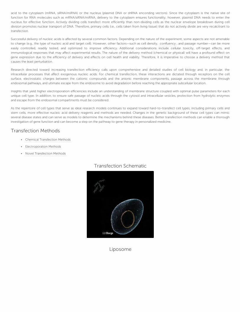
Transfection Schematic
Liposome
confluency

Cell Confluency
Transfection MethodsThe two main avenues for non-viral delivery of nucleic acids are chemical transfection and electroporation. An increased knowledge of cell and
membrane biology, expansion of chemical libraries, and the introduction of new instruments will also aid the development of novel methods for
transfection.
Chemical Methods for Transfection
Chemical transfection methods typically rely on electrostatic interactions to bind nucleic acid and to target cell membranes. These
methods utilize compounds such as calcium phosphate, polycations (e.g., ), and liposomes, as well as more current
technologies such as cationic lipids, polymers, dendrimers, and nanoparticles, to name a few.
represents the oldest and most inexpensive chemical method for transfecting nucleic acids. This technique
utilizes a solution of HEPES-buffered saline (HeBS) that contains phosphate ions (e.g., Na3PO4), which, upon addition of CaCl2, forms
a fine precipitate that binds DNA. The resulting complexes are added to cells, attach to the cell surface, and are taken up through
endocytosis. Additional modifications to the calcium phosphate method that can achieve higher efficiencies include glycerol shock and/or
chloroquine treatment . Despite the simple and cost-effective nature of the calcium phosphate method, it is ineffective for hard-to-transfect cells, is
very sensitive to changes in pH, often lacks reproducibility, and requires large quantities of DNA. These disadvantages necessitate more robust methods
for nucleic acid delivery.
More efficient transfection methods use organic compounds such as liposomes, charged lipids, and polymers. Liposomes can encapsulate DNA and
requires extrusion, a method for forming lipid bilayers by forcing lipids through a membrane filter, or lipsomes can coat the nucleic acid and trap it
between positively charged liposomes and does not require extrusion. Charged lipids and polymers form complexes with nucleic acids to generate
lipoplexes and polyplexes, respectively. Additionally, lipid, polymer, and nucleic acids can combine to form a lipopolyplex. All of these complexes exhibit a
net positive charge after binding to DNA, which enables association with the cell membrane through electrostatic interactions. Cellular uptake occurs via
endocytosis.
DEAE-dextran
Calcium phosphate
Liposome-mediated techniques were first introduced by Felgner and have become some of the most widely studied non-viral approaches for nucleic

Types of Vesicles
Despite the proven utility of cationic liposomes as nucleic acid delivery vehicles, lipofection can be quite toxic to cells, an obvious deterrent for many
researchers. Lipofection-mediated toxicity has been demonstrated through cytotoxicity and cell proliferation assays, including those that measure cell
morphology, mitochondrial activity, DNA synthesis, and cell viability . Transfection-mediated toxicity usually leads to changes in gene expression, even
in the absence of any visual toxicity. Therefore, it is important to choose a transfection method that can mitigate both observed as well as non-apparent
cellular toxicity.
Cationic lipids and polymers also form complexes with DNA through electrostatic interactions. However, lipoplexes and polyplexes cause less toxicity
than liposomal formulations. A few well-characterized cationic lipids used to form lipoplexes include:
,
,
, and
is a neutral helper lipid often used in conjunction with cationic lipids because of its membrane-destabilizing
effects at low pH, which facilitates endosomal escape.
Cationic polymers commonly used for transfection include and . These polymers condense DNA into
positively charged particles that can bind to the anionic cell surface. Subsequent uptake into the cell also occurs via endocytosis.
After cellular uptake, successful transfection requires release of the nucleic acid from the endosome to avoid degradation, and localization to the
cytoplasm (for RNA) or to the nucleus (for DNA). Various strategies have been used to promote endosomal release. Chemicals, such as chloroquine, or
the intrinsic ability of certain cationic polymers can prevent acidification of the endosome by absorbing protons through a process known as the ‘proton
sponge’ effect . As protons are absorbed, an influx of chloride ions and water causes osmotic swelling and eventual lysis of the endosome, which
promotes the release of its contents.
Following endosomal release, the final localization of the transfected nucleic acid depends on properties of both the nucleic acid and the transfection
reagent. Cytoplasmic delivery is sufficient for mRNA and siRNA or miRNA. In the cytoplasm, mRNA can access ribosomes for translation, and siRNA or
miRNA can associate with the RNA-induced silencing complex (RISC) in order to target endogenous RNA sequences for gene silencing. In contrast to
RNA molecules, plasmid DNA must enter the nucleus in order for transcription to occur. The nuclear envelope presents a huge barrier that may be
circumvented by natural nuclear envelope breakdown and reformation during cell division. Nuclear localization sequences (NLS) or targeting ligands have
been used to promote localization to the nucleus but without consistent, reproducible efficacy.
acid delivery. The spherical liposome structures contain hydrophilic and hydrophobic regions imparted by the lipid polar head and hydrophobic tail groups,
respectively. The amphipathic properties of these lipids lead to the spontaneous formation of bilayer structures that make up the basic structure of
liposomes. For recruitment to the cell, liposomes must have a slight net positive charge. Their sizes can vary from smaller unilamellar vesicles of 20–200
nm to larger vesicles of 200 nm–1 µμm, and upwards of 1 µμm for giant unilamellar vesicles. The latter can also exist as multilamellar vesicles, consisting of
multiple lipid bilayers. Ultimately, size is dictated by the thermodynamic properties of the lipid used in the system.
1,2-di-O-octadecenyl-3-trimethylammonium propane (DOTMA)
[N-[1-(2,3-dioleoyloxy)propyl]-N,N,N- trimethylammonium methyl sulfate] (DOTAP)
3β[N-(N’,N’-dimethylaminoethane)-carbamoyl] cholesterol (DCChol)
dioctadecylamidoglycylspermine (DOGS).
Dioleoylphosphatidylethanolamine (DOPE)
polyethylenimine (PEI) polylysine (ε-poly-L-lysine)

Calcium Phosphate Method
Electroporation
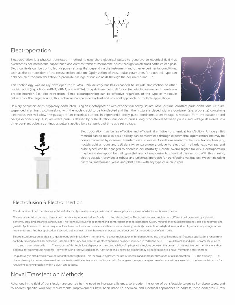
Electroporation is a physical transfection method. It uses short electrical pulses to generate an electrical field that
overcomes cell membrane capacitance and creates transient membrane pores through which small particles can pass.
Electrical fields can be controlled via pulse settings that depend on the instrument and other experimental conditions,
such as the composition of the resuspension solution. Optimization of these pulse parameters for each cell type can
enhance electropermeabilization to promote passage of nucleic acids through the cell membrane .
This technology was initially developed for in vitro DNA delivery but has expanded to include transfection of other
nucleic acids (e.g., oligos, mRNA, siRNA, and miRNA), drug delivery, cell-cell fusion (i.e., electrofusion), and membrane
protein insertion (i.e., electroinsertion). Since electroporation can be effective regardless of the type of molecule
delivered or the target source, this technique can provide a robust and universal approach for multiple applications.
Delivery of nucleic acids is typically conducted using an electroporator with exponential decay, square wave, or time-constant pulse conditions. Cells are
suspended in an inert solution along with the nucleic acid to be transfected and then the mixture is placed within a container (e.g., a cuvette) containing
electrodes that will allow the passage of an electrical current. In exponential-decay pulse conditions, a set voltage is released from the capacitor and
decays exponentially. A square-wave pulse is defined by pulse duration, number of pulses, length of interval between pulses, and voltage delivered. In a
time-constant pulse, a continuous pulse is applied for a set period of time at a set voltage.
Electroporation can be an effective and efficient alternative to chemical transfection. Although this
method can be toxic to cells, toxicity can be minimized through experimental optimization and may be
counterbalanced by increased transfection efficiencies. Conditions similar to chemical transfection (e.g.,
nucleic acid amount and cell density) or parameters unique to electrical methods (e.g., voltage and
pulse types) can be changed to decrease cell mortality. Despite overall higher toxicity, electroporation
may be a viable option for cell types that are not responsive to chemical transfection. With this in mind,
electroporation provides a robust and universal approach for transfecting various cell types—including
bacterial, mammalian, yeast, and plant cells—with any type of nucleic acid.
Electrofusion & Electroinsertion
The disruption of cell membranes with brief electrical pulses has many in vitro and in vivo applications, some of which are discussed below.
The use of electrical pulses to disrupt cell membranes induces fusion of cells , i.e., electrofusion. Electrofusion can combine both different cell types and cytoplasmic
contents, including organelles and nuclei. This technique involves alignment and compression of cells, membrane fusion, maturation of fused membranes, and cell recovery and
growth. Applications of this technique include fusion of tumor and dendritic cells for immunotherapy, antibody production via hybridomas, and fertility or animal propagation via
nuclear transfer. Another application is somatic cell nuclear transfer between an oocyte and donor cell for the production of stem cells.
Electroinsertion uses electrical charges to transiently break down membranes to allow implantation of foreign proteins into the cell membrane. Potential applications range from
antibody binding to cellular detection. Insertion of extraneous proteins via electroporation has been reported in red blood cells , multilamellar and giant unilamellar vesicles
, and mammalian cells . The success of this technique depends on the compatibility of hydrophobic regions between the protein of interest, the cell membrane and on
potential for autoimmune response. However, with effective application, fully functional and stable proteins may be integrated into a novel membrane environment.
Drug delivery is also possible via electroporation through skin. This technique bypasses the use of needles and improper absorption of oral medication . The efficacy of
chemotherapy increases when used in combination with electroporation of tumor cells. Some gene therapy strategies use electroporation across skin to deliver nucleic acids for
regulating gene expression within a given target tissue.
Novel Transfection Methods
Advances in the field of transfection are spurred by the need to increase efficiency, to broaden the range of transfectable target cell or tissue types, and
to address specific workflow requirements. Improvements have been made to chemical and electrical approaches to address these concerns. A few

examples are highlighted in this section.
One modification to chemical transfection is the implementation of to increase transfection efficiency . Nucleic acid
complexes are formed with magnetic nanoparticles, and a magnetic field is applied to bring the complexes into close proximity with the target cell. The
complexes are then taken up via endocytosis as in chemical transfection. This approach is thought to increase transfection efficiency by bringing
complexes closer to their cellular targets.
Another interesting approach is the biolistic particle delivery system, more commonly referred to as a gene gun . This method was first introduced for
transfection of plant cells but has since been expanded for use in various organisms, such as bacteria, yeasts, and mammalian cells—particularly hard-to-
transfect cells, e.g., primary cells. In addition, specific organelles (e.g., chloroplasts and mitochondria) can also be targeted. Biolistic delivery works by
binding complexes of nucleic acids to metal atoms, such as tungsten or gold, and accelerating the nucleic acid/metal complex through a vacuum for
delivery to a target cell or tissue. This approach can also deliver DNA vaccines in vivo for gene therapy. This biolistic delivery approach can save time by
circumventing cellular endosomal processes and can make the gene product readily available within the cell.
Continuous flow electroporation (CFE) was developed for optimal process workflow and high-throughput applications. In CFE, cells are mixed with the
compound to be delivered (e.g., nucleic acid or therapeutic agent) and allowed to flow through an electroporation chamber. As the mixture of cells and
compound pass through the chamber in a continuous stream, they are uniformly subjected to an electrical field, which provides for consistent delivery of
the compound and allows for uniform transfection of a large number of cells.
Three-dimensional (3D) tissue culture and transfection has been developed for experiments that more
accurately mimic normal cell morphology and functionality. 3D culture methods are generally categorized as
solid structural scaffolds or hydrogels of varying composition that provide structural support more akin to in
vivo tissue. Chemical transfection can be used in this context to study differentiation, tissue generation, cancer
invasion and migration, and drug target screening.
With a burgeoning interest in transfection, particularly for therapeutic purposes, a wide variety of new
transfection methods and developments are expected. These advances will most certainly be prompted by a
need to address new targets, increase transfection efficiency, and expand workflow. Ultimately, these developments will provide better avenues to study
existing and new disease states, leading to improved treatments.
Magnetic-Mediated Delivery
magnetic-mediated delivery
Applications for TransfectionTransfection enables a wide variety of applications in addition to transient gene expression including:
Gene silencing
Stable cell line generation
Virus production
Large-scale protein production
Stem cell reprogramming and differentiation
It is important to consider both the efficiency of transfection and the level of cytotoxic effects when a transfection methodology or reagent is selected.
Higher transfection efficiencies favor greater success whether the goal is to effectively knock down gene expression, create a stable cell line, increase
virus titers, achieve maximum protein yields, or address stem cell research. However, a low-toxicity approach must not be overlooked, since highly
cytotoxic approaches can lead to unwanted effects in the form of visible morphology changes, as well as unknown changes in gene expression or stress-
response pathways.
EXOGENOUSSEQUENCES OF siRNAsand miRNAs...can be designed and introduced into
cells through transfection to knock
down relevant gene expression.
Gene Silencing
The ability to silence genes plays an important role in molecular and cell biology and can be readily applied through transfection. Gene expression can be
effectively reduced or eliminated by introducing small noncoding RNA molecules that inhibit RNA translation though a process termed RNA interference
(RNAi). RNA molecules that take part in RNAi pathways include: (i) small interfering RNAs (siRNA), short (20–25 base pairs) double-stranded RNAs; and (ii)
microRNAs (miRNAs), a separate class of short single-stranded RNAs (20–22 nucleotides). RNAi-based approaches rely on the inherent cellular
machinery, shared among several eukaryotic organisms, to inhibit mRNA translation. RNAi pathways play in important role in regulating gene expression
and are also believed to provide a mechanism to protect cells from extraneous nucleotide sequences (e.g., viruses and transposons).
RNAi pathways are elicited through cleavage of double-stranded RNAs (dsRNAs) to produce siRNAs, or by processing of noncoding RNAs to produce
miRNAs. These separate RNAi pathways rely on cellular machinery such as the ribonuclease protein DICER and the RNA-induced silencing complex
(RISC). DICER initiates the RNAi pathway by processing dsRNA to form siRNAs or mature miRNAs. These RNA molecules can bind to complementary
sequences of mRNA within the RISC, and the mRNA can be cleaved by the catalytic component, Argonaute, which ultimately prevents translation.
Apart from this natural occurrence, exogenous sequences of siRNAs and miRNAs can be designed and introduced into cells through transfection to
knock down relevant gene expression. This application serves as a tool to elucidate genetic pathways, determine protein function, or uncover new gene
targets for biotherapeutic and pharmaceutical applications. Larger libraries of these RNA molecules are also available to perform larger genome RNAi
analysis via high-throughput screening.
Because siRNA and miRNA differ in size and structure from plasmid DNA, transfection reagents can be
optimized and formulated separately for delivery of these RNA molecules. In addition, delivery of siRNA and
miRNA to the cytoplasm for incorporation into the RISC complex is sufficient for gene knockdown. Selection
of the appropriate transfection methodology or reagent must first be considered, followed by further
optimization for efficient siRNA and miRNA delivery and subsequent gene knockdown.
An alternative approach that takes advantage of RNAi to knock down gene expression is the use of short
hairpin RNA (shRNA). These short RNA sequences can be expressed via viral or non-viral vectors. shRNA
expression mimics a pathway similar to siRNA/miRNA since the expression product must be processed by
DICER and ultimately incorporated into the RISC complex for targeted degradation of mRNA.
Although gene silencing via siRNA/miRNA and shRNA rely on similar RNAi pathways, the optimal method may depend on factors such as cell type, time
constraints and transient vs. stable expression. A high and reliable level of knockdown can be achieved through siRNA transfection via a variety of quality
transfection reagents and validated siRNA libraries. However, disadvantages with siRNA mediated knockdown are the chances of off-target effects and
transient knockdown through siRNA dilution after multiple cell divisions. Alternatively, the use of shRNA can be carried out to generate stable knockdown
cell lines, but this approach can be time-consuming, and cell types such as primary cells may yield lower transfection efficiencies through shRNA plasmid
based transfection as opposed to siRNA delivery.
Another vehicle for shRNAs is viral transduction through adeno-associated virus (AAV), adenovirus and lentivirus. Expression through AAV or adenovirus
can decrease the chance of insertional mutagenesis since these vectors are more likely to remain episomal, but this approach leads to more transient
expression since the vectors are lost through multiple rounds of cell division. Lentivirus provides a stable solution through chromosomal integration, but
this also presents the risk of insertional mutagenesis.
Regardless of whether shRNA is implemented via viral or non-viral methods, transfection plays a role to facilitate gene silencing via this approach. Non-
viral vectors can be introduced via chemical reagents optimized for plasmid transfection, or they can be delivered via electroporation. For viral based
Mirus Transfectopedia™

approaches, virus generation can be carried out via transfection (see Virus Production). Under either delivery, shRNA mediated knockdown can provide a
more stable means of RNAi than siRNA/miRNA with less turnover.
Whether shRNA, siRNA or miRNA mediated RNAi approaches are implemented, the overall goal of the experiment needs to be established. One must
also consider the gene targets and cell types used, design the proper sequence for specificity, determine duration of expression and select the most
effective means of delivery to ensure success.
For more information on transfection for RNAi applications, additional resources can be found on the Mirus Bio website:
Optimize siRNA Transfection
Deliver microRNA (miRNA) Effectively
Reverse Transfection Protocol for siRNA/miRNA
siRNA Mediated Pathway
miRNA Mediated Pathway
shRNA Mediated Pathway
Generation of Stable Cell Lines
The creation of a stable cell line provides many advantages over transient gene expression, such as the elimination of the need
for repeated transfections, establishment of more uniform gene expression within a cell population, and provision of a system for
long-term experiments. These experiments may include prolonged expression of a target gene or permanent silencing of a
particular gene.
To generate a stable cell line, introduced DNA must integrate into the host genome, which can be a rare event. This process
requires patience and diligence in addition to quality transfection systems. Creating a stable cell line requires the following:

STABLE CELL LINEGENERATION...eliminates the need for repeated
transfections, establishes a more
uniform gene expression within a cell
population, and provision of a system
for long-term experiments.
Delivery to the cell
Inclusion into the nucleus
Integration into the chromosome
Integration into the chromosome is typically accomplished through the inclusion of a homologous DNA sequence that flanks the inserted DNA element.
Very high transfection efficiency is required in order to maximize the chance of a rare chromosomal
integration event. Additionally, a positive selection marker such as an antibiotic (e.g., hygromycin, neomycin, or
zeocin) is required for proper selection of a stable cell line expressing the gene of interest. The use of a
linearized plasmid can also increase the likelihood of integration of the gene of interest.
Following multiple rounds of selection to isolate cells with the integrated plasmid containing the drug
resistance gene and the gene of interest, a mixed population of drug-resistant cells can be used directly for
further clonal isolation. The use of a mixed population, or batch culture, saves time but suffers from the
disadvantage of using an undefined and genotypically mixed culture. A clonal isolate can be generated using
serial dilution plating to isolate single cells or by picking individual colonies with rings or pipettes. The selection
process is then applied to these clonal isolates to verify integration.
Final verification of the stable cell line typically includes an assay to verify expression of the gene of interest. These methods include detection through
Western blot and sequencing of genomic DNA to verify integration.
More details on stable cell line generation can be found at our Tips from the Bench: Transfection Tip – Generate Stable Cell Lines.
VIRUSES PROVIDE...an alternative to nucleic acid delivery
when a specific cell type is refractory
to chemical transfection or
electroporation methods… regardless
of the application, transfection is a
helpful tool that is commonly used to
create functional viruses.
Virus Production
Virus applications from basic virology research to viral transduction and gene therapies all require the ability to develop high
titers of functional virus. Recombinant viruses can be generated to study all aspects of virology including viral life cycles,
structure, and mode of infection. Viruses provide an alternative to nucleic acid delivery when a specific cell type is refractory to
chemical transfection or electroporation methods. Gene therapies have also called upon viral vectors to introduce genes into
cells in vivo. Regardless of the application, transfection is a helpful tool that is commonly used to create functional viruses.
Whether the virus is needed for viral transduction purposes or for the study of the
virus itself — its life cycle,structure, or health risk, an essential requirement is the
generation of a functional virus in sufficient amounts. This process entails delivery of
genetic material in the form of DNA or RNA that contains the blueprint for the viral life cycle, including
replication, packaging, and — in some cases — genomic integration. Viral transduction is still a commonly used
methodology to deliver genetic material into cells when the following apply:
Viral transduction is not a safety or regulatory concern.
Cells are refractory to other nucleic acid delivery methods (e.g., chemical transfection or
electroporation).
Higher transfection efficiencies are necessary compared to those afforded by chemical transfection.
Virus Production Process
1. Nucleic Acid Preparation
2. Transfection
3. Harvest
4. Titer
Commonly Used Viruses and Applications
Virus Nucleic Acid Characteristics Examples
Retrovirus RNA
- Integrative (random)
- Replication-competent or replication-defective
- Requires actively dividing cells for infection
MMLV – Moloney murine leukemia virus
Lentivirus RNA - Integrative (random)
- Can infect nondividing cells
- HIV – Human immunodeficiency virus
- SIV – Simian immunodeficiency virus
- FIV – Feline immunodeficiency virus
Various; depend on host and type
Transient transfection is part of the general workflow for the production of most viruses, but the details will be specific to each type of virus produced.
Stable cell lines can also be generated to actively produce viral vectors that are not included in the Table 1. The following key steps are characteristic of
virus generation through transient transfection:
Nucleic Acid Preparation » Transfection » Viral Harvest » Titer
1. Nucleic Acid Preparation
In most cases, it is necessary to co-transfect two or more plasmids—the vector plasmid and one or more helper plasmids. The vector plasmid
contains the gene of interest designed for homologous recombination; the helper (i.e., packaging) plasmid(s) encodes the necessary structural,
regulatory, and replication genes to produce functional virus. Together, these plasmids will generate a virus that can transduce the gene of interest
for subsequent genomic integration.
2. Transfection
Multiple plasmids can be transiently transfected through the use of calcium phosphate or chemical transfection reagents. Calcium phosphate
provides an economical means for generating virus in this manner but can also exhibit low reproducibility and low virus titer yields. Chemical
transfection reagents are employed for more consistency and higher virus titers.
A packaging cell line is also required for generating the viral vector. In many cases, the human embryonic kidney cell line, HEK 293, or derivatives
such as HEK 293T or HEK 293A, can be suitable hosts since they readily expresses a variety of viral genes. HEK 293 cells can be transfected readily,
and—in the cases of the derivative cell lines—may be deficient in packaging virus without the use of a helper plasmid (e.g.,HEK 293T for retrovirus)
or may produce a necessary viral protein (e.g., HEK 293A for adenovirus).
When using chemical transfection reagents for DNA delivery, the total amount of DNA required from all plasmid sources must be considered in
order to determine the optimal amount of reagent for high-efficiency transfection.
For example, if a 3:1 ratio of µμl of transfection reagent per µμg of DNA is optimal for high-efficiency transfection in the packaging cell line, and 3 µμg
of vector plasmid and 1 µμg of helper plasmids are necessary (i.e., 4 µμg of DNA total), then a total of 12 µμl of reagent will be needed for efficient
transfection. Complex formation is facilitated by incubation of the reagent and DNA for 15 to 30 minutes. Complexes are then added directly to
cells for transfection
3. Harvest
After transfection, harvest of the virus can vary depending on the virus produced and the protocol followed. Protocols can require multiple harvests
of supernatant and medium changes within 24 to 48 hours after transfection. Harvests are typically filtered, aliquoted, and stored at –80°C.
4. Titer
It is generally recommended to titer the virus in order to determine the effective concentration for infection. Viral titers are determined by
infecting cell stocks with serial dilutions of the stock virus. After infection, most viral vectors contain a fluorescent reporter to quantify infection
through flow cytometry analysis.
Viral transduction provides a powerful and effective means to deliver nucleic acids into a variety of cell types, including primary cells. However, in
order to produce virus, introduction of viral genes into a packaging host is still necessary. Transfection provides a robust and reproducible means to
generate high-titer yields in contrast to calcium phosphate.
For more information on the use of transfection for high-titer virus production, additional resources can be found on the Mirus Bio website:
TransIT® Reagents for High-titer Virus Production
High-Titer Production of Recombinant Lentivirus in HEK 293FT Cells: Calcium phosphate vs. Lipopolyplex Transfection VIEW PDF
Large-scale Protein Production
Adenovirus DNA- Non-integrative
- Requires actively dividing cells for infection
- HAdV-B and C (Human respiratory disease)
- HAdV-B and D (Human conjunctivitis)
- HAdV-F types 40 and 41 (Human gastroenteritis)
- CadV-2 (Canine adenovirus)
- Equine adenovirus 1 (Horse adenovirus)
Adeno-associated
viruses (AAV)DNA
- Integrative (wildtype is site-specific)
- Can infect nondividing cells
- Nonpathogenic
AAVS1 (wildtype integrates at chromosome 19)

EFFECTIVE USE OFTRANSIENTTRANSFECTION...for large-scale protein production
requires a reagent that can transfect in
multiple media formulations, can be
effectively scaled up to transfect larger
volumes, and can reproducibly
generate large amounts of protein.
Transient transfection can be used in mammalian cell-culture systems to generate high yields of functional
protein for biotherapeutic applications. Mammalian cell lines, such as Chinese hamster ovary (CHO) and human
embryonic kidney (HEK 293) cells, are attractive hosts for biotherapeutic protein production because they
provide the correct post-translational modifications for biologically relevant use. In addition, these cell lines are
well characterized and are already amenable to Food and Drug Administration (FDA) clearance.
During the cumbersome process of generating a suitable stable cell line for biotherapeutic protein production,
which can take several weeks or even months, large amounts of protein can be produced from suspension
CHO or HEK 293 cells via transient transfection. Effective use of transient transfection for large-scale protein
production requires a reagent that can transfect in multiple media formulations, can be effectively scaled up to transfect larger volumes, and can
reproducibly generate large amounts of protein. In addition, the formulation of such a reagent should be animal product-free to address any Food and
Drug Administration (FDA) regulatory concerns.
One key consideration in such large-scale applications can also be cost. Linear PEI is a seemingly cost-effective
option for carrying out large-scale transfections in suspension CHO and HEK 293 cells. However, this
compound is incompatible with multiple media formulations and is not as effective at generating high protein
yields as other transfection reagents. Low protein yields will increase the cost of protein production because of
the time, wages, and materials associated with producing comparable protein amounts.
For more information on large-scale protein production in suspension cells, additional resources can be found on the Mirus Bio website:
Selecting a Transfection Reagent for Large Scale Protein Production in Suspension 293 Cell Types VIEW PDF
Maximize Protein Expression in CHO Suspension Cells VIEW PDF
Stem Cell Applications
Stem cells hold the promise of revolutionizing therapy for a myriad of diseases. Biologically relevant models can be generated through reprogramming
and differentiation. These techniques can be accomplished via the introduction of transcription factors through the use of small molecules, recombinant
virus transduction, or transfection of proteins or nucleic acid. Transfection provides a nonviral approach that is more efficient than protein transfection
and safer than the use of viruses for infection.
Entry Points for Transfection
Entry Points for Transfection
Transfection can be used at many points throughout the course of a reprogramming or stem cell differentiation experiment. Transfection of nucleic
acids, such as plasmid DNA, mRNA, and siRNA or miRNA, serve as vital tools in stem cell reprogramming and differentiation. Adult fibroblast cells can be
transfected or transduced via several methods (e.g., recombinant virus, plasmid, protein, mRNA, small molecules, or miRNA) with a combination of
transcription factors, including KLF4, SOX2, c-MYC, NANOG, OCT-4, or LIN-28, to reprogram the cells to a pluripotent state. Induced pluripotent stem

(iPS) cells can then be differentiated to a myriad of cell types through growth factor addition and/or transfection of selection markers driven by cell type-
specific promoters. Cell types derived from stem cells—such as cardiomyocytes, adipocytes, neural cells, pancreatic beta cells, and hematopoietic
progenitor cells—provide researchers with relevant models for their experiments.
For more information and examples highlighting transfection for stem cell applications, additional resources can be found on the Mirus Bio website:
From Reprogramming to Differentiation – Transfection Applications for Stem Cell Research VIEW PDF
High Efficiency Transfection of iCell® Cardiomyocytes (Cellular Dynamics International, Inc.) and Stem cell Relevant Sources VIEW PDF
Optimized transfection protocol for iCell® Cardiomyocytes (Cellular Dynamics International, Inc.) using TransIT-TKO Transfection Reagent: VIEW PROTOCOL PDF

Mirus Bio Transfection SolutionsOur focus: Your solution.
Every transfection product offered by Mirus Bio has been developed by an in-house team of organic chemists, molecular biologists, cell biologists, and
biochemists. Mirus Bio scientists continue to develop state-of-the-art, nonviral, self-assembling complexes (transfection complexes) that serve as DNA or
RNA carriers. These complexes are a mixture of a nucleic acid (DNA or RNA) with the carefully selected, appropriate transfection formulation to provide
highly efficient and reproducible delivery into a wide variety of cells. The delivery mechanism mimics virus delivery in several key ways, including active
transport into the cell (endocytosis) and endosomal destabilization and release.
All Mirus Bio products are developed and manufactured in our Madison, Wisconsin laboratories.
Lipid and Polymer Transfection of DNA
Mirus Bio offers a very comprehensive, selection of non-liposomal TransIT® Transfection Reagents. These reagents are based on blends of polymers
and/or lipids developed for DNA transfection with minimal toxicity and are all compatible with serum-containing media. TransIT Transfection Reagents
have been developed for the following applications: transfection of both DNA and/or siRNA in multiple cell types; broad-spectrum delivery into a wide
variety of cells; 3D cell culture formats; and increased protein production. The TransIT product line also includes a panel of cell type-specific DNA delivery
reagents.
TransIT-X2® for Transfection of DNA and siRNA into Multiple Cell Types
For optimal delivery of either DNA or siRNA into a variety of cell types, Mirus offers the TransIT-X2® Dynamic Delivery System.
This novel formulation relies on a nonliposomal, polymeric chemistry system for efficient delivery of nucleic acids through
cellular endosomal processes. The TransIT-X2® Dynamic Delivery System provides multiple advantages over outdated
liposomal methods, including better DNA delivery, more efficient delivery of siRNA, and less toxicity across multiple cell types.
TransIT® Reagent for Broad-spectrum DNA Delivery
Mirus offers two reagents, TransIT®-LT1 and TransIT®-2020 , for plasmid DNA delivery into a wide variety of cell types.
TransIT-LT1 has been Mirus Bio's flagship reagent since 1996 and offers a Low Toxicity solution for all nucleic acid delivery needs. More recently, TransIT-
2020 was developed for increased performance across a multitude of cell types including hard-to-transfect cell types such as primary and stem cells.
For high-throughput applications involving DNA transfection, TransIT®-Express provides a broad-spectrum reagent optimized for 96-well plate
applications. This reagent provides the same low cellular toxicity and high efficiency as other TransIT reagents.
TransIT® Reagent for 3D Cell Culture
The 3D Transfection System , which contains the TransIT®-3D Transfection Reagent and alvetex® 12-well tissue culture plates (Reinnervate, UK),
enables DNA delivery to cells grown in 3D cell culture. The TransIT-3D Transfection Reagent is a broad-spectrum transfection reagent optimized for
plasmid DNA delivery into cells grown in 3D matrices. alvetex tissue culture plates consist of a 3D polystyrene scaffold that provides a 3D environment in
which cells can migrate, proliferate, and differentiate.
3D culture methods are generally categorized as solid-structure scaffolds, such as alvetex tissue culture plates, or hydrogels of varying composition.
TransIT-3D Transfection Reagent can also be used for transfecting cells in hydrogel matrices. 3D cell culture provides an environment more similar to in
vivo conditions than do traditional two-dimensional (2D) plate formats by providing a support structure for greater cellular organization. In 3D culture
systems, cells can maintain their 3D morphology and form complex interactions with adjacent cells to transmit and receive signals.
TransIT® Reagent for Protein Production
The Transfection Experts at Mirus developed the TransIT-PRO® Transfection Kit to address the need to increase protein production for use in
biotherapeutics. The TransIT-PRO Transfection Kit consists of a DNA transfection reagent and optional boost reagent combination specifically developed
Mirus Transfectopedia™

for mammalian protein production in suspension HEK 293- and CHO-derived cells. The PRO Boost Reagent enhances
expression in certain media formulations. TransIT-PRO Transfection Reagent and PRO Boost Reagent are comprised of animal
origin-free components that are compatible with many chemically defined media formulations. The TransIT-PRO Transfection
Kit eliminates the need for a culture medium change after transfection and is suitable for both transient and stable
transfections.
TransIT® Reagent for Cell Line–Specific Transfection
With almost two decades of gene delivery experience, The Transfection Experts at Mirus have amassed a large collection of cell type-
specific reagents with all of the same low-toxicity and high-performance qualities of the broad-spectrum reagents. These reagents are
suitable for transfection of the most common cell types:
TransIT®-293 Reagent for HEK 293 cells
TransIT-HeLaMONSTER® Kit for HeLa cells
TransIT®-CHO Kit for CHO cells
Additionally, a variety of other reagents are available to address groups of commonly used but hard-to-transfect cell lines, including:
TransIT®-Insect Transfection Reagent for transient transfection and baculovirus production in insect cells, including Sf9, High Five™, and
Drosophila S2
TransIT®-BrCa Transfection Reagent specifically for breast cancer cell types, including MCF-7, MDA-MB-231, MDA-MB-453, MDA-MB-468,
and T47D
TransIT®-Jurkat Reagent for Jurkat and other hard-to-transfect cells
TransIT®-Keratinocyte Reagent for delivery into keratinocytes, such as NIKS (Near-Diploid Immortalized Keratinocytes)
TransIT-Neural® Reagent for various neural cell types, including SK-N-MC, Neuro-2a, DI-TNC1, and human astrocytes
Lipid and Polymer Transfection of RNA and Oligos
Mirus Bio also offers TransIT Transfection Reagents for RNA and oligonucleotide (oligo) delivery into any cell type. These reagents address delivery of
large RNA, siRNA/miRNA, and short oligos with minimal toxicity and are all compatible with serum-containing media. These reagents are based on blends
of polymers and/or lipids suited for broad-spectrum delivery applications.
TransIT® Reagent for Large RNA (e.g., mRNA and viral RNA)
The TransIT®-mRNA Transfection Kit delivers large RNA directly to the cytoplasm for expression, which avoids transcriptional regulation effects. The
TransIT-mRNA Transfection Kit can be used to deliver a variety of RNA molecules, including mRNAs and viral RNAs (2–10 kb). This kit can be used for
multiple applications, such as short-term protein expression, viral production, and replication studies.
The TransIT-mRNA Transfection Kit contains two components: TransIT-mRNA Reagent and the mRNA Boost Reagent. This kit is compatible with serum-
containing media; transfection efficiency is optimal when transfections are performed in the presence of serum, with no medium change required.
TransIT® Reagent for siRNA and miRNA
TransIT-TKO® Reagent and TransIT-siQUEST® Reagent are broad-spectrum siRNA transfection reagents that enable high efficiency siRNA delivery
and knockdown of target gene expression in many cell types, including primary cells. TransIT-TKO was introduced in 2001 and was the first commercially
available siRNA transfection reagent. Cotransfection of siRNA and DNA is also feasible with TransIT-TKO and has been tested across a variety of cell types.
TransIT-siQUEST was later developed to further supplement the number of cell lines that can be transfected with siRNA or miRNA. Transfections with
either TransIT-TKO or TransIT-siQUEST does not require medium changes and can be carried out in serum-containing medium. Each unique formulation
provides high-efficiency, broad-spectrum siRNA delivery.
TransIT® Reagent for Oligos
The TransIT®-Oligo Transfection Reagent provides maximal transfection performance for DNA and RNA oligonucleotides while maintaining low cellular
toxicity. Oligonucleotides tested include:
phosphodiester DNA
phosphothioate DNA (ssDNA)
phosphothioate RNA (sRNA)
2'-OMe RNA and 2'-OMe RNA/sDNA chimerics

Morpholino/DNA duplexes
TransIT-Oligo provides all the advantages of the trusted TransIT series of transfection reagents: high transfection efficiency, low toxicity, serum
compatibility, simplicity of use and reproducibility.
Transfection Solutions
Transfection of DNA
Transfection of DNA and siRNA
Broad-spectrum DNA Delivery
3D Cell Culture
Protein Production
Cell Line–Specific Transfection
Transfection of RNA and Oligos
Large RNA (e.g., mRNA and viral RNA)
siRNA and miRNA
Oligos
Protocols
TransIT® Products
Electroporation
Protocols
TransIT® Products
All TransIT® Transfection Reagents and Kits provide high performance and low toxicity. Moreover, they are all serum-compatible, which eliminates the
need for a medium change. This simplicity enables a more streamlined, user-friendly protocol that leads to faster results.
A complete protocol is available for each TransIT product. Each protocol is divided into the following sections:
Introduction: brief summary of specific product
Specifications: storage and warranty details
Materials:
Contents provided within the product package
Materials required but not supplied
Before You Start: suggestions to consider to ensure successful transfections
Seeding densities and cell confluency at transfection
Volumes of reagent and boost reagent (if applicable)
Nucleic acid considerations
Complex formation conditions
Cell culture conditions
Presence of antibiotics
Post-transfection incubation times
Transfection Protocol: specific transfection protocol details
Experimental set-up, including cell culture considerations
Table of recommended starting conditions for multiple plate formats
Troubleshooting Guide: recommendations to help troubleshoot problems
Related Products: list of related products
Reagent Agent: information regarding the online transfection database
Contact Information
This section of the Mirus Transfectopedia™ provides an overview of the TransIT products, including schematic diagrams of workflows and a
product decision tree . Please refer to the individual product protocols for more detailed experimental conditions.
The following TransIT products and associated protocols are available for chemical transfection:
DNA and siRNA/miRNA
TransIT-X2™ Dynamic Delivery System
DNA
Broad Spectrum
TransIT®-2020 Transfection Reagent
TransIT®-LT1 Transfection Reagent
TransIT®-Express Transfection Reagent
3D Transfection
3D Transfection System
TransIT®-3D Transfection Reagent
Protein Production
TransIT-PRO® Transfection Kit
Cell Line-Specific
TransIT®-293 Transfection Reagent
TransIT®-Insect Transfection Reagent
TransIT®-BrCa Transfection Reagent
TransIT®-CHO Transfection Kit
TransIT-HeLaMONSTER® Transfection Kit
TransIT®-Jurkat Transfection Reagent
TransIT®-Keratinocyte Transfection Reagent
TransIT-Neural® Transfection Reagent
RNA
TransIT®-mRNA Transfection Kit
TransIT-TKO® Transfection Reagent
TransIT-siQUEST® Transfection Reagent
Oligo
TransIT®-Oligo Transfection Reagent


Electroporation
Mirus Bio has developed the Ingenio® Electroporation Solution and Kits to facilitate efficient and reliable delivery of nucleic acids to eukaryotic cells that
are traditionally resistant to chemical transfection. Ingenio is a broad-spectrum solution that supports high-efficiency electroporation with minimal
toxicity. It replaces standard electroporation solutions, including phosphate-buffered saline (PBS) and serum-free media. Ingenio is compatible with most
conventional instruments and facilitates a wide range of applications that require nucleic acid delivery to cells. The Ingenio solution is available both as a
single product and as part of a complete kit with cuvettes and cell droppers.
Protocols for Electroporation
Ingenio Kits and Solution are compatible with multiple conventional electroporation instruments including the Amaxa® Nucleofector® , Bio-Rad® Gene
Pulser, and Harvard-BTX® electroporators. Ingenio Kits and Solution can be used for electroporation with both exponential decay and square wave pulse
conditions. Product protocols address pulse conditions in addition to cell culture and nucleic acid considerations.
Each Ingenio protocol is divided into the following sections:
Introduction: brief summary of the specific product
Specifications: storage and warranty details
Materials:
Contents provided within the product package
Materials required but not supplied
Before You Start: suggestions to consider to ensure successful transfections
Cell density and passage number
Nucleic acid considerations for DNA and RNA

Optimization of pulse conditions
Exponential-decay wave form
Square wave form
Lonza-amaxa Nucleofector
Post-electroporation incubation times
Electroporation Protocol: specific transfection protocol details
Experimental set-up, including cell seeding considerations
Preparation of Ingenio Solution/nucleic acid/cell mixture
Electroporation details
Tables with recommended settings for particular cell types and instruments
Troubleshooting Guide: recommendations to help troubleshoot problems
Related Products: list of related products
Reagent Agent: information regarding the online transfection database
Contact Information

Web Resources
Mirus Solutions
An interactive module to identify the best Mirus Bio solution for your experiments. Follow each step of the flowchart by selecting the nucleic acid you are
transfecting, which will lead you to the most appropriate product offered by The Transfection Experts. Each listing will guide you to the respective
product web page for more information including data, protocols and how to sample for free.
ApplicationLearn more about specific application areas where Mirus Bio products are being used
Virus Production
Genome Editing Using CRISPR/Cas
Stem Cell Transfection
Large Scale Protein Production
Stable Cell Line Generation
High Throughput Transfection
Low Toxicity Transfection
Cotransfection
Tips From The Bench
Transfection Tips
Electroporation Tips
Nucleic Acid Labeling Tips
Mirus Literature and Videos
Technical Product Literature
Brochures
Reports and Papers
Posters and Presentations
Video Library
Citations Database
The Mirus Bio citations database has over 1600 published citations using Mirus' TransIT® in vitro electroporation and transfection reagents, on 492 unique
cell lines and primary cells. Search Citation Database
Reagent AgentTransfection Database
Reagent Agent is a tool designed to help you determine the best delivery solution for any nucleic acid into any cell type including hard-to-transfect cell
lines and primary cells.
Recommendations are based on extensive in-house transfection and electroporation data, customer feedback, and citations that are all continually
updated.
Search Reagent Agent
Mirus Transfectopedia™

Ordering and Contact Information
Customer Support and Product Orders
Phone: 888.530.0801 (Toll-free within the U.S.) or +1.608.441.2852
Sales Fax: +1.608.441.2849
Email: [email protected]
Web: Online Store
Product Technical Support
Phone: 888.530.0801 (Toll-free within the U.S.) or +1.608.441.2852
Email: [email protected]
General Questions
Email: [email protected]
International Distributors
International Distributor Contact Information
Mailing Address
Mirus Bio LLC
545 Science Dr.
Madison, WI 53711 USA
Customer Testimonial
"Excellent technical support, they are very knowledgeable."
Review published in Select Science®
He Song Sun
University of Toronto Scarborough
Cell and Molecular Biology
Mirus Transfectopedia™

IndexAdeno-associated viruses (AAV) 1 , 2 , 3
Adenovirus 1 , 2 , 3 , 4
Argonaute 1
Biolistic delivery 1 , 2 , 3
Broad-spectrum 1 , 2 , 3 , 4 , 5 , 6 , 7 , 8 , 9
c-Myc 1
Calcium phosphate method 1 , 2 , 3 , 4 , 5 , 6 , 7
Cationic compound 1 , 2
Cell density 1 , 2 , 3
Cell division 1 , 2 , 3 , 4
Cell types
CHO cells 1 , 2 , 3 , 4 , 5 , 6 , 7
HEK 293 cells 1 , 2 , 3 , 4 , 5 , 6
HEK 293A cells 1
HEK 293T cells 1
HeLa cells 1 , 2
Keratinocyte cells 1 , 2
Primary cells 1 , 2 , 3 , 4 , 5 , 6 , 7 , 8
Stem cells 1 , 2 , 3 , 4 , 5 , 6 , 7 , 8 , 9 , 10 , 11 , 12
Cellular toxicity/cytotoxic effect/cytotoxicity 1 , 2 , 3 , 4 , 5 , 6 , 7 , 8 , 9 , 10 , 11 , 12 , 13 , 14 , 15 , 16 , 17 , 18
Chemical transfection 1 , 2 , 3 , 4 , 5 , 6 , 7 , 8 , 9 , 10 , 11 , 12 , 13 , 14
Chloroquine 1 , 2
CHO cells 1 , 2 , 3 , 4 , 5 , 6 , 7
Confluency 1 , 2
Continuous flow electroporation (CFE) 1
DCChol 1
DICER 1 , 2
DNA vaccine 1
DOGS 1
DOPE 1
DOTAP 1
DOTMA 1
Double-stranded RNAs (dsRNAs) 1 , 2
Electrofusion 1 , 2
Electroinsertion 1 , 2
Electropermeabilization 1 , 2
Electroporation 1 , 2 , 3 , 4 , 5 , 6 , 7 , 8 , 9 , 10 , 11 , 12 , 13 , 14 , 15 , 16 , 17 , 18 , 19 , 20 , 21 , 22 , 23 , 24 , 25
Endocytosis 1 , 2 , 3 , 4 , 5 , 6
Mirus Transfectopedia™

Endosome/Endosomal pathway 1 , 2 , 3 , 4 , 5 , 6 , 7 , 8 , 9
Episomal 1
Exponential decay 1 , 2 , 3
Food and Drug Administration (FDA) 1 , 2
Gene therapy 1 , 2 , 3 , 4
Genome editing 1 , 2
Glycerol shock 1
HEK 293 cells 1 , 2 , 3 , 4 , 5 , 6 7
HEK 293A cells 1
HEK 293T cells 1
HeLa cells 1 , 2
Helper plasmid 1 , 2 , 3
HEPES-buffered saline (HeBS) 1
Homologous recombination 1 , 2
Hydrogel 1 , 2
Immune response/immunological response 1 , 2
Induced pluripotent stem cells/iPS 1
Ingenio® Electroporation Kit and Solution 1 , 2 , 3 , 4 , 5 , 6 , 7 , 8 , 9
Keratinocyte cells 1 , 2
KLF4 1
Knockdown 1 , 2 , 3 , 4 , 5
Large-scale protein production 1 , 2 , 3 , 4 , 5 , 6 , 7 , 8
Lentivirus 1 , 2 , 3
LIN-28 1
Lipid 1 , 2 , 3 , 4 , 5 , 6
Lipid bilayer 1 , 2 , 3
Lipofection 1 , 2 , 3 , 4
Lipoplex 1 , 2
Liposome/liposomal 1 , 2 , 3 , 4 , 5 , 6 , 7 , 8 , 9
Lonza-amaxa Nucleofector 1 , 2
Microinjection 1 , 2
miRNA 1 , 2 , 3 , 4 , 5 , 6 , 7 , 8 , 9 , 10 , 11 , 12 , 13 , 14 , 15 , 16
mRNA 1 , 2 , 3 , 4 , 5 , 6 , 7 , 8 , 9
NANOG 1
Nanoparticle 1 , 2
Noncoding RNA 1 , 2
Nuclear envelope 1 , 2 , 3
Nuclear localization sequence (NLS) 1
Nucleic acids for transfection
mRNA 1 , 2 , 3 , 4 , 5 , 6 , 7 , 8 , 9
miRNA 1 , 2 , 3 , 4 , 5 , 6 , 7 , 8 , 9 , 10 , 11 , 12 , 13 , 14 , 15 , 16
Oligonucleotides (oligos) 1 , 2 , 3 , 4 , 5

Plasmid DNA (pDNA) 1 , 2 , 3 , 4 , 5 , 6 , 7 , 8 , 9 , 10 , 11 , 12 , 13 , 14 , 15
siRNA 1 , 2 , 3 , 4 , 5 , 6 , 7 , 8 , 9 , 10 , 11 , 12 , 13 , 14 , 15 , 16 , 17 , 18
OCT-4 1
Off-target effect 1 , 2 , 3
Oligonucleotide/oligo 1 , 2 , 3 , 4 , 5
Oncogenic effect 1
Passage number 1 , 2
PEI 1 , 2
Personalized medicine 1
Phosphate-buffered saline (PBS) 1
Plasmid DNA (pDNA) 1 , 2 , 3 , 4 , 5 , 6 , 7 , 8 , 9 , 10 , 11 , 12 , 13 , 14 , 15
Pluripotent 1
Polymer 1 , 2 , 3 , 4 , 5 , 6 , 7 , 8 , 9 , 10
Polyplex 1 , 2 , 3
Primary cell 1 , 2 , 3 , 4 , 5 , 6 , 7 , 8
Pulse parameter 1 , 2
Recombinant virus 1 , 2 , 3 , 4
Retrovirus 1 , 2
RNA interference (RNAi) 1 , 2 , 3 , 4 , 5 , 6 , 7 , 8
RNA-induced silencing complex (RISC) 1 , 2 , 3 , 4
shRNA 1 , 2 , 3 , 4 , 5 , 6
siRNA 1 , 2 , 3 , 4 , 5 , 6 , 7 , 8 , 9 , 10 , 11 , 12 , 13 , 14 , 15 , 16 , 17 , 18
SOX2 1
Square wave 1 , 2 , 3 , 4
Stable cell line generation 1 , 2 , 3 , 4 , 5
Stem cell 1 , 2 , 3 , 4 , 5 , 6 , 7 , 8 , 9 , 10 , 11 , 12
Stem cell reprogramming and differentiation 1 , 2
Stem cell research 1 , 2 , 3
Three-dimensional (3D) 1 , 2 , 3
Time-constant pulse 1 , 2
Transcription factor 1 , 2
TransIT® Transfection Reagents and Kits
TransIT®-293 Reagent 1
TransIT®-CHO Kit 1
TransIT®-Jurkat Reagent 1
TransIT®-Insect Transfection Reagent 1
TransIT®-BrCa Transfection Reagent 1
TransIT®-Keratinocyte Reagent 1
TransIT®-mRNA 1
TransIT®-2020 1
TransIT®-3D Transfection Reagent 1
TransIT®-Express 1

TransIT-HeLaMONSTER® Kit 1
TransIT®-LT1 1
TransIT-Neural® Reagent 1
TransIT®-Oligo Reagent 1
TransIT-PRO® 1
TransIT-siQUEST® Reagent 1
TransIT-TKO® Reagent 1
TransIT-X2® Dynamic Delivery System 1
Transposon 1
Two-dimensional (2D) 1
Viral recombination 1
Virus titer 1 , 2 , 3
Viral transduction 1 , 2 , 3 , 4 , 5 , 6 , 7
Viral vector 1 , 2 , 3 , 4 , 5 , 6
Virus production 1 , 2 , 3 , 4 , 5 , 6 , 7
©2014 Mirus Bio LLC. All Rights Reserved.