mirnas as mediators of drug resistance
TRANSCRIPT

369
Review
ISSN 1750-191110.2217/EPI.12.39 © 2012 Future Medicine Ltd Epigenomics (2012) 4(4), 369–381
miRNAs as mediators of drug resistance
Coordinated and accurate gene expression is essential for the development and survival of cells. Although genetic information (the genotype) is static and specifies the affiliation of an organism to its species and tribe, gene mutations, amplifi-cations or deletions in the DNA sequence may alter DNA processing. The individual activity of genes (the phenotype) is triggered by epigenetic programming that comprises of histone modifica-tions, DNA methylation and miRNA-mediated post-transcriptional gene silencing. Epigenetic modification is subject to environmental influences that reflect an individual’s unique life-acquired biological imprinting [1].
It is widely accepted that alongside genetic predisposition, epigenetic alterations are associ-ated with diseases such as cancer [2], metabolic [3] and neurological disorders [4]. Moreover, there is increasing evidence that exposition to medi-cations may modulate drug efficacy through alterations of DNA epigenetics [5]. Recently some approved drugs have been developed to modify disease-related epigenetic alterations [6]. Finally, circulating miRNAs are intended to be utilized as biomarkers for diseases, drug response and safety [7].
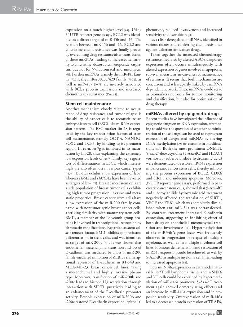
Our review addresses the major epigenetic mechanisms, including the role of miRNA expres-sion contributing to drug resistance and the role of epigenetic drugs to overcome nonresponse arising under conventional chemotherapy (Figure 1).
Histone & DNA modificationsThe nucleosome, which is the smallest pack-ing unit of the chromatin, comprises a core
of four different histone (H2A, H2B, H3 and H4) dimers. Histone protein H1 is respon-sible for proper localization of DNA wrapped by 146-bp long loops around the histone octamer [8]. Each histone exhibits a globular C-terminal region and an N-terminal tail. The latter is subject to several modifications such as acetylation, methylation, phosphorylation, ubiquitin ation, sumoylation, citrullination and ADP-ribosylation. These modifications form the epigenetic bookmarks regulating the structure and organization of chromatin, which is decisive for the accessibility of the genetic information to the transcription machinery. Depending on the type and the amino acid modified at the N-terminus of the different histone proteins, activation or suppression of transcription is initi-ated. Typical repressive marks are trimethylation of lysine residues at position 9 and 27 of histone H3 (H3K9me3 and H3K27me3), leading to a more dense packaging of histones and an inhibi-tion of the access of the transcription machinery to DNA. Trimethylation of H3K4me3 as well as lysine acetylation are associated with tran-scriptional activation. Histone modifications are dynamically mediated by enzymes such as histone acetyltransferases, histone deacetylases (HDACs), histone methyltransferases (HMTs) and histone demethylases [9]. DNA methylation is a further mechanism of epigenetic modifica-tion, influencing the activity of gene expression. Global hypomethylation leading to chromo-somal instability is often observed in various malignancies. However, hypermethylation of cytosine–phosphate–guanine dinucleotide rich
Chemoresistance of tumors is often reported to be due to overexpression of efflux transporters or genetic alterations of signaling pathways. More recently, there is increasing evidence that epigenetic modification contributes to the phenomenon of drug resistance. Despite alteration of DNA methylation or histone modifications, deregulated miRNA expression patterns of tumor cells have been identified as interfering with drug response. Attempts to modify the expression of selected miRNAs have partly led to intriguing improvements of chemotherapy response. This review focuses on the major epigenetic mechanisms, including the role of miRNA expression contributing to drug resistance and the role of epigenetic drugs to overcome nonresponse arising under conventional chemotherapy.
KEYWORDS: CpG islands n DNA methylation n drug transport n gene silencing n histone acetylation n miRNA
Sierk Haenisch1 & Ingolf Cascorbi*1
1Institute of Experimental & Clinical Pharmacology, University Hospital Schleswig-Holstein, Campus Kiel, Arnold-Heller-Street 3, Building 30, D-24105 Kiel, Germany *Author for correspondence: Tel.: +49 431 597 3501Fax: +49 431 597 [email protected]
part of
For reprint orders, please contact: [email protected]

Epigenomics (2012) 4(4)370 future science group
Review Haenisch & Cascorbi
sequences (CpG islands) typically occur in pro-moter regions of certain genes (e.g., tumor-sup-pressor genes) and is mainly associated with transcriptional silencing [10]. DNA methyl-transferases (DNMTs) enzymatically transfer a methyl-group from 5´-adenosylmethionine to the 5 -́residue of cytosine within the CpG di nucleotide. DNMTs can be guided by HMTs (e.g., K9-HMT or EZH2, a H3K27 HMT) to chromatin regions subject to transcriptional inac-tivation [11]. CpG methylation in turn increases the affinity of promoter regions for binding of methyl-CpG-binding domain (MBD) proteins. MBDs (e.g., MECP2 and MBD2) again exhibit
the ability to recruit HDACs thus interpreting the methylation moieties on DNA for the access of chromatin silencing enzymes [12] (Figure 1B).
This complex network of DNA methylation, chromatin modification and transcriptional suppression or activation is often disrupted in diseases. This observation raises the idea of a pharmacological intervention to modulate such epigenetic aberrations. So called epigenetic drugs, a heterogeneous group of compounds interacting with chromatin or DNA-modifying enzymes represent an interesting and encour-aging way to alter pathological epigenetic aberrations.
miRNA mRNA
miRNAgenes
Effluxtransporters
Disease-related genes
Effluxtransporters
Disease-related genes
K4-HMTK4me3
Ac AcAc
Ac
Ac
Ac
HAT
Protein
DNMT
MBD
HMT
HMTK9me3
K27me3
mRNA
ProteinDrug resistance
Disease progression
mCpG
CpG
Drug administration
Environment
Disease
Chromatin and DNA modifications
Epigenetic drugs,miRNA/lncRNA-based therapeutics
HDAC
Epigenomics © Future Science Group (2012)
Figure 1. Chromatin structure and its impact on gene expression. (A) Chromatin structure in balanced gene expression. (B) Chromatin and DNA modifications caused by disease, environmental influences or drug treatment can alter miRNA gene expression leading to disease-related altered gene expression, drug resistance or disease progression. Ac: Acetyl; DNMT: DNA methyltransferase; HAT: Histone acetyltransferase; HDAC: Histone deacetylase; HMT: Histone methyltransferase; MBD: Methyl-CpG-binding domain protein; mCpG: Methyl CpG.

www.futuremedicine.com 371future science group
miRNAs as mediators of drug resistance Review
Chromatin & DNA-modifying drugs & their influence on drug resistanceModification of the activity of histone-modifying enzymes as well as for DNMTs may significantly affect the rate of mRNA transcription and sub-sequently of cell proliferation and apoptotic processes. Hence, substances interacting with DNA-modifying enzymes are considered as highly interesting candidates for the treatment of malignant diseases [13], but interestingly also of neurological disorders [14].
Firstly DNMT-inhibitors (DNMTI) like 5-azacytidine (5-azaC) and decitabine (5-aza-2 -́deoxycytidine) were developed and approved for the treatment of myelodysplastic syndromes. The monotherapy with DNMT inhibitors was, however, limited by low complete and partial response rates and median response durations [15].
However, such approaches are most advanced regarding HDAC inhibitors (HDACIs). The inhibition of histone deacetylation leads to a hyperacetylation of histone tails and sub-sequently to induction of various genes (e.g., E-cadherin and p21), which are involved in the suppression of metastatic and invasive processes. Most of the current HDACIs inhibit the HDAC classes I and II; only benzamides are known to inhibit class III so far.
There are a number of clinical trials investi-gating HDACIs in various malignancies. Most of them are still in Phase I or II; some have been terminated due to weak effectiveness. Vorinostat is one of the most advanced drugs. It was granted ‘orphan designation’ by the EMA in 2005 and was approved in October 2006 by the US FDA for the treatment of cutaneous T-cell lymphoma in patients with progressive, persistent or recur-rent disease, and who failed at least two prior systemic therapies [16]. In 2011, vorinostat was granted by the EMA for designation as an orphan drug for the treatment of malignant mesotheli-oma and for multiple myeloma. In 2009, another HDACI namely romidepsin was approved for the t reatment of cutaneous T-cell lymphoma.
The well-established anticonvulsant valproic acid, a first line drug for the treatment of focal and idiopathic generalized epilepsies, has also been found to act also as a HDACI [17]. This resulted in a number of preclinical investigations and clinical trials of malignancies [18–20].
Histone methylation is also an interesting putative target to modify transcriptional silenced DNA regions. Hence, HMT inhibitors came into focus. The thiodioxopiperazine chaetocin, a natural mycotoxin produced by Chaetomium
and related fungi [21], was identified to be a spe-cific inhibitor of the HMT SU(VAR)3–9 [22]. Thiodioxopiperazine chaetocin is not only an interesting agent for the treatment of leukemias [23], but causes also induction of integrated HIV-1 without producing a T-cell response [24], thus being a putative drug to purge cells of latent HIV-1 for further anti-HIV treatment.
Methylated histone H3K4 also plays a major role in the regulation of tumor-suppressor genes. Inhibition of LSD1, the first identified histone demethylase involved in H3K4 demethylation, may therefore lead to a silencing of malignancies [25,26]. Interestingly, tranylcypromine, an irre-versible nonspecific monoamine oxidase inhibi-tor, earlier used in the treatment of depression, turned out to inhibit LSD1 [27]. Like valproate, this is a further example of previously unknown pleiotropic effects of drugs for which mode of action was believed to be understood.
miRNAs, biogenesis & functionmiRNAs are evolutionarily conserved small ncRNA molecules of 20–22 nucleotides encoded and processed from single genes or gene clusters as polycistronic transcripts [28]. miRNA genes are located intergenic as well as in introns or cod-ing regions of genes, explaining cotranscription with colocated genes or miRNAs. miRNAs are firstly transcribed by RNA polymerase II as up to several hundred basepairs long primary tran-scripts, exhibiting multiple hairpin structures (pri-miRNAs). In the nucleus pri-miRNAs are cleaved by RNase III Drosha and the dsRNA binding protein, DGCR8, into 70–100 nucleo-tides long precursor miRNAs (pre-miRNAs), having one hairpin structure. After transport into cytoplasma by exportin 5 and its cofactor Ran-GTP, pre-miRNAs are further cleaved by RNase III endonuclease dicer and its ds-RNA binding partner, the immunodeficiency virus TRBP into miRNA/miRNA* or miRNA-5p/ miRNA-3p duplexes. By recruiting EIF2C2, also known as AGO2, into the dicer complex, the minimal RNA-induced silencing complex is formed. After degradation of one of the RNA strands, the remaining mature miRNA strand guides the silencing complex to the 3 -́UTR of the target mRNA. Depending on the grade of complementarity, the target mRNA stability and/or translation is suppressed [29,30].
However, beside this well accepted mecha-nism of post-transcriptional gene silencing by miRNA/mRNA-3´-UTR interference, some more functions of miRNAs have recently been uncovered. Loss of miR-328 expression was

Epigenomics (2012) 4(4)372 future science group
Review Haenisch & Cascorbi
found in blast crisis myelogenous leukemia and was associated with inhibition of myeloid differ-entiation. It was shown, that the mRNA-5́ -UTR binding and post-transcriptional gene regulator hnRNP E2 is captured by binding to miRNA-328. In consequence, C/EBPα mRNA is released from its regulator and translated. Interestingly, C/EBPα is an important tran-scription factor for miR-328, and is therefore keeping up its own translation via a so called ‘decoy effect’. Restoration of miR-328 expression rescued myeloid maturation and differentiation [31]. Bao et al. postulated from investigations in Arabidopsis that by interacting with binding sites in nascent target mRNAs, miRNAs could guide methylating protein complexes to the DNA tem-plate leading to hypermethylation downstream of the miRNA binding site [32]. Moreover, it was postulated for the tumor-suppressor gene PTEN and its pseudogene PTENP1 (lost in human cancers by chromosomal aberrations) that the non translated pseudogene acts as a miRNA scav-enger, preventing and controlling immoderate miRNA binding to the mRNA-3 -́UTR of the coding target gene [33]. In conclusion, miRNAs are not only responsible for the physiological bal-ance of gene expression, miRNAs can alter the ability of cells to commence fundamental biolog-ical processes such as development, pro liferation, differentiation, apoptosis and maintenance of tissue specificity. In various diseases miRNAs are deregulated as predominantly shown in can-cer [34], but also in metabolic [35] or neurological disorders [36].
miRNAs involved in drug resistanceSince the expression of miRNAs depends on the complex interplay of DNA methylation and chromatin modifications, and since in turn certain miRNAs (epi-miRNAs) influence the expression of histone modifying or DNA-methylating enzymes [37,38], miRNAs may represent an interesting linking part between pathological epigenetic alterations and final disease-related deregulation of protein expres-sion. Therefore, miRNAs may not only be used as disease biomarkers [39], they could also rep-resent indicators of drug response. Moreover, the knowledge about which kind of miRNAs are altered in their expression by the use of a certain epigenetic drug – and about miRNAs altered in drug resistance – could give a hint as to which kind of epigenetic drug could be used in an adjuvant way to overcome drug resistance.
Inadequate response or decreasing efficacy under treatment is a major limitation of drug
therapy of various diseases; this phenomenon is especially known in pharmacological treatment of cancer, but also in neurological diseases as epilepsy. Limited bioavailability at the drug at target site may be caused by increased expression of efflux transporters or by alterations of drug target- or disease-related genes, and is regarded to be involved in therapy resistance.
The role of miRNAs in regulation of ATP-binding cassette transportersRecently several miRNAs have been described to be connected to drug resistance potentially changing the expression of ATP-binding cassette (ABC) efflux transporters. The most prominent representative of the ABC transporter family and most commonly associated with multidrug resis-tance is P-gp, also known as MDR1 encoded for by the ABCB1 gene. Kovalchuck et al. confirmed that miRNA-451 targets ABCB1 mRNA and is inversely correlated to doxo rubicin resistance of MCF7 breast cancer cells [40]. By contrast, downregulation of miR-27a and a simultane-ously upregulation of the co repressor HIPK2 was shown to decrease the expression of ABCB1 and to increase the sensitivity of the paclitaxel-resis-tant ovarian cancer cell line A2780 [41]. In line with P-gp downregulation, the sensitivity of doxorubicin-resistant A2780 cells was increased by inhibition of miR-27a [42] and, contrary to the observations of Kovalchuck et al., by inhibition of miR-451. In esophageal cancer cells, down-regulation of miR-27a was also associated with decreased expression of P-gp, anti apoptotic pro-tein, BCL-2, but upregulation of proapoptotic protein BAX, leading to increased accumulation and efficacy of doxorubicin [43]. By contrast, in K562 chronic myelogenous leukemia (CML) cells down regulation of miR-27a and miR-331-5p was associated with increased expression of P-gp and gradually increasing resistance to doxorubicin, which implies a direct interaction of both miRNAs with ABCB1 mRNA [44].
The half-transporter ABCG2 (also known as BCRP) is another important member of the ABC transporter family often involved in drug resistance. It was shown that miR-519c, miR-520h, miR-328 and miR-212 target ABCG2 mRNA [45–50]. Interestingly, compared with the parental S1 colon cancer cell line, the mitoxantrone-resistant counterpart S1MI80 and several other drug-resistant cell lines cells seem to elude miRNA-mediated control of ABCG2 expression by shortening the length of 3 -́UTR and loss of miR-519c binding site [48,49]. Moreover, miR-520h was less abundant in resistant cells
www.futuremedicine.com 373future science group
miRNAs as mediators of drug resistance Review
than in their respective parental cells [49]. Also miR-328 was demonstrated to be inversely regu-lated in MCF7 and MCF7/MX70 breast can-cer cells. The resistant cells exhibited a signifi-cantly lower expression of miR-328 and a higher ABCG2 mRNA and protein expression. Direct miRNA/mRNA interference was confirmed by 3 -́UTR reporter gene assays. Consequently after ectopic expression of miR-328 resistant MCF7 breast cancer cells developed higher sensitivity to mitoxantrone [47]. Partly similar observations were made in K562 cells resistant to increasing concentrations of imatinib. ABCG2 mRNA and protein was inversely correlated to miR-328 and miR-212. In this study, the latter was confirmed to target ABCG2 3 -́UTR [50]. In the MCF7 breast cancer cell line and its etoposid-resistant counterpart (MCF7/VP) ABCC1 was inversely regulated to miRNA-326. A negative regulative effect of miR-326 on ABCC1 expression was proven by reporter gene assays, as well as by an increase in sensitivity to etoposid and doxoru-bicin, but not to mitoxantrone, which is sug-gested to be transported by ABCG2 rather than by ABCC1. Interestingly, inverse correlation of miRNA and ABCC1 expression was found in early nonmetastatic compared with metastatic advanced breast cancer tissues [51]. Moreover, after transfection of miR-7 and miR-345, resis-tant MCF7/CDDP cells revealed lower IC
50
values for cisplatin. Reporter gene assays con-firmed a miRNA/mRNA interaction [52]. In glioma stem cells it was shown that inhibitor of differentiation (ID4) indirectly enhances SOX2 protein expression by suppressing miRNA-9*, which targets the 3 -́UTR of SOX2. Therefore, SOX2 an important transcription factor for stem cell maintenance in the CNS mediates not only stemness potential, but also chemo resistance to carmustine by upregulation of ABCC3 and ABCC6 [53]. It was shown that breast tumor initiating cells (BT-IC) having stem cell-like features, and being more resistant against che-motherapy than their adjacent differentiated cancer cells, exhibit significantly lower expres-sion of miR-128. Direct targeting of ABCC5 and BMI-1 by miR-128 was concomitant with decreased protein expression and increased doxorubicin sensitivity due to enhanced DNA damage and apoptosis in BT-ICs. Moreover, in breast cancer patients high miR-128 expression was correlated with elevated clinical response to chemotherapy and prolonged survival [54].
miRNAs also play a role in the regula-tion of drug-metabolizing enzymes. Pan et al. identified two miRNAs, namely miR-27b and
mmu-miR-298, targeting CYP3A4 [55]. Ectopic expression of both miRNAs lead to decreased sensitivity of the pan creatic cancer cell line PANC1 to cyclo phosphamide [55], a prodrug that has to be activated by CYP3A4, as well as by CYP2C9 and CYP2C19 (TaBle 1).
Apoptosis & cell survivalResistance to chemotherapy is multifactorial and not only the result of deregulation of miRNAs targeting drug eff lux transporter. Many miRNAs are related to apoptotic pathways and cell survival [56], and cells having altered expres-sion profiles of pro- and anti-apoptotic miRNAs frequently also exhibit increased resistance to cytotoxic agents. Tomokuni et al. showed an increased expression of miR-146a in hepatocel-lular carcinoma cell lines resistant to IFN-α. Further invest igations revealed a negative regu-lating effect of miR-146a on SMAD4, there-fore inhibiting IFN-α-mediated apoptosis [57]. miR-21, often upregulated in malignancies, was shown to target PTEN, a important tumor sup-pressor, which acts as an phosphatidylinositol-3,4,5-tris phosphate 3-phosphatase, thereby inhibiting the Akt/PKB pathway. Doxorubicin-resistant breast cancer cells (MCF7/ADR) did not only show higher expression of miR-21, they also exhibited lower protein content of PTEN than their parental cell line (MCF7) [58]. Similar findings were reported for the CML cell line K562 and its daunorubicin-resistant counter-part [59], as well as for HER2-positive breast cancer cell lines, resistant to trastuzumab [60]. miR-34a is a well-known proapoptotic miRNA transactivated by p53 [61], and often decreased in various tumors due to chromosomal deletion [62] or hypermethylation [63]. In the hormone-refractory prostate cancer cell line PC3 down-regulation of miR-34a was associated with pacli-taxel resistance and upregulation of HUR, an RNA-binding protein controlling the stability of mRNAs, the HDAC SIRT1 and the antiap-optotic mitochondrial protein BCL2. As proof-of-concept, transfection of miR-34a precursor molecules into PC3PR cells resulted in attenu-ated paclitaxel resistance. It further decreased the expression of BCL2 and SIRT1 by directly targeting the the 3 -́UTR, as well as indirectly by targeting HUR, that controls the expression of BCL2 and SIRT1 [64]. MCF7 breast can-cer cells resistant to 4-hydroxytamoxifen were shown to have an over expression of the miRNAs miR-21, miR-181, miR-222, miR-342 and miR-489. miR-221/222 was also over expressed in HER2/neu-positive primary tumors known

Epigenomics (2012) 4(4)374 future science group
Review Haenisch & Cascorbi
Table 1. Interaction of miRNAs with selected target genes, site of expression and major resulting functional consequences†.
miRNA Target gene Drug resistance Tissue/disease Consequence of miRNA inhibition
Effect of epigenetic drug
Ref.
let-7 family HRAS and HMGA2 Breast tumor initiating cells HRAS↑, HMGA2↑ [76]
miRNA-9* SOX2 Carmustine Induced human glioma stem-like cells
ABCC3↑, ABCC6↑ [53]
miR-15b and miR-16
BCL2 Vincristine, doxorubicin, etoposide and cisplatin
Gastric cancer cell line SGC7901
BCL2↑ [69]
miR-21 PTEN Doxorubicin
Daunorubicin andtrastuzumab
Breast cancer cell line MCF7CML cell line K562HER2+ breast cancer cells BT474, SKBR3, MDA-MB-453
PTEN↑ [58]
[59][60]
miR-27a HIPK2
ABCB1
Paclitaxel
Doxorubicin
Ovarian cancer cell line A2780Esophageal cancer cell lines ECA109 and TE13CML cell line K562
ABCB1↓
ABCB1↓, BAX↑, BCL2↓ABCB1↑
LAQ824 (dacinostat miR-27a↓)
[41]
[43]
[44]
miR-27b, mmu-miR-298
CYP3A4 Cyclophosphamide Pancreatic cancer cell line PANC1
CYP3A4↑ LAQ824 (dacinostat miR-27b↓)
[47]
miR-34a HUR, SIRT1BCL2,VEGFZEB1 c-Met, NOTCH1 and NOTCH2
c-Myc
Paclitaxel Prostate cancer cell line PC3Pancreatic cancer stem cells
Human glioblastoma tissues, medulloblastoma cell linesProstate cancer cell line PC3
HUR↑, SIRT1↑BCL2↑ VEGF↑,ZEB1↑,E-cadherin↓c-Met↑, NOTCH1↑ and NOTCH2↑, CDK6↑c-Myc↑
5-Aza-dC, vorinostat, (miR-34a↑)
[64]
[81]
[94]
[95]
miR-125b-2 Temozolomide GBMSCs BAX↑, BCL2↓ [96]
miR-128 ABCC5, BMI-1 Doxorubicin Breast tumor initiating cells ABCC5↑, BMI-1↑ [54]
miR-146a SMAD4
TRAF6
IFN-α
Etoposide
Hepatocellular carcinoma cell linesNKTL tissues, NKTL and NK cell lines
SMAD4↑
TRAF6↑IκB↓
5-Aza-dC (miR-146a↑)
[57]
[83]
miR-181 family BCL2 DaunorubicinCisplatin andvincristine
CML cell line K562 Gastric and lung cancer cell lines SGC7901 and A549
BCL2↑BCL2↑
[70][71]
miR-200a,b,c/ 429
BCL2, XIAP
BMI1
SIRT1, ZEB1
Vincristine and cisplatin
Gastric and lung cancer cell lines SGC7901 and A549BCSCs, normal mammary stem/progenitor cellsBreast cancer cell lines BT-549 and MDA-MB-231
BCL2↑, XIAP↑
BMI1↑
SIRT1↑, ZEB1↑E-cadherin↓
[72]
[77]
[78]
†Epigenetic drugs interfering with miRNA expression are also listed.5-Aza-dC: 5-aza-2´-deoxycytidine; BCSC: Breast cancer stem cell; CML: Chronic myelogenous leukemia; ER: Estrogen receptor; GBMSC: Glioblastoma multiforme tissues and stem cell; N/A: No epigenetic drugs have yet been idetified to influence expression of this RNA; NK: Natural killer; NKTL: NK/T cell lymphoma.

www.futuremedicine.com 375future science group
miRNAs as mediators of drug resistance Review
to be resistant to endocrine therapy. This miRNA targets p27Kip1, and indeed the expres-sion of this protein was reduced in the resistant MCF7 cells [65]. Vice versa, the resistance could be overcome by overexpression of p27Kip1 in resistant cells, leading to increased sensitivity to tamoxifen [66]. There is also an association of miR-221/222 with estrogen receptor (ER) expression. Inhibition of the highly upregu-lated miR-221/222 expression observed in ERα-negative breast cancer cell lines and primary tumors caused an increased tamoxifen-mediated cell growth arrest and apoptosis [67]. In line with these findings, Rao et al. reported an associa-tion of increased miR-221 and -222 expression
and resistance against fulvestrant, a selective ER downregulator. The author further report an activation of β-catenin and a repression of TGF-β-induced cell growth control [68]. These studies underline that miR-221 and -222 can act as important antiapoptotic key players in breast cancer, targeting several cellular signaling path-ways, and thereby circumventing the efficacy of tamoxifen, as well as fulvestrant, treatment.
Other well-known proapoptotic miRNAs, such as miR-15b and-16, are involved in chemo-resistance as well. The investigation by Xia et al. revealed that the vincristine resistant gastric can-cer cell line SGC7901/VCR have low expressions of miR-15b and -16 and inversely a BCL2 protein
Table 1. Interaction of miRNAs with selected target genes, site of expression and major resulting functional consequences† (cont.).
miRNA Target gene Drug resistance Tissue/disease Consequence of miRNA inhibition
Effect of epigenetic drug
Ref.
miR-203 ABL and BCR–ABL
SOCS3 Cisplatin
CML cell line KCL-22, K562
Human breast cancer tissues, breast cancer cell line MCF7
ABL↑, BCR–ABL↑SOCS3↑,p53↑, p21↑, BAX↑
5-Aza-dC plus 4-phenylbutyrate (miR-203↑)
[86]
[87]
miR-212 ABCG2 Imatinib CML cell line K562 ABCG2↑ N/A [50]
miR-221/222 p27Kip1
ERα
p27Kip1ERα
Tamoxifen
Tamoxifen
Fulvestrant
HER2/neu-positive primary human breast cancer tissues, breast cancer cell line MCF7ERα-negative breast cancer cell lines and primary tumorsBreast cancer cell line MCF7
p27Kip1↑
ERα↑
β-catenin↓, TGFβ-pathway↑
N/A [66]
[67]
[68]
miRNA-326 ABCC1 Etoposide and doxorubicin
Breast cancer cell line MCF7
ABCC1↑ N/A [51]
miR-328 ABCG2
ABCG2
Mitoxantrone
Imatinib
Breast cancer cell line MCF7CML cell line K562
ABCG2↑
ABCG2↑
N/A [47]
[50]
miR-7 and miR-345
ABCC1 Cisplatin Breast cancer cell line MCF7
ABCC1↑ N/A [52]
miR-331-5p ABCB1 Doxorubicin CML cell line K562 ABCB1↑ N/A [44]
miR-451 ABCB1
c-Myc
Doxorubicin
Doxorubicin
Breast cancer cell line MCF7A2780T-cell acute lymphoblastic leukemia
ABCB1↑
ABCB1↓c-Myc↑
N/A [40]
[42][97]
miR-497 BCL2 Vincristine and cisplatin
Gastric and lung cancer cell lines SGC7901 and A549
BCL2↑ N/A [73]
miR-519c, miR-520h
ABCG2 Mitoxantrone Colon cancer cell line S1 ABCG2↑ N/A [48,49]
†Epigenetic drugs interfering with miRNA expression are also listed.5-Aza-dC: 5-aza-2´-deoxycytidine; BCSC: Breast cancer stem cell; CML: Chronic myelogenous leukemia; ER: Estrogen receptor; GBMSC: Glioblastoma multiforme tissues and stem cell; N/A: No epigenetic drugs have yet been idetified to influence expression of this RNA; NK: Natural killer; NKTL: NK/T cell lymphoma.
Epigenomics (2012) 4(4)376 future science group
Review Haenisch & Cascorbi
expression on a much higher level [69]. Using 3 -́UTR reporter gene assays, BCL2 was identi-fied as a direct target of miR-15b and -16. The relation between miR-15b and -16, BCL2 and vincristine chemoresistance was finally proven by overcoming drug resistance after transfection of these miRNAs, leading to increased sensitiv-ity to vincristine, doxorubicin, etoposide, cispla-tin, but not for 5́ -fluorouracil and mitomycin [69]. Further miRNAs, namely the miR-181 fam-ily [70,71], the miR-200abc/429 family [70,72], as well as miR-497 [70,73] are inversely associated with BCL2 protein expression and increased chemotherapy resistance (TaBle 1).
Stem cell maintenanceAnother mechanism closely related to occur-rence of drug resistance and tumor relapse is the ability of cancer cells to reconstitute an embryonic stem cell (ESC)-like miRNA expres-sion pattern. The ESC marker lin-28 is regu-lated by the key transcription factors of stem cell maintenance, namely OCT-4, NANOG, SOX2 and TCF3, by binding to its promoter region. In turn, let-7g is inhibited in its matu-ration by lin-28, thus explaining the extremely low expression levels of let-7 family, key regula-tors of differentiation in ESCs, which interest-ingly are also often lost in various cancer types [74,75]. BT-ICs exhibit a low expression of let-7, whereas HRAS and HMGA2 have been revealed as targets of let-7 [76]. Breast cancer stem cells are a side population of breast tumor cells exhibit-ing high tumor progression, invasive and meta-static properties. Breast cancer stem cells have a low expression of the miR-200 family com-pared with nontumorigenic breast cancer cells, a striking similarity with mammary stem cells. BMI1, a member of the Polycomb group pro-teins is involved in transcriptional repression by chromatin modifications. Regarded as stem cell self-renewal factor, BMI1 inhibits apoptosis and differentiation in stem cells, and was identified as target of miR-200c [77]. It was shown that endothelial–mesenchymal transition and loss of E-cadherin was mediated by a loss of miR-200 family-mediated inhibition of ZEB1, a transcrip-tional repressor of E-cadherin in BT-549 and MDA-MB-231 breast cancer cell lines, having a mesenchymal and highly invasive pheno-type. Moreover, transfection of miR-200b and -200c leads to histone H3 acetylation through interaction with SIRT1, putatively leading to an enhancement of the E-cadherin promoter activity. Ectopic expression of miR-200b and -200c restored E-cadherin expression, epithelial
phenotype, reduced invasiveness and increased s ensitivity to doxorubicin [78].
TaBle 1 lists deregulated miRNAs, identified in various tissues and conferring chemoresistance against different anticancer drugs.
Taken together the increased chemotherapy resistance mediated by altered ABC-transporter expression often occurs simultaneously with altered expression of genes involved in apop tosis, survival, metastasis, invasiveness or maintenance of stemness. It seems that both mechanisms are concurrent and at least partly linked by a miRNA dependent network. Thus, miRNAs could serve as biomarkers not only for tumor monitoring and classification, but also for optimization of drug therapy.
miRNAs altered by epigenetic drugsRecent studies have investigated the influence of epigenetic drugs on miRNA expression, attempt-ing to address the question of whether adminis-tration of those drugs can be used to reprogram expression of deregulated miRNAs by altering DNA methylation [79] or chromatin modifica-tions [80]. Both the most prominent DNMTI, 5-aza-2 -́deoxycytidine (5-Aza-dC) and HDACI vorinostat (suberoylanilide hydroxamic acid) were demonstrated to restore miR-34a expression in pancreatic cancer stem cells, thereby inhibit-ing the protein expression of BCL2, CDK6 and SIRT1 and inducing apoptosis. Moreover, 3 -́UTR reporter gene assays, performed in pan-creatic cancer stem cells, showed that 5-Aza-dC and suberoylanilide hydroxamic acid treatment negatively affected the translation of SIRT1, VEGF and ZEB1, which was completely dimin-ished when anti-miR-34a was cotransfected. By contrast, treatment increased E-cadherin expression, suggesting an inhibiting effect of both drugs on endothelial–mesenchymal tran-sition and invasiveness [81]. Hypermethylation of the miR34b/c gene locus was frequently observed in progression or relapse of multiple myeloma, as well as in multiple myeloma cell lines. Promoter demethyl ation and restoration of miR34b expression could be achieved, as well by 5-Aza-dC in multiple myeloma cell lines leading to increased apoptosis [82].
Low miR-146a expression in extranodal natu-ral killer/T cell lymphoma tissues and in SNK6 and YT cells could be explained by hypermeth-ylation of miR-146a promoter. 5-Aza-dC treat-ment again showed demethylating effects and an increase in miR-146a expression and in eto-poside sensitivity. Overexpression of miR-146a led to a decreased protein expression of TRAF6,

www.futuremedicine.com 377future science group
miRNAs as mediators of drug resistance Review
a reported target of miR-146a. This was concur-rent with an increase of IκB and an inhibition of BCL2 expression. Since the transcription factor NFκB activates miR-146a, a hypermethlyation of miR-146a promoter could disrupt the miR146a/TRAF6/NFkB feed-back loop, physiologically mediating cell proliferation in natural killer/T cell lymphoma cells, and leading to inhibition of apopotosis and increased chemoresistance [83].
The potent hydroxamic acid HDACI, LAQ824 (dacinostat), was reported to downregulate miR-27a and miR-27b in the breast cancer cell line SKBr3 [84]. In the precursor B-cell acute lym-phoblastic leukemia cell line NALM-6 another hydroxamic acid HDACI, trichostatin A, upregu-lated miR-22, which was explained by a drug-induced accumulation of H3 acetylation and a decreased H3K27 trimetyl ation [85]. The combi-nation treatment of 5-Aza-dC and the HDACI, 4-phenylbutyrate, caused demethylation of upstream region of miR-203 in CML cell lines KCL-22 and K562, and decay in ABL as well as BCR–ABL protein expression. Direct RNA interference was proven by cotransfection of miR-203 and a reporter gene construct containing the ABL-3 -́UTR target sequence. Moreover, ectopic expression of miR-203 in CML cell lines signifi-cantly inhibited cell growth [86]. Interestingly, in breast cancer tissues miR-203 is significantly overexpressed. In MCF7 cells antagonizing miR-203 conferred chemosensitivity to cisplatin via upregulation of the target gene, SOCS3, which was in consequence concurrent with induction of proapoptotic genes p53, p21 and BAX [87].
This example again emphasizes that different diseases or cancer types exhibit distinct miRNA patterns. This raises the question whether epigen-etic drugs globally altering chromatin and DNA structure can be used universally as adjuvant to improve therapy response in diseases exhibit-ing deregulated miRNA expression, or whether administration of epigenetic drugs can even accelerate the progression of the disease.
In this context it should be mentioned that well established and long-term administered drugs such as valproic acid [88], rifampicin [89] and metformin [90] can alter the miRNA expres-sion and therefore could offer new therapeutic options beyond their classical indications.
ConclusionmiRNAs are important regulators of various cellular processes. Deregulation of the physio-logical miRNA expression can provoke malig-nancies as well as metabolic and neurological dis-orders. Moreover, many studies being currently
conducted show that drug resistance, a phenom-enon often occurring under treatment and charac-terized by relapse or attenuation of drug efficacy, is at least partially mediated by an alteration of the expression of miRNAs which interfere with drug transport, cell survival and stem cell maintenance. The expression of miRNA genes is influenced by DNA or histone modifications, and vice versa DNA or histone modifying enzymes are regulated by miRNAs (epi-miRNAs). Therefore, by using epigenetic drugs or approaches that directly alter the miRNA expression, miRNAs can not only be used as biomarkers to classify malignancies or to monitor disease progression, they may also repre-sent a new therapeutic starting point to optimize therapy and overcome pharmacoresistance.
Future perspectiveIt is now unquestionable that in many diseases miRNAs are deeply involved in the transition from a physiological balance to an abnormal and malignant cell function. Since a single miRNA controls dozens of genes of multiple cellular pathways, the attempt to restore the physiological miRNA expression could provide a more successful and sustainable strategy than to target the function of a single protein spe-cifically. Several approaches have been tested to influence the expression of miRNAs not only in vitro but also in vivo. One approach is to use oligonucleotides (antisense oligonucleotides or precursor miRNAs) to directly modify the deregulated expression of candidate miRNAs. The other attempts to indirectly alter the miRNA expression by epigenetic drugs such as DNMTIs or HDACIs. The main challenge of the first approach will be the tissue-specific, therapeutically reasonable and sufficient delivery of RNA molecules. Chemical modifications such as locked nucleic acid, 2 -́O-methyl or phospho-rothioate oligonucleotides have increased in vivo stability and half-life. Polymer formulations as well as nanoparticles, micropherese and hydro-gels exhibit sustained and prolonged delivery with less toxic effects than liposomes. In order to increase the tissue specificity or to penetrate, for example, the blood–brain barrier, nano particles could be tagged with antibodies targeting spe-cific cell surface proteins [91]. The latter approach using epigenetic drugs bears the risk of side effects caused by global and unspecific altera-tion of chromatin structure not only correcting deregulated gene expression, but maybe also affecting normal gene expression. Nevertheless, currently hundreds of Phase I and II trials are ongoing to investigate the safety and efficacy of

Epigenomics (2012) 4(4)378 future science group
Review Haenisch & Cascorbi
epigenetic drugs alone, as well as in combination with conventional chemotherapeutics. A recent study revealed promising results concerning the outcome of the use of 5-AzaC and entinostat (MS-275) in refractory metastatic non-small-cell lung cancer, which was as effective as the conventional therapy with erlotinib [92]. A very recent and interesting approach includes the observation that lncRNAs can recruit and guide genome-specific chromatin modifying protein complexes. Replacing or antagonizing disease-specific lncRNAs could therefore increase the arsenal of RNA-based therapeutics [93].
Considering the increasing knowledge about epigenetic alterations in various diseases and the promising approaches to reverse them, it is very
likely that in the near future a new era will be heralded, in which conventional drug regimes will be enriched by epigenetic drugs, as well as RNA oligonucleotide therapeutics, to optimize therapy and overcome drug resistance.
Financial & competing interests disclosureThe authors have no relevant affiliations or financial involvement with any organization or entity with a finan-cial interest in or financial conflict with the subject matter or materials discussed in the manuscript. This includes employment, consultancies, honoraria, stock ownership or options, expert testimony, grants or patents received or pend-ing, or royalties.
No writing assistance was utilized in the production of this manuscript.
Executive summary
� Epigenetics has emerged as an extremely interesting field contributing a lot to the explanation of gene–environmental interactions.
� Previously unknown modes of action of existing drugs can now partly be explained by epigenetic modification or interaction with ncRNAs.
� DNA methylation, histone modifications and miRNA expression are frequently altered in diseases, particularly in malignancies.
� Novel epigenetic drugs are under development and some, such as vorinostat, have already been approved for use in humans. This heterogeneous group of compounds represents a highly interesting and encouraging way to alter pathological epigenetic aberrations and miRNA expression patterns that have been identified to contribute to disease progression and chemoresistance.
� However, the consequences of an altered miRNA expression are much more complex. miRNAs may act as tumor suppressors or oncogenes.
� Furthermore, the miRNA expression of some highly malignant tumor cells is similar to that observed in stem cells, inhibiting the differentiation of cells.
� Suppression or restoration of deregulated miRNAs could help to overcome chemoresistance.
� Attempts to influence the epigenetics of cells through epigenetic drugs could also directly result in an alteration of miRNA pattern.
� Since epigenetic drugs act mostly nonspecifically, the improvement of specificity, maybe by the use of miRNA-based therapeutics, could bear a challenging but promising way for an advanced therapy of malignancies.
ReferencesPapers of special note have been highlighted as:n of interestnn of considerable interest
1 Szyf M. The early life social environment and DNA methylation: DNA methylation mediating the long-term impact of social environments early in life. Epigenetics 6(8), 971–978 (2011).
2 Lima SC, Hernandez-Vargas H, Herceg Z. Epigenetic signatures in cancer: implications for the control of cancer in the clinic. Curr. Opin. Mol. Ther. 12(3), 316–324 (2010).
3 Pirola L, Balcerczyk A, Okabe J, El-Osta A. Epigenetic phenomena linked to diabetic complications. Nat. Rev. Endocrinol. 6(12), 665–675 (2010).
4 Marques SC, Oliveira CR, Pereira CM, Outeiro TF. Epigenetics in neurodegeneration: a new layer of complexity. Prog. Neuropsychopharmacol. Biol. Psychiatry 35(2), 348–355 (2011).
5 Dong E, Grayson DR, Guidotti A, Costa E. Antipsychotic subtypes can be characterized by differences in their ability to modify GABAergic promoter methylation. Epigenomics 1(1), 201–211 (2009).
6 Crea F, Nobili S, Paolicchi E et al. Epigenetics and chemoresistance in colorectal cancer: an opportunity for treatment tailoring and novel therapeutic strategies. Drug Resist. Updat. 14(6), 280–296 (2011).
7 Starkey Lewis PJ, Dear J, Platt V et al. Circulating microRNAs as potential markers of human drug-induced liver injury. Hepatology 54(5), 1767–1776 (2011).
8 Hashimoto H, Takami Y, Sonoda E et al. Histone H1 null vertebrate cells exhibit altered nucleosome architecture. Nucleic Acids Res. 38(11), 3533–3545 (2010).
9 Sharma S, Kelly TK, Jones PA. Epigenetics in cancer. Carcinogenesis 31(1), 27–36 (2010).
10 Kulis M, Esteller M. DNA methylation and cancer. Adv. Genet. 70, 27–56 (2010).
11 Vire E, Brenner C, Deplus R et al. The Polycomb group protein EZH2 directly controls DNA methylation. Nature 439(7078), 871–874 (2006).
12 Sansom OJ, Maddison K, Clarke AR. Mechanisms of disease: methyl-binding domain proteins as potential therapeutic targets in cancer. Nat. Clin. Pract. Oncol. 4(5), 305–315 (2007).
13 Szyf M. Epigenetics, DNA methylation, and chromatin modifying drugs. Annu. Rev. Pharmacol. Toxicol. 49, 243–263 (2009).
n Overview of the classification and mechanisms of epigenetic drugs.
14 Suzuki Y, Yanagisawa M, Ariga T, Yu RK. Histone acetylation-mediated glycosyltransferase gene regulation in mouse brain during development. J. Neurochem. 116(5), 874–880 (2011).
15 Gore SD. Combination therapy with DNA methyltransferase inhibitors in hematologic

www.futuremedicine.com 379future science group
miRNAs as mediators of drug resistance Review
379www.futuremedicine.com
malignancies. Nat. Clin. Pract. Oncol. 2(Suppl. 1), S30–S35 (2005).
16 Mann BS, Johnson JR, Cohen MH, Justice R, Pazdur R. FDA approval summary: vorinostat for treatment of advanced primary cutaneous T-cell lymphoma. Oncologist 12(10), 1247–1252 (2007).
17 Hrzenjak A, Moinfar F, Kremser ML et al. Valproate inhibition of histone deacetylase 2 affects differentiation and decreases proliferation of endometrial stromal sarcoma cells. Mol. Cancer Ther. 5(9), 2203–2210 (2006).
18 Mohammed TA, Holen KD, Jaskula-Sztul R et al. A pilot Phase II study of valproic acid for treatment of low-grade neuroendocrine carcinoma. Oncologist 16(6), 835–843 (2011).
19 Scherpereel A, Berghmans T, Lafitte JJ et al. Valproate-doxorubicin: promising therapy for progressing mesothelioma. A Phase II study. Eur. Respir. J. 37(1), 129–135 (2011).
20 Thomas S, Thurn KT, Bicaku E, Marchion DC, Munster PN. Addition of a histone deacetylase inhibitor redirects tamoxifen-treated breast cancer cells into apoptosis, which is opposed by the induction of autophagy. Breast Cancer Res. Treat. 130(2), 437–447 (2011).
21 Sekita S, Yoshihira K, Natori S et al. Mycotoxin production by Chaetomium spp. and related fungi. Can. J. Microbiol. 27(8), 766–772 (1981).
22 Greiner D, Bonaldi T, Eskeland R, Roemer E, Imhof A. Identification of a specific inhibitor of the histone methyltransferase SU(VAR)3–9. Nat. Chem. Biol. 1(3), 143–145 (2005).
23 Chaib H, Nebbioso A, Prebet T et al. Anti-leukemia activity of chaetocin via death receptor-dependent apoptosis and dual modulation of the histone methyl-transferase SUV39H1. Leukemia 26(4), 662–674 (2011).
24 Bernhard W, Barreto K, Saunders A, Dahabieh MS, Johnson P, Sadowski I. The Suv39H1 methyltransferase inhibitor chaetocin causes induction of integrated HIV-1 without producing a T cell response. FEBS Lett. 585(22), 3549–3554 (2011).
25 Wang J, Lu F, Ren Q et al. Novel histone demethylase LSD1 inhibitors selectively target cancer cells with pluripotent stem cell properties. Cancer Res. 71(23), 7238–7249 (2011).
26 Chen Y, Jie W, Yan W, Zhou K, Xiao Y. Lysine-specific histone demethylase 1 (LSD1): A potential molecular target for tumor therapy. Crit Rev. Eukaryot. Gene Expr. 22(1), 53–59 (2012).
27 Singh MM, Manton CA, Bhat KP et al. Inhibition of LSD1 sensitizes glioblastoma
cells to histone deacetylase inhibitors. Neuro. Oncol. 13(8), 894–903 (2011).
28 Racz Z, Kaucsar T, Hamar P. The huge world of small RNAs: regulating networks of microRNAs (review). Acta Physiol Hung. 98(3), 243–251 (2011).
29 Skalsky RL, Cullen BR. Viruses, microRNAs, and host interactions. Annu. Rev. Microbiol. 64, 123–141 (2010).
30 Wiemer EA. The role of microRNAs in cancer: no small matter. Eur. J. Cancer 43(10), 1529–1544 (2007).
31 Eiring AM, Harb JG, Neviani P et al. miR-328 functions as an RNA decoy to modulate hnRNP E2 regulation of mRNA translation in leukemic blasts. Cell 140(5), 652–665 (2010).
n Demonstrates the pleiotropic function of miRNAs. This study describes miRNA that traps the translational regulator poly(rC)-binding protein hnRNP E2 and thereby maintains the translation of CEBPA mRNA.
32 Bao N, Lye KW, Barton MK. MicroRNA binding sites in Arabidopsis class III HD-ZIP mRNAs are required for methylation of the template chromosome. Dev. Cell 7(5), 653–662 (2004).
33 Poliseno L, Salmena L, Zhang J, Carver B, Haveman WJ, Pandolfi PP. A coding-independent function of gene and pseudogene mRNAs regulates tumour biology. Nature 465(7301), 1033–1038 (2010).
n Demonstrates the impact of pseudogene mRNAs on miRNA function. A nontranslated mRNA of the pseudogene PTENP1 is reported as acting as a miRNA scavenger and regulates the expression of the tumor suppressor PTEN.
34 Lovat F, Valeri N, Croce CM. MicroRNAs in the pathogenesis of cancer. Semin. Oncol. 38(6), 724–733 (2011).
35 Ferland-McCollough D, Ozanne SE, Siddle K, Willis AE, Bushell M. The involvement of microRNAs in Type 2 diabetes. Biochem. Soc. Trans. 38(6), 1565–1570 (2010).
36 Salta E, De Strooper B. Non-coding RNAs with essential roles in neurodegenerative disorders. Lancet Neurol. 11(2), 189–200 (2012).
37 Hatziapostolou M, Iliopoulos D. Epigenetic aberrations during oncogenesis. Cell Mol. Life Sci. 68(10), 1681–1702 (2011).
38 Smits M, Mir SE, Nilsson RJ et al. Down-regulation of miR-101 in endothelial cells promotes blood vessel formation through reduced repression of EZH2. PLoS ONE 6(1), e16282 (2011).
39 Nana-Sinkam SP, Croce CM. Non-coding RNAs in cancer initiation and progression and as novel biomarkers. Mol. Oncol. 5(6), 483–491 (2011).
40 Kovalchuk O, Filkowski J, Meservy J et al. Involvement of microRNA-451 in resistance of the MCF-7 breast cancer cells to chemotherapeutic drug doxorubicin. Mol. Cancer Ther. 7(7), 2152–2159 (2008).
41 Li Z, Hu S, Wang J et al. MiR-27a modulates MDR1/P-glycoprotein expression by targeting HIPK2 in human ovarian cancer cells. Gynecol. Oncol. 119(1), 125–130 (2010).
42 Zhu H, Wu H, Liu X et al. Role of MicroRNA miR-27a and miR-451 in the regulation of MDR1/P-glycoprotein expression in human cancer cells. Biochem. Pharmacol. 76(5), 582–588 (2008).
43 Zhang H, Li M, Han Y et al. Down-regulation of miR-27a might reverse multidrug resistance of esophageal squamous cell carcinoma. Dig. Dis. Sci. 55(9), 2545–2551 (2010).
44 Feng DD, Zhang H, Zhang P et al. Down-regulated miR-331–5p and miR-27a are associated with chemotherapy resistance and relapse in leukaemia. J. Cell Mol. Med. 15(10), 2164–2175 (2011).
45 Li X, Pan YZ, Seigel GM, Hu ZH, Huang M, Yu AM. Breast cancer resistance protein BCRP/ABCG2 regulatory microRNAs (hsa-miR-328, -519c and -520h) and their differential expression in stem-like ABCG2+ cancer cells. Biochem. Pharmacol. 81(6), 783–792 (2011).
46 Liao R, Sun J, Zhang L et al. MicroRNAs play a role in the development of human hematopoietic stem cells. J. Cell Biochem. 104(3), 805–817 (2008).
47 Pan YZ, Morris ME, Yu AM. MicroRNA-328 negatively regulates the expression of breast cancer resistance protein (BCRP/ABCG2) in human cancer cells. Mol. Pharmacol. 75(6), 1374–1379 (2009).
48 To KK, Zhan Z, Litman T, Bates SE. Regulation of ABCG2 expression at the 3´ untranslated region of its mRNA through modulation of transcript stability and protein translation by a putative microRNA in the S1 colon cancer cell line. Mol. Cell Biol. 28(17), 5147–5161 (2008).
49 To KK, Robey RW, Knutsen T, Zhan Z, Ried T, Bates SE. Escape from hsa-miR-519c enables drug-resistant cells to maintain high expression of ABCG2. Mol. Cancer Ther. 8(10), 2959–2968 (2009).
50 Turrini E, Haenisch S, Laechelt S, Diewock T, Bruhn O, Cascorbi I. MicroRNA profiling in K-562 cells under imatinib treatment: influence of miR-212 and miR-328 on ABCG2 expression. Pharmacogenet. Genomics 22(3), 198–205 (2012).

Epigenomics (2012) 4(4)380 future science group
Review Haenisch & Cascorbi
51 Liang Z, Wu H, Xia J et al. Involvement of miR-326 in chemotherapy resistance of breast cancer through modulating expression of multidrug resistance-associated protein 1. Biochem. Pharmacol. 79(6), 817–824 (2010).
52 Pogribny IP, Filkowski JN, Tryndyak VP, Golubov A, Shpyleva SI, Kovalchuk O. Alterations of microRNAs and their targets are associated with acquired resistance of MCF-7 breast cancer cells to cisplatin. Int. J. Cancer 127(8), 1785–1794 (2010).
53 Jeon HM, Sohn YW, Oh SY et al. ID4 imparts chemoresistance and cancer stemness to glioma cells by derepressing miR-9*-mediated suppression of SOX2. Cancer Res. 71(9), 3410–3421 (2011).
nn Demonstrates how a deregulated miRNA expression concurrently evokes stemness as well as chemoresistance via induction of ABC transporters in glioma cells.
54 Zhu Y, Yu F, Jiao Y et al. Reduced miR-128 in breast tumor-initiating cells induces chemotherapeutic resistance via Bmi-1 and ABCC5. Clin. Cancer Res. 17(22), 7105–7115 (2011).
55 Pan YZ, Gao W, Yu AM. MicroRNAs regulate CYP3A4 expression via direct and indirect targeting. Drug Metab. Dispos. 37(10), 2112–2117 (2009).
56 Subramanian S, Steer CJ. MicroRNAs as gatekeepers of apoptosis. J. Cell Physiol 223(2), 289–298 (2010).
57 Tomokuni A, Eguchi H, Tomimaru Y et al. miR-146a suppresses the sensitivity to interferon-alpha in hepatocellular carcinoma cells. Biochem. Biophys. Res. Commun. 414(4), 675–680 (2011).
58 Wang ZX, Lu BB, Wang H, Cheng ZX, Yin YM. MicroRNA-21 modulates chemosensitivity of breast cancer cells to doxorubicin by targeting PTEN. Arch. Med. Res. 42(4), 281–290 (2011).
59 Bai H, Xu R, Cao Z, Wei D, Wang C. Involvement of miR-21 in resistance to daunorubicin by regulating PTEN expression in the leukaemia K562 cell line. FEBS Lett. 585(2), 402–408 (2011).
60 Gong C, Yao Y, Wang Y et al. Up-regulation of miR-21 mediates resistance to trastuzumab therapy for breast cancer. J. Biol. Chem. 286(21), 19127–19137 (2011).
61 Tarasov V, Jung P, Verdoodt B et al. Differential regulation of microRNAs by p53 revealed by massively parallel sequencing: miR-34a is a p53 target that induces apoptosis and G1-arrest. Cell Cycle 6(13), 1586–1593 (2007).
62 Zenz T, Mohr J, Eldering E et al. miR-34a as part of the resistance network in chronic lymphocytic leukemia. Blood 113(16), 3801–3808 (2009).
63 Vogt M, Munding J, Gruner M et al. Frequent concomitant inactivation of miR-34a and miR-34b/c by CpG methylation in colorectal, pancreatic, mammary, ovarian, urothelial, and renal cell carcinomas and soft tissue sarcomas. Virchows Arch. 458(3), 313–322 (2011).
64 Kojima K, Fujita Y, Nozawa Y, Deguchi T, Ito M. MiR-34a attenuates paclitaxel-resistance of hormone-refractory prostate cancer PC3 cells through direct and indirect mechanisms. Prostate 70(14), 1501–1512 (2010).
65 le Sage C, Nagel R, Egan DA et al. Regulation of the p27(Kip1) tumor suppressor by miR-221 and miR-222 promotes cancer cell proliferation. EMBO J. 26(15), 3699–3708 (2007).
66 Miller TE, Ghoshal K, Ramaswamy B et al. MicroRNA-221/222 confers tamoxifen resistance in breast cancer by targeting p27Kip1. J. Biol. Chem. 283(44), 29897–29903 (2008).
67 Zhao JJ, Lin J, Yang H et al. MicroRNA-221/222 negatively regulates estrogen receptor alpha and is associated with tamoxifen resistance in breast cancer. J. Biol. Chem. 283(45), 31079–31086 (2008).
68 Rao X, Di Leva G, Li M et al. MicroRNA-221/222 confers breast cancer fulvestrant resistance by regulating multiple signaling pathways. Oncogene 30(9), 1082–1097 (2011).
69 Xia L, Zhang D, Du R et al. miR-15b and miR-16 modulate multidrug resistance by targeting BCL2 in human gastric cancer cells. Int. J. Cancer 123(2), 372–379 (2008).
70 Li H, Hui L, Xu W. miR-181a sensitizes a multidrug-resistant leukemia cell line K562/A02 to daunorubicin by targeting BCL-2. Acta Biochim. Biophys. Sin. (Shanghai) 44(3), 269–277 (2012).
71 Zhu W, Shan X, Wang T, Shu Y, Liu P. miR-181b modulates multidrug resistance by targeting BCL2 in human cancer cell lines. Int. J. Cancer 127(11), 2520–2529 (2010).
72 Zhu W, Xu H, Zhu D et al. miR-200bc/429 cluster modulates multidrug resistance of human cancer cell lines by targeting BCL2 and XIAP. Cancer Chemother. Pharmacol. 69(3), 723–731 (2012).
73 Zhu W, Zhu D, Lu S et al. miR-497 modulates multidrug resistance of human cancer cell lines by targeting BCL2. Med.Oncol. 29(1), 384–391 (2012).
74 Gunaratne PH. Embryonic stem cell microRNAs: defining factors in induced pluripotent (iPS) and cancer (CSC) stem cells? Curr. Stem Cell Res. Ther. 4(3), 168–177 (2009).
75 Liu C, Tang DG. MicroRNA regulation of cancer stem cells. Cancer Res. 71(18), 5950–5954 (2011).
76 Yu F, Yao H, Zhu P et al. let-7 regulates self renewal and tumorigenicity of breast cancer cells. Cell 131(6), 1109–1123 (2007).
77 Shimono Y, Zabala M, Cho RW et al. Downregulation of miRNA-200c links breast cancer stem cells with normal stem cells. Cell 138(3), 592–603 (2009).
78 Tryndyak VP, Beland FA, Pogribny IP. E-cadherin transcriptional down-regulation by epigenetic and microRNA-200 family alterations is related to mesenchymal and drug-resistant phenotypes in human breast cancer cells. Int. J. Cancer 126(11), 2575–2583 (2010).
79 Choudhry H, Catto JW. Epigenetic regulation of microRNA expression in cancer. Methods Mol. Biol. 676, 165–184 (2011).
80 Rhodes LV, Nitschke AM, Segar HC et al. The histone deacetylase inhibitor trichostatin A alters microRNA expression profiles in apoptosis-resistant breast cancer cells. Oncol. Rep. 27(1), 10–16 (2012).
81 Nalls D, Tang SN, Rodova M, Srivastava RK, Shankar S. Targeting epigenetic regulation of miR-34a for treatment of pancreatic cancer by inhibition of pancreatic cancer stem cells. PLoS ONE 6(8), e24099 (2011).
nn Example of a methodological straightforward approach to elucidate the interaction between 5-aza-2 -́deoxycytidine and suberoylanilide hydroxamic acid treatment, miR34a expression and regulation of stem cell characteristics in human pancreatic cancer stem cells.
82 Wong KY, Yim RL, So CC, Jin DY, Liang R, Chim CS. Epigenetic inactivation of the MIR34B/C in multiple myeloma. Blood 118(22), 5901–5904 (2011).
83 Paik JH, Jang JY, Jeon YK et al. MicroRNA-146a downregulates NFkappaB activity via targeting TRAF6 and functions as a tumor suppressor having strong prognostic implications in NK/T cell lymphoma. Clin. Cancer Res. 17(14), 4761–4771 (2011).
84 Scott GK, Mattie MD, Berger CE, Benz SC, Benz CC. Rapid alteration of microRNA levels by histone deacetylase inhibition. Cancer Res. 66(3), 1277–1281 (2006).
85 Li X, Liu J, Zhou R, Huang S, Huang S, Chen XM. Gene silencing of MIR22 in acute lymphoblastic leukaemia involves histone modifications independent of promoter DNA methylation. Br. J. Haematol. 148(1), 69–79 (2010).

www.futuremedicine.com 381future science group
miRNAs as mediators of drug resistance Review
381www.futuremedicine.com
86 Bueno MJ, Pérez de Castro I, Gómez de Cedrón M et al. Genetic and epigenetic silencing of microRNA-203 enhances ABL1 and BCR-ABL1 oncogene expression. Cancer Cell 13(6), 496–506 (2008).
87 Ru P, Steele R, Hsueh EC, Ray RB. Anti-miR-203 Upregulates SOCS3 expression in breast cancer cells and enhances cisplatin chemosensitivity. Genes Cancer 2(7), 720–727 (2011).
88 Zhou R, Yuan P, Wang Y et al. Evidence for selective microRNAs and their effectors as common long-term targets for the actions of mood stabilizers. Neuropsychopharmacology 34(6), 1395–1405 (2009).
89 Haenisch S, Laechelt S, Bruckmueller H et al. Down-regulation of ATP-binding cassette C2 protein expression in HepG2 cells after rifampicin treatment is mediated by microRNA-379. Mol. Pharmacol. 80(2), 314–320 (2011).
90 Bao B, Wang Z, Ali S et al. Metformin inhibits cell proliferation, migration and invasion by attenuating CSC function mediated by deregulating miRNAs in pancreatic cancer cells. Cancer Prev. Res. (Phila.) 5(3), 355–364 (2012).
91 Garzon R, Marcucci G, Croce CM. Targeting microRNAs in cancer: rationale, strategies and challenges. Nat. Rev. Drug Discov. 9(10), 775–789 (2010).
n Overview of how miRNAs and miRNA-based therapeutics could enlarge the arsenal of therapy options in cancer.
92 Juergens RA, Wrangle J, Vendetti FP et al. Combination epigenetic therapy has efficacy in patients with refractory advanced non-small cell lung cancer. Cancer Discov. 1(7), 598–607 (2011).
93 Moskalev EA, Schubert M, Hoheisel JD. RNA-directed epigenomic reprogramming: an emerging principle of a more targeted
cancer therapy? Genes Chromosomes. Cancer 51(2), 105–110 (2012).
94 Li Y, Guessous F, Zhang Y et al. MicroRNA-34a inhibits glioblastoma growth by targeting multiple oncogenes. Cancer Res. 69(19), 7569–7576 (2009).
95 Yamamura S, Saini S, Majid S et al. MicroRNA-34a modulates c-Myc transcriptional complexes to suppress malignancy in human prostate cancer cells. PLoS ONE 7(1), e29722 (2012).
96 Shi L, Zhang S, Feng K et al. MicroRNA-125b-2 confers human glioblastoma stem cells resistance to temozolomide through the mitochondrial pathway of apoptosis. Int. J. Oncol. 40(1), 119–129 (2012).
97 Li X, Sanda T, Look AT, Novina CD, von Boehmer H. Repression of tumor suppressor miR-451 is essential for NOTCH1-induced oncogenesis in T-ALL. J. Exp. Med. 208(4), 663–675 (2011).