mir-34c enhances mouse spermatogonial stem cells differentiation by targeting nanos2
TRANSCRIPT
miR‐34c Enhances Mouse Spermatogonial Stem CellsDifferentiation by Targeting Nanos2Meng Yu, Hailong Mu, Zhiwei Niu, Zhili Chu, Haijing Zhu, and Jinlian Hua*College of Veterinary Medicine, Shaanxi Centre of Stem Cells Engineering & Technology, Key Lab for AnimalBiotechnology of Agriculture Ministry of China, Northwest A&F University, Yangling, Shaanxi 712100, China
ABSTRACTmiRNAs are expressed in many mammalian cells, acting specific roles in regulating gene expression or mediating special mRNAs cleavage bytargeting their 30‐untranslated region (30UTR). Some miRNAs are essential and important for animal development. However, it is still unclearwhat the relationship is betweenmiR‐34c andmammalian spermatogonial stem cells (SSCs). We found that a conserved microRNA‐34c throughits target‐Nanos2, regulating SSCs0 differentiation in mouse. Immunohistochemistry analysis of Nanos2 and miR‐34c FISH results revealed theopposite expression trends between them. Seven bioinformatics websites and programs predicted that miR‐34c has interaction sites in Nanos20s30UTR. Dual‐luciferase reporter vector and mutated dual‐luciferase reporter vector analysis validated that they are interacted. After transfectionmiR‐34c mimics into mouse SSCs, or miR‐34c lentiviral vector in vitro co‐cultivation with seminiferous tubules, and Western blot analysisdemonstrated that miR‐34c over‐expression could suppress Nanos2 expression in post‐transcription level. Our experiments identified that miR‐34c may promote meiosis process by interacting with Nanos2 leading up‐regulation of Stra8 in mouse spermatogonial stem cells. J. Cell.Biochem. 115: 232–242, 2014. � 2013 Wiley Periodicals, Inc.
KEY WORDS: miR‐34c; NANOS2; SPERMATOGONIAL STEM CELLS (SSCs); DIFFERENTIATION
Spermatogenesis is a highly mediated process of germ celldifferentiation. Taking protamine (Prm1) as an example,
meiosis is known for their high transcriptional and low translationalactivities during spermatogenesis in male germline stem cells(mGSCs). Therefore, post‐transcriptional regulation is significantfor mammalian spermatogonial stem cells (SSCs) [Wu et al., 2011].MicroRNAs (miRNAs) are involved in nearly every biological processexamined to date, but little is known of the identity or function ofmiRNAs in their potential involvement in spermatogenesis [Hua andZhang, 2010; Curry et al., 2011]. There are some miRNAs, which playcritical roles in the process of spermatogenesis. For instance, miR‐383is associated with male infertility and promoted embryonal testicularcarcinoma cell proliferation [Lize et al., 2010]. MicroRNA‐184 down‐regulates nuclear receptor co‐repressor 2 in mouse spermatogenesis[Wu et al., 2011]. Additionally, miR‐34 family is conserved amongvarious species, functioning in the processes of proliferation,apoptosis, and differentiation [Corney et al., 2007]. miR‐34c could
play an essential role in late spermatogenesis process [Bouhallieret al., 2010]. In 2011, Brinster and colleagues found that miR‐34cprohibited the most abundantly in SSC‐enriched germ cell culturesby small RNA libraries construction and analysis [Niu et al., 2011].These studies highlighted the importance of miR‐34c expression incontrolling SSCs0 growth and differentiation. SSCs are necessary forspermatogenesis, although they constitute one in thousand cells intestis [Niu et al., 2011]. Nanos2 belongs to NANOS family whichcontains evolutionarily conserved zinc‐finger motif encoding RNA‐binding proteins, required in mouse SSCs for maintaining their self‐renewal by preventing differentiation [Shen and Xie, 2010]. In mousemGSCs, Nanos2 suppresses meiosis and in turn is required inmaintaining SSCs [Suzuki and Saga, 2008; Sada et al., 2012]. Stra8 isa proved signature of entering meiosis in both male and female germcells; Nanos2 can suppress the expression of Stra8 inmouse stem cells[Suzuki and Saga, 2008; Cannell et al., 2010]. miR‐34c could up‐regulate Stra8 expression in dairy goat mGSCs [Li et al., 2013].
232
Grant sponsor: National Natural Science Foundation of China; Grant number: 31272518; Grant sponsor: Ministry ofScience and Technology of China; Grant number: 2013CB947902; Grant sponsor: Ministry of Education of China;Grant number: RFDP, 20120204110030; Grant sponsor: Ministry of Education of China; Grant number: NCET‐09‐0654;Grant sponsor: Fundamental Research Funds for the Central Universities; Grant number: QN2011012.
*Correspondence to: Dr. Jinlian Hua, College of Veterinary Medicine, Shaanxi Center of Stem Cells Engineering&Technology, Key Lab for Animal Biotechnology of Agriculture Ministry of China, Northwest A&F University,Yangling, Shaanxi 712100, China. E‐mail: [email protected].
Manuscript Received: 25 March 2013; Manuscript Accepted: 14 August 2013
Accepted manuscript online in Wiley Online Library (wileyonlinelibrary.com): 22 August 2013
DOI 10.1002/jcb.24655 � � 2013 Wiley Periodicals, Inc.
Journal of CellularBiochemistry
ARTICLEJournal of Cellular Biochemistry 115:232–242 (2014)

Nanos2 interacts with Cnot1, a component of CCR4‐NOT dead-enylation complex, which could be co‐localized with processingbodies (P‐bodies). NANOS2‐interacting RNAs may be recruited toP‐bodies and degraded by the enzymes contained therein throughNANOS2‐mediated deadenylation [Cannell et al., 2010; Lianget al., 2012].
In P‐bodies, Dicer enzymes recognize specific double‐strandedRNA, producing small fragment RNAwhose 30 end has two prominentbases. Double‐stranded miRNAs nuclease combine to form the RNA‐induced silencing complex (RISC), miRNAs open double‐stranded toactivate RISC by base pairing with mRNA combination, then makemRNA decay or translational repression [Olszewska et al., 2012].However, there were little information on miR‐34 effect on mouseSSCs and the real mechanism. To explore the relationship betweenNanos2, miR‐34c, and Stra8, we investigated the expression patternsof miR‐34c and found that miR‐34c could play critical roles inregulation of mSSCs0 meiosis differentiation through suppression itstarget‐Nanos2, simultaneously, up‐regulation of Stra8, Scp3.
MATERIALS AND METHODS
ANIMALS AND ISOLATION OF mSSCsICR mice were purchased from the animal lab center of FMMU (Xian,China) and were maintained under controlled light, temperature (22–24°C) and humidity (60–70%), with a 12 h light/dark cycle. The micewere killed specifically for our experiment and all the procedures wereapproved by Northwest A&F University and Shaanxi Centre of StemCells Engineering & Technology.
The day on the pups were born was marked as 1 dpp and thecounting continued to the appropriate age for the experiments. Testesof Day 6 postnatal mice were harvested and the tunica albuginea werepeeled under the stereomicroscope. Digested in collagenase IV(Invitrogen, Carlsbad, CA) for 15min and pipetted 5min each time,then centrifuged. The seminiferous tubules were isolated from mousetestes as described above. The germ cells were separated using asecond enzymatic digestionwith 4mg/ml collagenase IV (Invitrogen),2mg/ml trypsin (Invitrogen) and 1mg/ml DNase I (Invitrogen). Thecell suspension was placed into culture dish and incubated for 1 h at37°C with gelatin and 1 h at 37°C in laminin coated dish in order toeliminate residual adherent Sertoli cells (differential plating). Thenthe cells were collected by trypsin digesting and centrifuging at1,400 rpm for 5min. Cells were seeded at a density of 1.5� 105 cellsper well on 12‐well plates without feeders. SSC cultures weremaintained in a serum‐free a‐MEM medium, supplemented with20 ng/ml glial cell line‐derived neurotrophic factor (GDNF) (Pepro-tech, NJ), and 2 ng/ml basic fibroblast growth factor (FGF2, Millipore,Billerica, MA) 2.5mM SB202190 (Sigma, St. Louis, MO), 2.5mMSB216763 (Sigma), 0.5mM PD0325901 (Sigma) and incubated at37 °C in 5% CO2 balance air atmosphere for first three passages.After three passages, SSCs were seeded in plates with mitoticallyinactivated mouse epidermal fibroblasts. The medium was replacedby DMEM/F12 (Invitrogen) supplemented with 15% FBS (Hyclone,Logan, UT) and 1,000 U/ml leukemia inhibitory factors (LIF;Millipore) every 2 days, and cultures were routinely passaged atintervals of 3 days. The in vitro work described in this study was
generated from at least three independent cultures from separategroups of mice.
miR‐34c DETECTION BY FLUORESCENCE IN SITU HYBRIDIZATION(ISH)Mmu‐miR‐34c‐5p detection probe, scrambled probe, and detectionkits were purchased from Focobio Corporation (Guangzhou, China).Mouse testicular tissue derived from 14, 21, 28 dpp and adult testiswere fixed in 4% formaldehyde. The testicular tissues were embeddedin paraffin and cut with the thickness of 5mm. The slides were dried at65°C for 5–6 h. The sections were dewaxed in Xylenes I and II for10min each. Then the slides were put in 100%, 75%, 50% ethanol for3min each. After washing using PBS for three times and 10min each,the slides were incubated in solution A for 20min at RT and weretreated with solution B for 15min at RT. After that, the slides werewashed in PBS for 10min, fixed in 4% formaldehyde in PBS for15min. Before pre‐hybridization with 200ml C solution added intothe section at 37°C, the slides were washed in PBS for 10min.Removed the C solution, added 10ml hybridization solutioncontaining 1.5–2.0mM miR‐34c probe, the slides were hybridizedovernight at 40–42°C in the incubator. In the second day, removed theprobe, the slides were washed in washing buffer I for 15min at RTthen rinsed in washing buffer II twice for 15min each. The slides werewashed in 75%, 100% ethanol for 2min, respectively and air‐dried for10min, which was followed by adding 10ml DAPI for 10min. Theslides could be analyzed by Axio Observer Z1 fluorescencemicroscope (Zeiss, Germany).
BIOINFORMATICS PREDICTIONTo predict the targets of miR‐34c in the process of spermatogenesis,miRWalk database were utilized for integration information [Dweepet al., 2011]. Then miRDB database [Wang, 2008] was used to identifyand find the precise interaction sites.
CELL TRANSFECTIONmiR‐34cmimics and inhibitors were purchased fromGenepharma Co.(Shanghai, China). mSSCs were transfected with miR‐34c mimics,miR‐34c inhibitor in a 48‐well plate, and scrambled oligonucleotides(NC) as a control. miR‐34c mimics/inhibitors were diluted to 0.2 ng in50ml Opti‐MEM (Invitrogen) reduced serum medium. Mixed gently,then added 0.5ml PLUS™ Reagent (Invitrogen) directly to the dilutedRNAs and incubated the mixed medium for 5min at roomtemperature (RT). LipofectamineTM LTX Reagent (Invitrogen) wasmixed gently before use, then 1ml was added directly to the dilutedDNA. Mixed gently and incubated for 30min at RT. The 50ml DNA—Lipofectamine™ LTX complexes were added, and incubated the cellsfor 4–6 h at 37°C in a CO2 incubator. The transfection medium wasreplaced 4 h later by fresh growth medium, and the cells wereobserved after 48 h under Evos f1 fluorescence microscope (AMG,America).
Nanos2 siRNA ANALYSISNanos2 siRNA duplexes were synthesized by Genepharma Co. (sense:CUGGAUGUCUGCCUACCAUTT; anti‐sense: AUGGUAGGCAGAC-
JOURNAL OF CELLULAR BIOCHEMISTRY miR-34c ENHANCING SSCs DIFFERENTATION 233

AUCCAGTT), as well as si‐NC andmimic NC. The si‐Nanos2, miR‐34cmimic, si‐NC, and mimic NC (all these small RNAs were 0.2 ng) werepermutated and combined into four different groups. These combinedsmall RNAs were transfected into SSCs using LipofectamineTM LTXReagent (Invitrogen) according to the described previously [Zhanget al., 2011; Li et al., 2013].
RT‐PCR AND QRT‐PCRTotal RNA was extracted with RNAiso reagent (Tiangen, China) frommSSCs. Single strand cDNAs were prepared from 0.5mg RNA using areverse transcription Kit (Fermentas). RT‐PCR and QRT‐PCR werebased on our previous study [Cao et al., 2012].
The QRT‐PCR reactions were set up in 15ml reaction mixturescontaining 7.5ml 2� BioEasy SYBR Green Mix (Bioer Technology,Hangzhou, China), 0.3ml sense primers, 0.3ml antisense primers,6.6ml distilled water, 0.5ml template, and 0.1ml Taq DNApolymerase. Reaction conditions were as follows: 94°C for 5min,and then 40 cycles 94°C for 20 s, 58°C for 30 s, and 70°C for10 s. Allexpression levels were normalized to b‐actin in each well. The doubleDCt method was used to measure the expression alteration [Zhu et al.,2012]. The fluorescence signal was collected every 0.5°C for 10 s. TheQRT‐PCR primers are listed in Table I.
SEMINIFEROUS TUBULE CULTURING IN VIRUS PARTICLES ANDWHOLE MOUNT STAININGAfter removing from the tunica albuginea, the seminiferous tubuleswere untangled surgically. Single seminiferous tubule was picked intoculturing plate (12‐well or 24‐well) and stretched flatly [Chuet al., 2013]. After the tubules adherenced on plate for 2–3min,culture medium (negative group: DMEMþ 10% FBS; positive group:DMEMþ 10% FBSþ pLL3.7‐miR‐34c‐lentiviral virus particles) wasadded into the well. The pLL3.7‐miR‐34c‐lentiviral particles wereconstructed as described in previous work [Liu et al., 2012a]. Afterculturing for 4–5 days, the seminiferous tubules were prepared forwhole mount staining or fluorescence analysis.
Whole‐mount staining was processed according to the methods ofSada0s paper [Sada et al., 2012]. The tubules were fixed with 4%formaldehyde for 2 h at 4°C. After washing with PBS‐T (0.3%TritonX‐100/PBS), the specimens were incubated with 3% skim milk/PBS‐T for 1 h at RT and subsequently with primary antibodies at RTovernight. The following day, the tubules were washed with PBS‐Tand were incubated with secondary antibodies at 4°C overnight. After
that, they were washed with PBS‐T before mounting. The specimenswere examined using an eclipse TI‐U inverted fluorescence micro-scope (Nikon, Japan).
Primary antibodies were used at the following dilutions: Oct4(1:500, Santa Cruz Biotechnology, CA), NANOS2 (1:200, Abcam,Cambridge, MA), DAZL (1: 500, Abcam), NANOS3 (1:200, SantaCruz), SCP3 (1:200, Santa Cruz), STRA8 (1:300, Abcam). Allsecondary antibodies were used at a 1:250 dilution: anti‐rabbit oranti‐mouse IgG antibodies conjugated with either Alexa‐488 orAlexa‐594 (Molecular Probes, Carlsbad, CA).
ALKALINE PHOSPHATASE (AP) STAININGThe mSSCs were cultured in 48‐well plate in normal conditions. Todetect the AP activity, the cells were fixed with 4% formaldehyde andwere washed three times with PBS. Then the cells were stained withnaphtol AS‐MX phosphate (200mg/ml, Sigma) and Fast Red TR salt(1mg/ml, Sigma) in 100mM Tris‐buffer (pH 8.2–8.4) for 15�20minat RT. Then, the mSSCs were washed several times with PBS toterminate staining. The results were analyzed by eclipse TI‐U invertedfluorescence microscope (Nikon, Japan) [Cao et al., 2012].
IMMUNOFLUORESCENCE STAININGThe cells (transfected withmiR‐34cmimics or not) cultured in 48‐wellplate werefixedwith 4% formaldehyde 10min at RT andwere washedwith PBS for two times, 5min each. The cells were permeabilized by0.1% Triton X‐100 for 10min at RT and were blocked for minimum30min with 1% BSA at RT. Then the cells were incubated withprimary antibodies specific against GFRa1 (1:100, Santa Cruz),NANOS2 (1:200, Abcam), PLZF (1:200, Santa Cruz), CD90 (1:100, BD),VASA (1: 200, Abcam), DAZL (1: 500, Abcam), SCP3 (1:200, SantaCruz), respectively for overnight at 4°C. The appropriate FITC‐ orTRITC‐conjugated secondary antibodies were used followingthe manufacturer0s manual (1:500, Chemicon, Temecula, CA).
TABLE I. PCR Primers
Gene Forward primer Reverse primer Annealing temp (°C) Product Size (bp)
b‐Actin GCGGCATCCACGAAACTAC TGATCTCCTCTGCATCCTGTC 104 58CD90 GATCCAGGACTGAGCTCTCGG TCACGGGTCAGACTGAACTCATAC 195 58Vasa GCTGGCGTAATAGCGAAG GCACAGATGCGTAAGGAGAAAA 107 58c‐Myc CTGGTGGGCGAGATCATCA CACTGCCATGAATGATGTTCC 304 54Dazl CAAGTTCACCAGTTCAGG GACAACGGAGTTTCTCAGTCTATTC 299 58Nanos2 AACTTCTGCAAGCACAATGG CCGAGAAGTCATCACCAG 220 58Nanos3 CCAGCAAGCCAGCAAGGAAT GACTCGCCATTGTGTTTGCAG 51 58Oct‐4 CTTTCCTCTGGCCCCAGG CTCAGTTTGAATGCATRGGAGAGC 152 58Plzf CACCGCAACAGCCAGCACTAT CAGCGTACAGCAGGTCATCCAG 127 58Stra8 AGCAGCTTAGAGGAGGTCAAGA TACTCGGAACCTCACTTTTGTC 111 585S CTGGTTAGTACTTGGACGGGAGAC GTGCAGGGTCCGAGGT 50 58miR‐34c GCAGCCAGGCAGTGTAGTTAGC GTGCAGGGTCCGAGGT 50 58
TABLE II. Primers for Constructed Dual‐Luciferase Reporter Vectors
Name Sequences
Primers forNanos2‐30UTR
F ATTgcggccgcTTGAGACCCTGTGAGGTACCTGTCR CCGctcgagGTAGACTAACAACGCTTTATTCAGCAG
Primers forMut‐Nanos2‐30UTR
Mut‐F1 CCGctcgagTTGAGACCCTGTGAGGTACCTGTCAMut‐F2 ACTGATCCTGATCGTTAGCGCTAMut‐R1 CCAACATCCACTTCTTAGCGCTAMut‐R2 ATTgcggccgcAACAACGCTTTATTCAGCAGCAGAC
234 miR-34c ENHANCING SSCs DIFFERENTATION JOURNAL OF CELLULAR BIOCHEMISTRY

Concurrently, the negative controls were stained with conjugatedsecondary antibodies alone: goat anti‐rabbit IgG and goat anti‐mouse IgG. The nuclei of cells were stained by Hoechst 33342.Images were captured with Evos f1 fluorescence microscope (AMG,America).
LUCIFERASE REPORTER ASSAYThe dual‐luciferase reporter gene vectors constructs were generatedby cloning the entire 30UTR or themutant30UTR of Nanos2 into pmiR‐RB‐Report™ vector (Ruibo, Guangzhou, China) at the site which wasdigested by NotI and XhoI enzyme. The Nanos2 30UTR fragment
Fig. 1. Characteristics of cultured mouse SSCs. A: The morphology of SSCs formed colonies (scale bar¼ 100mm). B: PCR analysis reveals the mRNA level expression of Oct‐4,CD90, c‐Myc, Nanos2, Vasa, Nanos3 in mSSCs, MEF cells as a negative control. C: Immunofluorescence reveals the expression of GFRa1, NANOS2, PLZF, VASA, and CD90 in mSSCs,nuclei were stained with Hoechst 33342 (blue) (a–j scale bars¼ 10mm; k and l scale bars¼ 5mm).
JOURNAL OF CELLULAR BIOCHEMISTRY miR-34c ENHANCING SSCs DIFFERENTATION 235

cloning was performed using PCR (Table II). According to thepredicted interaction sites, miR‐34c had seven seed bases paired withNanos2 30UTR. Among them, four bases were mutated by PCR andenzyme digestion methods to construct the Mut‐Nanos2‐30UTR. TheFirefly luciferase vector was used for internal reference. Constructionwith mutated 30UTR of Nanos2 (Mut‐Nanos2) was used as negativecontrol. A total of 50 ng of pmiR‐RB‐ReportTM‐Nanos2 30UTR(Nanos2 30UTR) or pmiR‐RB‐ReportTM‐Mut‐Nanos2 30UTR and miR‐34c mimics were co‐transfected into HeLa cells in a 48‐well plateusing Lipofectamine 2000 (Invitrogen). After 48 h, all the targetvalidation assays were performed with the dual‐luciferase reportersystem (Vigorous Biotechnology, Beijing) according to the man-ufacturer0s instructions and previous work [Li et al., 2013]. Theactivities were measured by a BHP9504 optical analyze reader(Hamamatsu, Japan).
WESTERN BLOTTotal cell extracts were prepared from mSSCs in the transfected miR‐34c or NC, and proteins were extracted in 1� SDS–PAGE sampleloading buffer. Total cell proteins were resolved by SDS–PAGE,transferred to 0.22mm PVDF membrane about 55min in 80V, andprobed with b‐actin (1:1,000, Beyotime, China), Stra8 (1:1,000,Abcam), Scp3 (1:1,000, Santa Cruz). Horseradish peroxidase‐conjugated anti‐rabbit was used as a secondary antibody (1:1,000,Beyotime). The detection was performed using the Thermo ScientificPierce ECL Western blot substrate (Thermo Scientific) [Cao et al.,2011]. The results were analyzed by Tanon‐410 automatically gelimaging system (Tanon Corporation, China).
STATISTICAL ANALYSISThe data are presented as mean� SEM. Differences in the expressionof specific markers were evaluated using Student0s t‐test (Excel,Microsoft Corporation). Results of the different treatments wereconsidered significantly different when a P‐value <0.05 wasobtained.
RESULTS
CHARACTERIZATION OF SSCs DERIVED FROM 6‐ TO 12‐DAYPOSTNATAL MOUSE TESTISSSCs were derived from 6‐ to 12‐day postnatal Kunming mouse. Atfirst, the cultured SSCs were presented paired or aligned, or 4–8 singlecells aggregated colonies (Fig. 1A). PCR indicated that the culturedSSCs were positive for Oct4, CD90, Nanos2, while MEF cells werenegative for the SSC0s markers (Fig. 1B). They formed typical coloniesat second passage. From third or fourth passages, SSCs were platedonto MEF layers. Most SSCs were positive for GFRa1, CD90,NANOS2, PLZF (SSC markers) by immunofluorescence (IF) assay.VASA positive staining demonstrated that SSCs presented male germcells characters (Fig. 1C).
miR‐34c WAS HIGHLY EXPRESSED IN THE ADULT MOUSE TESTISTo localize miR‐34c expression in the developing testis, a miR‐FISHprobe in 14, 21, and 28 dpp and adult mouse testis were used. Ascrambled probe was used as a negative control. The results showedthat the miR‐34c signal exhibited strongest, and the percentage ofmiR‐34c positive spermatogonia reached the top in adult murine
Fig. 2. Expression profiles of miR‐34c in mice testis. A: FISH analysis reveals the expression profile of miR‐34c. In adult testis, miR‐34c probe tests up to the highest amongvarious age testes (scale bar¼ 20mm). Arrowheads indicate the leptotene spermatocytes and pachytene spermatocytes. B: Quantification of miR‐34c FISH results (Scalebars¼ 20mm, error bars indicate �SD of three technical replicates, ��P< 0.01).
236 miR-34c ENHANCING SSCs DIFFERENTATION JOURNAL OF CELLULAR BIOCHEMISTRY
testis (Fig. 2A,B). In contrast, the signal intensity and the percentageof miR‐34c positive in 2 dpp, 7 dpp mouse testis were significantlyweaker compared than that in adult testis (data not shown).According to the FISH results, in the adult mouse testis, thehybridization signal for miR‐34c was detected in pachytenespermatocytes and round spermatids (arrowheads in Fig. 2A). Theseresults were in consistent with previous studies [Bouhallieret al., 2010; Liang et al., 2012; Zhang et al., 2012].
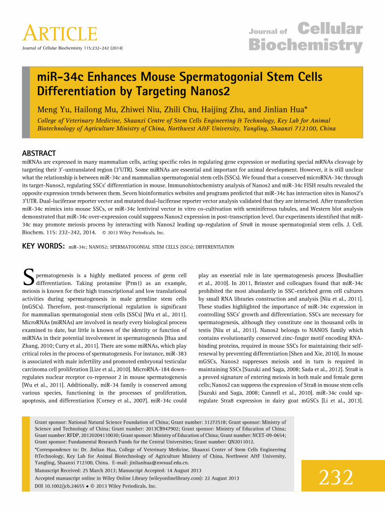
NANOS2 IS A DIRECT TARGET OF miR‐34cIn order to explore how miR‐34c regulates mSSCs, we computation-ally predicted that Nanos2 was the candidate of miR‐34c targets frommiRwalk database (http://www.ma.uni‐heidelberg.de/apps/zmf/mir-walk/). Then miRDB (http://mirdb.org/miRDB/) provided the detailedinteraction information between miR‐34c and Nanos2 (Fig. 3A,B). Itwas validated that they did have interaction analyzed by luciferasereporter assay. The predicted binding site, 30UTR of Nanos2 was then
Fig. 3. Target genes of miR‐34c was predicted and verified. A: Target genes of miR‐34c was predicted by miRwalk website. B: Nanos2 is a target of miR‐34c. Seed sequence ofmiR‐34c and the 30UTR of Nanos2 is correctly base paired. C: The construction schema of pmiR‐RB‐REPORT‐Nanos2‐30UTR and pmiR‐RB‐REPORT‐Mut‐Nanos2‐30UTR vectors. D:Relative luminescence intensity detected by a Hamamatsu optical analyze reader after miR‐34cmimics and dual‐luciferase vectors were co‐transfected into Hela cells (��P< 0.01).(scale bars¼ 100mm, error bars indicate þSD of three technical replicates, ��P< 0.01).
JOURNAL OF CELLULAR BIOCHEMISTRY miR-34c ENHANCING SSCs DIFFERENTATION 237
inserted downstream from the Renilla luciferase coding region in thereporter vector (Fig. 3C). Each reporter construct was separately co‐transfected with the miR‐34c mimics into Hela cells. Compared to themut‐Nanos2‐30UTR control, the luciferase activity declined by about37.5% after transfection with miR‐34c mimics and Nanos2‐30UTRreporter vector (Fig. 3D). The luciferase analysis showed that ectopicover‐expression of miR‐34c reduced Nanos2 protein expression viadirectly binding to Nanos2‐30UTR, indicating that Nanos2 is onetarget of miR‐34c. These results demonstrated that miR‐34c directlyregulates Nanos2 protein expression through its binding to the 30UTRregion of Nanos2.
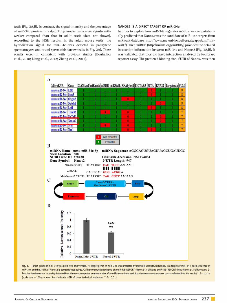
miR‐34c OVER‐EXPRESSION INHIBITED NANOS2, AND PROMOTEDMEIOSIS IN mSSCsTo further investigate the effects of miR‐34c on mSSCs, negativecontrol small RNAs, miR‐34c mimic, miR‐34c inhibitor, and incombination with miR‐34c mimic and its inhibitor were transfectedinto mSSCs, QRT‐PCR results manifested that miR‐34c were trans-
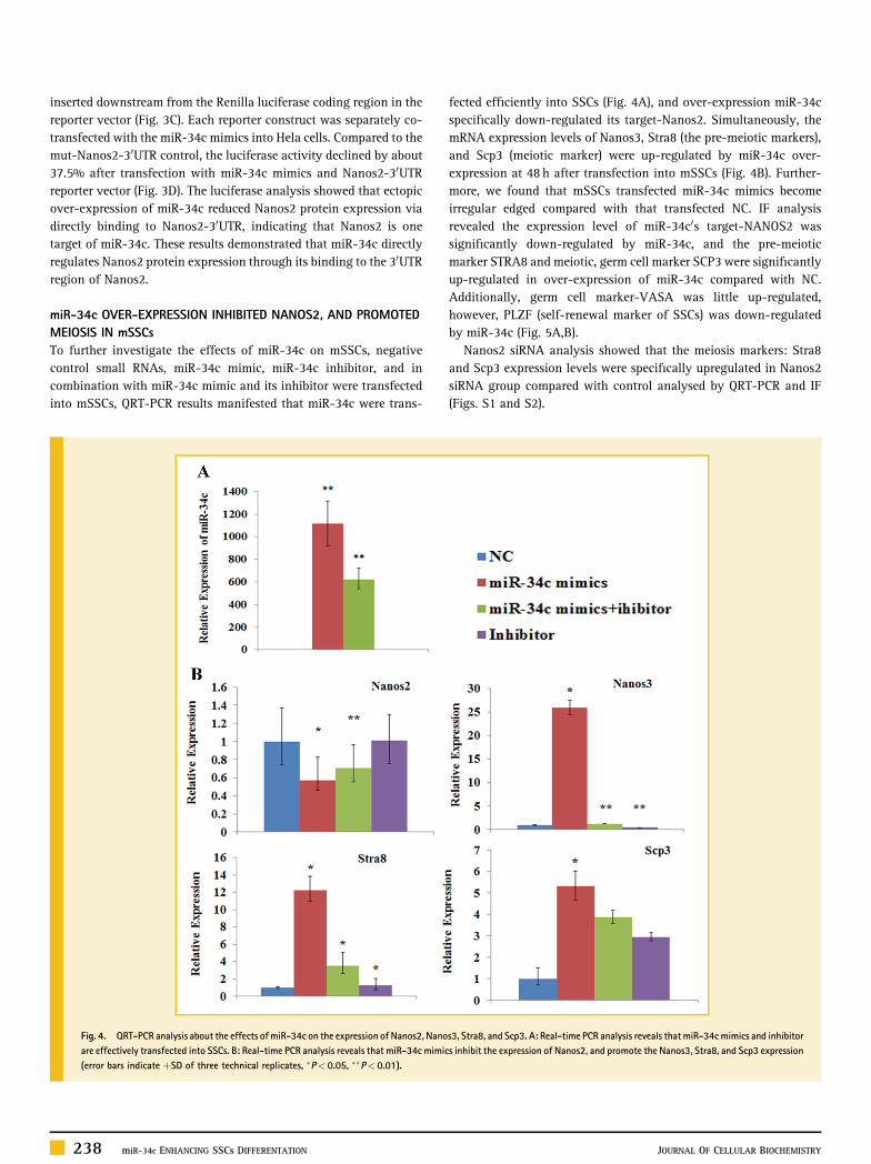
fected efficiently into SSCs (Fig. 4A), and over‐expression miR‐34cspecifically down‐regulated its target‐Nanos2. Simultaneously, themRNA expression levels of Nanos3, Stra8 (the pre‐meiotic markers),and Scp3 (meiotic marker) were up‐regulated by miR‐34c over‐expression at 48 h after transfection into mSSCs (Fig. 4B). Further-more, we found that mSSCs transfected miR‐34c mimics becomeirregular edged compared with that transfected NC. IF analysisrevealed the expression level of miR‐34c0s target‐NANOS2 wassignificantly down‐regulated by miR‐34c, and the pre‐meioticmarker STRA8 and meiotic, germ cell marker SCP3 were significantlyup‐regulated in over‐expression of miR‐34c compared with NC.Additionally, germ cell marker‐VASA was little up‐regulated,however, PLZF (self‐renewal marker of SSCs) was down‐regulatedby miR‐34c (Fig. 5A,B).
Nanos2 siRNA analysis showed that the meiosis markers: Stra8and Scp3 expression levels were specifically upregulated in Nanos2siRNA group compared with control analysed by QRT‐PCR and IF(Figs. S1 and S2).
Fig. 4. QRT‐PCR analysis about the effects ofmiR‐34c on the expression of Nanos2, Nanos3, Stra8, and Scp3. A: Real‐time PCR analysis reveals that miR‐34cmimics and inhibitorare effectively transfected into SSCs. B: Real‐time PCR analysis reveals that miR‐34c mimics inhibit the expression of Nanos2, and promote the Nanos3, Stra8, and Scp3 expression(error bars indicate þSD of three technical replicates, �P< 0.05, ��P< 0.01).
238 miR-34c ENHANCING SSCs DIFFERENTATION JOURNAL OF CELLULAR BIOCHEMISTRY
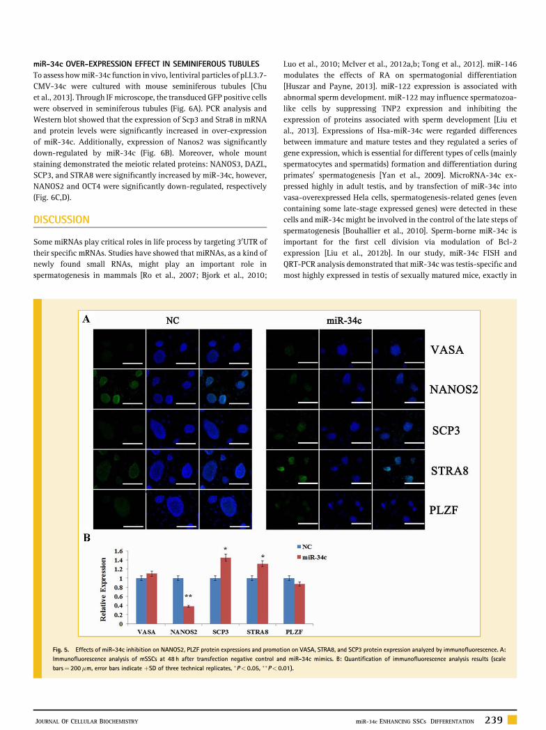
miR‐34c OVER‐EXPRESSION EFFECT IN SEMINIFEROUS TUBULESTo assess howmiR‐34c function in vivo, lentiviral particles of pLL3.7‐CMV‐34c were cultured with mouse seminiferous tubules [Chuet al., 2013]. Through IFmicroscope, the transduced GFP positive cellswere observed in seminiferous tubules (Fig. 6A). PCR analysis andWestern blot showed that the expression of Scp3 and Stra8 in mRNAand protein levels were significantly increased in over‐expressionof miR‐34c. Additionally, expression of Nanos2 was significantlydown‐regulated by miR‐34c (Fig. 6B). Moreover, whole mountstaining demonstrated the meiotic related proteins: NANOS3, DAZL,SCP3, and STRA8 were significantly increased by miR‐34c, however,NANOS2 and OCT4 were significantly down‐regulated, respectively(Fig. 6C,D).
DISCUSSION
Some miRNAs play critical roles in life process by targeting 30UTR oftheir specific mRNAs. Studies have showed that miRNAs, as a kind ofnewly found small RNAs, might play an important role inspermatogenesis in mammals [Ro et al., 2007; Bjork et al., 2010;
Luo et al., 2010; McIver et al., 2012a,b; Tong et al., 2012]. miR‐146modulates the effects of RA on spermatogonial differentiation[Huszar and Payne, 2013]. miR‐122 expression is associated withabnormal sperm development. miR‐122 may influence spermatozoa‐like cells by suppressing TNP2 expression and inhibiting theexpression of proteins associated with sperm development [Liu etal., 2013]. Expressions of Hsa‐miR‐34c were regarded differencesbetween immature and mature testes and they regulated a series ofgene expression, which is essential for different types of cells (mainlyspermatocytes and spermatids) formation and differentiation duringprimates0 spermatogenesis [Yan et al., 2009]. MicroRNA‐34c ex-pressed highly in adult testis, and by transfection of miR‐34c intovasa‐overexpressed Hela cells, spermatogenesis‐related genes (evencontaining some late‐stage expressed genes) were detected in thesecells and miR‐34c might be involved in the control of the late steps ofspermatogenesis [Bouhallier et al., 2010]. Sperm‐borne miR‐34c isimportant for the first cell division via modulation of Bcl‐2expression [Liu et al., 2012b]. In our study, miR‐34c FISH andQRT‐PCR analysis demonstrated that miR‐34c was testis‐specific andmost highly expressed in testis of sexually matured mice, exactly in
Fig. 5. Effects of miR‐34c inhibition on NANOS2, PLZF protein expressions and promotion on VASA, STRA8, and SCP3 protein expression analyzed by immunofluorescence. A:Immunofluorescence analysis of mSSCs at 48 h after transfection negative control and miR‐34c mimics. B: Quantification of immunofluorescence analysis results (scalebars¼ 200mm, error bars indicate þSD of three technical replicates, �P< 0.05, ��P< 0.01).
JOURNAL OF CELLULAR BIOCHEMISTRY miR-34c ENHANCING SSCs DIFFERENTATION 239

spermatogenic cells. miR‐34c might play an important role inmammalian spermatogenesis [Bouhallier et al., 2010; Liu et al.,2012a]. These results were almost in consistent with previous studies[Bouhallier et al., 2010; Niu et al., 2011; Zhang et al., 2012].
NANOS2 is a Nanos family protein that mediates a pivotal role inSSC0s self‐renewal and differentiation [Sada et al., 2009, 2012].Bioinformatics analysis and Luciferase reporter assay evidenced thatNanos2 30UTR has a specific miR‐34c binding sequence. Further, themorphology of mouse SSCs ectopic over‐expressed miR‐34c couldnot maintain the typical colony formation, but promoted SSCsdifferentiation trend. The expression levels of meiotic prophasemarker and germ cell markers in mRNA level were up‐regulated,accompanied with the down‐regulation of Nanos2. In contrast, thesemarkers exhibited downward expression patterns compared with NCgroup in treated with miR‐34c inhibitor. These results furtherindicated that Nanos2 is one target of miR‐34c, and over‐expressionmiR‐34c influenced SSCs0 differentiation by suppressing Nanos2expression, and promoting the expression genes associated withmeiosis, including Nanos3, Scp3, and Stra8.
Stra8 (stimulated by retinoic acid gene 8), which is required formeiotic initiation in both sexes [Koubova et al., 2006; Anderson et al.,2008; Zhou et al., 2008]. miR‐34c mimics were synthesized and
transfected into mSSCs. Moreover, miR‐34c lentiviral vector wasconstructed, virus particles were collected, and cultured withseminiferous tubules in vitro. RT‐PCR, Western blot, and wholemounting of seminiferous tubules demonstrated that miR‐34c over‐expression promoted the expression of meiosis associated markers,including Nanos3, Scp3, and Stra8, through suppressing its target‐Nanos2 expression.
NANOS3 have been directly shown to function in germ celldevelopment across diverse species from flies, worms, frogs, andmiceto humans [Julaton et al., 2011]. NANOS3was expressed in germ cellsthroughout spermatogenesis and oogenesis [Kee et al., 2009]. DAZLand SCP3 are meiosis regulated genes [Koubova et al., 2006;Anderson et al., 2008; Jørgensen et al., 2013]. In our study, theevidences in vivo and in vitro demonstrated that miR‐34c plays acritical role in regulation SSC0s differentiation, through NANOS2.Thus, we first summarized the function model for the role of miR‐34cin regulating mouse spermatogenesis (Fig. 7). In undifferentiatedspermatogonia, NANOS2 play a role in inhibiting NANOS3, SCP3,and Stra8 expression to make SSC or spermatogonia maintain anundifferentiated state. When mouse testis is mature, miR‐34cabundance removes the suppression of NANOS3, SCP3, and STRA8by targeting NANOS2, and promotes SSC or spermatogonia transition
Fig. 6. Effects of miR‐34c on the culturing seminiferous tubules. A: The morphology and immunofluorescence of the seminiferous tubules after transduction with pLL3.7‐U6‐miR‐34c lentiviral vectors, pLL3.7‐EGFP lentiviral vectors were as control. B: Nanos2 was decreased and Scp3, Stra8 in mRNA level increased after transduction with miR‐34clentiviral vectors. C: SCP3, STRA8 were increased after transduction with miR‐34c lentiviral vectors analyzed by Western blot. D: Whole mount staining analysis reveals thatNANOS2, OCT4 were decreased and NANOS3, SCP3, STRA8, DAZL increased after transduction with miR‐34c lentiviral vectors (scale bars¼ 200mm).
240 miR-34c ENHANCING SSCs DIFFERENTATION JOURNAL OF CELLULAR BIOCHEMISTRY

to a differentiating state. This study further extends the mechanism ofmeiosis in mammalian spermatogenesis.
Taken together, our study first shows miR‐34c functions bytargeting the Nanos2 in SSCs meiosis differentiation, providing anovel mechanism with involvement of miRNAs in the regulation ofmale germ cell differentiation.
ACKNOWLEDGMENTSThe authors appreciate the editors and the anonymous reviewers fortheir critical review and excellent comments.
REFERENCESAnderson EL, Baltus AE, Roepers‐Gajadien HL, Hassold TJ, de Rooij DG, vanPelt AM, Page DC. 2008. Stra8 and its inducer, retinoic acid, regulate meioticinitiation in both spermatogenesis and oogenesis in mice. Proc Natl Acad SciUSA 105:14976–14980.
Bjork JK, Sandqvist A, Elsing AN, Kotaja N, Sistonen L. 2010. miR‐18, amember of Oncomir‐1, targets heat shock transcription factor 2 inspermatogenesis. Development 137:3177–3184.
Bouhallier F, Allioli N, Lavial F, Chalmel F, Perrard MH, Durand P, Samarut J,Pain B, Rouault JP. 2010. Role of miR‐34c microRNA in the late steps ofspermatogenesis. RNA 16:720–731.
Cannell IG, Kong YW, Johnston SJ, Chen ML, Collins HM, Dobbyn HC, Elia A,Kress TR, Dickens M, Clemens MJ, Heery DM, Gaestel M, Eilers M, Willis AE,Bushell M. 2010. p38 MAPK/MK2‐mediated induction of miR‐34c followingDNA damage prevents Myc‐dependent DNA replication. Proc Natl Acad SciUSA 107:5375–5380.
Cao H, Chu Y, Lv X, Qiu P, Liu C, Zhang H, Li D, Peng S, Dou Z, Hua J. 2012.GSK3 inhibitor‐BIO regulates proliferation of immortalized pancreaticmesenchymal stem cells (iPMSCs). PLoS ONE 7:e31502.
Cao H, Chu Y, Zhu H, Sun J, Pu Y, Gao Z, Yang C, Peng S, Dou Z, Hua J. 2011.Characterization of immortalized mesenchymal stem cells derived from foetalporcine pancreas. Cell Prolif 44:19–32.
Chu Z, Liu C, Bai Y, Zhu H, Hu Y, Hua J. 2013. A three‐dimensional (3D)environment to keep the integrity of mouse testicular can occurrence ofmeiosis. J Inter Agri 12:1481–1488.
Corney DC, Flesken‐Nikitin A, Godwin AK, Wang W, Nikitin AY. 2007.MicroRNA‐34b andMicroRNA‐34c are targets of p53 and cooperate in controlof cell proliferation and adhesion‐independent growth. Cancer Res 67:8433–8438.
Curry E, Safranski TJ, Pratt SL. 2011. Differential expression of porcine spermmicroRNAs and their association with sperm morphology and motility.Theriogenology 76:1532–1539.
Dweep H, Sticht C, Pandey P, Gretz N. 2011. miRWalk–database: Prediction ofpossible miRNA binding sites by “walking” the genes of three genomes. JBiomed Inform 44:839–847.
Hua J, Zhang S. 2010. MicroRNAs and mammalian germ cell development.Chinese J Anat 33:281–285.
Huszar JM, Payne CJ. 2013. MicroRNA 146 (Mir146) modulatesspermatogonial differentiation by retinoic acid in mice. Biol Reprod88:15.
JørgensenA, Nielsen JE, AlmstrupK, Toft BG, Petersen BL, Rajpert‐DeMeyts E.2013. Dysregulation of the mitosis‐meiosis switch in testicular carcinoma insitu. J Pathol 229:588–598.
Julaton VT, Reijo Pera RA. 2011. NANOS3 function in human germ celldevelopment. Hum Mol Genet 20:2238–2250.
Kee K, Angeles VT, Flores M, Nguyen HN, Reijo Pera RA. 2009. Human DAZL,DAZ and BOULE genes modulate primordial germ‐cell and haploid gameteformation. Nature 462:222–225.
Koubova J, Menke DB, Zhou Q, Capel B, GriswoldMD, Page DC. 2006. Retinoicacid regulates sex‐specific timing of meiotic initiation in mice. Proc Natl AcadSci USA 103:2474–2479.
Li M, Yu M, Liu C, Zhu H, He X, Peng S, Hua J. 2013. miR‐34c worksdownstream of p53 leading to dairy goat male germline stem‐cell (mGSCs)apoptosis. Cell Prolif 46:223–231.
Liang X, Zhou D, Wei C, Luo H, Liu J, Fu R, Cui S. 2012. MicroRNA‐34cenhances murine male germ cell apoptosis through targeting ATF1. PLoS ONE7:e33861.
Liu C, Yu M, Zhu HJ, Li MZ, Hua JL. 2012. Construction of miR‐34c lentiviralexpression vector and its infection in dairy goat germ line stem cells. ScientiaAgricultura Sinica 45:3414–3421.
LiuWM, Pang RT, Chiu PC, Wong BP, Lao K, Lee KF, YeungWS. 2012. Sperm‐
borne microRNA‐34c is required for the first cleavage division in mouse. ProcNatl Acad Sci USA 109:490–494.
Fig. 7. Models for the role of miR‐34c in regulating mouse spermatogenesis. In undifferentiated spermatogonia, NANOS2 play a role in inhibiting NANOS3, SCP3, and STRA8expression to make SSCs or spermatogonia stay an undifferentiated state. miR‐34c abundance removes the suppression of NANOS3, SCP3, and STRA8 by NANOS2, and promotesSSC or spermatogonia transition to a differentiating state.
JOURNAL OF CELLULAR BIOCHEMISTRY miR-34c ENHANCING SSCs DIFFERENTATION 241
Liu T, Huang Y, Liu J, Zhao Y, Jiang L, Huang Q, Cheng W, Guo L. 2013.MicroRNA‐122 influences the development of sperm abnormalities fromhuman iPS cells by regulating TNP2 expression. Stem Cells Dev 22:1839–1850.
Lize M, Pilarski S, Dobbelstein M. 2010. E2F1‐inducible microRNA 449a/bsuppresses cell proliferation and promotes apoptosis. Cell Death Differ 17:452–458.
Luo L, Ye L, Liu G, ShaoG, Zheng R, Ren Z, Zuo B, XuD, LeiM, Jiang S, Deng C,Xiong Y, Li F. 2010. Microarray‐based approach identifies differentiallyexpressed microRNAs in porcine sexually immature and mature testes. PLoSONE 5:e11744.
McIver SC, Roman SD, Nixon B, McLaughlin EA. 2012a. miRNA andmammalian male germ cells. Hum Reprod Update 18:44–59.
McIver SC, Stanger SJ, Santarelli DM, Roman SD, Nixon B, McLaughlin EA.2012b. A unique combination of male germ cell miRNAs coordinates gonocytedifferentiation. PLoS ONE 7:e35553.
Niu Z, Goodyear SM, Rao S, Wu X, Tobias JW, Avarbock MR, Brinster RL.2011. MicroRNA‐21 regulates the self‐renewal of mouse spermatogonial stemcells. Proc Natl Acad Sci USA 108:12740–12745.
Olszewska M, Bujarski JJ, Kurpisz M. 2012. P‐bodies and their functionsduring mRNA cell cycle: Mini‐review. Cell Biochem Funct 30:177–182.
Ro S, Song R, Park C, Zheng H, Sanders KM, Yan W. 2007. Cloning andexpression profiling of small RNAs expressed in the mouse ovary. RNA13:2366–2380.
Sada A, Suzuki A, Suzuki H, Saga Y. 2009. The RNA‐binding protein NANOS2is required to maintain murine spermatogonial stem cells. Science 325:1394–1398.
Sada A, Hasegawa K, Pin PH, Saga Y. 2012. NANOS2 acts downstream of glialcell line‐derived neurotrophic factor signaling to suppress differentiation ofspermatogonial stem cells. Stem Cells 30:280–291.
Shen R, Xie T. 2010. NANOS:A germline stem cell0s GuardianAngel. JMol CellBiol 2:76–77.
Suzuki A, Saga Y. 2008. Nanos2 suppresses meiosis and promotes male germcell differentiation. Genes Dev 22:430–435.
Tong MH, Mitchell DA, McGowan SD, Evanoff R, Griswold MD. 2012. TwomiRNA clusters, Mir‐17–92 (Mirc1) and Mir‐106b‐25 (Mirc3), are involved inthe regulation of spermatogonial differentiation in mice. Biol Reprod 86:72.
Wang X. 2008. miRDB: A microRNA target prediction and functionalannotation database with a wiki interface. RNA 14:1012–1017.
Wu J, Bao J,Wang L, Hu Y, Xu C. 2011.MicroRNA‐184 downregulates nuclearreceptor corepressor 2 in mouse spermatogenesis. BMC Dev Biol 11:64.
Yan N, Lu Y, Sun H, QiuW, Tao D, Liu Y, Chen H, Yang Y, Zhang S, Li X, Ma Y.2009. Microarray profiling of microRNAs expressed in testis tissues ofdeveloping primates. J Assist Reprod Genet 26:179–186.
Zhang S, Sun J, Pan S, Zhu H, Wang L, Hu Y, Wang J, Wang F, Cao H, Yan X,Hua J. 2011. Retinol (vitamin A) maintains self‐renewal of pluripotent malegermline stem cells (mGSCs) from adult mouse testis. J Cell Biochem 112:1009–1021.
Zhang S, Yu M, Liu C, Wang L, Hu Y, Bai Y, Hua J. 2012. MIR‐34c regulatesmouse embryonic stem cells differentiation into male germ‐like cells throughRARg. Cell Biochem Funct 30:623–632.
Zhou Q, Nie R, Li Y, Friel P, Mitchell D, Hess RA, Small C, Griswold MD. 2008.Expression of stimulated by retinoic acid gene 8 (Stra8) in spermatogenic cellsinduced by retinoic acid: an in vivo study in vitamin A‐sufficient postnatalmurine testes. Biol Reprod 79:35–42.
Zhu H, Liu C, Sun J, Li M, Hua J. 2012. Effect of GSK‐3 inhibitor on theproliferation of multipotent male germ line stem cells (mGSCs) derived fromgoat testis. Theriogenology 77:1939–1950.
SUPPORTING INFORMATION
Additional supporting information may be found in the onlineversion of this article at the publisher0s web‐site.
242 miR-34c ENHANCING SSCs DIFFERENTATION JOURNAL OF CELLULAR BIOCHEMISTRY